Embed Size (px)
Citation preview

Environmental Research 96 (2004) 62–71
ARTICLE IN PRESS
�Correspond
E-mail addr
0013-9351/$ - se
doi:10.1016/j.en
In vitro effects on macrophages induced by noncytotoxic doses ofsilica particles possibly relevant to ambient exposure
M. Balduzzi,a M. Diociaiuti,b,� B. De Berardis,b S. Paradisi,c and L. Paolettib
aSezione di Tossicologia e Scienze Biomediche, ENEA, Via Anguillarese 301, Roma, ItalybDipartimento di Tecnologie e Salute, Istituto Superiore di Sanita, Viale Regina Elena 299, 00161 Roma, Italy
cDipartimento di Biologia Cellulare e Neuroscienze, Istituto Superiore di Sanita, Viale Regina Elena 299, 00161 Roma, Italy
Received 3 June 2003; received in revised form 14 October 2003; accepted 14 November 2003
Abstract
The RAW 246.7 macrophage cell line was exposed in vitro to aged crystalline silica particles of respirable size for 24 h at a range of
doses starting from 15mg/2� 106 cells, which is a realistic exposure level of macrophages in the airways of ambiently exposed
individuals. The particle sample used for the experiments was prepared to mimic some aspects of ambient crystalline silica particles:
size distribution, morphology, and surface reactivity. Our purpose was to determine whether a nontoxic quartz load comparable to
that of ambient exposure would be able to induce macrophage activation and impairment of the phagocytic ability, factors altering
the lung’s capacity to deal with increased particle loads (as occurs during high-pollution episodes) or infections and affecting the
local and systemic responses through the release of biologically active compounds (cytokines, reactive oxygen species, NO,
isoprostanes). Exposure of RAW 264.7 cells to aged silica particles induced macrophage activation (evidenced by the morphological
features observed with scanning electron microscopy and by the release of TNF-a and IL-6) and impairment of phagocytosis of testparticles, even at noncytotoxic doses. The reduction of the phagocytic function of the cells after silica treatment was dose-dependent,
as evidenced by an increase of the population of unphagocytic cells, paralleled by a decrease of the actively phagocytizing cell
population. We evaluated the oxidative stress induced by aged silica particles, quantifying the peroxidation products (8-
isoprostanes) in the culture media of treated cells, and found a strong release at low doses. Isoprostanes are a complex family of
compounds which have been used as in vivo markers of lipid peroxidation in human disorders, but that, as far as we know, have
never been evaluated in relation to airborne particulate matter exposure. Lipid peroxides are involved in various cellular events in
the inflammatory response, and isoprostanes are also supposed to exert important biological actions on airway and pulmonary
vascular smooth muscles and on platelets.
r 2003 Elsevier Inc. All rights reserved.
Keywords: Silica; RAW 264.7; Citotoxicity; Lipid peroxidation; Inflammatory mediators; Electron microscopy
1. Introduction
Several epidemiological studies indicate a consistentassociation between particulate air pollution and acutehealth effects, including increased cardiopulmonarymorbidity and mortality, in susceptible populations(Schwartz et al., 1996; Roemer et al., 2000), but eitherthe components casually related to or the mechanismsunderlying the observed health effects have not beenfully elucidated (Harrison and Yin, 2000).Crystalline silica particles are present in inhalable
ambient particulate matter (PM10) as a fractional
ing author. Fax: 39-6-4938-7140.
ess: [email protected] (M. Diociaiuti).
e front matter r 2003 Elsevier Inc. All rights reserved.
vres.2003.11.004
component of ambient particulate air pollution; theyare preferentially deposited and retained in the lung,where they come in direct contact with target cells, buttheir possible contribution to particulate matter toxicityis still undetermined.Epidemiological and pathological studies have shown
that workers chronically exposed to high concentrationsof silica can develop silicosis, a form of interstitial lungdisease caused by chronic inflammation in the lungparenchyma (Mossman and Churg, 1998). Chroniclower levels of silica dust exposure are thought to causepathological changes in the lung, leading to thedevelopment of chronic obstructive pulmonary diseaseand silicosis (Hnizdo and Vallyathan, 2003). In additionto well-documented pulmonary toxicity, there are

ARTICLE IN PRESSM. Balduzzi et al. / Environmental Research 96 (2004) 62–71 63
indications that occupational exposure to crystallinesilica is associated with immune dysfunction, bothsystemically and in the lung (Weissman et al., 1996),and also with autoimmune diseases (Ding et al, 2002;EPA, 1996; Cooper et al., 2002).Based on evidence obtained from both animal models
and epidemiological studies, the International Agencyfor Research on Cancer (IARC, 1997) has concludedthat there is sufficient evidence that inhaled crystallinesilica from occupational sources, in the form of quartzor cristobalite, is carcinogenic to humans. The IARCworking group pointed out the difficulties met in theevaluation of the hazard posed by quartz due toconflicting results in the assessment of carcinogenicityto humans exposed to different silica-containing dusts.In reviewing the IARC evaluations, some authors haveargued that crystalline silica is not a single hazard entityand that its toxicological properties are dependent on itssurface characteristics, which are supposed to bedifferent in mixed dusts due to the presence ofsubstances affecting surface reactivity (Donaldson andBorm, 1998).The hazard posed by inhaled crystalline silica
particulate has long been considered mainly occupa-tional; concern about nonoccupational or ambientcrystalline silica exposure has emerged only recently,and data on noncancer health effects of inhaled silicahave been extensively reviewed (EPA, 1996).Crystalline silica, notably in the form of quartz, is
widespread in nature, being a component of both soiland many rocks. Ambient silica particles are emittedinto the general environment by natural, industrial, andfarming activities and constitute a fractional componentof inhalable ambient particulate matter. Direct measure-ments of ambient crystalline silica levels in the respirablerange are limited. It has been reported that quartz levelsin US metropolitan areas average around 3 mg/m3 andgenerally do not exceed 8 mg/m3 (Davis et al., 1984). In astudy conducted in an urban area of Rome, thecontribution of quartz mass to the inhalable fractionof PM has been found to vary from 1.7% to 3.4%,corresponding to 0.6 and 1.5 mg/m3 (Puledda et al.,1999).Despite of this small contribution, it has been shown
that crystalline silica particles are largely represented inthe bulk of particles retained in the lung of environmen-tally exposed people and that crytalline silica is thesingle largest species retained in the airways of adultnonsmokers from either a low (Churg and Brauer, 2000)or high polluted area (Brauer et al., 2001). Theseobservations are consistent with the finding that crystal-line silica is among the most biopersistent of nonfibrousmineral particles (Pairon et al., 1994). Biopersistence ofmineral particles and direct contact with target lung cellsare important determinants for long-term effects inexposed animals (Johnston et al., 2000).
The pathways arising from oxidative stress are knownto be important in quartz-induced inflammation in vivo(Vallyathan and Shi, 1997; Castranova, 1998; Shi et al.,2001; MacNee, 2001). Most in vitro studies, which havedealt with freshly fractured crystalline silica, have shownthat it is highly cytotoxic and that its cytotoxicproperties are related to the presence of highly reactiveradicals on the surface of the particles. Some authorshave proposed that those highly reactive radicals exerttheir action at the level of the cytoplasmic membrane(Razzaboni and Bolsaitis, 1990), inducing lipid perox-idation and protein denaturation. Experiments per-formed by electron microscopy and dielectricspectroscopy techniques on human erythrocytes, as asimplified model of the cellular membrane, have shownthat freshly fractured silica particles are able to damagecellular membranes, modify their dielectric properties,and modify the whole erythrocyte morphology (Dio-ciaiuti et al., 1999). The same authors have demon-strated that the effects are directly correlated with theparticle surface rather than with the particle weight.This surface-related behavior has been described inmany recent papers and is generally accepted (Tranet al., 2000).Silica surface-derived reactive oxygen species are
much higher on freshly fractured quartz than on agedspecimens, as surface radicals decay with time (Fubini,1998a); in experimental animals, freshly fractured silicahas been shown to be more toxic than aged silica(Castranova et al., 1996, 1997; Porter et al., 2002).Ambient silica particles are likely to be aged (the half-life of surface radical activity decay is approximately30 h), while in occupational settings workers can beexposed either to freshly fractured or to aged silica,depending on the type of industry. Moreover, crystallinesilica particles in the general environment occur at levelsconsiderably lower than those in occupational settings;consequently, the health effects from ambient exposureare expected to be different. Lung macrophages are thehost defense cells most important to the host’s effortsagainst inhaled pollutants, and an impairment of theirphagocytic ability, together with the release of reactiveoxygen species and a number of cytokines, may lead toaltered efficiency in controlling airway inflammationand infection.In the present in vitro study the macrophage response
to increasing silica particle concentrations was evaluatedon the mouse monocyte-macrophage RAW 264.7 cellline cultured and exposed under well-defined conditions.Chemically homogeneous crystalline silica particles ofrespirable size were selected by sedimentation of grindedMin-U-Sil crystals. The particle sample used forexperiments mimicked some of the properties of thequartz that occur as a part of human exposure (crystal-linity, size, shape and aging). Number of cells andparticle mass range were calculated in order to roughly

ARTICLE IN PRESSM. Balduzzi et al. / Environmental Research 96 (2004) 62–7164
correspond to ambient exposure conditions. For thispurpose, the total quartz amount found in normal lungsof environmentally exposed people over total macro-phage lung population was taken as baseline ratio. Ourpurpose was to determine if a nontoxic quartz load,comparable to the ambient exposure, was able to inducemacrophage activation and impairment of the phago-cytic ability (as factors altering the lung capacity to dealwith increased particle loads, as occurs during highpollution episodes, or infections). The release ofbiological active compounds (cytokines, NO, isopros-tanes) was also evaluated for their possible role in theonset and progression of local and systemic inflamma-tory responses that are associated with the adversehuman health effects of ambient particulate matter.
2. Materials and methods
2.1. Silica particle suspension and characterization
Silica particles were produced by grinding Min-U-Sil5 quartz in ethanol for 30min with a micronizing mill(McCrone, London, UK). The ethanol suspension wasdried at about 50�C and the powder resuspended in PBSand sonicated in order to obtain a single particlesuspension. The largest particles were then settled bykeeping the suspension vertical for 10min, and theupper part, containing the smallest particles, wascharacterized and used in the experiments.The silica sample sat for the month preceding the
experiments to aged. The preparation, free of endotoxinas determined by the LAL assay, was sonicated for 2 hand then stored at 4�C. Thirty microliters of thesuspension was filtered on a polycarbonate membrane.A portion of the membrane was mounted on scanningelectron microscopy (SEM) stubs and coated with a thingold film, deposited by sputtering, for observation. Thespecimen was analyzed by a SEM Philips XL30equipped with a thin-window EDAX DX4 system forX-ray microanalysis by energy-dispersion spectrometry(EDX). The silica particles were automatically detectedby an increase in the secondary electron (SE) videosignal above a preset video threshold. At least 5000particles were analyzed, and the size distribution andmean diameter were calculated.
2.2. Cell culture and exposure to silica particle suspension
RAW 264.7 cells, a mouse peritoneal macrophage cellline, were obtained from the American Type CultureCollection. The cells were maintained in RPMI 1640with 10% fetal bovine serum (FBS), 100U/mL peni-cillin/streptomycin, and 2mM glutamine. For stimula-tion experiments, cells were plated in 24-well plates andallowed to grow for 24 h until almost confluent. For
SEM analysis, cells were grown on Thermanox (NalgeNunc International) coverslips. Prior to experiments,the culture medium was replaced with a fresh mediumsupplemented with 1% FBS. The stock silica particlesuspensions were added to the culture wells to produce adose of 15–120 mg per well in a final volume of 1mL.The cells were exposed to particles for 24 h, and then theculture media and cells were isolated for assays. Forphagocytosis experiments, cells were grown and treatedin suspension at the same conditions. Experiments wereperformed in triplicate at least three times.
2.3. Macrophage morphology by SEM
Thermanox coverslips were rinsed three times with0.1M phosphate-buffered saline (PBS, pH 7.4), fixed in2% glutaraldehyde in 0.1M cacodylate for 20–40min,and postfixed in 1% osmium tetroxide in 0.1Mcacodylate buffer for 1 h. After a thorough rinsing in0.1M phosphate buffer and dehydration in a gradedethanol series, the samples were transferred into a CO2
critical-point dryer, mounted on SEM stubs, andsputter-coated with gold. To demonstrate phagocytosisof silica particles, Si X-ray maps were performed ontreated macrophages. We defined the Si X-ray region ofinterest in the EDX spectrum and selected onlycharacteristic X rays of the chosen element to obtainthe image of the sample related to Si. Morphologicalanalysis was semiautomatically performed by compar-ing the increased SE video signal with a preset videothreshold, and for each cell the average and maximumdiameters, area, perimeter, and shape factor weredetermined.
2.4. Cytotoxicity analysis
The extracellular leakage of the cytoplasmic markerlactate dehydrogenase (LDH) was used as cytotoxicityendpoint. LDH activities were determined in the culturemedia and in the cell monolayers using a two-stepenzymatic test (Sigma LDH Kit): in the first step NAD+
was reduced to NADH/H+ by the LDH-catalyzedconversion of lactate to pyruvate; in the second step thecatalyst (diaphorase) transferred H/H+ from NADH/H+ to the tetrazolium salt, which was reduced toformazan. The formazan dye formed was quantified bya determination of the spectral photometric absorbanceat 492 nm on an ELISA reader (Bio-Rad microplatereader). The absorbance values were used to calculatethe percentage of cytotoxicity as follows:
% Citotoxicity ¼ A � K
B;
where A is the experimental value of cell supernatant, K
is the control value of the cell supernatant (untreated

ARTICLE IN PRESSM. Balduzzi et al. / Environmental Research 96 (2004) 62–71 65
cells), and B is the experimental value of the correspond-ing cell monolayer.
2.5. ELISA assays for TNF-a and IL-6
TNF-a and IL-6 proteins were determined by ELISAkits (Amersham Pharmacia Biotech) on cell culturemedia that were collected at various time points afterexposure to silica particles and stored at �80�C untilassay. Values are reported as picograms per milliliter (in1mL there were about 106 cells).
2.6. Assay for NO
Nitric oxide production was quantified by measuringthe accumulation of nitrite, a stable NO oxidationproduct, in the culture media collected 24 h afterexposure to silica particles and stored at �20�Cuntil assay. Nitrite was quantified spectrophoto-etrically using the Griess reaction by a modifiedprocedure (Green et al., 1982). The detection limit was0.3 mM.
2.7. 8-Isoprostane immunoassay
8-Isoprostane was quantified in conditioned mediumfrom treated cells by a commercially available immu-noassay performed according to the instructions of themanufacturer (Cayman Chemical). The concentration ofthe isoprostanes produced was calculated from astandard curve and expressed in picograms/milliliter(in 1mL there were about 106 cells). This immunoassayshould be considered semiquantitative.
2.8. Phagocytic assay
Phagocytic function in RAW 264.7 was measured byflow cytometry (Mebouta Nkamgueu et al., 2000). Cellswere treated with silica particles at concentrations of 15,30, 60, and 120 mg/mL for 24 h. Following silicaexposure, samples of 2� 106 cells/mL were incubatedfor 1 h at 37�C with 2-mm-diameter fluorescein isothio-cyanate-labeled latex beads (Fluoresbrite, Polyscience),at a ratio of at least 10 beads/cell. Afterward, cellswere washed three times with cold PBS to stopphagocytosis and to eliminate freely floatingparticles. A FACScan flow cytometer (BectonDickinson), using CellQuest Software, was used todetermine the fluorescence emitted by the cells thathad engulfed the beads. Analysis was performed onabout 10,000 cells. Maintenance of membrane integritywas evaluated by propidium iodide exclusion. Thehistograms shown are representative of three differentexperiments.
2.9. Statistical analyses
All data were expressed as mean7SD. The differencesin measured variables between experimental and controlgroups were analyzed by Student’s t test. Variationswere considered statistically significant at a P valueo0.05.
3. Results and discussion
The purpose of the present experiments was to study,in a panel of biological assays, the effects induced on theRAW 264.7 monocyte-macrophage cell line by crystal-line silica particles (Min-U-Sil) fractured to respirablesize and aged to mimic some of the properties ofambient particles, namely dimension, morphology, andsurface reactivity, which are important determinants ofparticle toxicity (Donaldson et al., 1996; Harrison andYin, 2000).SEM analysis showed that we had prepared a silica
particle sample with a dimensional distribution similarto that of the actual PM10 crystalline silica component,as assessed by a comparison with the results of the studyperformed by Puledda et al. (1999). This result wasobtained using a simple sedimentation procedure basedon the Stoke law and used in our previous work(Diociaiuti et al., 1999).SEM characterization indicated that the particle sizes
of our sample ranged from 0.16 to 11.2 mm (with a meanparticle size of 0.83 mm). Seventy-six percent of theparticles possessed dimensions below 1 mm, 18.8%dimensions between 1 and 2.5 mm, 4.3% dimensionsbetween 2.6 and 4.9 mm, and only 0.9% dimensionsbetween 5 and 11.2 mm. Mechanical fracturegenerated irregular particles similar in shape to theambient quartz particles (Fig. 1), but with sharper edges(Fig. 1a). Moreover, we used an aged silica samplebecause the general population is more likely to beexposed to aged particles. Crystalline silica fromdifferent sources exhibits great variability in its surfaceproperties (Fubini, 1998b) and inflammogenicity (Clou-ter et al., 2001), so our sample reproduced only someaspects of ambient silica particles. Moreover, quartzcannot be considered a single hazard entity because itssurface chemistry can be modified easily by ambientconditions (Donaldson and Borm, 1998; Fubini, 1998),and quartz samples in a nonquartz matrix are likely tovary in the extent of biological effects and even in themechanism of action (Borm, 1997; Clouter et al., 2001;Donaldson et al., 2001). Therefore, the contribution ofquartz to the toxicity of mixed dusts is difficult topredict.For our experiments, 2� 106 macrophage cells were
challenged for 24 h with aged quartz particles at a rangeof doses from 15 to 120 mg. This loading correspond to
ARTICLE IN PRESS
Fig. 1. Comparison by SEM images of silica particles used in the study
(a) and an ambient silica particle (arrow) present in a PM10 sample
collected in Rome (b). Images are at the same magnification and show
that the particles look alike, even considering that the prepared
particles have sharper edges.
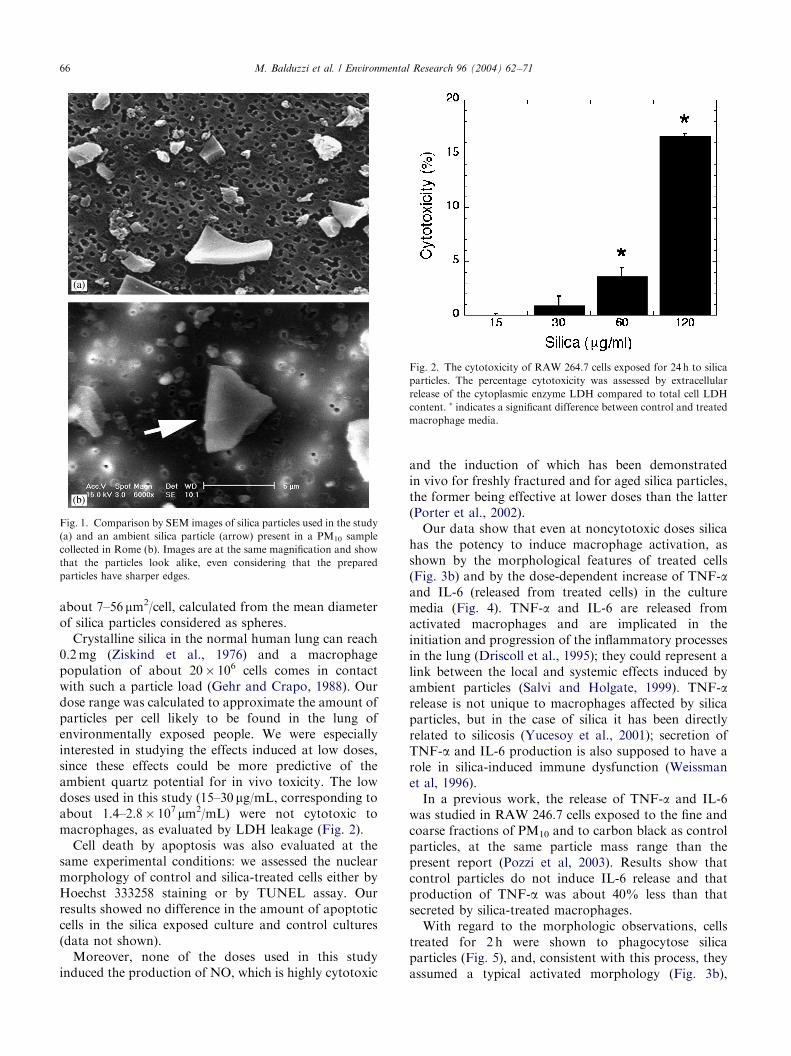
Fig. 2. The cytotoxicity of RAW 264.7 cells exposed for 24 h to silica
particles. The percentage cytotoxicity was assessed by extracellular
release of the cytoplasmic enzyme LDH compared to total cell LDH
content. � indicates a significant difference between control and treated
macrophage media.
M. Balduzzi et al. / Environmental Research 96 (2004) 62–7166
about 7–56 mm2/cell, calculated from the mean diameterof silica particles considered as spheres.Crystalline silica in the normal human lung can reach
0.2mg (Ziskind et al., 1976) and a macrophagepopulation of about 20� 106 cells comes in contactwith such a particle load (Gehr and Crapo, 1988). Ourdose range was calculated to approximate the amount ofparticles per cell likely to be found in the lung ofenvironmentally exposed people. We were especiallyinterested in studying the effects induced at low doses,since these effects could be more predictive of theambient quartz potential for in vivo toxicity. The lowdoses used in this study (15–30 mg/mL, corresponding toabout 1.4–2.8� 107 mm2/mL) were not cytotoxic tomacrophages, as evaluated by LDH leakage (Fig. 2).Cell death by apoptosis was also evaluated at the
same experimental conditions: we assessed the nuclearmorphology of control and silica-treated cells either byHoechst 333258 staining or by TUNEL assay. Ourresults showed no difference in the amount of apoptoticcells in the silica exposed culture and control cultures(data not shown).Moreover, none of the doses used in this study
induced the production of NO, which is highly cytotoxic
and the induction of which has been demonstratedin vivo for freshly fractured and for aged silica particles,the former being effective at lower doses than the latter(Porter et al., 2002).Our data show that even at noncytotoxic doses silica
has the potency to induce macrophage activation, asshown by the morphological features of treated cells(Fig. 3b) and by the dose-dependent increase of TNF-aand IL-6 (released from treated cells) in the culturemedia (Fig. 4). TNF-a and IL-6 are released fromactivated macrophages and are implicated in theinitiation and progression of the inflammatory processesin the lung (Driscoll et al., 1995); they could represent alink between the local and systemic effects induced byambient particles (Salvi and Holgate, 1999). TNF-arelease is not unique to macrophages affected by silicaparticles, but in the case of silica it has been directlyrelated to silicosis (Yucesoy et al., 2001); secretion ofTNF-a and IL-6 production is also supposed to have arole in silica-induced immune dysfunction (Weissmanet al, 1996).In a previous work, the release of TNF-a and IL-6
was studied in RAW 246.7 cells exposed to the fine andcoarse fractions of PM10 and to carbon black as controlparticles, at the same particle mass range than thepresent report (Pozzi et al, 2003). Results show thatcontrol particles do not induce IL-6 release and thatproduction of TNF-a was about 40% less than thatsecreted by silica-treated macrophages.With regard to the morphologic observations, cells
treated for 2 h were shown to phagocytose silicaparticles (Fig. 5), and, consistent with this process, theyassumed a typical activated morphology (Fig. 3b),

ARTICLE IN PRESS
Fig. 3. SEM micrographs of RAW 264.7 macrophages cultured for 24 h in the absence of silica and then exposed to 30mg/106 cells for 2 h (b) and
24 h (d). Control cells were cultured in the absence of silica and fixed at the same time than 2 h treated cells (a) and 24 h treated cells (c). 2 h treated
cells (b) are enlarged and show rougher surface as respect to control cells (a). 24 h treated cells (d) have almost resumed the same morphological
appearance than control cells (c).
M. Balduzzi et al. / Environmental Research 96 (2004) 62–71 67
showing rough surfaces with prominent filopodia, blebs,and rufflings. After 24 h, the cells almost resumed theirnormal appearance (Fig. 3d), but their phagocyticcapacity was decreased with respect to that of controlcells (see Fig. 6).Occupational exposure to silica particles predisposes
workers to bacterial infections (Wagner, 1997; Dinget al., 2002), and impairment of the macrophage-dependent host defense is one of the biological mechan-isms that may lead to the reported increase of lunginfections due to elevations of particulate pollutionlevels (Becker and Soukup, 1998). Silica has beenreported to decrease the phagocytosis of alveolarmacrophages in vitro (Zimmerman et al., 1986), butbactericidal activity was increased in rats following silicainhalation, probably as a consequence of an increasedproduction of reactive oxygen species from lungphagocytes and macrophage phagocytosis (Antoniniet al., 2000a, b).The application of flow cytometry enabled us to
compare the effects on RAW 264.7 cell of treatmentwith different concentrations of silica particles byevaluating their ability to phagocytose latex beads. Asreported in Fig. 6a, in control cultures 18.5% of cells
were nonphagocytic (Region I), and the activelyphagocytosing cells comprised 69% of cells (RegionIII). After treatment with 120 mg/mL silica, thesepercentages became, respectively, 46% and 33% (Fig.6b). The percentage of cells capable of ingesting onlyone bead (Region II) increased in treated cells to 21%(in control cells it was 12.5%). These data indicate thatsilica treatment permitted only limited phagocyticfunction, with the ability to phagocytose two or morebeads markedly reduced. In Fig. 6c are cumulative datafrom cells treated with the four different doses of silicaused: one observes that the phagocytic ability limited toone bead increased in a dose-dependent manner andthat the activity against two or more beads decreased atthe same time. In conclusion, the exposure of RAW264.7 cells to aged silica particles was detrimental totheir phagocytic activity even in the absence of an effecton cell viability.Since the pathways arising from oxidative stress are
known to be important determinants in the biologicalactivity of particles (Donaldson et al., 1996), in lunginflammation (MacNee, 2001), and in particulate pollu-tion-induced inflammation (including silica particles)(Becker et al., 2002), and since the inflammogenic

ARTICLE IN PRESS
Fig. 4. Release of IL-6 (A) and TNF-a (B) by control and silica
challenged RAW 264.7macrophages. Cells were incubated with silica
particles (15-30-60-120mg/106 cells) for 24 h in culture medium
supplemented with 1% FBS. After incubation, the culture media were
collected and stored at �80�C until analysis. TNF-a and IL-6 were
quantitated as described in Materials and Methods. Results are
expressed per 1� 106 cells. Data are means of three experiments, each
using three experimental points. � indicates a significant difference
between control and treated macrophage media.
Fig. 5. X-ray elemental map showing silica particles (in yellow)
ingested by a macrophage. Bar, 10 mm.
M. Balduzzi et al. / Environmental Research 96 (2004) 62–7168
potency of silica has been better related to lipidperoxidation than to hydroxyl radical generation andsecretion (Vallyathan, 1994), we quantified the oxidativestress marker 8-isoprostanes in the culture media ofsilica treated cells as a lipid stable peroxidation productoccurring as a result of ROS reaction at the cellmembrane level.Isoprostanes are a complex family of compounds used
as in vivo markers of lipid peroxidation in many humandisorders, such as pulmonary inflammation, asthma,heart failure, and cigarette smoking conditions, with arole in pathological states that needs to be elucitated
(Cracowski et al., 2002; Morrow and Roberts 2002). Inaddition to their cytotoxic properties, isoprostanes areincreasingly recognized as being important in signaltransduction for a number of events in the inflammatoryresponse. The 15-series F2-isoprostanes possess bronch-oconstrictor and vasoconstrictor properties that mightplay a role in the pathophysiology of lung disease(Janssen, 2001). In contrast to free radicals, lipidperoxidation products can diffuse from the site offormation and exert their action on target organs.In general, free radicals can be released either by
quartz particles or by phagocytes. As for the quartzparticles, they release radicals according to two differentmechanisms: one is a Fenton-like process activated byiron, even in traces; the other is dependent on theradicals formed during the grinding process, whichdecay during the aging process. The aged silica sampleused in this study was pure quartz carrying iron in traceamounts, a condition that is common with ambientmineral particles (Fenoglio et al., 2001); therefore, it ispossible that in our system iron, besides macrophages,could generate free radicals.Our results (Fig. 7) show that the oxidative stress
induced by aged silica particle exposure results in stronglipid peroxidation. This effect is relevant at lownoncytotoxic doses and seems to reach a plateau atdoses higher than 120 mg/mL. As far as we know,isoprostanes have never been evaluated in relation toairborne particulate matter exposure.
4. Conclusion
The role that ambient crystalline silica may play in thehealth risk in the general population exposed to PM10
has not been fully assessed. It seems that its silicoticeffects are not likely to contribute to ambient chroniclung disease risk (EPA, 1996). The ability of silica toinduce silicotic fibrosis has been related to its cytotoxi-city to alveolar macrophages (Mossman and Churg,

ARTICLE IN PRESS
Fig. 7. Effect of silica particles on 8-isoprostane production in RAW
264.7 cells. Cells were incubated with silica particles (15, 30, 60, and
120mg/106 cells) for 24 h in culture medium supplemented with 1%
FBS. After incubation, the culture media were collected and
immediately assayed. Results are expressed per 1� 106 cells. Data
are the means of three experiments, each using three experimental
points. � indicates a significant difference between control and treated
macrophage media.
Fig. 6. Analysis by flow cytometry of phagocytosis of RAW 264.7
cells. Representative fluorescence histograms of control cells (a) and of
cells treated with 15 mg/ml silica (b). The fluorescence expressed is
related to the number of latex bead ingested by cells; region I: no beads
ingested; region II: one bead ingested; region III: two or more beads
ingested. Comparison of phagocytosis of cells treated with different
silica concentration (c); the percentages are from a representative
experiment.
M. Balduzzi et al. / Environmental Research 96 (2004) 62–71 69
1998), so it is possible that in ambient exposureconditions silica does not exert its cytotoxic effects,due either to the low doses or to the efficacy of thedefense mechanisms. However, this observation doesnot exclude a possible role for silica particles in thegeneral toxicity of ambient particulate matter.
In previous experiments we found differences in thebiological effects induced by the two PM components(fine and coarse) on both erythrocytes and macrophages(Diociaiuti et al., 2001). In another work we performedexperiments on the possible contributions of the carbonand silica components of PM2.5 (fine), the most activecomponent, to the effects induced on erythrocytes(hemolysis and morphological alterations) (De Berardiset al., 2003). Finally, we performed the present experi-ments in order to assess the possible role of silica in thetoxicological properties of PM. In the present study wetried to reproduce some features of realistic exposure tosilica particles and assessed macrophage responses thatcould be of some relevance to the mechanisms leading tothe health effects of ambient particles, such as thedecrease in phagocyting activity and the release ofinflammatory and lipid mediators.Our results on phagocytic activity clearly show that it
is possible that the interaction in the airways of thehighly biopersistent crystalline silica particles withresident macrophages could impose critical conditions,especially in the presence of compromised lung func-tions, leading to a decrease of the defense mechanismsand an enhancing of the magnitude of effects induced byan additional particle load.The role of oxidative stress in silica-induced lung
injury has been well established here; in good agreementwith our previous observations, we found that silicaparticles induced the formation of lipid peroxidation

ARTICLE IN PRESSM. Balduzzi et al. / Environmental Research 96 (2004) 62–7170
products, the isoprostanes, even at doses at whichcytotoxicity was not revealed.Lipid peroxides are involved in various cellular events
in the inflammatory response, and isoprostanes are alsosupposed to exert important biological actions onairway and pulmonary vascular smooth muscles andon platelets (Janssen, 2001). Exposure to airborneparticulate matter has been associated with a varietyof diseases, including lung and cardiovascular diseases,in humans and experimental animals, and the biologicalresponses evoked by isoprostanes are consistent withthese effects. It therefore would be useful to investigatethe involvement of isoprostanes in the health outcomesassociated with particulate pollution, with an eye towardpossible therapeutic design.
References
Antonini, J.M., Roberts, J.R., Yang, H.M., Barger, M.W., Ramsey,
D., Castranova, V., Ma, J.Y.C., 2000a. Effect of silica inhalation
on the pulmonary clearance of a bacterial pathogen in Fischer 344
Rats. Lung 178, 341–350.
Antonini, J.M., Yang, H.M., Ma, J.Y.C., Roberts, J.R., Barger,
M.W., Butterworth, L., Charron, T.G., Castranova, V., 2000.
Subchronic silica exposure enhances respiratory defense mechan-
isms and the pulmonary clearance of Listeria monocytogenes in
rats. Inhalation Toxicol. 12, 1017–1036.
Becker, S., Soukup, J.M., 1998. Decreased CD11B expression,
phagocytosis, and oxidative burst in urban particulate pollution-
exposed human monocytes and alveolar macrophages. J. Toxicol.
Environ. Health 55, 455–477.
Becker, S., Soukup, J.M., Gallagher, J.E., 2002. Diffrential particulate
air pollution induced oxidant stress in human granulocytes,
monocytes and alveolar macrophages. Toxicol. in vitro 16,
209–218.
Borm, P.J.A., 1997. Toxicity and occupational health hazards of coal
fly ash (CFA9): a review of data and comparison to coal mine dust.
Ann. Occup. Hyg. 41, 659–676.
Brauer, M., Avila-Casado, C., Fortoul, T.I., Vedal, S., Stevens, B.,
Churg, A., 2001. Air Pollution and Retained Particles in the lung.
Environ. Health Perspect. 109, 1039–1043.
Castranova, V., 1998. Particulates and the airways: basic biological
mechanisms of pulmonary pathogenicity. Appl. Occup. Environ.
Hyg. 13, 613–616.
Castranova, V., Pailes, W.H., Ma, J.Y., Miles, P.R., Bowman, L.,
Vallyathan, V., Pack, D., Weber, K., Hubbs, A., Schwegler-Berry,
D., Xiang, J., Dey, R., Blackford, J., Ma, J.Y., Barger, M.,
Shoemaker, D., Pretty, J., Ramsey, D., McLaurin, J., Khan, A.,
Baron, P., Childress, C., Stettler, L., Teass, A., 1996. Enhanced
pulmonary response to the inhalation of freshly fractured silicas
compared with aged dust exposure. Appl. Occup. Environ. Hyg.
11, 937–941.
Castranova, V., Vallyathan, V., Ramsey, D.M., McLaurin, J L., Pack,
D., Leonard, S., Barger, M.W., Ma, J.Y., Dalal, N.S., Teass, A.,
1997. Augmentation of pulmonary reaction to quartz inhalation by
trace amounts of iron containing particles. Environ. Health
Perspect. 105, 1319–1924.
Cooper, G.S., Miller, F.W., Germolec, D.R., 2002. Occupational
exposures and autoimmune diseases (Review). Int. Immunophar-
macol. 2, 303–313.
Churg, A., Brauer, M., 2000. Ambient atmospheric particles in the
airways of human lungs. Ultrastruct. Pathol. 24, 353–361.
Clouter, A., Brown, D., Hohr, Borm, P., Donaldson, K., 2001.
Inflammatory effects of rerspirable quartz collected in workplaces
versus standard DQ12 quartz: particle surface correlates. Toxicol.
Sci. 63, 90–98.
Cracowski, J., Durand, T., Bessard, G., 2002. Isoprostanes as a
biomarker of lipid peroxidation in humans: physiology,
pharmacology and clinical implications. Trends Pharmacol. Sci.
23, 360–366.
Davis, B.L., Johnson, L.R., Stevens, R.K., Courtney, W.J., Safriet,
D.W., 1984. The quartz content and elemental composition of
aerosols from selected sites of the EPA inhalable particulate
network. Atmos. Environ. 18, 771–782.
De Berardis, B., Balduzzi, M., Diociaiuti, M., Paoletti, L., 2003.
Differences in the biological activity of two PM3.3 components:
carbonaceous and silica particles. Ann.’Istit. Superiore di
Sanita. 39 (3).
Ding, M., Shi, X., Yucesoy, B., Mossman, B., Vallyathan, V., 2002.
Diseases caused by silica: mechanisms of injury and disease
development (review). Int. Immunopharmacol. 2, 173–182.
Diociaiuti, M., Bordi, F., Gataleta, L., Baldo, G., Crateri, P.,
Paoletti, L., 1999. Morphological and functional alteration of
human erythrocytes induced by SiO2 particles: An electron
microscopy and dielectric spectroscopy study. Environ. Res. 80,
197–207.
Diociaiuti, M., Balduzzi, M., De Berardis, B., Cattani, G., Stacchini,
G., Ziemacki, G., Marconi, A., Paoletti, L., 2001. The two PM2.5
(fine) and PM2.5–10 (coarse) fraction: evidence of different
biological activity. Environ. Res. 86, 254–262.
Donaldson, K., Beswick, P.H., Gilmour, P.S., 1996. Free radical
activity associated with the surface of particles: a unifying factor in
determining biological activity? Toxicol. Lett. 88, 293–298.
Donaldson, K., Borm, P., 1998. The quartz hazard: A variable entity.
Ann. Occup. Hyg. 42, 287–294.
Donaldson, K., Stone, V., Duffin, R., Clouter, A., Schins, R., Borm,
P., 2001. The quartz hazard: effects of surface a matrix in
inflammogenic activity. J. Environ. Pathol. Toxicol. Oncol. 20
(Suppl. 1), 109–118.
Driscoll, K.E., Hassenbein, D.G., Carter, J.M., Kunkel, S.L., Quinlan,
T.R., Mossman, B.T., 1995. TNF alpha and increased chemokine
expression in rat lung after particles exposure. Toxicol. Lett. 82,
483–489.
EPA (U.S. Envirommental Protection Agency), 1996. Ambient Levels
and Noncancer Health Effects of Inhaled Crystalline and
Amorphous Silica. EPA/600/R-95/115. EPA, Research Triangle
Park, NC, USA.
Fenoglio, I., Prandi, L., Tomatis, M., Fubini, B., 2001. Free radical
generation in the toxicity of inhaled mineral particles: the role of
iron speciation at the surface of asbestos and silica. Redox Rep. 6,
235–241.
Fubini, B., 1998a. In: Legrand, A.P. (Ed.), The Surface Properties of
Silicas. Wiley, Chichester, UK, pp. 415–464.
Fubini, B., 1998b. Surface chemistry and quartz hazard. Ann. Occup.
Hyg. 42, 521–530.
Gehr, P., and Crapo, J.D., 1988. Morphometric analysis of the gas
exchange region of the lung. Toxicology of the Lung. Raven Press,
NY, pp. 1–42.
Green, L.C., Wagner, D.A., Glogowssky, J., Skipper, P.L., Wishnok,
J.S., Tannenbaum, S.R., 1982. Analysis of nitrate, nitrite and (15N)
nitrate in biological fluids. Anal. Biochem. 126, 131–138.
Harrison, R.M., Yin, J., 2000. Particulate matter in the atmosphere:
which particle properties are important for its effects on health?
Sci. Total Environ. 249, 85–101.
Hnizdo, E., Vallyathan, V., 2003. Chronic obstructive pulmonary
disease due to occupational exposure to silica dust: a review of
epidemiological and pathological evidence. Occup. Environ. Med.
60, 237–243.

ARTICLE IN PRESSM. Balduzzi et al. / Environmental Research 96 (2004) 62–71 71
International Agency for Research on Cancer (IARC), 1997. IARC
Monograph on the Evaluation of the Carcinogenic Risk of
Chemicals to Humans: Silica, Some Silicates, Coal Dust and
Para-Aramid Fibrils, Vol. 68. IARC Press, Geneva.
Janssen, L.J., 2001. Isoprostanes: an overview and putative roles in
pulmonary pathophysiology. Am. J. Physiol. Lung Cell Mol.
Physiol. 280, L1067–1082.
Johnston, C.J., Driscoll, K.E., Finkelstein, J.N., Baggs, R., O’Reilly,
M.A., Carter, J., Gelein, R., Oberdorster, G., 2000. Pulmonary
chemokine and mutagenic responses in rats after subchronic
inhalation of amorphous and crystalline silica. Toxicol. Sci. 56,
405–413.
MacNee, W., 2001. Oxidative stress and lung inflammation in airway
disease. Eur. J. Pharmacol. 429, 195–207.
Mebouta Nkamgueu, E., Adnet, J-J., Bernard, J., Zierold, K., Kilian,
L., Jallot, E., Benhayoune, H., Bonhomme, P., 2000. In vitro
effects of zirconia and alumina particles on human blood
monocyte-derived macrophages: X-ray microanalysis and flow
cytometric studies. J. Biomed. Mater. Res. 52, 587–594.
Morrow, J.D., Roberts, L.J., 2002. The isoprostanes: their role as an
index of oxidant stress status in human pulmonary disease. Am. J.
Respir. Crit. Care Med. 166, S25–S30.
Mossman, B.T., Churg, A., 1998. Mechanisms in the pathogenesis of
asbestosis and silicosis. Am. J. Respir. Crit. Care Med. 157,
1666–1680.
Pairon, J.C., Billon-Galland, M.A., Iwatsubo, Y., Bernstein, M.,
Gaudichet, A., Bignon, J., Brocherd, P., 1994. Biopersistence of
nonfibrous mineral particles in the respiratory tracts of subjects
following occupational exposure. Environ. Health Perspect. 102
(Suppl. 5), 269–275.
Porter, D.W., Barger, M., Robinson, V.A., Leonard, S.S., Landsittel,
D., Castranova, V., 2002. Comparison of low doses of aged and
freshly fractured silica on pulmonary inflammation and damage in
the rat. Toxicology 175, 63–71.
Pozzi, R., De Berardis, B., Paoletti, L., Guastadisegni, C., 2003.
Inflammatory mediators induced by coarse (PM2.5–10) and fine
(PM2.5) urban particles in RAW 264.7 cells. Toxicology 183,
243–254.
Puledda, S., Paoletti, L., Ferdiandi, M., 1999. Airborne quartz
concentration in an urban site. Environ. Pollut. 104, 441–448.
Razzaboni, B., Bolsaitis, L., 1990. Evidence of an oxidative mechanism
for the hemolitic activity of silica particles. Environ. Health
Perspect. 87, 337–337.
Roemer, W., Hoek, G., Brunekreef, B., Clench-Aas, J., Forsberg, B.,
Pekkanen, J., Schutz, A., 2000. PM10 elemental composition and
acute respiratory health effects in European children (PEACE
project). Eur. Respir. J. 15, 553–559.
Salvi, S., Holgate, T., 1999. Mechanisms of particulate matter toxicity.
Clin. Exp. Allergy 29, 1187–1194.
Schwartz, J., Dockery, D.W., Neas, L.M., 1996. Is daily mortality
associated specifically with the fine particles? J. Air Waste Manage.
Assoc. 46, 927–993.
Shi, X., Ding, M., Ding, M., Wang, L., Rojanasakul, Y., Vallyathan,
V., Castranova, V., 2001. Reactive oxygen species and molecular
mechanism of silica-induced injury. J. Environ. Pathol. Toxicol.
Oncol. 20 (Suppl. 1), 85–93.
Tran, C.L., Buchanan, D., Cullen, R.T., Searl, A., Jones, A.D.,
Donaldson, K., 2000. Inhalation of poorly soluble particles II.
Influence of particle surface area on inflammation and clearance.
Inhalation Toxicol. 12, 101–115.
Vallyathan, V., 1994. Generation of oxygen radicals by minerals and
its correlation to cytotoxicity. Environ. Health Perspect. 102
(Suppl. 10), 111–115.
Vallyathan, V., Shi, X., 1997. The role of oxygen free radicals in
occupational and environmental lung disease. Environ. Health
Perspect. 105 (Suppl. 1), 1138–1144.
Weissman, D.N., Ma, J.H.K., Rojanasakul, Y., Hubbs, A.F., 1996.
Immune dysfunction in silicosis: a hypothesis. Appl. Occup.
Environ. Hyg. 11, 962–965.
Yucesoy, B., Vallyathan, V., Landsittel, D.P., Sharp, D.S., Weston,
A., Burleson, G.R., Semenova, P., McKinstry, M., Luster, M.I.,
2001. Association of tumor necrosis factor-a and interleukin-1
gene plimorphisms with silicosis. Toxicol. Appl. Pharmacol. 172,
75–82.
Ziskind, M., Jones, R.N., Weill, H., 1976. Silicosis. Am. Rev. Respir.
Dis. 113, 643–665.


