Embed Size (px)
Citation preview

Our reference: JPB 9144 P-authorquery-v8
AUTHOR QUERY FORM
Journal: JPB
Article Number: 9144
Please e-mail or fax your responses and any corrections to:
E-mail: [email protected]
Fax: +31 2048 52799
Dear Author,
Please check your proof carefully and mark all corrections at the appropriate place in the proof (e.g., by using on-screen annotation in the PDFfile) or compile them in a separate list. To ensure fast publication of your paper please return your corrections within 48 hours.
For correction or revision of any artwork, please consult http://www.elsevier.com/artworkinstructions.
Any queries or remarks that have arisen during the processing of your manuscript are listed below and highlighted by flags in the proof. Clickon the ‘Q’ link to go to the location in the proof.
Location inarticle
Query / Remark: click on the Q link to goPlease insert your reply or correction at the corresponding line in the proof
Q1 Please provide 3–5 Research highlights. For more information, see www.elsevier.com/researchhighlights.
Thank you for your assistance.

1
2
3
4
5
678
9
1 1
1213141516
171819202122
2 3
49
50
51
52
53
54
55
56
57
58
Journal of Photochemistry and Photobiology B: Biology xxx (2011) xxx–xxx
JPB 9144 No. of Pages 8, Model 5G
12 February 2011
Contents lists available at ScienceDirect
Journal of Photochemistry and Photobiology B: Biology
journal homepage: www.elsevier .com/locate / jphotobiol
In vitro toxicity testing of zinc tetrasulfophthalocyanines in fibroblastand keratinocyte cells for the treatment of melanoma cancer by photodynamictherapy
K. Maduray a,b,⇑, A. Karsten b, B. Odhav a, T. Nyokong c
a Durban University of Technology, Department of Biotechnology and Food Technology, 41 Centenary Road, Durban 4001, South Africab Biophotonics, National Laser Centre, CSIR, PO Box 395, Pretoria 0001, South Africac Rhodes University, Chemistry Department, Rhodes University, Grahamstown 6140, South Africa
a r t i c l e i n f o a b s t r a c t
2425262728293031323334
Q1
Article history:Received 5 October 2010Received in revised form 17 December 2010Accepted 21 January 2011Available online xxxxKeywords:Photodynamic therapyZinc tetrasulfophthalocyaninesMelanoma cancerPhthalocyanines
35363738394041424344454647
1011-1344/$ - see front matter � 2011 Elsevier B.V. Adoi:10.1016/j.jphotobiol.2011.01.020
⇑ Corresponding author at: Durban University ofBiotechnology and Food Technology, 41 Centenary Roa
E-mail address: [email protected] (K. Maduray
Please cite this article in press as: K. Maduray etreatment of melanoma cancer by photodynam
A series of water-soluble tetrasulfonated metallophthalocyanines (MPcs) dyes have been studied to beused as a drug or photosensitizer (PS) in photodynamic therapy (PDT) for the treatment of cancers. Dur-ing PDT the PS is administrated intravenously or topically to the patient before laser light at an appropri-ate wavelength is applied to the cancerous area to activate the PS. The activated PS will react with oxygentypically present in the cancerous tissue to generate reactive oxygen species for the destruction of thecancerous tissue. This in vitro study aimed at investigating the cytotoxic effects of different concentra-tions of zinc tetrasulfophthalocyanines (ZnTSPc) activated with a diode laser (k = 672 nm) on melanoma,keratinocyte and fibroblast cells. To perform this study 3� 104 cells/ml were seeded in 24-well plates andallowed to attach overnight, after which cells were treated with different concentrations of ZnTSPc. After2 h, cells were irradiated with a constant light dose of 4.5 J/cm2. Post-irradiated cells were incubated for24 h before cell viability was measured using the CellTiter-Blue Viability Assay. Data indicated high con-centrations of ZnTSPc (60–100 lg/ml) in its inactive state are cytotoxic to the melanoma cancer cells.Also, results showed that photoactivated ZnTSPc (50 lg/ml) was able to reduce the cell viability of mel-anoma, fibroblast and keratinocyte cells to 61%, 81% and 83% respectively. At this photosensitizing con-centration the efficacy the treatment light dose of 4.5 J/cm2 against other light doses of 2.5 J/cm2, 7.5 J/cm2 and 10 J/cm2 on the different cell lines were analyzed. ZnTSPc at a concentration of 50 lg/ml acti-vated with a light dose of 4.5 J/cm2 was the most efficient for the killing of melanoma cancer cells withreduced killing effects on healthy normal skin cells in comparison to the other treatment light doses. Mel-anoma cancer cells after PDT with a photosensitizing concentration of 50 lg/ml and a treatment lightdose of 4.5 J/cm2 showed certain apoptosis characteristics such as chromatin condensation and fragmen-tation of the nucleus. This concludes that low concentrations of ZnTSPc activated with the appropriatelight dose can be used to induce cell death in melanoma cells with the occurrence of minimal damageto surrounding healthy tissue.
� 2011 Elsevier B.V. All rights reserved.
48
59
60
61
62
63
64
65
66
67
68
1. Introduction
Cancer is one of the major health problems in South Africa. TheCancer Association of South Africa (CANSA) states: One in six SouthAfrican men and one in seven South African women will get cancerduring their lives [1]. South Africa has a high incidence for skincancer because ambient ultraviolet radiation levels in South Africaare high throughout the year [2]. It appears that South Africa isamong the top ten countries with high mortality rates for mela-noma skin cancer. The other countries are New Zealand, Australia,
69
70
71
72
ll rights reserved.
Technology, Department ofd, Durban 4001, South Africa.).
t al., In vitro toxicity testing ofic therapy, J. Photochem. Photo
Norway, Denmark, Sweden, Switzerland, Kazakhstan, CzechRepublic and United States [3].
The standard oncology treatment for melanoma cancer is surgi-cal excision (e.g. Moh’s surgery) and adjuvant (after-surgery) treat-ment is sometimes offered to prevent recurrences of the cancerwhich solely depends on the stage of the melanoma cancer. Themost common adjuvant cancer treatments are radiation therapyand chemotherapy [4]. These therapies are also used as a primarytreatment when surgery is not feasible [4]. It is known that mela-noma tends to resist radiation treatment so patients are given aradiation dose exceeding the healthy normal tissue tolerance[5,6]. While post-operative chemotherapy is associated with liverand kidney toxicity. It is also difficult to achieve adequate chemo-therapy drug concentrations in the areas which have reduced
zinc tetrasulfophthalocyanines in fibroblast and keratinocyte cells for thebiol. B: Biol. (2011), doi:10.1016/j.jphotobiol.2011.01.020

73
74
75
76
77
78
79
80
81
82
83
84
85
86
87
88
89
90
91
92
93
94
95
96
97
98
99
100
101
102
103
104
105
106
107
108
109
110
111
112
113
114
115
116
117
118
119
120
121
122
123
124
125
126
127
128
129
130
131
132
133
134
135
136
137
138
139
140
141
142
143
144
145
146
147
148
149
150
151
152
153
154
155
156
157
158
159
160
161
162
163
164
165
166
167
168
169
170
171
172
173
174
175
176
177
178
179
180
181
182
183
184
185
186
187
188
189
190
191
192
193
194
195
196
197
198
199
200
2 K. Maduray et al. / Journal of Photochemistry and Photobiology B: Biology xxx (2011) xxx–xxx
JPB 9144 No. of Pages 8, Model 5G
12 February 2011
perfusion due to resecting [6]. The current available adjuvant treat-ment options for patients at high risk of recurrences of melanomacancer are High dose interferon (alfa, beta and gamma), Interleu-kin-2, Granulocytes-macrophage colony-stimulating factor andCancer vaccine therapy [7,8]. High doses of interferon are recom-mended for high-risk resected melanoma since low doses are inef-fective [7]. The major concern coupled with high doses ofinterferon during therapy is toxicity. Whilst, there has been long-term complete remission of melanoma cancer with high doses ofinterleukin-2 but the drawbacks of this treatment option are onceagain toxicity and cost [7]. The adjuvant use of granulocytes-mac-rophage colony-stimulating factor has also a few adverse effects ofmild toxicity, transient myalgias, patient feels weak, mild fatigueand skin reactions (erythema) at the site of injection, but no detri-mental long-term health effects on the patients [7].
For, the cancer vaccine therapy a melanoma vaccine is requiredcontaining antigens expressed by melanoma cells and not healthynormal cells to stimulate the immune system to attack and destroythe cancer cells [8]. Unfortunately, some melanoma antigens arealso shared by healthy normal cells known as melanoma associ-ated antigens (MAA) [9]. The cancer vaccines need to be made fromthe patient’s own cancer cells or from cells that are grown in a lab-oratory, and the treatment dosage depends on the type of cancerbeing treated. However, there is limited availability of tumor cellsand sterile laboratories for vaccine preparations [9]. The possibleside effects of cancer vaccine therapy include skin reaction at thesite of the injection (rash) and mild flu-like symptoms [8].
A major obstacle to an effective treatment especially in the caseof cancer vaccines is tumor heterogeneity. Melanomas consist ofnumerous cell populations with a variety of antigens and thesemelanoma cells have the ability to secrete various cytokines andgrowth factors [9]. Future primary and adjuvant cancer treatmentsunder investigation must address the heterogeneity of melanomacancer [9]. Therefore, the above mentioned cancer treatments stillneeds considerable improvements due to the given record of toxic-ity, side effects and the need of high drug doses for therapeutic effi-cacy [10].
Photodynamic therapy (PDT) is a promising primary or adju-vant treatment for various cancers. It aims at offering a cancertreatment which is selective and localized [11]. PDT is a processthat involves the laser activation of a drug/dye that is systemicallyor topically administrated to the patient depending on the type ofcancer or disease to be treated [11–13]. This inactive drug istermed as a photosensitizer (PS) and absorbs light from a lasersource at a specific excitation wavelength [11–13]. With absorp-tion of a photon from a laser source the PS molecule is excited oractivated. When the activated PS reacts with the molecular oxygenpresent in biological tissue it leads to the generation of reactiveoxygen species (ROS) or singlet oxygen. ROS are beneficial as theyact as signalling molecules of central processes such as prolifera-tion, apoptosis (active or programmed cell death) and necrosis(passive or accidental cell death). This makes PDT a potentiallyemerging therapeutic method of treatment for many cancers as itcan selectively cause the destruction of cancerous cells or tissuevia apoptosis or necrosis, provided that the appropriate PS concen-tration and light dose are administrated [11–13].
In several countries, Photofrin� was the first photosensitizer toreceive approval by governmental regulatory agencies for clinicaltreatment of lung, oesophageal, bladder, cervical and gastric cancer[11,14]. This first-generation photosensitizer achieved great clini-cal success in the field of PDT although it had some undesirablecharacteristics. For example, it is a complex mixture. Secondly,its absorption spectrum is around 630 nm and light at this wave-length penetrate tissue to a maximum depth of 5 mm. This proto-col is suitable for the treatment of superficial lesions while thetreatment of deep-seated or larger tumors require photosensitizers
Please cite this article in press as: K. Maduray et al., In vitro toxicity testing oftreatment of melanoma cancer by photodynamic therapy, J. Photochem. Photo
that absorb light at longer wavelengths for greater tissue penetra-tion depth [14–17]. In addition, Photofrin� has proven to be inef-fective for certain cancers such as pigmented melanoma cancerbecause the absorption spectra of Photofrin� and melanin in themalignant tissue overlap [18]. Finally, Photofrin is associated withthe severe side effect of prolonged photosensitivity due to the factthat this photosensitizer retains in cutaneous tissue for up to10 weeks post-injection [18]. These undesirable characteristics ofPhotofrin� led to the discovery of numerous pure compounds thatabsorbs light at longer wavelengths and these compounds areknown as second-generation photosensitizers [17,18]. A promisinggroup of second-generation photosensitizers for PDT are phthalo-cyanines (Pcs). In general phthalocyanines exhibit effective tissuepenetration because their suitable light absorption region is be-tween 600 nm and 800 nm. On the contrary, most of these com-pounds visibly aggregate in solution making them insoluble inwater [18,19]. Recently, researchers have synthesized effectivewater-soluble phthalocyanines by incorporating tetra sulfonatogroups into the compound. This helped in producing tetrasulfopht-halocyanines that will not aggregate in blood (water-based med-ium) when administrated intravenously to patients during PDT.Thus, allowing the photosensitizer to effectively accumulate inthe tumor. Tetrasulfophthalocyanines can be further modified toenhance its photodynamic action. By adding central metal ions(Al3+, Zn2+, Ga3+) to the tetrasulfophthalocyanines compound theimportant photophysical properties such as high triplet quantumyield and long life-time are increased for this excited photosensi-tizer during PDT [18–20]. Most importantly, the metals (e.g. zinc)used for medical applications such as PDT should be biocompatibleand diamagnetic [21]. These are essential properties a photosensi-tizer should possess as they are required for the production of sin-glet oxygen, which is regarded as the cytoxic species in PDT [22].
The rationale for this study is that research on the photody-namic effect of ZnTSPc (synthesized by Professor Nyokong fromRhodes University, South Africa) on melanoma cancer cells, healthynormal skin fibroblast and keratinocytes have not been studiedpreviously. However, recent reports have demonstrated the photo-dynamic effectiveness of a different photosensitizer namely hyper-icin on melanoma cells and healthy normal skin cells [23]. Thisstudy was based on the three main objectives. Firstly, to determinethe optimum concentration of ZnTSPc activated with a continuouswave laser (CW) at a wavelength of 672 nm to kill approximately50% of melanoma cancer cells with minimal toxicity in healthynormal fibroblast and keratinocyte cells. Since, toxicity and distri-bution of photosensitizers in cancerous tissue as well as adjacenthealthy normal tissue are some of the major concerns associatedwith the ideal photosensitizer concentration to be administratedduring PDT treatment. Secondly, to compare the efficacy of thetreatment light dose of 4.5 J/cm2 and exposure time of the CW laserby photosensitizing the melanoma, fibroblast and keratinocytecells with the optimal ZnTSPc concentration, and exposing the cellsto other light doses. Lastly, to evaluate the mechanism of cell deathby the optimal photoactivated ZnTSPc concentration using mela-noma cancer cells.
2. Materials and methods
2.1. Toxicity screening of ZnTSPc
2.1.1. Cell cultureMelanoma cancer cells (UACC62; Human malignant melanoma
from NCI) were grown in RPMI-1640 medium (Lonza, Walkersville,USA) supplemented with 10% Foetal Bovine Serum (FBS; Gibco –Invitrogen), 1% Penicillin/Streptomycin (Lonza, Walkersville, USA)and 1% Non-essential amino acids (NEAA; Sigma, St. Louis, USA).
zinc tetrasulfophthalocyanines in fibroblast and keratinocyte cells for thebiol. B: Biol. (2011), doi:10.1016/j.jphotobiol.2011.01.020

201
202
203
204
205
206
207
208
209
210
211
212
213
214
215
216
217
218
219
220
221
222
223
224
225
226
227
228
229
230
231
232
233
234
235
236
237
238
239
240
241
242
243
244
245
246
247
248
249
250
251
252253
255255
256
257
258
259
260
261
262
263
264
265
266
267
268
269
270
271
272
273
274
275
276
277
278
279
280
281
282
283
284
285
286
287
288
289
290
291
292
293
294
295
296
297
298
299
300
301
302
303
304
305
306
307
308
309
310
311
K. Maduray et al. / Journal of Photochemistry and Photobiology B: Biology xxx (2011) xxx–xxx 3
JPB 9144 No. of Pages 8, Model 5G
12 February 2011
Immortalized epidermal keratinocyte cells were kindly providedby Dr. Lester Davids from University of Cape Town and these cellswere grown in Eagles Minimum Essential Media (EMEM – Lonza,Walkersville, USA) supplemented with 10% FBS and 1% Penicillin/Streptomycin. Primary skin dermal fibroblast cells were isolatedfrom human skin biopsies acquired from patients at the Universityof Limpopo under ethical approval (MREC/M/63/2009: IR) and theisolated fibroblast cells were grown in Fibroblast Basal Medium(Lonza, Walkersville, USA). Each cell line was maintained at 37 �Cin a 5% CO2 incubator. 80% confluent cells were seeded in 24-wellplates at a cell density of 3 � 104 cells/ml. Cells were allowed to at-tach overnight at 37 �C in a 5% CO2 incubator.
2.1.2. Preparation of photosensitizerThe zinc tetrasulfophthalocyanines were synthesized using
known methods [24]. A stock solution of ZnTSPc (100 lg/ml) wasprepared in either RPMI-1640 medium without L-Glutamine, Fibro-blast Basal or EMEM medium depending on whether melanomacells, fibroblast cells or keratinocyte cells were to be treatedaccordingly. This stock solution was further diluted to attain con-centrations of the PS in range of 10 lg/ml–100 lg/ml. The PS wasprepared under light-restricted conditions.
2.1.3. Addition of photosensitizer to cellsAfter 24 h of cell growth the culture medium from each well
was removed and the cells were washed twice with Phosphate Buf-fered Saline (PBS: Sigma, St. Louise, USA). The PS solutions (1 ml) ofeach dilution were added to the cells. Cells containing no PS and nolaser irradiation were used as a control (untreated cells) duringeach set of experiments. Also, a negative control (medium only)was set-up. Each concentration was tested in triplicate. The plateswere wrapped in aluminum foil and incubated at 37 �C in 5% CO2
incubator for 2 h. Preliminary experiments were conducted withphotosensitization incubation times of 2 h, 4 h, 18 h and 24 h.The 4 h, 18 h and 24 h incubation periods were potentially cytotox-icity to the cells without laser activation.
A dark toxicity study was conducted simultaneously to deter-mine if ZnTSPc in its inactive state (without any laser irradiation)has cytotoxic effects on the melanoma, fibroblast and keratinocytecells.
2.1.4. IrradiationAfter 2 h, each well was irradiated with a red light diode laser
(CW) emitting a wavelength at 672 nm. The output power of thelaser varied for each experiment and the beam was measuredusing a power meter (Nova, Ophir) for each experiment. The outputpower was between 20 and 30 mW and the irradiation time (s) wascalculated to deliver a light dose of 4.5 J/cm2. A beam of 1 cm indiameter was used to deliver a light dose of 4.5 J/cm2 to the cells.After irradiation the plates were incubated at 37 �C in 5% CO2 for24 h before cell viability was measured using the CellTiter-Blue�
Viability Assay. The EC50 (effective concentration that reduced cellviability to ±50%) graph was constructed by calculating the cell via-bility (CV) percentage (%) using the data from the CellTiter-Blue As-say and the following equation:
Fluorescence SignalSample � Fluorescence SignalMedia
Fluorescence SignalUntreated Cells � Fluorescence SignalMedia
� �� 100
ð1Þ
312313
314
315
316
317
318
2.1.5. ControlsA negative control containing culture media (without cells) was
used for each set of experiments to detect for background fluores-cence signals that contribute to the fluorescence signal readings ofthe PDT treated samples. The untreated cells contained 0 lg/ml of
Please cite this article in press as: K. Maduray et al., In vitro toxicity testing oftreatment of melanoma cancer by photodynamic therapy, J. Photochem. Photo
the PS and were not irradiated. This control would be an indicationof the amount of viable cells present before PDT treatment. Cells(in the absence of photosensitizer) exposed to the laser served asanother control (laser control) and this control was compared tothe untreated cells to rule out the possibility that the laser in theabsence of the PS is responsible for the decrease in cell viabilityof the PDT treated cells. The laser control and untreated controlwere also used to indicate the uptake of the photosensitizer(dye) by cells by comparing them to the photosensitizered cellsand PDT treated cells under an inverted microscope.
2.2. Determining the effective treatment light dose
The optimal concentration of ZnTSPc (50 lg/ml) was preparedusing RPMI-1640 medium, Fibroblast Basal or EMEM medium fortreating melanoma, fibroblast or keratinocyte cells accordingly.The cells were grown in 24-well plates and washed with PBS as de-scribed previously. Cells were photosensitized with ZnTSPc for 2 hbefore cells were irradiated with a diode laser emitting a wave-length of 672 nm. The output power of the laser varied between20 and 30 mW and the irradiation time was calculated to deliverlight doses of 2.5 J/cm2, 7.5 J/cm2 or 10.5 J/cm2. An additional setof controls were prepared for each treatment light dose experi-ment, control cells that were irradiated in the absence of the pho-tosensitizer. After irradiation the plates were placed at 37 �C in a5% CO2 incubator for 24 h before cell viability was measured usingthe CellTiter-Blue� Viability Assay.
2.3. Cell death mechanism induced by photoactivatedZnTSPc – transmission electron microscopy (TEM)
Melanoma cancer cells were grown in T-25 culture flask con-taining 5 ml of RPMI-1640 medium supplemented with 10% FBS,1% Penicillin/Streptomycin and 1% NEAA. 80% confluent cells werewashed with PBS before photosensitization with ZnTSPc (50 lg/ml). A flask of untreated cells were prepared simultaneously. After2 h, the ZnTSPc treated flask was irradiated with diode laser emit-ting a wavelength of 672 nm. The output power of the CW laserwas 28.71 mW and the output power was measured using a powermeter (Nova, Ophir). A beam of 1.5 cm in diameter was used to de-liver light doses of 4.5 J/cm2 in 4 min 38 s. After irradiation theflasks were incubated at 37 �C in a 5% CO2 incubator for 24 h. Sam-ples were processed for TEM by detaching cells from the flask witha cell scraper and centrifuging at 1000 rpm for 5 min. The pellet foreach sample was sealed in microcapillary tubes before placingthem into gold coated chambers. Then samples were immediatelyloaded on a high pressure freezing device (EMPACT 2, Leica). Fixedsamples were embedded in resin before thin sections were cut tobe placed onto copper grids. The sections were stained with uranylacetate and lead citrate Post-stained copper grids were examinedusing a JEOL 2100F (200 kW) TEM and digital images were cap-tured for examination.
3. Results
3.1. Dark toxicity assay – photosensitization of cells with no lightactivation
In Fig. 1 the cell viability percentage for each cell line photosen-sitized with the different concentrations of ZnTSPc was calculatedusing a negative control (background fluorescence that may bepresent in fluorescence signal readings of the samples) and the un-treated cells. There is a significant difference (Fig. 1; P < 0.001) inthe cell viability of melanoma cancer cells, healthy normal fibro-blast and keratinocyte cells photosensitized with ZnTSPc under
zinc tetrasulfophthalocyanines in fibroblast and keratinocyte cells for thebiol. B: Biol. (2011), doi:10.1016/j.jphotobiol.2011.01.020

319
320
321
322
323
324
325
326
327
328
329
330
331
332
333
334
335
336
337
338
339
340
341
342
343
344
345
346
347
348
349
350
351
352
353
354
355
356
357
358
359
360
361
362
363
364
365
366
367
368
369
370
371
372
373
374
375
376
Fig. 1. Changes in the cell viability (%) of melanoma, fibroblast and keratinocytecells photosensitized with different concentrations of ZnTSPc without lasertreatment. Cell viability was measured using the CellTiter-Blue� Viability Assayand original cell viability is expressed as a percentage of the untreated cells. Datapoints represent the mean ± standard deviation, n = 3.
4 K. Maduray et al. / Journal of Photochemistry and Photobiology B: Biology xxx (2011) xxx–xxx
JPB 9144 No. of Pages 8, Model 5G
12 February 2011
experimental conditions in the absence of light activation. Highconcentrations of ZnTSPc (without laser activation) such as60 lg/ml, 70 lg/ml and 100 lg/ml were able to reduce the cell via-bility of melanoma cancer cells to 75%, 62% and 55% respectively.Negligible cytotoxicity was observed in fibroblast and keratinocytecells photosensitized with the different concentrations of ZnTSPc.
3.2. Toxicity screening of ZnTSPc with laser activation
Results in this experiment indicate activated ZnTSPc with alight dose of 4.5 J/cm2 can effectively kill melanoma cancer cellsas illustrated in Fig. 2. It is significantly evident that ZnTSPc in itsactivated state was more successful in reducing the cell viabilityof melanoma cancer cells than ZnTSPc photosensitization withoutlaser activation (P < 0.001). PDT is shown to be a concentration-dependent treatment because as the ZnTSPc concentration in-creased the cell viability of each cell line proportionally decreasedas illustrated in Fig. 2. Results also clearly show that melanomacancer cells photosensitized with a ZnTSPc concentration of
Fig. 2. The dose-dependent effect of the different ZnTSPc concentrations photoac-tivated with a light dose of 4.5 J/cm2 from a CW laser on the cell viability ofmelanoma, fibroblast and keratinocyte cells. Data points represent the mean ± stan-dard deviation, n = 3.
Please cite this article in press as: K. Maduray et al., In vitro toxicity testing oftreatment of melanoma cancer by photodynamic therapy, J. Photochem. Photo
60 lg/ml, 70 lg/ml and 100 lg/ml in combination with a laserlight dose of 4.5 J/cm2 at a wavelength of 672 nm was able to de-crease cell viability of melanoma cancer cells to 55%, 50% and 3%respectively. For this study, ZnTSPc used at a concentration of50 lg/ml was chosen to be optimum concentration although it re-duced the cell viability of melanoma cancer cells to 61% and not50% because the dark toxicity associated with high concentrationsof ZnTSPc (60–100 lg/ml) which was seen in Fig. 1 was taken intoconsideration.
In the case of fibroblast cells treated with photoactivatedZnTSPc concentrations of 50 lg/ml, 60 lg/ml, 70 lg/ml and100 lg/ml there was accordingly a 81.32%, 79.32%, 61.57% and43.46% cell viability. For keratinocyte cells treated with photoacti-vated ZnTSPc at concentrations of 50 lg/ml, 60 lg/ml, 70 lg/mland 100 lg/ml the cell viability was 83.32%, 72.23%, 72.05% and62.68% respectively.
The post-irradiated melanoma, fibroblast and keratinocyte cellsthat were not exposed to the photosensitizer but treated with thelaser (laser control) showed an average cell viability of 96% whencompared to the untreated cells and this helped to exclude the pos-sibility that the laser was responsible for the decrease in cell viabil-ity without the photosensitizer.
3.3. Treatment light dose study
Fig. 3 shows the effect of different light doses (2.5 J/cm2, 4.5 J/cm2, 7.5 J/cm2 and 10.5 J/cm2) delivered from a CW laser at a wave-length of 672 nm on the cell viability of melanoma cancer cells andhealthy normal cells (fibroblast and keratinocyte cells) photosensi-tized with ZnTSPc (50 lg/ml). The greatest reduction in cell viabil-ity of melanoma cells was achieved by exposure of photosensitizedmelanoma cells to a light dose of 4.5 J/cm2. It was observed that thephotoactivation of fibroblast cells treated with 50 lg/ml of ZnTSPc,with a treatment dose of 2.5 J/cm2 killed less healthy normal fibro-blast cells in comparison to a light dose of 4.5 J/cm2. Thereafter, thecell viability of fibroblast cells decreased as the treatment lightdose increased. The cell viability of the post–irradiated keratino-cyte cells indicated that a treatment light dose of 2.5 J/cm2 killedmore keratinocyte cells in comparison to a treatment light doseof 4.5 J/cm2. A further decrease in cell viability with a treatmentlight dose of 7.5 J/cm2 was observed. There was a slight increasein cell viability with a treatment light dose of 10.5 J/cm2 in com-parison to treatment light doses of 4.5 J/cm2 and 7.5 J/cm2.
Fig. 3. The graph comparing the cell viability (%) of melanoma, fibroblast andkeratinocyte cells treated with the optimal ZnTSPc concentration (50 lg/ml) and atreatment light dose of 4.5 J/cm2 to other treatment light doses of 2.5 J/cm2, 7.5 J/cm2 and 10.5 J/cm2. Data points represent the mean ± standard deviation, n = 3.
zinc tetrasulfophthalocyanines in fibroblast and keratinocyte cells for thebiol. B: Biol. (2011), doi:10.1016/j.jphotobiol.2011.01.020
377
378
379
380
381
382
383
384
385
386
387
388
389
390
391
392
393
394
395
396
397
398
399
400
401
402
403
404
405
K. Maduray et al. / Journal of Photochemistry and Photobiology B: Biology xxx (2011) xxx–xxx 5
JPB 9144 No. of Pages 8, Model 5G
12 February 2011
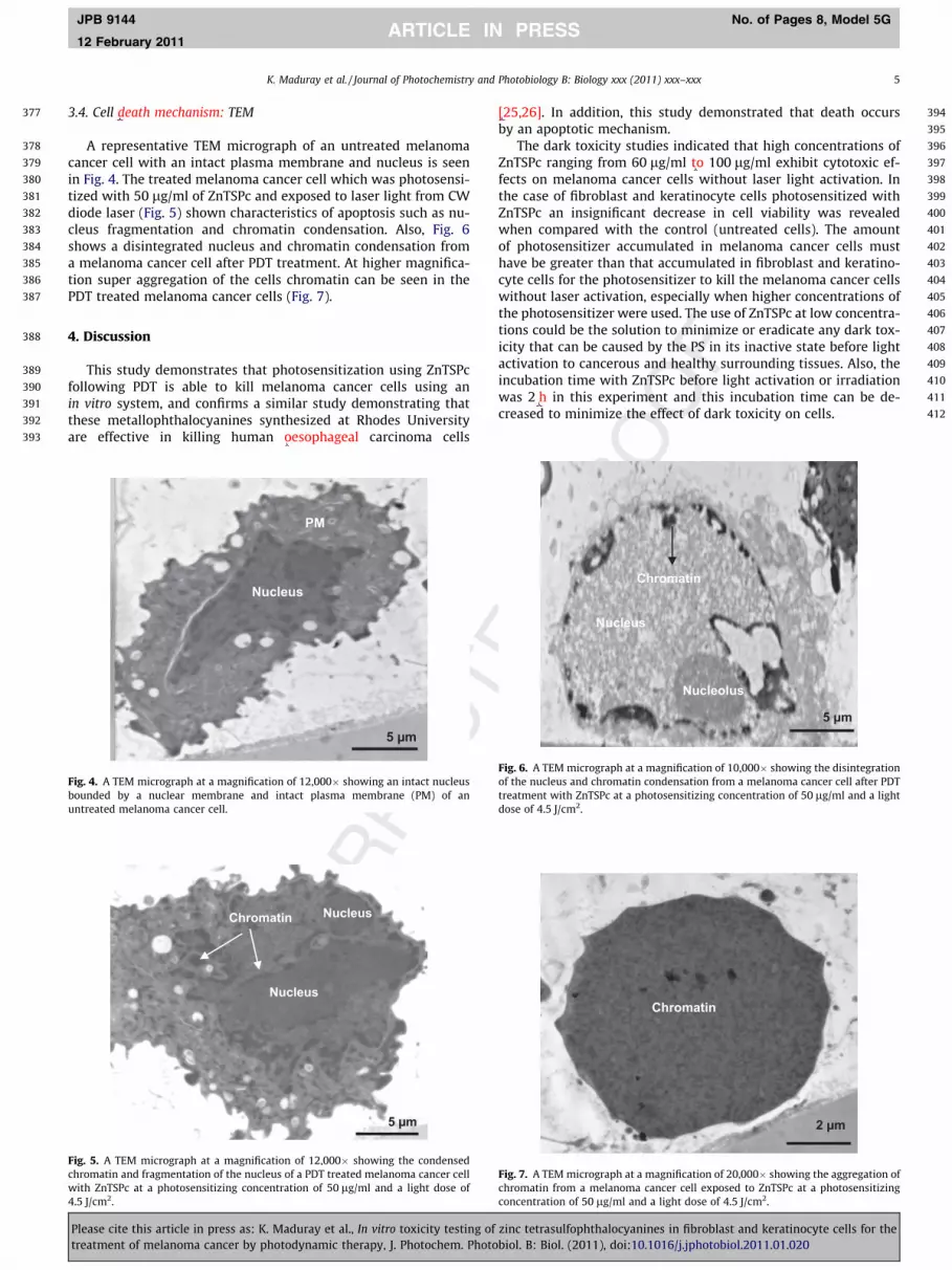
3.4. Cell death mechanism: TEM
A representative TEM micrograph of an untreated melanomacancer cell with an intact plasma membrane and nucleus is seenin Fig. 4. The treated melanoma cancer cell which was photosensi-tized with 50 lg/ml of ZnTSPc and exposed to laser light from CWdiode laser (Fig. 5) shown characteristics of apoptosis such as nu-cleus fragmentation and chromatin condensation. Also, Fig. 6shows a disintegrated nucleus and chromatin condensation froma melanoma cancer cell after PDT treatment. At higher magnifica-tion super aggregation of the cells chromatin can be seen in thePDT treated melanoma cancer cells (Fig. 7).
406
407
408
409
410
411
412
4. Discussion
This study demonstrates that photosensitization using ZnTSPcfollowing PDT is able to kill melanoma cancer cells using anin vitro system, and confirms a similar study demonstrating thatthese metallophthalocyanines synthesized at Rhodes Universityare effective in killing human oesophageal carcinoma cells
5 µm
Nucleus
PM
Fig. 4. A TEM micrograph at a magnification of 12,000� showing an intact nucleusbounded by a nuclear membrane and intact plasma membrane (PM) of anuntreated melanoma cancer cell.
Nucleus
Chromatin Nucleus
5 µm
Fig. 5. A TEM micrograph at a magnification of 12,000� showing the condensedchromatin and fragmentation of the nucleus of a PDT treated melanoma cancer cellwith ZnTSPc at a photosensitizing concentration of 50 lg/ml and a light dose of4.5 J/cm2.
Please cite this article in press as: K. Maduray et al., In vitro toxicity testing oftreatment of melanoma cancer by photodynamic therapy, J. Photochem. Photo
[25,26]. In addition, this study demonstrated that death occursby an apoptotic mechanism.
The dark toxicity studies indicated that high concentrations ofZnTSPc ranging from 60 lg/ml to 100 lg/ml exhibit cytotoxic ef-fects on melanoma cancer cells without laser light activation. Inthe case of fibroblast and keratinocyte cells photosensitized withZnTSPc an insignificant decrease in cell viability was revealedwhen compared with the control (untreated cells). The amountof photosensitizer accumulated in melanoma cancer cells musthave be greater than that accumulated in fibroblast and keratino-cyte cells for the photosensitizer to kill the melanoma cancer cellswithout laser activation, especially when higher concentrations ofthe photosensitizer were used. The use of ZnTSPc at low concentra-tions could be the solution to minimize or eradicate any dark tox-icity that can be caused by the PS in its inactive state before lightactivation to cancerous and healthy surrounding tissues. Also, theincubation time with ZnTSPc before light activation or irradiationwas 2 h in this experiment and this incubation time can be de-creased to minimize the effect of dark toxicity on cells.
5 µm
Nucleus
Chromatin
Nucleolus
Fig. 6. A TEM micrograph at a magnification of 10,000� showing the disintegrationof the nucleus and chromatin condensation from a melanoma cancer cell after PDTtreatment with ZnTSPc at a photosensitizing concentration of 50 lg/ml and a lightdose of 4.5 J/cm2.
2 µm
Chromatin
Fig. 7. A TEM micrograph at a magnification of 20,000� showing the aggregation ofchromatin from a melanoma cancer cell exposed to ZnTSPc at a photosensitizingconcentration of 50 lg/ml and a light dose of 4.5 J/cm2.
zinc tetrasulfophthalocyanines in fibroblast and keratinocyte cells for thebiol. B: Biol. (2011), doi:10.1016/j.jphotobiol.2011.01.020

413
414
415
416
417
418
419
420
421
422
423
424
425
426
427
428
429
430
431
432
433
434
435
436
437
438
439
440
441
442
443
444
445
446
447
448
449
450
451
452
453
454
455
456
457
458
459
460
461
462
463
464
465
466
467
468
469
470
471
472
473
474
475
476
477
478
479
480
481
482
483
484
485
486
487
488
489
490
491
492
493
494
495
496
497
498
499
500
501
502
503
504
505
506
507
508
509
510
511
512
513
514
515
516
517
518
519
520
521
522
523
524
525
526
527
528529530531532533534535536537538539540541542543
6 K. Maduray et al. / Journal of Photochemistry and Photobiology B: Biology xxx (2011) xxx–xxx
JPB 9144 No. of Pages 8, Model 5G
12 February 2011
The exposure of photosensitized melanoma cancer cells to redlight from continuous irradiation at a wavelength of 672 nm re-sulted in a further decrease in cell viability for each of the differentPS concentrations in comparison to the dark toxicity data [27]. Foreffective PDT treatment the photosensitizer in combination withlaser light is necessary. Results demonstrated that as the ZnTSPcconcentration increased the cell viability of melanoma, fibroblastand keratinocyte cells proportionally decreased. For this study,the use of 50 lg/ml of ZnTSPc in combination with a laser lightdose of 4.5 J/cm2 at a wavelength of 672 nm are optimum condi-tions for the effective killing of melanoma cancer cells. The darktoxicity results and the adverse killing effects on normal healthycells after PDT treatment for each of the different ZnTSPc concen-trations were taken into consideration before regarding the ZnTSPcof concentration 50 lg/ml the EC50 value or the optimal photosen-sitizing concentration for this in vitro study.
Statistically, in this study there is a significant difference in thecell viability between absence of laser activation and laser activa-tion for each cell line photosensitized with ZnTSPc, (P < 0.0001)and adjusted for all variables. It is more likely that melanoma cells(P = 0.0016), fibroblast cells (P < 0.0001) or keratinocyte cells(P < 0.0001) would be killed with laser exposure than without laserexposure.
The PDT effect at the tumor site depends on the PS concentra-tion and the radiant energy density at the site which togetherdetermines the energy absorbed per unit volume at the target site.Knowledge of light dose and PS concentrations is therefore essen-tial for the safe and effective treatment. Therefore, the influence ofdifferent light doses (2.5 J/cm2, 7.5 J/cm2 and 10.5 J/cm2) on the cellviability of melanoma, fibroblast and keratinocyte cells photosen-sitized with the optimum ZnTSPc concentration was evaluatedduring the light dose study. It was noted that the optimum ZnTSPc(50 lg/ml) concentrations and the light dose of 4.5 J/cm2 was themost lethal for the melanoma cancer cells in comparison to theother light doses (2.5 J/cm2, 7.5 J/cm2 and 10.5 J/cm2). The cell via-bility of healthy normal fibroblast cells photosensitized withZnTSPc (50 lg/ml) decreased with the increase in light dose. Kerat-inocyte cells photosensitized with ZnTSPc (50 lg/ml) showed thehighest cell viability with the light dose of 2.5 J/cm2 and 4.5 J/cm2. This indicates that the low light doses of 2.5 J/cm2 and 4.5 J/cm2 using the output power between 20 and 30 mW would be abetter combination with the optimum PS concentrations than7.5 J/cm2 and 10.5 J/cm2 for the killing of melanoma cancer cells.
During this study the light doses 7.5 J/cm2 and 10.5 J/cm2 withthe output power between 20 and 30 mW required irradiationtimes in range of 3–7 min. Many photosensitizers can undergo aprocess called photobleaching (photodegradation) duringprolonged irradiation. Photobleaching or photodegradation is thedegradation of the photosensitizer for the production of photo-products by specific photochemical reactions. Studies have shownthat second-generation photosensitizers (e.g. Foscan�) can be morereadily bleached than first-generation photosensitizers (e.g. Photo-frin) [15,28,29]. Patients treated with Foscan� demonstrated that75% of the photosensitizer in the tumor is bleached at the end ofan irradiation light dose treatment with only 10 J/cm2. Photoble-aching has its advantages of theoretically helping to increase thetherapeutic effects of PDT providing that the photosensitizer levelsare higher in the tumors than surrounding healthy tissue[15,28,29].
In this study, the possibility of the photosensitizer (ZnTSPc)photodegrading with the irradiation times associated with thelight doses of 7.5 J/cm2 and 10.5 J/cm2 could be the reason forthe ineffective killing of the melanoma cancer cells. Unfortunately,healthy normal fibroblast and keratinocyte cells were not affectedby PS degradation during long irradiation times because as thelight dose increased cell viability decreased in most cases. There-
Please cite this article in press as: K. Maduray et al., In vitro toxicity testing oftreatment of melanoma cancer by photodynamic therapy, J. Photochem. Photo
fore, the most effective light dose would be 4.5 J/cm2 for the killingof most of the melanoma cancer cells while sparing most of thehealthy normal fibroblast and keratinocyte cells.
Lastly, ultrastructural features of apoptosis were clearly identi-fied in post-irradiated melanoma cancer cells treated with ZnTSPcafter 24 h. Chromatin condensation (Figs. 5 and 6), nucleus frag-mentation (Fig. 5), nucleus disintegration (Fig. 6) and chromatinaggregation (Fig. 7) are detectable morphological changes of lateapoptosis. The TEM can also provide some information on the nat-ure of the biochemical pathways because the morphological orultrastructural changes in apoptotic cells are initiated by certainspecific apoptosis proteins or factors [30,31]. For example, theTEM image in Fig. 6 shows characteristics of caspase-independentapoptosis, namely lumpy incomplete chromatin (electron denseregions or matter) condensation and disintegrated nucleus withtightly packed partially condensed micronuclei [30–33]. ParallelDNA fragmentation and morphological studies were conducted,which reported that apoptosis was induced in melanoma cancercells after PDT treatment with ZnTSPc [34].
5. Conclusion
This in vitro study has shown that ZnTSPc mediated photody-namic therapy is an effective treatment option for melanoma can-cer. 50 lg/ml of ZnTSPc with the treatment light dose of 4.5 J/cm2
from a CW diode laser source with a wavelength of 672 nm wasadequate to destroy melanoma cancer cells via apoptosis withlow killing effects on healthy normal skin cells. There are still sev-eral questions on the detailed effects of photobleaching that stillneeds to be answered in order to understand its role in PDT. PDTas a primary treatment and an adjuvant therapy with either sur-gery or other treatment modalities for melanoma cancer needs tobe further investigated in a clinical setting.
6. Abbreviations
ZnTSPc zinc tetrasulfophthalocyaninePDT Photodynamic therapyPS photosensitizerPcs phthalocyaninesCW continuous wave
Acknowledgements
The authors would like to thank Dr Lester Davids (Departmentof Human Biology, University of Cape Town) for the keratinocytes,Natasha Kolesnikova (Biosciences, CSIR) for the melanoma cancercells and Dr Alan Hall (Laboratory for microscopy and microanaly-sis, University of Pretoria) for your assistance with the Transmis-sion Electron Microscopy.
References
[1] H. McLeod, The impact of cancer on the future, 2009. <http://www.imsa.org.za/files/Library/NHI/policy>.
[2] S. Human, V.B. Bajic, Contributions to skin cancer prevention in South Africa:modelling the UV index utilizing imprecise data, Austrian Journal of Statistics31 (2002) 169–175.
[3] A.C. Geller, S.M. Swetter, K. Brooks, M.F. Demierre, A.L. Yaroch, Screening, earlydetection, and trends for melanoma: current status (2000–2006) and futuredirections, American Academy of Dermatology 57 (2007) 555–572.
[4] C. Garbe, K. Peris, A. Hauschild, P. Saiag, M. Middleton, A. Spatz, J.J. Grob, J.Malvehy, J. Newton-Bishop, A. Stratigos, H. Pehamberger, A. Eggermont,Diagnosis and treatment of melanoma: European consensus-basedinterdisciplinary guidelines, European Journal of Cancer 46 (2010) 270–283.
[5] L.B. Berk, Radiation therapy as primary and adjuvant treatments for local andregional melanoma, Cancer Control 15 (2008) 233–237.
[6] P. Baas, L. Murrer, F.A.N. Zoetmulder, F.A. Stewart, H.B. Ris, N. Van Zandwijk, J.L.Peterse, E.J.T.H. Rutgers, Photodynamic therapy as adjuvant therapy in
zinc tetrasulfophthalocyanines in fibroblast and keratinocyte cells for thebiol. B: Biol. (2011), doi:10.1016/j.jphotobiol.2011.01.020

544545546547548549550551552553554555556557558559560561562563564565566567568569570571572573574575576577578579580581582583584585586
587588589590591592593594595596597598599600601602603604605606607608609610611612613614615616617618619620621622623624625626627628
629
K. Maduray et al. / Journal of Photochemistry and Photobiology B: Biology xxx (2011) xxx–xxx 7
JPB 9144 No. of Pages 8, Model 5G
12 February 2011
surgically treated pleural malignancies, British Journal of Cancer 76 (1997)819–826.
[7] D.H. Lawson, Choices in adjuvant therapy of melanoma, Cancer Control 12(2005) 236–241.
[8] S. Prasanthi, T. Kranthi, N.L. S Bharani, V. Radha Rani, B. Syamala, K. Srinivasulu,Cancer vaccines: a mile stone in cancer therapy, International Journal ofBiotechnology and Biochemistry 6 (2010) 259–269.
[9] E.G. Elias, J.H. Hasskamp, B.K. Sharma, Biology of human cutaneous melanoma,Cancers 2 (2010) 165–189.
[10] M.S. Sabel, V.K. Sondak, Pros and cons of adjuvant interferon in the treatmentof melanoma, The Oncologist 8 (2003) 451–458.
[11] S. Banfi, E. Caruso, L. Buccafurni, R. Ravizza, M. Gariboldi, E. Monti, Zincphthalocyanines-mediated photodynamic therapy induces cell death inadenocarcinoma cells, Journal of Organometallic Chemistry 692 (2007)1269–1276.
[12] A.H.A. Machado, C.P. Soares, N.S. Da Silva, K.C.M. Moraes, Cellular andmolecular studies of the initial process of photodynamic therapy in Hep-2cells using LED light source and two different photosensitizers, Cell BiologyInternational 33 (2009) 785–795.
[13] J.P. Celli, B.Q. Spring, I. Rizvi, C.L. Evans, K.S. Samkoe, S. Verma, B.W. Pogue, T.Hasan, Imaging and photodynamic therapy: mechanisms, monitoring andoptimization, Chemical Reviews 110 (2010) 2795–2838.
[14] C. Hopper, Photodynamic therapy: a clinical reality in the treatment of cancer,The Lancet Oncology 1 (2000) 4212–4219.
[15] K. Plaetzer, B. Krammer, J. Berlanda, F. Berr, T. Kiesslich, Photophysics andphotochemistry of photodynamic therapy: fundamental aspects, Laser MedicalScience 24 (2009) 259–268.
[16] M. Triesscheijn, P. Baas, J.H.M. Schellens, F.A. Stewart, Photodynamic therapyin oncology, The Oncologist 11 (2006) 1034–1044.
[17] Y. You, L. Gibson, R. Hilf, S.R. Davies, A.R. Oseroff, I. Roy, T.Y. Ohulchanskyy, E.J.Bergey, M.R. Detty, Water soluble, core-modified Porphyrins. 3. Synthesis.Photophysical properties and, in vitro studies of photosensitization, uptake,and localization with carboxylic acid-substituted derivatives, Journal ofMedicinal Chemistry 46 (2003) 3734–3747.
[18] C.M. Allen, W.M. Sharman, J.E. Van Lier, Current status of phthalocynanines inthe photodynamic therapy of cancer, Journal of Porphyrins andPhthalocyanines 5 (2001) 161–169.
[19] A. Ogunsipe, M. Durmus�, D. Atilla, A.G. Gürek, V. Ahsen, T. Nyokong, Synthesis,photophysical and photochemical studies on long chain zincphythalocyanines, Synthetic Metals 158 (2008) 839–847.
[20] M. Idowu, T. Nyokong, Synthesis, photophysical and photochemical studies ofwater soluble cationic zinc phthalocyanine derivatives, Polyhedron 28 (2009)416–424.
Please cite this article in press as: K. Maduray et al., In vitro toxicity testing oftreatment of melanoma cancer by photodynamic therapy, J. Photochem. Photo
[21] W. Chidawanyika, T. Nyokong, The synthesis and photophysicochemicalproperties of low-symmetry zinc phthalocyanine analogues, Journal ofPhotochemistry and Photobiology A: Chemistry 206 (2009) 169–176.
[22] A. ErdoGmuS�, A. Ogunsipe, T. Nyokong, Synthesis, photophysics andphotochemistry of novel tetra (quinoxalinyl) phythalocyaninato zinc (II)complexes, Journal of Photochemistry and photobiology A: Chemistry 205(2009) 12–18.
[23] L.M. Davids, B. Kleemann, D. Kacerovská, K. Pizinger, S.H. Kidson, Hypericinphototoxicity induces different modes of cell death in melanoma and humanskin cells, Journal of Photochemistry and Photobiology B: Biology 91 (2008)67–76.
[24] J. Weber, D.H. Busch, Inorganic Chemistry 4 (1965) 469.[25] T.L. Kresfelder, M.J. Cronjé, H. Abrahamse, The effect of two
metallophthalocyanines on the cell viability and proliferation of anesophageal cancer cell line, Photomedicine and Laser Surgery 27 (2009)625–631.
[26] I. Seotsanyana-Mokhosi, T. Kresfelder, H. Abrahamse, T. Nyokong, The effect ofGe, Si and Sn phthalocyanine photosensitizers on cell proliferation andviability of human esophageal carcinoma cells, Journal of Photochemistryand Photobiology B: Biology 83 (2006) 55–62.
[27] R. Decreau, M.J. Richard, P. Verrando, M. Chanon, M. Julliard, Photodynamicactivities of silicon phthalocyanines against achromic M6 melanoma cells andhealthy human melanocytes and keratinocytes, Journal of Photochemistry andPhotobiology B: Biology 48 (1999) 48–56.
[28] I.J. Macdonald, T.J. Dougherty, Basic principles of photodynamic therapy,Journal of Porphyrins and Phthalocyanines 5 (2001) 105–129.
[29] F. Stewart, P. Baas, W. Star, What does photodynamic therapy have to offerradiation oncologists (or their cancer patients)?, Radiotherapy and Oncology48 (1998) 233–248
[30] F. Doonan, T. G Cotter, Morphological assessment of apoptosis, Methods 44(2008) 200–204.
[31] G. Häcker, The morphology of apoptosis, Cell Tissue Research 301 (2000)5–17.
[32] F. Luchetti, A. Di Baldassarre, A.R. Mariani, M. Columbaro, C. Cinti, E. Falcieri,Apoptotic cell death induced by different triggering agents may follow acommon enzymatic pathway, Scanning Microscopy 12 (1998) 351–360.
[33] U. Ziegler, P. Groscurth, Morphological features of cell death, News inPhysiological Science 19 (2004) 124–128.
[34] K. Maduray, A. Karsten, B. Odhav, T. Nyokong, The photodynamic therapyeffect of aluminum and zinc tetrasulfophthalocyanines on melanomacancer cells, Proceedings of SPIE 7376 (2010) 73760A–1, doi:10.1117/12.871055.
zinc tetrasulfophthalocyanines in fibroblast and keratinocyte cells for thebiol. B: Biol. (2011), doi:10.1016/j.jphotobiol.2011.01.020


