Embed Size (px)
Citation preview
Inactivation of the Median Raphe Nucleus Increases Intake ofSucrose Solutions: A Microstructural Analysis
David Wirtshafter1, John D. Davis2, and Thomas R. Stratford1
1Laboratory of Integrative Neuroscience and Department of Psychology (M/C 285), University ofIllinois at Chicago, 1007 W. Harrison St., Chicago, IL 60607-71372810 Maderia Circle, Tallahassee, FL 32312-1815
AbstractPrevious studies have shown that microinjections of the GABA-A agonist muscimol into themedian raphe nucleus (MR) result in large increases in the intake of solid foods. In the currentstudy, we used microstructural techniques to characterize the effects of intra-MR muscimolinjections on the consumption of either a 0.05 M or a 0.29 M sucrose solution. After injections ofeither saline or muscimol, animals consumed more of the 0.29 M than the 0.05 M solution, aneffect which resulted primarily from increases in the initial rate of consumption with no change inthe rate at which licking decayed across the test session. In contrast, intra-MR muscimol injectionshad little effect on the initial licking rate, but greatly increased meal duration, indicating that thistreatment affected ingestion in a different way than did altering the sucrose concentration.Muscimol injections produced a significantly larger increase in the intake of the 0.29 M than ofthe 0.05 M solution. Intra-MR muscimol injections did not alter the within burst rate of licking,suggesting that they did not affect the functioning of the licking pattern generator. In contrast,these injections did increase the number of licks contained within “clusters”, that is groups of licksseparated from each other by intervals of more than 0.5 sec. These findings show that inactivationof the MR produces a powerful effect on the intake of liquid diets, and that the nature of this effectis different than that produced here by changes in sucrose concentration and from those reportedafter pharmacological manipulations of a number of other brain systems. We additionally discussseveral theoretical issues arising in the interpretation of microstructural data
KeywordsFeeding; Ingestive behavior; serotonin; GABA; nucleus centralis superior; microstructure;lickometer
IntroductionThe median raphe nucleus (MR) is a prominent structure located on the midline of themidbrain and pons that sends both serotonergic and nonserotonergic projections to thehypothalamus and other forebrain sites (Vertes, Fortin & Crane, 1999). In previous studies,we have shown that pronounced alterations in food intake can be produced by injections of a
Correspondence to: David Wirtshafter.The following manuscript is the final accepted manuscript. It has not been subjected to the final copyediting, fact-checking, andproofreading required for formal publication. It is not the definitive, publisher-authenticated version. The American PsychologicalAssociation and its Council of Editors disclaim any responsibility or liabilities for errors or omissions of this manuscript version, anyversion derived from this manuscript by NIH, or other third parties. The published version is available atwww.apa.org/pubs/journals/bne
NIH Public AccessAuthor ManuscriptBehav Neurosci. Author manuscript; available in PMC 2012 August 1.
Published in final edited form as:Behav Neurosci. 2011 August ; 125(4): 529–540. doi:10.1037/a0024372.
NIH
-PA Author Manuscript
NIH
-PA Author Manuscript
NIH
-PA Author Manuscript
variety of drugs into the MR (Wirtshafter, 2000). For example, marked hyperphagia can beproduced by intra-MR microinjections of several glutamate antagonists (Wirtshafter &Krebs, 1990; Wirtshafter & Trifunovic, 1988), whereas injections of glutamate agonistssuppress deprivation induced feeding (Wirtshafter & Krebs, 1990). Increases in food intakeare also seen following injections of both the inhibitory GABA-A agonist muscimol(Fletcher, 1994; Klitenick & Wirtshafter, 1988; Klitenick & Wirtshafter, 1989; Paris,Mitsushio & Lorens, 1991) and the GABA-B agonist baclofen (Wirtshafter et al, 1993). Theresponse to muscimol has been studied in the greatest detail, and we have found that theeffects obtained from the MR are much larger than those seen after injections into a numberof adjacent regions, including the ventral tegmental area and the dorsal raphe nucleus(Klitenick & Wirtshafter, 1988). These findings all suggest that treatments which decreaseneural activity within the MR, or its immediate vicinity, lead to large increases in foodintake. Although the MR is a major source of serotonergic projections to the forebrain(Moore, 1981; Steinbusch & Nieuwenhuys, 1983), several lines of evidence suggest that theorexigenic effects of muscimol at this site cannot be entirely accounted for by their effectson serotonin containing neurons (Wirtshafter, 2000). It is likely, therefore, that cells usingother transmitters play a major role in these effects.
The ability of various pharmacological treatments to influence the amount of food eatenmust be secondary to the effects of these drugs on the ingestive behavior of animals. It isclear, however, that simply measuring the amount of food eaten over a given period of timedoes not provide direct information about the way in which the behavior of the animal hasbeen altered. For example, it has been demonstrated that a variety of pharmacologicalagents, at doses which produce very similar changes in total intakes, may affect ingestivebehavior in very different ways (Asin, Davis & Bednarz, 1992; Hsiao & Deupree, 1983;Hsiao & Spencer, 1983; Moran, Carrigan, Schwartz & Ladenheim, 1996). In order todetermine exactly what behavioral effects are produced by a given experimentalmanipulation, it is necessary to actually examine the subject’s behavior, rather than relyingon inferences from the amount consumed. In the current study, we attempted to characterizethe behavioral basis of the hyperphagia produced by injections of muscimol into the MRusing microstructural analysis. In this method, our approach to which has been described indetail in a number of earlier publications (Davis, 1989; Davis & Perez, 1993; Davis &Smith, 1992), the time of occurrence of individual licks is recorded and their distributionover the test period analyzed. We examined the effects of muscimol injections on the intakesof solutions containing two different concentrations of sucrose in order to determine whetherthe muscimol treatments interacted with the properties of the ingestate. In particular, wewere interested in determining whether the effects of muscimol resembled those producedby alterations in sucrose concentration, or those reported after administration of severalother orexigenic agents.
MethodsSubjects
Subjects were 17 adult male Sprague-Dawley derived rats obtained from a colonymaintained by the University of Illinois at Chicago. Animals weighted between 270 and 330g at the time of surgery and were housed individually in wire mesh cages on a 12:12 hr lightdark cycle with food (Wayne LabBlox) and water available ad libitum, except as noted. Allprocedures were in accordance with NIH Guidelines for the Care and Use of LaboratoryAnimals using methods approved by the IACUC.
Wirtshafter et al. Page 2
Behav Neurosci. Author manuscript; available in PMC 2012 August 1.
NIH
-PA Author Manuscript
NIH
-PA Author Manuscript
NIH
-PA Author Manuscript
SurgeryAnimals were anesthetized with sodium pentobarbital (50 mg/kg) and prepared with chronic22 gauge stainless steel guide cannula aimed to terminate 2 mm dorsal to the MR (AP: −0.3,H: 3.2, L: 0.0; mm from interaural line with incisor bar at +6.8 mm). The cannulae werelowered in the sagittal plane following retraction of the superior sagittal sinus (Wirtshafter etal, 1979), and were fixed in place with skull screws and dental cement. A 28 gauge stainlesssteel obturator which extended 2 mm past the end of the guide cannula was then inserted.
ApparatusTesting was conducted in wire mesh cages, identical to those in which the rats were housed,measuring 24 cm wide X 29 cm deep X 20 cm high. A stainless steel drinking spout,attached to a 60 ml calibrated tube, was mounted 6 cm above the floor with the tip flush withthe inside of the chamber. The tube was connected to an amplifier such that a current of lessthan 60 nA was passed through the spout each time the animal licked the tube (DiLogInstruments, Tallahassee, FL). Times of licks were recorded to the nearest millisecond andanalyzed using Quick Lick software (DiLog Instruments).
Microinjection procedureObturators were gently removed and replaced by 28 gauge stainless steel injection cannulae,trimmed to terminate 2 mm beyond the end of the guide cannulae. Injections were made in avolume of 0.5 ul at a rate of 0.25 ul/min using a motor driven microsyringe connected to theinjectors by means of a length of polyethylene tubing. Injectors were removed 30 secfollowing the completion of the injection, and the obturators replaced. Subjects receivedinjections of either normal saline, or 25 ng of muscimol (Klitenick & Wirtshafter, 1988;Klitenick & Wirtshafter, 1989) (Sigma, St. Louis) dissolved in saline. After the injectionwas completed, subjects were placed in the test cages and 2–3 min later the drinking spoutswere inserted.
ProcedureAnimals were allowed 10 days to recover from surgery, during which time they werefrequently handled. Animals were divided randomly into high and low concentration sucrosegroups containing 8 and 9 animals respectively. On the following day, food was removedfrom their cages and 24 hr later the animals were placed in the test cages for a 1 hr session;animals in the low concentration sucrose group received a 0.05 M sucrose solution, andsubjects in the high concentration sucrose group a 0.29 M solution. These concentrationswere chosen based on previous experience in our laboratory indicating that they produceddependably different intakes and that the 0.05M solution was near the lower limit forreliably inducing drinking. At the end of the hour, animals were returned to their home cagesand the volume consumed measured to the nearest ml. On subsequent days, food wasremoved 2 hr prior to testing, to ensure that animals had not just eaten a meal. Subjects inboth the low and high concentration sucrose groups received 7 daily sessions to allow theirintakes to become stable. At this time, approximately half of the subjects in each of thesucrose conditions received an injection of saline and the remaining subjects an injection ofmuscimol prior to being placed in the lickometer for their 60 min. test session. The next day,they received another baseline run and, the day after that, animals were tested followinginjections of the solutions complimentary to those given on the first treatment day.
HistologyFollowing the completion of behavioral testing, all subjects were transcardially perfused,under deep sodium pentobarbital anesthesia, with saline followed by 10% formalin. Several
Wirtshafter et al. Page 3
Behav Neurosci. Author manuscript; available in PMC 2012 August 1.
NIH
-PA Author Manuscript
NIH
-PA Author Manuscript
NIH
-PA Author Manuscript
days later, the brainstems were sectioned at a thickness of 60 um, and the sections stainedwith cresyl violet to evaluate the injection site.
ResultsHistology
Histological examination indicated that all cannula tracks terminated within the medianraphe nucleus at sites similar to those we have examined in previous studies (Fig. 1).
Macrostructural variablesTotal intakes across the test period for animals drinking the low and high concentrations ofsucrose are shown in the upper left panel of Fig. 2 where it can be seen that rats consumedmore of the 0.29 M than of the 0.05 M sucrose solution, and that muscimol injectionsincreased intakes of both solutions, but tended to produce a larger increase of the moreconcentrated ingestate. These conclusions were supported by a 2-way (sucrose concentrationX muscimol) analysis of variance (ANOVA), with repeated measures on the muscimolfactor, which indicated significant effects of sucrose concentration (F(1,15)=99.9; p<0.001),of muscimol (F(1,15)=65.1; p<0.001) and of the sucrose X muscimol interaction(F(1,15)=9.0; p<0.01), indicating that muscimol produced a significantly larger increase infeeding in animals consuming the more concentrated than the less concentrated solution. Atest of simple main indicated that muscimol significantly increased intake even in subjectstested on the low concentration of sucrose (F(1,15)=13.6; p<0.002). As can be seen in theupper right panel of Fig. 2, a similar pattern was seen when total numbers of licks wereexamined, and ANOVA again indicated significant effects of both sucrose (F91,15)=153.5;p<0.001)), muscimol (F(1,15)=38.7; p<0.001), and of the sucrose X muscimol interactionF(1,15)=7.2; p<0.02).
Although the muscimol-induced increase in fluid consumption resulted primarily fromincreases in the number of licks executed, this may not have been the only factor involved.The middle left panel of Fig. 2 indicates that muscimol injections produced a small, butconsistent, decrease in the number of licks required to consume a milliliter of fluid(F(1,15)=5.38; p<0.05). This effect was of similar magnitude at both concentrations ofsucrose, as indicated by the failure of the sucrose X muscimol interaction to approachsignificance (F>1). In contrast, licks/milliliter was actually reduced slightly at the high, ascompared to the low, concentration of sucrose (F(1,15)=6.27; p<0.025). Analogousstatistical results were obtained when lick efficiency (i.e., mls/lick) was analyzed (notshown).
The increases in total fluid consumption observed could result from increases in the amountof time that animals spent drinking or from increases in the overall rate at which fluid wasconsumed. The middle right hand panel of Fig. 2 displays the total duration of licking; thatis the time from the first lick to the last lick made within the session, irrespective of anypauses taken. It can be seen that muscimol injections greatly prolonged the period of timeover which animals licked (F(1,15)=50.4; P<0.001), whereas sucrose concentration waswithout effect on this variable (F<1). The lower left hand panel of Fig. 2 shows the averagerate at which animals drank; that is, the total number of licks divided by duration of theperiod during which licking occurred. Overall licking rate was significantly greater foranimals consuming the higher concentration of sucrose (F(1,15)=63.5; p<0.001), but wasalso slightly increased by injections of muscimol (F(1,15)=4.5; p≤0.05). The sucrose Xmuscimol interaction was not significant (F<1), however, indicating that the effects ofsucrose concentration and of muscimol injections on overall licking rates were additive.
Wirtshafter et al. Page 4
Behav Neurosci. Author manuscript; available in PMC 2012 August 1.
NIH
-PA Author Manuscript
NIH
-PA Author Manuscript
NIH
-PA Author Manuscript
Latency to initiate licking was not significantly affected either by sucrose concentration ormuscimol injections (data not shown).
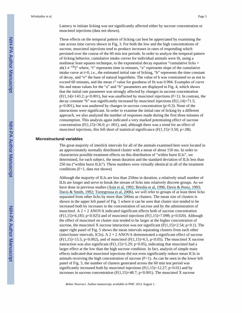
These effects on the temporal pattern of licking can best be appreciated by examining therate across time curves shown in Fig. 3. For both the low and the high concentrations ofsucrose, muscimol injections tend to produce increases in rates of responding whichpersisted over the course of the 60 min test periods. In order to analyze the temporal patternof licking behavior, cumulative intake curves for individual animals were fit, using anonlinear least squares technique, to the exponential decay equation “cumulative licks =ab(1-e−t/b)” where, “t” represents time in minutes, “a” represents slope of the cumulativeintake curve at t=0, i.e., the estimated initial rate of licking, “b” represents the time constantof decay, and “e” the base of natural logarithms. The value of b was constrained so as not toexceed 60 minutes, and the mean r2 value for goodness of fit was 0.994. Examples of curvefits and mean values for the “a” and “b” parameters are displayed in Fig. 4, which showsthat the initial rate parameter was strongly affected by changes in sucrose concentration(F(1,14)=143.2; p<0.001), but was unaffected by muscimol injections (F<1). In contrast, thedecay constant “b” was significantly increased by muscimol injections (F(1,14)=71.5;p<0.001), but was unaltered by changes in sucrose concentration (p>0.3). None of theinteractions were significant. In order to examine the initial rate of licking by a differentapproach, we also analyzed the number of responses made during the first three minutes ofconsumption. This analysis again indicated a very marked potentiating effect of sucroseconcentration (F(1,15)=36.0; p<.001), and, although there was a trend for an effect ofmuscimol injections, this fell short of statistical significance (F(1,15)=3.50; p<.08).
Microstructural variablesThe great majority of interlick intervals for all of the animals examined here were located inan approximately normally distributed cluster with a mean of about 150 ms. In order tocharacterize possible treatment effects on this distribution of “within burst ILIs”, wedetermined, for each subject, the mean duration and the standard deviation of ILIs less than250 ms (“within burst ILIs”). These numbers were virtually identical in all of the treatmentconditions (F<1, data not shown)
Although the majority of ILIs are less than 250ms in duration, a relatively small number ofILIs are longer and serve to break the stream of licks into relatively discrete groups. As wehave done in previous studies (Asin et al, 1992; Breslin et al, 1996; Davis & Perez, 1993;Davis & Smith, 1992; Torregrossa et al, 2006), we will refer to groups of at least three licksseparated from other licks by more than 500ms as clusters. The mean size of clusters isshown in the upper left panel of Fig. 5 where it can be seen that cluster size tended to beincreased both by increases in the concentration of sucrose and by the administration ofmuscimol. A 2 × 2 ANOVA indicated significant effects both of sucrose concentration(F(1,15)=6,183; p<0.025) and of muscimol injections (F(1,15)=7.098; p<0.020). Althoughthe effect of muscimol on cluster size tended to be larger at the higher concentration ofsucrose, the muscimol X sucrose interaction was not significant (F(1,15)=2.54; p>0.1). Theupper right panel of Fig. 5 shows the mean intervals separating clusters from each other(intercluster intervals, ICIs). A 2 × 2 ANOVA demonstrated a significant effect of sucrose(F(1,15)=15.5; p<0.002), and of muscimol (F(1,15)=6.5, p<0.05). The muscimol X sucroseinteraction was also significant (F(1,15)=5.29; p<0.05), indicating that muscimol had alarger effect at the low than the high sucrose condition. In fact, analysis of simple maineffects indicated that muscimol injections did not even significantly reduce mean ICIs inanimals receiving the high concentration of sucrose (F<1). As can be seen in the lower leftpanel of Fig. 5, the number of clusters generated across the 60 min test period wassignificantly increased both by muscimol injections (F(1,15)=12.27; p<0.01) and byincreases in sucrose concentration (F(1,15)=48.7; p<0.001). The muscimol X sucrose
Wirtshafter et al. Page 5
Behav Neurosci. Author manuscript; available in PMC 2012 August 1.
NIH
-PA Author Manuscript
NIH
-PA Author Manuscript
NIH
-PA Author Manuscript
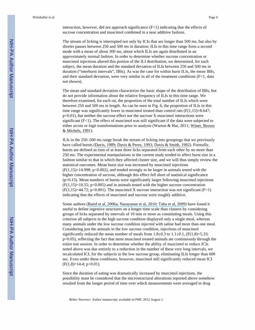
interaction, however, did not approach significance (F<1) indicating that the effects ofsucrose concentration and muscimol combined in a near additive fashion.
The stream of licking is interrupted not only by ICIs that are longer than 500 ms, but also byshorter pauses between 250 and 500 ms in duration; ILIs in this time range form a secondmode with a mean of about 300 ms, about which ILIs are again distributed in anapproximately normal fashion. In order to determine whether sucrose concentration ormuscimol injections altered this portion of the ILI distribution, we determined, for eachsubject, the mean duration and the standard deviation of ILIs between 250 and 500 ms induration (“interburst intervals”, IBIs). As was the case for within burst ILIs, the mean IBIs,and their standard deviation, were very similar in all of the treatment conditions (F<1, datanot shown).
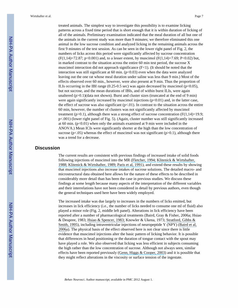
The mean and standard deviation characterize the basic shape of the distribution of IBIs, butdo not provide information about the relative frequency of ILIs in this time range. Wetherefore examined, for each rat, the proportion of the total number of ILIs which werebetween 250 and 500 ms in length. As can be seen in Fig. 6, the proportion of ILIs in thistime range was significantly lower in muscimol treated than control rats (F(1,15)=8.647;p<0.01), but neither the sucrose effect nor the sucrose X muscimol interactions weresignificant (F<1). The effect of muscimol was still significant if the data were subjected toeither arcsin or logit transformations prior to analysis (Warton & Hui, 2011; Winer, Brown& Michels, 1991).
ILIs in the 250–500 ms range break the stream of licking into groupings that we previouslyhave called bursts (Davis, 1989; Davis & Perez, 1993; Davis & Smith, 1992). Formally,bursts are defined as runs of at least three licks separated from each other by no more than250 ms. The experimental manipulations in the current study tended to affect burst size in afashion similar to that in which they affected cluster size, and we will thus simply review thestatistical outcomes. Mean burst size was increased by muscimol injections(F(1,15)=14.998; p<0.002), and tended strongly to be larger in animals tested with thehigher concentration of sucrose, although this effect fell short of statistical significance(p=0.15). Mean numbers of bursts were significantly larger following muscimol injections(F(1,15)=10.55; p<0.005) and in animals tested with the higher sucrose concentration(F(1,15)=44.72; p<0.001). The muscimol X sucrose interaction was not significant (F<1)indicating that the effects of muscimol and sucrose were roughly additive.
Some authors (Baird et al, 2006a; Narayanan et al, 2010; Taha et al, 2009) have found ituseful to define ingestive structures on a longer time scale than clusters by consideringgroups of licks separated by intervals of 10 min or more as constituting meals. Using thiscriterion all subjects in the high sucrose condition displayed only a single meal, whereasmany animals under the low sucrose condition injected with saline had more than one meal.Considering just the animals in the low sucrose condition, injections of muscimolsignificantly reduced the mean number of meals from 1.8±0.3 to 1.1±0.1, (F(1,8)=5.33;p<0.05), reflecting the fact that most muscimol treated animals ate continuously through theentire test session. In order to determine whether the ability of muscimol to reduce ICIsnoted above was due entirely to a reduction in the number of these very long intervals, werecalculated ICI, for the subjects in the low sucrose group, eliminating ILIs longer than 600sec. Even under these conditions, however, muscimol still significantly reduced mean ICI(F(1,8)=14.4; p<0.01).
Since the duration of eating was dramatically increased by muscimol injections, thepossibility must be considered that the microstructural alterations reported above somehowresulted from the longer period of time over which measurements were averaged in drug
Wirtshafter et al. Page 6
Behav Neurosci. Author manuscript; available in PMC 2012 August 1.
NIH
-PA Author Manuscript
NIH
-PA Author Manuscript
NIH
-PA Author Manuscript
treated animals. The simplest way to investigate this possibility is to examine lickingpatterns across a fixed time period that is short enough that it is within duration of licking ofall of the animals. Preliminary examination indicated that the meal duration of all but one ofthe animals in the current study was more than 9 minutes; we therefore eliminated this oneanimal in the low sucrose condition and analyzed licking in the remaining animals across thefirst 9 minutes of the test session. As can be seen in the lower right panel of Fig. 2, thenumbers of licks across this period were significantly affected by sucrose concentration(F(1,14)=72.87; p<0.001) and, to a lesser extent, by muscimol (F(1,14)=7.69; P<0.02) but,in marked contrast to the situation across the entire 60 min test period, the sucrose Xmuscimol interaction did not approach significance (F<1). (It should be noted that theinteraction was still significant at 60 min. (p<0.03) even when the data were analyzedleaving out the one rat whose meal duration under saline was less than 9 min.) Most of theeffects observed over 60 min., however, were also present at 9 min. Thus the proportion ofILIs occurring in the IBI range (0.25-0.5 sec) was again decreased by muscimol (p<0.05),but not sucrose, and the mean durations of IBIs, and of within burst ILIs, were againunaltered (p>0.1)(data not shown). Burst and cluster sizes (truncated at the end of 9 min)were again significantly increased by muscimol injections (p<0.01) and, in the latter case,the effect of sucrose was also significant (p<.01). In contrast to the situation across the entire60 min, however, the number of clusters was not significantly affected by muscimoltreatment (p>0.1), although there was a strong effect of sucrose concentration (F(1,14)=19.9;p<.001) (lower right panel of Fig. 5). (Again, cluster number was still significantly increasedat 60 min. (p<0.01) when only the animals examined at 9 min were included in theANOVA.) Mean ICIs were significantly shorter at the high than the low concentration ofsucrose (p<.05) whereas the effect of muscimol was not significant (p>0.1), although therewas a trend for a decrease.
DiscussionThe current results are consistent with previous findings of increased intake of solid foodsfollowing injections of muscimol into the MR (Fletcher, 1994; Klitenick & Wirtshafter,1988; Klitenick & Wirtshafter, 1989; Paris et al, 1991), and extend these results by showingthat muscimol injections also increase intakes of sucrose solutions. The detailed macro- andmicrostructural data obtained here allows for the nature of these effects to be described inconsiderably more detail than has been the case in previous studies. We discuss thesefindings at some length because many aspects of the interpretation of the different variablesand their interrelations have not been considered in detail by previous authors, even thoughthe general techniques used here have been widely employed.
The increased intake was due largely to increases in the numbers of licks emitted, butincreases in lick efficiency (i.e., the number of licks needed to consume one ml of fluid) alsoplayed a minor role (Fig. 2, middle left panel). Alterations in lick efficiency have beenreported after a number of pharmacological treatments (Baird, Gray & Fisher, 2006a; Hsiao& Deupree, 1983; Hsiao & Spencer, 1983; Knowler & Ukena, 1973; Stratford, Gibbs &Smith, 1995), including intraventricular injections of neuropeptide Y (NPY) (Baird et al,2006a). The physical basis of the effect observed here is not clear since there is littleevidence that muscimol injections alter the basic pattern of licking behavior. It is possiblethat differences in head positioning or the duration of tongue contact with the spout mayhave played a role. We also observed that licking was less efficient in subjects consumingthe high rather than the low concentration of sucrose. Although not always seen, similareffects have been reported previously (Genn, Higgs & Cooper, 2003) and it is possible thatthey might reflect alterations in the viscosity or surface tension of the ingestate.
Wirtshafter et al. Page 7
Behav Neurosci. Author manuscript; available in PMC 2012 August 1.
NIH
-PA Author Manuscript
NIH
-PA Author Manuscript
NIH
-PA Author Manuscript
The most dramatic effect of the intra-MR muscimol injections was a prolongation of theamount of time animals spent licking. In contrast, the effect of sucrose concentration wasprimarily to change the initial rate at which animals drank. It is especially instructive tocompare the rate across time as shown in Fig. 3 for the saline injected rats drinking the highconcentration of sucrose and the muscimol-injected animals drinking the low concentrationof sucrose. Mean intakes under the two conditions were similar, but the temporal pattern ofdrinking shown by the subjects was drastically different. Thus, animals receiving salineinjections and the high concentration of sucrose began licking at a high rate that rapidlydecayed, whereas the subjects receiving muscimol injections and the low concentration ofsucrose began drinking at a modest rate that declined only slowly throughout the session.This illustrates the point that measures of total amount consumed contain only a smallproportion of the available information, and demonstrates that, despite the similarities inoverall intake, the behavior of muscimol treated animals consuming a low concentration ofsucrose does not resemble that of control subjects consuming a higher concentration ofsucrose.
Previous studies have shown that cumulative intake vs. time curves can be well fit by twoparameter exponential functions of the form “cumulative licks = ab(1-e−t/b)” (Davis &Levine, 1977; Davis & Perez, 1993; Genn et al, 2003), an observation that was confirmed inthe current experiment. Earlier studies have also shown that the value of the initial rateparameter “a” is strongly influenced by the sensory properties of the ingestate (Breslin,Davis & Rosenak, 1996; Davis, 1998; Davis & Levine, 1977; Genn et al, 2003), and thisfinding was also confirmed here in that values for the rate parameter were substantiallyhigher in animals consuming the more concentrated sucrose solution. In contrast, muscimolinjections did not significantly alter the initial rate parameter, or the numbers of licks madein the first 3 min, although there was a small trend in both cases. These results indicate thatchanges in the initial rate of drinking are not the major mechanism through which intra-MRmuscimol exerts its effects on intake.
The time constant parameter “b” reflects the rate at which licking declines from its initialvalue and is often related to inhibitory feedback arising from the act of licking, or itspostingestive consequences (Davis, 1998; Davis & Levine, 1977). In the current study thetime constant was not affected by changes in the concentration of sucrose, a result which issomewhat surprising given that previous studies have found that this value is inverselyproportional to carbohydrate concentration (Davis & Levine, 1977). It is possible that thisdifference may reflect the fact that sucrose concentration was a between subject variable inthe current study, whereas individual subjects were tested on multiple carbohydrateconcentrations in previous reports. At any account, in contrast to the lack of effect of sucroseconcentration, muscimol injections produced a marked increase in the time constant,reflecting the fact that licking persisted for a much longer time in these subjects than it did insaline-treated animals. The simplest explanation of these findings is that inactivation of theMR reduced the sensitivity of animals to some form of licking-induced feedback. A similarsuggestion has been made with respect to neuropeptide Y (Lynch, Hart & Babcock, 1994),which also increases meal durations. In this case, however, it has been shown that NPY stillexerts this effect in sham feeding animals (Torregrossa, Davis & Smith, 2006), suggestingthat its actions cannot result entirely from a blockade of post-ingestive feedback. Furtherstudies will be needed to determine whether a similar conclusion holds for the case of intra-MR injections of muscimol. It should also be pointed out that care should be exerted ininterpreting the effects of drug injections on the temporal pattern of licking; the actions ofmost drugs vary as a function of time since injection, and such “time-course effects” mightwell combine in unknown ways with changes in licking rate naturally occurring across thecourse of a meal.
Wirtshafter et al. Page 8
Behav Neurosci. Author manuscript; available in PMC 2012 August 1.
NIH
-PA Author Manuscript
NIH
-PA Author Manuscript
NIH
-PA Author Manuscript
In the current experiment, muscimol injections produced a significantly larger increase inintake in animals consuming the high than the low concentration of sucrose. Although onemight be tempted to suggest that this reflects a synergistic effect of sucrose concentrationand raphe inactivation on the vigor of licking, there is little in the data to support thiscontention. Analysis of the time course of drinking allows for much simpler explanation.Increasing the sucrose concentration produces a large increase in the mean rate of licking,whereas muscimol injections primarily increase the duration of meals. Since the totalamount consumed is equal to the mean rate of ingestion multiplied by the duration oflicking, an equivalent increase in meal duration naturally will produce a larger increase intotal consumption in animals licking a high rather than a low concentration solution. If thisexplanation were correct, one would expect the synergy between sucrose concentration andmuscimol injections to be greatly reduced under conditions in which meal duration cannotvary; the current results support this viewpoint because the effects of muscimol and sucroseconcentration were statistically additive when numbers of licks were measured across justthe first 9 min of access. It is interesting that the orexigenic compound NPY has beensuggested to alter oral sensory processing based on findings that the magnitude of its effectvaries with the palatability of the ingestate. However, since NPY, like intra-MR muscimol,tends to lengthen meals (Lynch et al, 1994; Torregrossa et al, 2006), the considerationsdiscussed here may provide a simpler explanation of these findings.
In normal rats licking sapid solutions, the great majority of interlick intervals (ILIs) fall inthe vicinity of 150 ms (Corbit & Luschei, 1969; Stellar & Hill, 1952) and have beenproposed to reflect the operation of a brainstem licking pattern generator (Davis & Smith,1992; Travers, Dinardo & Karimnamaze, 1997; Wiesenfeld, Halpern & Tapper, 1977). Themean of these “within burst ILIs” is typically unaltered by deprivation (Corbit & Luschei,1969), although small and inconsistent changes have sometimes been observed (Davis &Perez, 1993; Spector et al, 1998). Alterations in within burst ILIs have also been reportedafter systemic or intracranial administration of a number of compounds (Asin, Davis &Bednarz, 1992; Baird, Rios, Gray, Walsh, Fischer & Pecora, 2006b; Knowler & Ukena,1973; Stratford et al, 1995). In the current study, however, the shape and location of thisdistribution were not altered by muscimol injections, suggesting that inactivation of the MRdoes not directly influence the licking pattern generator,
In agreement with many previous studies (Corbit & Luschei, 1969; Davis & Smith, 1992;Gramling, Fowler & Collins, 1984; Hsiao & Deupree, 1983; Hsiao & Spencer, 1983), wenoted that a second, much smaller, peak in the ILI distribution could be observed with amean close to 300 msec. Some authors have reported that additional peaks can be observedwith means close to 450 and 600 msec (Corbit & Luschei, 1969; Gramling et al, 1984), butthese were not seen in the current experiment. Previous studies have shown that ILIsbetween 250 and 500 ms, which we will refer to as inter-burst intervals (IBIs), aredistributed normally (Davis & Smith, 1992). Mean IBI has been reported to be unaffected byeither deprivation or sucrose concentration (Davis & Perez, 1993), and we confirmed thelatter result in the current report. We also found that the mean and standard deviation of thedistribution of IBIs was not affected by inactivation of the MR, suggesting that thismanipulation did not alter the nature of the behavior responsible for these brief pauses inrecorded licking. In contrast, the relative frequency at which IBIs occurred was significantlyreduced by muscimol injections. Alterations in the relative frequency of IBIs have also beenreported after several other pharmacological treatments (Baird et al, 2006b; Gramling et al,1984; Hsiao & Deupree, 1983; Hsiao & Spencer, 1983; Taha, Katsura, Noorvaash, Seroussi,& Fields,, 2009). The exact significance of IBIs is not currently understood; one possibilityis that they may simply reflect standard licks during which the tongue did not makeelectrical contact with the spout (Corbit & Luschei, 1969). Alternatively, they might resultfrom alterations in the actual rate of licking (Hsiao & Deupree, 1983), or reflect some other
Wirtshafter et al. Page 9
Behav Neurosci. Author manuscript; available in PMC 2012 August 1.
NIH
-PA Author Manuscript
NIH
-PA Author Manuscript
NIH
-PA Author Manuscript
behavior, such as a lateral tongue movement (Davis & Smith, 1992), which coincidentallytakes about twice as long as a standard lick. Although not conclusive, the current results aremore easily accommodated within the “separate behavior viewpoint”; for example,inactivation of the MR could either reduce the frequency of the behavior responsible forIBIs, or could increase the tendency to lick to the point that any sort of interruption of thelicking rhythm became less probable. In contrast, it is not immediately apparent how theprobability of missed licks could be altered, although possibly this could result from changesin positioning of the head with respect to the spout. High-speed video analysis of lickingwould be useful in deciding between these possibilities.
The occurrence of intermittent pauses serves to divide the stream of licks into discrete bouts.Using the terminology of Davis and his coworkers (Davis, 1998; Davis & Perez, 1993;Davis & Smith, 1992), bouts of licking separated from each other by pauses of at least 0.25sec. are referred to as bursts, whereas bouts isolated by pauses of more than 0.5 seconds(“intercluster intervals,” ICIs) are referred to as “clusters”. (Some workers (Spector, Klumpp& Kaplan, 1998) have suggested that a cutoff of 1.0 sec be used, but this is a matter of littlepractical importance since, under the current conditions, ILIs between 0.5 and 1.0 secondswere extremely rare.) Alterations in the size of clusters therefore simply reflect alterations inthe relative frequency of ILIs longer than 0.5 sec (the mean cluster size being approximatelyequal to the reciprocal of the probability of an ICI). In contrast, burst size is related to theprobabilities of both IBIs and of ICIs, being approximately equal to one over the sum of theprobabilities of IBIs and ICIs. As a result of these relations, changes in the frequency ofpauses between 0.25 and 0.5 sec in duration would be expected to alter burst size alone,whereas changes in the frequency of pauses longer than 0.5 sec would influence both burstand cluster size. Burst size is thus not independent of cluster size, a consideration whichsuggests that the relative frequency of IBIs may provide a more discrete measure of theseinterruptions than does burst size. In the current study, cluster size was significantlyincreased both by injections of muscimol and by increases in the concentration of sucrose.Burst size was also significantly increased by muscimol injections, as would be expectedgiven the increases in cluster size and the decrease in the relative frequency of IBIsdiscussed above.
Many previous studies have demonstrated effects of tastant concentration on cluster or burstsize, and it has been suggested that these parameters may reflect the palatability of theingestates. Our current observation that inactivation of the MR increases both cluster andburst size is consistent with the notion that this structure may influence hedonic processingof taste related stimuli. Alterations in the perceived palatability of the test solutions would,however, also be expected to alter the initial rate of licking, an effect which was notobserved in the current experiment. Other experiments (Baird et al, 2006a; Torregrossa et al,2006) have also found that effects on initial rate and cluster size can be dissociated,suggesting that factors in addition to palatability can also influence these variables. It isinteresting in this context that lesions of the MR result in perseverative behavior undercertain conditions (Asin, Wirtshafter & Kent, 1979; Wirtshafter & Asin, 1983; Wirtshafter& Asin, 1986); if a similar effect occurred after muscimol injections, it is possible that itcould play a role in the observed prolongation of the bouts of licking.
Given that the median raphe is a major source of serotonergic projections to the forebrain, itis striking that the serotonergic anorexics fluoxetine and fenfluramine have been reported todecrease burst and cluster size (Asin et al, 1992), effects opposite to those observed herewith intra-MR injections of muscimol, which are known to reduce serotonin release (Shim,Javaid & Wirtshafter, 1997; Wirtshafter & Trifunovic, 1992). Although inhibition ofserotonin neurons does not appear to be the only mechanism underlying the orexigeniceffects of intra-MR muscimol (Wirtshafter, 2000), it is possible that serotonin may be
Wirtshafter et al. Page 10
Behav Neurosci. Author manuscript; available in PMC 2012 August 1.
NIH
-PA Author Manuscript
NIH
-PA Author Manuscript
NIH
-PA Author Manuscript
involved in effects on burst and cluster size. A number of feeding-inducing manipulations,including intraventricular injections of either orexin or NPY (Baird et al, 2006a; Baird,Choe, Loveland, Beck, Mahoney, Lord & Grigg, 2009; Torregrossa et al, 2006), andinjections of muscimol or DAMGO into the accumbens shell (Stratford & Wirtshafter, 2007;Taha et al., 2009), do not affect cluster size, showing that the these treatments affect feedingin a different way than does intra-MR muscimol. In contrast, increases in cluster size havebeen reported after systemic administration of benzodiazepines (Higgs & Cooper, 2000).These different patterns illustrate the ability of microstructural analysis to distinguishbetween different types of effects on ingestive behavior that could not be detected byexamining intake data alone. Since intra-MR injections of benzodiazepines have been shownto produce several behavioral effects (Gonzalez, Quagazzal & File, 1998; Sainati & Lorens,1983), which are presumably mediated through enhanced GABAergic transmission, aninteresting possibility is that the MR may be one of the sites at which systemicallyadministered benzodiazepines act to influence feeding.
Increasing the concentration of sucrose from 0.05 to 0.29M significantly increased thenumbers of clusters generated across the 60 min session, and a similar effect was foundwhen only the first 9 min were examined. In contrast, muscimol injections significantlyincreased the number of clusters when measured across the entire test session, but this effectwas absent when only the first 9 min were examined. These divergent results highlightcertain logical differences between the constraints on microstructural variables that occurwhen intake is measured over fixed or potentially varying time periods. Since changing thesucrose concentration did not alter the duration of eating, the only way that animals couldgenerate more clusters would be if they were to shorten the time between them. Thisinference was in fact empirically confirmed, because mean ICIs were significantly shorter atthe high than the low sucrose concentration. This effect was apparent both across the entiretest session and during the first 9 min. Muscimol, in contrast, produced a large increase inthe total duration of eating; increasing meal duration allows for more clusters to be producedeven in the absence of changes in mean ICIs. In fact, mean ICIs were significantly reducedby muscimol injections only at the low concentration of sucrose, and even here the effectwas much smaller than that produced by increases in sucrose concentration. When, licking isevaluated over the first 9 min alone, a period of time shorter than the smallest observedmeal, changes in the total duration of eating can no longer play a role, and the number ofclusters generated is related simply to the mean duration of clusters and of ICIs. Under theseconditions, sucrose concentration, but not muscimol, had an effect on mean ICIs, with theresult that the number of clusters was significantly influenced by sucrose concentration, butnot by muscimol. (In fact, muscimol actually tended to reduce the number of clusters seen inthe first nine min by animals consuming the high concentration of sucrose, presumablybecause cluster size had become so large under these conditions that there was insufficienttime remaining in the test to accommodate more clusters.) These results further highlight thedifferent nature of the effects of muscimol injections and increased sucrose concentration;the latter manipulation increases the numbers of clusters because it tends to reduce the timebetween them, whereas muscimol has relatively small effects on the ICI and increases thenumber of clusters primarily by increasing the duration of eating. In fact, the relativelymodest effects of MR inactivation on ICIs during the early part of the drinking period maybe one reason why initial rate is affected to a much smaller extent by these injections than bychanges in sucrose concentration.
In summary, the current results demonstrate that while intake can be augmented both byincreasing sucrose concentrations and by injecting muscimol into the MR, the detailednature of these two effects differ in a large number of particulars. The data thus provide nosupport for the view that the MR produces its effects by causing animals to treat sucrosesolutions as though they are more concentrated than they really are. Our findings also
Wirtshafter et al. Page 11
Behav Neurosci. Author manuscript; available in PMC 2012 August 1.
NIH
-PA Author Manuscript
NIH
-PA Author Manuscript
NIH
-PA Author Manuscript
suggest that the effects of MR inactivation on intake differ substantially from those reportedafter a number of other orexigenic treatments. For example, intraventricular injections ofmelanocyte concentrating hormone (MCH) resemble intra-MR muscimol in that they tend toincrease cluster size and decrease the proportion of IBIs; in contrast to intra-MR muscimol,however, these injections do not alter meal duration and their effect on intake is primarilydue to increases in the within meal rate of eating (Baird et al, 2006b). On the other hand,meal duration is increased by intraventricular injections of orexin or NPY, but theseinjections either have no effect on, or, in the case of NPY, sometimes actually decrease,cluster size (Baird et al, 2006a; Baird et al, 2009; Torregrossa et al, 2006). Although thesecomparisons must be made with caution, since the various results have been obtained indifferent laboratories using varying methods, they do certainly suggest that the effects ofinactivation of the MR are distinct from those of these other treatments. In other studies(unpublished observations) we have observed that intra-MR muscimol produces a very largeincrease in Fos expression within the lateral hypothalamus (LH) which involves bothorexinergic cells, and neurons not containing this peptide, and we have suggested elsewherethat some of the effects of MR inactivation on ingestive behavior may be mediated throughthe LH (Stratford & Wirtshafter, 2000; Wirtshafter, 2000). One interesting possibility is thatthese injections may simultaneously activate multiple peptidergic cell groups, and perhapsother types of cells as well, to produce a more complex pattern of effects on licking behaviorthan can be produced by manipulation of any one of these in isolation. Of course, it ispossible that projections to targets other than the LH may be critically involved in the effectsobtained from the MR – clearly a great deal more work needs to be done to understand thepowerful influence of the paramedian tegmentum in the control of ingestive behavior.
AcknowledgmentsThis publication was supported by grants 0641943 from the National Science Foundation, R01DK071738 from theNational Institute of Diabetes and Digestive and Kidney Diseases, and R03DA020802 from the National Institutefor Drug Abuse. The content is solely the responsibility of the authors and does not necessarily represent theofficial views of the National Science Foundation or the National Institutes of Health.
ReferencesAsin KE, Davis JD, Bednarz L. Differential effects of serotonergic and catecholaminergic drugs on
ingestive behavior. Psychopharmacology. 1992; 109:415–421. [PubMed: 1365856]Asin KE, Wirtshafter D, Kent EW. Straight alley acquisition and extinction and open field activity
following discrete electrolytic lesions of the mesencephalic raphe nuclei. Behavioral and NeuralBiology. 1979; 25:242–256. [PubMed: 464976]
Baird JP, Choe A, Loveland JL, Beck J, Mahoney CE, Lord JS, Grigg LA. Orexin-A hyperphagia:Hindbrain participation in consummatory feeding responses. Endocrinology. 2009; 150:1202–1216.[PubMed: 19008313]
Baird JP, Gray NEE, Fischer SG. Effects of neuropeptide Y on feeding microstructure: Dissociation ofappetitive and consummatory actions. Behavioral Neuroscience. 2006a; 120:937–951. [PubMed:16893299]
Baird JP, Rios C, Gray NE, Walsh CE, Fischer SG, Pecora AL. Effect of melanin-concentratinghormone on licking microstructure and brief-acess taste responses. American Journal of Physiology,Regulatory, Integrative and Comparative Physiology. 2006b; 294 R12656-R1274.
Breslin PAS, Davis JD, Rosenak R. Saccharin increases the effectiveness of glucose in stimulatingingestion in rats but has little effect on negative feedback. Physiology and Behavior. 1996; 60:411–416. [PubMed: 8840899]
Corbit JD, Luschei ES. Invarience of the rat's rate of drinking. Journal of Comparative andPhysiological Psychology. 1969; 69:119–125. [PubMed: 5347358]
Davis JD. The microstructure of ingestive behavior. Annals of the New York Academy of Sciences.1989; 575:106–119. [PubMed: 2699182]
Wirtshafter et al. Page 12
Behav Neurosci. Author manuscript; available in PMC 2012 August 1.
NIH
-PA Author Manuscript
NIH
-PA Author Manuscript
NIH
-PA Author Manuscript
Davis JD. A model for the control of ingestion - 20 years later. Prog.Psychobiol.Physiol.Psychol.1998; 17:127–173.
Davis JD, Levine MW. A model for the control of ingestion. Psychological Review. 1977; 84:379–412. [PubMed: 327497]
Davis JD, Perez MC. Food deprivation- and palatability-induced microstructural changes in ingestivebehavior. American Journal of Physiology, Regulatory, Integrative and Comparative Physiology.1993; 264:R97–R103.
Davis JD, Smith GP. Analysis of the microstructure of the rhythmic tongue movements of ratsingesting maltose and sucrose solutions. Behavioral Neuroscience. 1992; 106:217–228. [PubMed:1554433]
Fletcher PJ. Effects of 8-OH-DPAT, 5-CT and muscimol on behavior maintained by a DRL20schedule of reinforcement, following microinjection into the dorsal or median raphe nuclei.Behavioural Pharmacology. 1994; 5:326–336. [PubMed: 11224282]
Genn RF, Higgs S, Cooper SJ. The effects of 7-OH-DPAT, quinpirole and raclopride on licking forsucrose solutions in the non-deprived rat. Behavioural Pharmacology. 2003; 14:609–617.[PubMed: 14665978]
Gonzalez LE, Quagazzal AM, File SE. Stimulation of benzodiazepine receptors in the dorsalhippocampus and median rephe reveals differential GABAergic control in two animal tests ofanxiety. European Journal of Neuroscience. 1998; 10:3673–3680. [PubMed: 9875346]
Gramling SE, Fowler LJ, Collins KR. Some effects of pimozide on nondeprived rats licking sucrosesolutions in an anhedonia paradigm. Pharmacology, Biochemistry and Behavior. 1984; 21:617–624.
Higgs S, Cooper SJ. The effect of the dopamine D2 receptor antagonist raclopride on the pattern oflicking microstructure induced by midazolam in the rat. European Journal of Pharmacology. 2000;409:73–80. [PubMed: 11099702]
Hsiao S, Deupree D. Cholecystokinin and bombesin effects on rewarded and nonrewarded operants.Peptides. 1983; 4:1–3. [PubMed: 6866804]
Hsiao S, Spencer R. Analysis of licking responses in rats: Effects of cholecystokinin and bombesin.Behavioral Neuroscience. 1983; 97:234–235. [PubMed: 6849686]
Klitenick MA, Wirtshafter D. Comparative studies of the ingestive behaviors produced bymicroinjections of muscimol into the midbrain raphe nuclei or the ventral tegmental area of the rat.Life Sci. 1988; 42:775–782. [PubMed: 3339955]
Klitenick MA, Wirtshafter D. Elicitation of feeding, drinking, and gnawing following microinjectionsof muscimol into the median raphe nucleus of rats. Behavioral and Neural Biology. 1989; 51:436–441. [PubMed: 2730501]
Knowler CW, Ukena TE. The effects of chlorpromazine, pentobarbital, chlordiazepoxide and d-amphetamine on rates of licking in the rat. Journal of Pharmacology and ExperimentalTherapeutics. 1973; 184:385–397. [PubMed: 4688176]
Lynch WC, Hart P, Babcock AM. Neuropeptide Y attenuates satiety: evidence from a detailed analysisof patterns ingestion. Brain Research. 1994; 626:28–34. [PubMed: 8156407]
Moore, KE. The anatomy of central serotonin neuron systems in the rat brain. In: Jacobs, BL.;Gelpern, A., editors. Serotonin Neurotransmission and Behavior. Cambridge: MIT Press; 1981.
Moran TH, Carrigan TS, Schwartz GJ, Ladenheim EE. Bombesin and cholecystokinin differentiallyaffect ingestive microstructural variables whether given alone or in combination. BehavioralNeuroscience. 1996; 110:1110–1116. [PubMed: 8919013]
Narayanan NS, Guarnieri DJ, DiLeone RJ. Metabolic hormones, dopamine circuits, and feeding.Frontiers in Neuroendocrinology. 2010; 31:104–112. [PubMed: 19836414]
Paris JM, Mitsushio H, Lorens SA. Intra-midbrain raphe injections of the neurokinin-3 agonistsenktide inhibit food and water intake in the rat. Pharmacology, Biochemistry and Behavior. 1991;38:223–226.
Sainati S, Lorens SA. Intra-raphe benzodiazepines enhance rat locomotor activity: Interactions withGABA. Pharmacology, Biochemistry and Behavior. 1983; 18:407–414.
Wirtshafter et al. Page 13
Behav Neurosci. Author manuscript; available in PMC 2012 August 1.
NIH
-PA Author Manuscript
NIH
-PA Author Manuscript
NIH
-PA Author Manuscript
Shim I, Javaid J, Wirtshafter D. Dissociation of hippocampal serotonin release and locomotor activityfollowing pharmacological manipulations of the median raphe nucleus. BehaviouralBrain.Research. 1997; 89:191–198. [PubMed: 9475626]
Spector AC, Klumpp PA, Kaplan JM. Analytical issues in the evaluation of food deprivation andsucrose concentration effects on the microstructure of licking behavior in the rat. BehavioralNeuroscience. 1998; 112:678–694. [PubMed: 9676983]
Steinbusch, HWM.; Nieuwenhuys, R. The raphe nuclei of the rat brainstem: A cytoarchitectonic andimmunohistochemical study. In: Emson, PC., editor. Chemical Neuroanatomy. New York: RavenPress; 1983. p. 131-208.
Stellar E, Hill JH. The rat's rat of drinking as a function of water deprivation. Journal of Comparativeand Physiological Psychology. 1952; 45:95–102.
Stratford TR, Gibbs J, Smith GP. Microstructural analysis of licking behavior following peripheraladministration of bombesin or gastrin-releasing peptide. Peptides. 1995; 16:903–909. [PubMed:7479333]
Stratford TR, Wirtshafter D. Forebrain lesions differentially affect drinking elicited by dipsogenicchallenges and injections of muscimol into the median raphe nucleus. Behavioral Neuroscience.2000; 114:760–771. [PubMed: 10959535]
Stratford, TR.; Wirtshafter, D. Activation of GABA-A receptors in the nucleus accumbens shell elicitsopposite effects on consumption of sucrose and saccharin solutions; Neuroscience MeetingPlanner, Program No. 630.5; 2007.
Taha SA, Katsuura Y, Noorvaash D, Seroussi A, Fields HL. Convergent, not serial, striatal and pallidalcircuits regulate opioid-induced food intake. Neuroscience. 2009; 161:718–733. [PubMed:19336249]
Torregrossa AM, Davis JD, Smith GP. Orosensory stimulation is sufficient and postingestive negativefeedback is not necessary for neuropeptide Y to increase sucrose intake. Physiology and Behavior.2006; 87:773–780. [PubMed: 16540131]
Travers JB, Dinardo LA, Karimnamazi H. Motor and premotor mechanisms of licking. Neuroscienceand Biobehavioral Reviews. 1997; 21:631–647. [PubMed: 9353796]
Vertes RP, Fortin WJ, Crane AM. Projections of the median raphe nucleus in the rat. Journal ofComparative Neurology. 1999; 407:555–582. [PubMed: 10235645]
Warton DI, Hui FKC. The arcsin is asinine: the analysis of proportions in ecology. Ecology. 2011;92:3–13. [PubMed: 21560670]
Wiesenfeld Z, Halpern BP, Tapper DN. Licking behavior: Evidence of hypoglossal oscillator. Science.1977; 196:1122–1124. [PubMed: 558653]
Winer, BJ.; Brown, DR.; Michels, KM. Statistical Principles in Experimental Design. 3rd Ed.. NewYork: McGraw Hill; 1991. p. 356-357.
Wirtshafter D. The control of ingestive behavior by the median raphe nucleus. Appetite. 2000; 36:99–105. [PubMed: 11161350]
Wirtshafter D, Asin KE. Impaired radial maze peformance in rats with electrolytic median raphelesions. Experimental Neurology. 1983; 79:412–421. [PubMed: 6822272]
Wirtshafter D, Asin KE. Discrimination learning and reversal following electrolytic lesions of themedian raphe nucleus. Physiology and Behavior. 1986; 79:213–219. [PubMed: 3737730]
Wirtshafter D, Asin KE, Kent EW. Simple technique for midline stereotaxic surgery in the rat.Physiology and Behavior. 1979; 23:409–410. [PubMed: 504430]
Wirtshafter D, Krebs J. Control of food intake by kainate/quisqualate receptors in the median raphenucleus. Psychopharmacology. 1990; 101:137–141. [PubMed: 2160664]
Wirtshafter D, Stratford TR, Pitzer MR. Studies on the behavioral activation produced by stimulationof GABA-B receptors in the median raphe nucleus. Behavioural Brain Research. 1993; 59:83–93.[PubMed: 8155296]
Wirtshafter D, Trifunovic R. Stimulation of ingestive behaviors following injections of excitatoryamino acid antagonists into the median raphe nucleus. Pharmacology, Biochemistry and Behavior.1988; 30:529–533.
Wirtshafter D, Trifunovic R. Nonserotonergic control of nucleus accumbens dopamine metabolism bythe median raphe nucleus. Pharmacology, Biochemistry and Behavior. 1992; 41:501–505.
Wirtshafter et al. Page 14
Behav Neurosci. Author manuscript; available in PMC 2012 August 1.
NIH
-PA Author Manuscript
NIH
-PA Author Manuscript
NIH
-PA Author Manuscript
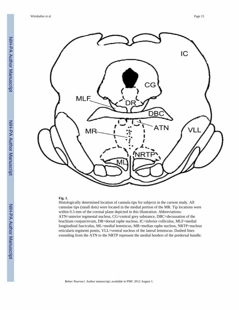
Fig. 1.Histologically determined location of cannula tips for subjects in the current study. Allcannulae tips (small dots) were located in the medial portion of the MR. Tip locations werewithin 0.3 mm of the coronal plane depicted in this illustration. Abbreviations:ATN=anterior tegmental nucleus, CG=central grey substance, DBC=decussation of thebrachium conjunctivum, DR=dorsal raphe nucleus, IC=inferior colliculus, MLF=mediallongitudinal fasciculus, ML=medial lemniscus, MR=median raphe nucleus, NRTP=nucleusreticularis tegmenti pontis, VLL=ventral nucleus of the lateral lemniscus. Dashed linesextending from the ATN to the NRTP represent the medial borders of the predorsal bundle.
Wirtshafter et al. Page 15
Behav Neurosci. Author manuscript; available in PMC 2012 August 1.
NIH
-PA Author Manuscript
NIH
-PA Author Manuscript
NIH
-PA Author Manuscript
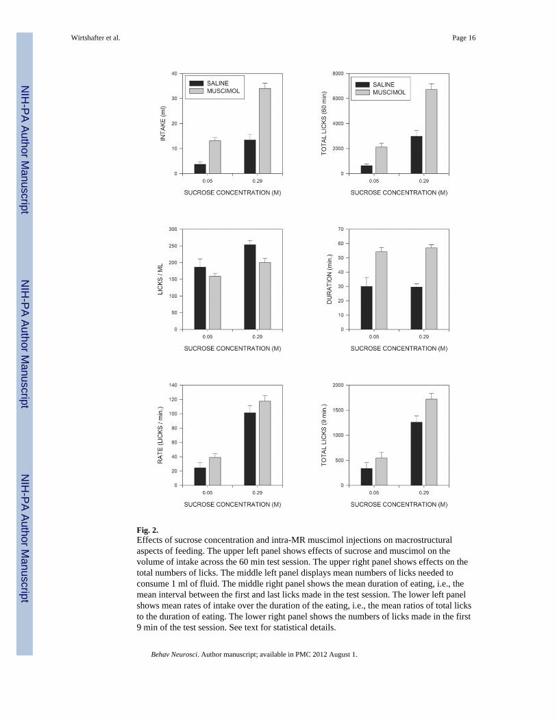
Fig. 2.Effects of sucrose concentration and intra-MR muscimol injections on macrostructuralaspects of feeding. The upper left panel shows effects of sucrose and muscimol on thevolume of intake across the 60 min test session. The upper right panel shows effects on thetotal numbers of licks. The middle left panel displays mean numbers of licks needed toconsume 1 ml of fluid. The middle right panel shows the mean duration of eating, i.e., themean interval between the first and last licks made in the test session. The lower left panelshows mean rates of intake over the duration of the eating, i.e., the mean ratios of total licksto the duration of eating. The lower right panel shows the numbers of licks made in the first9 min of the test session. See text for statistical details.
Wirtshafter et al. Page 16
Behav Neurosci. Author manuscript; available in PMC 2012 August 1.
NIH
-PA Author Manuscript
NIH
-PA Author Manuscript
NIH
-PA Author Manuscript
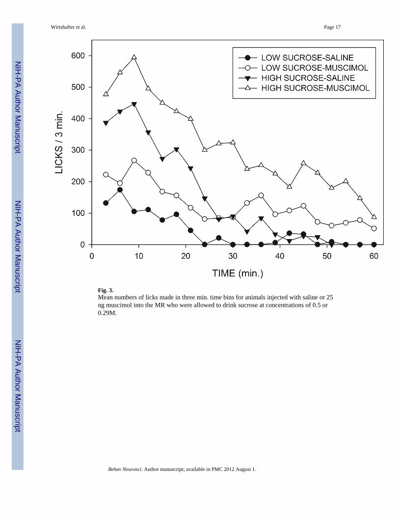
Fig. 3.Mean numbers of licks made in three min. time bins for animals injected with saline or 25ng muscimol into the MR who were allowed to drink sucrose at concentrations of 0.5 or0.29M.
Wirtshafter et al. Page 17
Behav Neurosci. Author manuscript; available in PMC 2012 August 1.
NIH
-PA Author Manuscript
NIH
-PA Author Manuscript
NIH
-PA Author Manuscript
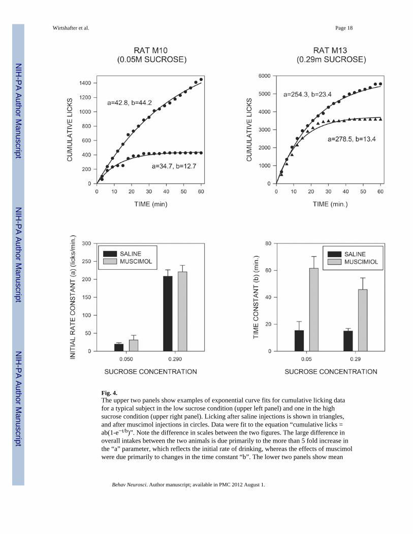
Fig. 4.The upper two panels show examples of exponential curve fits for cumulative licking datafor a typical subject in the low sucrose condition (upper left panel) and one in the highsucrose condition (upper right panel). Licking after saline injections is shown in triangles,and after muscimol injections in circles. Data were fit to the equation “cumulative licks =ab(1-e−t/b)”. Note the difference in scales between the two figures. The large difference inoverall intakes between the two animals is due primarily to the more than 5 fold increase inthe “a” parameter, which reflects the initial rate of drinking, whereas the effects of muscimolwere due primarily to changes in the time constant “b”. The lower two panels show mean
Wirtshafter et al. Page 18
Behav Neurosci. Author manuscript; available in PMC 2012 August 1.
NIH
-PA Author Manuscript
NIH
-PA Author Manuscript
NIH
-PA Author Manuscript
values of the initial rate constant and the time constant after saline and muscimol injectionsfor animals in the two sucrose conditions.
Wirtshafter et al. Page 19
Behav Neurosci. Author manuscript; available in PMC 2012 August 1.
NIH
-PA Author Manuscript
NIH
-PA Author Manuscript
NIH
-PA Author Manuscript
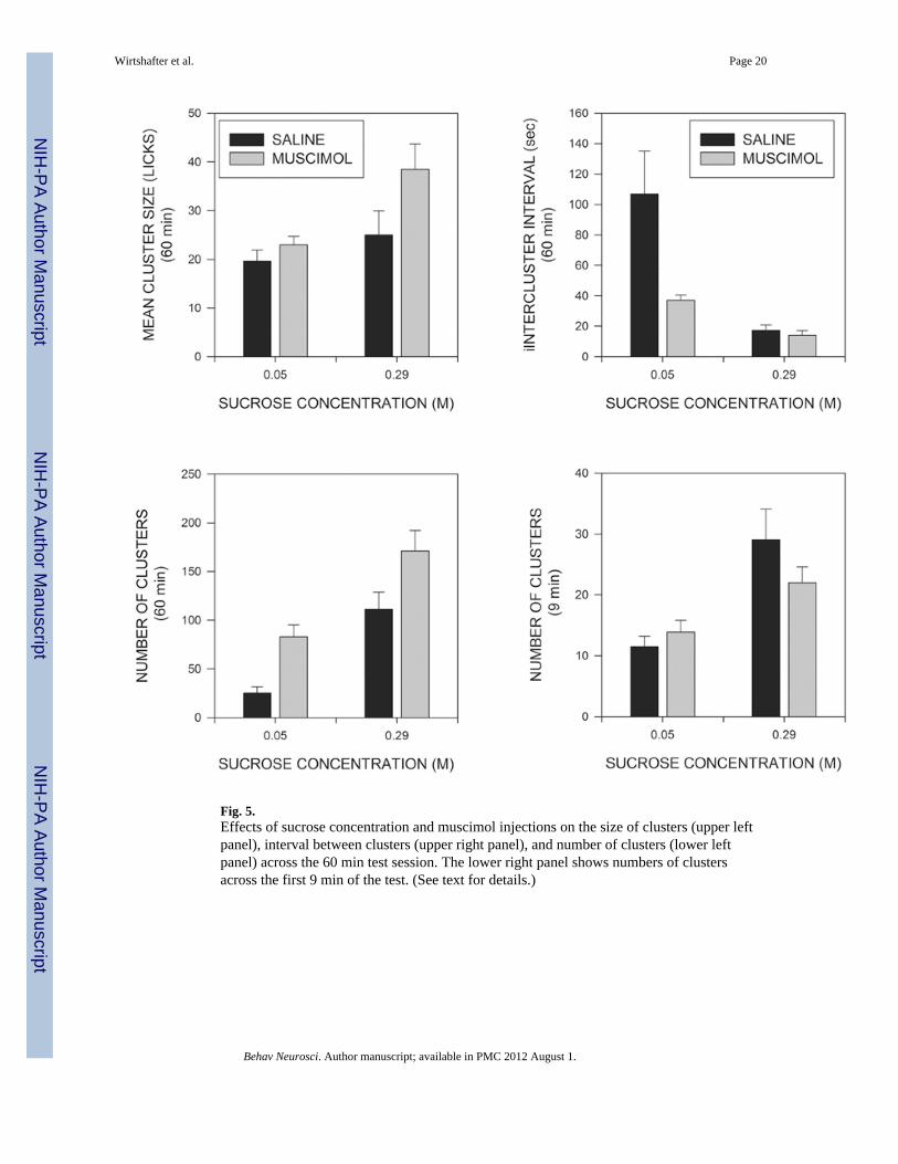
Fig. 5.Effects of sucrose concentration and muscimol injections on the size of clusters (upper leftpanel), interval between clusters (upper right panel), and number of clusters (lower leftpanel) across the 60 min test session. The lower right panel shows numbers of clustersacross the first 9 min of the test. (See text for details.)
Wirtshafter et al. Page 20
Behav Neurosci. Author manuscript; available in PMC 2012 August 1.
NIH
-PA Author Manuscript
NIH
-PA Author Manuscript
NIH
-PA Author Manuscript
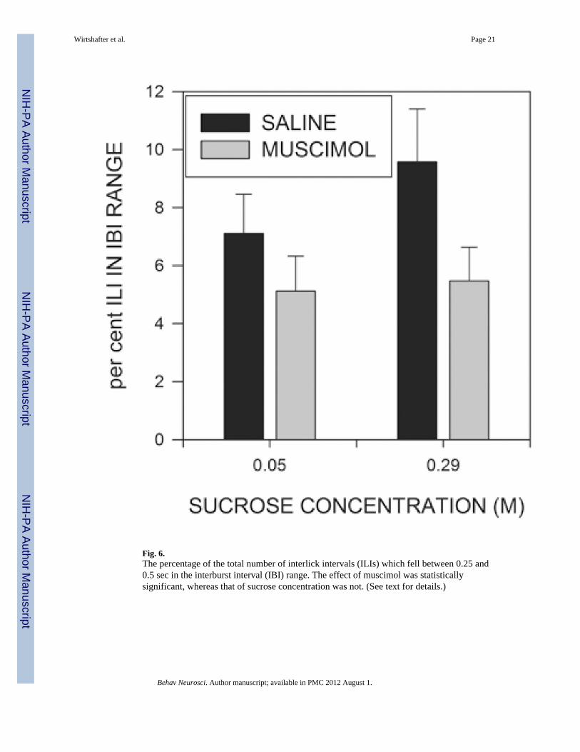
Fig. 6.The percentage of the total number of interlick intervals (ILIs) which fell between 0.25 and0.5 sec in the interburst interval (IBI) range. The effect of muscimol was statisticallysignificant, whereas that of sucrose concentration was not. (See text for details.)
Wirtshafter et al. Page 21
Behav Neurosci. Author manuscript; available in PMC 2012 August 1.
NIH
-PA Author Manuscript
NIH
-PA Author Manuscript
NIH
-PA Author Manuscript


![Median Shapes arXiv:1802.04968v2 [math.DG] 9 Dec 2018](https://img.pdfslide.net/doc/110x75/631a1b5b1a1adcf65a0eefdd/median-shapes-arxiv180204968v2-mathdg-9-dec-2018.jpg)


