Embed Size (px)
Citation preview

Induction of Erythroid Differentiation of Human K562Cells by Cisplatin Analogs
Nicoletta Bianchi,* Federico Ongaro,* Cristiano Chiarabelli,* Licia Gualandi,†Carlo Mischiati,* Paola Bergamini† and Roberto Gambari*‡
DEPARTMENTS OF *BIOCHEMISTRY AND MOLECULAR BIOLOGY AND
†CHEMISTRY, UNIVERSITY OF FERRARA, 44100 FERRARA, ITALY
ABSTRACT. Human leukemic K562 cells can be induced in vitro to erythroid differentiation by a variety ofchemical compounds, including hemin, butyric acid, 5-azacytidine, and cytosine arabinoside. Differentiation ofK562 cells is associated with an increase in the expression of embryo–fetal globin genes, such as the z-, e-, andg-globin genes. Therefore, the K562 cell line has been proposed as a very useful in vitro model system fordetermining the therapeutic potential of new differentiating compounds as well as for studying the molecularmechanism(s) regulating changes in the expression of embryonic and fetal human globin genes. Inducers oferythroid differentiation that stimulate g-globin synthesis could be considered for possible use in theexperimental therapy of hematological diseases associated with a failure in the expression of adult b-globingenes. In this paper, we analyzed the effects of a series of cisplatin analogs on both cell growth and differentiationof K562 cells. Among seven cisplatin analogs studied, three were found to be potent inducers of erythroiddifferentiation. Erythroid differentiation was associated with an increase in the accumulation of (a) hemoglobinsGower 1 and Portland and (b) g-globin mRNA. BIOCHEM PHARMACOL 60;1:31–40, 2000. © 2000 ElsevierScience Inc.
KEY WORDS. K562 cells; erythroid differentiation; DNA-binding drugs; cisplatin; cisplatin analogs
Pharmacologically mediated regulation of the expression ofhuman g-globin genes has been proposed as a potentialtherapeutic strategy in hematological disorders, includingb-thalassemia [1–4]. It has been suggested that even mod-est increases in fetal Hb§ levels can be clinically beneficialin sickle cell disease [5], while a greater increase in HbF isnecessary to reduce the severity of b-thalassemia [3].Therefore, recent studies have been focused on the searchfor compounds able to stimulate g-globin gene expression[6–10]. In this respect, the human leukemic K562 cell linehas been proposed as a very useful in vitro model system forstudying the molecular mechanism(s) regulating the ex-pression of embryonic and fetal human globin genes [11–17], as well as for determining the therapeutic potential ofnew differentiating compounds [3, 11]. This cell line,isolated and characterized by Lozzio and Lozzio [18] from apatient with chronic myelogenous leukemia in blast crisis,exhibits a low proportion of hemoglobin-synthesizing cellsunder standard cell growth conditions, but is capable ofundergoing erythroid differentiation when treated with a
variety of compounds, including hemin [11], ara-C [13],butyric acid [13, 17], 5-azacytidine [15], and chromomycinand mithramycin [19]. Following erythroid induction ofK562 cells, a sharp increase in cytoplasmic accumulation ofHb Portland (z2g2) and Hb Gower 1 (z2e2) is observed,accompanied by an increase in the expression of human e-and g-globin genes [14–17]. In vitro studies demonstratethat known inducers of K562 erythroid differentiation, suchas hydroxyurea, erythropoietin, butyrates, and 5-azacyti-dine, are also capable of inducing fetal hemoglobin produc-tion when administered singularly or in combination tonormal erythroid cells [1, 6, 8, 9, 20]. With respect to thispoint, butyric acid and 5-azacytidine have been the objectof recent reports focused on in vivo treatment of b-thalas-semia.
Among possible biological response modifiers, one of themost interesting classes of compounds are DNA-bindingdrugs displaying sequence selectivity [21–24]. DNA-bind-ing drugs are capable of interfering with the DNA-bindingactivity of a variety of transcription factors in a sequence-dependent manner [22–28]. With respect to capacity toinduce erythroid differentiation, we have recently demon-strated that the G 1 C-selective DNA-binding compoundsmithramycin and chromomycin are able to stimulate HbPortland production and g-globin mRNA accumulation intreated K562 cells [19].
Among DNA-binding compounds, cisplatin (1) hasbeen the object of a number of studies. Since the discovery
‡Corresponding author: Prof. Roberto Gambari, Department of Biochem-istry and Molecular Biology, Via Luigi Borsari 46, 44100 Ferrara, Italy. Tel.139-532-291440; FAX 139-532-202723; E-mail: [email protected]
§ Abbreviations: Hb, hemoglobin; HbF, fetal hemoglobin; DACH,diaminocyclohexane; THMP, trihydroxymethyl phosphine; H2O2, hydro-gen peroxide; FT-IR, Fourier transformed-infrared; ara-C, 1-b-arabino-furanosylcytosine.
Received 18 May 1999; accepted 18 November 1999.
Biochemical Pharmacology, Vol. 60, pp. 31–40, 2000. ISSN 0006-2952/00/$–see front matter© 2000 Elsevier Science Inc. All rights reserved. PII S0006-2952(00)00297-5

of its cytostatic activity in 1964 by Rosenberg [29] and itsproposed use as anticancer drug since 1979 [30], cisplatin isroutinely employed for the treatment of testicular andovarian cancer and is being increasingly used againstcervical, bladder, and head/neck tumors [30]. The mecha-nism of action of cisplatin is based on the intrastrandcross-linking of the cis-Pt(NH3)2 unit to cellular DNA attwo neighboring guanine bases [30]. With respect to apossible use as differentiating agent, cisplatin was shown toinduce erythroid differentiation of K562 cells [31]. How-ever, despite being one of the most active chemotherapeu-tic agents currently available [30–33] and despite having alarge clinical use [30], cisplatin exhibits severe side-effects,the most significant of which is acute and chronic nephro-toxicity [34, 35]. In addition, optic neuropathy [36] andototoxicity [37] of cisplatin have been reported. Therefore,cisplatin analogs are currently being designed, synthesized,and tested in order to find compounds retaining biologicalactivity without exhibiting nephrotoxic effects. In the mostsuccessful second generation cisplatin analogs, the chlorideligands have been replaced by carboxylate (e.g. carboplatin,9). Despite the fact that the structural variation of carbo-platin (9) seems to be responsible for a decreased toxicity ofthis compound [38], to our knowledge no information isavailable in the literature on the possible capacity ofcarboplatin to induce erythroid differentiation of K562cells.
Platinum(1,2-DACH) complexes, including the proto-type [PtCl2(1R,2R-DACH)] (2), have also attracted con-siderable interest. In fact, (2) and other 1,2-DACH deriv-atives showed good in vitro activity [32] and in vivo efficacyagainst cisplatin-resistant tumors [33]; low aqueous solubil-ity and molecular instability have been the major impedi-ments to clinical development of Pt(DACH) analogs.Following this perspective, we prepared the new com-pounds 3 and 4 cis-[Pt(OOCR)2(NH3)2] (3, RCOO 5cholate; 4, RCOO 5 deoxycholate), bearing bile acids asanionic ligands. In this paper, we report on the synthesis ofthese cisplatin analogs (see Fig. 1 for chemical structures)and their biological effects on human leukemic K562 cells.The activity of compounds 3 and 4 was compared to that ofcisplatin (1), carboplatin (9), compound 2, and a parallelgroup of complexes where the neutral ligand NH3 isreplaced by S-bonded DMSO: cis-[PtCl2(DMSO)2] (5),cis-[Pt(OOCR)2(DMSO)2] (6, RCOO 5 cholate; 7,RCOO 5 deoxycholate). Finally, we also tested a phos-phinic compound (8) cis-[PtCl2(THMP)2].
Erythroid differentiation of K562 cells was studied by thebenzidine/H2O2 reaction [16, 17], hemoglobin productionby cellulose acetate gel electrophoresis of postmitochon-drial supernatants [39], and g-globin mRNA accumulationwas analyzed by Northern blotting [40]. The results ob-tained demonstrated that three of the newly synthesizedplatinum complexes are potent inducers of erythroid differ-entiation of K562 cells. Erythroid differentiation by thesecisplatin analogs was associated with (a) an increase in the
content of embryo–fetal hemoglobins and (b) an increasein g-globin mRNA accumulation.
MATERIALS AND METHODSSynthesis of Cisplatin and Cisplatin Analogs ContainingChloride Ligands
cis-[PtCl2(NH3)2] (cisplatin) (1) [41], [PtCl2(1R,2R-DACH)] (2) [42], cis-[PtCl2(DMSO)2] (5) [43], and cis-[PtCl2(THMP)2] (8) [44] were prepared following reportedmethods and characterized by elemental analyses and spec-troscopic techniques (i.r. and NMR). Carboplatin (9) wasobtained from Sigma Chemical Co.
Synthesis of cis-[Pt(OOCR)2(NH3)2] (3, RCOO 5cholate; 4, RCOO 5 deoxycholate)
Five hundred milligrams of cis-[PtI2(NH3)2] (1 mmol) wassuspended in 2.5 mL of H2O. With the suspension main-tained under stirring, 0.34 g (2 mmol) of AgNO3 dissolvedin 2.5 mL of H2O was added. The reaction mixture wasstirred for 20 min at 65° in the dark. The colloidalsuspension of AgI was then filtered through a Milliporemembrane and a clear solution thus obtained. Sodiumcholate (for product 3) or sodium deoxycholate (for prod-uct 4) (2.2 mmol in 10 mL of water) was added dropwiseunder stirring. The reaction mixture containing the prod-uct as a white precipitate was cooled for 2 hr, then the solidwas filtered, washed three times with water, and dried undervacuum.
(3): 70% yield. FT-IR (KBr, n cm21, selected data): 3300(OH, NH); 1572 (COO asym); 1377 (COO sym). 1H-NMR (CD3OD, d ppm): 0.7 (6H, s, CH3); 0.9 (6H, s,CH3); 1.1 (6 H, d, 3JHH 5 6 Hz, CHCH3); 1.1–2.4 (48 H,m); 3.4 (2 H, br s, HOC3H); 3.9 (2 H, br s, HOC7H); 4.0(2 H, br s, HOC12H); in DMSO-d6, 4.3 ppm (6 H, br,NH3). Elemental analysis: calculated values forC48H84N2O10Pt: C% 5 55.25; H% 5 8.05; N% 5 2.68;found: C% 5 55.56; H% 5 8.06; N% 5 2.60.
(4): 68% yield. FT-IR (KBr, n cm21, selected data): 3300(OH, NH); 1556 (COO asym); 1381 (COO sym). 1H-NMR (CD3OD, d ppm): 0.7 (6 H, s, CH3); 0.9 (6 H, s,CH3); 1.0 (6 H, d, 3JHH 5 6 Hz, CHCH3); 1.0–2.4 (52 H,m); 3.5 (2 H, br s, HOC3H); 3.9 (2 H, br s, HOC12H).Elemental analysis: calculated value for C48H84N2O8Pt:C% 5 56.97; H% 5 8.31; N% 5 2.77; found: C% 5 56.99;H% 5 8.60; N% 5 2.65.
Synthesis of cis-[Pt(OOCR)2(DMSO)2] (6, RCOO 5cholate; 7, RCOO 5 deoxycholate)
Two hundred milligrams (2.37 z 1024 mol) of cis-[PtCl2(DMSO)2] was dissolved in 40 mL of CH2Cl2, and4.74 z 1024 mol of silver salt (silver cholate for 6, silverdeoxycholate for 7) was added. The brown suspension waskept under stirring in the dark for 18 hr and the precipitate
32 N. Bianchi et al.

of AgCl then filtered. The clear yellow filtrate was taken tocomplete dryness, leaving the product as a cream solid.
(6): 80% yield. IR (CsI, n cm21, selected data): 3422(OH); 1566 (COO asym), 1381 (COO sym), 1145 (n SO).1H NMR (CD3OD, d ppm): 0.6 (6 H, s, CH3); 0.9 (6 H, s,CH3); 1.0 (6 H, d, 3JHH 5 6 Hz, CHCH3) 1.1–2.4 (48 H,m); 3.4 (2 H, br s, HOC3H); 3.45 (12 H, s, 3JHPt 5 13 Hz,OSCH3); 3.8 (2H, br s, HOC7H); 3.9 (2H, br s,HOC12H). {1H}195Pt-NMR (CD3OD, d ppm): 2452. Ele-mental analysis: calculated values for C52H90O12PtS2: C% 553.58; H% 5 7.72; S% 5 5.49; found: C% 5 53.8; H% 57.87; S% 5 5.73.
(7): 81% yield. IR(KBr, n cm21, selected data): 3414(OH); 1558 (COO asym); 1385 (COO sym), 1033 (SO),439 (PtS). 1H NMR (CD3OD, d ppm): 0.7 (6 H, s, CH3);0.9 (6 H, s, CH3); 1.0 (6 H, d, 3JHH 5 6 Hz, CHCH3);
1.2–2.4 (52 H, m); 3.4 (12 H, s, 3JHPt 5 13 Hz, OSCH3);3.5 (2H, br s, HOC3H); 4.0 (2H, br s, HOC12H).{1H}195Pt-NMR (CD3OD, d ppm): 2454.22. Elementalanalysis: calculated values for C52H90O10PtS2: C% 5 55.07;H% 5 7.94; S% 5 5.65; found: C% 5 53.06; H% 5 8.74;S% 5 5.06.
Cell Lines and Culture Conditions
Human erythroleukemia K562(S) cells [11] were culturedin a humidified atmosphere at 5% CO2 in RPMI-1640(Flow Laboratories) supplemented with 10% fetal bovineserum (FBS, Celbio), 50 units/mL of penicillin, and 50mg/mL of streptomycin [16]. Cell growth was studied bydetermining the cell number/mL after different days of invitro cell culture [14, 15, 17]. Stock solutions of cisplatin
FIG. 1. Chemical structures of cisplatin and the cisplatin analogs employed in this study.
Induction of Erythroid Differentiation by Cisplatin Analogs 33
(10 mM) and ara-C (10 mM) were stored at 220° in thedark and diluted immediately before use. Treatment withthe indicated concentrations of cisplatin and cisplatinanalogs was carried out by adding the appropriate drugconcentrations at the beginning of the experiment. Themedium was not changed during the induction period.The chemical structures of cisplatin and cisplatin ana-logs are shown in Fig. 1.
Hemoglobin Determination
K562 cells containing heme or hemoglobin were detectedby specific reaction with a benzidine/hydrogen peroxidesolution as reported elsewhere [15, 16]. The final concen-tration of benzidine was 0.2% in 5 M glacial acetic acid,10% H2O2 [15, 16]. In order to analyze hemoglobinproduction by erythroid-induced K562 cells, 2 mL of totalfresh postmitochondrial cell lysates was electrophoresed oncellulose acetate strips (Poliphorm) in Tris–EDTA–boratebuffer [17, 39]. After an electrophoresis of 30 min at 5 mA,the gels were stained with benzidine/hydrogen peroxide(1% benzidine in 4.3 M acetic acid, 3% H2O2) andphotographed [39].
Northern Blotting
Total RNA was phenol-chloroform-extracted from cyto-plasms of uninduced K562 cells or K562 cells induced toerythroid differentiation with cisplatin and cisplatin ana-logs [40]. Fifteen micrograms of total RNA was loaded onto1% agarose gel, electrophoresed, transferred to a nylonmembrane (GeneScreen plus), and hybridized with the32P-labeled g-pUCA probe. This probe is a 3.3-kb HindIIIfragment of the human A-g-globin gene cloned in theHindIII site of pUC18 [45].
RESULTSCell Growth and Differentiation of K562 Cells Culturedin the Presence of Newly Synthesized Cisplatin Analogs
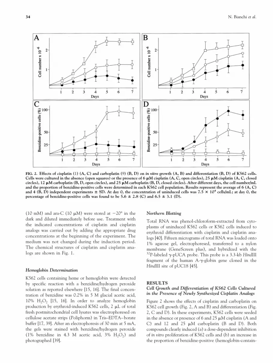
Figure 2 shows the effects of cisplatin and carboplatin onK562 cell growth (Fig. 2, A and B) and differentiation (Fig.2, C and D). In these experiments, K562 cells were seededin the absence or presence of 6 and 25 mM cisplatin (A andC) and 12 and 25 mM carboplatin (B and D). Bothcompounds clearly induced (a) a dose-dependent inhibitionof in vitro proliferation of K562 cells and (b) an increase inthe proportion of benzidine-positive (hemoglobin-contain-
FIG. 2. Effects of cisplatin (1) (A, C) and carboplatin (9) (B, D) on in vitro growth (A, B) and differentiation (B, D) of K562 cells.Cells were cultured in the absence (open squares) or the presence of 6 mM cisplatin (A, C, open circles), 25 mM cisplatin (A, C, closedcircles), 12 mM carboplatin (B, D, open circles), and 25 mM carboplatin (B, D, closed circles). After different days, the cell number/mLand the proportion of benzidine-positive cells were determined in each K562 cell population. Results represent the average of 6 (A, C)and 4 (B, D) independent experiments 6 SD. At day 0, the concentration of uninduced cells was 7.5 3 104 cells/mL; at day 0, thepercentage of benzidine-positive cells was found to be 5.6 6 2.8 (C) and 6.5 6 3.1 (D).
34 N. Bianchi et al.
ing) cells. It should be noted that high concentrations ofcisplatin (25–50 mM, see also Fig. 3A) were highly toxic,leading to a strong inhibition of cell growth, associated witha blocking of erythroid differentiation of treated K562 cells.Trypan blue exclusion tests demonstrated that a highnumber of K562 cells treated with 25 mM cisplatin (60.8 63.8%; N 5 6) efficiently incorporated the dye, thusconfirming the high cytotoxicity of treatment with cispla-tin. By contrast, carboplatin was found to be less cytotoxicat the same concentrations, the cells being positive to thetrypan blue exclusion test 7.5 6 2.5% (N 5 4). Figure 3shows a comparison of the effects of cisplatin and cisplatinanalogs on K562 cell growth and differentiation. For all thecompounds, experiments similar to those reported in Fig. 2were conducted and the results obtained after 7 days of cellculture were compared. Figure 3 (open symbols) shows thatcisplatin and cisplatin analogs 2, 3, 4, and 8 caused adose-dependent decrease in the proliferation efficiency ofK562 cells. Fifty percent inhibition of cell growth (IC50)occurred when K562 cells were cultured for 7 days in thepresence of 4 mM cisplatin (1), 20 mM (2), 35 mM (8), 8mM (3), and 20 mM (4), respectively. On the contrary,compounds 5, 6, and 7 exhibited much lower inhibitoryactivity on K562 in vitro cell growth. Trypan blue exclusiontests, analysis of caspase activity, and the dUTP nick endlabeling (TUNEL) assay demonstrated that the antiprolif-erative activity of compounds 2, 3, 4, and 8 was notassociated with cytotoxicity, but rather with activation ofapoptosis.* Figure 3 (closed symbols) also demonstrates thatcisplatin and cisplatin analogs displayed a clearly differentability to induce a significant increase in the proportion ofbenzidine-positive (hemoglobin-containing) K562 cells af-ter 7 days of induction. Cisplatin and compounds 2, 3, and4 were found to be effective inducers of K562 erythroiddifferentiation. By contrast, compounds 8, 5, 6, and 7 didnot induce erythroid differentiation. This lack of inductionability was particularly significant for compound 8, which,unlike 5, 6, and 7, exhibited antiproliferative activity.
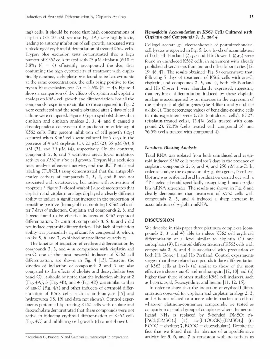
The kinetics of induction of erythroid differentiation bycompounds 2, 3, and 4 in comparison with cisplatin andara-C, one of the most powerful inducers of K562 celldifferentiation, are shown in Fig. 4 [13]. Therein, thekinetics of induction of compounds 2 and 3 are alsocompared to the effects of cholate and deoxycholate (seepanel C). It should be noted that the induction ability of 2(Fig. 4A), 3 (Fig. 4B), and 4 (Fig. 4B) was similar to thatof ara-C (Fig. 4A) and other inducers of erythroid differ-entiation of K562 cells, such as mithramycin [19] andhydroxyurea ([8, 19] and data not shown). Control exper-iments performed by treating K562 cells with cholate anddeoxycholate demonstrated that these compounds were notactive in inducing erythroid differentiation of K562 cells(Fig. 4C) and inhibiting cell growth (data not shown).
Hemoglobin Accumulation in K562 Cells Cultured withCisplatin and Compounds 2, 3, and 4
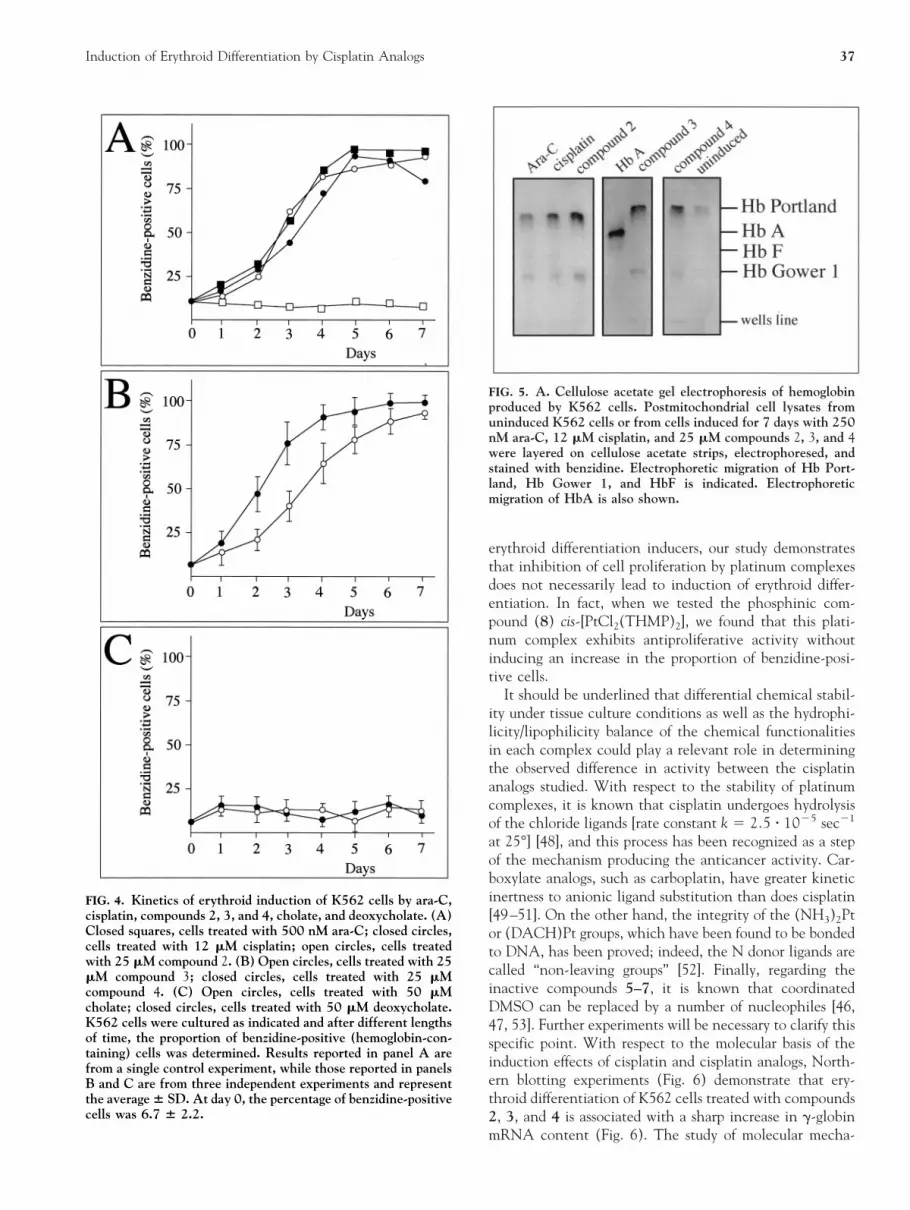
Cellogel acetate gel electrophoresis of postmitochondrialcell lysates is reported in Fig. 5. Low levels of accumulationof both Hb Portland (z2g2) and Hb Gower 1 (z2e2) werefound in uninduced K562 cells, in agreement with alreadypublished observations from our and other laboratories [12,19, 46, 47]. The results obtained (Fig. 5) demonstrate that,following 7 days of treatment of K562 cells with ara-C,cisplatin, and compounds 2, 3, and 4, both Hb Portlandand Hb Gower 1 were abundantly expressed, suggestingthat erythroid differentiation induced by these cisplatinanalogs is accompanied by an increase in the expression ofthe embryo–fetal globin genes (the b-like e and g and thea-like z). The percentage values of benzidine-positive cellsin this experiment were 6.5% (uninduced cells), 85.2%(cisplatin-treated cells), 75.4% (cells treated with com-pound 2), 72.3% (cells treated with compound 3), and76.5% (cells treated with compound 4).
Northern Blotting Analysis
Total RNA was isolated from both uninduced and eryth-roid-induced K562 cells treated for 7 days in the presence ofcisplatin, compounds 2, 3, and 4, and 250 nM ara-C. Inorder to analyze the expression of g-globin genes, Northernblotting was performed and hybridization carried out with a32P-labeled plasmid specifically recognizing human g-glo-bin mRNA sequences. The results are shown in Fig. 6 andclearly demonstrate that treatment of K562 cells withcompounds 2, 3, and 4 induced a sharp increase inaccumulation of g-globin mRNA.
DISCUSSION
We describe in this paper three platinum complexes (com-pounds 2, 3, and 4) able to induce K562 cell erythroiddifferentiation at a level similar to cisplatin (1) andcarboplatin (9). Erythroid differentiation of K562 cells withcompounds 2, 3, and 4 is associated with production ofboth Hb Gower 1 and Hb Portland. Control experimentssuggest that these related compounds induce differentiationof K562 cells at levels (a) similar to those of the mosteffective inducers ara-C and mithramycin [12, 18] and (b)higher than those of other studied K562 cell inducers, suchas butyric acid, 5-azacytidine, and hemin [11, 12, 15].
In order to show that the induction of erythroid differ-entiation observed for cisplatin and cisplatin analogs 2, 3,and 4 is not related to a mere administration to cells ofwhatever platinum-containing compounds, we tested ascomparison a parallel group of complexes where the neutralligand NH3 is replaced by S-bonded DMSO: cis-[PtCl2(DMSO)2] (5), cis-[Pt(OOCR)2(DMSO)2] (6,RCOO 5 cholate; 7, RCOO 5 deoxycholate). Despite thefact that we found that the absence of antiproliferativeactivity for 5, 6, and 7 is consistent with no activity as* Mischiati C, Bianchi N and Gambari R, manuscript in preparation.
Induction of Erythroid Differentiation by Cisplatin Analogs 35

FIG. 3. Effects of cisplatin and cisplatin analogs on in vitro growth (open symbols) and differentiation (closed symbols) of K562 cells.Cells were cultured in the absence (0) or the presence of different concentrations of cisplatin and cisplatin analogs. After 7 days, thecell number/mL and the proportion of benzidine-positive (hemoglobin-containing) cells were determined in each K562 cell population.The data values of cell number/mL in treated cells were compared to those of untreated cultures and expressed as % of control. Resultsrepresent the average 6 SD of four independent experiments. After 7 days’ culture of untreated K562 cells, the percentage ofbenzidine-positive cells was 7.9 6 2.7.
36 N. Bianchi et al.
erythroid differentiation inducers, our study demonstratesthat inhibition of cell proliferation by platinum complexesdoes not necessarily lead to induction of erythroid differ-entiation. In fact, when we tested the phosphinic com-pound (8) cis-[PtCl2(THMP)2], we found that this plati-num complex exhibits antiproliferative activity withoutinducing an increase in the proportion of benzidine-posi-tive cells.
It should be underlined that differential chemical stabil-ity under tissue culture conditions as well as the hydrophi-licity/lipophilicity balance of the chemical functionalitiesin each complex could play a relevant role in determiningthe observed difference in activity between the cisplatinanalogs studied. With respect to the stability of platinumcomplexes, it is known that cisplatin undergoes hydrolysisof the chloride ligands [rate constant k 5 2.5 z 1025 sec21
at 25°] [48], and this process has been recognized as a stepof the mechanism producing the anticancer activity. Car-boxylate analogs, such as carboplatin, have greater kineticinertness to anionic ligand substitution than does cisplatin[49–51]. On the other hand, the integrity of the (NH3)2Ptor (DACH)Pt groups, which have been found to be bondedto DNA, has been proved; indeed, the N donor ligands arecalled “non-leaving groups” [52]. Finally, regarding theinactive compounds 5–7, it is known that coordinatedDMSO can be replaced by a number of nucleophiles [46,47, 53]. Further experiments will be necessary to clarify thisspecific point. With respect to the molecular basis of theinduction effects of cisplatin and cisplatin analogs, North-ern blotting experiments (Fig. 6) demonstrate that ery-throid differentiation of K562 cells treated with compounds2, 3, and 4 is associated with a sharp increase in g-globinmRNA content (Fig. 6). The study of molecular mecha-
FIG. 4. Kinetics of erythroid induction of K562 cells by ara-C,cisplatin, compounds 2, 3, and 4, cholate, and deoxycholate. (A)Closed squares, cells treated with 500 nM ara-C; closed circles,cells treated with 12 mM cisplatin; open circles, cells treatedwith 25 mM compound 2. (B) Open circles, cells treated with 25mM compound 3; closed circles, cells treated with 25 mMcompound 4. (C) Open circles, cells treated with 50 mMcholate; closed circles, cells treated with 50 mM deoxycholate.K562 cells were cultured as indicated and after different lengthsof time, the proportion of benzidine-positive (hemoglobin-con-taining) cells was determined. Results reported in panel A arefrom a single control experiment, while those reported in panelsB and C are from three independent experiments and representthe average 6 SD. At day 0, the percentage of benzidine-positivecells was 6.7 6 2.2.
FIG. 5. A. Cellulose acetate gel electrophoresis of hemoglobinproduced by K562 cells. Postmitochondrial cell lysates fromuninduced K562 cells or from cells induced for 7 days with 250nM ara-C, 12 mM cisplatin, and 25 mM compounds 2, 3, and 4were layered on cellulose acetate strips, electrophoresed, andstained with benzidine. Electrophoretic migration of Hb Port-land, Hb Gower 1, and HbF is indicated. Electrophoreticmigration of HbA is also shown.
Induction of Erythroid Differentiation by Cisplatin Analogs 37

nisms underlying the switch between e- and g-globin genesis crucial for experiments aimed at the induction of g-glo-bin gene expression in adults [2]. Pharmacologically medi-ated regulation of the expression of human g-globin genescould be of interest in the search for potential therapeuticagents in hematological disorders, including b-thalassemia[3, 6, 8, 9, 54–57], as recently published observationsdemonstrate that hormones, cytotoxic agents, hemopoieticcytokines, and short fatty acids are agents capable ofaugmenting fetal hemoglobin levels in humans [2]. Inparticular, butyric acid and 5-azacytidine have been theobject of recent reports focusing on in vivo treatment ofb-thalassemia patients [1, 2, 9]. This is a major issue in thisfield, since it is well established that an increase in HbFproduction as low as 30% leads to a significant improve-ment in the clinical status [3]. Accordingly, our data shouldencourage studies on possible effects of the cisplatin analogsused on g-globin gene expression on normal erythroidprecursor cells from peripheral blood as well as from bonemarrow.
Despite the fact that our data do not provide informationon the mechanism of action of the studied platinumcomplexes, in agreement with the proposed mechanism ofaction of cisplatin [58–60], in vitro DNase I footprintingexperiments demonstrate that these DNA-binding drugsare able to interact with the g-globin promoter of humangenomic DNA (data not shown); in vivo footprintingexperiments could clarify this specific point. Molecularanalyses are in this case very important, as it is known thatstructurally related compounds could induce hemoglobinsynthesis in K562 cells by distinct mechanisms [61, 62].
Whatever the mechanism of action of compounds 2, 3,and 4 may be, our results demonstrate that these com-pounds are potent new inducers of K562 erythroid differ-entiation, leading to accumulation of g-globin mRNA andproduction of embryo–fetal hemoglobins.
This work was supported by CNR PF Biotecnologie, by AIRC, and byPRIN-98. Partial support from the Associazione per la Guarigione delBambino Thalassemico (Ferrara) and the Associazione Veneta per laLotta alla Thalassemia (Rovigo) is also acknowledged. C.M. and N.B.are recipients of AIRC and FIRC fellowships, respectively. We thankProf. Michael Antoniou, National Institute for Medical Research, TheRidgeway, Mill Hill, London, for the g-globin probe used in theseexperiments.
References
1. Lowrey CH and Nienhuis AW, Brief report: Treatment withazacytidine of patients with end-stage b-thalassemia. N EnglJ Med 329: 845–848, 1993.
2. Rodgers GP and Rachmilewitz EA, Novel treatment optionsin the severe b-globin disorders. Br J Haematol 91: 263–268,1995.
3. Rochette J, Craig JE and Thein SL, Fetal hemoglobin levels inadults. Blood Rev 8: 213–224, 1994.
4. Ley TJ, The pharmacology of hemoglobin switching: Of miceand men. Blood 77: 1146–1152, 1991.
5. Platt OS, Thorington BD, Brambilla DJ, Milner PF, RosseWF, Vichinsky E and Kinney TR, Pain in sickle cell disease.Rates and risk factor. N Engl J Med 325: 11–16, 1991.
6. Dover GJ, Brusilow S and Samid D, Increased fetal hemoglo-bin in patients receiving sodium 4-phenylbutyrate. N EnglJ Med 327: 569–570, 1992.
7. Fibach E, Prasanna P, Rodgers GP and Samid D, Enhancedfetal hemoglobin production by phenylacetate and 4-phenyl-butyrate in erythroid precursors derived from normal donorsand patients with sickle cell anemia and b-thalassemia. Blood82: 2203–2209, 1993.
8. Rodgers GP, Dover GJ, Uyesaka N, Noguchi CT, SchechterAN and Nienhuis AW, Augmentation by erythropoietin ofthe fetal-hemoglobin response to hydroxyurea in sickle celldisease. N Engl J Med 328: 73–80, 1993.
9. Ikuta T, Atweh G, Boosalis V, White GL, Da Fonseca S,Boosalis M, Faller DV and Perrine SP, Cellular and moleculareffects of a pulse butyrate regimen and new inducers of globingene expression and hematopoiesis. Ann N Y Acad Sci 850:87–99, 1998.
10. Perrine SP, Ginder GD, Faller DV, Dover GH, Ikuta T,Witkowska HE, Cai SP, Vichinsky EP and Olivieri NF, Ashort-term trial of butyrate to stimulate fetal-globin-geneexpression in the b-globin disorders. N Engl J Med 328:81–86, 1993.
FIG. 6. Northern blotting analysis. Total RNA was isolatedfrom uninduced and erythroid-induced K562 cells. Inductionwas carried out for 7 days in the presence of 250 nM ara-C, 6mM or 12 mM cisplatin, and 25 mM compounds 2, 3, and 4, asindicated. Fifteen micrograms of total RNA was electropho-resed, Northern blotting was performed, and hybridization wascarried out with a 32P-labeled g-globin probe. Ethidium-bromidestaining of the gel is shown in panel A; autoradiography isshown in panel B.
38 N. Bianchi et al.

11. Rutherford TR, Clegg JB and Weatherall DJ, K562 humanleukaemic cells synthesise embryonic haemoglobin in re-sponse to haemin. Nature 280: 164–165, 1979.
12. Cioe L, McNab A, Hubbell HR, Meo P, Curtis P and RoveraG, Differential expression of the globin genes in humanleukemia K562(S) cells induced to differentiate by hemin orbutyric acid. Cancer Res 41: 237–243, 1981.
13. Bianchi Scarra GL, Romani M, Coviello DA, Garre C,Ravazzolo R, Vidali G and Ajmar F, Terminal erythroiddifferentiation in the K-562 cell line by 1-b-D-arabinofurano-sylcytosine by c-myc messenger RNA decrease. Cancer Res46: 6327–6332, 1986.
14. Gambari R, Raschella G, Biagini R, Tripodi M, Farace MG,Romeo A and Fantoni A, Predominant expression of z and eglobin genes in human leukemia K-562(S6) variant cell line.Experientia 39: 415–416, 1983.
15. Gambari R, del Senno L, Barbieri R, Viola L, Tripodi M,Raschella G and Fantoni A, Human leukemia K-562 cells:Induction of erythoid differentiation by 5-azacytidine. CellDiffer 14: 87–97, 1984.
16. Gambari R, Amelotti F and Piva R, Efficient cell proliferationand predominant accumulation of e-globin mRNA in humanleukemic K562 cells which produce mostly Hb Gower 1.Experientia 41: 673–675, 1985.
17. Gambari R, Barbieri R, Buzzoni D, Bernardi F, Marchetti G,Amelotti F, Piva R, Viola L and del Senno L, Humanleukemic K562 cells: Suppression of hemoglobin accumula-tion by a monoclonal antibody to human transferrin receptor.Biochim Biophys Acta 886: 203–213, 1986.
18. Lozzio CB and Lozzio BB, Human chronic myelogenousleukemia cell-line with positive Philadelphia chromosome.Blood 45: 321–334, 1975.
19. Bianchi N, Osti F, Rutigliano C, Corradini FG, Borsetti E,Tomassetti M, Mischiati C, Feriotto G and Gambari R, TheDNA-binding drugs mithramycin and chromomycin are pow-erful inducers of erythroid differentiation of human K562cells. Br J Haematol 104: 258–263, 1999.
20. Al-Khatti A, Papayannopoulou T, Knitter G, Fritsch EF andStamatoyannopoulos G, Cooperative enhancement of F-cellformation in baboons treated with erythropoietin and hy-droxyurea. Blood 72: 817–819, 1988.
21. Dervan PB, Design of sequence-specific DNA binding mole-cules. Science 232: 464–471, 1986.
22. Bianchi N, Passadore M, Feriotto G, Mischiati C, Gambari Rand Piva R, Alteration of the expression of human estrogenreceptor gene by distamycin. J Steroid Biochem Mol Biol 54:211–215, 1995.
23. Bianchi N, Passadore M, Rutigliano C, Feriotto G, MischiatiC and Gambari R, Targeting of the Sp1 binding sites ofHIV-1 long terminal repeat with chromomycin. Disruption ofnuclear factor. DNA complexes and inhibition of in vitrotranscription. Biochem Pharmacol 52: 1489–1498, 1996.
24. Vaquero A and Portugal J, Modulation of DNA–proteininteractions in the P1 and P2 c-myc promoters by twointercalating drugs. Eur J Biochem 251: 435–442, 1998.
25. Ray R, Snyder RC, Thomas S, Koller CA and Miller DM,Mithramycin blocks protein binding and function of theSV40 early promoter. J Clin Invest 83: 2003–2007, 1989.
26. Snyder RC, Ray R, Blume S and Miller DM, Mithramycinblocks transcriptional initiation of the c-myc P1 and P2promoters. Biochemistry 30: 4290–4297, 1991.
27. Welch JJ, Rausher FJ 3rd and Beerman TA, TargetingDNA-binding drugs to sequence-specific transcription factorDNA complexes. Differential effects of intercalating andminor groove binding drugs. J Biol Chem 269: 31051–31058,1994.
28. Feriotto G, Mischiati C and Gambari R, Sequence-specific
recognition of the HIV-1 long terminal repeat by distamycin:A DNase I footprinting study. Biochem J 299: 451–458, 1994.
29. Rosenberg B, Vancamp L and Krigas T, Inhibition of celldivision in Escherichia coli by electrolysis products from aplatinum electrode. Nature 205: 698–699, 1965.
30. Sigel A and Sigel H (Eds.), Metal Ions in Biological Systems,vol. 32. Marcel Dekker, New York, 1996.
31. Parodi MT, Tonini GP, Bologna R, Franchini E and Corna-glia-Ferraris P, Cisplatin-induced erythroid differentiation inK562 cells: Modulation of transferrin receptor. Boll Ist SieroterMilan 67: 142–148, 1988.
32. Eastman A and Illenye S, Murine leukemia L1210 cell lineswith different patterns of resistance to platinum coordinationcomplexes. Cancer Treat Rep 68: 1189–1190, 1984.
33. Burchenal JH, Kalaher K, O’Toole T and Chisholm J, Lack ofcross-resistance between certain platinum coordination com-plexes in mouse leukemia. Cancer Res 37: 3455–3457, 1977.
34. Jones MM, Basinger MA and Holsher MA, Control of thenephrotoxicity of cisplatin by clinically used sulfur-containingcompounds. Fundam Appl Toxicol 18: 181–188, 1992.
35. Verplanke AJ, Herber RF, de Wit R and Veenhof CH,Comparison of renal function parameters in the assessment ofcis-platin induced nephrotoxicity. Nephron 66: 267–272,1994.
36. Caraceni A, Martini C, Spatti G, Thomas A and Onofrj M,Recovering optic neuritis during systemic cisplatin and car-boplatin chemotherapy. Acta Neurol Scand 96: 260–261,1997.
37. Qie WX, Experimental study on ototoxicity of cisplatin.Chung Hua Erh Pi Yen Hou Ko Tsa Chih 254: 195–198, 1990.
38. Hay RW and Miller S, Reactions of platinum(II) anticancerdrugs. Kinetics of acid hydrolysis of cis-diammine (cyclobu-tane-1,1-dicarboxylato)platinum(II) “carboplatin”. Polyhe-dron 17: 2337, 1998.
39. Weatherall DJ and Clegg JB, The laboratory diagnosis of thethalassaemia syndromes. In: The Thalassemia Syndromes (Eds.Weatherau DJ and Clegg JB), pp. 744–769. Blackwell Scien-tific Publications, Oxford, 1981.
40. Sambrook J, Fritsch EF and Maniatis T, Extraction, purifica-tion and analysis of messenger RNA from eukaryotic cells. In:Molecular Cloning, 2nd Edn (Nolan C Ed.), pp. 7.43–7.45.Cold Spring Harbor Laboratory, Cold Spring Harbor, NewYork, 1981.
41. Dhara SC, A rapid method for the synthesis of cis-[Pt(NH3)2Cl2]. Indian J Chem 8: 194, 1970.
42. Khokhar AR, Krakoff IH, Hacker MP and McCormack JJ,The synthesis and antitumor properties of a series of watersoluble carboxylato-(1,2-diaminocyclohexane)platinum(II)complexes. Inorg Chim Acta 108: 63, 1985.
43. Romeo R and Monsu Scolaro L, (2,29:69,20-Terpyridine)methylplatinum (II) chloride and (1,10-phenanthroline)-methylchloroplatinum(II). Inorg Synth 32: 153, 1998.
44. Ellis JW, Harrison KN, Hoye PA, Orpen AG, Pringle PG andSmith MB, Water-soluble tris(hydroxymethyl)phosphinecomplexes with nickel, palladium and platinum. Inorg Chem31: 3026–3033, 1992.
45. Fraser P, Pruzina S, Antoniou M and Grosveld F, Eachhypersensitive site of the human beta-globin locus controlregion confers a different developmental pattern of expressionon the globin genes. Genes Dev 7: 106–113, 1993.
46. Bitha P, Morton GO, Dunne TS, De los Santos EF, Lin Y,Boone SR, Haltiwanger RC and Pierpont CG, (Malonato-)bis[sulfinylbis[methane]-S]platinum(II) compounds: Versa-tile synthons for a new general synthesis of antitumor sym-metrical and dissymmetrical (malonato)platinum(II) com-plexes. Inorg Chem 29: 645–652, 1990.
47. Clement O, Roszak AW and Buncel E, Synthesis, character-ization and X-ray crystal structure determination of platinu-
Induction of Erythroid Differentiation by Cisplatin Analogs 39

m(II)–diaminoalkane complexes. Inorg Chim Acta 253: 53–63, 1996.
48. Reishus JW and Martin DS Jr, cis-Dichlorodiammine plati-num(II). Acid hydrolysis and isotopic exchange of the chlo-ride ligands. J Am Chem Soc 83: 2457–2462, 1961.
49. Neidle S, Ismail IM and Sadler PJ, The structure of theantitumor complex cis(diammino)(1,1-cyclobutanedicarboxy-lato)-Pt(II): X ray and NMR studies. J Inorg Biochem 13:205–212, 1980.
50. Frey U, Ranford JD and Sadler PJ, Ring-opening reactions ofthe anticancer drug carboplatin: NMR characterization ofcis-[Pt(NH3)2(CBDA-O)(59-GMP-N7)] in solution. InorgChem 32: 1333–1340, 1993.
51. Corden BJ, Reaction of platinum(II) antitumor agents withsulfhydryl compounds and the implications for nephrotoxic-ity. Inorg Chim Acta 137: 125–130, 1987.
52. Lippard SJ, Metals in medicine. In: Bioinorganic Chemistry(Bertini I, Grey HB, Lippard SJ and Valentine JS Eds.), pp.505–583. University Science Books, Sausalito CA, 1994.
53. Annibale G, Bonivento M, Cattalini L and Tobe ML, A 1Hnuclear magnetic resonance study of the mechanism of thereaction between cis-dichlorobis(dimethyl sulfoxide)-platinu-m(ii) and nitrogen donors J Chem Soc Dalton Trans 3433–3438, 1992.
54. Olivieri NF and Weatherall DJ, The therapeutic reactivationof fetal hemoglobin. Hum Mol Genet 7: 1655–1658, 1998.
55. Olivieri NF, Rees DC, Ginder DG, Thein SL, Waye JS,Chang L, Brittenham GM and Weatherall DJ, Elimination of
transfusion through induction of fetal hemoglobin synthesisin Cooley’s anemia. Ann N Y Acad Sci USA 850: 100–109,1998.
56. Swank RA and Stamatoyannopoulos G, Fetal gene reactiva-tion. Curr Opin Genet Dev 8: 366–370, 1998.
57. Ginder GD, Singal R, Little JA, Dempsey N, Ferris R andWang SZ, Silencing and activation of embryonic globin geneexpression. Ann N Y Acad Sci 30: 70–79, 1998.
58. Sherman SE and Lippard SJ, Structural aspects of platinumanticancer drug interactions with DNA. Chem Rev 86:1153–1181, 1987.
59. Takahara PM, Rosenzweig AC, Frederick CA and Lippard SJ,Crystal structure of double-stranded DNA containing themajor adduct of the anticancer drug cisplatin. Nature 377:649–652, 1995.
60. Mymryk JS, Zaniewski E and Archer TK, Cisplatin inhibitschromatin remodeling, transcription factor binding, and tran-scription from the mouse mammary tumor virus promoter invivo. Proc Natl Acad Sci USA 92: 2076–2080, 1995.
61. Morceau F, Aries A, Lahlil R, Devy L, Jardillier JC, Jeannes-son P and Trentesaux C, Evidence for distinct regulationprocesses in the aclacinomycin- and doxorubicin-mediateddifferentiation of human erythroleukemia cells. Biochem Phar-macol 51: 839–845, 1996.
62. Aries A, Trentesaux C, Ottolenghi S, Jardillier JC, Jeannes-son P and Doubeikovski A, Activation of erythroid-specificpromoters during anthracycline-induced differentiation ofK562 cells. Blood 87: 2885–2890, 1996.
40 N. Bianchi et al.


