Embed Size (px)
Citation preview

lable at ScienceDirect
Biomaterials 32 (2011) 579e586
Contents lists avai
Biomaterials
journal homepage: www.elsevier .com/locate/biomater ia ls
Infarct stabilization and cardiac repair with a VEGF-conjugated, injectablehydrogel
Jun Wu, Faquan Zeng, Xi-Ping Huang, Jennifer C.-Y. Chung, Filip Konecny, Richard D. Weisel*, Ren-Ke Li*
Division of Cardiovascular Surgery, University Health Network and Department of Surgery, University of Toronto, Toronto, Ontario M5G 1L7, Canada
a r t i c l e i n f o
Article history:Received 9 August 2010Accepted 29 August 2010Available online 6 October 2010
Keywords:AngiogenesisCardiac tissue engineeringCytokine (VEGF)HydrogelIn vivo testThermally responsive material
* Corresponding authors. MaRS Centre, Toronto Me#3-702, 101 College St., Toronto, Ontario M5G 1L7, Cafax: þ1 416 581 7493.
E-mail address: [email protected] (R.-K.
0142-9612/$ e see front matter � 2010 Elsevier Ltd.doi:10.1016/j.biomaterials.2010.08.098
a b s t r a c t
Injectable scaffolds made of biodegradable biomaterials can stabilize a myocardial infarct and promotecardiac repair. Here,wedescribe the synthesis of a new, temperature-sensitive, aliphatic polyesterhydrogel(HG) conjugatedwith vascular endothelial growth factor (VEGF) and evaluate its effects on cardiac recoveryafter a myocardial infarction (MI). Seven days after coronary ligation in rats, PBS, HG, or HG mixed orconjugated with VEGF (HG þ VEGF or HG-VEGF, respectively) was injected around the infarct (n ¼ 8e11/group). Function was evaluated by echocardiography at multiple time points. Pressureevolumemeasurements were taken and infarct morphometry and blood vessel density were assessed at 35 daysafter injection. HG-VEGF provided localized, sustained VEGF function. Comparedwith outcomes in the PBSgroup, fractional shortening, ventricular volumes, preload recruitable stroke work, and end-systolic ela-stancewere all preserved (p<0.05) in theHGandHGþVEGFgroups, and further preserved in theHG-VEGFgroup. Conjugated VEGF also produced the highest blood vessel density (p< 0.05). The infarct thinned anddilated after PBS injection, but was smaller and thicker in hearts treated with HG (p < 0.05). Our temper-ature-sensitive HG attenuated adverse cardiac remodeling and improved ventricular function wheninjected after an MI. VEGF delivery enhanced these effects when the VEGF was conjugated to the HG.
� 2010 Elsevier Ltd. All rights reserved.
1. Introduction
After a myocardial infarction (MI) [1], biodegradable biomate-rials can be used, alone or in conjunction with cardiac cell orcytokine therapy, to stabilize and preserve elasticity in the infarctarea [2]. For instance, previous studies found that alginates [3] orfibrin glue [4] were well-tolerated by the heart and preventedadverse remodeling by stabilizing the infarct and the matrixstructure of the border zone. Some materials also providea substrate for the attachment of injected or recruited cells.
Injectable materials that incorporate a gelation mechanism; forexample, aqueous alginates (that gel in the presence of Ca2þ by ioniccross-linking [5]) or macromonomers (that can be cross-linked byUV light [6]), improve the efficiency of the repair because theyassume a solid form in vivo and so are more readily retained at theinjection site. Of these materials, temperature-sensitive and biode-gradable gels are preferred for cardiac repair because they undergoa mild solidification process under physiological conditions that
dical Discovery Tower, Roomnada. Tel.: þ1 416 581 7492;
Li).
All rights reserved.
allows them to maintain cell viability and molecular bioactivitywhile avoiding damage to the surrounding tissue. Hydrogel (HG) isone such temperature-sensitive and injectable material that hasbeen used successfully to inhibit ventricular remodeling andimprove cardiac function after an experimental MI [7]. The HG’sfunctional effects can be enhanced by co-injecting progenitor cellsor growth factors [8,9]. One study recorded increased angiogenesisin the infarct area after treatmentwith amixtureofHGandplasmidscarrying genes for the well-characterized angiogenic cytokinevascular endothelial growth factor (VEGF) [10].
We expected that the beneficial effects of HG-mediated VEGFdeliverywould be improved by controlling the release of VEGF fromthe biomaterial. Specifically, conjugating VEGF to the HG couldachieve sustained local release, extending the duration of proteinactivity and the resultant angiogenesis. This approach to cytokinedelivery may be more effective than either local bolus delivery orsystemic administration [11] (since both are limited by transientcytokine levels at the site of delivery) and could improve proteinstability, solubility, and biocompatibility [12]. To test this theory, wesynthesized and evaluated a temperature-sensitive, aliphaticpolyester HG [Poly (d-valerolactone)-block-poly (ethylene glycol)-block-poly (d-valerolactone) (PVL-b-PEG-b-PVL)] that permits theconjugation of cytokines. This polymer dissolves in water at room

OO C
O
OC
O
O HHm n m
HO O H +O
O
Trifluoromethanesulfonic aciddichloromethane
2m
(PVL-PEG-PVL)
n
OO C
O
OC
O
CH2O HHm n m
PVL PEG PVL COOHHOOC
PVL PEG PVL CC
OO
VEGF VEGF
(PVL-PEG-PVL)
O OO
VEGF
A
B
C HG-VEGFkDa
37
50
100
CH2 CH2 CH2
CH2 CH2 CH2 CH2
CH2 CH2
CH2 CH2 CH2 CH2 CH2 CH2
CH2 CH2 CH2 CH2 CH2 CH2
CH2 CH2CH2 CH2
NH2
NH2 CH2 CH2 CH2 CH2 NH2
MW VEGF HG
Fig. 1. Hydrogel synthesis. (A,B) Diagrams illustrating the synthesis of (A) HG and (B) HG-VEGF. (C) Western blot was used to assess VEGF protein expression in VEGF, HG, and HG-VEGF samples. Arrow indicates the presence of VEGF expression in VEGF and HG-VEGF. MW ¼ molecular weight.
J. Wu et al. / Biomaterials 32 (2011) 579e586580
temperature, but gels at 37 �C. In a rat MI model, we measuredblood vessel density and cardiac repair after injection of the newHG alone, HG mixed with VEGF, or HG conjugated with VEGF.
2. Materials and methods
2.1. PVL-b-PEG-b-PVL synthesis
The triblock copolymer was synthesized using a metal-free cationic method.Briefly, 0.5 g polyethylene glycol (PEG) (0.33 mmol) and 1.2 g of d-valerolactone (VL,12 mmol) were dissolved in 5 mL dichloromethane. Trifluorimethanesulfonic acid[catalyst, 61 mL (0.67 mmol)] was added to the mixture at 0 �C. The reaction was
maintained for 3 h, and terminated by adding 0.2 g NaHCO3 and then filtering themixture. The copolymer was collected after precipitation in hexane. The molecularweight of the poly-VL (PVL) block was calculated from 1H nuclear magnetic reso-nance, with the known molecular weight of the PEG macroinitiator used asa reference and CHCl3 as the internal standard.
2.2. N-Hydroxysuccinimide (NHS) terminated PVL-b-PEG-b-PVL synthesis
The carboxyl-terminated block copolymer was synthesized by reacting thehydroxyl-terminated triblock copolymer with succinic anhydride as previouslydescribed [13]. Briefly, 0.5 g dicarboxy-terminated block copolymer (0.128 mmol),0.0396 g N,N-dicyclohexylcarbodiimide (1.5� excess, 0.192 mmol), 0.0221 g NHS(1.5� excess, 0.192 mmol), and 5 mL dichloromethane were mixed. The reactionwas

J. Wu et al. / Biomaterials 32 (2011) 579e586 581
maintained for 24 h at room temperature, and then the reaction mixture wasfiltered. NHS-PVL-b-PEG-b-PVL-NHS was precipitated in cold diethyl ether.
2.3. VEGF conjugate synthesis
VEGF conjugates were synthesized as we previously described [14]. Briefly,10mg NHS-PVL-b-PEG-b-PVL-NHS was added to a solution of 100 ng VEGF in 0.5 mLphosphate buffered saline (PBS; pH ¼ 7.4, equiv. 200 ng/mL). The reaction wasmaintained for 24 h at room temperature. To remove the uncoupled VEGF, thereaction mixture was dialyzed against water using Spectra/Por 2 dialysis membranetubing with a molecular weight cut-off of 12e14 kDa for 48 h. The reaction products(VEGF conjugates) were analyzed using sodium dodecyl sulfate-polyacrylamide gelelectrophoresis and the VEGF proteins were stained with Coomassie Brilliant Blue.
2.4. Hydrogel and VEGF-conjugated hydrogel preparation
To prepare the temperature-sensitive HG, 0.2 g PVL-b-PEG-b-PVL was added to1.0 mL PBS. The mixture was heated to 60 �C and stirred until the polymer wascompletely dissolved, and then cooled to 10 �C. A clear polymer solution formed. Thegelling temperature was determined by increasing the temperature by 5 �C per minuntil a gel formed [15]. VEGF-conjugated HG (HG-VEGF) was prepared by addingVEGF conjugates to PVL-b-PEG-b-PVL solution at 10 �C.
Fig. 2. Hydrogel: Physical properties and degradation. (A) HG exists as a clear solution(dissolves in water) at ambient temperature, but gels at 37 �C. (B) Representativescanning electron micrograph illustrating that HG has a honeycomb-like structure withpore size w1 mm (C) HG degradation in vivo. Different doses of the gel solution (10, 20,30 mL) were injected subcutaneously, and then nodule diameters were measured over42 days after implantation. At all 3 doses, the gel the nodule size initially increased asthe gel absorbed water, and then decreased beyond 7 days after implantation. By 42days, the nodules were completely degraded.
2.5. In vitro and in vivo degradation assays
In vitro: Pellets of HG solution were dropped onto a cell culture dish (10, 20, or30 mL per dish) and gelled at 37 �C. The pellets were soaked in culture media andincubated at 37 �C for 35 days. The diameter of each pellet was measured initially,and then at 7 day intervals (with several time points during the first week) to assessHG degradation.
In vivo: HG solution was injected subcutaneously (10, 20, or 30 mL per injection)in rats. The diameters of the resultant nodules were measured at regular intervalsover a period of 42 days.
2.6. Myocardial infarction
We used Sprague Dawley rats (body weight¼ 200e250 g). All experiments wereperformed in accordance with the principles of laboratory animal care formulatedby the guide for the care and use of laboratory animals by the Institute of LaboratoryAnimal Resources (Commission on Life Sciences, National Research Council). Allanimal procedures were approved by the University Health Network Animal CareCommittee. Detailed surgical procedures for MI (coronary artery ligation) were aswe previously described [16]. Cardiac function was evaluated using echocardiog-raphy at 7 days after MI; at this point, rats with no infarct or with a very large infarctwere excluded from the study. The remaining animals were randomly assigned to 4groups (n ¼ 8e11/group).
2.7. Hydrogel injection
Under general anesthesia with ventilation, the heart was exposed througha thoracotomy. Next, 100 mL of PBS, HG, HG mixed with VEGF (40 ng/rat)(HG � VEGF), or HG-VEGF (40 ng VEGF/rat) was injected into 4 sites around theinfarct with a 28-gauge insulin syringe (25 mL/injection), and the incisionwas closed.All animals received post-operative care.
2.8. Cardiac functional measurements
Function was evaluated using echocardiography immediately before MI,immediately before gel or PBS injection, and at 7, 14, 21, and 35 days after injection[16]. Cardiac function was also assessed at the end of the study (35 days aftertreatment) with a pressureevolume catheter as we previously described [17].
2.9. Morphometric and histological studies
After the pressureevolume analysis was complete, hearts were rapidly excisedand fixed in 10% formaldehyde. Morphometry (measuring scar size and thickness)and immunohistochemical staining (measuring blood vessel density) were per-formed as we previously described [16,17].
2.10. Statistical analyses
All data were expressed as mean � SD. Analyses were performed usingGraphPad version 4.1 software with the critical a-level set at p < 0.05. One-wayanalyses of variance (ANOVA) compared the effects of treatment (PBS, HG,HG þ VEGF, HG-VEGF) on pressureevolume cardiac function, infarct size and
0 7 14 21 28 35 42 49
20
30
40
50
60
PBS HG
HG+VEGF
Days after myocardial infarction
Fractio
nal sh
orten
in
g (%
)
P<0.01
P<0.01
HG
injection
HG-VEGF
Fig. 3. Cardiac function by echocardiography. Percent fractional shortening in heartsimplanted with PBS, HG, HG þ VEGF, or HG-VEGF over 42 days after MI (35 days afterinjection). Cardiac function was significantly improved (vs. PBS) after the injection ofHG or HG þ VEGF, and further improved after HG-VEGF.
0
15
30
45
Ejectio
n F
ractio
n (%
)
0
2000
4000
6000
8000
Dp
/d
t m
ax (m
mH
g/sec
)
-8000
-6000
-4000
-2000
0
Dp
/d
t m
in
(m
mH
g/sec)
0
10
20
Tau
_W
(m
sec)
0
100
200
300
PBS HG HG+VEGF HG-VEGF PBS HG HG+VEGF HG-VEGF
PBS HG HG+VEGF HG-VEGF PBS HG HG+VEGF HG-VEGF
PBS HG HG+VEGF HG-VEGF
0
100
200
300
400
** ****
**
* **
** **
**
**#**#
**#
**
**#**#** ** **
End Systolic Volume (µL)
E F
G H
I J
En
d s
ys
to
lic
p
re
ss
ure
(m
mH
g)
100
250 300250200150300150 200
AHG+VEGF
CHG
B
300250200150
PBS
75
50
25
125
0
HG-VEGFD
300250200150
PBS HG HG+VEGF HG-VEGF
En
d S
ysto
lic V
olu
me (µL
)
En
d D
iasto
lic V
olu
me (µL
)
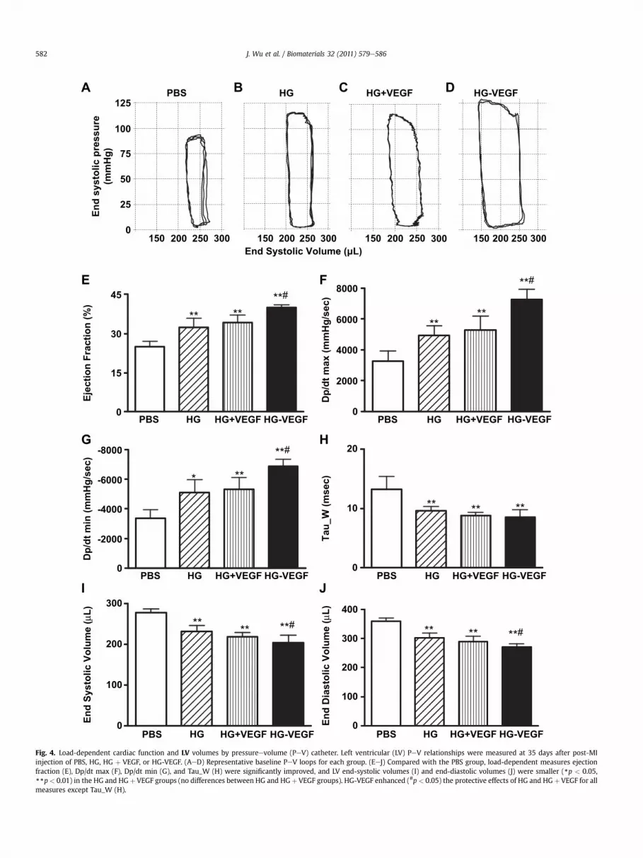
Fig. 4. Load-dependent cardiac function and LV volumes by pressureevolume (PeV) catheter. Left ventricular (LV) PeV relationships were measured at 35 days after post-MIinjection of PBS, HG, HG þ VEGF, or HG-VEGF. (AeD) Representative baseline PeV loops for each group. (EeJ) Compared with the PBS group, load-dependent measures ejectionfraction (E), Dp/dt max (F), Dp/dt min (G), and Tau_W (H) were significantly improved, and LV end-systolic volumes (I) and end-diastolic volumes (J) were smaller (*p < 0.05,**p< 0.01) in the HG and HG þ VEGF groups (no differences between HG and HG þ VEGF groups). HG-VEGF enhanced (#p < 0.05) the protective effects of HG and HG þ VEGF for allmeasures except Tau_W (H).
J. Wu et al. / Biomaterials 32 (2011) 579e586582

150 175 200 225 250 275 300
50
60
70
80
90
100
110
120
PBS
HG
HG+VEGF
HG-VEGF
ESPVR
End Systolic Volume ( µµL)
En
d S
ysto
lic P
ressu
re (m
mH
g)
PRSW
175 200 225 250 275 300 325 350
2000
3000
4000
5000
6000
7000
8000
9000
PBS
HG
HG+VEGF
HG-VEGF
End Diastolic Volume ( µL)
Stro
ke W
ork (m
mH
g* µ
L)
HG-VEGF
HG PBS
HG+VEGF
D C
B
E F
*
**
**
**
150 140 130 120 110 100 90 80 70 60 50 40 30 20 10 0
150 140 130 120 110 100 90 80 70 60 50 40 30 20 10 0
150 140 130 120 110 100 90 80 70 60 50 40 30 20 10 0
150 140 130 120 110 100 90 80 70 60 50 40 30 20 10 0
Pres
sure
(mm
Hg)
Pres
sure
(mm
Hg)
Pres
sure
(mm
Hg)
Pres
sure
(mm
Hg)
180 201 222 243 264 285 Volume (µl)
195 221 247 273 299 325 Volume (µl)
165 190 215 240 265 290 Volume (µl)
195 229 263 297 331 365Volume (µl)
ig. 5. Load-independent cardiac function by pressureevolume (PeV) catheter. (AeD) Representative series of PeV loops obtained during vena cava occlusion at 35 days after post-I injection of HG-VEGF (A), HG þ VEGF (B), HG (C), or PBS (D). (E,F) Load-independent measures end-systolic pressureevolume relationship (ESPVR, E) and preload recruitabletroke work (PRSW, F) were significantly improved (**p < 0.01) in the HG and HG þ VEGF groups compared with the PBS group (no differences between HG and HG þ VEGFroups). HG-VEGF enhanced (*p < 0.05, **p < 0.01) the protective effects of HG and HG þ VEGF.
J. Wu et al. / Biomaterials 32 (2011) 579e586 583
A
FMsg

0
10
20
Scar area / L
V area (%
)
30
40
PBS HG HG+VEGF HG-VEGF
0.00
0.25
0.50
0.75
1.00
* **
**
**
**
HG-VEGF
HG+VEGF
PBS
HG
A
B
C
PBS HG HG+VEGF HG-VEGF
Scar th
ickn
ess (m
m)
Fig. 6. Infarct morphometry by planimetry. (A) Representative heart slices obtained at35 days after post-MI injection of PBS, HG, HG þ VEGF, or HG-VEGF. Arrows indicatethe location of the infarct in individual slices. (B, C) LV scar area (as a percentage oftotal LV area, B) was decreased and scar thickness (C) was increased after the injectionof HG or HG þ VEGF (*p < 0.05, **p < 0.01 vs. PBS for both measures). Scar area wassmallest and scar thickness was greatest after the injection of HG-VEGF (#p < 0.05 vs.HG and HG þ VEGF for both measures).
J. Wu et al. / Biomaterials 32 (2011) 579e586584
thickness, and vascular density. A two-way ANOVA compared the effects of treat-ment and time on fractional shortening (echocardiography). When the F ratio wassignificant, differences were specified using Tukey’s multiple range tests.
3. Results
3.1. Synthesis and characterization of the VEGF polymer
The triblock copolymer (PEG-PVL) was synthesized by poly-merizing PEG with VL using a metal-free cationic polymerizationmethod (Fig. 1A). Triblock copolymers are water soluble at roomtemperature. HG-VEGF was synthesized by coupling the NHSfunctionalized polymer with VEGF protein in PBS solution (Fig. 1B).Successful conjugation was verified using a Western blot (Fig. 1C).We observed a clear VEGF expression band in the HG-VEGF sample.Unconjugated VEGF was not observed.
PEG-PVL is a clear solution inwater at ambient temperature. Thebiomaterial gels at a concentration of 200 mg/mL, and forms an HGwithin 10 min at 37 �C (Fig. 2A). Scanning electron microscopeanalysis revealed a honeycomb structure with a pore size ofapproximately 1 mm, which may facilitate the diffusion of proteinsor peptides (Fig. 2B).
3.2. Hydrogel degradation
In vitro: HG (10, 20, or 30 mL) pellets dropped onto a cell culturedish formed a gel at 37 �C. The pellet diameter did not change over5 weeks (data not shown), indicating that the gel was stable in vitro.
In vivo: HG injected subcutaneously (10, 20, or 30 mL) formednodules that initially increased in size as the nodules absorbedwater at all 3 tested concentrations. The nodules decreased in sizebeyond 7 days after implantation, and were completely degradedafter 42 days (Fig. 2C).
3.3. In vivo study
3.3.1. AnimalsA total of 44 rats were initially included in the study. Three rats
died during or immediately after undergoing the MI procedure, and2 rats died during the second surgical procedure (gel or PBSinjection). There were no further deaths. Of the remaining 39animals, 10 received HG-VEGF, 10 received HG þ VEGF, 11 receivedHG, and 8 received PBS control.
3.3.2. Cardiac function3.3.2.1. Echocardiography. Coronary artery ligation producedsignificant left ventricle (LV) dilatation, evidenced by increased LVdiameter and decreased LV function in all animals. There were nodifferences among the groups in any of the echocardiographicparameters at 7 days after MI (Fig. 3). Overall, HG-VEGF contributedmore functional preservation than HG and HG þ VEGF, though allHG treatments increased fractional shortening (p< 0.01) comparedto PBS. Specifically, both HG and HG þ VEGF preserved function(p < 0.01 vs. PBS) beyond 21 days after injection, while HG-VEGFincreased fractional shortening earlier, by 14 days after injection(p < 0.01 vs. PBS, HG, HG þ VEGF).
3.3.2.2. Pressureevolume catheter. Ventricular volumes and cardiacfunction were evaluated under load-dependent (Fig. 4) and load-independent (Fig. 5) conditions. Compared with PBS, HG andHG þ VEGF improved ejection fraction (EF), Dp/dt max, Dp/dt min,and Tau (load-dependent indices), as well as end-systolic elastance(ESPVR) and preload recruitable stroke work (PRSW) (load-inde-pendent indices). HG-VEGF further improved EF, Dp/dt max, Dp/dtmin, ESPVR, and PRSW (p < 0.01 vs. PBS, p < 0.05 vs. HG and
HG þ VEGF). End-systolic and end-diastolic volumes were smallestin the HG-VEGF group, followed by the HG þ VEGF and HG groups.All HG treatments reduced end-systolic and end-diastolic volumesrelative to PBS (p < 0.01 for all groups).
3.3.3. Infarct morphometryMyocardial scar tissue was observed in all animals (Fig. 6A).
Computerized planimetry (performed at 42 days after MI on explan-ted hearts fixed at physiologic pressures) showed that infarcts werelarger and infarcted tissue was thinner in animals that received PBSrather than HG or HG þ VEGF (p < 0.05). Scar size was smallest and

Fig. 7. Vascular density. (AeD) Representative micrographs illustrating vascular formations (arrows ¼ Factor VIII þ structures) in the peri-infarct area at 35 days after post-MIinjection of PBS, HG, HG þ VEGF, or HG-VEGF. (E) Capillary density was significantly greater after the injection of HG or HG þ VEGF (*p < 0.05 vs. PBS), and highest after HG-VEGF(#p < 0.05 vs. HG and HG þ VEGF).
J. Wu et al. / Biomaterials 32 (2011) 579e586 585
scar thickness greatest in animals that receivedHG-VEGF (p< 0.05 vs.HG and HG þ VEGF for both measures) (Fig. 6B,C). These findingssuggest that conjugating VEGF with the biomaterial stabilized theinfarct and prevented scar expansion and ventricular dilatation.
3.3.4. Blood vessel density
Blood vessels expressing Factor VIII were quantified immunohis-tochemically in the infarcted and peri-infarcted myocardium (Fig. 7).Vascular densities were increased in the hearts of animals thatreceivedHG orHGþVEGF (p< 0.05 vs. PBS), and further increased inthose that received HG-VEGF (p < 0.05 vs. HG and HG þ VEGF).
4. Discussion
This study reports the synthesis of a temperature-sensitive HGthat is an injectable liquid at room temperature and becomesa biodegradable solid at physiological temperature (37 �C). Our HGalso permits the conjugation of cytokines before myocardial
implantation, which might extend the period of delivered cytokineactivity. We demonstrated that intramyocardial injection of the HGsignificantly preserved scar thickness, attenuated adverse cardiacremodeling, and improved ventricular function (compared withPBS injection) for up to 35 days after an MI in rats. Further,compared with HG alone or HG mixed with VEGF, HG withconjugated VEGF boosted regional angiogenesis.
After an MI, cardiomyocyte loss triggers matrix degradation,fibrosis, and the progression to heart failure. Injectable biomaterialsmitigate these effects by providing a simple method to physicallystabilize the infarct (prevent ventricular remodeling) and preservematrix structure [2e4]. Kelley et al. [18] first demonstrated thatsuturing a polypropylene mesh to the myocardium to restrain theinfarcted wall preserves LV geometry and prevents cardiac func-tional decline. Our HG may have the same effect if applied soonafter a coronary occlusion. An advantage of injectable biomaterialsover external restraints is their ability to increase the recruitment ofmarrow progenitor cells and induce angiogenesis [2]. In this study,the temperature-sensitive HG (vs. PBS) increased scar thickness,

J. Wu et al. / Biomaterials 32 (2011) 579e586586
prevented LV dilation, improved cardiac function, and enhancedangiogenesis. It may also have prevented paradoxical systolicbulging by gelling immediately within the infarct.
The damaging effects of an MI can be further moderated by genetransfection, cytokine treatment, or implanted stem cells [19].However, the infarcted myocardium is stiff and the marginal regionis enriched with blood vessels, and so genes, cytokines, or cellsinjected directly into the scar often leak into the epicardial space orthe circulation (for example, we found that cell retention was lessthan 50% immediately after implantation into a myocardial infarct[20]). Unsuccessful strategies to increase retention have includedmultiple small volume injections, sealing the injection holes, andencapsulating genes, proteins or cells. However, there has beensome success with injectable biomaterials, which e beyondproviding structural support for the infarcted heart e also act asagents of therapeutic cytokine delivery. For instance, biologically-derived materials such as fibrin, collagen, alginates, and self-assembling peptides [2,3,21] can provide a platform to increase thedelivery and/or support of cells or cytokines e enhancing bloodflow and preventing necrosis [22,23]. Here, we tested a PVL-b-PEG-b-PVL HG that provides additional advantages since it is relativelyinert (unlikely to induce rejection), easy to manufacture (andtherefore, more cost-efficient than the biologically-derived mate-rials), and it is synthetic, with a very uniform, honeycomb-likestructure that may facilitate the diffusion of proteins or peptides.
PVL-b-PEG-b-PVL gels at a physiological temperature (37 �C)and degrades within 6 weeks. Temperature-sensitive HGs areparticularly attractive candidates for injectable therapeuticsbecause they can easily incorporate biological elements while inliquid form (i.e., at room temperature) that are retained in the heartwhen the polymer gels rapidly at body temperature. In this way, thegel effectively “traps” therapeutic cells or cytokines and thenprolongs their effects by releasing them gradually at the injectionsite as the biomaterial slowly degrades. Indeed, in this study,conjugating VEGF to the HG (vs. mixing VEGF and HG) increasedregional angiogenesis in vivo e perhaps because the biologicalactivity of VEGF was extended over the w40 days of biomaterialdegradation (post-MI healing phase). Boosting cell recruitment andangiogenesis at the site of myocardial repair is particularly impor-tant for older, debilitated patients because the regenerativecapacity of endogenous stem cells declines with increasing age.
5. Conclusions
We synthesized a temperature-sensitive, biodegradable HG thatpreserved ventricular function after an MI by stabilizing the infarctand inducing angiogenesis. Many investigators are attempting todevelop methods that will improve the delivery efficiency of VEGFand other cytokines at sites of ischemic injury. We found thatconjugating VEGF to the HGmaterial (rather than mixing VEGF andHG) before injection optimized the extent of myocardial andfunctional recovery e possibly by extending the angiogenic effectsof the “trapped” VEGF throughout the period of HG degradation.
Funding sourcesThis research was supported by grants from the Canadian
Institutes of Health Research (MOP14795) and the Heart and StrokeFoundation of Ontario (T6604) to R-KL. The authors wish toacknowledge the generous support of Eileen Mercier and INGCanada Inc.
Conflict of interest disclosuresNone declared.
Acknowledgements
R-KL is a Career Investigator of the Heart and Stroke Foundationof Canada, and holds a Canada Research Chair in cardiac regener-ation. We thank Heather McDonald Kinkaid for writing and editingassistance.
References
[1] McMurray J, Pfeffer MA. New therapeutic options in congestive heart failure:part I. Circulation 2002;105:2099e106.
[2] Christman KL, Lee RJ. Biomaterials for the treatment of myocardial infarction.J Am Coll Cardiol 2006;48:907e13.
[3] Leor J, Amsalem Y, Cohen S. Cells, scaffolds, and molecules for myocardialtissue engineering. Pharmacol Ther 2005;105:151e63.
[4] Christman KL, Vardanian AJ, Fang Q, Sievers RE, Fok HH, Lee RJ. Injectablefibrin scaffold improves cell transplant survival, reduces infarct expansion,and induces neovasculature formation in ischemic myocardium. J Am CollCardiol 2004;44:654e60.
[5] Kuo CK, Ma PX. Ionically crosslinked alginate hydrogels as scaffolds for tissueengineering: part 1. Structure, gelation rate and mechanical properties.Biomaterials 2001;22:511e21.
[6] Ono K, Saito Y, Yura H, Ishikawa K, Kurita A, Akaike T, et al. Photocrosslinkablechitosan as a biological adhesive. J Biomed Mater Res 2000;49:289e95.
[7] Wang T, Wu DQ, Jiang XJ, Zhang XZ, Li XY, Zhang JF, et al. Novel thermo-sensitive hydrogel injection inhibits post-infarct ventricle remodelling. Eur JHeart Fail 2009;11:14e9.
[8] LuWN, Lu SH,Wang HB, Li DX, Duan CM, Liu ZQ, et al. Functional improvementof infarcted heart by co-injection of embryonic stem cells with temperature-responsive chitosan hydrogel. Tissue Eng Part A 2009;15:1437e47.
[9] Wang H, Zhang X, Li Y, Ma Y, Zhang Y, Liu Z, et al. Improved myocardialperformance in infarcted rat heart by co-injection of basic fibroblast growthfactor with temperature-responsive chitosan hydrogel. J Heart Lung Trans-plant 2010;29:881e7.
[10] Kwon JS, Park IK, Cho AS, Shin SM, Hong MH, Jeong SY, et al. Enhancedangiogenesis mediated by vascular endothelial growth factor plasmid-loadedthermo-responsive amphiphilic polymer in a rat myocardial infarction model.J Control Release 2009;138:168e76.
[11] Sellke FW, Tofukuji M, Laham RJ, Li J, Hariawala MD, Bunting S, et al.Comparison of VEGF delivery techniques on collateral-dependent microvas-cular reactivity. Microvasc Res 1998;55:175e8.
[12] Heredia KL, Maynard HD. Synthesis of protein-polymer conjugates. Org Bio-mol Chem 2007;5:45e53.
[13] Li J, Kao WJ. Synthesis of polyethylene glycol (PEG) derivatives and PEGylated-peptide biopolymer conjugates. Biomacromolecules 2003;4:1055e67.
[14] Zeng F, Lee H, Allen C. Epidermal growth factor-conjugated poly(ethyleneglycol)-block-poly(delta-valerolactone) copolymermicelles for targeteddeliveryof chemotherapeutics. Bioconjug Chem 2006;17:399e409.
[15] Lee J, Bae YH, Sohn YS, Jeong B. Thermogelling aqueous solutions of alter-nating multiblock copolymers of poly(L-lactic acid) and poly(ethylene glycol).Biomacromolecules 2006;7:1729e34.
[16] Kan CD, Li SH, Weisel RD, Zhang S, Li RK. Recipient age determines the cardiacfunctional improvement achieved by skeletal myoblast transplantation. J AmColl Cardiol 2007;50:1086e92.
[17] Sun Z, Wu J, Fujii H, Wu J, Li SH, Porozov S, et al. Human angiogenic cellprecursors restore function in the infarcted rat heart: a comparison of celldelivery routes. Eur J Heart Fail 2008;10:525e33.
[18] Kelley ST, Malekan R, Gorman III JH, Jackson BM, Gorman RC, Suzuki Y, et al.Restraining infarct expansion preserves left ventricular geometry and func-tion after acute anteroapical infarction. Circulation 1999;99:135e42.
[19] Sato K, Wu T, Laham RJ, Johnson RB, Douglas P, Li J, et al. Efficacy of intra-coronary or intravenous VEGF165 in a pig model of chronic myocardialischemia. J Am Coll Cardiol 2001;37:616e23.
[20] Yasuda T, Weisel RD, Kiani C, Mickle DA, Maganti M, Li RK. Quantitativeanalysis of survival of transplanted smooth muscle cells with real-timepolymerase chain reaction. J Thorac Cardiovasc Surg 2005;129:904e11.
[21] Davis ME, Motion JP, Narmoneva DA, Takahashi T, Hakuno D, Kamm RD, et al.Injectable self-assembling peptide nanofibers create intramyocardial micro-environments for endothelial cells. Circulation 2005;111:442e50.
[22] Richardson TP, Peters MC, Ennett AB, Mooney DJ. Polymeric system for dualgrowth factor delivery. Nat Biotechnol 2001;19:1029e34.
[23] Silva EA, Mooney DJ. Spatiotemporal control of vascular endothelial growthfactor delivery from injectable hydrogels enhances angiogenesis. J ThrombHaemost 2007;5:590e8.


![[Dissecting aortic aneurysm simulating an acute myocardial infarct]](https://img.pdfslide.net/doc/110x75/63558f23922cbb7c550ca86c/dissecting-aortic-aneurysm-simulating-an-acute-myocardial-infarct.jpg)


