Embed Size (px)
Citation preview

Palaeogeography, Palaeoclimatology, Palaeoecology 310 (2011) 427–441
Contents lists available at SciVerse ScienceDirect
Palaeogeography, Palaeoclimatology, Palaeoecology
j ourna l homepage: www.e lsev ie r.com/ locate /pa laeo
Influence of atrypid morphological shape on Devonian episkeletobiont assemblagesfrom the lower Genshaw formation of the Traverse Group of Michigan: A geometricmorphometric approach
Rituparna Bose a,⁎, Chris L. Schneider b, Lindsey R. Leighton c, P. David Polly a
a Department of Geological Sciences, Indiana University, 1001 East 10th Street, Bloomington, IN 47405, United Statesb Alberta Geological Survey, Energy Resources Conservation Board, 4999 98th Ave, Edmonton, Alberta, Canada T6B 2X3c Department of Earth and Atmospheric Sciences, 1–26 Earth Sciences Building, University of Alberta, Edmonton, Alberta, Canada T6G 2E3
⁎ Corresponding author. Tel.: +1 812 606 8815; fax:E-mail addresses: [email protected] (R. Bose), chri
(C.L. Schneider), [email protected] (L.R. Leighton), pd
0031-0182/$ – see front matter © 2011 Elsevier B.V. Aldoi:10.1016/j.palaeo.2011.08.004
a b s t r a c t
a r t i c l e i n f oArticle history:Received 21 May 2011Received in revised form 9 August 2011Accepted 10 August 2011Available online 16 August 2011
Keywords:AtrypidaGivetianMorphologyLandmarksPalaeoecologyEpiskeletobionts
Atrypids examined from the lower Genshaw Formation of the Middle Devonian (early middle Givetian)Traverse Group include a large assemblage of Pseudoatrypa bearing a rich fauna of episkeletobionts. Weidentified two species of Pseudoatrypa – Pseudoatrypa lineata and Pseudoatrypa sp. A based on ornamentationand shell shape. Qualitative examination suggested that the former had fine-medium size ribbing, narrowhinge line, widened anterior, gentle to steep mid-anterior fold, a more domal shaped dorsal valve, and aninflated ventral valve in contrast to the coarse ribbing, widened hinge line, narrow anterior, gentle mid-anterior fold, arched shape dorsal valve, and flat ventral valve of the latter. Geometric morphometric analysissupported two statistically different shapes (pb0.01) for the two distinct species.This study further examined these atrypids to investigate the influence of morphology on episkeletobiontsettlement on the two Pseudoatrypa species. Among the 343 atrypid hosts examined, nearly 50% were encrustedby episkeletobionts. Common encrusters included microconchids, bryozoan sheets, and hederellids. Lesscommon encrusters included auloporid corals, cornulitids, tabulate corals, Ascodictyon, craniid brachiopods, andfenestrate bryozoans. Hederellids, auloporid corals, cornulitids, and tabulate corals encrusted a few livingPseudoatrypa hosts, but determination of pre- or post-mortemencrustation by themajority of episkeletobionts isequivocal. In a very few cases, episkeletobionts crossed the commissure indicating the death of the host.Some episkeletobionts, microconchids and the sheet bryozoans, were more common on Pseudoatrypa lineata,which exhibited more dorsal-ventral convexity than Pseudoatrypa sp. A. Perhaps, P. lineatamay have provided alarger surface area for episkeletobiont settlement relative to Pseudoatrypa sp. A.In both the host species, encrustation was heaviest on the convex dorsal valve. This suggests that most of theencrustation occurred in a reclining, dorsal-valve-up life orientation of both species, in which the convex dorsalvalve was exposed in the water column and the ventral valve remained in contact with the substrate. However,life orientations of these atrypid species could not be confidently predicted simply from the location preferencesof episkeletobionts alone, as the life orientation of the host would also have been a hydrodynamically stableorientation of the articulated shell after death.Most episkeletobionts encrusted the posterior region of both dorsal and ventral valves of the two species, whichsuggests that the inflated areas of these valves, when exposed, favored the settlement of most episkeletobiontlarvae.
+1 812 855 [email protected]@indiana.edu (P.D. Polly).
l rights reserved.
© 2011 Elsevier B.V. All rights reserved.
1. Introduction
Pseudoatrypa is a common brachiopod from the Givetian to lateFrasnian of North America. This genus occurs throughout much of theTraverse Group in the Michigan Basin, including the GenshawFormation (Kelly and Smith, 1947; Koch, 1978). Here we focus onmaterial from the lower Genshaw Formation to: 1) analyze morpho-
logical shape patterns in two Pseudoatrypa species, and, 2) investigateepiskeletobiont interactions with these species to determine how thedistinct morphological shapes of the two species may have influencedtheir settlement.
Pseudoatrypa is frequently encountered in Devonian Midcontinentbasins. Webster (1921) first described the taxon as Atrypa devonianafrom the late Frasnian Independence Shale of Iowa; his specimenswere later designated as the type species of the new genusPseudoatrypa (Copper, 1973; Day and Copper, 1998). Pseudoatrypaalso occurs in the Traverse Group of the Michigan Basin (Stumm,1951; Copper, 1973; Koch, 1978) and in the Silica Formation of Ohio

428 R. Bose et al. / Palaeogeography, Palaeoclimatology, Palaeoecology 310 (2011) 427–441
and equivalent rocks from northern Indiana (Wiedman, 1985).Webster (1921) described the species Atrypa lineata from the lateMiddle Devonian (late Givetian) upper Osage Springs Member of theLithograph City Formation of the Upper Cedar Valley Group of Iowa,which was later included in Pseudoatrypa by Day and Copper (1998).Fenton and Fenton (1935) described a subspecies and growth variantforms of this species from the late Givetian Cedar Valley of Illinois.Herein we test both qualitatively and quantitatively whether distinctspecies exist within Pseudoatrypa from the Lower Genshaw Formationof Michigan, and whether these external morphology influencesepiskeletobiont assemblages.
Episkeletobionts are organisms that adhere to, or encrust, the surfaceof a shell (Taylor and Wilson, 2002). Episkeletobionts are useful asecological and life status indicators of their hosts—whether the hostwasliving at the time of encrustation orwas dead (Watkins, 1981; Andersonand Megivern, 1982; Brezinski, 1984; Gibson, 1992; Lescinsky, 1995;Sandy, 1996; Sumrall, 2000; Morris and Felton, 2003; Schneider, 2003,2009a; Zhan and Vinn, 2007; Rodrigues et al., 2008). Episkeletobiontshave been used to infer the life orientation of brachiopods (Rudwick,1962; Hurst, 1974; Pitrat and Rogers, 1978; Kesling et al., 1980;Spjeldnaes, 1984; Lescinsky, 1995), the preferred orientation of hostwater currents (e.g., Kesling et al., 1980), potential camouflage for hosts(Schneider, 2003, 2009a), the attracting or antifouling nature ofornamentation (Richards and Shabica, 1969; Richards, 1972; Carrera,2000; Schneider, 2003, 2009a; Schneider and Leighton, 2007), and thefunction of the valve punctae (Thayer, 1974; Curry, 1983; Bordeaux andBrett, 1990). Brachiopod hosts are useful for investigating hostinfluences on episkeletobiont preferences such as shell texture(Schneider and Webb, 2004; Rodland et al., 2004; Schneider andLeighton, 2007), size of host (Ager, 1961; Kesling et al., 1980), andantifouling strategies (Schneider and Leighton, 2007). Although otherPaleozoic marine organisms were frequently encrusted, brachiopodsremain one of the best understood hosts for Paleozoic episkeletobionts.
In the present study, the settlement of a live episkeletobiontduring the life of the host is called a live–live association (pre-mortemencrustation) and the settlement of a live encruster on a dead host iscalled a live-dead association (post-mortem encrustation). Taylor andWilson (2003) provided the following criteria for distinguishingbetween pre- and post-mortem associations: (a) If the episkeleto-biont fossil overgrows (crosses) the commissural margin, or if there isevidence of internal valve encrustation, then the brachiopod host wasdead when the organism overgrew the commissure - this is evidenceof a live-dead association; (b) if the episkeletobiont and the host bothhave a similar degree of preservation, and if there is evidence of scarsrepresenting the repair of damage inflicted by episkeletobionts, thenthe brachiopod host was alive during encrustation - this is evidence oflive-live association; and (c) if certain episkeletobionts repeatedlyencrust specific locations on host shells, e.g., if branching fossils, likeauloporid corals or hederellids, branch towards or are aligned parallelto the commissure or if solitary organisms, such as cornulitids, growwith their apertures pointing towards the commissure, then hosts andtheir episkeletobionts experienced live-live associations. In othercases, there is no way to tell for certain whether the host was alive ordead at the time of encrustation.
Our purpose herein is twofold: (1) to quantitatively assessputatively distinct species of Pseudoatrypa from the GenshawFormation, herein described as Pseudoatrypa lineata and Pseudoatrypasp. A; and (2) to examine the influence of species morphology onencrustation by episkeletobionts. We structured the study by testingthe following hypotheses:
1) two species of Pseudoatrypa - Pseudoatrypa lineata and Pseudoa-trypa sp. A, previously distinguished on qualitative features, musthave statistically distinct shell shapes and are validly differentspecies;if not they will be considered growth variants of the samespecies;
2) episkeletobionts are influenced by morphology and will preferen-tially encrust the two species differently; and
3) given the preferred orientation of atrypid adult shells—convexdorsal valve raised into the water column and flat ventral valve incontact with the substrate (as inferred by Fenton and Fenton(1932)) —the extent of encrustation coverage on the ventral valvewill be limited by physical contact with the substrate and thusstatistically less than on the dorsal valve.
These hypotheses were tested using geometric morphometricassessment of shell shape and statistical analysis of the location ofencrusting organisms.
2. Geologic setting
Pseudoatrypa brachiopods were collected from the lower GenshawFormation of the Middle Devonian Traverse Group. The TraverseGroup ranges in thickness from ~25.0–169.5 meters (Ehlers andKesling, 1970; Wylie and Huntoon, 2003), with depositional environ-ments ranging from shallow water carbonate lagoons with coral-stromatoporoid reefs to storm-dominated mixed carbonate-siliciclastic shelf deposits and offshore muddy shelf to slopeenvironments (Ehlers and Kesling, 1970). The Genshaw Formationwas named by Warthin and Cooper (1935) for strata overlying theirFerron Point Formation and underlying their Killians Limestone, laterrevised by the same stratigraphers to include the Killians Limestone asthe upper member of the Genshaw Formation (Warthin and Cooper,1943). Warthin and Cooper (1943) placed the new upper contact atthe base of the overlying Newton Creek Limestone. The GenshawFormation remained one of the least studied units of the lowerTraverse Group until recently, when the LaFarge Quarry in the Alpenaarea began to mine into this unit and exposed nearly the entireFormation (Bartholomew, 2006). The Genshaw Formation accumu-lated during the highstand of a third-order sea level sequence (Wylieand Huntoon, 2003; Brett et al., 2010).
The Genshaw Formation, which is ~30.0 meters thick (Fig. 1), issubdivided into informal lower, middle, and upper (formerly KilliansMember) portions (Wylie and Huntoon, 2003). The lower unit of theGenshaw Formation begins with a 0.5 m-thick crinoidal grainstone,which locally contains burrows on its lower surface. Overlying thisbasal bed of the Genshaw Formation is a thin, argillaceous successioncapped by a limestone-rich interval to the top of the lower Genshaw.The brachiopods used in this study were collected from theargillaceous beds of this lower unit (Fig. 1).
3. Materials and methods
3.1. Sampling
Samples examined were collected by A. Bartholomew of StateUniversity of New York, New Paltz from the northeastern outcrop ofthe Lafarge Alpena Quarry, Alpena County, Michigan (Fig. 1). Heextensively sampled all brachiopods from a shale bed of the lowerGenshaw Formation. The 185 well-preserved atrypids examined forencrustation in this study have been deposited in the IndianaUniversity Paleontology Collection (IU 100059 – IU 100243). Use ofammonium chloride spray in a dry environment helped distinguishmorphological features of the species.
3.2. Species recognition
The atrypid sample was first divided into two populations basedon the qualitative traits examined in this study. The two populationsare similar in that they have an apical foramen, hinge line withincurved extremities, orbicular to subquadrate shell outline, ribbingwith implantations and bifurcations, and somewhat similar spacing

Fig. 1. A) Map showing Michigan surface exposures of the Middle Devonian Traverse Group and the location of the La Farge Quarry, Alpena area, fromwhich the samples used in thisstudy were collected, B) Simplified stratigraphic section of the Traverse Group at La Farge Quarry showing the horizons of the Genshaw Formation where the specimens used in thisstudy were collected (after Bartholomew, 2006).
429R. Bose et al. / Palaeogeography, Palaeoclimatology, Palaeoecology 310 (2011) 427–441
between growth lamellae or frills, with frills crowding more at theanterior. However, Pseudoatrypa lineata is different from Pseudoatrypasp. A in having a) a smooth, arcuate, domal curvature to the dorsalvalve as opposed to the arched shape in the latter, b) slightly inflatedventral valve with an inflation near the umbo as opposed to a moreflattened ventral valve in the latter, c) relatively lower dorsal valvecurvature height, d) fine to medium closely spaced ribs in contrast tocoarse ribs in the latter, e) angular to subrounded hinge line asopposed to the widened hinge in the latter, f) widened mediandeflection (fold and sulcus) on the commissure as opposed to thenarrow deflection in the latter, and g) gentle to steep mid-anteriorfold as opposed to the gentler fold in the latter.
3.3. Morphometrics
We performed geometric morphometric analysis to determinemorphological variation within and between the two qualitativelydistinguished populations assigned to Pseudoatrypa lineata and Pseu-doatrypa sp. A. No crushed specimens were used for morphometrics.
Geometric morphometrics is the analysis of geometric landmarkcoordinates points on specific parts of an organism (Bookstein, 1991;MacLeod and Forey, 2002; Zelditch et al., 2004). We based ourmorphometric analysis on the use of landmarks to capture shape(Rohlf and Marcus, 1993); landmarks represent discrete geometricpoints on each specimen that correspondamong forms (sensuBookstein,1991). In this study, we used 10 two-dimensional landmark points onthe external shell to capture the most meaningful shape differences(Fig. 2).When selecting landmarks for analyses, we selected points thatnot only characterized body shape accurately, but also representedsomeaspect of the inferred ecological niche. All landmarkswere definedby geometric position on host shells (1=beak tip on brachial valve; 2and 8=intersection points of the commissure and the hingeline; alsoregion for food intake from inhalant currents; 3 and7=lengthmidpointprojected onto the commissure; these points are perpendicular to, andcrosses the midline of the shell; 4 and 6=lowest point of mediandeflection (fold and sulcus) on the commissure; 5=middle point ofcommissure; 9=tip of umbo on pedicle valve; 10=maximum height
on brachial valve). These landmarks are appropriate for analysesattempting to capture shape changes or function. For this study, weconducted four different analyses operating on ten landmarks in fourdifferent orientations of the shell. Two analyses were conducted in thex-y plane of the dorsal and ventral valves; these analyses capture onlythe view in that plane (Fig. 2A-B). These analyses of the dorsal andventral valve views, included nine landmarks, which were selected toencompass the outline of the entire specimen in the x-y plane. Althoughbrachiopods are bilaterally symmetrical, landmarkswere included fromboth the left and right sides of the specimens to record the functionalresponse of these hosts to the then existing ecological conditions and toencrusting episkeletobionts. Capturing both the postero-lateral distalextremities of the hingeline and the lowest points of median deflectionon the commissure, even of a bilaterally symmetrical organismmay beimportant for determining shape changes in the host species, as each ofthese locations may possess unique specific abundances of distinctepiskeletobiont assemblages (Bookstein, 1991; Kesling et al., 1980).Two separate analyses operating on landmarks in the y-z planewere conducted from the anterior and posterior views of the shells(Fig. 2C-D). For posterior and anterior regions of the shell, landmarkmeasurements (four landmarks on posterior and three landmarks onanterior) were taken only on half of the specimen (anterior/posteriorview left or right) (Fig. 2C-D). Overall, these two orientations measurenot only the shape of the valves, but also capture the shape of thebrachiopod lophophore support, the spiralia (Bookstein, 1991; Haneyet al., 2001). The four views (dorsal, vental, anterior, andposterior)wereanalyzed separately.
Procrustes analysis (Rohlf, 1990; Rohlf and Slice, 1990; Rohlf,1999; Slice, 2001) was performed on original shape data, rotating,translating and scaling all landmarks to remove all size effects, whilemaintaining their geometric relationships (Procrustes superimposi-tion). Principal component analysis of the covariance matrix of theresiduals of the Procrustes superimposed coordinates was performedto determine the morphological variation of the two species alongtheir major principal component axes (1 and 2) in the shape morpho-space (Fig. 2) and to provide a set of uncorrelated shape variables forfurther statistical analysis. Procrustes distances, which are the sum of
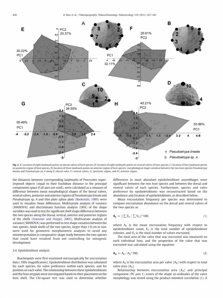
Fig. 2. A) Location of eight landmark points on dorsal valves of host species, B) location of eight landmark points on ventral valves of host species, C) location of four landmark pointson posterior region of host species, D) location of three landmark points on anterior region of host species; morphological shape variation between the two host species Pseudoatrypalineata and Pseudoatrypa sp. A along E) dorsal vales, F) ventral valves, G) posterior region, and H) anterior region.
430 R. Bose et al. / Palaeogeography, Palaeoclimatology, Palaeoecology 310 (2011) 427–441
the distances between corresponding landmarks of Procrustes super-imposed objects (equal to their Euclidean distance in the principalcomponents space if all axes are used), were calculated as a measure ofdifference between mean morphological shapes of the dorsal valves,ventral valves, posterior andanterior regionsof Pseudoatrypa lineata andPseudoatrypa sp. A and thin plate spline plots (Bookstein, 1989) wereused to visualize those differences. Multivariate analysis of variance(MANOVA) and discriminant function analysis (DFA) of the shapevariableswasused to test for significant shell shape differences betweenthe two species along the dorsal, ventral, anterior and posterior regionsof the shells (Hammer and Harper, 2005). Multivariate analysis ofvariance (MANOVA)wasperformed to test shape variationbetween thetwo species. Adult shells of the two species, larger than 1.9 cm in size,were used for geometric morphometric analyses to avoid anymisinterpretation in comparative shape study between the two speciesthat could have resulted from not controlling for ontogenicdevelopment.
3.4. Episkeletobiont analysis
Brachiopods were first examined microscopically for encrustationdata (100x magnification). Episkeletobiont distribution was tabulatedfor each species, for valve preference within each species, and forposition on each valve. The relationship between these episkeletobiontsand thehost atrypidswere investigatedbasedon their placement on thehost shell. The Chi-square test was used to determine whether
differences in most abundant episkeletobiont assemblages weresignificant between the two host species and between the dorsal andventral valves of each species. Furthermore, species and valvepreference by episkeletobionts was reconstructed based on theabundance and location of episkeletobionts, as described below.
Mean encrustation frequency per species was determined tocompare encrustation abundance on the dorsal and ventral valves ofthe two species as
AC = ∑ET =∑VEð Þ½ ��100; ð1Þ
where AC is the mean encrustation frequency with respect toepiskeletobiont count, ET is the total number of episkeletobiontcolonies, and VE is the total number of valves encrusted.
The total area of the valve that was encrusted was measured oneach individual host, and the proportion of the valve that wasencrusted was calculated using the equation:
AA = AE = AV�100; ð2Þ
where AA is the encrustation area per valve (AE) with respect to totalvalve area (AV).
Relationship between encrustation area (AA) and principalcomponent (PC axis 1) scores of the shape co-ordinates of the valvemorphology was tested using the product-moment correlation (r). A

431R. Bose et al. / Palaeogeography, Palaeoclimatology, Palaeoecology 310 (2011) 427–441
p-value was also reported for this correlation method to determine if‘r’ was significantly different from 0.0.
Following the methods previously established by Bose et al.(2010), each valve of the atrypid hosts was divided into six regions(Fig. 3). The six regions were defined as postero-left lateral (PLL),posteromedial (PM), postero-right lateral (PRL), antero-left lateral(ALL), anteromedial (AM), and antero-right lateral (ARL). These sixdivisions, i.e., six different surface areas of host, were selected suchthat they represent biologically functional grids for both the host andthe episkeletobiont. The PLL and PRL regions were selected based onthe idea that host inhalant currents in those regions may attractepiskeletobionts, and these currents may also partially influenceepiskeletobiont settling along the PM region. Similarly, the AM regionwas selected based on the host exhalant current criteria, which mayalso partially influence the ALL and ARL regions. Thus, selecting thesesix regions and recording the frequency of episkeletobionts on each ofthese regions will help infer host-episkeletobiont relationships inlive-live associations.
Area ratios for each region of the shell was determined using thefollowing equation (Fig. 3):
R = AR = AT; ð3Þ
where R is the area ratio, AR is the area of each region, and AT is thetotal area.
The frequency of encrustation in each region was recorded bycounting individual colonies asoneoccurrenceand then summing for allatrypid hosts. For comparison with the actual frequency of episkeleto-biosis, expected episkeletobiosis for each region was calculated by:
E = NRi; ð4Þ
in which the expected number of episkeletobionts (E) is calculated bymultiplying the total number of episkeletobionts (N) for allPseudoatrypa specimens by the area ratio (R) for a given region on
Fig. 3. Pseudoatrypa specimendivided into six regions for episkeletobiont frequency study;PLL=postero-left lateral, PM=posteromedial; PRL=postero-right lateral; ALL=antero-left lateral, AM=anteromedial; andARL=antero-right lateral. Numbers represent thearearatios of each grid across the Pseudoatrypa lineata and Pseudoatrypa sp. A host valve. Scalebar0.5 cm.Note that thearea ratios are slightly different for P. lineatahost specieswhich areas follows: PLL=0.18, PM=0.138, PRL=0.18, ALL=0.147, AM=0.208, ARL=0.147.Significantlygreaterobserved episkeletobiont frequency thanexpected isdenotedby aplussymbol and smaller observed episkeletobiont frequency than expected is denoted by aminus symbol across the six regions of the valve; this is described for A) dorsal valve ofP. lineata, B) dorsal valve of Pseudoatrypa sp. A, C) ventral valve of P. lineata, and D) ventralvalve of Pseudoatrypa sp. A.
the atrypid shell (i). The null hypothesis used here for developing theexpected value is as follows: Given that we have six regions for onevalve, and each region has a chance of being encrusted based on theirproportion of available surface area (assuming as a null hypothesis arandom distribution of encrusters), then if one region accounts for x%of the surface area on the valve, then the expected value for thatregion is x% of the total number of episkeletobionts encrusting thatvalve (all six regions) for that species. The same approach is used todetermine the expected value for the other regions. Colonialepiskeletobionts—sheet-like and branching—posed a problem forcalculations because these specimens often crossed borders intoadjacent regions. For these specimens, colonization of an episkeleto-biont that extended into two or more regions was divided among thetotal number of regions it inhabited. For example, branchingauloporid corals and hederellids that were observed in all the threeanterior regions were counted as 1/3 for each region.
We then quantified common episkeletobionts (i.e., microconchids,bryozoans sheets, and hederellids) for their distribution on six regionsof the shell of both dorsal and ventral valves of each host, usingEquation 2 above. Distribution of rare episkeletobionts on host shellswas also examined, but only for dorsal valves, as episkeletobiontabundance of rare encrusters is negligible on ventral valves. A chi-square test was also performed for the total observed and expectedepiskeletobiont activity along the six regions to determine theepiskeletobiont location preference on the atrypid valve within eachspecies.
4. Results
4.1. Morphology and morphometrics
Two-hundred and thirty two specimens of atrypids were assignedto Pseudoatrypa lineata and 111 specimens of atrypids were assignedto Pseudoatrypa sp. A. Representatives of the two species from thisstudy are shown in Fig. 4. The first two principal component axes(axes 1 and 2) explained a total of 50.2% of the variation in dorsalvalves, 52.9% of the variation in ventral valves, 82.0% of the variationin the posterior region and 98.0% of the variation in the anterior region(Fig. 2). Principal component analysis of dorsal and ventral valvesindicates that there is considerable shape variation within eachspecies and that the two species overlap considerably in themorphology of both valves in the x-y plane (Fig. 2E-F). Procrustesdistances between themean shape of the two species are 0.023 for thedorsal valves and 0.028 for ventral valves, suggesting that ventralvalves show slightly greater morphological differentiation than dorsalvalves. Principal component analysis of posterior and anterior regionsalso indicates that there is considerable variation in morphologybetween the two species in the y-z plane (Fig. 2G-H). Procrustesdistances between the mean shape of the two species are 1.69 for theposterior region and 1.53 for anterior region.. MANOVA detects asmall significant difference in mean shape of dorsal, ventral, posteriorand anterior regions (pb0.01). DFA detects a small but significantdifference in mean shape of dorsal and ventral valves (pb0.01) and alarge significant difference in mean shape of the posterior andanterior regions (pbb0.01) (Fig. 5). The significance of the MANOVAand the DFA demonstrates that the two populations can bedistinguished as separate species, based on shell shape.
Thin plate spline visualisation plots show the mean morphologicalshapes of these two species are different (Fig. 6). Dorsal valves show adifference in the shape of the posterior hinge line and anteriorcommissure. The distances between the umbo tip and the posteriorleft and right lateral margins in the dorsal valve plots are less inPseudoatrypa lineata than in Pseudoatrypa sp. A, confirming theobservation of a more widened hinge line in the latter. Similarly, thedistances between themiddle point of commissure and the lowest pointof the median deflection (fold and sulcus) on both halves of the

Fig. 4. Pseudoatrypa lineata A) Dorsal view, B) ventral view, C) posterior view, D) anterior view (IU#100069); Pseudoatrypa sp. A E) dorsal view, F) ventral view, G) posterior view andH) anterior view (IU#100220). The small inset illustrations next to dorsal and ventral views of P. lineata represent the type specimen of Atrypa lineata var. inflata as described inFenton and Fenton, 1935 and the posterior and anterior views represent the type specimen of Atrypa lineata as described in Day and Copper, 1998.
432 R. Bose et al. / Palaeogeography, Palaeoclimatology, Palaeoecology 310 (2011) 427–441
specimen suggesting a widening of the deflection in Pseudoatrypalineata and narrowing in Pseudoatrypa sp. A. Ventral valves, however, donot show much significant difference in shape, except for the widenedhinge line in Pseudoatrypa sp. A relative to the narrow hinge line inPseudoatrypa lineata. The posterior region plots show greater distance
Fig. 5. Discriminant function analysis showing themorphological distinctness between P. lineDFA) B) ventral valves: Hotelling's t2 P=0.002113 (ventral valves DFA), C) posterior reHotelling's t2 P=1.973×10⁻¹² (anterior region DFA).
between the dorsal umbo and ventral beak tip and lesser distancebetweendorsal umbo andmaximumcurvaturepoint in P. lineata than inPseudoatrypa sp. A, consistent with the visual observation of a domal,relatively shallower dorsal valve and inflated ventral valve in P. lineataand arched, relatively deeper dorsal valve and flattened ventral valve in
ata and Pseudoatrypa sp. A for A) dorsal valves: Hotelling's t2 P=0.00759 (Dorsal valvesgion: Hotelling's t2 P=6.304×10⁻⁹ (posterior region DFA), and D) anterior region:

Fig. 6. Thin Plate Spline visualisation plots for mean morphological shape of A) dorsal valves of P. lineata, B) dorsal valves of Pseudoatrypa sp. A, C) ventral valves of P. lineata, D) ventralvalves of Pseudoatrypa sp. A, E) posterior region of P. lineata, F) posterior region of Pseudoatrypa sp. A, G) anterior region of P. lineata and H) anterior region of Pseudoatrypa sp. A.
433R. Bose et al. / Palaeogeography, Palaeoclimatology, Palaeoecology 310 (2011) 427–441
Pseudoatrypa sp. A. The mean shape plots for the anterior region showless distance between the mid-anterior and right anterior margin inP. lineata than Pseudoatrypa sp. A and the lateral margin is higher inPseudoatrypa sp. A than in P. lineata. This demonstrates that the twospecies are substantially different in morphological shape (Fig. 6).
4.2. Frequency of episkeletobionts
Pseudoatrypa lineata and Pseudoatrypa sp. A are hosts to manycolonial episkeletobionts – hederellids, sheet-like bryozoans, tabulatecorals, fenestrate bryozoans and Ascodictyon – as well as many solitaryepiskeletobionts - microconchids, craniid brachiopods, and Cornulites(Fig. 7). Episkeletobionts encrusted 155 specimens (out of 232 totalspecimens) of Pseudoatrypa lineata and 30 specimens (out of 111 totalspecimens) of Pseudoatrypa sp. A, for a total of 185 encrustedspecimens. Episkeletobionts encrusted more frequently on Pseudoa-trypa lineata than Pseudoatrypa sp. A. On Pseudoatrypa lineata, 125dorsal valves (81%) and 65 ventral valves (42%) out of 155 encrustedspecimens were encrusted, compared with 30 dorsal valves (100%)and 22 ventral valves (74%) out of 30 encrusted specimens ofPseudoatrypa sp. A.
A total of 354 episkeletobionts encrusted Pseudoatrypa lineata dorsalvalves (AC=2.83%) and 74 episkeletobionts encrusted ventral valves(AC=1.14%). On Pseudoatrypa sp. A, 152 episkeletobionts encrusteddorsal valves (AC=5.07%)and61 encrustedventral valves (AC=2.77%).Dorsal valves are more heavily encrusted for both species (Fig. 8;Table 1). However, average encrustation frequency (AA) was onlyweakly correlated with the principal component (PC1) scores for bothPseudoatrypa lineata (dorsal view: r=−0.08, p=0.36; posterior view:
r=0.08, p=0.40; anterior view: r=0.01, p=0.87) and Pseudoatrypasp. A (dorsal view: r=0.09, p=0.63; posterior view: r=−0.06,p=0.75; anterior view: r=−0.10, p=0.62), implying that episkele-tobionts did not have a strict preference for shape.
Microconchids, hederellids and sheet bryozoans were the mostabundant epizoans, while tabulate corals, auloporid corals, craniidbrachiopods, fenestrate bryozoans, Cornulites and Ascodictyon werepresent but rarer. Overall, dorsal valves of both species wereencrusted more frequently by microconchids, sheet bryozoans andhederellid colonies (Chi-square test, pb0.05) (Table 2). Frequencies ofeach episkeletobiont taxon on both valves of the two species areillustrated in Table 2 and Fig. 8. For each episkeletobiont taxon, meanfrequency based on encrustation count (AC) is presented in Table 1.
4.3. Location of episkeletobionts
The frequency of biotic interactions varies among the six regions onboth valves of the Pseudoatrypa lineata and Pseudoatrypa sp. A hosts;dorsal valves of each are illustrated in Fig. 9 and 10. Dorsal valves aremore heavily encrusted than the ventral valves with relatively greaterepiskeletobiont concentration on all the grids (pb0.01).
4.3.1. Dorsal valvesIn dorsal valves, the posteromedial region contains the most
frequent occurrence of episkeletobionts on both Pseudoatrypa lineataand Pseudoatrypa sp. A. Microconchids, the most frequent episkele-tobiont, is noticeably abundant on all of the six shell regions of bothspecies (Fig. 9).

Fig. 7. Different types of episkeletobionts on P. lineata and Pseudoatrypa sp. A hosts – A-B) (IU#100196, IU#100211) Microconchid tube-worms; C-D) (IU#100179, IU#100122)tabulate sheet corals; E-J) (IU#100061, IU#100109, IU#100164, IU#100177, IU#100241) auloporid coral colonies; K) (IU#100222) craniid brachiopod; L) (IU#100174) bryozoanssheet; M) (IU#100077) hederellid colony; N-O) (IU#100138, IU#100226) mutual co-occurrences of hederellid, bryozoan sheet andmicroconchid tube worms. Black arrows indicatethe episkeletobiont extension to the posterior or anterior edges of the host valve.
434 R. Bose et al. / Palaeogeography, Palaeoclimatology, Palaeoecology 310 (2011) 427–441
The episkeletobiont distribution was non-random on dorsal valves.The observed frequencyof total episkeletobionts across all regionsof thetwo Pseudoatrypa species is significantly different than expected ifepiskeletobionts randomly encrusted any portion of the shell (Chi-square; pbb0.01) (Table 3). Specifically, the antero-left lateral region ofP. lineata was encrusted at a lower rate than expected (Chi-square,pbb0.01) whereas the postero-right lateral and antero-left lateralregions were encrusted at a lower than expected rate on Pseudoatrypasp. A (Chi-square, p=0.04, pbb0.01) respectively (Table 4; Fig. 3).Conversely, the diagonally opposite posteromedial and antero-rightlateral regions were encrusted at a greater frequency than expected inboth the species (Chi-square, p=0.015 and pb0.027 in P. lineata;pbb0.01 and p=0.08 in Pseudoatrypa sp. A) (Table 4; Fig. 3). Theremaining regions do not show any significant difference betweenexpected and observed episkeletobiont frequency (Table 4; Fig. 10).
4.3.2. Ventral valvesOn ventral valves, the postero-left lateral region bears abundant
episkeletobionts on both species. Microconchids are common to all ofthe six shell regions of P. lineata, but absent in postero-right lateraland antero-right lateral regions of Pseudoatrypa sp. A.
The ventral valve episkeletobiont distribution was too low to inferwhether the distribution was random or non-random in the sixregions. The observed frequency of total episkeletobionts across allregions of the two Pseudoatrypa species for ventral valves is notsignificantly different than expected (Chi-square, P. lineata, p=0.13and Pseudoatrypa sp. A, p=0.29) (Table 3). However, the postero-leftlateral region of P. lineata was encrusted at a greater rate thanexpected (Chi-square, p=0.03) and the antero-left lateral region wasencrusted at a lower rate than expected rate on P. lineata (Chi-square,p=0.03) (Table 4; Fig. 3). The remaining regions do not show any

Fig. 8. Total episkeletobiont count on A) P. lineata and B) Pseudoatrypa sp. A.
435R. Bose et al. / Palaeogeography, Palaeoclimatology, Palaeoecology 310 (2011) 427–441
significant difference between expected and observed episkeletobiontfrequency on P. lineata (Table 4). None of the six regions of the ventralvalve show any significant difference between expected and observedepiskeletobiont activity in Pseudoatrypa sp. A (Table 4).
5. Discussion
5.1. Morphology of Pseudoatrypa lineata and Pseudoatrypa sp. A
The two species Psueudoatrypa lineata and Pseudoatrypa sp. aredifferent enough to warrant splitting into separate species. Thesesamples show significant morphological differences, especially intheir dorsal convexity, that are sufficient to designate them as twodistinct species based on qualitative traits and significant morpho-metric shape. Thus, in addition to the visually distinguishingcharacteristic differences in shell shape,morphometric results suggestthat the two morphotypes deserve species-level distinction. In this
Table 1Mean encrustation frequency (AC) results of each episkeletobiont type on Pseudoatrypalineata and Pseudoatrypa sp. A.
Episkeletobionttype
Pseudoatrypalineata(dorsal)
Pseudoatrypalineata(ventral)
Pseudoatrypasp. A(dorsal)
Pseudoatrypasp. A(ventral)
Hederellids 0.3 0.05 1.44 1.21Auloporids 0.02 0 0.33 0.3Microconchus 0.89 0.43 9.00 3.64Cornulites 0.03 0.02 0.22 0.15Bryozoan sheet 0.54 0.17 4.22 3.18Fenestrate Bryozoan 0.02 0.07 0.67 0.45Ascodictyon 0.01 0 0.89 0.3Tabulate corals 0.02 0 0 0Craniid brachiopods 0.01 0 0.11 0
study, morphological shape variation exists within each species.Principal component analysis indicates morphologies of the twospecies overlap when dorsal and ventral valves are considered.However, the morphological differences between the two species isbest observed in the posterior and anterior views, which is alsoobserved in overall qualitative traits of these species (Figs. 2, 4, 5). Themorphological differences between the two species are clearly visiblein the thin plate spline plots and the differences in mean aresignificant using MANOVA, when the posterior and anterior regionsare assessed. The two species are separable by DFA (Fig. 5). Thesedifferenceswere in the shape of the hinge line and commissure, heightof dorsal valve curvature, and ventral valve inflation (Figs. 4 and 5).Thus, we consider the two morphotypes to be different species.
Pseudoatrypa lineata (Webster, 1921), was described by Fentonand Fenton (1935) and Day and Copper (1998) as a medium-largesized atrypide with dorsibiconvex to convexoplanar shells withinflated dome-like dorsal valves. Pseudoatrypa lineata from CedarValley of Iowa possessed fine radial, subtubular to tubular ribstructure (1-2/1 mm at anterior margin), irregularly spaced concen-tric growth lamellae (more like wrinkles or lines) crowding towardsthe anterior and lateral margins in larger adults (20 mm length), withvery short projecting frills or almost absent. Day and Copper (1998)grouped Atrypa lineata, a growth variant form of A. lineata and asubspecies of this species, Atrypa lineata var. inflata (described earlierby Fenton and Fenton (1935)), all under Pseudoatrypa lineata. In ourstudy, one of the species sampled from the Givetian age TraverseGroup resemble the overall shape and morphology of P. lineatadescribed previously from the late Givetian Cedar Valley of Iowa byFenton and Fenton (1935) and hence are called P. lineata for thepurpose of this study.
In contrast, the other species do not resemble P. lineata typespecimens in that they possess a highly arched dorsal valve in contrastto the dome shaped dorsal valve, a flat ventral valve in contrast to theslightly inflated ventral valve, coarser ribs in contrast to the fine-medium ribs, and a subquadrate shell outline in contrast to therounded outline of P. lineata. P. devoniana has an elongated shelloutline and is Late Frasnian in age (Day and Copper, 1998). P. witzkeihas a rounded shell outline, a shallow dorsal valve profile and ismiddle Frasnian in age (Day and Copper, 1998). Desquamatia(Independatrypa) scutiformis has a subrounded shell outline, shallowdorsal valve and highly imbricate tubular ribs in contrast to thesubtubular ribs in this species. Thus, the speciesdescribed in this study isreferred to Pseudoatrypa sp. A, as it does not resemble P. lineata or otherspecies of late Givetian time.
P. lineata diagnosed in this study clearly resembles A. lineata var.inflata of Fenton and Fenton (1935) in having similar shell size (2.1-2.4±0.2 cm), shell thickness and convexity (slightly convex ventralvalve), ribs with implantations and bifurcations, and numerousgrowth lines crowding at the anterior. However, Pseudoatrypa sp. Aresembles A. lineata described by Fenton and Fenton (1935) in havinglarger sized shells (2.3-3.3±0.2 cm), flattened ventral valve, etc. butis significantly different from A. lineata in its dorsal valve shape. Thus,both P. lineata and Pseudoatrypa sp. A in this study are described astwo distinct species of Pseudoatrypa (Fig. 4) based on ornamentationand overall shell shape differences (Figs. 4–6).
Several studies have speculated possible causes (e.g., sedimenta-tion rates, current stimuli, oxygen level, substrate conditions, etc.)behind the observed morphologies for brachiopod shell shape(Lamont, 1934; Bowen, 1966; Copper, 1967; Alexander, 1975;Richards, 1969, 1972; Leighton, 1998). Copper (1973) suggestedPseudoatrypa was a soft muddy bottom inhabitant favoring quieterwater, though in later studies (Day, 1998; Day and Copper, 1998),Pseudoatrypa has been reported from carbonate environments of theUpper Devonian Cedar Valley group of Iowa. In this study, the samplesof the two species were collected from a thin succession of argillaceousshales representingmiddle shelf environments. These argillaceous beds

Table 2Summary of encrustation by episkeletobiont type. The number of brachiopods encrusted by each episkeletobiont (shells encrusted) and the percentage of encrusted shells that hadthat particular episkeletobiont (%) are reported. The total number of encrusters of each episkeletobiont are reported for each valve. p values report the probability that the rate ofencrustation is the same on dorsal and ventral valves. Grand totals give the total number of shells encrusted by each episkeletobiont in both species and the total number ofencrustations by each episkeletobiont.
Pseudoatrypa lineata Pseudoatrypa sp. A Grand Totals
Episkeletobiont Shellsencrusted
% Dorsalencrusters
Ventralencrusters
P Shellsencrusted
% Dorsalencrusters
Ventralencrusters
P Shells Encrusters
Hederellids 23 14.8 59 5 b0.05 5 16.7 13 8 b0.05 28 85Auloporids 4 2.6 4 0 2 6.7 3 2 6 9Microconchus 55 35.5 173 43 b0.05 9 30.0 81 24 b0.05 64 321Cornulites 2 1.3 6 2 2 6.7 2 1 4 11Bryozoan sheet 64 41.3 104 17 b0.05 6 20.0 38 21 b0.05 70 180Fenestrate Bryozoan 3 1.9 3 7 2 6.7 6 3 5 19Ascodictyon 1 0.7 1 0 2 6.7 8 2 3 11Tabulate corals 2 1.3 3 0 1 3.3 0 0 3 3Craniid brachiopods 1 0.7 1 0 1 3.3 1 0 2 2Total 155 354 74 30 152 61 185 641
436 R. Bose et al. / Palaeogeography, Palaeoclimatology, Palaeoecology 310 (2011) 427–441
contained large atrypidswith encrusters attached on both valves,whichsuggests that these shells might have been subject to agitated currentsfrom time to time that were capable of occasionally flipping the shellsover in a moderate-low water energy conditions, thus enabling thegrowth of encrusters on both sides. Thus, as the P. lineata andPseudoatrypa sp. A both existed in the same sedimentological regimeandwere exposed to similar environmental conditions (similar oxygen-level, energy, and substrate conditions), the causes of the difference inshape of the two species aremore likely genetic rather thanecomorphic.
Fig. 9. Total standardized frequencyof eachepiskeletobiont activity ondorsal valvesacrosseach region for A) 125 P. lineata and B) 30 Pseudoatrypa sp. A hosts. (Note: Standardizedfrequency=Frequency of colonized episkeletobionts on host species). The six regions areas follows: PLL=postero–left lateral region; PM=posteromedial region; PRL=postero–right lateral region; ALL=antero–left lateral region; AM=anteromedial region; andARL=antero–right lateral region.
5.2. Species preference of episkeletobionts
Episkeletobionts more frequently encrusted Pseudoatrypa lineataover Pseudoatrypa sp. A (Tables 1–2). In particular, microconchids andsheet bryozoans, the two most abundant episkeletobionts, were more
Fig. 10. Observed versus expected episkeletobiont activity in dorsal valves across eachregion. Observed values are the actual frequency of encrustation for each region of theshell; expected values are calculated as described in the text. A) 125 count P. lineatahosts and B) 30 count Pseudoatrypa sp. A hosts. The six regions are as follows:PLL=postero–left lateral region; PM=posteromedial region; PRL=postero–rightlateral region; ALL=antero–left lateral region; AM=anteromedial region; andARL=antero–right lateral region.

Table 3Sum of the observed and expected number of episkeletobiont activity across six shellregions of a) dorsal valves and b) ventral valves of Pseudoatrypa lineata andPseudoatrypa sp. A. The six regions are as follows: PLL postero–left lateral region; PMposteromedial region; PRL postero–right lateral region; ALL antero–left lateral region;AM anteromedial region; ARL antero–right lateral region. Note that in ventral valves,many rare episkeletobionts were absent, so only the most abundant episkeletobionts isconsidered.
Regions Pseudoatrypa lineata Pseudoatrypa sp. A
Observed Expected Observed Expected
DORSALPLL 68.4 65.0 32.584 32.544PM 65.7 49.8 46.25 24.26PRL 61.9 65.0 22.08 32.54ALL 28.7 53.1 4.333 19.43AM 68.4 75.1 20.17 24.41ARL 68.0 53.1 25.25 18.38SUM 361.1 361.1 150.667 151.564
VENTRALPLL 15 9.18 11 6.696PM 6 7.038 7 5.006PRL 10 9.18 4 6.696ALL 2 7.497 2 3.782AM 9 10.608 4.83 5.022ARL 9 7.497 2.17 3.782SUM 51 51 31 30.984
437R. Bose et al. / Palaeogeography, Palaeoclimatology, Palaeoecology 310 (2011) 427–441
common on P. lineata than Pseudoatrypa sp. A. No strict speciespreference was observed for the third-most abundant taxon, heder-ellids, or for any of the rarer episkeletobionts (Cornulites, auloporidcorals, tabulate corals, fenestrate bryozoans, Ascodictyon, craniidbrachiopods).
Among the abundant episkeletobionts, the greatest episkeletobiontactivity on the two species can be attributed to the calcareous
Table 4Results of Chi-square test of observed versus expected episkeletobiont activity forPseudoatrypa lineata and Pseudoatrypa sp. A a) dorsal and b) ventral valves. A p-valueb0.05indicates either more or less biological activity in that shell region than expected, asindicated.
p values Observed is — than expected
A) Pseudoatrypa lineataPLL vs. sum of other areas 0.6410PM vs. sum of other areas 0.0150 MOREPRL vs. sum of other areas 0.6710ALL vs. sum of other areas 0.0003 LESSAM vs. sum of other areas 0.3850ARL vs. sum of other areas 0.0270 MORE
A) Pseudoatrypa sp. APLL vs. sum of other areas 0.9300PM vs. sum of other areas 9.4×10⁻⁷ MOREPRL vs. sum of other areas 0.042 LESSALL vs. sum of other areas 0.0003 LESSAM vs. sum of other areas 0.3639ARL vs. sum of other areas 0.0822 MORE
B) Pseudoatrypa lineataPLL vs. sum of other areas 0.0300 MOREPM vs. sum of other areas 0.6700PRL vs. sum of other areas 0.5400ALL vs. sum of other areas 0.0300 LESSAM vs. sum of other areas 0.5800ARL vs. sum of other areas 0.5500
B) Pseudoatrypa sp. APLL vs. sum of other areas 0.0600PM vs. sum of other areas 0.3300PRL vs. sum of other areas 0.2400ALL vs. sum of other areas 0.3200AM vs. sum of other areas 0.9200ARL vs. sum of other areas 0.3700
spirorbiform microconchid tube worms. These spiral worm tubes(0.5-5.0 mm) on Pseudoatrypa hosts resemble those encrusting theMiddle Devonian Hamilton Group brachiopod hosts (Bordeaux andBrett, 1990: Fig. 2). A few tubeworms, as shown in Fig. 7,were very large(N3 mm).
Bryozoan sheets, the second most abundant episkeletobiont,generally encrusted Pseudoatrypa hosts over large surface areas(Fig. 7L, N, O). They weremore common on P. lineata than Pseudoatrypasp. A. The surface covering patterns and colonial morphology of thetrepostomebryozoanswas similar to those encrustingMiddle Devonianbrachiopods from the Kashong Shale (Bordeaux and Brett, 1990). In afew instances, bryozoans even overgrew spirorbiform microconchidsand hederellids (Fig. 7N-O).
Hederellids, the third most abundant episkeletobiont, werecommon on both species and did not show a strict preference foreither host. Hederellids, originally defined as suborder Hederolloidea(Bassler, 1939), are characterized by tubular, calcitic branches.Hederellids have been traditionally referred to bryozoans, but thetrue affinity of hederelloids has been called into question by somerecent workers (Wilson and Taylor, 2001; Taylor and Wilson, 2008).These Genshaw Formation hederellids (Fig. 7M, O) resemble thehederellid species Hederella canadensis that encrusted brachiopodsfrom the Devonian Silica Formation of northwestern Ohio (Hoare andSteller, 1967; Pl. 1), brachiopods from the Middle Devonian KashongShale of New York (Bordeaux and Brett, 1990, Fig. 2) and Paraspiriferbownockeri from the Michigan Basin (Sparks et al., 1980).
Coarse ribs and spines on brachiopod shells have been consideredanti-predatory and anti-fouling tools (Richards and Shabica, 1969;Vermeij, 1977; Alexander, 1990; Leighton, 1999, 2003; Carrera, 2000;Dietl and Kelley, 2001; Schneider, 2003, 2009a; Schneider andLeighton, 2007; Voros, 2010) and in some cases have been avoidedby epizoans (Richards, 1972). Consistent with this hypothesis, finerribbed taxa in modern (Rodland et al., 2004) and Devonian (Hurst,1974; Thayer, 1974; Anderson and Megivern, 1982; Schneider andWebb, 2004; Zhan and Vinn, 2007; Schneider, 2009b) brachiopodassemblages experienced greater encrustation frequency than morecoarsely ribbed taxa. . In this study, there may be a similar preferencefor finer ribs. As there is no relationship between shell shape andencrustation, but episkeletobionts did prefer P. lineata, the data suggestthat ornamentation may have been the determining factor in encrusterpreference for hosts. The fine-medium rib structure of P. lineata mayhave attracted more episkeletobionts than the coarsely ribbed Pseudoa-trypa sp. A specimens.
Thus, althoughmost episkeletobionts do not exhibit a preference forone of the species, the microconchids, and sheet bryozoans clearlyexhibited a preference for P. lineata. Surprisingly, Pseudoatrypa sp. A,which possesses a relatively larger shell size than P. lineata, is lesspreferred by the most abundant episkeletobionts. One possibleexplanation for this could be the greater surface area provided by theinflated geometry of the dorsal valve of P. lineata, despite its smalleroverall shell size. In other words, P. lineatamay have facilitated heavierencrustation by providing a larger surface area for settlement.
5.3. Live-dead and live-live associations
Both live and dead hosts could be used as a substrate forepiskeletobiont settlement. While live hosts might attract encrustersthrough their feeding currents, dead hosts, obviously, can only be usedas a substrate for encrustation.
For some episkeletobionts, hosts would serve merely as hardsubstrates. In an epizoan ecology study performed byWatkins (1981),it was observed that some epizoans had a very weak preference forlive hosts. There is evidence of hederellids and sheet bryozoaencrusting dead hosts or wood (Thayer, 1974). There is also evidenceof microconchids encrusting dead brachiopod hosts from the UpperDevonian Cerro Gordo Member of the Lime Creek Formation of Iowa

438 R. Bose et al. / Palaeogeography, Palaeoclimatology, Palaeoecology 310 (2011) 427–441
(Anderson and Megivern, 1982). In contrast, in other studies (e.g.,Ager, 1961; Hoare and Steller, 1967; Richards, 1974; Kesling andChilman, 1975; Morris and Felton, 1993), auloporids, hederellids andcornulitids frequently displayed preferential growth along or towardthe commissure, particularly on Devonian alate spiriferides and largeatrypides, possibly in order to take advantage of feeding andrespiration currents actively generated by the host's lophophore.
Because post-mortem encrustation cannot be ruled out, it is criticalto interpret whether the brachiopod host was alive concurrently withthe episkeletobionts. We observed that in rare cases, episkeletobiontsoriented themselves on the brachiopod host in specific directions orencrusted particular regions to benefit from feeding currents (Fig. 7).These instances may indicate live-live associations.
Sparks et al. (1980), suggested a commensal relation betweenParaspirifer bownockeri hosts and the spirorbiform microconchids,whereas Barringer (2008) noted no preferred location or orientationof microconchids on host valves, and suggested that they simplyinfested the hard substrates of the brachiopods. In the present study,microconchids randomly encrusted both valves (Fig. 7A-B), with noparticular concentration along the commissure area nor any particularorientation of their apertures, thus leaving the host-episkeletobiontrelationship ambiguous. Their random orientation on the host valvesmay indicate that they fed from ambient water currents, rather thanrequiring currents induced by live brachiopods, a result consistentwith other studies (Ager, 1961; Pitrat and Rogers, 1978; Hurst, 1974;Kesling et al., 1980; Fagerstrom, 1996). Encrusting bryozoans rarelyindicate live-live episkeletobiont-host interactions (Fagerstrom,1996). Microconchids or sheet bryozoa in this study could haveencrusted the two species whether live or dead, possibly because ofthe availability of their hard substrate.
Hederellid colonies that encrusted brachiopods with their aper-tures oriented towards the anterolateral commissure may have beenin that position to benefit from host exhalant currents as described byBordeaux and Brett (1990). Hederella has been reported to have acommensal relationship with its host by Sparks et al. (1980), wherebythe episkeletobiont benefited from the hard surface for attachmentand from the elevation above the soft substrate but this would be alsotrue for a dead host. In our study, orientation of the apertures ofhederellid colonies towards the postero-left lateral end of the hostand their termination towards the lateral margin could indicate thatthe hederellid was taking advantage of host-induced currents(Fig. 7M). Such an orientation of hederellids indicates that theseorganisms may have either benefited from host's feeding inhalantcurrents, and possibly may have harmed the host by taking in toomuch of the host's food supply, thus implying a commensal, orparasitic relationship. In one particular instance, however, hederellidcolonies were found to parallel the anterior commissure, whichwouldsupport a commensal relation with the host (Fig. 7O). Thus, some ofthese associations of hederellids and host brachiopods provideevidence of a real, biological interaction.
Some of the rare episkeletobionts also possibly encrusted livehosts. Although auloporid corals were rare, four colonies grew fromthe medial region of the host towards the anterior commissuresuggesting the possibility of mutualism in which corals would benefitfrom the host feeding currents and protect the host from predators byusing possible stinging cells (Fig. 7E). Another colony grew parallel toits host's commissure, suggesting a commensal relationship in whichthe encruster may have benefited from host feeding currents(Fig. 7G). On another specimen (Fig. 7H), the hinge-proximal locationof the corallites with their termination towards the postero-left lateralend of the host suggests possible live-live association with amutualistic relation between the host and the episkeletobiont. Inone instance, the tabulate coral colony, located along the anteriorcommissure and extending upright suggests a possible live-liveassociation (commensal relation) between the host and the episke-letobiont, where the encruster may have benefited from host exhalant
currents (Fig. 7D). Cornulites attached along the anterior commissureof one host, suggesting encrustation of a live host. Based on theencruster location preference, these instances suggest a live-liveassociation between the host and the episkeletobiont, thus providingfurther evidence of real, biological interactions. For other host-encruster associations (Ascodictyon, fenestrate bryozoans, craniidbrachiopods), the live or dead status of the host remains unknown.
Overall, the preferred location of the most abundant episkeleto-bionts (microconchids, sheet bryozoans and hederellids) and the rareepiskeletobionts that show possible evidence of live-live associations(Cornulites, auloporid corals, and tabulate corals) is for the posteriorregion of both dorsal and ventral valves, regardless of host species(described in next section), suggesting that larval settlement of theepiskeletobionts may have occurred at the highest point of inflationon brachiopod valves, regardless of whether the brachiopod was deador alive.
5.4. Distribution of episkeletobionts on P. lineata and Pseudoatrypa sp. A
Distribution of episkeletobionts has assisted in interpretations oflife orientation of brachiopod hosts in the past (Hurst, 1974).Encrustation frequency (AC) on hosts assessed from encrustationcount (Table 2) were preferentially greater for dorsal valves thanventral valves for both species. This suggests that the dorsal valvesprobably facilitated greater encrustation due to their domal shellgeometry relative to the flattened ventral valve, or possibly because thedorsal valve was “up” and somore exposed to settlement by encrusters.
Episkeletobionts may have settled randomly or non-randomly onthe host surface of both valves. In this study, episkeletobiont occurrencevaries among the six regions sampled on the dorsal valve of the hosts ofthe two species (Fig. 10). On dorsal valves, microconchids had nopreference for posterior or anterior, sheet bryozoans preferred theposterior region over the anterior, and hederellids also preferred theposterior region. Microconchids had a high preference for postero-medial and second-most preference for the antero-medial region ondorsal valves of both species and a high preference for postero-leftlateral region and secondmost preference for postero-medial region onventral valves. Sheet bryozoans frequently encrusted the postero-rightlateral region of the dorsal valve of both species with a secondarycoverage on postero-left lateral, postero-right lateral and antero-rightlateral regions (Fig. 9). Sheet bryozoans frequented the postero-left andpostero-right lateral regions and secondarily encrusted the anterome-dial and antero-right lateral regions of the ventral valves. Hederellidsoften occupied a vast area along the brachiopod host, often along theposterior edge (Fig. 7M, O) in both hosts. Although the ventral valve hada comparatively lower concentration of episkeletobionts as compared todorsal valves, the most abundant episkeletobionts showed similarpreferences for posterior versus anterior regions on both valves.
The rare episkeletobionts, Cornulites, auloporids and tabulate corals,preferred the anterior regions while Ascodictyon, fenestrate bryozoansand craniid brachiopods had no strict preference for anterior orposterior regions of dorsal valves. Tabulate corals and craniid brachio-pods were absent on ventral valves, and Ascodictyon, fenestratebryozoans, Cornulites and auloporid corals were too low in abundanceto determine their preference for a specific region on the valve.Cornulitids, where present, dominated the anterior (antero-medial andantero-right lateral) commissural margin of the host valves with theiraperture pointing away from the hinge margin. Auloporid corals mostfrequently grew their branching colonies along the anterior commissure(antero-left lateral and antero-right lateral) ofdorsal valves odbothhostspecieswith someoccurrences in thepostero-medial region (Fig. 9); thesame pattern is also observed for ventral valves. This could simply be apreference of auloporids for settling and growing near their hosts’exhalant currents, a suggested phenomenon for other brachiopod-auloporid associations (e.g., Shou-Hsin, 1959; Pitrat and Rogers, 1978;Alvarez and Taylor, 1987; Taylor and Wilson, 2003; Zapalski, 2005).
439R. Bose et al. / Palaeogeography, Palaeoclimatology, Palaeoecology 310 (2011) 427–441
Tabulate sheet-like corals, craniid brachiopod, Ascodictyon, and fenes-trate bryozoans were too low in abundance to determine whether theywere random or non-randomly distributed along the grids of the dorsalvalve of the two species. Overall, the greater abundance of episkeleto-bionts on the posterior region of dorsal and ventral valves may havebeen due to the greater chances of encruster larval settlement on thehighest point of the shell geometry, simply because they are higherabove the substrate and somore likely tobe encounteredfirst by settlinglarvae. Alternatively, episkeletobiont settlers possibly selected thoseregions to benefit from host feeding currents. This pattern held true forboth hederellid colonies and sheet bryozoans. In addition, hederellidsmay have selected the posterior edges of the host to benefit from hostfeeding currents. Although fewer in abundance, the preference ofauloporid corals, tabulate corals and cornulitids along the anteriorregion of the hosts suggest that these episkeletobionts may haveselected that region possibly to benefit from the host feeding (exhalant)currents. In general, most episkeletobionts preferred margins ofposterior and anterior areas of the valves, an encrustation patternworth noting. Thus, this nonrandom distribution of episkeletobionts onPseudoatrypa is a real, biological signal.
5.5. Inference of life orientation in P. lineata and Pseudoatrypa sp. A.
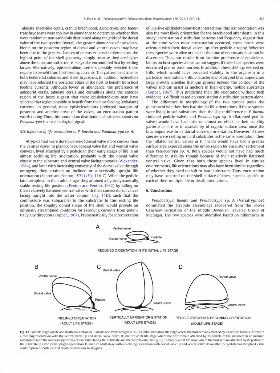
Atrypids that were dorsibiconvex (dorsal valve more convex thanthe ventral valve) to planoconvex (dorsal valve flat and ventral valveconvex), lived attached by a pedicle in their early stages of life in analmost reclining life orientation, probably with the dorsal valveclosest to the substrate and ventral valve facing upwards (Alexander,1984), and later with increasing convexity of the dorsal valve throughontogeny, they attained an inclined or a vertically upright lifeorientation (Fenton and Fenton, 1932) (Fig. 11A-C). When the pediclelater atrophied in their adult stage, they attained a hydrodynamicallystable resting life position (Fenton and Fenton, 1932) by falling ontheir relatively flattened ventral valve with their convex dorsal valvesfacing upright into the water column (Fig. 11D), such that thecommissure was subparallel to the substrate. In this resting lifeposition, the roughly domal shape of the shell would provide anoptimally streamlined condition for receiving currents from poten-tially any direction (Copper, 1967). Problematically for interpretation
Fig. 11. Possible stages of life and death orientation in P. lineata and Pseudoatrypa sp. A – A) ina reclining orientation with the ventral valve up and dorsal valve down, B) mature adult lorientation with the increasingly convex dorsal valve facing the substrate and the ventral vathe substrate in a vertically upright orientation, D) mature adult stage with a reclining orientcould represent both life and death orientations in atrypids.
of live-live episkeletobiont-host interactions, this last orientation wasalso the most likely orientation for the brachiopod after death. In thisstudy, encrustation distribution patterns and frequency suggest that,at the time when most encrustation occurred, these hosts wereoriented with their dorsal valves up after pedicle atrophy. Whetherthese species were alive or dead at the time of encrustation cannot bediscerned. Thus, our results from location preference of episkeleto-bionts on host species alone cannot suggest if these host species wereencrusted pre- or post-mortem. In addition, these shells preserved nofrills, which would have provided stability to the organism in aparticular orientation. Frills, characteristic of atrypid brachiopods, arelarge growth lamellae that can project beyond the contour of thevalves and can assist as anchors in high energy, mobile substrates(Copper, 1967). Thus predicting their life orientation without suchevidence is difficult based on encrustation distribution pattern alone.
The difference in morphology of the two species poses thequestion of whether they had similar life orientations. If these specieswere living in soft substrates, then the shape difference in P. lineata(inflated pedicle valve) and Pseudoatrypa sp. A (flattened pediclevalve) would have had little or almost no effect in their stabilitypatterns in life or in availability of cryptic surface area, once thebrachiopod was in its dorsal-valve-up orientation. However, if thesespecies were resting on hard substrates in the same orientation, thenthe inflated ventral valves in P. lineata would have had a greatersurface area exposed along the umbo region for encruster settlementthan Pseudoatrypa sp. A. Both species would not have had muchdifference in stability though because of their relatively flattenedventral valve. Given that both these species lived in similarenvironments, life orientations may also have been similar regardlessof whether they lived on soft or hard substrates. Thus, encrustationmay have occurred on the shell surface of these species specific toeach of their multiple life or death orientations.
6. Conclusions
Pseudoatrypa lineata and Pseudoatrypa sp. A (Variatrypinae)dominated the atrypide assemblage recovered from the LowerGenshaw Formation of the Middle Devonian Traverse Group ofMichigan. The two species were identified based on differences in
itial immature life stage where the host remain attached by its pedicle to the substrate inife stage where the host remain attached by its pedicle to the substrate in an inclinedlve facing up, C) mature adult life stage where the host remain attached by its pedicle toation with dorsal valve up and ventral valve down after the pedicle has atrophied – this
440 R. Bose et al. / Palaeogeography, Palaeoclimatology, Palaeoecology 310 (2011) 427–441
qualitative traits and statistical shape analysis. Pseudoatrypa lineatadiffers from Pseudoatrypa sp. A in having a relatively smaller shellsize, domal shape with a relatively shallower dorsal valve curvature,slightly convex ventral valve with inflation near the umbo, narrowerhinge line, wider commissure with a pronounced gentle to steepfold, and fine-medium sized closely spaced ribs. Statisticallysignificant shape values and large morphological distances betweenthe two species, supports the distinct shapes of the two speciesidentified.
Of the 343 Pseudoatrypa hosts examinedfrom both species, 185 ofthem bore episkeletobionts. The most abundant episkeletobionts werethe microconchids, hederellids and the sheet bryozoans. Auloporidcorals, Cornulites, tabulate corals, Ascodictyon, craniid brachiopods, andfenestrate bryozoans were very rare. Several episkeletobionts in thisstudy provide evidence of encrusting a live host based on the locationpreference of the episkeletobionts. Hederellids, auloporid corals,tabulate corals, and Cornulites had a live-live episkeletobiont-hostrelationship. The majority of other episkeletobionts, notably micro-conchids, sheet bryozoans, and Ascodictyon, were enigmatic indetermining whether their relationship was with a live or a deadhost. Very few epizoans crossed the commissure of the host after thehost's death.
Most episkeletobionts (microconchids and sheet bryozoans)preferred P. lineata, despite the fact that this species is generallysmaller. This differential effect in epibiosis could be due to the natureof ribbing structure (fine to medium) and greater exposed areafacilitated by the shell shape of P. lineata. Overall, the episkeletobiontpreference for one species over another strongly suggests that theoverall episkeletobiont distribution was influenced by shape andornamentation variation in atrypid samples. Abundant encrustingorganisms – microconchids, sheet bryozoans and hederellids, had apreference for P. lineata dorsal valves. This greater abundance ofepiskeletobionts on dorsal valves and lower abundance on ventralvalves is suggestive of most of the encrustation occurring when thehost species were oriented with their convex dorsal valves up andventral valves down with most of the ventral valve surface in contactwith the sediment substrate. Whether encrustation was pre- or post-mortem was challenging to discern for the majority of host-episkeletobiont associations as life orientation of the host wouldalso be a hydrodynamically stable orientation of the articulated shellafter death. Additionally, the most abundant episkeletobionts showeda preference for the posterior region on both dorsal and ventral valvesof both species. This suggests that the posterior umbonal region mayhave provided an inflated surface that remained exposed, thus,favoring the settlement of most episkeletobiont larvae in that region.
The present study of the Genshaw Formation documents epibiosison two species of atrypids, which significantly enhances ourunderstanding of morphological influence on episkeletobiontdistribution.
Acknowledgements
This research was funded by the Theodore Roosevelt MemorialResearch Grant received from the American Museum of NaturalHistory. We would especially like to thank Alex Bartholomew forproviding stratigraphic information of the Traverse Group of MichiganBasin and for providing access to his brachiopod collections, which arenow deposited at the Indiana University Paleontology Collections. Wewould also like to acknowledge insightful discussions on atrypidmorphology with Jed Day from Illinois State University. The firstauthor is grateful to Erle Kauffman for suggestions on morphologicalvariation within the species investigated and Claudia Johnson for acareful review of this manuscript. Finally, a special thanks to Mark A.Wilson and Alycia L. Stigall, the two reviewers, and Editor Dave Bottjerfor their careful reviews.
References
Ager, D.V., 1961. The epifauna of a Devonian spiriferid. Quarterly Journal of theGeological Society of London 117, 1–10.
Alexander, R.R., 1975. Phenotypic lability of the brachiopod Rafinesquina alternata(Ordovician) and its correlation with the sedimentologic regime. Journal ofPaleontology. 49, 607–618.
Alexander, R.R., 1984. Comparative hydrodynamic stability of brachiopod shells oncurrent-scoured arenaceous substrates. Lethaia 17, 17–32.
Alexander, R.R., 1990. Mechanical strength of shells of selected extant articulatebrachiopods: Implications for paleozoic morphologic trends. Historical Biology:International Journal of Paleobiology 3, 169–188.
Alvarez, F., Taylor, P.D., 1987. Epizoan ecology and interactions in the Devonian ofSpain. Palaeogeography, Palaeoclimatology, Palaeoecology 61, 17–31.
Anderson, W.I., Megivern, K.D., 1982. Epizoans from the Cerro Gordo Member of theLime Creek Formation (Upper Devonian), Rockford, Iowa. Proceeding of the IowaAcademy of Sciences 89, 71–80.
Barringer, J. E., 2008. Analysis of the occurrence of microconchids on middle Devonianbrachiopods from theMichigan basin: implications for microconchid and brachiopodautecology. Unpubl. M. S. Thes., Michigan State University, Michigan, pp. 1–127.
Bartholomew, J. B., 2006. Middle Devonian Faunas of the Michigan and AppalachianBasins: Comparing Patterns of Biotic Stability and Turnover between twoPaleobiogeographic Subprovinces. Unpubl. M. S. Thes., University of Cincinnati,Ohio, pp. 1–300.
Bassler, R.S., 1939. The Hederelloidea. A suborder of Paleozoic cyclostomatous Bryozoa.Proceedings of the United States National Museum 87, 25–91.
Bookstein, F.L., 1989. Principal warps: thin-plate splines and the decomposition ofdeformations. IEEE Transactions on Pattern Analysis and Machine Intelligence 11,567–585.
Bookstein, F.L., 1991. Morphometric tools for landmark data: geometry and biology.Cambridge University Press, New York.
Bordeaux, Y.L., Brett, C.E., 1990. Substrate specific associations of epibionts on middleDevonian Brachiopods: Implications for Paleoecology. Historical Biology 4, 203–220.
Bose, R., Schneider, C., Polly, P.D., Yacobucci, M.M., 2010. Ecological interactionsbetween Rhipidomella (Orthides, Brachiopoda) and its endoskeletobionts andpredators from the Middle Devonian Dundee Formation of Ohio, United States.Palaios 25, 196–210.
Bowen, Z.P., 1966. Intraspecific variation in the Brachial Cardinalia of Atrypa reticularis.Journal of Paleontology 40, 1017–1022.
Brett, C.E., Baird, G.C., Bartholomew, A.J., DeSantis, M.K., Straeten, C.A., 2010. Sequencestratigraphy and a revised sea-level curve for theMiddle Devonian of eastern NorthAmerica. Palaeogeography, Palaeoclimatology, Palaeoecology 304, 21–53.
Brezinski, D.K., 1984. Upper Mississippian epizoans and hosts from southwesternPennsylvania. Proceedings of the Pennsylvania Academy of Sciences 58, 223–226.
Carrera, M.G., 2000. Epizoan-sponge interactions in the Early Ordovician of theArgentine Precordillera. Palaios 15, 261–272.
Copper, P., 1967. Adaptations and life habits of Devonian atrypid brachiopods.Palaeogeography, Palaeoclimatology, Palaeoecology 3, 363–379.
Copper, P., 1973. New Siluro-Devonian atrypoid brachiopods. Journal of Paleontology47, 484–500.
Curry, G.B., 1983. Brachiopod caeca—A respiratory role? Lethaia 16, 311–312.Day, J., 1998. Distribution of latest Givetian-Frasnian Atrypida (Brachiopoda) in central
and western North America. Acta Palaeontologica Polonica 43, 205–240.Day, J., Copper, P., 1998. Revision of latest Givetian-Frasnian Atrypida (Brachiopoda)
from central North America. Acta Palaeontologica Polonica 43, 155–204.Dietl, G.P., Kelley, P.H., 2001. Mid-Paleozoic latitudinal predation gradient: Distribution of
brachiopod ornamentation reflects shifting Carboniferous climate. Geology 29,111–114.
Ehlers, G.M., Kesling, R.V., 1970. Devonian strata of Alpena and Presque Isle Counties,Michigan. Guidebook for field trips. Michigan Basal of the Geological Society 1–130.
Fagerstrom, J.A., 1996. Paleozoic brachiopod symbioses: Testing the limits of modernanalogues in paleoecology. Geological Society of America Bulletin 108, 1393–1403.
Fenton, C.L., Fenton, M.A., 1932. Orientation and Injury in the Genus Atrypa. TheAmerican Midland Naturalist 13, 63–74.
Fenton, C.L., Fenton, M.A., 1935. Atrypae described by Clement L. Webster and relatedforms (Devonian, Iowa). Journal of Paleontology 9, 369–384.
Gibson, M.A., 1992. Some epibiont-host and epibiont-epibiont relationships from theBirdsong Shale Member of the Lower Devonian Ross Formation (west-centralTennessee, U.S.A.). Historical Biology 6, 113–132.
Hammer, Ø., Harper, D., 2005. Paleontological data analysis. Blackwell Publishing,Oxford, United Kingdom.
Haney, R.A., Mitchell, C.E., Kim, K., 2001. Geometric Morphometric Analysis of Patternsof Shape Change in the Ordovician Brachiopod Sowerbyella. Palaios 16, 115–125.
Hoare, R.D., Steller, D.L., 1967. A Devonian brachiopod with epifauna. The Ohio Journalof Science 67, 291–297.
Hurst, J.M., 1974. Selective epizoan encrustation of some Silurian brachiopods fromGotland. Palaeontology 17, 423–429.
Kelly, A.W., Smith, G.W., 1947. Stratigraphy and structure of Traverse group in Afton-Onaway area, Michigan. American Association of Petroleum Geologists Bulletin 31,447–469.
Kesling, R.V., Chilman, R.B., 1975. Strata and megafossils of the middle Devonian SilicaFormation. Univ. Mich. Mus. Paleontol., Papers on Paleontol. 8, Ann Arbor, Michigan,pp. 1–408.
Kesling, R.V., Hoare, R.D., Sparks, D.K., 1980. Epizoans of the Middle Devonianbrachiopod Paraspirifer bownockeri: Their relationships to one another and totheir host. Journal of Paleontology 54, 1141–1154.

441R. Bose et al. / Palaeogeography, Palaeoclimatology, Palaeoecology 310 (2011) 427–441
Koch, W. F., 1978. Brachiopod paleoecology, paleobiogeography, and biostratigraphy inthe upper middle Devonian of Eastern North America: an ecofacies model for theAppalachian, Michigan, and Illinois basins. Unpubl. PhD. Thes., Oregon StateUniversity, Oregon, pp. 1–311.
Lamont, A., 1934. Brachiopodmorphology in relation to environment. Cement Lime andGravel 8, 216–219.
Leighton, L.R., 1998. Constraining functional hypotheses: controls on themorphology ofthe concavo-convex brachiopod Rafinesquina. Lethaia 3, 293–307.
Leighton, L.R., 1999. Possible latitudinal predation gradient in middle Paleozoic oceans.Geology 27, 47–50.
Leighton, L.R., 2003. Predation on brachiopods. In: Kelley, P.H., Kowalewski, M., Hansen,T.A. (Eds.), Predator-Prey Interactions in the Fossil record: Topics in Geobiology, 20.Kluwer/Plenum, New York, pp. 215–237.
Lescinsky, H.L., 1995. The life orientation of concavo-convex brachiopods: Overturningthe paradigm. Paleobiology 21, 520–551.
Macleod, N., Forey, P.L., 2002. Morphology, shape, and phylogeny. Taylor and Francis,New York.
Morris, R.W., Felton, S.H., 1993. Symbiotic Association of Crinoids, PlatyceratidGastropods, and Cornulites in the Upper Ordovician (Cincinnatian) of theCincinnati, Ohio Region. Palaios 8, 465–476.
Morris, R.W., Felton, S.H., 2003. Paleoecologic associations and secondary tiering ofCornulites on crinoids and bivalves in the Upper Ordovician (Cincinnatian) ofsouthwesternOhio, southeastern Indiana, andnorthernKentucky. Palaios18, 546–558.
Pitrat, C.W., Rogers, F.S., 1978. Spinocyrtia and its epizoans in the Traverse Group(Devonian) of Michigan. Journal of Paleontology 52, 1315–1324.
Richards, R.P., 1969. Biology and ecology of Rafinesquina alternata (Emmons). GeologicalSociety of Australia Abstract Progress, 6. North Central Section, pp. 41–42.
Richards, R.P., 1972. Autecology of Richmondian brachiopods (Late Ordovician ofIndiana and Ohio). Journal of Paleontology 46, 386–405.
Richards, R.P., 1974. Ecology of the Cornulitidae. Journal of Paleontology 48, 514–523.Richards, R.P., Shabica, C.W., 1969. Cylindrical living burrows in Ordovician dalmanellid
brachiopod beds. Journal of Paleontology 43, 838–841.Rodland, D.L., Kowalewski, M., Carroll, M., Simoes, M.G., 2004. Colonization of a ‘Lost
World’: Encrustation patterns in modern subtropical brachiopod assemblages.Palaios 19, 381–395.
Rodrigues, S.C., Simoes, M.G., Kowalewski, M., Petti, M.A.V., Nonato, E.F., Martinez, S.,Rio, C.D.J., 2008. Biotic interaction between spionid polychaetes and bouchardiidbrachiopods: Paleoecological, taphonomic and evolutionary implications. ActaPalaeontologica Polonica 53, 657–668.
Rohlf, F.J., 1990. Fitting curves to outlines. In: Rohlf, F.J., Bookstein, F.L. (Eds.), Proc.Mich. Morphometrics Workshop: Special Publication no. 2. Univ. of MichiganMuseum of Zoology, pp. 167–178.
Rohlf, F.J., Marcus, L.F., 1993. A revolution in morphometrics. Trends in Ecology &Evolution 8, 129–132.
Rohlf, F.J., Slice, D.E., 1990. Extensions of the Procrustes method for the optimalsuperimposition of landmarks. Systematic Zoology 39, 40–59.
Rohlf, F.J., 1999. Shape statistics: Procrustes superimpositions and tangent spaces.Journal of Classifications 16, 197–223.
Rudwick, M.J.S., 1962. Filter-feeding mechanisms in some brachiopods from NewZealand. Journal of the Linnean Society of London Zoology 44, 592–615.
Sandy, M.R., 1996. Oldest record of peduncular attachment of brachiopods to crinoidstems, Upper Ordovician, Ohio, USA (Brachiopods; Atrypida: Echinodermata;Crinoidea). Journal of Paleontology 70, 532–534.
Schneider, C.L., 2003. Hitchhiking on Pennsylvanian echinoids: Epibionts on Archae-ocidaris. Palaios 18, 435–444.
Schneider, C.L., 2009a. Epibionts on Late Carboniferous through Early Permian echinoidspines from Texas, USA, Echinoderms 2006. Proceedings of the 12th InternationalEchinoderm Conference, 7–11 August 2006, Durham, New Hampshire, U.S.A. CRCPress, Boca Raton, pp. 1–679.
Schneider, C.L., 2009b. Substrate preferences of Middle and Late Devonian Hederellafrom the midcontinent USA. In: Hageman, S.J., Key Jr., M.M., Winston, J.E. (Eds.),
Bryozoan Studies 2007: Proceedings of the 14th IBA Conference, Boone, NorthCarolina, July 1–8, 2007, 15. Virginia Museum of Natural History SpecialPublication, Martinsville, Virginia, pp. 295–300.
Schneider, C.L., Leighton, L.R., 2007. The influence of spiriferide microornament onDevonian epizoans. Geological Society of Australia Abstract Progress 39, 531.
Schneider, C.L., Webb, A., 2004. Where have all the encrusters gone? Encrustingorganisms on Devonian versus Mississippian brachiopods. Geological Society ofAustralia Abstract Progress 36, 111.
Shou-Hsin, C., 1959. Note on the palaeoecological relation between Aulopora andMucrospirifer. Acta Palaeontologica Sinica 7, 502–504.
Slice, D.E., 2001. Landmark Coordinates Aligned by Procrustes Analysis Do Not Lie inKendall's Shape Space. Systems Biology 50, 141–149.
Sparks, D.K., Hoare, R.D., Kesling, R.V., 1980. Epizoans on the brachiopod Paraspiriferbowneckeri (Stewart) from the Middle Devonian of Ohio. University of MichiganMuseum of the Paleontology Papers on Paleontology 23, 1–50.
Spjeldnaes, N., 1984. Epifauna as a tool in autecological analysis of Silurian Brachiopods.Special Papers in Paleontology 32, 225–235.
Stumm, E.C., 1951. Check list of fossil invertebrates described from the middleDevonian Traverse group of Michigan. Continental Museum of PaleontologyUniversity of Michigan 9, 1–44.
Sumrall, C.D., 2000. The biological implications of an edrioasteroid attached to apleurocystitid rhombiferan. Journal of Paleontology 74, 67–71.
Taylor, P.D., Wilson, M.A., 2002. A new terminology for marine organisms inhabitinghard substrates. Palaios 17, 522–525.
Taylor, P.D., Wilson, M.A., 2003. Palaeoecology and evolution of marine hard substratecommunities. Earth Science Reviews 62, 1–103.
Taylor, P.D., Wilson, M.A., 2008. Morphology and affinities of hederelloid "bryozoans".In: Hageman, S.J., Key Jr., M.M., Winston, J.E. (Eds.), Bryozoan Studies 2007:Proceedings of the 14th International Bryozoology Conference, Boone, NorthCarolina, July 1–8, 2007, 15. Virginia Museum of Natural History SpecialPublication, pp. 301–309.
Thayer, C.W., 1974. Substrate specificity of Devonian epizoan. Journal of Paleontology48, 881–894.
Vermeij, G.J., 1977. The Mesozoic marine revolution: evidence from molluscs,predation, and grazing. Paleobiology 3, 245–258.
Voros, A., 2010. Escalation reflected in ornamentation and diversity history ofbrachiopod clades during the Mesozoic marine revolution: Palaeogeogr. Palaeocli-matol. Palaeoecol. 291, 474–480.
Warthin, A.S., Cooper, G.A., 1935. New Formation Names in the Michigan Devonian.Journal of the Washington Academy of Sciences 25, 524–526.
Warthin, A.S., Cooper, G.A., 1943. Traverse rocks of Thunder bay region, Michigan.American Association of Petroleum Geologists Bulletin 27, 571–595.
Watkins, R., 1981. Epizoan ecology of the type Ludlow Series (Upper Silurian). NewEngland Journal of Paleontology 55, 29–32.
Webster, C.L., 1921. Notes on the genus Atrypa, with description of new species. TheAmerican Midland Naturalist 7, 13–26.
Wiedman, L.A., 1985. Community Paleoecological Study of the Silica Shale Equivalent ofNortheastern Indiana. Journal of Paleontology 59, 160–182.
Wilson, M.A., Taylor, P.D., 2001. “Pseudobryozoans” and the problem of encrusterdiversity in the Paleozoic. PaleoBios. 21, 134–135.
Wylie, A.S., Huntoon, J.E., 2003. Log curve amplitude slicing - visualization of log datafrom the Devonian Traverse Group in the Michigan Basin. American Association ofPetroleum Geologists Bulletin 87, 581–608.
Zapalski, M.K., 2005. Paleoecology of Auloporida: an example from the Devonian of theHoly Cross Mts. Poland Geobios 38, 677–683.
Zelditch, M.L., Swiderski, D.L., Sheets, H.D., Fink, W.L., 2004. Geometric Morphometricsfor Biologists, A Primer. Elsevier Academic Press, New York.
Zhan, R., Vinn, O., 2007. Cornulitid epibionts onbrachiopod shells from the Late Ordovician(middle Ashgill) of East China. Estonian Journal Earth Sciences 56, 101–108.