Embed Size (px)
Citation preview

Influence of GLP-1 on Myocardial Glucose Metabolism inHealthy Men during Normo- or HypoglycemiaMichael Gejl1*, Susanne Lerche1, Annette Mengel2, Niels Møller2, Bo Martin Bibby8, Kamille Smidt1,
Birgitte Brock1,3, Hanne Søndergaard9, Hans Erik Bøtker4, Albert Gjedde5,6, Jens Juul Holst7, Søren
Baarsgaard Hansen5, Jørgen Rungby1,2,10
1 Department of Biomedicine, Aarhus University, Aarhus, Denmark, 2 Department of Endocrinology, Aarhus University Hospital, Aarhus, Denmark, 3 Department of Clinical
Biochemistry, Aarhus University Hospital, Aarhus, Denmark, 4 Department of Cardiology, Aarhus University Hospital, Skejby, Aarhus, Denmark, 5 PET Centre, Aarhus
University Hospital, Aarhus, Denmark, 6 Department of Neuroscience and Pharmacology, University of Copenhagen, Copenhagen, Denmark, 7 Department of Biomedical
Sciences, University of Copenhagen, Copenhagen, Denmark, 8 Department of Biostatistics, Aarhus University, Aarhus, Denmark, 9 Department of Medicine, Viborg
Regional Hospital, Viborg, Denmark, 10 Department of Endocrinology, Rigshospitalet, Copenhagen, Denmark
Abstract
Background and Aims: Glucagon-like peptide-1 (GLP-1) may provide beneficial cardiovascular effects, possibly due toenhanced myocardial energetic efficiency by increasing myocardial glucose uptake (MGU). We assessed the effects of GLP-1on MGU in healthy subjects during normo- and hypoglycemia.
Materials and Methods: We included eighteen healthy men in two randomized, double-blinded, placebo-controlled cross-over studies. MGU was assessed with GLP-1 or saline infusion during pituitary-pancreatic normo- (plasma glucose (PG):4.5 mM, n = 10) and hypoglycemic clamps (PG: 3.0 mM, n = 8) by positron emission tomography with 18fluoro-deoxy-glucose (18F-FDG) as tracer.
Results: In the normoglycemia study mean (6 SD) age was 2563 years, and BMI was 22.660.6 kg/m2 and in thehypoglycemia study the mean age was 2362 years with a mean body mass index of 2362 kg/m2. GLP-1 did not changeMGU during normoglycemia (mean (+/2 SD) 0.15+/20.04 and 0.16+/20.03 mmol/g/min, P = 0.46) or during hypoglycemia(0.16+/20.03 and 0.13+/20.04 mmol/g/min, P = 0.14). However, the effect of GLP-1 on MGU was negatively correlated tobaseline MGU both during normo- and hypoglycemia, (P = 0.006, r2 = 0.64 and P = 0.018, r2 = 0.64, respectively) and changesin MGU correlated positively with the level of insulin resistance (HOMA 2IR) during hypoglycemia, P = 0.04, r2 = 0.54. GLP-1mediated an increase in circulating glucagon levels at PG levels below 3.5 mM and increased glucose infusion rates duringthe hypoglycemia study. No differences in other circulating hormones or metabolites were found.
Conclusions: While GLP-1 does not affect overall MGU, GLP-1 induces changes in MGU dependent on baseline MGU suchthat GLP-1 increases MGU in subjects with low baseline MGU and decreases MGU in subjects with high baseline MGU. GLP-1preserves MGU during hypoglycemia in insulin resistant subjects.
ClinicalTrials.gov registration numbers: NCT00418288: (hypoglycemia) and NCT00256256: (normoglycemia).
Citation: Gejl M, Lerche S, Mengel A, Møller N, Bibby BM, et al. (2014) Influence of GLP-1 on Myocardial Glucose Metabolism in Healthy Men during Normo- orHypoglycemia. PLoS ONE 9(1): e83758. doi:10.1371/journal.pone.0083758
Editor: Jose A. L. Calbet, University of Las Palmas de Gran Canaria, Spain
Received July 11, 2013; Accepted November 6, 2013; Published January 6, 2014
Copyright: � 2014 Gejl et al. This is an open-access article distributed under the terms of the Creative Commons Attribution License, which permits unrestricteduse, distribution, and reproduction in any medium, provided the original author and source are credited.
Funding: Supported by grants from The Danish Strategic Research Council (the DD2 Study), Desire and Niels Ydes Foundation. The funders had no role in studydesign, data collection and analysis, decision to publish, or preparation of the manuscript.
Competing Interests: The authors have declared that no competing interests exist.
* E-mail: [email protected]
Introduction
Type 2 diabetes (T2D) is estimated to affect approximately 400
million people before 2030 [1]. The main hormonal and metabolic
abnormalities in T2D are dysfunctional insulin secretion, glucagon
excess, and insulin resistance [2], eventually beta-cell failure [3]
and defects in glucagon-like peptide-1 (GLP-1) secretory responses
[4].
Because patients with T2D have elevated circulating free fatty
acids (FFA) they also have increased beta oxidation of FFAs at the
expense of reduced glucose oxidation. [5]. Compared to glucose
oxidation, reliance on fatty acid oxidation for ATP production
results in higher costs in mitochondrial oxygen consumption [6],
and may become deleterious during myocardial ischemia, which
occurs more frequently in diabetic than in non-diabetic subjects
due to an increased prevalence of coronary artery disease.
Consequently, pharmacological means to switch from free fatty
acid to glucose oxidation, when energy demands are challenged,
are highly warranted.
The glucagon-like peptide-1 (GLP-1) receptor agonists are
validated anti-diabetic medications. They mimic a range of
physiological actions of native GLP-1 on glucose metabolism
including stimulation of insulin secretion, inhibition of glucagon
PLOS ONE | www.plosone.org 1 January 2014 | Volume 9 | Issue 1 | e83758

secretion, inhibition of gastric emptying and reduction of appetite
and food intake [7]. Native GLP-1 and GLP-1 analogues are
reported to have several significant cardiovascular effects provid-
ing the opportunity to address the multifactorial issues involved in
increased mortality and morbidity associated with T2D as well as
in patients with cardiac disease and no diabetes [8]. The
potentially beneficial impact of GLP-1 receptor stimulation may
rely on, among others, improved myocardial glucose metabolism
[9].
We hypothesized that GLP-1 increases glucose availability and
utilization and aimed to assess the effects of GLP-1 receptor (GLP-
1R) activation on myocardial glucose uptake (MGU) in healthy
subjects with hypo- and normoglycemia.
Materials and Methods
The protocol for these trials and supporting CONSORT
checklist are available as supporting information; see Checklist S1
and Protocol S1, S2, S3, S4.
Study groupsIn the normoglycemia study ten non-smoking, healthy, Cauca-
sian male subjects were included from November 2005 through
January 2007. Mean (6 SD) age was 2563 years and BMI was
22.660.6 kg/m2. In the hypoglycaemia study eight non-smoking,
healthy, Caucasian males with a mean age of 2362 years and a
mean body mass index of 2362 kg/m2 participated from January
to October 2007. They had a normal physical examination, no
history of diabetes or cardiovascular disease, and received no
medication. The studies were conducted in accordance with the
Declaration of Helsinki and the protocol was approved by the
official Regional Science Ethics Committee of Region Midtjylland
and County of Aarhus. The participants in both studies were
recruited from notices at Aarhus University. All participants
received both oral and written information and signed an
approved informed consent form before entering the study.
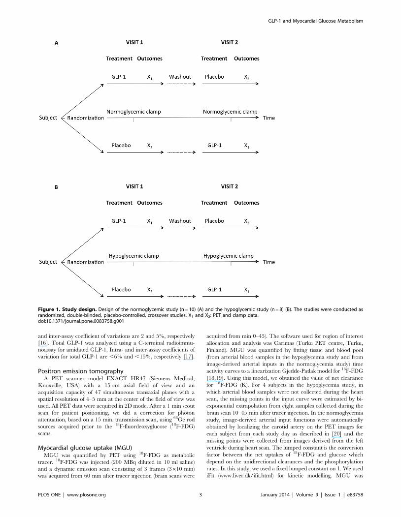
Study designThe studies were conducted as randomized, double-blinded,
placebo-controlled, crossover studies (figure 1 A and B). Each
subject was studied twice in random order with GLP-1 and
placebo infusion. Positron emission tomography (PET) sessions
were separated by an interval of 2–6 weeks. Both sessions
commenced at 9.00 hours after an overnight fast. The subjects
did not exercise 24 hours prior to the sessions. Subjects were
placed in bed and two catheters were inserted for infusion of clamp
hormones and for the infusion of GLP-1 or placebo. A third
catheter was placed in an arterialized (heated) dorsal hand vein for
blood sampling.
An arterial catheter was placed in the radial artery of the right
arm in order to draw blood samples for measuring input
radioactivity during PET in the hypoglycemia study. In the
normoglycemia study a pancreatic-pituitary clamp was performed
as described in [10]. In the hypoglycemia study a stepwise
hypoglycaemic pancreatic-pituitary clamp was performed accord-
ing to principles previously described [10,11]. In brief, somato-
statin (Ferring GmbH, D-24109 Kiel) was infused at a rate of
300 mg/hour to suppress the endogenous insulin, glucagon,
growth hormone and GLP-1 production. Human glucagon
(Glucagen, Novo Nordisk A/S, Copenhagen, Denmark) 0.6 ng/
kg/min, growth hormone (Genotropin Miniquik 0.2 mg, Pfizer
ApS, Ballerup, Denmark) 2 ng/kg/min were infused with the aim
of maintaining near-basal levels (0–360 min). In the normoglyce-
mia study, insulin (Actrapid, Novo Nordisk A/S, Copenhagen,
Denmark) was infused at a rate of 0.12 mU/kg/min and in the
hypoglycemia study insulin (Actrapid, Novo Nordisk A/S,
Copenhagen, Denmark) was infused at a rate of 0.8 mU/kg/
min. Glucose (200 g/l) was infused at a variable rates to clamp
plasma glucose (PG) at 4.5 mM in the normoglycemic setup and in
the hypoglycemia study glucose was infused at variable rates to
initially clamp PG at 4.5 mM (0–150 minutes). From 150 to
180 minutes, PG was lowered to 4.0 mM and maintained at this
level until 210 minutes. From 210 minutes to 240 min PG was
lowered to 3.5 mM and maintained at this level until 270 minutes.
From 270 min to 300 min PG was lowered to a nadir of 3.0 mM
and maintained at this level during PET (360–400 min). GLP-1 or
placebo infusion was initiated at time 60 minutes and maintained
during the entire session.
The subjects received either intravenous synthetic GLP-1 (7–36
amide) or placebo at a rate of 1.2 pmol/kg/min (60–360 min)
[12]. Recombinant human GLP-1 (7–36 amide) was a kind gift
from BioNebraska Inc., Lincoln, NE (USA) and was tested and
found positive for sterility and negative for bacterial endotoxins
before use. It was dissolved in a sterile buffer containing 600 mg of
acetic acid, 50.7 g of mannitol and sterile water added up to
1000 g and had a pH of 4.5. The concentration of GLP-1(7–
36amide) was 1 mg/ml and vials of 0.25 ml were stored at 2206Celsius. The test solution consisted of 0.25 ml GLP-1, 20 ml of
human albumin ‘‘ZLB’’ 5% and sodium chloride (9 g/l) to yield
100 ml of solution. The placebo solution consisted of the above
mentioned buffer solution containing human albumin and saline.
One subject experienced transient nausea and vomiting during
hypoglycemia and GLP-1 infusion with onset at 20 min after
initiation of the infusion, lasting approximately 1K hour. Another
subject experienced headache during placebo infusion after
2 hours of infusion, lasting 1 hour.
PG was measured in duplicate every 10 min until PG reached
3.0 mM and then every fifth minute. In the normoglycemia study,
PG was measured every 10 min. Blood for measuring insulin, c-
peptide, glucagon, GLP-1 (total and intact), growth hormone,
FFA, cortisol and ghrelin was drawn every 30 min. Blood for
measuring epinephrine and norepinephrine was drawn at 0, 150
and 240 min and every 30 min during the PET scan. Arterial
blood for measuring input radioactivity was drawn at predeter-
mined intervals during PET (1265 s, 8630 s, 86300 s and
46600 s).
AssaysPlasma glucose was measured immediately after sampling on a
Beckman glucose analyzer (Beckman, Palo Alto, CA). All other
blood samples were stored at 220uC (C-peptide at 280uC) until
assay. Serum NEFAs were analyzed by a commercial kit (Wako
Chemicals, Neuss, Germany). Serum GH was analyzed using
chemiluminescence technology (IDS-iSYS Multi-Discipline Auto-
mated Analyzer; Immunodiagnostic Systems Nordic, Herlev,
Denmark). Serum insulin and C-peptide was analyzed using
time-resolved fluoroimmunoassay assay (AutoDELFIA PerkinEl-
mer, Turku, Finland). Serum cortisol was measured using a DRG
ELISA kit (DRG Instruments, Marburg, Germany). Serum
ghrelin (total levels) was measured in duplicate by an in-house
assay [13]. Plasma catecholamines were measured by liquid
chromatography [14]. Plasma glucagon was measured by radio-
immunoassay [15]. All coefficients of variation (inter- and intra-
assay) were ,9.5%. The assay for intact GLP-1 is an enzyme-
linked immunosorbent assay using unextracted plasma, which was
collected and stored in the presence of a dipeptidyl peptidase-IV
inhibitor (valine-pyrrolidide, 0.01 mmol/l, final concentration
added to the blood sample immediately after collection). Intra-
GLP-1 and Myocardial Glucose Metabolism
PLOS ONE | www.plosone.org 2 January 2014 | Volume 9 | Issue 1 | e83758
and inter-assay coefficient of variations are 2 and 5%, respectively
[16]. Total GLP-1 was analyzed using a C-terminal radioimmu-
noassay for amidated GLP-1. Intra- and inter-assay coefficients of
variation for total GLP-1 are ,6% and ,15%, respectively [17].
Positron emission tomographyA PET scanner model EXACT HR47 (Siemens Medical,
Knoxville, USA) with a 15 cm axial field of view and an
acquisition capacity of 47 simultaneous transaxial planes with a
spatial resolution of 4–5 mm at the center of the field of view was
used. All PET data were acquired in 2D mode. After a 1 min scout
scan for patient positioning, we did a correction for photon
attenuation, based on a 15 min. transmission scan, using 68Ge rod
sources acquired prior to the 18F-fluordeoxyglucose (18F-FDG)
scans.
Myocardial glucose uptake (MGU)MGU was quantified by PET using 18F-FDG as metabolic
tracer. 18F-FDG was injected (200 MBq diluted in 10 ml saline)
and a dynamic emission scan consisting of 3 frames (3610 min)
was acquired from 60 min after tracer injection (brain scans were
acquired from min 0–45). The software used for region of interest
allocation and analysis was Carimas (Turku PET centre, Turku,
Finland). MGU was quantified by fitting tissue and blood pool
(from arterial blood samples in the hypoglycemia study and from
image-derived arterial inputs in the normoglycemia study) time
activity curves to a linearization Gjedde-Patlak model for 18F-FDG
[18,19]. Using this model, we obtained the value of net clearance
for 18F-FDG (K). For 4 subjects in the hypoglycemia study, in
which arterial blood samples were not collected during the heart
scan, the missing points in the input curve were estimated by bi-
exponential extrapolation from eight samples collected during the
brain scan 10–45 min after tracer injection. In the normoglycemia
study, image-derived arterial input functions were automatically
obtained by localizing the carotid artery on the PET images for
each subject from each study day as described in [20] and the
missing points were collected from images derived from the left
ventricle during heart scan. The lumped constant is the conversion
factor between the net uptakes of 18F-FDG and glucose which
depend on the unidirectional clearances and the phosphorylation
rates. In this study, we used a fixed lumped constant on 1. We used
iFit (www.liver.dk/ifit.html) for kinetic modelling. MGU was
Figure 1. Study design. Design of the normoglycemic study (n = 10) (A) and the hypoglycemic study (n = 8) (B). The studies were conducted asrandomized, double-blinded, placebo-controlled, crossover studies. X1 and X2: PET and clamp data.doi:10.1371/journal.pone.0083758.g001
GLP-1 and Myocardial Glucose Metabolism
PLOS ONE | www.plosone.org 3 January 2014 | Volume 9 | Issue 1 | e83758

calculated as: MGU = PG*K/rm, where PG is plasma glucose
during scan, K is clearance of 18F-FDG and rm is myocardial
density.
Insulin sensitivityInsulin resistance homeostasis model of assessment 2 (HOMA
2IR) was computed as described in ref. [21].
Calculations and statistical analysisPrimary outcome was MGU.
The PET data were analyzed using a paired student’s t-test. The
plasma-data were analysed using a linear mixed effects model with
subject and all interactions involving subject, including the
interaction between subject and time and the interaction between
subject and treatment, as random effects. Treatment (GLP-1 vs.
placebo), time, and the interaction between the two were included
in the analysis as fixed effects. A linear regression model was used
for regression analysis of K and MGU and Pearson’s r test was
used to evaluate K, MGU and insulin resistance (HOMA2 IR).
The statistical software used was GraphPadPrism (GraphPad
Software) and Stata (StataCorp LP, College Station, Texas, USA)
with a significance level of 5%. Data are presented as mean 6 SD.
The study was planned as preliminary exploration and thus the
number of subjects that entered the study did not derive from a
power calculation.
Results
PETNormoglycemia study (figure 2). GLP-1 did not affect
MGU (placebo: 0.1760.05 and GLP-1: 0.1460.04 mmol/g/min,
P = 0.21) (figure 2A) nor K (placebo: 0.0460.01 and GLP-1:
0.0360.01 ml/cm3/min, P = 0.25) (figure 2C). Regression analysis
showed a linear relationship (P = 0.009, r2 = 0.59) between the
baseline (placebo) K and the alteration in K after GLP-1 infusion
(figure 2G). GLP-1 increased K of 18F-FDG and glucose of
cardiomyocytes in subjects with low baseline K suggesting
decreased K in subjects with high baseline K. Correspondingly,
there was a linear relationship between the baseline MGU and the
GLP-1-induced alterations in MGU (P = 0.006, r2 = 0.64)
(figure 2E), an enhanced response was found with low baseline
MGU and a decreased response with high baseline MGU.
Hypoglycemia study (figure 2). GLP-1 did not affect MGU
(placebo: 0.1360.04 and GLP-1: 0.1660.03 mmol/g/min,
P = 0.14) (figure 2B) nor K (placebo: 0.04560.01 and GLP-1:
0.05360.009 ml/cm3/min, P = 0.17) (figure 2 D). Regression
analysis showed a linear relationship (P = 0.016, r2 = 0.65) between
the baseline (placebo) K and the alteration in K after GLP-1
infusion (figure 2H). GLP-1 increased K of 18F-FDG and glucose
of cardiomyocytes in subjects with low baseline K suggesting
decreased K in subjects with high baseline K. Correspondingly,
there was a linear relationship between the baseline MGU and the
GLP-1-induced alterations in MGU (P = 0.018, r2 = 0.64)
(figure 2F), a higher response was found with low baseline MGU
and a decreased response with high baseline MGU. The
alterations of K and MGU correlated positively with HOMA
2IR (P = 0.046 and 0.037, r2 = 0.51 and 0.54) (figure 2I and 2J).
Hormones and metabolitesNormoglycemia study (figure 3). GLP-1infusion increased
circulating GLP-1 concentrations as shown in figure 3. Concen-
trations differed between study days regarding intact and total
GLP-1, both P,0.001. PG and glucose infusion rates were the
same with placebo and GLP-1 (all P.0.4). The plasma C-peptide
and FFA levels were suppressed and no differences were found
between placebo and GLP-1 (P = 0.58 and 0.74). There were no
significant differences in circulating growth hormone, insulin or
glucagon (P = 0.51, 0.99 and 0.36) between sessions. Ghrelin (data
not shown, p = 0.26), norepinephrine (data not shown, p = 0.20),
and epinephrine levels were also similar during sessions, p = 0.39.
Serum cortisol concentrations were significantly higher with GLP-
1 at 120 and 150 min (P,0.001), but similar during PET scans,
P.0.28.
Hypoglycemia study (figure 4). GLP-1 infusion increased
circulating GLP-1 concentrations. Concentrations differed be-
tween study days regarding intact and total GLP-1, both P,0.001.
During the PET scan, the PG concentration remained at
3.1560.09 (GLP-1) and 3.1460.16 (placebo) mM, P = 0.67. The
glucose infusion rate increased with GLP-1 infusion, P = 0.01. The
plasma C-peptide and FFA levels were suppressed and no
differences were found between groups (P = 0.73 and 0.67). There
were no significant differences in circulating growth hormone
(P = 0.14) between sessions. Glucagon increased in the GLP-1
group during clamp step PG,3.5 mM (time 270, 330, 360 and
420 min.), P,0.05. Counter-regulatory hormones rose as expect-
ed when PG declined. Insulin levels were similar between sessions,
P = 0.52. Ghrelin (data not shown, p = 0.30), norepinephrine (data
not shown, p = 0.75), and epinephrine levels were also similar
during sessions, p = 0.98. Serum cortisol concentrations were
significantly higher with GLP-1 at PG steps of 4.5 and 4.0 mM,
P = 0.0001–0.04 but were similar at PG steps of 3.5 and 3.0 mM.,
P = 0.86, GLP-1 vs. placebo.
Discussion
GLP-1 did not change overall MGU or K during normo- or
hypoglycemia. The GLP-1 conditioned MGU is negatively
correlated to baseline MGU such that GLP-1 increased MGU
in subjects with low baseline MGU and decreased MGU in
subjects with high baseline MGU during both normo- and
hypoglycemia. This alteration correlated with the grade of insulin
resistance of the subjects in the hypoglycemia setup. Moreover, the
glucose infusion rate was increased with GLP-1 despite increased
glucagon secretion during hypoglycemia.
In a healthy heart, the substrate preferences are flexible. In
normal conditions a minimum of 60% of ATP is derived from the
oxidation of free fatty acids, and the rest from the oxidation of
glucose. In the diabetic heart, ATP is predominantly derived from
myocardial fatty acid oxidation [5]. This has been associated with
increased myocardial oxygen consumption and decreased me-
chanical efficiency [22]. During hypoxia, energy metabolism shifts
toward a greater oxidation of glucose to maintain myocardial
viability, as the oxidation of glucose consumes less oxygen than
oxidation of free fatty acids. Clinical and experimental studies
have shown that increased glucose uptake during acute myocardial
ischemia is associated with preserved cardiac function [23,24].
Hyperglycaemia is an independent predictor of cardiovascular
risk, infarct sizes are reported to be directly related to the severity
of hyperglycaemia and the glycometabolic state is associated with
the mortality risk in T2D patients with acute myocardial infarction
[5]. The beneficial effect of lowering blood glucose to very low
levels, however, is controversial, one of the major problem being
the increase in the number of hypoglycemic events [25]. However,
the studies also showed that the currently available anti-diabetic
treatments fail to reach acceptable glycemic control in the majority
of patients. Therefore treatment strategies with beneficial effects
toward reducing macrovascular complications (e.g., stroke, myo-
cardial infarction) and with a low risk of adverse hypoglycemic
GLP-1 and Myocardial Glucose Metabolism
PLOS ONE | www.plosone.org 4 January 2014 | Volume 9 | Issue 1 | e83758

GLP-1 and Myocardial Glucose Metabolism
PLOS ONE | www.plosone.org 5 January 2014 | Volume 9 | Issue 1 | e83758

events are being tested and although hypoglycemic events are
reported in patients with heart failure [26,27], GLP-1 analogues
are considered agents with low risk of hypoglycaemia. GLP-1
inhibits glucagon secretion from the pancreatic a-cells at fasting
and elevated glucose concentrations, whereas hypoglycemia
induced glucagon secretion is not inhibited - the mechanisms are
not fully understood [28]. GLP-1 infusion resulted in pharmaco-
logically relevant plasma concentrations of the intact hormone in
both studies [12]. Slightly higher glucagon levels during hypogly-
cemia in the GLP-1 group compared to the control experiments
were previously found in studies with native GLP-1 [29], with the
GLP-1 analogue exenatide [10] and the the DPP-IV inhibitor
vildagliptin [30]. We found an increased secretion of glucagon
during hypoglycemia despite somatostatin infusion with GLP-1
infusion and furthermore glucose infusion rates increased in the
hypoglycemia study with GLP-1. Together the latter indicate a
possible mechanism for alleviation of hypoglycemia by GLP-1. In
the PET studies, increased levels of cortisol were observed with
GLP-1 infusion during the clamp from time 120–150 min after
which cortisol levels were similar (figure 3 and 4). This increase in
cortisol is consistent with previous observations [31,32] and may
indicate an activation of hypothalamic neuroendocrine neurons by
GLP-1 [33]. Central administration of GLP-1 activates the
hypothalamo-pituitary-adrenocortical axis primarily through stim-
ulation of corticotropin releasing hormone neurons [34]. Myocar-
dial glucose uptake is not correlated to cortisol levels as reported in
[35], and cortisol might even decrease MGU primarily by
stimulating lipolysis and by elevating FFA concentration. Howev-
er; cortisol levels were similar 1 hour prior to and during the PET
scans and therefore the observed effects of GLP-1 on myocardial
observed are most likely not influenced by cortisol.
GLP-1 exerts its actions through the GLP-1R. The GLP-1R
may be present in cells of many tissues including the vascular
endothelium, cardiomyocytes, endocardium and smooth muscle
cells. Some cardiac effects of GLP-1R agonists may be exerted via
other receptors [36]. Prior studies have demonstrated the presence
of GLP-1R in the myocardium [36,37], but recent papers [38–40]
have questioned the interpretation of data obtained using standard
antisera to detect the authentic GLP-1R.
Growing evidence, reviewed by Chinda K. et al [41],
demonstrate cardioprotective effects of GLP-1 and GLP-1
analogues during ischemia-reperfusion in both animal and clinical
models despite inconsistent reports. The exact mechanisms have
never been fully elucidated, although many mechanisms have been
proposed (e.g. increased MGU, reduction of oxidative stress and
proapoptotic kinase, activation of prosurvival kinase, and atten-
uation proinflammatory cell activation) [41]. The GLP-1R
agonists reduce ischemia-reperfusion injury by reducing infarct
size and improve left ventricle function [41], and long term studies
show that patients treated with the GLP-1 agonist exenatide are
less likely to record a CVD event [42]. However, the impact of
these so-called cardioprotective effects of GLP-1 is still less well
established. The most comprehensive meta-analysis of the GLP-1
analogues did not find any significant GLP-1 effect on CVD events
as a whole [43]. Therefore, although several possible mechanisms
have been put forward suggesting that GLP-1s could have a
protective effect on the cardiovascular risk profile (e.g., reductions
of blood glucose, body weight, and blood pressure, improvement
in left ventricular ejection fraction, changes in cardiac metabolism,
lipid metabolism, arthrosclerosis development and endothelial
function and the response for ischemia-reperfusion injury [44]),
reasons for a (possible) decreased risk of developing CVD are still
poorly understood. Interestingly, Kim et al [39] demonstrated that
the receptor is localized predominantly in the cardiac atria and
that GLP-1 stimulates the secretion of atrial natriuretic peptide
(ANP). In rat neonatal cardiac myocytes, hypoxia significantly
increased glucose uptake stimulated by ANP, but ANP did not
affect basal glucose uptake under normoxic conditions [45] as
examined in the present study and in a similar study of patients
with T2D [11] where no direct effect on MGU are demonstrated
despite a 24% increase in myocardial blood flow. The T2D study
revealed that the acute stimulation of GLP-1R did alter the
myocardial glucose metabolism dependent on the baseline
myocardial glucose metabolism and hence level of insulin
resistance [11], consistent with the findings in the present study.
A recent paper reports an increase in MGU in healthy lean
subjects with GLP-1 infusion [46]. The study differs from the
present studies by not having clamped the participants and
consequently not having completely comparable PG, insulin, and
FFA between the groups as well as non-suppressed FFA.
Furthermore, they infused a GLP-1 at a higher rate (1.5 pmol/
kg/min).Hypoglycemia is associated with increased mortality rates
in diabetic patients after ischemia and reperfusion and the
underlying mechanisms may involve reduced capacity for precon-
ditioning. As the salvage of the myocardium is associated with
increased MGU during reperfusion, cardioprotection is thought to
be linked to glucose metabolism [47]. An important mechanism of
the beneficial cardiovascular effect of GLP-1 may be the increase
of MGU in the subjects with the lowest baseline MGU – the most
insulin resistant subjects, regardless of glycemia. Glucose mainly
enters myocardial cells via the facilitative glucose transporters,
GLUT1 and GLUT4. As most of the glucose transported across
the GLUT1 and GLUT4 into the myocardium is metabolized due
to high hexokinase affinity, our results support that transport
across the cell membranes is affected by acute infusion of GLP-1 as
indicated in [11]. The regression estimates indicate that GLP-1
raises GLUT translocation in subjects with low baseline GLUT
activity and lowers the activity in subjects with high baseline
GLUT activity. Thus, it seems that the baseline activity of glucose
transporters and hence level of insulin resistance influences the
GLP-1 mediated action of K and MGU in cardiomyocytes.
Furthermore, the effects of GLP-1 on K and MGU regress
positively with HOMA2 IR (figure 2I and J). This is in accordance
with observations in models of T2D where reduced translocation
of GLUT1 and GLUT4 is found in the heart [48] and, the
GLUT1 and GLUT4 protein levels in the membrane are related
to fasting glucose levels, the higher the fasting glucose are, the
lower glut concentration in the membranes of the cardiomyocytes
[49]. Fasting glucose and IR are highly correlated. Growing
evidence [37,50,51] indicates that GLP-1 and analogues enhance
GLUT1 translocation in myocardial cell membranes and GLUT4
protein levels of the myocardium, but no reports support
Figure 2. Positron emission tomography. Myocardial glucose uptake (MGU) during normoglycemia and hypoglycemia (A and B). 18F-FDGclearance (K) during normoglycemia and hypoglycemia (C and D). Relation between placebo MGU and change in MGU during GLP-1 infusion in thenormoglycemia study (E), placebo MGU and change in MGU during GLP-1 infusion in the hypoglycemia study (F). Relation between placebo K andchange in K during GLP-1 infusion in the normoglycemia study (G), placebo K and change in K during GLP-1 infusion in the hypoglycemia study (H).HOMA 2IR and the change of MGU during GLP-1 infusion in the hypoglycemia study (I). HOMA 2IR and the change of K during GLP-1 infusion in thehypoglycemia study (J). Data are mean 6 SD. Regression lines with 95% confidence intervals.doi:10.1371/journal.pone.0083758.g002
GLP-1 and Myocardial Glucose Metabolism
PLOS ONE | www.plosone.org 6 January 2014 | Volume 9 | Issue 1 | e83758

hexokinase activity to be directly influenced by GLP-1 in contrast
to glucokinase activity in b-cell lines [52].
Although the mechanism is not clear, it is speculated that
signaling through the GLP-1R facilitates activation of Akt or
AMP-activated protein kinase (AMPK) and consequently GLUT4
translocation [50]. Alternatively that stimulation of translocation
of GLUT1 and GLUT4 is mediated by increased myocardial NO
production [53,54] - facilitated by activation of the GLP-1R [37].
Figure 3. Hormones and metabolites - normoglycemia study. Plasma glucose, glucose infusion rates (GIR), total GLP-1, insulin, cortisol, freefatty acid (FFA), glucagon and epinephrine concentrations during GLP-1 (black dots) and placebo infusion (white dots). Data are means 6 SEM.* P#0.05.doi:10.1371/journal.pone.0083758.g003
GLP-1 and Myocardial Glucose Metabolism
PLOS ONE | www.plosone.org 7 January 2014 | Volume 9 | Issue 1 | e83758

Figure 4. Hormones and metabolites - hypoglycemia study. Plasma glucose, glucose infusion rates (GIR), total GLP-1, insulin, cortisol, freefatty acid (FFA), glucagon and epinephrine concentrations during GLP-1 (black dots) and placebo infusion (white dots). Data are means 6 SEM.* P#0.05.doi:10.1371/journal.pone.0083758.g004
GLP-1 and Myocardial Glucose Metabolism
PLOS ONE | www.plosone.org 8 January 2014 | Volume 9 | Issue 1 | e83758

GLUT1 is reported to be controlled partly by levels of plasma
glucose and GLP-1 in other tissues, e.g. in the blood brain barrier
in various cerebral regions [29,55]. The mechanism of the dual
effect of GLP-1 and the analogue exenatide remains unclear but
may be caused by receptor-mediated changes or may be a general
feature of intracellular IR. The duality may be beneficial because
glucose transport and uptake are stabilized by GLP-1R activation
in subjects with low insulin sensitivity.
The practical implications of these findings are most impor-
tantly that GLP-1 preserves myocardial glucose metabolism during
hypoglycemia in insulin resistant subjects.
Limitations: In the intact human organism, the cardiac lumped
constant varies with the metabolic condition [56,57]. Lumped
constant was predefined to 1. This may underestimate the MGU,
but the underestimations are the same in the groups due to the
cross-over design of the study and as the subjects were clamped in
both settings. GLP-1 and the analogue exenatide do not seem to
affect lumped constant in the preceding brain scan or in previous
myocardial 18F-FDG scans [11,29,55]. Comparisons of absolute
MGU between the normoglycemia and the hypoglycemia state
could be compromised by the different methods used for
determination of the input curve. This was not the case for
comparisons between GLP-1 and placebo within the normo- or
hypoglycemia groups.
In conclusion GLP-1 does not enhance MGU overall. GLP-1
increases MGU in subjects with low baseline MGU and decreases
MGU in subjects with high baseline MGU. During hypoglycemia
the most insulin resistant subjects increased their MGU. The GLP-
1 induced increased secretion of glucagon and glucose infusion
rates during hypoglycemia despite somatostatin indicate a possible
alleviation of hypoglycemia by GLP-1. Studies addressing MGU
in both the hypoxic state (under possible influence of ANP) and
long-term studies are needed to provide more profound knowledge
regarding the potential beneficial impact of GLP-1R stimulation
on MGU.
Supporting Information
Protocol S1 Normoglycemia.
(DOCX)
Protocol S2 Hypoglycemia.
(DOCX)
Protocol S3
(DOC)
Protocol S4
(DOC)
Checklist S1 CONSORT checklist.
(DOC)
Acknowledgments
We thank Elin Carstensen and the staff at the PET Center, Aarhus
University Hospital, for outstanding technical assistance.
Author Contributions
Conceived and designed the experiments: SL NM BB HEB AG JR.
Performed the experiments: SL AM. Analyzed the data: MG BMB KS HS
SBH. Contributed reagents/materials/analysis tools: JJH. Wrote the
paper: SL AM NM BMB KS BB HS HEB AG JJH SBH JR.
References
1. Smyth S, Heron A (2006) Diabetes and obesity: the twin epidemics. Nat Med 12:
75–80. nm0106-75 [pii];10.1038/nm0106-75 [doi].
2. DeFronzo RA (2004) Pathogenesis of type 2 diabetes mellitus. Med Clin NorthAm 88: 787–835, ix.
3. Guz Y, Nasir I, Teitelman G (2001) Regeneration of pancreatic beta cells fromintra-islet precursor cells in an experimental model of diabetes. Endocrinology
142: 4956–4968.
4. Nauck MA, Vardarli I, Deacon CF, Holst JJ, Meier JJ (2011) Secretion ofglucagon-like peptide-1 (GLP-1) in type 2 diabetes: what is up, what is down?
Diabetologia 54: 10–18. 10.1007/s00125-010-1896-4 [doi].
5. van den Brom CE, Bulte CS, Loer SA, Bouwman RA, Boer C (2013) Diabetes,perioperative ischaemia and volatile anaesthetics: consequences of derangements
in myocardial substrate metabolism. Cardiovasc Diabetol 12: 42. 1475-2840-12-
42 [pii];10.1186/1475-2840-12-42 [doi].
6. Taegtmeyer H, McNulty P, Young ME (2002) Adaptation and maladaptation of
the heart in diabetes: Part I: general concepts. Circulation 105: 1727–1733.
7. Holst JJ (2004) On the Physiology of GIP and GLP-1. Horm Metab Res 36:747–754.
8. Lonborg J, Vejlstrup N, Kelbaek H, Botker HE, Kim WY, et al. (2012)
Exenatide reduces reperfusion injury in patients with ST-segment elevationmyocardial infarction. Eur Heart J 33: 1491–1499. ehr309 [pii];10.1093/
eurheartj/ehr309 [doi].
9. Ravassa S, Zudaire A, Diez J (2012) GLP-1 and cardioprotection: from bench tobedside. Cardiovasc Res 94: 316–323. cvs123 [pii];10.1093/cvr/cvs123 [doi].
10. Degn KB, Brock B, Juhl CB, Djurhuus CB, Grubert J, et al. (2004) Effect of
intravenous infusion of exenatide (synthetic exendin-4) on glucose-dependentinsulin secretion and counterregulation during hypoglycemia. Diabetes 53:
2397–2403. 53/9/2397 [pii].
11. Gejl M, Sondergaard HM, Stecher C, Bibby BM, Moller N, et al. (2012)Exenatide alters myocardial glucose transport and uptake depending on insulin
resistance and increases myocardial blood flow in patients with type 2 diabetes.J Clin Endocrinol Metab 97: E1165–E1169. jc.2011-3456 [pii];10.1210/
jc.2011-3456 [doi].
12. Toft-Nielsen MB, Madsbad S, Holst JJ (1999) Continuous subcutaneous infusionof glucagon-like peptide 1 lowers plasma glucose and reduces appetite in type 2
diabetic patients. Diabetes Care 22: 1137–1143.
13. Espelund U, Hansen TK, Hojlund K, Beck-Nielsen H, Clausen JT, et al. (2005)Fasting unmasks a strong inverse association between ghrelin and cortisol in
serum: studies in obese and normal-weight subjects. J Clin Endocrinol Metab 90:741–746. jc.2004-0604 [pii];10.1210/jc.2004-0604 [doi].
14. Eriksson BM, Persson BA (1982) Determination of catecholamines in rat hearttissue and plasma samples by liquid chromatography with electrochemical
detection. J Chromatogr 228: 143–154.
15. Orskov H, Thomsen HG, Yde H (1968) Wick chromatography for rapid and
reliable immunoassay of insulin, glucagon and growth hormone. Nature 219:193–195.
16. Wilken M, Larsen FS, Buckley D, Holst JJ (1999) New highly specificimmunoassays for glucacon-like peptide 1 (GLP-1). Diabetologia 42: A196.
17. Orskov C, Rabenhoj L, Wettergren A, Kofod H, Holst JJ (1994) Tissue and
plasma concentrations of amidated and glycine-extended glucagon-like peptide I
in humans. Diabetes 43: 535–539.
18. Gjedde A (1981) High- and low-affinity transport of D-glucose from blood tobrain. J Neurochem 36: 1463–1471.
19. Gjedde A (1982) Calculation of cerebral glucose phosphorylation from brainuptake of glucose analogs in vivo: a re-examination. Brain Res 257: 237–274.
20. Lerche S, Brock B, Rungby J, Botker HE, Moller N, et al. (2008) Glucagon-like
peptide-1 inhibits blood-brain glucose transfer in humans. Diabetes 57: 325–
331. db07-1162 [pii];10.2337/db07-1162 [doi].
21. Wallace TM, Levy JC, Matthews DR (2004) Use and abuse of HOMAmodeling. Diabetes Care 27: 1487–1495. 27/6/1487 [pii].
22. Lopaschuk GD, Ussher JR, Folmes CD, Jaswal JS, Stanley WC (2010)Myocardial fatty acid metabolism in health and disease. Physiol Rev 90: 207–
258. 90/1/207 [pii];10.1152/physrev.00015.2009 [doi].
23. Owen P, Dennis S, Opie LH (1990) Glucose flux rate regulates onset of ischemic
contracture in globally underperfused rat hearts. Circ Res 66: 344–354.
24. Mallet RT, Hartman DA, Bunger R (1990) Glucose requirement forpostischemic recovery of perfused working heart. Eur J Biochem 188: 481–493.
25. Finfer S, Chittock DR, Su SY, Blair D, Foster D, et al. (2009) Intensive versusconventional glucose control in critically ill patients. N Engl J Med 360: 1283–
1297. NEJMoa0810625 [pii];10.1056/NEJMoa0810625 [doi].
26. Sokos GG, Nikolaidis LA, Mankad S, Elahi D, Shannon RP (2006) Glucagon-
like peptide-1 infusion improves left ventricular ejection fraction and functionalstatus in patients with chronic heart failure. J Card Fail 12: 694–699. S1071-
9164(06)01109-2 [pii];10.1016/j.cardfail.2006.08.211 [doi].
27. Nielsen R, Wiggers H, Halbirk M, Botker H, Holst JJ, et al. (2012) Metabolic
effects of short-term GLP-1 treatment in insulin resistant heart failure patients.Exp Clin Endocrinol Diabetes 120: 266–272. 10.1055/s-0032-1304605 [doi].
28. Nauck MA, Heimesaat MM, Behle K, Holst JJ, Nauck MS, et al (2002) Effects of
glucagon-like peptide 1 on counterregulatory hormone responses, cognitivefunctions, and insulin secretion during hyperinsulinemic, stepped hypoglycemic
GLP-1 and Myocardial Glucose Metabolism
PLOS ONE | www.plosone.org 9 January 2014 | Volume 9 | Issue 1 | e83758

clamp experiments in healthy volunteers. J Clin Endocrinol Metab 87: 1239–
1246.29. Gejl M, Lerche S, Egefjord L, Brock B, Moller N, et al. (2013) Glucagon-like
peptide-1 (GLP-1) raises blood-brain glucose transfer capacity and hexokinase
activity in human brain. Front Neuroenergetics 5: 2. 10.3389/fnene.2013.00002[doi].
30. Ahren B, Schweizer A, Dejager S, Dunning BE, Nilsson PM, et al. (2009)Vildagliptin enhances islet responsiveness to both hyper- and hypoglycemia in
patients with type 2 diabetes. J Clin Endocrinol Metab 94: 1236–1243. jc.2008-
2152 [pii];10.1210/jc.2008-2152 [doi].31. Vella A, Shah P, Basu R, Basu A, Camilleri M, et al. (2001) Effect of glucagon-
like peptide-1(7–36)-amide on initial splanchnic glucose uptake and insulinaction in humans with type 1 diabetes. Diabetes 50: 565–572.
32. Vella A, Shah P, Reed AS, Adkins AS, Basu R, et al. (2002) Lack of effect ofexendin-4 and glucagon-like peptide-1-(7,36)-amide on insulin action in non-
diabetic humans. Diabetologia 45: 1410–1415.
33. Ryan AS, Egan JM, Habener JF, Elahi D (1998) Insulinotropic hormoneglucagon-like peptide-1-(7–37) appears not to augment insulin-mediated glucose
uptake in young men during euglycemia. J Clin Endocrinol Metab 83: 2399–2404.
34. Larsen PJ, Tang-Christensen M, Jessop DS (1997) Central administration of
glucagon-like peptide-1 activates hypothalamic neuroendocrine neurons in therat. Endocrinology 138: 4445–4455.
35. Choi Y, Brunken RC, Hawkins RA, Huang SC, Buxton DB, et al. (1993) Factorsaffecting myocardial 2-[F-18]fluoro-2-deoxy-D-glucose uptake in positron
emission tomography studies of normal humans. Eur J Nucl Med 20: 308–318.36. Ban K, Noyan-Ashraf MH, Hoefer J, Bolz SS, Drucker DJ, et al. (2008)
Cardioprotective and vasodilatory actions of glucagon-like peptide 1 receptor are
mediated through both glucagon-like peptide 1 receptor-dependent and-independent pathways. Circulation 117: 2340–2350. CIRCULATIO-
NAHA.107.739938 [pii];10.1161/CIRCULATIONAHA.107.739938 [doi].37. Bhashyam S, Fields AV, Patterson B, Testani JM, Chen L, et al. (2010)
Glucagon-like peptide-1 increases myocardial glucose uptake via p38alpha MAP
kinase-mediated, nitric oxide-dependent mechanisms in conscious dogs withdilated cardiomyopathy. Circ Heart Fail 3: 512–521. CIRCHEARTFAI-
LURE.109.900282 [pii];10.1161/CIRCHEARTFAILURE.109.900282 [doi].38. Panjwani N, Mulvihill EE, Longuet C, Yusta B, Campbell JE, et al. (2013) GLP-
1 receptor activation indirectly reduces hepatic lipid accumulation but does notattenuate development of atherosclerosis in diabetic male ApoE(2/2) mice.
Endocrinology 154: 127–139. en.2012-1937 [pii];10.1210/en.2012-1937 [doi].
39. Kim M, Platt MJ, Shibasaki T, Quaggin SE, Backx PH, et al. (2013) GLP-1receptor activation and Epac2 link atrial natriuretic peptide secretion to control
of blood pressure. Nat Med. nm.3128 [pii];10.1038/nm.3128 [doi].40. Pyke C, Knudsen LB (2013) The glucagon-like peptide-1 receptor–or not?
Endocrinology 154: 4–8. 154/1/4 [pii];10.1210/en.2012-2124 [doi].
41. Chinda K, Chattipakorn S, Chattipakorn N (2012) Cardioprotective effects ofincretin during ischaemia-reperfusion. Diab Vasc Dis Res 9: 256–269.
1479164112440816 [pii];10.1177/1479164112440816 [doi].42. Best JH, Hoogwerf BJ, Herman WH, Pelletier EM, Smith DB, et al. (2011) Risk
of cardiovascular disease events in patients with type 2 diabetes prescribed theglucagon-like peptide 1 (GLP-1) receptor agonist exenatide twice daily or other
glucose-lowering therapies: a retrospective analysis of the LifeLink database.
Diabetes Care 34: 90–95. dc10-1393 [pii];10.2337/dc10-1393 [doi].
43. Sun F, Yu K, Wu S, Zhang Y, Yang Z, et al. (2012) Cardiovascular safety and
glycemic control of glucagon-like peptide-1 receptor agonists for type 2 diabetes
mellitus: A pairwise and network meta-analysis. Diabetes Res Clin Pract. S0168-
8227(12)00308-7 [pii];10.1016/j.diabres.2012.09.004 [doi].
44. Sivertsen J, Rosenmeier J, Holst JJ, Vilsboll T (2012) The effect of glucagon-like
peptide 1 on cardiovascular risk. Nat Rev Cardiol 9: 209–222. nrcardio.
2011.211 [pii];10.1038/nrcardio.2011.211 [doi].
45. Kudoh A, Katagai H, Takazawa T (2002) Atrial natriuretic peptide increases
glucose uptake during hypoxia in cardiomyocytes. J Cardiovasc Pharmacol 40:
601–610.
46. Moberly SP, Mather KJ, Berwick ZC, Owen MK, Goodwill AG, et al. (2013)
Impaired cardiometabolic responses to glucagon-like peptide 1 in obesity and
type 2 diabetes mellitus. Basic Res Cardiol 108: 365. 10.1007/s00395-013-0365-
x [doi].
47. Nishino Y, Miura T, Miki T, Sakamoto J, Nakamura Y, et al. (2004) Ischemic
preconditioning activates AMPK in a PKC-dependent manner and induces
GLUT4 up-regulation in the late phase of cardioprotection. Cardiovasc Res 61:
610–619. 10.1016/j.cardiores.2003.10.022 [doi];S0008636303006874 [pii].
48. Kainulainen H, Breiner M, Schurmann A, Marttinen A, Virjo A, et al. (1994) In
vivo glucose uptake and glucose transporter proteins GLUT1 and GLUT4 in
heart and various types of skeletal muscle from streptozotocin-diabetic rats.
Biochim Biophys Acta 1225: 275–282.
49. Hall JL, Sexton WL, Stanley WC (1995) Exercise training attenuates the
reduction in myocardial GLUT-4 in diabetic rats. J Appl Physiol (1985 ) 78: 76–
81.
50. Vyas AK, Yang KC, Woo D, Tzekov A, Kovacs A, et al. (2011) Exenatide
improves glucose homeostasis and prolongs survival in a murine model of dilated
cardiomyopathy. PLoS One 6: e17178. 10.1371/journal.pone.0017178 [doi].
51. Zhao T, Parikh P, Bhashyam S, Bolukoglu H, Poornima I, et al. (2006) Direct
effects of glucagon-like peptide-1 on myocardial contractility and glucose uptake
in normal and postischemic isolated rat hearts. J Pharmacol Exp Ther 317:
1106–1113. jpet.106.100982 [pii];10.1124/jpet.106.100982 [doi].
52. Ding SY, Nkobena A, Kraft CA, Markwardt ML, Rizzo MA (2011) Glucagon-
like peptide 1 stimulates post-translational activation of glucokinase in pancreatic
beta cells. J Biol Chem 286: 16768–16774. M110.192799 [pii];10.1074/
jbc.M110.192799 [doi].
53. Van Dyke DA, Walters L, Frieswyk D, Kokmeyer D, Louters LL (2003) Acute
effects of troglitazone and nitric oxide on glucose uptake in L929 fibroblast cells.
Life Sci 72: 2321–2327. S002432050300119X [pii].
54. Etgen GJ Jr, Fryburg DA, Gibbs EM (1997) Nitric oxide stimulates skeletal
muscle glucose transport through a calcium/contraction- and phosphatidylino-
sitol-3-kinase-independent pathway. Diabetes 46: 1915–1919.
55. Gejl M, Egefjord L, Lerche S, Vang K, Bibby BM, et al. (2012) Glucagon-like
peptide-1 decreases intracerebral glucose content by activating hexokinase and
changing glucose clearance during hyperglycemia. J Cereb Blood Flow Metab.
jcbfm2012118 [pii];10.1038/jcbfm.2012.118 [doi].
56. Botker HE, Bottcher M, Schmitz O, Gee A, Hansen SB, et al. (1997) Glucose
uptake and lumped constant variability in normal human hearts determined
with [18F]fluorodeoxyglucose. J Nucl Cardiol 4: 125–132.
57. Botker HE, Goodwin GW, Holden JE, Doenst T, Gjedde A, et al. (1999)
Myocardial glucose uptake measured with fluorodeoxyglucose: a proposed
method to account for variable lumped constants. J Nucl Med 40: 1186–1196.
GLP-1 and Myocardial Glucose Metabolism
PLOS ONE | www.plosone.org 10 January 2014 | Volume 9 | Issue 1 | e83758


![Integration of [U‐13C] glucose and 2H2O for quantification of hepatic glucose production and gluconeogenesis](https://img.pdfslide.net/doc/110x75/632943157c79cafe67097368/integration-of-u13c-glucose-and-2h2o-for-quantification-of-hepatic-glucose.jpg)
![Integration of [U-13C]glucose and2H2O for quantification of hepatic glucose production and gluconeogenesis](https://img.pdfslide.net/doc/110x75/63331f8b4e0143040300f789/integration-of-u-13cglucose-and2h2o-for-quantification-of-hepatic-glucose-production.jpg)


