Embed Size (px)
Citation preview
Influence of postprandial triglyceride-rich lipoproteinson lipid-mediated gene expression in smooth musclecells of the human coronary artery
Beatriz Bermudez1†, Sergio Lopez1†, Yolanda M. Pacheco1, Jose Villar2, Francisco J.G. Muriana1,Joerg D. Hoheisel3, Andrea Bauer3, and Rocıo Abia1*
1Cellular and Molecular Nutrition, Instituto de la Grasa (CSIC), Avda. Padre Garcıa Tejero, 4, 41012 Seville, Spain; 2Serviceof Internal Medicine, Hospitales Universitarios Virgen del Rocıo, Seville, Spain; and 3Department of Functional GenomeAnalysis, German Cancer Research Center, Heidelberg, Germany
Received 14 September 2007; revised 6 March 2008; accepted 17 March 2008; online publish-ahead-of-print 21 March 2008
Time for primary review: 27 days
Aims Postprandial triglyceride-rich lipoproteins (TRL) have a direct effect on vascular smooth musclecells (SMC) and they increase the risk of atherogenesis. Here, we have tested the hypothesis that thedifferent fatty acid composition of TRL is capable of differentially modifying gene expression inhuman coronary artery SMC (CASMC). In addition, the effect of TRL on cell proliferation and transcrip-tion factor activation was also evaluated.Methods and results TRL were prepared from plasma of healthy volunteers after the ingestion of mealsenriched in refined olive oil (ROO), butter or a mixture of vegetable and fish oils (VEFO). We use cDNAmicroarrays to determine the genes differentially expressed in TRL-treated CASMC. Correspondencecluster analysis demonstrated that TRL-butter, -ROO and -VEFO provoked different transcriptional pro-files in CASMC. Sixty-six genes were regulated by TRL-butter, 55 by –ROO, and 47 by -VEFO. The datarevealed that TRL-butter predominantly activated genes involved in the regulation of cell proliferationand inflammation. Likewise, TRL-VEFO induced the expression of genes implicated in inflammation,while TRL-ROO promoted a less atherogenic gene profile.Conclusion The pathophysiological contribution of TRL to the development of atherosclerosis and thestability of atherosclerotic plaques may depend on the fatty acid composition of TRL. Our findingssuggest a role for macrophage-inhibiting cytokine-1 (MIC-1) in coronary artery cardiovascular events.
KEYWORDSPostprandial lipoproteins;
Gene array analysis;
Coronary disease;
Fatty acids;
Smooth muscle cells
1. Introduction
Postprandial lipemia is characterized by a rise oftriglyceride-rich lipoproteins (TRL) after a rich fat mealand elevated levels of remnant TRL in fasting plasma, andit is associated with an increased risk of coronary arterydisease (CAD).1 TRL can penetrate the artery wall2 andmay have a direct effect on atherosclerosis. While theeffect of TRL on macrophages and endothelial cells hasbeen well characterized, little is known about the effectof these lipoproteins on the smooth muscle cells (SMC) ofcoronary vessels. TRL induce proliferation,3 enhance coron-ary vasospastic activity through up-regulation of Rho-kinase4
and they have been suggested to participate in immuneresponses by inducing monocyte chemoattractant protein-1
expression in SMC.5 The role of SMC in the stability ofplaques in response to postprandial triglyceridemia is there-fore of key importance to understand the progression ofCAD. In recent years, we have shown that the fatty acidcomposition of TRL modulates the pathogenesis of postpran-dial triglyceridemia,6 and that it may regulate certainthrombogenic and fibrinolytic markers during the postpran-dial state in healthy subjects.7 Hence, we tested thehypothesis that the fatty acid composition of postprandialTRL influences how they modify coronary artery SMC(CASMC) gene expression, which was analyzed using cDNAmicroarrays. In this way, we have been able to develop anew hypothesis concerning the cellular responses to TRL.Identifying the genes activated in CASMC by TRL with differ-ent lipid compositions helps to understand how plaque stab-ility is established, and may lead to the development ofeffective nutrient-based preventive and therapeutic strat-egies to combat diet-related diseases.
† B.B. and S.L. contributed equally to this work.
* Corresponding author. Tel: þ34 954 611550; fax: þ34 954 616790.E-mail address: [email protected]
Published on behalf of the European Society of Cardiology. All rights reserved. & The Author 2008.For permissions please email: [email protected].
Cardiovascular Research (2008) 79, 294–303doi:10.1093/cvr/cvn082
by guest on June 26, 2016D
ownloaded from
2. Methods
2.1 Study design and postprandial lipoproteinisolation
Healthy males participated in this study with a BMI (in kg/m2) of23.9+1.9 (mean+ SD) and a mean age of 27+7. The details ofthe subjects and the study design have been reported previously.7
For TRL isolation, the subjects were administered a fat-rich mealcontaining, refined olive oil (ROO) (Monteolivo), butter (Puleva),or a mixture of vegetable and fish oils (VEFO) (Puleva). Bloodsamples were drawn 3 h after the ingestion of the meals, and theTRL were immediately isolated from plasma by ultracentrifugationand identified as described.6 The TRL from the individuals in eachgroup were mixed and the pools used for cell culture studies. Theendotoxin concentration in the TRL fractions was ,5 pg/mL whendetermined by the Limulus Amebocyte Lysate assay (Hycult Biotech-nology, The Netherlands), far less than that which might induce thegene pattern observed here. Triglycerides (TG) and fatty acid com-position in the TRL were determined as described.8 The study wascarried out in accordance with the principles outlined in the HelsinkiDeclaration.
2.2 Cell culture
The human CASMC (Clonetics, Walkersville, ML) were used up to theeighth passage. CASMC were grown in SMC basal medium (SmBMw)supplemented with 0.5 mg/mL hEGF, 5 mg/mL insulin, 1 mg/mLbFGF, 50 mg/mL gentamicin and 5% FBS. Cells were arrested whensubconfluent by maintaining them in serum-free medium for 48 h(quiescent), before they were stimulated with TRL at different con-centrations and for different times.
2.3 Microarray analysis: sample preparation andhybridization
A human cDNA microarray was used to study the gene expressionprofile of CASMC in response to the different TRL. All cDNA spotswere present in duplicate and each slide contained the polymerasechain reaction (PCR)-products from the cDNAs of 4376 known genes.Cells were stimulated for 24 h with 100 mg TG/mL of TRL-ROO,TRL-butter, or TRL-VEFO, and quiescent cells were used as acontrol. Total RNA was extracted from the cells using the RNeasyMini Kit (Qiagen, Hilden, Germany) and quantified using the Nano-drop system (Nucliber, Wilmington, USA). Fluorescently labelledcDNA-samples were prepared from 10 mg total RNA in the presenceof either Cy3- or Cy5- labelled dCTP (Amersham Bioscience,Freiburg, Germany) and using 600 U of Superscript II reverse tran-scriptase (Invitrogen, Carlsbad, USA). Each labelling reaction wasperformed twice. Hybridization was carried out in SlideHybTM
hybridization buffer 1 (Ambion, Huntingdon, UK) under a coverslipin a humidified hybridization chamber9 (Hauser Prazionstechnik,Vohringen, Germany).
2.4 Microarray analysis: detection and data analysis
Fluorescence signals were detected using a ScanArray5000 confocallaser scanner (PackardBioChip Technologies, Billerica, USA). At leasteight data points were accumulated per gene and individual experi-mental condition (duplicate spots on each array, four hybridizationsper sample, inclusive dye-swaps). The signal intensities were quan-tified with GenePix Pro 4.1 analysis software (Axon Instruments,Union City, USA). Data quality assessment, normalization, and corre-spondence cluster analysis were performed by procedures that meetor exceed the MIAME-criteria of microarray analysis10 and using datawarehouse software M-CHiPS (http://www.mchips.org).
Significance of signal variations was assessed by the highly strin-gent ‘min-max separation’ criterion,11 which is calculated bytaking the minimum distance between all data points of two con-ditions. For further analysis, only genes that exhibited significant
changes between TRL samples and the control were selected.Cluster analyses were performed using the Correspondence Analysisclustering and projection method, which plots the genes and hybrid-ization conditions in the same space. In the resulting plot, thex2-distance displayed is a measure of association between genesand hybridizations. Profiles are similar to the average profile plotnear the centroid of the map; while greater the distance to the cen-troid, the greater is the dissimilarity to the average profile. Thus,the more similar two profiles were, the smaller the distance ofthe data points in the plot. Points with anti-correlated profiles arelocated on opposite sides of the centroid.12
2.5 Quantitative real-time polymerase chainreaction
The mRNA levels for specific genes were determined by real-timePCR in a MX3000P system (Stratagene, La Jolla, USA). Reverse tran-scription was performed using 3 mg RNA and RevertAidTM M-MuLV RT(Fermentas, Ontario, Canada) according to the manufacturer’sinstructions. The cDNA template was added to Brilliant SYBRgreen QPCR Master mix (Stratagene) containing the primer pairsfor the genes (Table 1). Reactions were performed in triplicateand the change in mRNA expression was calculated using a standardcurve. All data were normalized to the endogenous reference P0and GAPDH genes, and expressed as the change with respect tothe controls.
2.6 Proliferation assay
Cell proliferation was analyzed in cells seeded at a density of �40%with a colorimetric bromodeoxyuridine (BrdU) ELISA kit (Roche,Mannheim, Germany) according to manufactur’s instructions. Quies-cent cells were stimulated with 50, 100 and 200 mg/mL of TG–TRLfor 48 h and the cells were incubated with 10 mM BrdU 15 h priorto analysis.
2.7 Flow cytometric analysis
Quiescent cells were incubated with 100 mg/mL TG–TRL for 12 h.Cell cycle distribution was evaluated by flow cytometry (FACSCANTOII flow cytometer, Beckton Dickinson, Franklin Lakes, USA) using pro-pidium iodide (Sigma, Cedex, France) to label the DNA. Cells weretreated and analyzed for cell cycle distribution with FACS DIVA Soft-ware (Beckton Dickinson) as described previously.8
2.8 Immunoblotting
Quiescent CASMC were stimulated with 100 mg/mL TG–TRL fordifferent times (6, 12, 18, 24, 36, and 42 h) and the protein extractswere analyzed by immunoblotting as described previously13 with anantibody against cyclin D1 (A-12) (SantaCruz Biotechnology, SantaCruz, USA), mouse anti-PCNA (proliferating cell nuclear antigen)(SantaCruz) and Rb (Becton Dickinson, Heidelberg, Germany).Specific antigen-antibody complexes were detected with the ECL
Table 1 Gene-specific oligonucleotides used for quantitativereverse transcription polymerase chain reaction (RT–PCR) forseven selected genes
Gene Forward primer Reverse primer
HSPA1A AAGATCTGCGTCTGCTTGGT CGACCTGAACAAGAGCATCAMIC-1 AGAGATACGCAGGTGCAGGT CTCCAGATTCCGAGAGTTGCIL-8 ATTGCATCTGGCAACCCTAC CTGCGCCAACACAGAAATTASOCS-5 CCCACAGTATCCTGCAACCT ACCCAGAGTTCATTGGATGCMCP-1 TGGAATCCTGAACCCACTTC CCCCAGTCACCTGCTGTTATTIMP-1 TGCAGTTTTCCAGCAATGAG AATTCCGACCTCGTCATCAGPOU2F-1 GGAGTGGAGGTGGTCTGTGT CAGTGCAGCAACTACCCTCA
Gene expression in human vascular cells is postprandially lipid-dependent 295
by guest on June 26, 2016D
ownloaded from
detection kit (Supersignal West Pico Chemiluminescent Substrate,Pierce, USA).
2.9 Electrophoretic mobility shift assay
Quiescent cells were incubated with 100 mg/mL of TG–TRL for 2 h.Nuclear proteins extracts and EMSA (electrophoretic mobility shiftassay) assays were performed as described previously.14 In brief,10 mg of nuclear protein extracts were incubated with 0.5 ng of oli-gonucleotide containing the NF-kB or AP-1 binding sequences in areaction buffer that contained 1 mg of Poly(dI-dC) as a non-specificcompetitor. Samples were analyzed using a PhosphoImager (Thy-phoon 9400, Amersham) (ImageQuant TI 2003.03). Competitionexperiments were performed by including a 50-fold excess of theunlabelled oligonucleotides (specific competitor) and mutantbinding oligonucleotides. The sequences of the double strandedoligonucleotides probes (labelled with T4 kinase and [g-32P]ATP)were: NF-kB (50-AGTTGAGGGGACTTTCCCAGGC-30) and AP-1(50-CGCTTGATGACTCAGCCGGAA-30) consensus oligonucleotides,and the mutant oligonucleotides were: NF-kB (50-AGTTGAGGCGACTTTCCCAGGC-30) and AP-1 (50-CGCTTGATGACTTGGCCGGAA-30)(SantaCruz).
2.10 Biochemical assessment of lipid peroxidation
Quiescent cells were stimulated with 100 mg/mL of TG–TRL. Then,the TRL-containing medium was collected at time 0 and 24 h andthe amount of lipid peroxidation was assessed by the analysis of2-thiobarbituric acid reactive substances (TBARS) as described.15
Human low-density lipoprotein (LDL) (100 mg/mL protein) wasused as a control.
2.11 Statistics
The results were expressed as the mean+ SD. Comparisons of thedifferences between these mean values were performed using theStudent’s t-test. The data were analyzed with STATVIEW v.5 forWINDOWS (SAS Institute, Cary, USA). The designated level of signifi-cance was P , 0.05. Microarray data analyzes are shown before.
3. Results
3.1 Fatty acid profile of postprandialtriglyceride-rich lipoproteins
The butter meal provided saturated fatty acids (SFA), result-ing in the enrichment of TRL-butter with palmitic andstearic acids (Table 2). The ROO meal provided monounsatu-rated fatty acids (MUFA), and TRL-ROO had high amounts ofoleic acid (OA). The percentage of MUFA in TRL-VEFO wasalso high; in addition it contained moderate amounts ofthe n-3 polyunsaturated fatty acids (PUFA), eicosapentae-noic (EPA) and docosahexaenoic (DHA) acids. Moreover,TRL-VEFO had a higher linoleic acid (LA) content thanTRL-ROO and TRL-butter. Fatty acids were not detectablein the TRL fraction after the control (non-fat) meal.
3.2 Correspondence cluster analysis of geneexpression profile of TRL-stimulated CASMC
A human cDNA expression array containing 4376 knowngenes that represent different functional classes was usedto identify the genes involved in TRL-mediated CASMC acti-vation. Housekeeping genes were included in the array asinternal positive controls. Transcriptional profiling wasperformed with samples obtained from controls (serum-starved cells) and after incubation with TRL-ROO,TRL-butter or TRL-VEFO for 24 h. The samples labelled
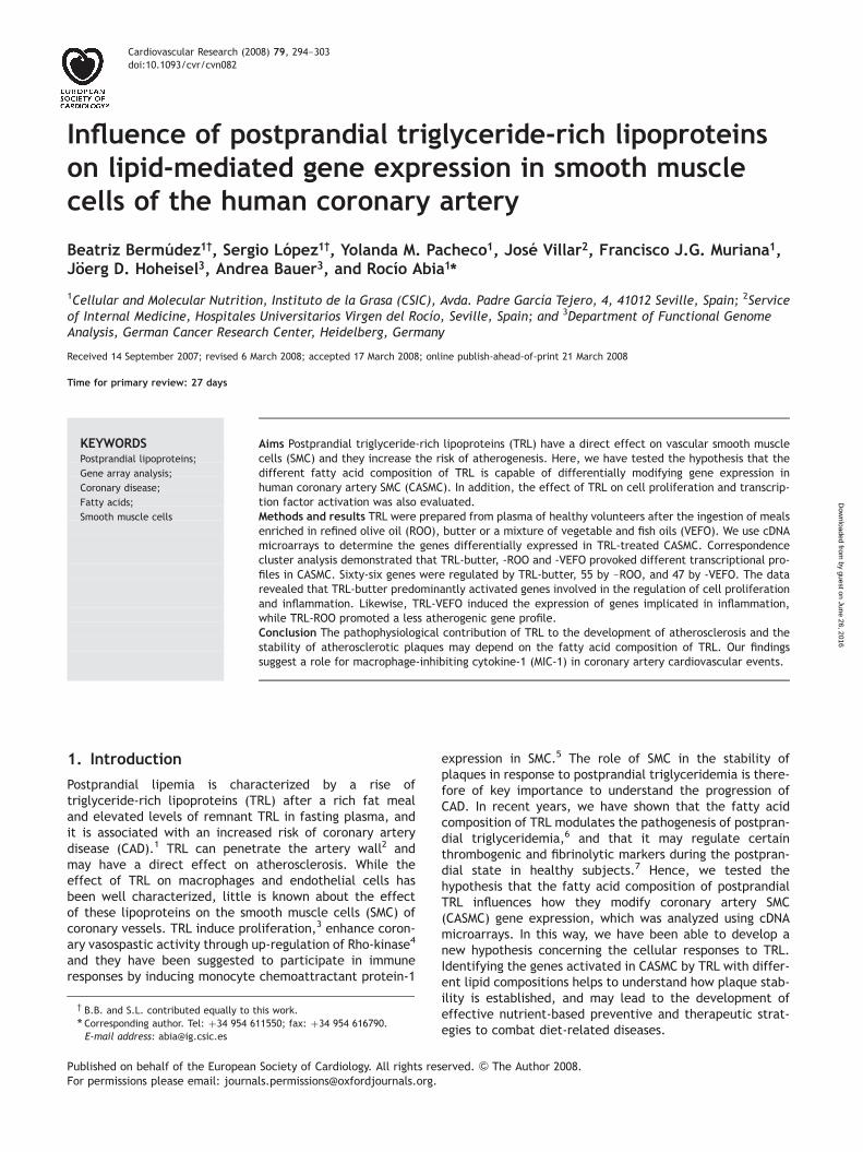
with either Cy3 or Cy5 were analyzed in competitive hybrid-izations, and after scanning and quantifying the spot inten-sities, the data were normalized and filtered. In total, 189PCR fragments contained a sequence that was differentiallytranscribed in the samples analyzed according to the highlystringent ‘min-max separation’ criterion.11 The results weresubjected to correspondence cluster analysis,12 which is anexplorative computational method to investigate the associ-ations between variables, such as genes and hybridizations,in a multi-dimensional space. This analysis simultaneouslydisplays data for two (or more) variables in a low-dimensional projection, thereby revealing associationsbetween them. The results of clustering the profilesobtained after incubation with the different TRL areshown as squares in Figure 1, in which the black dots rep-resent differentially transcribed genes. Genes locatedclose to the centroid of the guidelines are simultaneouslyco-expressed in all the different TRL while genes locatedclose to one of the guidelines and far from the centroidare key regulated genes for each TRL. The co-localizationof genes and conditions in the blot is indicative of a strongassociation between these elements. When classified bycorrespondence analysis, three distinct clusters were recog-nized corresponding to the differential gene expressionprofile produced by TRL-ROO, TRL-butter, and TRL-VEFO.Different TRL regimes produced statistically different tran-scriptional profiles in CASMC and the distances betweenTRL-butter and TRL-VEFO indicated particularly large differ-ences in the expression of certain genes.
3.3 Differential expression of specific functionalgroups of genes in triglyceride-richlipoproteins-stimulated coronary artery smoothmuscle cells
In each of the three clusters, genes were considered statisti-cally differentially expressed if they fulfilled the min-maxseparation criterion (see Methods) and the change in
Table 2 Fatty acid composition of lipid fractions in postprandialtriglyceride-rich lipoproteins (TRL) derived from butter, refinedolive oil (ROO), and a mixture of vegetable and fish oils (VEFO)
Fatty acid % by weight of total fatty acid
TRL-butter TRL-ROO TRL-VEFO
14:0 (MA) 3.7+0.716:0 (PA) 27.7+2.3a 14.2+1.2b 10.6+0.7c
18:0 (SA) 12.6+1.1a 5.4+0.2b 6.2+0.6c
18:1n-9 (OA) 35.5+5.2a 64.5+2.6b 59.7+2.8c
18:2n-6 (LA) 12.1+1.0a 9.6+0.5b 15.0+1.6c
20:5n-3 (EPA) 0.6+0.4a 0.6+0.2a 1.6+0.3b
22:6n-3 (DHA) 0.9+0.3a 0.8+0.3a 2.3+0.4b
Other 6.9+1.8 4.9+1.4 4.6+1.1SFA 45.5+2.1a 20.8+1.1b 17.8+0.7c
MUFA 39.9+3.4a 67.4+2.4b 62.4+3.2c
PUFA 14.7+0.6a 11.8+0.6a 19.8+1.5b
MA, miristic acid; PA, palmitic acid; SA, stearic acid; OA, oleic acid; LA,linoleic acid, EPA; eicosapentaenoic acid; DHA, docosahexaenoic acid.
SFA, saturated fatty acids; MUFA, monounsaturated fatty acids; PUFA,polyunsaturated fatty acids.
a,b,cValues in arrow with different letter are significantly different,P , 0.05. Values are means+ SD, n ¼ 3.
B. Bermudez et al.296
by guest on June 26, 2016D
ownloaded from
expression was at least two-fold. As a result 71 differentiallyexpressed genes were identified that exhibited moderate(two- to four-fold) to strong (greater than four-fold) differ-ential expression (Table 3). Of these genes, 59 wereup-regulated by TRL-butter, 51 by TRL-ROO and 45 byTRL-VEFO. Seven genes were down-regulated by TRL-butter,4 by TRL-ROO and 2 by TRL-VEFO in three different exper-iments. The functional classification of these genes revealedthat many of them encoded for proteins that were directlyor indirectly responsible for the regulation of cell prolifer-ation, migration and inflammatory responses. The strongestdifferentially expressed genes after exposure to TRL-butterwere associated with cell cycle regulation and motility.These included genes that encode for the cell cycle proteins(cyclin D1, cyclin E1), the TGFBR-2 and BMPR-2 receptors,intracellular signalling proteins (MAP3K1, DYRK1A, MKP-3,PLK-3), transcription factors and binding proteins (AP-1,GTF2H1, SP-1, NF-kB), cytoskeletal proteins and thoseimplicated in motility (RAF-1, ANXA2, VIM). Furthermore,INSIG-1 and ODC-1 were among the positive regulators ofcell proliferation strongly up-regulated by these TRL. Somegenes implicated in migration and/or inflammation werealso highly regulated by TRL-butter, such as MIC-1 andDDR-1. TRL-VEFO produced a strong response in genesmostly implicated in inflammation (MIC-1, IL-8, IL1B,MCP-1) and stress-responses (HSPA1L, SAPK-3, SAPK2A). Bycontrast, TRL-ROO induced changes in gene expression ingenes that belong to the functional classes of cell cycle
regulators, intracellular signalling, cytoskeleton, transcrip-tion factors, receptors and immune response. However,the induction of these genes was moderate when comparedwith the effects of TRL-butter and TRL-VEFO. All TRLstrongly induced the expression of HSP1A, a molecular cha-perone that is also involved in stress and apoptosis.
3.4 Confirmation of the microarray data byRT-PCR
Seven of the genes that were considered to be differentiallyexpressed were further analyzed by RT-PCR. The relativequantitative expression of the PCR products was consistentwith their differential expression profile in the cDNAarrays: HSPA1A (RT 9.1-fold vs. microarray seven-fold),MIC-1 (6.7 vs. 4.5), IL-8 (4.8 vs. 3.1), SOCS-5 (5.1 vs. 3.8),MCP-1 (4.0 vs. 2.9), TIMP-1 (23.0 vs. 22.3) and POU2F-1(23.2 vs. 22.3).
3.5 Effects of triglyceride-rich lipoproteins onproliferation, entry into S-phase and proteinexpression
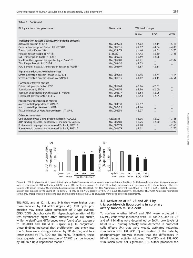
The influence of TRL on the proliferation of CASMC wasexamined by assessing the effect of TRL on the incorporationof BrdU as a measure of DNA synthesis. Furthermore, theability of TRL to stimulate quiescent cells to enter the cellcycle was analyzed by FACS analysis.
The incorporation of BrdU into CASMC was significantlyincreased 48 h after exposure to the TRL in a dose-dependentmanner. ELISA assay was carried out in quiescent cells follow-ing incubation with physiological concentrations of 50, 100 or200 mg/mL of TG–TRL. Accordingly, 50 mg/mL of TG wassufficient to promote cell proliferation while the maximalresponse was obtained at 100 mg/TG–TRL (Figure 2A), theconcentration used in the following experiments. Prolifer-ation was also dependent on the nature of dietary fats(Figure 2B) and TRL-butter induced significantly higher BrdUincorporation into CASMC than TRL-ROO and TRL-VEFO.
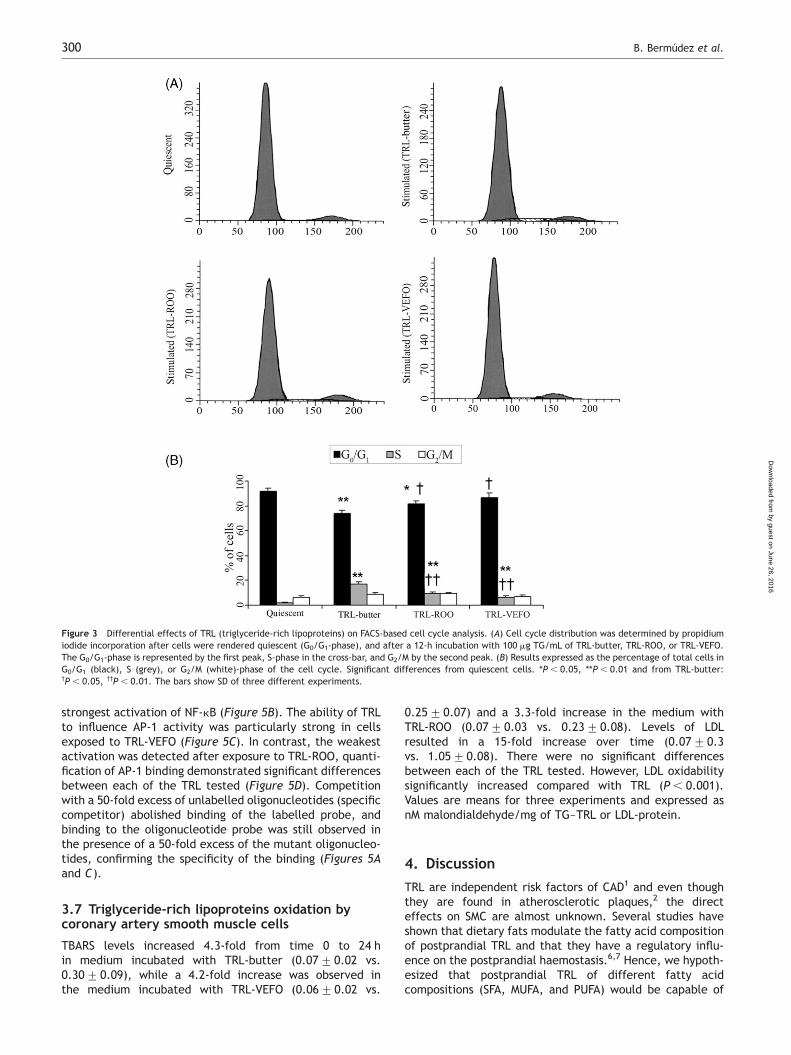
The ability of TRL to stimulate the entry of CASMC into thecell cycle was analyzed by FACS. We found that TRL releasedcells from growth arrest on a lipid dependent-manner(Figure 3A). When CASMC were rendered quiescent 92.0+2.1% of cells were in the G0/G1-phase, 1.9+0.6% were inthe S-phase, and 6.1+1.7% were in the G2/M-phase(Figure 3B). After stimulation with TRL-butter for 12 h, theproportion of cells in G0/G1-phase decreased to 74.2+2.0% while the proportion in S-phase increased to 17.0+1.9%. In cells incubated with TRL-ROO, the proportion ofcells in G0/G1-phase decreased to 81.4+2.8% and the pro-portion in the S-phase increased to 9.4+1.1%. TRL-VEFOdid not produce a significant increase in the cells in theS-phase (6.6+0.9%) when compared with TRL-ROO.
Cyclin D1 is necessary for entry and progression throughthe G1/S-phase of the cell cycle. Significantly, TRL producedthe transient induction of cyclin D1 within as little as 6 h(Figure 4A). The maximal effect was observed forTRL-butter at 12 h and while TRL-ROO induced significantlyless cyclin D1 expression, TRL-VEFO provoked the lowestlevels of expression (Figure 4A). DNA synthesis in theS-phase requires the DNA polymerase cofactor PCNA. PCNAprotein levels were significantly higher at 12 and 18 hafter TRL-butter stimulation than those found for
Figure 1 Biplot representation of the results of the correspondence analy-sis. Transcriptional profiling data were generated from serum-starved cells(Quiescent) cells treated with TRL (triglyceride-rich lipoproteins)-butter,TRL-ROO (refined olive oil), and TRL-VEFO (vegetable and fish oils). In theplot, each hybridization of the RNA following exposure to a specific TRL isdepicted as a square, where four hybridizations have been performed foreach experimental condition. Each gene differentially transcribed is shownas a black dot. As a consequence of the normalization process, only themedian of all the control hybridizations is shown in the diagram as a singlesquare (Quiescent) instead of the individual hybridization. The samplesfrom the different TRL and from the control form distinct clusters showingthat the effects of the TRL are clearly different from each other. Each differ-entially transcribed gene is shown as a black dot and the closer theco-localization of two spots (both genes and TRL); the higher is the degreeof association between them. The guidelines displayed in the diagram corre-spond to the position of genes whose transcription profiles would exhibit asignal in one condition only. The closer a gene lies to one of these guidelinesand the further its distance to the centroid, the better its expression isdescribed by the related ideal profile. All genes that are not differentiallytranscribed are located close to the centroid of the lines but are not shownfor clarity.
Gene expression in human vascular cells is postprandially lipid-dependent 297
by guest on June 26, 2016D
ownloaded from
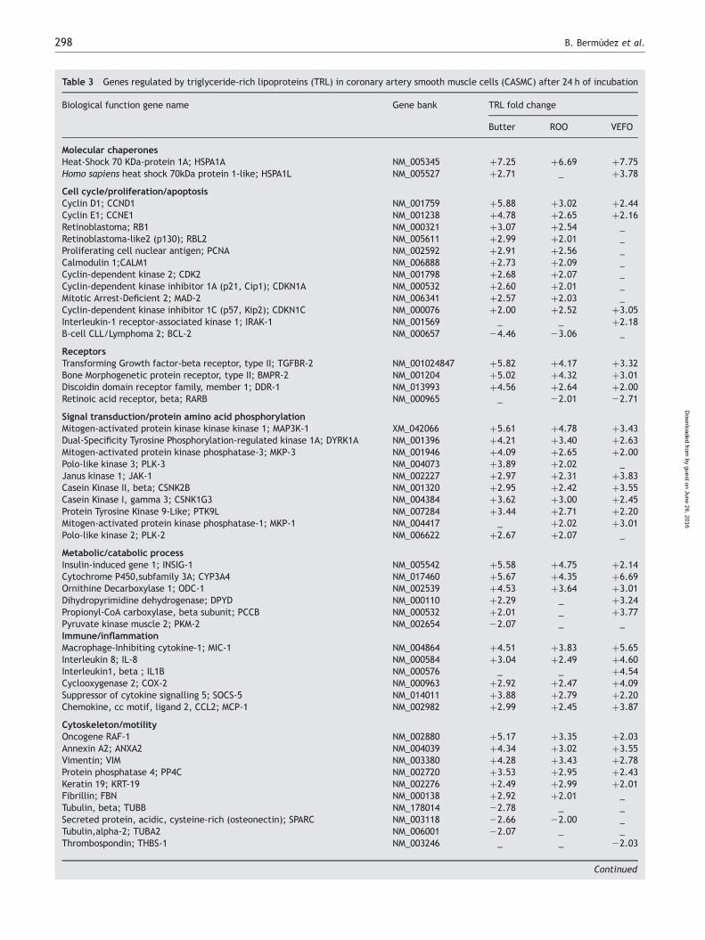
Table 3 Genes regulated by triglyceride-rich lipoproteins (TRL) in coronary artery smooth muscle cells (CASMC) after 24 h of incubation
Biological function gene name Gene bank TRL fold change
Butter ROO VEFO
Molecular chaperonesHeat-Shock 70 KDa-protein 1A; HSPA1A NM_005345 þ7.25 þ6.69 þ7.75Homo sapiens heat shock 70kDa protein 1-like; HSPA1L NM_005527 þ2.71 _ þ3.78
Cell cycle/proliferation/apoptosisCyclin D1; CCND1 NM_001759 þ5.88 þ3.02 þ2.44Cyclin E1; CCNE1 NM_001238 þ4.78 þ2.65 þ2.16Retinoblastoma; RB1 NM_000321 þ3.07 þ2.54 _Retinoblastoma-like2 (p130); RBL2 NM_005611 þ2.99 þ2.01 _Proliferating cell nuclear antigen; PCNA NM_002592 þ2.91 þ2.56 _Calmodulin 1;CALM1 NM_006888 þ2.73 þ2.09 _Cyclin-dependent kinase 2; CDK2 NM_001798 þ2.68 þ2.07 _Cyclin-dependent kinase inhibitor 1A (p21, Cip1); CDKN1A NM_000532 þ2.60 þ2.01 _Mitotic Arrest-Deficient 2; MAD-2 NM_006341 þ2.57 þ2.03 _Cyclin-dependent kinase inhibitor 1C (p57, Kip2); CDKN1C NM_000076 þ2.00 þ2.52 þ3.05Interleukin-1 receptor-associated kinase 1; IRAK-1 NM_001569 _ _ þ2.18B-cell CLL/Lymphoma 2; BCL-2 NM_000657 24.46 23.06 _
ReceptorsTransforming Growth factor-beta receptor, type II; TGFBR-2 NM_001024847 þ5.82 þ4.17 þ3.32Bone Morphogenetic protein receptor, type II; BMPR-2 NM_001204 þ5.02 þ4.32 þ3.01Discoidin domain receptor family, member 1; DDR-1 NM_013993 þ4.56 þ2.64 þ2.00Retinoic acid receptor, beta; RARB NM_000965 _ 22.01 22.71
Signal transduction/protein amino acid phosphorylationMitogen-activated protein kinase kinase kinase 1; MAP3K-1 XM_042066 þ5.61 þ4.78 þ3.43Dual-Specificity Tyrosine Phosphorylation-regulated kinase 1A; DYRK1A NM_001396 þ4.21 þ3.40 þ2.63Mitogen-activated protein kinase phosphatase-3; MKP-3 NM_001946 þ4.09 þ2.65 þ2.00Polo-like kinase 3; PLK-3 NM_004073 þ3.89 þ2.02 _Janus kinase 1; JAK-1 NM_002227 þ2.97 þ2.31 þ3.83Casein Kinase II, beta; CSNK2B NM_001320 þ2.95 þ2.42 þ3.55Casein Kinase I, gamma 3; CSNK1G3 NM_004384 þ3.62 þ3.00 þ2.45Protein Tyrosine Kinase 9-Like; PTK9L NM_007284 þ3.44 þ2.71 þ2.20Mitogen-activated protein kinase phosphatase-1; MKP-1 NM_004417 _ þ2.02 þ3.01Polo-like kinase 2; PLK-2 NM_006622 þ2.67 þ2.07 _
Metabolic/catabolic processInsulin-induced gene 1; INSIG-1 NM_005542 þ5.58 þ4.75 þ2.14Cytochrome P450,subfamily 3A; CYP3A4 NM_017460 þ5.67 þ4.35 þ6.69Ornithine Decarboxylase 1; ODC-1 NM_002539 þ4.53 þ3.64 þ3.01Dihydropyrimidine dehydrogenase; DPYD NM_000110 þ2.29 _ þ3.24Propionyl-CoA carboxylase, beta subunit; PCCB NM_000532 þ2.01 _ þ3.77Pyruvate kinase muscle 2; PKM-2 NM_002654 22.07 _ _Immune/inflammationMacrophage-Inhibiting cytokine-1; MIC-1 NM_004864 þ4.51 þ3.83 þ5.65Interleukin 8; IL-8 NM_000584 þ3.04 þ2.49 þ4.60Interleukin1, beta ; IL1B NM_000576 _ _ þ4.54Cyclooxygenase 2; COX-2 NM_000963 þ2.92 þ2.47 þ4.09Suppressor of cytokine signalling 5; SOCS-5 NM_014011 þ3.88 þ2.79 þ2.20Chemokine, cc motif, ligand 2, CCL2; MCP-1 NM_002982 þ2.99 þ2.45 þ3.87
Cytoskeleton/motilityOncogene RAF-1 NM_002880 þ5.17 þ3.35 þ2.03Annexin A2; ANXA2 NM_004039 þ4.34 þ3.02 þ3.55Vimentin; VIM NM_003380 þ4.28 þ3.43 þ2.78Protein phosphatase 4; PP4C NM_002720 þ3.53 þ2.95 þ2.43Keratin 19; KRT-19 NM_002276 þ2.49 þ2.99 þ2.01Fibrillin; FBN NM_000138 þ2.92 þ2.01 _Tubulin, beta; TUBB NM_178014 22.78 _ _Secreted protein, acidic, cysteine-rich (osteonectin); SPARC NM_003118 22.66 22.00 _Tubulin,alpha-2; TUBA2 NM_006001 22.07 _ _Thrombospondin; THBS-1 NM_003246 _ _ 22.03
Continued
B. Bermudez et al.298
by guest on June 26, 2016D
ownloaded from
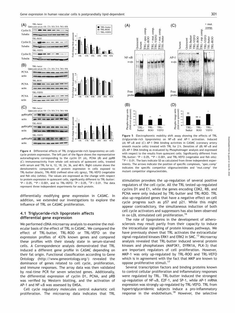
TRL-ROO, and at 12, 18, and 24 h they were higher thanthose induced by TRL-VEFO (Figure 4B). Cell cycle pro-gression may occur when complexes of D-type cyclins/CDK4/CDK6 phosphorylate Rb. Hyperphosphorylation of Rbwas significantly higher after stimulation of TRL-butter,while no significant differences were found after exposureto TRL-ROO and TRL-VEFO (Figure 4C). In conjuction,these findings indicated that proliferation and entry intothe S-phase were strongly induced by TRL-butter, and to alesser extent by TRL-ROO and TRL-VEFO. Therefore, thesedata suggests that proliferation of CASMC can be inducedby TRL in a lipid-dependent manner.
3.6 Activation of NF-kB and AP-1 bytriglyceride-rich lipoproteins in coronaryartery smooth muscle cells
To confirm whether NF-kB and AP-1 were activated inCASMC, cells were incubated with TRL for 2 h, and NF-kBand AP-1 binding were determined by EMSA. Low levels ofbasal NF-kB binding activity were detected in quiescentcells (Figure 5A) that were weakly activated followingstimulation with TRL-ROO. Quantification of the data byphosphoimager analysis showed that the differences inNF-kB binding activity following TRL-VEFO and TRL-ROOstimulation were not significant. TRL-butter produced the
Figure 2 TRL (triglyceride-rich lipoproteins) induced CASMC (coronary artery smooth muscle cells) proliferation. BrdU (bromodeoxyuridine) incorporation wasused as a measure of DNA synthesis in CASMC and in (A), the dose-response effect of TRL on BrdU incorporation in quiescent cells is shown (white). The cellstreated with serum (grey) or the indicated concentrations of TG–TRL (black) for 48 h. *Significantly different from 50 mg TG–TRL (P , 0.05). (B) BrdU incorpor-ation in cells exposed to 100 mg/mL of TRL-butter, TRL-ROO or TRL-VEFO (black) for 48 h. *P , 0.005 TRL-butter vs. TRL-ROO or TRL-VEFO. Values are normalizedto the BrdU incorporation in quiescent cells and the bars indicate the SD as calculated from three different experiments.
Table 3 Continued
Biological function gene name Gene bank TRL fold change
Butter ROO VEFO
Transcription factors activity/DNA-binding proteinsActivator protein 1; AP-1 NM_002228 þ4.02 þ2.11 þ5.18General transcription factor IIH; GTF2H1 NM_005316 þ4.97 þ4.54 þ4.00Transcription Factor SP-1 NM_138473 þ4.60 þ4.01 þ3.75Nuclear factor-kappa B; NF-kB L_26267 þ4.42 þ2.60 þ3.46E2F Transcription Factor 1; E2F-1 NM_005225 þ2.59 þ2.08 _Small mother against decapentaplegic; SMAD-2 NM_005901 þ2.71 _ þ2.04Zinc Finger Protein 91; ZNF-91 NM_003430 þ2.33 _ _POU domain, class 2, transcription factor 1; POU2F-1 NM_002697 22.67 22.11 _
Signal transduction/oxidative stressStress-activated protein kinase 3; SAPK-3 NM_002969 þ3.15 þ2.41 þ4.14Stress-activated protein kinase 2A; SAPK2A NM_001315 þ4.02 þ3.11 þ6.51
Hormones/growth factorsEpidermal growth factor; EGF NM_001963 þ3.76 þ2.31 _Stanniocalcin 1; STC-1 NM_003155 þ2.96 þ2.00 _Vascular endothelial growth factor B; VEGFB NM_003377 þ2.64 þ2.06 _Fibroblast growth factor; FGF-5 NM_004464 þ2.53 þ2.01 _
Proteolysis/extracellular matrixMatrix metallopeptidase 2; MMP-2 NM_004530 þ2.97 _ _Matrix metalloproteinase 1; MMP-1 NM_002421 þ2.66 _ _Tissue Inhibitor of Metalloproteinase 1; TIMP-1. NM_003254 22.37 _ _
Other or unknownCell division cycle 2-like protein kinase 6; CDC2L6 AB028951 þ3.06 þ2.02 þ3.85ATP-binding cassette, subfamily B, member 6; ABCB6 NM_005689 þ3.25 þ2.70 þ3.99Post-meiotic segregation increased 2-like 1; PMS2L1 NM_002679 þ2.28 – þ2.65Post-meiotic segregation increased 2-like 2; PMS2L2 NM_002679 þ2.01 _ þ2.75
Gene expression in human vascular cells is postprandially lipid-dependent 299
by guest on June 26, 2016D
ownloaded from
strongest activation of NF-kB (Figure 5B). The ability of TRLto influence AP-1 activity was particularly strong in cellsexposed to TRL-VEFO (Figure 5C). In contrast, the weakestactivation was detected after exposure to TRL-ROO, quanti-fication of AP-1 binding demonstrated significant differencesbetween each of the TRL tested (Figure 5D). Competitionwith a 50-fold excess of unlabelled oligonucleotides (specificcompetitor) abolished binding of the labelled probe, andbinding to the oligonucleotide probe was still observed inthe presence of a 50-fold excess of the mutant oligonucleo-tides, confirming the specificity of the binding (Figures 5Aand C).
3.7 Triglyceride-rich lipoproteins oxidation bycoronary artery smooth muscle cells
TBARS levels increased 4.3-fold from time 0 to 24 hin medium incubated with TRL-butter (0.07+0.02 vs.0.30+0.09), while a 4.2-fold increase was observed inthe medium incubated with TRL-VEFO (0.06+0.02 vs.
0.25+0.07) and a 3.3-fold increase in the medium withTRL-ROO (0.07+0.03 vs. 0.23+0.08). Levels of LDLresulted in a 15-fold increase over time (0.07+0.3vs. 1.05+0.08). There were no significant differencesbetween each of the TRL tested. However, LDL oxidabilitysignificantly increased compared with TRL (P , 0.001).Values are means for three experiments and expressed asnM malondialdehyde/mg of TG–TRL or LDL-protein.
4. Discussion
TRL are independent risk factors of CAD1 and even thoughthey are found in atherosclerotic plaques,2 the directeffects on SMC are almost unknown. Several studies haveshown that dietary fats modulate the fatty acid compositionof postprandial TRL and that they have a regulatory influ-ence on the postprandial haemostasis.6,7 Hence, we hypoth-esized that postprandial TRL of different fatty acidcompositions (SFA, MUFA, and PUFA) would be capable of
Figure 3 Differential effects of TRL (triglyceride-rich lipoproteins) on FACS-based cell cycle analysis. (A) Cell cycle distribution was determined by propidiumiodide incorporation after cells were rendered quiescent (G0/G1-phase), and after a 12-h incubation with 100 mg TG/mL of TRL-butter, TRL-ROO, or TRL-VEFO.The G0/G1-phase is represented by the first peak, S-phase in the cross-bar, and G2/M by the second peak. (B) Results expressed as the percentage of total cells inG0/G1 (black), S (grey), or G2/M (white)-phase of the cell cycle. Significant differences from quiescent cells. *P , 0.05, **P , 0.01 and from TRL-butter:†P , 0.05, ††P , 0.01. The bars show SD of three different experiments.
B. Bermudez et al.300
by guest on June 26, 2016D
ownloaded from
differentially modifying gene expression in CASMC. Inaddition, we extended our investigations to explore theinfluence of TRL on CASMC proliferation.
4.1 Triglyceride-rich lipoprotein affectsdifferential gene expression
We performed cDNA microarray analysis to examine the mol-ecular basis of the effect of TRL in CASMC. We compared theeffect of TRL-butter, TRL-ROO or TRL-VEFO on theexpression profiles of 4376 known genes and comparedthese profiles with their steady state in serum-starvedcells. A Correspondence analysis demonstrated that TRLinduced a different gene profile in CASMC depending ontheir fat origin. Functional classification according to GeneOntology (http://www.geneontology.org/) revealed thedominance of genes related to cell cycle, proliferation,and immune responses. The array data was then validatedby real-time PCR for seven selected genes. Additionally,the differential expression of cyclin D1, PCNA, and pRBwas verified by Western blotting, and the activation ofAP-1 and NF-kB was assessed by EMSA.
Cell cycle regulatory molecules control eukariotic cellproliferation. The microarray data indicates that TRL
stimulation provokes the up-regulation of several positiveregulators of the cell cycle. All the TRL tested up-regulatedcyclins D1 and E1, while the genes encoding CDK2, RB, andPCNA were only induced by TRL-butter and TRL-ROO. TRLalso up-regulated genes that have a negative effect on cellcycle progress such as p57 and p21. While this mightappear contradictory, the simultaneous induction of bothcell cycle activators and suppressors has also been observedin ox-LDL stimulated cell proliferation.16
The role of lipoproteins in the development of athero-sclerosis may result partly from their capacity to inducethe intracellular signalling of protein kinases pathways. Wehave previously shown that TRL activates the extracellularsignal-regulated kinases ERK1 and ERK2 in SMC.13 Microarrayanalysis revealed that TRL-butter induced several proteinkinases and phosphatases (MAP3K1, DYRK1A, PLK-3) thatare important regulators of cell proliferation. However,MKP-1 was only up-regulated by TRL-ROO and TRL-VEFOwhich is in agreement with the fact that MKP are known tooppose proliferative stimuli.17
Several transcription factors and binding proteins knownto control cellular proliferation and inflammatory responseswere regulated by TRL. TRL-butter induced the strongestup-regulation of NF-kB, E2F-1, and SP-1, while AP-1 mRNAexpression was strongly up-regulated by TRL-VEFO. TRL fromhypertriglyceridemic subjects induce a pro-inflammatoryresponse in the endothelium.18 However, the selective
Figure 4 Differential effects of TRL (triglyceride-rich lipoproteins) on cell-cycle protein expression. The left part of the figure shows the representativeautoradiograms corresponding to the cyclin D1 (A), PCNA (B) and ppRB(C) immunoreactivity from whole cell extracts of quiescent cells, treatedwith serum and TRL for 6, 12, 18, 24, 36, and 48 h. Right column shows thedensitometric comparisons of protein expression in cells exposed toTRL-butter (black), TRL-ROO (refined olive oil) (grey), TRL-VEFO (vegetableand fish oils) (white). The values are expressed as the change with respectto protein expression in quiescent cells, significantly different to TRL-butter:*P , 0.05, **P , 0.001, and to TRL-VEFO: †P , 0.05, ††P , 0.01. The datarepresent three independent experiments for each protein.
Figure 5 Electrophoretic mobility shift assay showing the effects of TRL(triglyceride-rich lipoproteins) on NF-kB and AP-1 activation. Induced(A) NF-kB and (C) AP-1 DNA binding activities in CASMC (coronary arterysmooth muscle cells) treated with TRL for 2 h. Densities of (B) NF-kB and(D) AP-1 DNA binding as evaluated by PhosphoImager analysis and expressedwith respect to the results from quiescent cells. Significantly different fromTRL-butter: *P , 0.05, **P , 0.001, and TRL-VEFO (vegetable and fish oils):††P , 0.01. The bars indicate SD as calculated from three independent exper-iments. The arrows indicate the position of specific complexes, ‘spec.comp’indicates the specific competitor oligonucleotides and ‘mut.comp’ themutant competitor oligonucleotides.
Gene expression in human vascular cells is postprandially lipid-dependent 301
by guest on June 26, 2016D
ownloaded from
effect of TRL according to their fatty acid composition hasyet to be demonstrated. TRL-VEFO increased mRNAexpression of inflammatory cytokines, chemokines, andadhesion molecules. Expression of the IL-8 gene wasup-regulated by all the TRL, although it was strongly acti-vated by TRL-VEFO. The higher LA content in TRL-VEFOcould account for this, since LA but not OA up-regulatesproduction of IL-8 in SMC.19 IL1B and COX-2, both genesthat are related to inflammation, were stronglyup-regulated by TRL-VEFO. However, TRL-ROO diminishedthe levels of inflammatory gene expression in CASMC.Since TRL-ROO does not contain antioxidant compounds,their anti-inflammatory effects could have been mediatedby OA. Indeed, MUFA-rich diets decrease the expression ofinflammatory genes in endothelial cells.20 Inhibitors of cyto-kine signalling such SOCS-5 were strongly up-regulated byTRL-butter, which is interesting since SOCS are negative reg-ulators of cytokine signalling and they also have a cleareffect on proliferation.21
Other proliferation-related genes that were up-regulatedby TRL-butter, and to a lesser extent by TRL-ROO andTRL-VEFO include, TGFBR-2, BMPR-2, and INSIG-1. Growthfactors involved in SMC proliferation, such as EGF, VEGFB,and FGF-5, were only up-regulated by TRL-butter andTRL-ROO, suggesting that the presence of n-3 PUFA couldimpede their induction by TRL-VEFO.22 Other functionalgroup include genes that encode heat shock proteins, allTRL strongly up-regulated HSPA1A while HSPA1L was onlyup-regulated by TRL-VEFO and TRL-butter. Failure ofTRL-ROO to activate the expression of this gene is in agree-ment with its potential role in attenuating inflammation,since HSPA1L influences the concentrations of cytokine inplasma.23
Finally, TRL up-regulated expression of macrophage-inhibiting cytokine-1 (MIC-1) by more than five-fold. MIC-1may constitute a marker of atherosclerosis as it is detectedin the serum of individuals suffering from myocardial infrac-tion.24 MIC-1 is expressed in human atherosclerotic carotidarteries and is induced by ox-LDL in human macrophages.25
Hence, it is significant that for the first time, we have shownthat TRL can up-regulate MIC-1 expression in CASMC.
4.2 Effects of triglyceride-rich lipoproteins oncell proliferation and transcriptional activation
TRL can force cells to enter S-phase of the cell cycle andinduce CASMC proliferation in the absence of any othermitogenic factor. Proliferation was dose-dependent up to aconcentration of 100 mg TG–TRL/mL and it was affectedby the nature of the TRL. TRL-butter had the strongesteffect on CASMC proliferation while the presence of moder-ate amounts of n-3 PUFA in TRL-VEFO was able to attenuateproliferation, although the decrease was not significantwhen compared with TRL-ROO. The presence of SMC dis-tinguishes progression-prone from progression-resistantlesions and in fact, progression-resistant lesions containfew SMC. TRL-butter increased the expression of cyclin D1and hyperphosphorylated pRb, while TRL-ROO andTRL-VEFO diminished their expression, probably due to thepresence of OA or EPA and DHA,26 respectively. Similarly,both human and animal diets rich in SFA have been shownto produce greater lymphocyte proliferation than dietsrich in OA, LA, or fish oil.27,28 For the first time, our
results suggest that the mitogenic effect of human TRL onCASMC depends on their fatty acid composition.
We confirmed that TRL differentially modulated AP-1 andNF-kB DNA binding activity in a lipid-dependent fashion inCASMC. TRL-butter induced the highest DNA bindingcapacity of NF-kB and indeed, postprandial studies showedthat the intake of butter but not olive oil-enriched mealsinduced activation of NF-kB in human monocytes.29 Wehave shown that postprandial TRL obtained after ROO inges-tion did not completely inhibit but rather attenuatedNF-kB-DNA binding in CASMC when compared with thebutter meal. TRL could influence NF-kB by releasing fattyacids from core TG and while OA has little or no effect,SFA as well as n-6 and n-3 PUFA appear to activate NF-kBin endothelial cells.30,31 Hence, the higher amount of OAcould explain the low NF-kB transcriptional activationfound after TRL-ROO incubation. Furthermore, we suggestthat OA could reduce Cyclin D1 promoter activity bydecreasing NF-kB-binding. While TRL-VEFO provoked thehighest AP-1 activation, the role of n-6 and n-3 PUFA onAP-1 activation is controversial. Although n-6 PUFA appearsto be most effective in activating AP-1 and in contributingto an inflammatory response,31 n-3 PUFA may induceeither AP-1 inhibition32 or activation.30 We suggest thatthe elevated ratio of n-6 to n-3 PUFA in TRL-VEFO may bea significant factor in mediating AP-1-DNA binding activity,and in the increase in inflammatory gene expression foundin CASMC. Additionally, the resistance of TRL to oxidationdid not depend on the fatty acid composition of the diet.Furthermore, TRL were more resistant to oxidation whencompared with LDL, which may suggest that the fatty acidcomposition of TRL may be more important than their oxi-dation state in their atherogenicity. Accordingly, remnantTRL induces SMC proliferation regardless of its oxidativestress.33
4.3 Limitation of the study
We assessed the effect of TRL on the regulation of geneexpression in human coronary artery SMC (CASMC) using acell-based in vitro model. Extrapolation of the results tothe human clinical situation remains uncertain.
In conclusion, the pathophysiological contribution of TRLto the development of atherosclerosis and the stability ofthe atherosclerotic plaque may depend on the fatty acidcomposition of TRL, indicating that the quality of theplaque rather than the quantity may determine the clinicalconsequences of atherosclerosis. Since enhanced SMCproliferation contributes to early lesion development, asdoes secretion of proinflamatory cytokines by SMC,TRL-butter could be considered a major determinant ofplaque instability. TRL-VEFO reduced proliferation ofCASMC, which could possibly lead to progression-resistantlesions, whilst inducing moderate gene expression of inflam-matory cytokines. Ingestion of diets with a lower ratio ofomega-6/omega-3 fatty acids could therefore offer protec-tion against atherosclerosis. TRL-ROO induced moderateproliferation and a less atherogenic gene profile, whichcould improve atherosclerotic-plaque stability and supportthe prescription of olive oil enriched-diets in secondary pre-vention of cardiovascular disease. The results also suggest arole of MIC-1 in coronary artery cardiovascular events.
B. Bermudez et al.302
by guest on June 26, 2016D
ownloaded from
Acknowledgements
We thank Melanie Bier for technical assistance and Kurt Fellenbergfor help with the data analysis. We thank Puleva Biotech SA,Granada, and aceites Monteolivo SL, Jaen, Spain, for supplying uswith the butter, VEFO and olive oil.
Conflict of interest: none declared.
Funding
This work was supported by the Spanish Ministry of Science andTechnology (AGL2005–03722). We also thank the Consejerıa deEducacion y Ciencia of the Junta de Andalucıa for financialsupport through a visiting fellowship.
References1. Cohn JS. Postprandial lipemia and remnant lipoproteins. Clin Lab Med
2006;26:773–786.2. Rapp JH, Lespine A, Hamilton RL, Colyvas N, Chaumeton AH,
Tweedie-Hardman J et al. Triglyceride-rich lipoproteins isolated byselected-affinity anti-apolipoprotein B immunosorption from humanatherosclerotic plaque. Arterioscler Thromb 1994;14:1767–1774.
3. Kawakami A, Tanaka A, Chiba T, Nakajima K, Shimokado K, Yoshida M.Remnant lipoprotein-induced smooth muscle cell proliferation involvesepidermal growth factor receptor transactivation. Circulation 2003;108:2679–2688.
4. Oi K, Shimokawa H, Hiroki J, Uwatoku T, Abe K, Matsumoto Y et al.Remnant lipoproteins from patients with sudden cardiac death enhancecoronary vasospastic activity through upregulation of Rho-kinase. Arter-ioscler Thromb Vasc Biol 2004;24:918–922.
5. Domoto K, Taniguchi T, Takaishi H, Takahashi T, Fujioka Y, Takahashi Aet al. Chylomicron remnants induce monocyte chemoattractantprotein-1 expression via p38 MAPK activation in vascular smooth musclecells. Atherosclerosis 2003;171:193–200.
6. Abia R, Perona JS, Pacheco YM, Montero E, Muriana FJG, Ruiz-Gutierrez V.Postprandial triacylglycerols from dietary virgin olive oil are selectivelycleared in humans. J Nutr 1999;129:2184–2191.
7. Pacheco YM, Bermudez B, Lopez S, Abia R, Villar J, Muriana FJG. Ratio ofoleic to palmitic acid is a dietary determinant of thrombogenic and fibri-nolytic factors during the postprandial state in men. Am J Clin Nutr 2006;84:342–349.
8. Lopez S, Bermudez B, Pacheco YM, Lopez-Lluch G, Moreda W, Villar Jet al. Dietary oleic and palmitic acids modulate the ratio of triacylgli-cerol to cholesterol in postprandial triacylglycerol-rich lipoproteins inmen and cell viability and cyclin in human monocytes. J Nutr 2007;137:1–7.
9. Esposito I, Bauer A, Hoheisel JD, Kleeff J, Friess H, Buchler MW et al.Microcystic tubulopapillary carcinoma of the pancreas: a new tumorentity. Virchows Arch 2004;444:447–453.
10. Brazma A, Hingamp P, Quackenbush J, Sherlock G, Spellman P, Stoeckert Cet al. Minimum information about a microarray experiment (MIAME)-toward standards for microarray data. Nat Genet 2001;29:365–371.
11. Beißbarth T, Fellenberg K, Brors B, Arribas-Prat R, Boer J, Hauser NCet al. Comparison and quality measures of DNA array hybridisationdata. Bioinformatics 2001;16:1014–1022.
12. Fellenberg k, Hauser NC, Brors B, Neutzner A, Hoheisel JD, Vingron M.Correspondence analysis applied to microarray data. Proc Natl Acad SciUSA 2001;98:10781–10786.
13. Pacheco YM, Abia R, Perona JS, Meier KE, Montero E, Ruiz-Gutierrez Vet al. Triacylglycerol-rich lipoproteins trigger the phosphorylation ofextracellular-signal regulated kinases in vascular cells. Life Sci 2002;71:1351–1360.
14. Metzler B, Abia R, Ahmad M, Wernig F, Pachinger O, Hu Y et al. Activationof heat shock transcription factor-1 in atherosclerosis. Am J Path 2003;162:1669–1676.
15. Miyoshi T, Tian J, Matsumoto AH, Shi W. Differential response of vascularsmooth muscle cells to oxidized-LDL in mouse strains with differentatherosclerosis susceptibility. Atherosclerosis 2006;189:99–105.
16. Zettler ME, Prociuk MA, Austria JA, Massaeli H, Zhong G, Pierce GN.OxLDL stimulates cell proliferation through a general induction of cellcycle proteins. Am J Physiol Heart Circ Physiol 2003;284:644–653.
17. Bueno OF, De Windtl LJ, Lim HW, Tymitz KM, UIT SA, Timbal TR et al. Thedual-specific phosphatase MKP-1 limits the cardiac hypertrophic responsein vitro and in vivo. Cir Res 2001;88:88–96.
18. Norata GD, Grigore L, Raselli S, Seccomandi PM, Hamsten A, Maggi FMet al. Triglyceride-rich lipoproteins from hypertriglyceridemicsubjects induce a pro-inflammatory response in the endothelium: Mol-ecular mechanisms and gene expression studies. J Mol Cell Cardiol2006;40:484–494.
19. Leik CE, Walsh SW. Linoleic acid, but not oleic acid, upregulates pro-duction of interleukin-8 by human vascular smooth muscle cells via ara-chidonic acid metabolites under conditions of oxidative stress. J SocGynecol Investig 2005;12:593–598.
20. Toborek M, Lee YW, Garrido R, Kaiser S, Hennig B. Unsaturated fatty acidsselectively induce an inflammatory environment in human endothelialcells. Am J Clin Nutr 2002;75:119–125.
21. Nicholson SE, Metcalf D, Sprigg NS, Columbus R, Walker F, Silva A et al.Suppressor of cytokine signaling (SOCS-5) is a potential negative regula-tor of epidermal growth factor signaling. Proc Natl Acad Sci USA 2005;102:2328–2333.
22. Tsuji M, Murota SI, Morita I. Docosapentaenoic acid (22:5) suppressedtube-forming activity in endothelial cells induced by vascular endothelialgrowth factor. Prostaglandins. Leukot Essent Fatty Acids 2003;68:337–342.
23. Schroder O, Schulte KM, Ostermann P, Roher HD, Ekkernkamp A, Laun RA.Heat shock protein 70 genotypes HSPA1B and HSPA1L influence cytokineconcentrations and interfere with outcome after major injury. CritCare Med 2003;31:73–79.
24. Brown DA, Breit SN, Buring J, Fairlie WD, Bauskin AR, Liu T et al. Concen-tration in plasma of macrophage inhibitory cytokine-1 and risk of cardio-vascular events in women: a nested case-control study. Lancet 2002;359:2159–2163.
25. Schlittenhardt D, Schober A, Strelau J, Bonaterra GA, Schmiedt W,Unsicker K et al. Involvement of growth differentiation factor-15/macro-phage inhibitory cytokine-1 (GDF-15/MIC-1) in oxLDL-induced apoptosisof human macrophages in vitro and in arteriosclerotic lesions. CellTissue Res 2004;318:325–333.
26. Bousserouel S, Raymondjean M, Brouillet A, Bereziat G, Andreani M.Modulation of cyclin-D1 and early growth response factor-1 geneexpression in interleukin-1beta-treated rat smooth muscle cells by n-6and n-3 polyunsaturated fatty acids. Eur J Biochem 2004;271:4462–4473.
27. Tinahones FJ, Gomez-Zumaquero JM, Monzon A, Rojo-Martinez G,Pareja A, Morcillo S et al. Dietary palmitic acid influences LDL-mediatedlymphocyte proliferation differently to other mono- and polyunsaturatedfatty acids in rats. Diabetes Nutr Metab 2004;17:250–258.
28. Thies F, Nebe-von-Caron G, Powell JR, Yaqoob P, Newsholme EA,Calder PC. Dietary supplementation with gamma-linolenic acid or fishoil decreases T lymphocyte proliferation in healthy older humans.J Nutr 2001;131:1918–1927.
29. Bellido C, Lopez-Miranda J, Blanco-Colio LM, Perez-Martınez P,Muriana FJG, Martın-Ventura JL et al. Butter and walnuts, but not oliveoil, elicit postprandial activation of nuclear transcription factor kappaBin peripheral blood mononuclear cells from healthy men. Am J ClinNutr 2004;80:1487–1491.
30. Maziere C, Conte MA, Degonville J, Ali D, Maziere JC. Cellular enrichmentwith polyunsaturated fatty acids induces an oxidative stress and activatesthe transcription factors AP1 and NFkappaB. Biochem Biophys ResCommun 1999;265:116–122.
31. Hennig B, Meerarani P, Ramadass P, Watkins BA, Toborek M. Fatty acid-mediated activation of vascular endothelial cells. Metabolism 2000;49:1006–1013.
32. Babcock TA, Kurland A, Helton WS, Rahman A, Anwar KN, Espat NJ. Inhi-bition of activator protein-1 transcription factor activation by omega-3fatty acid modulation of mitogen-activated protein kinase signalingkinases. J Parenter Enteral Nutr 2003;27:176–180.
33. Kawakami A, Tanaka A, Nakano T, Saniabadi A, Numano F. Stimulation ofarterial smooth muscle cell proliferation by remnant lipoprotein particlesisolated by immuno-affinity chromatography with anti-apo A-I andanti-apo B-100. Horm Metab Res 2001;33:67–72.
Gene expression in human vascular cells is postprandially lipid-dependent 303
by guest on June 26, 2016D
ownloaded from


