Embed Size (px)
Citation preview
Inhibition of ovarian cancer cell proliferation by a cell cycleinhibitory peptide fused to a thermally responsive polypeptidecarrier
Iqbal Massodi, Shama Moktan, Aruna Rawat, Gene L. Bidwell III, and Drazen Raucher*
Department of Biochemistry, University of Mississippi Medical Center, Jackson, MS 39216
AbstractCurrent treatment of solid tumors is limited by normal tissue tolerance, resulting in a narrowtherapeutic index. To increase drug specificity and efficacy and to reduce toxicity in normaltissues, we have developed a polypeptide carrier for a cell cycle inhibitory peptide, which has thepotential to be thermally targeted to the tumor site. The design of this polypeptide is based onelastin-like polypeptide (ELP). The coding sequence of ELP was modified by the addition of thecell penetrating peptide Bac-7 at the N-terminus and a 23 amino acid peptide derived from p21 atthe C-terminus (Bac-ELP1-p21). Bac-ELP1-p21 is soluble in aqueous solutions belowphysiological temperature (37°C) but aggregates when the temperature is raised above 39°C,making it a promising thermally responsive therapeutic carrier that may be actively targeted tosolid tumors by application of focused hyperthermia. While Bac-ELP1-p21 at 37°C did not haveany effect on SKOV-3 cell proliferation, the use of hyperthermia increased the antiproliferativeeffect of Bac-ELP1-p21 compared with a thermally unresponsive control polypeptide. Bac-ELP1-p21 displayed both a cytoplasmic and nuclear distribution in the SKOV-3 cells, with nuclear-localized polypeptide enriched in the heated cells, as revealed by confocal microscopy. UsingWestern blotting, we show that Bac-ELP1-p21 caused a decrease in Rb phosphorylation levels incells treated at 42°C. The polypeptide also induced caspase activation, PARP cleavage, and cellcycle arrest in S-phase and G2/M-phase. These studies indicate that ELP is a promisingmacromolecular carrier for the delivery of cell cycle inhibitory peptides to solid tumors.
KeywordsElastin-like polypeptide; p21; Hyperthermia; Thermal targeting; Drug delivery
IntroductionCurrent cancer therapy is limited by the extreme toxicity of chemotherapeutic drugs, whichcan cause extensive damage to non-cancerous tissues when administered in doses requiredto eradicate cancer cells. Therefore, it is necessary to develop new targeted therapeuticapproaches to make chemotherapeutics more site specific and less toxic to normal tissues.Numerous drug delivery systems have been previously devised for targeted drug deliveryincluding liposomes,1, 2 microspheres and nanospheres,3 macromolecular syntheticpolymers,4–8 etc. Many macromolecular carriers are already employed in the advancedstages of drug development due to the fact that they impart enhanced stability and
*Correspondence to: Department of Biochemistry, University of Mississippi Medical Center, 2500 North State Street, Jackson, MS39216, Phone: 601-984-1510, Fax: 601-984-1501.Novelty and impact: This study describes a novel thermally responsive macromolecular carrier for the targeted delivery of a cell cycleinhibitory peptide to solid tumors. The study can have an impact on the targeted therapy of solid tumors.
NIH Public AccessAuthor ManuscriptInt J Cancer. Author manuscript; available in PMC 2011 January 15.
Published in final edited form as:Int J Cancer. 2010 January 15; 126(2): 533–544. doi:10.1002/ijc.24725.
NIH
-PA Author Manuscript
NIH
-PA Author Manuscript
NIH
-PA Author Manuscript
therapeutic efficacy to low molecular weight drugs. Another advantage of usingmacromolecules in the treatment of solid tumors is that polymer-drug conjugatespreferentially accumulate in tumors as a result of the enhanced permeability and retentioneffect (EPR).9–12 EPR is a phenomenon in which macromolecular drugs are retained in thetumor due to their leaky vasculature and poor lymphatic drainage. In addition to the EPRmediated passive targeting by macromolecular carriers, active targeting can also be achievedby delivering the chemotherapeutic drug specifically to the tumor site. In addition to passiveand active accumulation of the drug at the tumor site, chemotherapy can also be improvedby developing therapeutic agents which are exclusively toxic to the cancer cells. The workdescribed here combines all of these approaches to develop a peptide inhibitor that can beactively targeted to the tumor site and is capable of arresting the cell cycle.
One approach in therapeutic drug design is to target cellular pathways that are oftenderegulated in cancer cells. Inhibition of the cell cycle is one of the attractive targets forcancer therapy. Numerous cell cycle inhibitors have been well characterized previously, andp21 and p16 mimetic peptides showing a great potential as chemotherapeutic agents.13–16However, peptide based drugs have limited cellular access and poor pharmacokineticparameters, rendering them ineffective in vivo. By conjugating peptides to cell penetratingpeptides and macromolecular carriers, cell permeability and pharmacokinetic characteristicsof therapeutic peptides may be dramatically improved.
In this study, we present a targeted drug delivery approach using the thermally responsivemacromolecular carrier elastin-like polypeptide (ELP) for intracellular delivery of atherapeutic peptide. ELP is a biopolymer derived from the structural motif found inmammalian elastin protein, which consists of pentapeptide Val-Pro-Gly-Xaa-Gly (VPGXG)repeated 150 times, where Xaa, a “guest residue”, is any amino acid except Pro.17, 18 ELPsare soluble in aqueous solution below their transition temperature (Tt). However, when thetemperature is raised above their Tt, they undergo a reversible phase transition, formingaggregates which appear as an insoluble cloudy solution. This cloudy solution turns clearagain on lowering the temperature below Tt. Based on this property, ELP has the potential tobe used as a thermally targeted drug delivery vector. Our hypothesis is that intravenouslydelivered anticancer therapeutics conjugated to ELP will accumulate at the tumor site wherelocal hyperthermia is applied (T>Tt). However, they will be cleared under normalphysiological conditions and temperature (T<Tt), where these ELP carriers will be in theirsoluble state. This hypothesis is supported by a previous in-vivo study of ELP delivered tohuman tumors implanted in nude mice. A 2-fold increase in ELP accumulation wasobserved in heated tumors as compared to non-heated tumors, on systemic administration ofELP.19, 20 The accumulation of ELP in the extravascular compartment was furtherenhanced by employing “thermal cycling”.21 Hyperthermia itself enhances the permeabilityand perfusion of tumor vasculature as compared to normal vasculature and may thereforefurther enhance the drug delivery.22–24 Therefore, the use of ELP as a therapeutic vectorcombines the advantages of passive targeting due to its macromolecular nature and activetargeting due to the accumulation of thermally responsive ELP upon application ofhyperthermia.
One of the problems in efficient delivery of drugs by macromolecular carriers is theirinability to efficiently translocate across the cell membrane because of its lipophilic nature.One way to overcome this problem is to conjugate these macromolecules to cell penetratingpeptides (CPPs). CPPs are short, 10–30 amino acid peptides that are able to efficientlytranslocate various cargo into the cells.25–28 In our previous study, we have shown thatdifferent CPPs (Antp, Tat, and MTS) were able to translocate ELP across the cellmembrane.29 In addition, Antp was used to deliver a p21 mimetic peptide capable ofinhibiting the proliferation of HeLa and SKOV-3 cells. We have also shown that the fusion
Massodi et al. Page 2
Int J Cancer. Author manuscript; available in PMC 2011 January 15.
NIH
-PA Author Manuscript
NIH
-PA Author Manuscript
NIH
-PA Author Manuscript
of the Tat peptide dramatically increases the internalization of thermally responsive ELP1and ELP1-GFLG-Dox upon application of hyperthermia.30, 31 More recently, we havemodified the coding sequence of the thermally responsive ELP at the N-terminus by additionof a transduction domain from Bac-7 32. Bac-7 belongs to the bactenecin family ofantimicrobial peptides, and we have shown that fusion of the Bac CPP to ELP causes aportion of the polypeptide in the cell to reach the nucleus.33 The current study expands theprevious work by using the Bac CPP to deliver ELP modified at its C-terminus with ap21WAF1/CIP1 derived peptide (p21) 34. This p21 derived peptide has been shown to mimicthe C-terminus of p21, interfere with PCNA function, and inhibit cyclin-CDK activity.15,34, 35 By conjugating the peptide to Bac-ELP, we show here that the polypeptide can belocalized to the nucleus of SKOV-3 cells, where it arrests the cell cycle, induces caspaseactivation, and inhibits Rb phosphorylation. Furthermore and most excitingly, theproliferation inhibitory effects of this polypeptide were enhanced when hyperthermia wasused to induce polypeptide aggregation and increase its cellular uptake. These resultssuggest that ELP-based therapeutics have great potential as targeted drug delivery systemsfor cell cycle inhibitory peptides such as p21.
Material and methodsDesign of constructs
pUC19-ELP1 and pUC19-ELP2 were synthesized as described previously.30, 36, 37 TheELP coding sequence was modified by the addition of the Bac(MRRIRPRPPRLPRPRPRPLPFPPRP) coding sequence to the N-terminus and the p21(GRKRRQTSMTDFYHSKRRLIFSKRKP) coding sequence to the C-terminus. Sequencesof all synthesized constructs were confirmed by DNA sequencing.
Polypeptide purificationpET25b+ expression vectors containing the desired constructs were transformed into E. coliBLR (DE3) (Novagen, Madison, WI) for protein hyperexpression.38 Cells were thenharvested by centrifugation and the protein was extracted as described previously.29, 37
Characterization of transition temperatureThe temperature-induced aggregation of the proteins was characterized by monitoringabsorbance at 350 nm as a function of temperature. Solutions of Bac-ELP1-p21 and Bac-ELP2-p21, or their rhodamine labeled counterparts, containing 20 µmol/ L protein in PBSwere heated or cooled at a constant rate of 1°C/ min in a temperature-controlled multicellholder in a UV-visible spectrophotometer (Cary 100, Varian instruments). The Tt wasdefined as the temperature at which the A350 reached 50% of the maximum turbidity. Inorder to determine the effect of polypeptide concentration on the Tt under experimentalconditions, a solution of 5, 10, 20, or 30 µM Bac-ELP1-p21 in cell culture media wasanalyzed as described above. In order to determine the therapeutic concentration window(the concentrations at which the Tt is between 37 and 42 °C), the Tt was plotted versuspolypeptide concentration, and this data was fit with a logarithmic equation.
Conjugation of polypeptides with fluorescent probesIn a conjugation reaction, proteins were diluted to 100–200 µM in PBS, and tris-(2-carboxyethyl) phosphine (TCEP) was added to a 10-fold molar excess. A thiol reactiveprobe (tetramethylrhodamine-5-iodoacetamide dihydroiodide; Molecular probes, Eugene,OR) was slowly added, while mixing to a final two-fold molar excess, and then incubatedwith continuous stirring for 2 h at room temperature or overnight at 4°C. The reaction wasthen quenched by adding an excess of β-mercaptoethanol, and the unreacted label was
Massodi et al. Page 3
Int J Cancer. Author manuscript; available in PMC 2011 January 15.
NIH
-PA Author Manuscript
NIH
-PA Author Manuscript
NIH
-PA Author Manuscript
removed by extensive dialysis in PBS followed by two to three thermal cycles. Efficiency ofthe labeling on the single cysteine residue was assessed by UV-visible spectrophotometery(UV-1600 Shimadzu). The molar label to protein ratio was 0.3–0.5.
Cell culture and polypeptide treatmentOvarian cancer cells SKOV-3 (ATCC, Manassas, VA) cells were grown in McCoy’s-5amedia (Invitrogen) supplemented with 10% fetal bovine serum (Sigma), 100 units/mlpenicillin, 100µg/ml streptomycin, and 25 µg/ml amphotericin B (Invitrogen, Carlsbad,CA). MCF-7 cells were grown in Minimal Essential Medium supplemented with 10% fetalbovine serum, 1 mM Sodium pyruvate, BME amino acids, 5 µg/ml insulin (Sigma), 100units/ml penicillin, 100 µg/ml streptomycin, and 25 µg/ml amphotericin B (Invitrogen).Panc-1 cells were grown in Dulbecco’s Minimal Essential Medium (Invitrogen)supplemented with 10% fetal bovine serum (Sigma), 100 units/ml penicillin, 100µg/mlstreptomycin, and 25 µg/ml amphotericin B (Invitrogen). Cultures were maintained at 37°Cin a humidified atmosphere + 5% CO2. For experiments, cells were removed from tissueculture flasks by brief treatment with 0.05% v/v trypsin-EDTA (Invitrogen) and plated in 6well plates (500,000 cells/ well) for flow cytometry, in 96 well plates (6000 cells/ well) forproliferation, and in 24 well plates (12,000 cells/well) for proliferation rate. Twenty-fourhours after plating, cells were treated with media containing the indicated concentration ofpolypeptides for 1 h, rinsed, and replaced with fresh media.
Cell proliferationSKOV-3 cells were plated at 6000/ well in 96 well plates and treated with Bac-ELP1-p21,Bac-ELP2-p21, Bac-ELP1, ELP1-p21 and p21 peptide at various concentrations for 1 h at37 °C or 42°C. Cells were washed, and fresh media was replaced. Cell proliferation wasmeasured at the indicated time using the MTS assay. The effect of polypeptides on theproliferation rate of SKOV-3 cells was determined by plating 12,000 cells/ well in a 24 wellplate. Cells were treated with Bac-ELP1-p21 or Bac-ELP1 (20 µM) for 1 h at 37 or 42°C,and the cell counts were made daily using a Coulter counter (Coulter, Fullerton, CA).
Laser scanning confocal fluorescence microscopySKOV-3 cells were plated in 6 well plates on coverslips and allowed to grow overnight to50% confluence. Cells were then treated with 20 µM rhodamine-conjugated Bac-ELP1-p21for 1 h at 37 or 42°C, rinsed with PBS at the indicated times, fixed with PBS + 4%paraformaldehyde, and permeabilized with PBS + 0.025% Triton X-100. Cells were blockedwith PBS + 1% BSA for 30 min at room temperature, stained with a monoclonal, FITC-conjugated α-tubulin antibody (1:50 dilution, Sigma) and visualized using a TCS SP2 laserscanning confocal microscope with a 100× oil immersion objective (Leica, Wetzlar,Germany).
Cell cycle analysisSKOV-3 cells were plated in 6 well plates (500,000 cells/ well). Cells were then treated with20 µM Bac-ELP1 or Bac-ELP1-p21 for 1 h at 37 or 42°C. After 1 h incubation, fresh mediawas added and cells were incubated at 37°C. After the indicated time intervals, the cellswere washed with PBS and detached with trypsin. To this cell suspension RNase A (Sigma)was added to a final concentration of 750 µg/ml for 5 min., followed by 75 µg/ml propidiumiodide (PI, Sigma) for 30 minutes at room temperature. The propidium iodide fluorescencewas measured in channel FL3 using a Cytomics FC 500 flow cytometer (Becton Dickinson,San Jose, CA). A scatter plot of forward scatter vs. FL3 intensity was used to exclude celldebris and cell aggregates from the analysis. Histograms of PI intensity were gated intoregions representing cell cycle phases to determine the percentage of cells in each phase of
Massodi et al. Page 4
Int J Cancer. Author manuscript; available in PMC 2011 January 15.
NIH
-PA Author Manuscript
NIH
-PA Author Manuscript
NIH
-PA Author Manuscript
the cell cycle using CXP software from Beckman Coulter. The data represents the averageof at least 3 experiments; error bars, SEM.
To confirm the results of the PI staining, a BrdU (5-bromo-2’-deoxyuridine) incorporationassay was combined with PI staining. Cells were plated and treated as described above. 24 hafter treatment, cells were exposed to 10 µM BrdU for 1 h. Cells were rinsed, harvested bytrypsinization, and fixed for 30 min on ice in 75% ethanol. Cells were suspended in 1 ml of2 N HCl / 0.5% Triton X-100 for 30 min at room temperature to denature the DNA. Cellswere spun and resuspended in 1 ml of 0.1 M Na2B4O7, pH 8.5, to neutralize the sample,then rinsed one time with PBS. After suspension in 75 µL PBS + 0.5% Tween 20 / 1% BSA,RNase A (Sigma) was added to a final concentration of 750 µg/ml, and mouse-anti-BrdU-Alexa 488 (Molecular Probes, Eugene, OR) was added to a final concentration of 100 µg/ml. The sample was incubated overnight at 4 °C with gentle agitation. Cells were rinsedonce with PBS and stained with 75 µg/ml propidium iodide (Sigma) for 30 minutes at roomtemperature. Alexa 488 fluorescence was measured in channel FL1 and propidiumfluorescence was measured in channel FL3 using a Cytomics FC 500 flow cytometer(Beckman Coulter, Fullerton, CA). A scatter plot of forward scatter vs. FL3 intensity wasused to exclude cell debris and cell aggregates from the analysis.
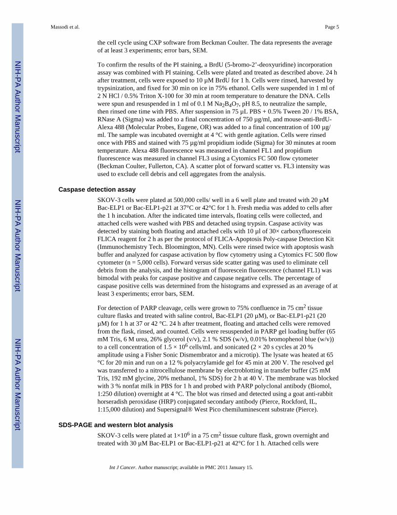
Caspase detection assaySKOV-3 cells were plated at 500,000 cells/ well in a 6 well plate and treated with 20 µMBac-ELP1 or Bac-ELP1-p21 at 37°C or 42°C for 1 h. Fresh media was added to cells afterthe 1 h incubation. After the indicated time intervals, floating cells were collected, andattached cells were washed with PBS and detached using trypsin. Caspase activity wasdetected by staining both floating and attached cells with 10 µl of 30× carboxyfluoresceinFLICA reagent for 2 h as per the protocol of FLICA-Apoptosis Poly-caspase Detection Kit(Immunochemistry Tech. Bloomington, MN). Cells were rinsed twice with apoptosis washbuffer and analyzed for caspase activation by flow cytometry using a Cytomics FC 500 flowcytometer (n = 5,000 cells). Forward versus side scatter gating was used to eliminate celldebris from the analysis, and the histogram of fluorescein fluorescence (channel FL1) wasbimodal with peaks for caspase positive and caspase negative cells. The percentage ofcaspase positive cells was determined from the histograms and expressed as an average of atleast 3 experiments; error bars, SEM.
For detection of PARP cleavage, cells were grown to 75% confluence in 75 cm2 tissueculture flasks and treated with saline control, Bac-ELP1 (20 µM), or Bac-ELP1-p21 (20µM) for 1 h at 37 or 42 °C. 24 h after treatment, floating and attached cells were removedfrom the flask, rinsed, and counted. Cells were resuspended in PARP gel loading buffer (65mM Tris, 6 M urea, 26% glycerol (v/v), 2.1 % SDS (w/v), 0.01% bromophenol blue (w/v))to a cell concentration of 1.5 × 10⁶ cells/mL and sonicated (2 × 20 s cycles at 20 %amplitude using a Fisher Sonic Dismembrator and a microtip). The lysate was heated at 65°C for 20 min and run on a 12 % polyacrylamide gel for 45 min at 200 V. The resolved gelwas transferred to a nitrocellulose membrane by electroblotting in transfer buffer (25 mMTris, 192 mM glycine, 20% methanol, 1% SDS) for 2 h at 40 V. The membrane was blockedwith 3 % nonfat milk in PBS for 1 h and probed with PARP polyclonal antibody (Biomol,1:250 dilution) overnight at 4 °C. The blot was rinsed and detected using a goat anti-rabbithorseradish peroxidase (HRP) conjugated secondary antibody (Pierce, Rockford, IL,1:15,000 dilution) and Supersignal® West Pico chemiluminescent substrate (Pierce).
SDS-PAGE and western blot analysisSKOV-3 cells were plated at 1×10⁶ in a 75 cm2 tissue culture flask, grown overnight andtreated with 30 µM Bac-ELP1 or Bac-ELP1-p21 at 42°C for 1 h. Attached cells were
Massodi et al. Page 5
Int J Cancer. Author manuscript; available in PMC 2011 January 15.
NIH
-PA Author Manuscript
NIH
-PA Author Manuscript
NIH
-PA Author Manuscript
washed with PBS and harvested using trypsin 24 h after treatment. The cells were lysed byflash freezing the pellet in liquid nitrogen, followed by grinding with a pestle in 200 µl lysisbuffer (PBS + 0.25% Triton X-100). This lysed cell solution was further rotated for 1 h at4°C. The cell lysate was cleared by centrifugation (13,000 × g for 2 min) and the totalprotein concentration was determined by a Bradford Assay. Equal amounts of total proteinfrom each lysate were loaded on a 12% polyacrylamide gel. Electrophoresis and transferwere carried out as described above. The membrane was blocked with 3 % nonfat milk inPBS for 1 h and probed with rabbit polyclonal antibodies against the Rb or pRb proteins(Molecular Probes, 1:250 dilution) overnight at 4°C. The blot was rinsed and detected usinga goat anti-rabbit horseradish peroxidase (HRP) conjugated secondary antibody andSupersignal® West Pico chemiluminescent substrate as described above. Equal loading wasinsured by reprobing the blot with mouse anti β-tubulin (Chemicon International, Temecula,CA, 1:10,000 dilution).
ResultsDesign and thermal properties of polypeptide
The drug carriers used in this study are based on elastin-like polypeptide (ELP), amacromolecular biopolymer composed of the pentapeptide repeat VPGXG, where “X” is aguest amino acid. ELPs are thermally responsive polypeptides that undergo an inversetemperature phase transition when the temperature is raised above its characteristic Tt. Twovariants of ELP were used in this study, ELP1 and ELP2. ELP1 consists of 150 pentapeptiderepeats with the guest position comprised of the amino acids V, G, and A in a 5:3:2 ratio.The group of polypeptides based on the ELP1 sequence has a Tt∼ 39–41°C. ELP2polypeptides contain 160 VPGXG repeats, where X is represented by V, G, and A in a 1:7:8ratio. ELP2-containing polypeptides are used as controls for the effect of hyperthermia alonesince they have a Tt ∼ 65 °C and therefore do not undergo a temperature transition at thehyperthermia temperature
To facilitate the entry of the ELP polypeptide across the cell membrane, ELP was fused tothe cell penetrating peptide Bac, a 24 amino acid peptide of the bactenecin family. Bac hasbeen shown to deliver cargo inside cells 32, 39, and we have used Bac to delivery ELP tothe nucleus 33. The coding sequence of ELP was further modified with an inhibitory peptidederived from p21waf1/CIP, which is the C-terminal part of the full length p21 proteinspanning amino acids 139–164.34 This C-terminal p21 fragment has been shown to exhibitcyclin-CDK inhibitory activity and also inhibit human cancer cell proliferation.16, 34, 40
Since these modifications of the ELP sequence often result in a change in the ELP Tt, wemeasured the Tt of Bac-ELP1-p21 and Bac-ELP2-p21. As the temperature of the ELPsolution is increased above its Tt, there is a structural rearrangement of the protein whichresults in the formation of aggregates. When the temperature is lowered below the Tt, theseaggregates re-solubilize. Formation of these aggregates in response to an increasingtemperature causes an increase in the turbidity of the solution. This increase in turbiditycorrelates with a scattering of light, which is used to characterize the phase transitionbehavior of these polypeptides. Figure 1A shows the turbidity of 10 µM Bac-ELP1-p21 andBac-ELP2-p21 polypeptide solutions as a function of temperature. Solutions of bothpolypeptides are clear below the physiological temperature, but as the temperature isincreased above 41°C, there is a sharp increase in the turbidity of Bac-ELP1-p21 which isreflected by the steep transition curve. At this concentration in PBS, Bac-ELP1-p21 has a Ttof 43 °C. Alternatively, the Tt of Bac-ELP2-p21 is 69 °C. Thus, Bac-ELP2-p21 and ELP2containing constructs serve as a useful control to differentiate the effect of hyperthermiafrom the effect of aggregated Bac-ELP1-p21 polypeptide. The transition of each polypeptide
Massodi et al. Page 6
Int J Cancer. Author manuscript; available in PMC 2011 January 15.
NIH
-PA Author Manuscript
NIH
-PA Author Manuscript
NIH
-PA Author Manuscript
was reversible (data not shown), which is consistent with previously reported thermalproperties of ELPs.19
The Tt of ELP is also dependent on the ELP concentration, as well as the concentration ofother co-solutes. Therefore, in order to determine what concentration range of ELP is usefulfor thermal targeting under cell culture conditions, we determined the Tt of a range ofconcentrations of Bac-ELP1-p21 in cell culture media containing 10% FBS. As shown inFigure 1B, the Tt is inversely related to the polypeptide concentration. When Tt is plottedversus polypeptide concentration, the resulting curve can be fit with a logarithmic equation.Using this fitting method and extrapolation (Figure 1C), we determined that the usefulwindow for Bac-ELP1-p21 thermal targeting (the concentration range in which the Tt liesbetween 37 and 42 °C) is between 12 µM and 52 µM.
The Tt of the rhodamine labeled Bac-ELP1-p21 was also determined and was within 2 °C ofthe unlabeled protein, insuring that the rhodamine label does not interfere with theaggregation of the polypeptide (data not shown).
Intracellular localization of Bac-ELP1-p21 by confocal microscopyConfocal microscopy of rhodamine-labeled Bac-ELP-p21 was used to confirm that the BacCPP was capable of delivering the polypeptide to the cell interior and to determine thepolypeptide’s subcellular localization. SKOV-3 cells were exposed to rhodamine labeledBac-ELP1-p21 (20 µM) for 1 h at 37°C or 42°C, and cells were stained with a tubulinantibody to mark the cytoplasm and imaged either immediately or 24 h after exposure to thepolypeptides. Images taken immediately after the 1 h polypeptide exposure showedpredominantly membrane and cytoplasmic staining (Figure 2A), and cells treated at 42 °Chad large polypeptide aggregates on the membrane and in the cytoplasm. Images taken 24 hlater showed both cytoplasmic and nuclear staining in heated and unheated cells (Figure2B). This observation is consistent with the previous study 33, in which Bac was shown todeliver ELP to the nucleus in a percentage of the cells, and the percentage of staining nucleiincreased with polypeptide concentration and time after exposure. To confirm that theobserved localization is not the product of a fixation artifact, confocal images of live cellswere also collected 24 h after polypeptide treatment. As shown in Figure 2C, the localizationin live cells agrees quite wiell with the observed localization in fixed cells.
Effect of polypeptides on cell proliferationIn order to examine the effect of polypeptides on cell proliferation, SKOV-3 cells weretreated with Bac-ELP1-p21 or Bac-ELP2-p21 for 1 h at 37°C or 42°C in a concentrationdependent manner. As shown in Figure 3A, treatment of SKOV-3 cells with Bac-ELP1-p21at 37°C did not have any effect at 10 and 20 µM, and only a slight effect at 30 µM.However, treatment of the cells with 20 and 30 µM of the same polypeptide at 42°C resultedin nearly 50% and 60% inhibition of cell proliferation, respectively. Similarly, SKOV-3cells treated for 1 h at 37 or 42 °C with different concentrations of polypeptides andanalyzed after 6 days showed an even greater inhibition of proliferation in heated samples ascompared to unheated samples (Figure 3B). We used the same 6 day assay to determine theeffects of control polypeptides (20 µM) lacking either the Bac CPP, the p21 peptide, or theELP polypeptide, and the Bac-ELP2-p21 non-thermally responsive control. As shown inFigure 3C, there was no effect on the cell proliferation on treatment with controlpolypeptides Bac-ELP1 (without the p21 inhibitory peptide), ELP1-p21 (without the BacCPP), Bac-ELP2-p21 (does not aggregate in response to hyperthermia at 42°C) or p21peptide at 37°C or 42°C. Hyperthermia itself also did not have any effect on the cellproliferation as untreated cells at 37°C or 42°C showed no difference in cell survival. Inorder to support the MTS proliferation assay, the proliferation rate of SKOV-3 cells was
Massodi et al. Page 7
Int J Cancer. Author manuscript; available in PMC 2011 January 15.
NIH
-PA Author Manuscript
NIH
-PA Author Manuscript
NIH
-PA Author Manuscript
measured using daily cell counts following treatment with Bac-ELP1-p21 or Bac-ELP1control at 20 µM for 1 h at 37 °C or 42 °C. Untreated and Bac-ELP1 treated cells showedapproximately 3 doublings during the 6 day experiment. Bac-ELP1-p21 treated cells at 37°Cshowed a proliferation pattern similar to control and untreated cells, whereas Bac-ELP1-p21treated cells combined with hyperthermia showed less than one doubling at the end of day 6(Figure 3D). These results suggest that the inhibition of cell proliferation by Bac-ELP1-p21in combination with hyperthermia is primarily due to its phase transition and nothyperthermia itself.
Effect of Bac-ELP1-p21 on cell cycle distributionPrevious studies have shown that p21 inhibits the growth of cells by interacting mainly withcyclinD/ E-CDK2/ 4 and negatively modulates cell cycle progression.41 Therefore, weexamined the effect of 20 µM Bac-ELP1-p21 treatment on the cell cycle in heated and non-heated cells. As shown in Figure 4, there was an increase in S-phase and G2/M-phase cellsat the expense of G1 phase cells in heated cells harvested 24 h (Figure 4A) and 48 h (Figure4B) after treatment, whereas no difference was observed in the distribution of the cell cyclephases in non-heated cells. Also, no effect was seen with the control polypeptide Bac-ELP1,which suggests that the ability to arrest the cell cycle is due to the p21 peptide moiety of thethermally delivered Bac-ELP1-p21 polypeptide.
In order to confirm these results and obtain a more accurate estimation of the fraction ofcells in S phase, the experiment was repeated using BrdU incorporation to mark the cells inS phase during a short BrdU pulse. As shown in Figure 4C, this assay confirms the resultsobtained by simple PI staining. Hyperthermia alone had no effect on the cell cycle, and thedistribution determined by the BrdU incorporation assay agreed will with the PI assay. Bac-ELP1-p21 treatment at 42 °C caused an increase in the number of cells in S phase and in G2/M, which was also in agreement with the PI results. The 37 °C treatments (not shown) alsoagreed with the results of the PI assay.
Western blot analysis of Rb and pRB proteinsOne of the important events in the cell cycle progression is hyperphosphorylation of theretinoblastoma protein.42 The p21 peptide has been shown to prevent the cyclin/CDK-induced hyperphosphorylation of the retinoblastoma protein Rb.15 Therefore, we measuredthe effect of polypeptide treatment on the hypo-phophorylated (Rb) and hyper-phosphorylated (pRb) levels at 42°C. As shown in Figure 5, 24 h after treatment with Bac-ELP1-p21 for 1 h at 42°C, cells showed a significant decrease in the pRb levels as comparedto untreated or Bac-ELP1 treated cells. The total Rb level was unchanged, and β-tubulinblotting was used to confirm accurate gel loading. These results suggest that Bac-ELP1-p21most likely blocks the cell cycle by inhibiting the phosphorylation of the Rb protein.
Induction of caspase activation by Bac-ELP1-p21Caspases play an important role in the apoptosis mediated cell death and have been shown tobe activated following cell cycle arrest by p21Waf1/Cip1.43 In order to determine if caspase-mediated apoptosis is responsible for cell death following cell cycle arrest by the Bac-ELP1-p21 inhibitory polypeptide, cells were treated for 1 h at 37 or 42°C with the indicatedpolypeptides. Cells were harvested at various time intervals to check for poly-caspaseactivity by incubation with a fluorescent-caspase inhibitor and flow cytometry analysis. Ahistogram of fluorescence intensity versus cell number showed two peaks. Cells under thehigh intensity peak were quantified as cells with active caspases, whereas cells with lowintensity represented caspase inactive cells. There was no effect of hyperthermia treatmentalone on caspase activity. Also, treatment with the control polypeptide lacking the p21peptide at both temperatures showed no caspase activity. As shown in Figure 6A, there was
Massodi et al. Page 8
Int J Cancer. Author manuscript; available in PMC 2011 January 15.
NIH
-PA Author Manuscript
NIH
-PA Author Manuscript
NIH
-PA Author Manuscript
a significant increase in the number of caspase positive cells on treatment with Bac-ELP1-p21 at 42°C. In addition, the percentage of cells with active caspases increased with time upto 72 h after Bac-ELP1-p21 and hyperthermia treatment. Only a slight increase in caspasepositive cells was observed in non-heated cells treated with Bac-ELP1-p21.
In order to confirm that Bac-ELP1-p21 was inducing caspase activity, we examined Bac-ELP1-p21 treated cells for cleavage of poly (ADP-ribose) polymerase (PARP), which iscleaved by caspase 3 from a 116 kDa form to an 85 kDa form. SKOV-3 cells were treatedwith 20 µM Bac-ELP1 or Bac-ELP1-p21 for 1 h at 37 or 42 °C. 24 h after treatment, cellswere harvested, and PARP cleavage was detected by Western blotting. As shown in Figure6B, there was minimal PARP cleavage in untreated or Bac-ELP1 treated cells at eithertreatment temperature. In contrast, about 50% of the cellular PARP was found in the cleavedform 24 h after 37 °C exposure to Bac-ELP1-p21, and, 24 h after 42 °C exposure, nearly allthe PARP existed in the cleaved 85 kDa form. These results support the flow cytometryassay and indicate that caspase-mediated apoptosis is a downstream effect of treatment withthe Bac-ELP1-p21 polypeptide.
Ability of Bac-ELP-p21 to inhibit the proliferation of other cell typesThis study was carried out using SKOV-3 cells because their lack of p53 expression makesthem an ideal model for human cancers in which p53 expression is lost. However, we alsowished to determine the ability of Bac-ELP1-p21 to inhibit the proliferation of other celltypes with varying p53 status. We repeated the 3 day cell proliferation assay describedabove with MCF-7 breast cancer cells (wild type p53) and Panc-1 pancreatic cancer cells(mutant (R273H) p53). As shown in Figure 7, 37 °C treatment with Bac-ELP1-p21 had littleeffect on either cell line. However, when Bac-ELP1-p21 treatment was combined withhyperthermia, proliferation of both cell lines was greatly inhibited. Furthermore, the potencyof Bac-ELP1-p21 is similar in both MCF-7 and Panc-1, and slightly more potent in thesecell lines than in SKOV-3. These results confirm that the p21 peptide acts downstream ofp53, and inhibition by this peptide is not dependent on the cell’s p53 status.
DiscussionPrevious in vivo studies have suggested that ELPs have potential as macromolecular carriersfor a variety of therapeutic molecules or peptides that can be conjugated to thesepolypeptides.36, 37 We have previously taken advantage of the fact that the ELP polymer isgenetically encoded and have introduced a therapeutic peptide which inhibits c-Myctranscriptional function and proliferation of MCF-7 breast cancer cells.37 We have alsoshown that CPP mediated intracellular delivery of ELP fused to the p21 peptide was capableof inhibiting cancer cell proliferation.29 The present study extends the pilot work with thep21 peptide by adding the Bac CPP, and demonstrating the ability of Bac-ELP-p21 to targetthe nucleus and inhibit proliferation by arresting the cell cycle and inducing apoptosis in atemperature dependent manner.
We report here that Bac-ELP1-p21 treatment at 37°C had no effect on human ovarian cancerSKOV-3 cell proliferation. However, application of hyperthermia caused a concentrationdependent decrease in the cell proliferation (Figure 3A & 3B). When hyperthermia isapplied with thermally unresponsive ELP2 based constructs (Tt > 60 °C), there was no effecton cell proliferation. This suggests that the inhibitory effect of Bac-ELP1-p21 is greatlyenhanced by the phase transition of ELP1. This is consistent with previous studies wherethermally responsive ELP has been used to deliver different therapeutics like a c-Myctranscriptional inhibitory peptide (H1-S6A,F8A) 37, 44 and doxorubicin 31, 45. The authorswere able to show an enhanced antiproliferative effect of Pen-ELP-H1 and Tat-ELP1-GFLG-Dox when treatment with each polypeptide was combined with hyperthermia in
Massodi et al. Page 9
Int J Cancer. Author manuscript; available in PMC 2011 January 15.
NIH
-PA Author Manuscript
NIH
-PA Author Manuscript
NIH
-PA Author Manuscript
different cell lines.31, 37, 44 Similar to the findings of the current study, the inhibition ofcell proliferation observed in the previous studies was enhanced greatly by the hyperthermiainduced phase transition of the ELP1 moiety, and not due to hyperthermia itself. The reasonfor this enhancement is that hyperthermia treatment induced ELP aggregation in the cellculture media. These aggregates bind to the cell surface, and are eventually internalized byendocytosis. Once the hyperthermia is removed, the polypeptides are free to re-dissolve anddiffuse to their intracellular site of action. Therefore, the role of the hyperthermia treatmentis simply to increase the amount of the polypeptide that is delivered into the cell 31, 33, 37.
One of the limitations of therapeutic macromolecular carriers is their poor penetrationcapability through cellular plasma membranes. To overcome this limitation we fused ELP tothe short cell penetrating peptide Bac. Cell penetrating peptides have been shown to delivervarious cargos across the cell membrane.27, 28 In our previous study, we were able to fusevarious CPPs to ELP and enhance the uptake of ELP inside the cells.29, 30 Since themolecular site of action of p21 is in the nucleus, in order to achieve its inhibitory effect oncell proliferation it is necessary to deliver the p21 mimicking peptide to the nuclearcompartment. In a previous study, we showed that the Bac CPP can deliver the ELPpolypeptide to the nucleus in a significant percentage of the cell population.33 Here, weshow that Bac can be used as a transduction domain to efficiently translocate the ELPmacromolecular carrier fused to the p21 mimetic peptide across the plasma membrane andinto the nuclear compartment. The polypeptide ELP1-p21 lacking the Bac transductiondomain did not show any cytotoxicity at either the control or hyperthermia temperatures(Figure 3C).
In previous studies, it has been shown that various p21 peptide fragments arrest cells in theG1and G2 stages, resulting in an inhibition of cell proliferation.15, 46, 47 In this work, wehave shown that Bac-ELP1-p21 arrests cells predominantly in the S-phase and the G2/M-phase (Figure 4). This cell cycle arrest may be due to the interaction of p21 with various cellcycle regulators, like cyclins and CDKs, that inhibits their complex formation.34 One of theregulators of the cell cycle progression is the Retinoblastoma (Rb) protein.42 In rapidlyproliferating cells, Rb is usually in the hyperphoshorylated form (pRb), thereby leaving thetranscription factor E2F free to activate transcription. Hypophosporylated Rb binds E2F,prevents transcription activation, and causes cell cycle arrest. Our results indicated that therewas a decrease in the pRb levels after treatment with Bac-ELP1-p21 in heated cells, withoutany significant change in Rb levels (Figure 5). Similarly, Ball et al. reported a decrease inhyperphosphorylated Rb protein on treatment with the p21 (141–160) peptide fused to theAntp cell penetrating peptide in human-keratinocyte-derived HaCaT cells.15 These resultssuggest that the mechanism of cell cycle arrest by Bac-ELP1-p21 is most likely mediated byinhibition of Rb phospohorylation.
In an attempt to determine the biological effect Bac-ELP1-p21, we investigated its role inmediating apoptosis. The role of p21 in apoptosis is controversial. While some studiessuggest that it prevents apoptosis, other studies demonstrate that it actually plays a role inapoptosis.48 Caspases play an important role in mediating apoptosis. Therefore, in thisstudy, we investigated the effect of Bac-ELP1-p21 on caspase activity because p21Waf1/Cip1
has been reported to be involved in the activation of caspases.43 As shown in Figure 6, anincrease in poly-caspase activity was observed in heated cells which had been treated withBac-ELP1-p21 for 1 h, and these results were confirmed with a PARP cleavage assay. Theseresults suggest that the observed inhibition of cell proliferation by Bac-ELP1-p21 may be aresult of apoptotic cell death through the activation of caspases. This observation isconsistent with the study of Baker et al., who reported that the C-terminus p21 conjugated toa Tat peptide was able to enter the nucleus of U251 cells and cause apoptosis, as visualizedby TUNEL staining. The authors were able to show the co-localization of Tat-p21 with
Massodi et al. Page 10
Int J Cancer. Author manuscript; available in PMC 2011 January 15.
NIH
-PA Author Manuscript
NIH
-PA Author Manuscript
NIH
-PA Author Manuscript
PCNA and therefore suggested that interaction of p21 with PCNA interferes with PCNA’sfunction, leading to apoptosis.49
This study provides further evidence that ELP is an effective vector for targeted delivery oftherapeutics. As shown here, the role of ELP is not limited to the delivery of oncogenicinhibitory peptides and conventional chemotherapeutic drugs, but it may also be used for thedelivery of cell cycle inhibitory peptides. In order show that Bac-ELP1-p21 mediatedcytotoxicity is not dependent on the p53 status of the cell line, we expanded our cell linescreen beyond the p53 null cell line SKOV-3. It was observed that Bac-ELP1-p21 treatmentfor 1 h at 42°C resulted in a significant inhibition of proliferation in a MCF-7 breastcarcinoma cell line (p53 wild type) and a p53 mutated Panc-1 cell line.
In the present study, we have shown that Bac-ELP1-p21 is a promising thermally responsivemacromolecular carrier which has the potential to be thermally targeted to the tumor site.Since no effect was seen at normal physiological temperature with Bac-ELP1-p21, it is veryunlikely that it will cause any adverse effects in non heated tissues. Another advantageassociated with macromolecular ELP is that it can overcome multidrug resistance.31 Its useas a vector for the delivery of therapeutic drugs and peptides is attractive because, apartfrom the passive targeting due to the macromolecular nature of ELP, it can be activelytargeted to the tumor site by application of heat. By attaching different CPPs to ELP it canalso be targeted specifically to either the cytoplasmic compartment or to the nucleus.Because it is genetically encoded, ELP may be easily applied as a targeted macromolecularcarrier for other inhibitors. Further in vivo studies of tumor reduction efficacy andpolypeptide-induced toxicity and immunogenicity are necessary to advance this technologytoward clinical evaluation, and these studies are currently underway.
In conclusion, the results presented here demonstrate that genetically engineered ELP, whichwas modified with the Bac CPP and a p21 mimetic peptide, inhibited the proliferation ofSKOV-3 cells in combination with hyperthermia. This Bac-ELP1-p21 polypeptide has atransition temperature in the range of 39–42°C, making it a very good candidate for thermaltargeting. We found that Bac-ELP1-p21 treatment at 42°C caused inhibition of Rbphosphorylation, arrest of the cell cycle, and activation of caspases. Such thermallyresponsive macromolecular biopolymers with antiproliferative activity would have greatpotential in cancer therapy for the thermally targeted treatment of solid tumors.
Abbreviations
CPP cell penetrating peptide
Bac CPP from bactenecin
EPR enhanced permeability and retention
Tat CPP from HIV-1 transcription factor
ELP Elastin-like polypeptide
PBS phosphate buffered saline
Rb retinoblastoma protein
Tt transition temperature
AcknowledgmentsWe wish to thank Ms. Rowshan Begum and Ms. Leslie Robinson for technical assistance and critical reading of themanuscript.
Massodi et al. Page 11
Int J Cancer. Author manuscript; available in PMC 2011 January 15.
NIH
-PA Author Manuscript
NIH
-PA Author Manuscript
NIH
-PA Author Manuscript
Grant sponsor: This study was supported by the National Institute of Health R21 CA113813-01A2 and Wendy WillCase Foundation grant.
References1. Allen TM. Liposomal drug formulation: rationale for development and what we can expect in the
future. Drugs. 1998; 56:747–756. [PubMed: 9829150]2. El-Aneed A. An overview of current delivery systems in cancer gene therapy. J Control Release.
2004; 94:1–14. [PubMed: 14684267]3. Torchilin VP. Polymer-coated long-circulating microparticulate pharmaceuticals. J.Microencapsul.
1998; 15:1–19. [PubMed: 9463803]4. Langer R. Drug delivery and targeting. Nature (Lond.). 1998; 392 Suppl:5–10. [PubMed: 9579855]5. Duncan R, Vicent MJ, Greco F, Nicholson RI. Polymer-drug conjugates: towards a novel approach
for the treatment of endrocine-related cancer. Endocr Relat Cancer. 2005; 12 Suppl 1:S189–S199.[PubMed: 16113096]
6. Kopecek J, Kopeckova P, Minko T, Lu Z. HPMA copolymer-anticancer drug conjugates: design,activity, and mechanism of action. Eur J Pharm Biopharm. 2000; 50:61–81. [PubMed: 10840193]
7. Peer D, Karp JM, Hong S, Farokhzad OC, Margalit R, Langer R. Nanocarriers as an emergingplatform for cancer therapy. Nat Nanotechnol. 2007; 2:751–760. [PubMed: 18654426]
8. Duncan R. Polymer conjugates as anticancer nanomedicines. Nat Rev Cancer. 2006; 6:688–701.[PubMed: 16900224]
9. Takakura Y, Fujita T, Hashida M, Sezaki H. Disposition characteristics of macromolecules intumor-bearing mice. Pharm. Res. 1990; 7:339–346. [PubMed: 1694582]
10. Maeda H, Seymour LW, Miyamoto Y. Conjugates of anticancer agents and polymers: advantagesof macromolecular therapeutics in vivo. Bioconjug.Chem. 1992; 3:351–362. [PubMed: 1420435]
11. Cassidy J, Duncan R, Morrison GJ, Strohalm J, Plocova D, Kopecek J, Kaye SB. Activity of N-(2-hydroxypropyl)methacrylamide copolymers containing daunomycin against a rat tumour model.Biochem. Pharmacol. 1989; 38:875–879. [PubMed: 2930589]
12. Yamaoka T, Tabata Y, Ikada Y. Distribution and tissue uptake of poly(ethylene glycol) withdifferent molecular weights after intravenous administration to mice. J. Pharm.Sci. 1994; 83:601–606. [PubMed: 8046623]
13. Gius DR, Ezhevsky SA, Becker-Hapak M, Nagahara H, Wei MC, Dowdy SF. Transducedp16INK4a peptides inhibit hypophosphorylation of the retinoblastoma protein and cell cycleprogression prior to activation of Cdk2 complexes in late G1. Cancer Res. 1999; 59:2577–2580.[PubMed: 10363976]
14. Hosotani R, Miyamoto Y, Fujimoto K, Doi R, Otaka A, Fujii N, Imamura M. Trojan p16 peptidesuppresses pancreatic cancer growth and prolongs survival in mice. Clin Cancer Res. 2002;8:1271–1276. [PubMed: 11948142]
15. Ball KL, Lain S, Fahraeus R, Smythe C, Lane DP. Cell-cycle arrest and inhibition of Cdk4 activityby small peptides based on the carboxy-terminal domain of p21WAF1. Curr Biol. 1997; 7:71–80.[PubMed: 8999999]
16. Bonfanti M, Taverna S, Salmona M, D'Incalci M, Broggini M. p21WAF1-derived peptides linkedto an internalization peptide inhibit human cancer cell growth. Cancer Res. 1997; 57:1442–1446.[PubMed: 9108443]
17. Urry DW, Luan C-H, Parker TM, Gowda DC, Prasad KU, Reid MC, Safavy A. Temperature ofPolypeptide Inverse Temperature Transition Depends on Mean Residue Hydrophobicity. J. Am.Chem. Soc. 1991; 113:4346–4348.
18. Tatham AS, Shewry PR. Elastomeric proteins: biological roles, structures and mechanisms. TrendsBiochem Sci. 2000; 25:567–571. [PubMed: 11084370]
19. Meyer DE, Kong GA, Dewhirst MW, Zalutsky MR, Chilkoti A. Targeting a GeneticallyEngineered Elastin-like Polypeptide to Solid Tumors by Local Hyperthermia. Cancer Res. 2001;61:1548–1554. [PubMed: 11245464]
Massodi et al. Page 12
Int J Cancer. Author manuscript; available in PMC 2011 January 15.
NIH
-PA Author Manuscript
NIH
-PA Author Manuscript
NIH
-PA Author Manuscript
20. Liu W, Dreher MR, Furgeson DY, Peixoto KV, Yuan H, Zalutsky MR, Chilkoti A. Tumoraccumulation, degradation and pharmacokinetics of elastin-like polypeptides in nude mice. JControl Release. 2006; 116:170–178. [PubMed: 16919353]
21. Dreher MR, Liu W, Michelich CR, Dewhirst MW, Chilkoti A. Thermal cycling enhances theaccumulation of a temperature-sensitive biopolymer in solid tumors. Cancer Res. 2007; 67:4418–4424. [PubMed: 17483356]
22. Fujiwara K, Watanabe T. Effects of hyperthermia, radiotherapy and thermoradiotherapy on tumormicrovascular permeability. Acta Pathol Jpn. 1990; 40:79–84. [PubMed: 2160185]
23. Gerlowski LE, Jain RK. Effect of hyperthermia on microvascular permeability to macromoleculesin normal and tumor tissues. Int J Microcirc Clin Exp. 1985; 4:363–372. [PubMed: 4086191]
24. Jain RK. Transport of molecules across tumor vasculature. Cancer Metastasis Rev. 1987; 6:559–593. [PubMed: 3327633]
25. Temsamani J, Guinot P. Antisense oligonucleotides: a new therapeutic approach. Biotechnology &Applied Biochemistry. 1997; 26:65–71. [PubMed: 9357101]
26. Snyder EL, Meade BR, Saenz CC, Dowdy SF. Treatment of terminal peritoneal carcinomatosis bya transducible p53-activating peptide. PLoS Biol. 2004; 2:E36. [PubMed: 14966535]
27. Nori A, Kopecek J. Intracellular targeting of polymer-bound drugs for cancer chemotherapy. AdvDrug Deliv Rev. 2005; 57:609–636. [PubMed: 15722167]
28. Temsamani J, Vidal P. The use of cell-penetrating peptides for drug delivery. Drug Discov Today.2004; 9:1012–1019. [PubMed: 15574317]
29. Massodi I, Bidwell GL 3rd, Raucher D. Evaluation of cell penetrating peptides fused to elastin-likepolypeptide for drug delivery. J Control Release. 2005; 108:396–408. [PubMed: 16157413]
30. Massodi I, Raucher D. A thermally responsive Tat-elastin-like polypeptide fusion protein inducesmembrane leakage, apoptosis, and cell death in human breast cancer cells. J Drug Target. 2007;15:611–622. [PubMed: 17968715]
31. Bidwell GL 3rd, Fokt I, Priebe W, Raucher D. Development of elastin-like polypeptide forthermally targeted delivery of doxorubicin. Biochem Pharmacol. 2007; 73:620–631. [PubMed:17161827]
32. Sadler K, Eom KD, Yang JL, Dimitrova Y, Tam JP. Translocating proline-rich peptides from theantimicrobial peptide bactenecin 7. Biochemistry. 2002; 41:14150–14157. [PubMed: 12450378]
33. Bidwell GL 3rd, Davis AN, Raucher D. Targeting a c-Myc inhibitory polypeptide to specificintracellular compartments using cell penetrating peptides. J Control Release. 2009; 135:2–10.[PubMed: 19095020]
34. Mutoh M, Lung FD, Long YQ, Roller PP, Sikorski RS, O'Connor PM. A p21(Waf1/Cip1)carboxyl-terminal peptide exhibited cyclin-dependent kinase-inhibitory activity andcytotoxicity when introduced into human cells. Cancer Res. 1999; 59:3480–3488. [PubMed:10416614]
35. Warbrick E, Lane DP, Glover DM, Cox LS. A small peptide inhibitor of DNA replication definesthe site of interaction between the cyclin-dependent kinase inhibitor p21WAF1 and proliferatingcell nuclear antigen. Curr Biol. 1995; 5:275–282. [PubMed: 7780738]
36. Chilkoti A, Dreher MR, Meyer DE. Design of thermally responsive, recombinant polypeptidecarriers for targeted drug delivery. Adv Drug Deliv Rev. 2002; 54:1093–1111. [PubMed:12384309]
37. Bidwell GL 3rd, Raucher D. Application of thermally responsive polypeptides directed against c-Myc transcriptional function for cancer therapy. Mol Cancer Ther. 2005; 4:1076–1085. [PubMed:16020665]
38. Meyer DE, Chilkoti A. Purification of Recombinant Proteins by Fusion with ThermallyResponsive Polypeptides. Nat.Biotechnol. 1999; 17:1112–1115. [PubMed: 10545920]
39. Tomasinsig L, Skerlavaj B, Papo N, Giabbai B, Shai Y, Zanetti M. Mechanistic and functionalstudies of the interaction of a proline-rich antimicrobial peptide with mammalian cells. J BiolChem. 2006; 281:383–391. [PubMed: 16257969]
40. Mattock H, Lane DP, Warbrick E. Inhibition of cell proliferation by the PCNA-binding region ofp21 expressed as a GFP miniprotein. Exp Cell Res. 2001; 265:234–241. [PubMed: 11302688]
Massodi et al. Page 13
Int J Cancer. Author manuscript; available in PMC 2011 January 15.
NIH
-PA Author Manuscript
NIH
-PA Author Manuscript
NIH
-PA Author Manuscript
41. Brugarolas J, Moberg K, Boyd SD, Taya Y, Jacks T, Lees JA. Inhibition of cyclin-dependentkinase 2 by p21 is necessary for retinoblastoma protein-mediated G1 arrest after gamma-irradiation. Proc Natl Acad Sci U S A. 1999; 96:1002–1007. [PubMed: 9927683]
42. Giacinti C, Giordano A. RB and cell cycle progression. Oncogene. 2006; 25:5220–5227. [PubMed:16936740]
43. Huo JX, Metz SA, Li GD. p53-independent induction of p21(waf1/cip1) contributes to theactivation of caspases in GTP-depletion-induced apoptosis of insulin-secreting cells. Cell DeathDiffer. 2004; 11:99–109. [PubMed: 12970678]
44. Bidwell GL 3rd, Raucher D. Enhancing the antiproliferative effect of topoisomerase II inhibitorsusing a polypeptide inhibitor of c-Myc. Biochem Pharmacol. 2006; 71:248–256. [PubMed:16316634]
45. Bidwell GL 3rd, Davis AN, Fokt I, Priebe W, Raucher D. A thermally targeted elastin-likepolypeptide-doxorubicin conjugate overcomes drug resistance. Invest New Drugs. 2007; 25:313–326. [PubMed: 17483874]
46. Tunnemann G, Martin RM, Haupt S, Patsch C, Edenhofer F, Cardoso MC. Cargo-dependent modeof uptake and bioavailability of TAT-containing proteins and peptides in living cells. Faseb J.2006; 20:1775–1784. [PubMed: 16940149]
47. Luo Y, Hurwitz J, Massague J. Cell-cycle inhibition by independent CDK and PCNA bindingdomains in p21Cip1. Nature. 1995; 375:159–161. [PubMed: 7753174]
48. Gartel AL, Tyner AL. The role of the cyclin-dependent kinase inhibitor p21 in apoptosis. MolCancer Ther. 2002; 1:639–649. [PubMed: 12479224]
49. Baker RD, Howl J, Nicholl ID. A sychnological cell penetrating peptide mimic of p21(WAF1/CIP1) is pro-apoptogenic. Peptides. 2007; 28:731–740. [PubMed: 17287047]
Massodi et al. Page 14
Int J Cancer. Author manuscript; available in PMC 2011 January 15.
NIH
-PA Author Manuscript
NIH
-PA Author Manuscript
NIH
-PA Author Manuscript
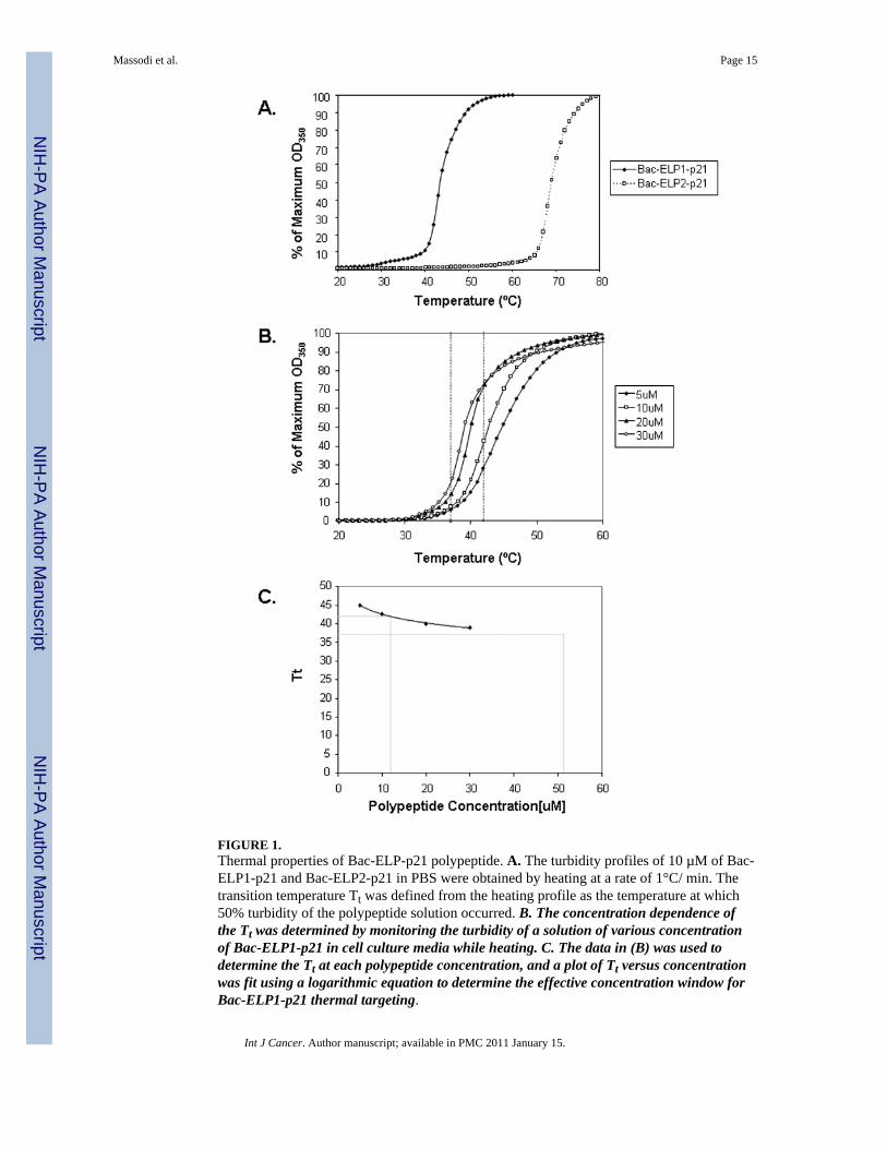
FIGURE 1.Thermal properties of Bac-ELP-p21 polypeptide. A. The turbidity profiles of 10 µM of Bac-ELP1-p21 and Bac-ELP2-p21 in PBS were obtained by heating at a rate of 1°C/ min. Thetransition temperature Tt was defined from the heating profile as the temperature at which50% turbidity of the polypeptide solution occurred. B. The concentration dependence ofthe Tt was determined by monitoring the turbidity of a solution of various concentrationof Bac-ELP1-p21 in cell culture media while heating. C. The data in (B) was used todetermine the Tt at each polypeptide concentration, and a plot of Tt versus concentrationwas fit using a logarithmic equation to determine the effective concentration window forBac-ELP1-p21 thermal targeting.
Massodi et al. Page 15
Int J Cancer. Author manuscript; available in PMC 2011 January 15.
NIH
-PA Author Manuscript
NIH
-PA Author Manuscript
NIH
-PA Author Manuscript
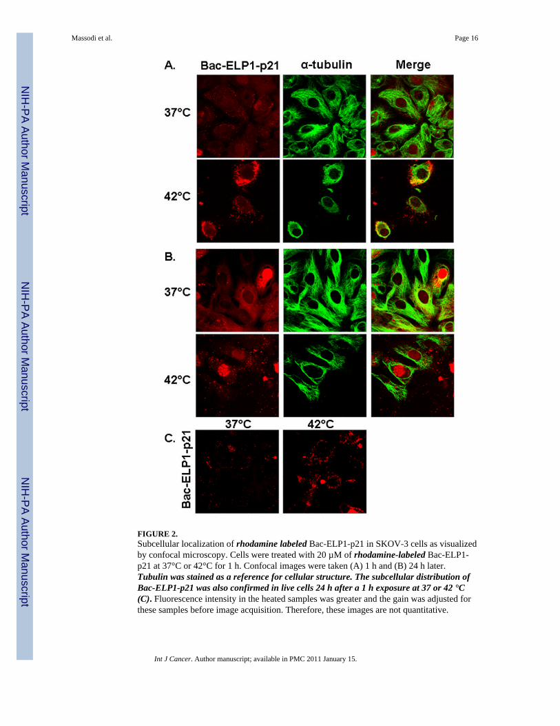
FIGURE 2.Subcellular localization of rhodamine labeled Bac-ELP1-p21 in SKOV-3 cells as visualizedby confocal microscopy. Cells were treated with 20 µM of rhodamine-labeled Bac-ELP1-p21 at 37°C or 42°C for 1 h. Confocal images were taken (A) 1 h and (B) 24 h later.Tubulin was stained as a reference for cellular structure. The subcellular distribution ofBac-ELP1-p21 was also confirmed in live cells 24 h after a 1 h exposure at 37 or 42 °C(C). Fluorescence intensity in the heated samples was greater and the gain was adjusted forthese samples before image acquisition. Therefore, these images are not quantitative.
Massodi et al. Page 16
Int J Cancer. Author manuscript; available in PMC 2011 January 15.
NIH
-PA Author Manuscript
NIH
-PA Author Manuscript
NIH
-PA Author Manuscript
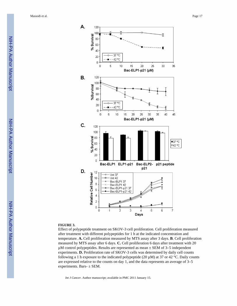
FIGURE 3.Effect of polypeptide treatment on SKOV-3 cell proliferation. Cell proliferation measuredafter treatment with different polypeptides for 1 h at the indicated concentration andtemperature. A. Cell proliferation measured by MTS assay after 3 days. B. Cell proliferationmeasured by MTS assay after 6 days. C. Cell proliferation 6 days after treatment with 20µM control polypeptides. Results are represented as mean ± SEM of 3–5 independentexperiments. D. Proliferation rate of SKOV-3 cells was determined by daily cell countsfollowing a 1 h exposure to the indicated polypeptide (20 µM) at 37 or 42 °C. Daily countsare expressed relative to the counts on day 1, and the data represents an average of 3–5experiments. Bars- ± SEM.
Massodi et al. Page 17
Int J Cancer. Author manuscript; available in PMC 2011 January 15.
NIH
-PA Author Manuscript
NIH
-PA Author Manuscript
NIH
-PA Author Manuscript
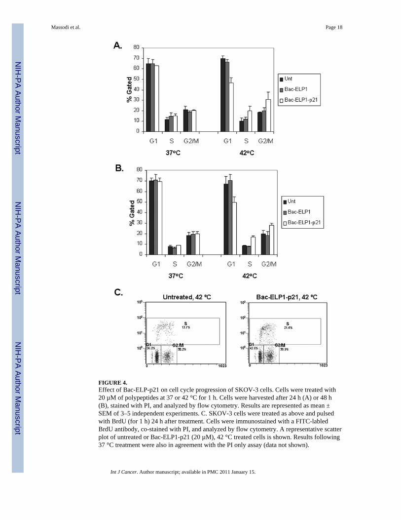
FIGURE 4.Effect of Bac-ELP-p21 on cell cycle progression of SKOV-3 cells. Cells were treated with20 µM of polypeptides at 37 or 42 °C for 1 h. Cells were harvested after 24 h (A) or 48 h(B), stained with PI, and analyzed by flow cytometry. Results are represented as mean ±SEM of 3–5 independent experiments. C. SKOV-3 cells were treated as above and pulsedwith BrdU (for 1 h) 24 h after treatment. Cells were immunostained with a FITC-labledBrdU antibody, co-stained with PI, and analyzed by flow cytometry. A representative scatterplot of untreated or Bac-ELP1-p21 (20 µM), 42 °C treated cells is shown. Results following37 °C treatment were also in agreement with the PI only assay (data not shown).
Massodi et al. Page 18
Int J Cancer. Author manuscript; available in PMC 2011 January 15.
NIH
-PA Author Manuscript
NIH
-PA Author Manuscript
NIH
-PA Author Manuscript
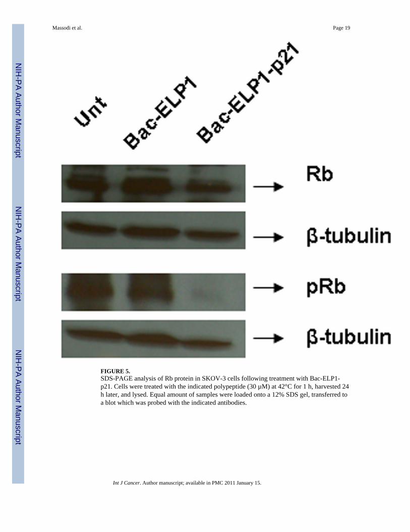
FIGURE 5.SDS-PAGE analysis of Rb protein in SKOV-3 cells following treatment with Bac-ELP1-p21. Cells were treated with the indicated polypeptide (30 µM) at 42°C for 1 h, harvested 24h later, and lysed. Equal amount of samples were loaded onto a 12% SDS gel, transferred toa blot which was probed with the indicated antibodies.
Massodi et al. Page 19
Int J Cancer. Author manuscript; available in PMC 2011 January 15.
NIH
-PA Author Manuscript
NIH
-PA Author Manuscript
NIH
-PA Author Manuscript
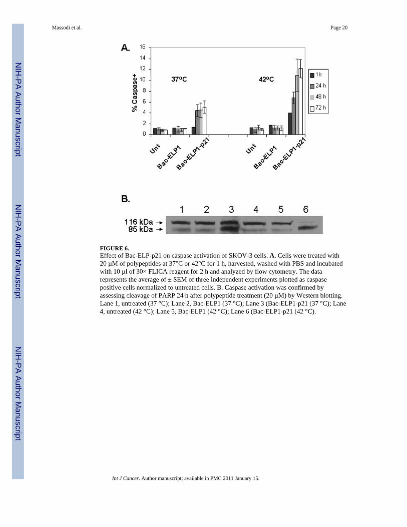
FIGURE 6.Effect of Bac-ELP-p21 on caspase activation of SKOV-3 cells. A. Cells were treated with20 µM of polypeptides at 37°C or 42°C for 1 h, harvested, washed with PBS and incubatedwith 10 µl of 30× FLICA reagent for 2 h and analyzed by flow cytometry. The datarepresents the average of ± SEM of three independent experiments plotted as caspasepositive cells normalized to untreated cells. B. Caspase activation was confirmed byassessing cleavage of PARP 24 h after polypeptide treatment (20 µM) by Western blotting.Lane 1, untreated (37 °C); Lane 2, Bac-ELP1 (37 °C); Lane 3 (Bac-ELP1-p21 (37 °C); Lane4, untreated (42 °C); Lane 5, Bac-ELP1 (42 °C); Lane 6 (Bac-ELP1-p21 (42 °C).
Massodi et al. Page 20
Int J Cancer. Author manuscript; available in PMC 2011 January 15.
NIH
-PA Author Manuscript
NIH
-PA Author Manuscript
NIH
-PA Author Manuscript
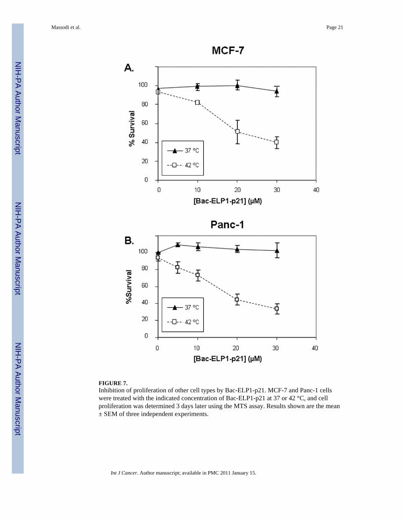
FIGURE 7.Inhibition of proliferation of other cell types by Bac-ELP1-p21. MCF-7 and Panc-1 cellswere treated with the indicated concentration of Bac-ELP1-p21 at 37 or 42 °C, and cellproliferation was determined 3 days later using the MTS assay. Results shown are the mean± SEM of three independent experiments.
Massodi et al. Page 21
Int J Cancer. Author manuscript; available in PMC 2011 January 15.
NIH
-PA Author Manuscript
NIH
-PA Author Manuscript
NIH
-PA Author Manuscript


