Embed Size (px)
Citation preview

Inhibitory Antibodies to Human Angiotensin-Converting Enzyme: Fine EpitopeMapping and Mechanism of Action†
Olga E. Skirgello,‡,§ Irina V. Balyasnikova,§,| Petr V. Binevski,‡ Zhu-Li Sun,| Igor I. Baskin,‡ Vladimir A. Palyulin,‡
Andrei B. Nesterovitch,⊥ Ronald F. Albrecht, II,| Olga A. Kost,‡,# and Sergei M. Danilov*,|,#
Department of Chemistry, Moscow State UniVersity, Russia, Department of Anesthesiology, UniVersity of Illinois at Chicago,Chicago, Illinois 60612, and Department of Orthopedic Surgery, Rush UniVersity Medical Center, Chicago, Illinois 60612
ReceiVed December 20, 2005; ReVised Manuscript ReceiVed February 16, 2006
ABSTRACT: Angiotensin I-converting enzyme (ACE), a key enzyme in cardiovascular pathophysiology,consists of two homologous domains (N and C), each bearing a Zn-dependent active site. We modeledthe 3D-structure of the ACE N-domain using known structures of the C-domain of human ACE and theACE homologue, ACE2, as templates. Two monoclonal antibodies (mAb), 3A5 and i2H5, developedagainst the human N-domain of ACE, demonstrated anticatalytic activity. N-domain modeling andmutagenesis of 21 amino acid residues allowed us to define the epitopes for these mAbs. Their epitopespartially overlap: amino acid residues K407, E403, Y521, E522, G523, P524, D529 are present in bothepitopes. Mutation of 4 amino acid residues within the 3A5 epitope, N203E, R550A, D558L, and K557Q,increased the apparent binding of mAb 3A5 with the mutated N-domain 3-fold in plate precipitationassay, but abolished the inhibitory potency of this mAb. Moreover, mutation D558L dramatically decreased3A5-induced ACE shedding from the surface of CHO cells expressing human somatic ACE. The inhibitionof N-domain activity by mAbs 3A5 and i2H5 obeys similar kinetics. Both mAbs can bind to the freeenzyme and enzyme-substrate complex, forming E‚mAb and E‚S‚mAb complexes, respectively; however,only complex E‚S can form a product. Kinetic analysis indicates that both mAbs bind better with theACE N-domain in the presence of a substrate, which, in turn, implies that binding of a substrate causesconformational adjustments in the N-domain structure. Independent experiments with ELISA demonstratedbetter binding of mAbs 3A5 and i2H5 in the presence of the inhibitor lisinopril as well. This effect canbe attributed to better binding of both mAbs with the “closed” conformation of ACE, therefore, disturbingthe hinge-bending movement of the enzyme, which is necessary for catalysis.
Angiotensin I-converting enzyme (ACE1) (EC 3.4.15.1,CD 143) is a zinc-metallopeptidase, responsible for theformation of the vasoconstrictor angiotensin II and theinactivation of the vasodilator bradykinin. The enzyme isalso involved in neuropeptide metabolism and reproductiveand immune functions (for reviews see refs1-4). Thesomatic isoform is expressed widely at surface-fluid inter-faces and plays an important role in blood pressure regula-tion, the development of vascular pathology, and endotheliumremodeling in some disease states. ACE has been assigned
as a CD marker: CD 143 (5, 6). The testicular isoform islimited to spermatozoa and is essential for male fertility (7).
Somatic ACE consists of two homologous N- and C-domains, each having a functional active site (8). The tertiarystructure of somatic ACE is still unknown. Testicular ACEis coded by the same gene as somatic ACE but from analternative promoter (9). This ACE is identical to theC-terminal domain of somatic enzyme, except for a shortN-terminal sequence of 36 amino acids, and thus containsonly one catalytic site (10). The crystal structure of thehuman C-terminal domain was recently described (11).Somatic and testicular ACEs are both type 1 integralmembrane-anchored proteins. Somatic ACE also exists as asoluble form, e.g., in plasma, cerebrospinal fluid, and seminalplasma (see reviews1-3), that lacks the transmembranedomain responsible for membrane attachment (12-16).
The “bridge sequence” between the two ACE domains canbe cleaved both in vivo and in vitro (17-21). Different heatstabilities of the domains allow selective inactivation of theC-domain and underlie the method of acquiring an activeN-domain with inactive C-domain within a full-length ACE(19, 21). These results suggest that the two domains withinthe somatic ACE molecule are rather separate and distinctivestructures.
† This work was supported in part by Russian Foundation for BasicResearch, Grant 03-04-48821 (to O.A.K.).
* Corresponding author. Mailing address: Anesthesiology ResearchCenter, University of Illinois at Chicago, 1819 W. Polk St. (M/C 519),Chicago, IL 60612. Phone: (312) 413-7526. Fax: (312) 996-9680.E-mail: [email protected].
‡ Moscow State University.§ Equally contributed to this work.| University of Illinois at Chicago.⊥ Rush University Medical Center.# Authors share equal seniority over this paper.1 Abbreviations: ACE, angiotensin converting enzyme; Hip-His-Leu,
hippuryl-L-histidyl-L-leucine; Z-Phe-His-Leu, benzyloxycarbonyl-phe-nylalanyl-L-histidyl-L-leucine; CHO, Chinese hamster ovary; mAb,monoclonal antibody; ELISA, enzyme-linked immunosorbent assay;CD markers, cluster designation markers; PBS, phosphate-bufferedsaline; (CHAPS), 3-[(3-cholamidopropyl)-dimethylammonio]-1-propa-nosulfonate; TMB, tetramethylbenzidine.
4831Biochemistry2006,45, 4831-4847
10.1021/bi052591h CCC: $33.50 © 2006 American Chemical SocietyPublished on Web 03/22/2006

Hydrolysis of various substrates by different ACE formsand, especially, the inhibition of ACE by inhibitors have beenstudied intensively (3, 20, 22-27). Special attention was paidto the differences in the function of N- and C-domains ofACE, as well as to the development of ACE inhibitorsspecific for each domain (20, 28-33). The two catalyticcenters within somatic ACE were long considered to functionindependently (22, 24, 28, 34). However, our recent studieswith both bovine and human ACE (20, 27) revealed theexistence of strong negative cooperativity between the N-and C-domain active centers functioning in full-length ACE,which implies tight proximity of the domains within thesomatic enzyme molecule.
Development of numerous mAbs to different epitopes ofthe ACE molecule which influence ACE functions, inconjunction with epitope mapping, provides a unique op-portunity to study ACE in fine structural detail. Thus, thestudy of the effect of mAbs on ACE shedding (35) or ACEdimerization in reverse micelles (36) plus epitope mappingof functionally active antibodies allowed us to identify theregion on the N-domain that determines a low rate of somaticACE shedding from the cell surface (36). One of the mAbs,3A5, which greatly influences ACE shedding (35), alsodemonstrated strong anticatalytic activity (25).
The presence of anti-ACE autoantibodies was indicatedrecently in several diseases with an autoimmune compo-nent: lupus erythrematosis, scleroderma, and rheumatoidarthritis (35, 38). The clinical significance of the presenceof these autoantibodies is still unclear. However, we mightassume that an epitope specificity of the autoantibodies toACE for these distinct diseases could be a characteristic ofthe pathology.
In this study we identified the epitopes for two mAbs thatexhibit inhibitory activity toward the N-domain of ACE.Detailed kinetic analysis of ACE inhibition by these mAbsallowed us to reveal their mechanisms of action. Modelingof the N-domain structure along with the knowledge of thesekinetic mechanisms of inhibition allowed us to hypothesizeabout a mechanism action of the enzyme.
MATERIALS AND METHODS
Chemicals.Benzyloxycarbonyl-L-phenylalanyl-L-histidyl-L-leucine (Z-Phe-His-Leu) was from Bachem (King ofPrussia, PA). Hippuryl-L-histidyl-L-leucine (Hip-His-Leu),N-(S-1-carboxy-3-phenylpropyl)-L-lysyl-L-proline (lisinopril)and other reagents (unless otherwise indicated) were obtainedfrom Sigma (St. Louis, MO).
Expression of Human ACE Constructs in CHO Cells.Stable cell lines of CHO cells expressing wild-type humansomatic ACE (clone 2C2) or single N-domain of ACE(D629-clone 4G6) were cultured as described previously (39,40). CHO-ACE cells at confluence were washed gently withPBS and incubated 24-48 h with “complete culture medium”(Mediatech, Inc., Herndon, VA) without FBS. Culturemedium was collected as a source of soluble ACE, whereasa cell lysate prepared with 8 mM CHAPS was a source ofthe membrane-bound form of ACE (35, 39).
Purified N-domain of human ACEwas obtained by limitedproteolysis of the parent pure somatic ACE after partialdenaturation in NH4OH solution as described in ref41.N-Domain preparations proved to be homogeneous according
to SDS-PAGE electrophoresis. Stoichiometric titration ofactive molecules in solutions of N-domain of ACE wasperformed with the specific competitive inhibitor lisinoprilas described in refs27, 41.
Antibodies.Properties of a set of monoclonal antibodiesdirected to different epitopes located on the N-domain ofACE were described in detail elsewhere (25, 35-37, 39, 40).
ACE actiVity was assayed fluorimetrically with differentsubstrates, Hip-His-Leu and Z-Phe-His-Leu, as described (42,43).
Inhibitory action of anti-ACE mAbswas tested using singleN-domain as well as numerous mutants of recombinanttruncated N-domain and somatic ACE, membrane-bound andsoluble. ACEs with enzymatic activity of around 10 mU/mL (with Hip-His-Leu or Z-Phe-His-Leu) were incubatedwith different concentrations of mAbs in 10 mM phosphatebuffer saline, pH 7.4, containing 150 mM NaCl with 0.1mg/mL bovine serum albumin, for 2 h at 25°C, then 20-40 µL of the reaction mixture was added to 200µL ofsubstrate (5 mM Hip-His-Leu or 2 mM Z-Phe-His-Leu) inthe potassium phosphate buffer (100 mM), pH 8.3, containing300 mM NaCl and 80 mM ZnS04 (PBS-I), and the residualACE activity was determined.
In some experiments the reaction mixtures (475µL)containing 0.1 nM N-domain and 0-7 nM mAbs in 50 mMphosphate buffer, pH 7.5, containing 150 mM KCl and 1µM ZnCl2 (PBS-II), were incubated for 2 h at 25°C. Residualenzyme activities were then determined by adding 25µL of3.2 mM Hip-His-Leu or 2 mM Z-Phe-His-Leu and measuringthe initial rates of hydrolysis. The influence of pH on theinhibitory action of mAbs on the N-domain of ACE wasdetermined at two mAb concentrations, 0.7 nM and 3.0 nM,in 15 mM acetate-15 mM phosphate-15 mM borate buffer,containing 0.15 M KCl and 1µM ZnCl2.
Kinetic studies of the inhibitory action of mAbs 3A5 andi2H5 were performed with the N-domain of ACE obtainedby limited proteolysis of somatic enzyme. The reactionmixture containing 0.1 nM N-domain and 0-10 nM mAbsin PBS-II was incubated for 2 h at 25°C. This preincubationtime was proved to be sufficient for establishing equilibriumbetween enzyme and mAb. The N-domain of ACE was foundto be stable under these conditions. Residual ACE activitywas then determined in duplicate with different concentra-tions (50-400 µM) of Hip-His-Leu. The mode of ACEinhibition by mAbs 3A5 and i2H5, as well as the values ofthe inhibition constants, was determined by data processingin Dixon coordinates, 1/V vs [mAb], and modified Cornish-Bowden coordinates, [S]/V vs [mAb] (44, 45).
Quantification of ACE Binding by Anti-ACE mAb (PlatePrecipitation Assay and ELISA).Microtiter plates bound withgoat-anti-mouse IgG were coated with different anti-ACEmAbs and were incubated with serum-free culture mediumobtained from CHO-ACE cells (wild-type or mutants). Insome experiments mAbs were adsorbed to the wells ofmicrotiter plates directly, without the goat-anti-mouse bridge(46). In some experiments the incubation of ACEs withimmobilized mAbs was performed in the presence of ACEinhibitor lisinopril or EDTA as well.
The amount of ACE precipitated by a given mAb (whichreflects the affinity of binding) was quantified by twomethods:
4832 Biochemistry, Vol. 45, No. 15, 2006 Skirgello et al.

(i) Precipitated ACE activity was estimated in the wellsusing Hip-His-Leu or Z-Phe-His-Leu as substrate as de-scribed previously (25). The minor background hydrolysisof the substrate in the wells coated by non-immune mouseIgG was subtracted from each value obtained with specificanti-ACE mAbs.
(ii) The amount of precipitated ACE protein was quantifiedby incubation with sheep-anti-ACE polyclonal antibodiesconjugated with horseradish peroxidase from ACE ELISAkit (Chemicon Int, Temecula, CA) followed by spectropho-tometric assay with tetramethylbenzidine (TMB) as a sub-strate at 450 or 620 nm.
Quantification of Anti-ACE mAb Binding by the N-Domainof ACE (Direct ELISA).Microtiter plates coated with purifiedN-domain (10µg/mL, 50 µL/well) were incubated withmAbs, 3A5 or i2H5. The amount of mAb (which reflectsthe affinity of binding) was quantified by incubation withrabbit-anti-mouse-IgG polyclonal antibodies conjugated withhorseradish peroxidase (Imtek, Moscow, Russia) followedby spectrophotometric assay with TMB. In a separate set ofexperiments the incubation of mAbs with immobilizedN-domain was performed in the presence of ACE inhibitorlisinopril.
Binding constants for mAb i2H5 and 3A5 were determinedusing purified N-domain adsorbed to plastic. In a separateexperiment the binding constant for mAb 3A5 was deter-mined using somatic ACE as well. The amount of adsorbedACE was preliminarily estimated from its activity in micro-titer wells and known kinetic parameters (27) for thehydrolysis of the substrate Z-Phe-His-Leu. The completecurves of ACE titration with mAbs were obtained. Concen-trations of the ACE-mAb complex and of the free enzymeand free mAb were calculated from the direct ELISA datafor each experimental point using the following relations:
wherePf is a peroxidase signal in the absence of mAb (whichcorresponds to the lower plateau value in the completetitration curve); Pb is a peroxidase signal at maximalconcentration of the enzyme-inhibitor complex (the upperplateau value in the titration curve);Pm are the peroxidasesignals determined at different concentrations of mAb; mAb0
and ACE0 are the total concentrations of mAb and of theN-domain, respectively;Cf and Cf(E) are the equilibriumconcentrations of the free mAb and enzyme; andCb is theconcentration of the ACE-mAb complex. The processingof Cb and Cf values in theCb/Cf versusCb coordinates,Scatchard’s coordinates (47), provides the dissociationconstant of the ACE-mAb complex.
Mutagenesis.For mutagenesis studies and following fineepitope mapping of mAbs 3A5 and i2H5, the truncated Nterminal domain of human ACE containing the first 629amino acid residues (D629) (and its cDNA) was used (40).
A series of 21 single or double amino acid substitutions incDNA for D629 was generated using a mutagenesis kit(Stratagene, La Jolla, CA). Mutants were identified andconfirmed by DNA sequence analysis. Mutated variants ofthe truncated N-domain were expressed in CHO cells usinglipofectamine as described (39). These constructs do notcontain a transmembrane anchor; therefore the expressedACE protein was secreted to the culture medium. Serum-free “complete culture medium” (see above) from thesetransfected cells was used as a source of the mutated variantsof the N terminal domain of human ACE. These mutantsare referred to by the single letter amino acid code for thewild-type protein (in our case D629), the position of thisamino acid in the amino acid sequence, followed by thevariant amino acid in single letter code, e.g., N203E is anasparagine (N) substitution by a glutamic acid (E) at position203. In a separate experiment the D558L mutation was alsogenerated in somatic, two domain ACE.
Cell ELISA.CHO-ACE cells were grown in 96-well platesuntil confluence, chilled on ice for at least 30 min, andwashed several times with cold PBS. Control mouse IgG oranti-ACE mAbs (10µg/mL in PBS-I-BSA) was added andincubated for 2 h on ice. After washing, cells were fixedwith 4% paraformaldehyde for 15 min at room temperatureand washed several times with PBS-I, before the boundmAbs (that reflect membrane-bound ACE) were quantifiedby incubation with alkaline phosphatase-conjugated anti-mouse IgG (Sigma, St. Louis, MO) followed by spectro-photometric assay withp-nitrophenylphosphate as a substrateat 405 nm.
ACE Shedding Assay.CHO-ACE cells were grown in 96-well microtiter plates. Once confluent, they were washed 3times with complete culture medium and then were incubatedfor 4-24 h with mAbs, diluted in the same medium. Basalshedding of ACE was calculated 24 h after culturing withfresh “complete medium”.
To determine cell-associated ACE activity as well as toquantify the rate of ACE release (by comparing the levelsof ACE in the culture medium and those on the cell surface),cells from each well were lysed with 100µL of 8 mMCHAPS. Both culture medium and cell lysates were centri-fuged, and aliquots (10-50 µL) were added to 200µL of 5mM Hip-His-Leu or 2 mM Z-Phe-His-Leu and incubatedfor the appropriate time at 37°C for the following determi-nation of ACE activity and calculation of the rate of ACEshedding according to ref35.
Modeling of the N-Domain Structure.The structures oftACE, PDB 1O86 (11), and ACE2, PDB 1R42 (48), havebeen used for predicting the structure of the N-domain ofhuman ACE (“closed” and “open” conformations, respec-tively). We have taken advantage of the 54% amino acidsequence identity (65% sequence similarity) between the N-and C-domains of human ACE and 43% amino acid sequenceidentity (64% sequence similarity) between the N-domainand ACE2. Sequences of the N- and C-domains of the humansomatic ACE and ACE2 were aligned using Clustal Xsoftware (49). These sequence alignments were used as abasis for homology modeling of the N-domain structure usingthe Biopolymer module in the Sybyl 6.9 molecular modelingpackage (Sybyl 6.9 Tripos Assoc., St. Louis, MO) on theSGI Octane workstation. In the course of modeling, “insilico” mutations were performed in the amino acid se-
Pm )PbCb + PfCf
Cb + Cf(1)
Cf(E) ) ACE0 - Cb (2)
Cf ) mAb0 - Cb (3)
Cb )Pm - Pf
Pb - PfACE0 (4)
Anti-ACE Inhibitory Antibodies Biochemistry, Vol. 45, No. 15, 20064833

quences of template proteins to the sequence of the goalN-domain by the following procedure. All aligned nonidenti-cal amino acids in the template tACE or ACE2 were mutatedto the corresponding amino acids in the N-domain bychanging their side chains while keeping the main chainatoms unchanged and retaining their original position inspace. The most appropriate internal torsion angles in theside chains of new amino acids were chosen for eachmutation site in a given secondary structure state. Allinsertions and deletions were handled by defining “loops”,containing all amino acid residues being inserted, and severalresidues neighboring to positions of insertion or deletion,followed by searching the Brookhaven Protein Data Bank(50) for them. The conformations of the loops wereconsidered as appropriate only when (1) the geometry of themain chain atoms neighboring to the deletion or insertionpositions underwent only minimal changes and (2) nounfavorable spatial hindrance between atoms belonging tothe loop and to the remaining part of the protein could beexpected. Then, all proline residues and the side chains ofall other amino acid residues were “fixed” by finding newappropriate values of several torsion angles so as to removespatial overlapping between atoms. The resultant models forthe N-domain were submitted to a steepest-descent energyminimization (1000 steps) using the Tripos force field. Thestereochemical quality of the N-domain models was assessedby the PROCHECK program (51) at the same resolution asthe 1O86 and 1R42 structures.
The domain movement between the “open” and “closed”conformations was analyzed using the program DynDom,available at http://www.cmp.uea.ac.uk/dyndom/main.jsp.
RESULTS
Previously we demonstrated that two mAbs (out of 8)directed to the N-domain of human ACE inhibited catalyticactivity of the N-domain within somatic ACE (25). In arecent study, based on the species cross-reactivity of thisset of mAbs to the N-domain and on their functionalproperties we proposed a location of the epitopes for someof the mAbs, including the epitope for inhibitory mAb 3A5(37). Here we performed a more detailed study of inhibitoryaction of two inhibitory mAbs, 3A5 and i2H5, using fineepitope mapping and detailed kinetic analysis.
Anticatalytic ActiVity of mAbs 3A5 and i2H5.We studiedinhibition of both truncated single N-domain of ACE andtwo-domain somatic ACE by these mAbs. The method ofobtaining the N-domain of ACE did not influence the results,as we observed similar inhibitory activity of these mAbstoward recombinant truncated N-domain D629 and theN-domain obtained by limited proteolysis of the parentsomatic ACE (data not shown).
Figure 1 (A and B) demonstrates that Z-Phe-His-Leuhydrolysis by the truncated N domain (having one activecenter) and by the somatic ACE (having N- and C-domainactive centers) was inhibited by both mAbs 3A5 and i2H5in a dose-dependent manner, but with different efficiencies:IC50 for mAb 3A5 inhibition of substrate hydrolysis by thetruncated N domain (0.14µg/mL) was significantly lower(more than 3-fold) than IC50 for i2H5 (0.54µg/mL) in theseexperimental conditions, i.e., at pH 8.3 and 37°C. The extentof inhibition of somatic ACE activity by both mAbs was
lower than for the N-domain, which is explained by thedifferent substrate specificity of the whole enzyme and itsdomains (22, 25, 27) and by the fact that mAbs 3A5 andi2H5 are directed to the epitopes located on the N-domain(25). The values of IC50 for inhibition of single N-domainby mAb 3A5 and i2H5 when Hip-His-Leu was a substratewere similar, whereas the extent of inhibition of Hip-His-Leu hydrolysis by somatic two-domain ACE by these mAbswas significantly lower (data not shown). The reason for thesubstantial difference in inhibitory action of mAbs 3A5 andi2H5 on Z-Phe-His-Leu and Hip-His-Leu hydrolysis by thewhole ACE molecule can be explained by the preferential
FIGURE 1: Anticatalytic effect of mAbs 3A5 and i2H5 on the Z-Phe-His-Leu hydrolysis by recombinant truncated N domain (A), wild-type somatic ACE (B), and the N-domain, obtained by limitedproteolysis of the parent somatic form (C). Experimental conditionsA and B: [ACE] ) 10 mU/mL, 100 mM potassium phosphatebuffer, containing 300 mM NaCl, 80 mM ZnSO4, pH 8.3, 37°C.Results are shown as mean+ SD of several (6-8) experiments.MAb 9B9, which does not inhibit ACE catalytic activity (25), wasused as a negative control. C: [ACE]) 0.1 nM, [mAb] ) 33 nM,15 mM acetate-15 mM phosphate-15 mM borate buffer, contain-ing 0.15 M KCl and 1µM ZnCl2, 25 °C. Residual ACE activity isexpressed as the percentage of ACE activity remaining after mAb,3A5 or i2H5, was added to the reaction mixture.
4834 Biochemistry, Vol. 45, No. 15, 2006 Skirgello et al.
hydrolysis of the Hip-His-Leu by the C-domain at pH 8.3(22, 25).
The efficiencies of inhibition of the N-domain of ACEby both mAbs 3A5 and i2H5 are dependent, however, onthe pH value of the reaction medium. Figure 1C demonstratesthe pH dependence of N-domain inhibition by both mAbsat a constant ratio [ACE]/[mAb]. The mode of the function“inhibitory effect- pH” is different for the two mAbs. Whileinhibitory activity of mAb i2H5 decreased with pH value,the curve of the dependence of inhibitory efficiency of mAb3A5 had an optimum near pH 8.5. Thus, the difference inthe inhibitory action of these mAbs is dependent on the pHvalue of the medium and is the most pronounced near pH8.5.
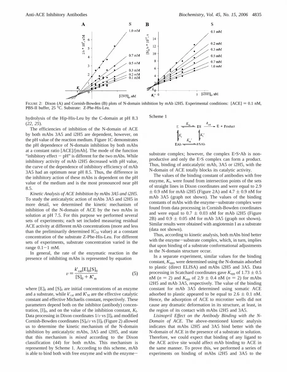
Kinetic Analysis of ACE Inhibition by mAbs 3A5 and i2H5.To study the anticatalytic action of mAbs 3A5 and i2H5 inmore detail, we determined the kinetic mechanism ofinhibition of the N-domain of ACE by the two mAbs insolution at pH 7.5. For this purpose we performed severalsets of experiments; each set included measuring residualACE activity at different mAb concentrations (more and lessthan the preliminarily determined IC50 value) at a constantconcentration of the substrate Z-Phe-His-Leu. For differentsets of experiments, substrate concentration varied in therange 0.1-1 mM.
In general, the rate of the enzymatic reaction in thepresence of inhibiting mAbs is represented by equation
where [E]0 and [S]0 are initial concentrations of an enzymeand a substrate, whilek′cat andK′M are the effective catalyticconstant and effective Michaelis constant, respectively. Theseparameters depend both on the inhibitor (antibody) concen-tration, [I]0, and on the value of the inhibition constant,KI.Data processing in Dixon coordinates 1/V vs [I]0 and modifiedCornish-Bowden coordinates [S]0/V vs [I]0 (Figure 2) allowedus to determine the kinetic mechanism of the N-domaininhibition by anticatalytic mAbs, 3A5 and i2H5, and statethat this mechanism ismixed according to the Dixonclassification (44) for both mAbs. This mechanism isrepresented by Scheme 1. According to this scheme, mAbis able to bind both with free enzyme and with the enzyme-
substrate complex; however, the complex E‚S‚Ab is non-productive and only the E‚S complex can form a product.Thus, binding of anticatalytic mAb, 3A5 or i2H5, with theN-domain of ACE totally blocks its catalytic activity.
The values of the binding constant of antibodies with freeenzyme,KI, were found from intersection points of the setsof straight lines in Dixon coordinates and were equal to 2.9( 0.9 nM for mAb i2H5 (Figure 2A) and 4.7( 0.9 nM formAb 3A5 (graph not shown). The values of the bindingconstants of mAbs with the enzyme-substrate complex werefound from data processing in Cornish-Bowden coordinatesand were equal to 0.7( 0.03 nM for mAb i2H5 (Figure2B) and 0.9( 0.05 nM for mAb 3A5 (graph not shown).Similar results were obtained with angiotensin I as a substrate(data not shown).
Thus, according to kinetic analysis, both mAbs bind betterwith the enzyme-substrate complex, which, in turn, impliesthat upon binding of a substrate conformational adjustmentsin the N-domain structure occur.
In a separate experiment, similar values for the bindingconstant,Kapp, were determined using the N-domain adsorbedto plastic (direct ELISA) and mAbs i2H5 and 3A5. Dataprocessing in Scatchard coordinates gaveKappof 1.73( 0.5nM (n ) 2) andKapp of 2.9 ( 0.4 nM (n ) 2) for mAbsi2H5 and mAb 3A5, respectively. The value of the bindingconstant for mAb 3A5 determined using somatic ACEadsorbed to plastic appeared to be equal to 2.6( 0.1 nM.Hence, the adsorption of ACE to microtiter wells did notcause any dramatic deformation in its structure, at least, inthe region of its contact with mAbs i2H5 and 3A5.
Lisinopril Effect on the Antibody Binding with the N-Domain of ACE.The above-mentioned kinetic analysisindicates that mAbs i2H5 and 3A5 bind better with theN-domain of ACE in the presence of a substrate in solution.Therefore, we could expect that binding of any ligand tothe ACE active site would affect mAb binding to ACE inthe same manner. To prove this, we performed a series ofexperiments on binding of mAbs i2H5 and 3A5 to the
FIGURE 2: Dixon (A) and Cornish-Bowden (B) plots of N-domain inhibition by mAb i2H5. Experimental conditions: [ACE]) 0.1 nM,PBS-II buffer, 25°C. Substrate: Z-Phe-His-Leu.
V )k′cat[E]0[S]0[S]0 + K′m
(5)
Scheme 1
Anti-ACE Inhibitory Antibodies Biochemistry, Vol. 45, No. 15, 20064835

N-domain of ACE and somatic ACE in different settings inthe presence of the specific ACE inhibitor, lisinopril.
MAbs binding to the N-domain, directly adsorbed tomicrotiter plate wells (direct ELISA), was significantlyenhanced in the presence of lisinopril (Figure 3, bar A): 140( 8% and 130( 20% (n ) 3) from control for mAbs i2H5and 3A5, respectively. The values of the binding constant,Kapp, for mAbs i2H5 and 3A5 with the N-domain adsorbedto the wells were estimated to be equal to 0.53( 0.06 nMand 0.9( 0.08 nM (n ) 3), respectively, which is much thesame as the values of the binding constants of these mAbswith the ACE-substrate complex determined by us insolution. The value of the binding constant for mAb 3A5with somatic ACE adsorbed to plastic was 1.0( 0.1 nM.
These values, however, significantly differ from thoseobtained for the free enzyme, both in solution (N-domain)and on plastic (N-domain and somatic ACE).
The influence of lisinopril on the binding of mAbs 3A5and i2H5 to the truncated N domain was studied by othertwo approaches as well. In one set of experiments mAbswere adsorbed to the microtiter plates coated via a goat anti-mouse IgG bridge, whereas in the second set mAbs werebound directly to uncoated plates. Precipitation of ACEs bymAbs 3A5 and i2H5 was estimated by two methods: (i)The amount of ACE protein precipitated by mAbs wasquantified with sheep polyclonal antibodies to human ACEconjugated with peroxidase (46); (ii) Enzymatic activityprecipitated by mAbs was estimated by direct measurementof ACE activity in the wells of microtiter plates byfluorimetric assay (25). As a negative control we used mAb9B9, which did not show a significant inhibitory potencytoward the N domain active site at least in solution.
These experiments (Figure 3) demonstrate the following:(1) The presence of lisinopril did not affect the amount of
ACE protein precipitated by mAb 3A5 bound to microtiterplates via goat anti-mouse IgG (Figure 3, mAb 3A5, barsB, C). However, binding of 3A5 with ACE complexed withlisinopril retained some inhibitor within the active center,because ACE activity precipitated by this mAb in thepresence of lisinopril was significantly (p < 0.05) lower(Figure 3, mAb 3A5, bars D, E);
(2) Precipitation of ACE protein by mAb i2H5, bound tomicrotiter plates via goat anti-mouse IgG, increased slightly
in the presence of lisinopril, albeit significantly: 113.5(1.9% of control,n ) 3, p < 0.05 (Figure 3, mAb i2H5, barC). Precipitated ACE activity, 84.1( 3.2% of control,n )3, p < 0.05 (Figure 3, mAb i2H5, bar E), did not follow theincrease in precipitation of ACE protein, which indicates thatbinding of mAb i2H5 to the complex of ACE with inhibitoralso restricts a natural dissociation of lisinopril from theactive center during washing steps in this assay.
(3) Both ACE protein and ACE activity precipitated bymAb 3A5 in the presence of lisinopril were much the sameregardless of how this mAb was immobilized on themicrotiter plate, via goat anti-mouse bridge or directly.However, the effect of lisinopril on mAb i2H5 binding wasgreatly affected by the mode of mAb immobilization:precipitation of ACE protein in the presence of lisinopril bymAb i2H5 directly bound to the plastic was stronger thanwhen mAb i2H5 was immobilized via goat-anti-mousebridge: 121.6( 0.8% of control (p < 0.05) compared to113.5% (Figure 3, mAb i2H5, bars B, C). ACE activityprecipitated in the presence of lisinopril by mAb i2H5directly bound to the microtiter plates was higher (143.5(9.6% of control,p < 005) compared with only 84.1% in thecase of mAb i2H5 immobilized via goat anti-mouse IgG(Figure 3, mAb i2h5, bars D and E, respectively).
These results also indicate that the greater flexibility ofmAb i2H5 fixed via goat-anti-mouse bridge provided moreinhibition of ACE activity by this mAb. Another fact tosupport a proposed effect of mAb i2h5 flexibility on the ACEinhibition is that the greatest extent of N-domain active centerinhibition was observed when both ACE and mAbs were insolution, i.e., at their maximal flexibility (See Figure 1A).
In a separate experiment, we used EDTA at concentrationsup to 5 mM, as an alternative inhibitor, so as to remove Zn2+
from the active site of ACE. We found that EDTA neitherincreases nor decreases the binding of mAbs 3A5 and i2H5with both recombinant human somatic ACE and truncatedN domain (data not shown).
Mutagenesis of the N-Domain of ACE.In a previous studywe defined broadly the epitopes for some mAbs to theN-domain of human ACE by examining the cross-reactivityof these mAbs, raised against human ACE, with naturallyoccurring variants of ACE, and the differences in functional
FIGURE 3: Effect of lisinopril on mAbs 3A5 and i2H5 binding with N-domain of ACE. A: Binding of mAbs to the N-domain, directlybound to microtiter plate wells. B: Precipitation of N-domain protein by mAbs, directly bound to microtiter plate wells. C: Precipitationof N-domain protein by mAbs, bound to microtiter plate wells via goat anti-mouse IgG bridge. D: Precipitation of N-domain ACE activity(with Z-Phe-His-Leu as a substrate) precipitated by mAbs, directly bound to microtiter plates. E: Precipitation of N-domain ACE activity(with Z-Phe-His-Leu as a substrate) precipitated by mAbs, bound to microtiter plates via goat anti-mouse IgG. Binding of mAbs to ACE,precipitation of ACE N-domain protein, and precipitation of the activity of the N-domain by mAbs in the absence of lisinopril were takenas 100%.
4836 Biochemistry, Vol. 45, No. 15, 2006 Skirgello et al.

properties of these mAbs (37). However, the residues thatcould be evaluated were limited by the availability of aminoacid sequences of naturally occurring variants. Replacementof specific amino acids in the N-terminal domain by site-directed mutagenesis allowed for a rationally designed,systematic and quantitative analysis of the interactionbetween antibodies and ACE.
Figure 4 demonstrates the effect of 21 mutations in theN-terminal domain on the binding with mAbs 3A5 and i2H5.The chosen amino acid residues in the N-domain weresubstituted by the corresponding amino acids in the C-domain, because neither mAb 3A5 nor i2H5 recognizesC-terminal domain.
The rationale for the choice of amino acid residues to bemutated for fine epitope mapping of mAb 3A5 was dictatedby the previously predicted region for the epitope 3A5, inparticular, amino acid residues A564, Q568, and L572 (37).
The region for the mAb i2H5 epitope (and choice of aminoacid residues for mutagenesis) was chosen based on thefollowing facts:
(i) Previously we made the K407S mutation in theN-terminal domain construct (D737), and found that thismutation almost completely abolished the binding of mAb6A12 (data not shown), which recognizes an epitope thathighly overlaps with the epitope for i2H5 (25). Binding of
another mAb, belonging to this group, 1G12, was diminishedsignificantly after mutation as well.
(ii) Binding of all mAbs belonging to this group (i2H5,1G12, and 6A12) was completely absent with chimpanzeeACE (52), which has a substitution in the same region:E403A and R413N (53).
Thus, we chose for mutagenesis those amino acid residuesthat fall into the region and within an area of<700-800Å2: the mean value of the surface area of the epitopes formAbs (54, 55) around amino acid residues already knownas counterparts of the epitope recognized by a definite mAb.Some amino acid residues were chosen on the basis ofanalysis of the N-domain model, described below. They arepredominantly “hills” on the protein surface, as this wouldmake them most favorable for interacting with antibody. Weconsidered the effect of mutation as positive only when achange of 10%, or more, occurred in the binding of mutatedACE with mAb.
Anticatalytic ActiVity of mAbs 3A5 and i2H5 on theN-Domain Mutants.The 21 mutants can be classified intothree subsets with respect to their effects on ACE precipita-tion by mAb 3A5: (1) those mutants where precipitation ofACE activity by mAb 3A5 did not change in comparisonwith wild-type ACE (in that case D629)sE315A-G316E,R413N, and R541A; (2) those mutants where precipitation
FIGURE 4: The effect of mutations on the precipitation of ACE activity by mAbs. ACE activity of culture fluid from CHO cells expressingeach mutant was equalized with ACE activity of truncated N-domain (D629), which was considered here as a wild-type ACE, in a rangeof 5-10 mU/mL (with Z-Phe-His-Leu as a substrate). Then precipitation of ACE activity of each mutant by mAbs 3A5 and i2H5 wasestimated by the plate precipitation assay. Results are expressed as a percentage of precipitated ACE activity from each mutant to that oftruncated N fragment and shown as mean( SD of 4-6 independent experiments, each in duplicate or triplicate.
Anti-ACE Inhibitory Antibodies Biochemistry, Vol. 45, No. 15, 20064837

of ACE activity by mAb 3A5 decreased in comparison withwild typesmost of the studied mutants; (3) those mutantswhere precipitation of ACE activity by mAb 3A5 increasedin comparison with wild typesN203E, R550A, K557Q, andD558L.
Of particular interest were the mutants belonging to thethird group. Because mAb 3A5 effectively inhibits thecatalytic activity of the truncated N-terminal domain (D629)(see Figure 1), the increase in the precipitation of ACEactivity by mAbs immobilized on microtiter plates might bedue to a dramatic increase (2-3-fold) in affinity of mAb3A5 binding with these mutants or to an abolition of theanticatalytic properties of this antibody.
In order to define the reason for the increase in precipita-tion of ACE activity we determined the anticatalytic effectof mAbs 3A5 and i2H5 on the N domain mutants in solution.
Figure 5 demonstrates the effect of mAbs 3A5 and i2H5on the catalytic activity of mutants representing a spectrumof the responses. One can see that the K407S mutation, whichled to a significant albeit weak decrease in the precipitationof ACE activity by mAb i2H5 (Figure 4), also showed asignificant decrease of mAb i2H5 inhibitory potency: IC50
increased more than 3-fold (Figure 5). K542T mutation, inwhich ACE activity precipitation by mAb 3A5 was signifi-cantly decreased (more than 2-fold: Figure 4), also demon-strated a dramatic decrease in anticatalytic activity (Figure5). Mutations Q286K and R413N, whose effects on theprecipitation of ACE activity by mAb i2H5 were dramatics>90% decrease (Figure 4)scompletely abolished the inhibi-tory potencies of mAb i2H5 as well (not shown). Other
mutants demonstrating a decrease in ACE precipitation bymAb 3A5 in the plate precipitation assay (Figure 4) alsoshowed a significant and corresponding decrease in theanticatalytic potency of mAb 3A5 (not shown).
In contrast, all four mutants, where precipitation of ACEactivity by mAb 3A5 was dramatically increasedsN203E,K557Q, D558L, and R550A (Figure 4)sdemonstrated aremarkable decrease in the anticatalytic effect of mAb 3A5(Figure 5). The effect of R550A on 3A5 inhibition, the sameas for three other mentioned mutations, is not shown.Therefore, the dramatic increase in precipitation of ACEactivity by mAb 3A5 obtained with these mutants shouldbe attributed not to better binding of mAb 3A5 but toabolition of its anticatalytic potency toward mutants withcritical amino acid substitutions N203E, R550A, K557Q, andD558L.
In most mutants, where binding of one mAb was changed,the binding of another mAb was not influenced, confirmingthat the epitopes for mAbs 3A5 and i2H5 are essentiallynonoverlapping (25).
Modeling of the N-Domain of ACE and Fine EpitopeMapping.Crystallization of the C-domain of human ACE(tACE) and resolution of its 3D structure (11) made possiblethe modeling of the N-domain of human ACE as well (26,33, 37). By combining knowledge of the 3D structure of theN-domain and the effects of site-directed mutagenesis webecame able to define epitope mapping of mAbs 3A5 andi2H5 in fine detail.
Kinetic analysis together with direct ELISA and activityprecipitation experiments demonstrate different affinities of
FIGURE 5: Anticatalytic effect of mAbs 3A5 and i2H5 on N domain mutants. Assay conditions were as described in Materials and Methodsand in the caption to Figure 1. Residual ACE activity is expressed as the percentage of ACE activity remaining after mAb 3A5 or i2H5 wasadded to the reaction mixture in comparison with ACE activity in the presence of normal, nonimmune mouse IgG, which was taken as100%. MAb 9B9, which did not inhibit ACE catalytic activity (25), was used as negative control for the truncated N-domain, which wasconsidered here as a wild-type ACE. The effect of mAb 9B9 on the catalytic activity of other mutants was absent and is not shown forclarity. Results are shown as mean( SD of 3-4 experiments, each in duplicate.
4838 Biochemistry, Vol. 45, No. 15, 2006 Skirgello et al.

mAbs 3A5 and i2H5 for the free and ligand-bound N-domainof ACE indicating, therefore, the possible existence of twoconformational states for this domain. The native C-domainstructure was reported to be nearly identical to the inhibitor-bound C-domain structure (11), suggesting that no ligand-dependent conformational change occurs for the C-domain.However, the reported C-domain structure actually appearedto contain two ligands, acetate andN-carboxyalanine, thatare bound to the active site of the enzyme, and, therefore,
might represent another ligand-bound structure. For thisreason we employed two models of the N-domain of ACE:the “closed” conformation was based on the structure of theC-domain of human ACE (PDB 1O86), while the “open”conformation was based on the structure of human ACE-related carboxypeptidase, ACE2 (PDB 1R42).
The aligned sequences of the N-domain, the C-domain,and ACE2 are presented in Figure 6. There are two gapswith a total of five inserted amino acids and 272 differences
FIGURE 6: Sequence alignment of the ACE N-domain with C-domain and ACE2. Identical residues between the N- and C-domains andbetween the N-domain and ACE2 are shown against a background. Secondary structure elements (R-helices andâ-strands) are representedon the top of the alignment according to the resolved tACE and ACE2 crystal structures. Zinc binding motif is marked by red. The numberingof ACE relates to mature somatic ACE (8).
Anti-ACE Inhibitory Antibodies Biochemistry, Vol. 45, No. 15, 20064839

between the N- and C-domains over the 595 aligned residues,which correspond to 54% of sequence identity. There are,however, five deletions and 3 insertions with total 21 deleted/inserted amino acid residues within 340 differences betweenthe N-domain and ACE2 (43% of sequence identity). Ahigher conservation of amino acid residues in ACE2,C-domain and N-domain structures is observed in thesequence 200-550 (numeration according to the N-domain).The central sequence containing and surrounding the HEMGHmotif is most conservative.
The secondary structure elements described for the tem-plates (tACE and ACE2) are mapped in the alignment (Figure6). The main elements of the secondary structures of thetemplate proteins coincide well providing feasible compari-son of the two models of the N-domain. The region D85-A94 in the N-domain, however, contains both deletion andinsertion in the templateR-helices. This could be the reasonfor some inexactitude in modeling of this particular region.
Analysis of the Ramachandran plots using the programPROCHECK (51) for both N-domain models showed 82%and 77% of the residues in the N-domain structures modeledwith the C-domain and ACE2 as templates, respectively, inthe most favorable region.
Fine epitope mapping of mAb 3A5 and i2H5 is presentedin Figure 7 and Figure 8, respectively, using both N-domainmodel structures. Both “open” and “closed” N-domainconformations exhibit similar relative dispositions ofR-he-lixes and amino acid residues within the epitopes for bindingmAbs 3A5 and i2H5.
The surface area of the epitope for mAb 3A5 wasestimated to be about 740 Å2. This area is slightly longer onthe surface of the “open” structure of the N-domain than onthe “closed” structure, e.g., amino acid residues N203 andL562 are 19.16 Å and 18.69 Å from each other in the “open”and “closed” structures, respectively. Most of the amino acidresidues that are counterparts of the epitope for mAb 3A5are charged residues (50%), such as 3 Arg, 7 Lys, 4 Tyr, 6Glu, 5 Asp, while hydrophobic amino acid residues representonly near 7% (Figure 7).
The surface area of the amino acid residues that take partin binding to antibody i2H5 was assigned at least 350 Å2
(Figure 8). Charged amino acid residues (1 Arg, 2 Lys, 2His, 3 Glu, 6 Asp) account for 38% of amino residues inthis epitope area, while hydrophobic residues represent 25%.We propose that the real surface area of the epitope for i2H5is much larger, because the minimal surface for published
FIGURE 7: Fine epitope mapping for mAb 3A5 (surface presentation). The structure of the N-domain of human ACE was modeled basedon the 3D structures of human C-domain (PDB 108A) and ACE2 (PDB 1R42).The various amino acid residues belonging to the epitopefor mAb 3A5 are color-coded. Amino acid residues, whose substitution results in significant changes in mAb 3A5 binding, are marked bypurple. Amino acid residues marked by green were recently predicted as belonging to the epitope for mAb 3A5 (37). Amino acid residuesmarked by blue were suggested to be in the epitope for mAb 3A5 because (i) they are located between amino acid residues crucial for mAb3A5 binding and (ii) the surface of the epitope for the mAb should be about 500-900 Å2 (54, 55). Amino acid residues around the putativeepitope for mAb 3A5 were marked by yellow for orientation. The rest of the surface is in gray. The first amino acid residues seen in themodeled N-domain structures are colored in red.
4840 Biochemistry, Vol. 45, No. 15, 2006 Skirgello et al.

FIGURE 8: Fine epitope mapping for mAb i2H5 (surface (A) and ribbon (B) presentations). The surface and ribbon presentations of theN-domain of human ACE were modeled on the basis of the 3D structures of human C-domain (PDB 108A) and ACE2 (PDB 1R42).Various amino acid residues belonging to the epitope for mAb i2H5 are color-coded. Amino acid residues whose substitution results insignificant changes in mAb i2H5 binding are purple. Amino acid residues in green were recently predicted as belonging to the epitope formAb i2H5 (52). Amino acid residues in blue were suggested to be in the epitope for mAb i2H5 because (i) they are located between aminoacid residues crucial for mAb i2H5 binding and (ii) the minimal surface area of the epitope for mAb is about 500 Å2. Amino acid residuesaround the putative epitope for mAb i2H5 are in yellow for orientation. Residues in white belong to the epitope for mAb 3A5. The rest ofthe surface is in gray.
Anti-ACE Inhibitory Antibodies Biochemistry, Vol. 45, No. 15, 20064841

epitopes is about is 500 Å2 (54, 55). It is worth noting thatthe epitope for mAb i2H5 is positioned in the region of a“chewing muscle” in the jaws of the “open” structure, whilein the “closed” structure it is located near one of the entrancesto the active site channel (Figure 8).
The regions of epitopes for mAbs i2H5 and 3A5 are partlyoverlapping. This partial overlap allows binding of mAb 3A5with the ACE-mAb i2H5 complex, whereas binding of mAbi2H5 with the ACE-mAb 3A5 complex is impossible (25).This could be explained by serious conformational changesin ACE structure upon binding mAb 3A5.
3A5-Induced Shedding of ACE from the Cell Surface.Previously we demonstrated that binding of mAb 3A5 tosomatic ACE on the cell surface led to a dramatic increase(3-4-fold) in ACE proteolytic cleavage from the cell surface- antibody-induced ACE shedding (35). We suggested thatbinding of mAb 3A5 induced a significant conformationchange in the ACE molecule on the cell surface, improvedthe accessibility of ACE for secretase, and, as a result, ledto increased ACE shedding (35-37). It is logical to suggestthat binding of mAb 3A5 with residues 203, 550, 557, 558might determine (to some extent) the significant ACEconformational changes observed on mAb binding.
In order to investigate the role of these amino acids in the3A5-induced ACE shedding, we generated D558L in thesomatic ACE and determined its effect on binding of anti-ACE mAbs to this two-domain enzyme, as well as on 3A5-induced ACE shedding (Figure 9). As is apparent, the bindingof mAb 3A5 (Figure 4), as well as mAbs i1A8 and 3G8(data not shown), to D558L is significantly decreased asrevealed by the plate precipitation assay. We also comparedthe rate of shedding of wild-type somatic ACE and D558Lmutant under identical conditions and found that the mutationalmost completely abolished 3A5-induced ACE sheddingfrom the cell surface (Figure 9D). The basal rate of ACEshedding (without any inhibitors or mAbs) was not changedas a result of this mutation (not shown). Interestingly, despitethe fact that D558L has significantly (albeit weakly) lowermAb 3G8 binding, the inhibitory effect of 3G8 on ACEshedding (35-37) was not diminished as demonstrated forthe NdelACE chimeric mutant (37). Therefore, we suggestthat covering of some critical amino acid residues on thesurface of the N-domain of ACE by mAb 3A5 leads tosignificant changes in ACE conformation, which, in turn,leads to allosteric inhibition of ACE activity and/or deter-mines the strong effect of mAb 3A5 on ACE shedding.
DISCUSSION
Monoclonal antibodies to ACE are extremely useful toolsfor investigation of enzyme functions, its structural topog-raphy, etc. Thus, mAbs to human ACE that recognizeconformational epitopes on the N-domain surface andsequential epitopes on the denatured C-domain have beensuccessfully used (i) for ACE quantification both in solutionby ELISA (46) and on the cell surface by flow cytometry(56); (ii) to study the structure and function of ACE (25,35-37); (iii) to deliver enzymes and genes to the pulmonaryendothelium (57-60); (iv) as a diagnostic tool for lung vesselvisualization (61, 62); and (v) for immunohistochemistry ofACE (40, 63-67).
In this work we performed fine epitope mapping for twomonoclonal antibodies to human ACE, 3A5 and i2H5, and
studied the influence of these mAbs on enzyme catalysis. Itwas shown previously (25) that mAbs 3A5 and i2H5 areproduced to the N-domain of human ACE and both exhibitanticatalytic activity. Measurement of the residual ACEactivity at different mAb concentrations demonstrated that,in general, mAb 3A5 inhibits the N-domain of ACE moreeffectively than mAb i2H5 in the pH range 7.1-9.5.However, the relative efficiency of inhibition by the twomAbs is dependent on pH, because the anticatalytic ef-fectiveness of mAb i2H5 decreases with pH, while theinhibitory activity of mAb 3A5 has an optimum near pH8.5.
For fine epitope mapping of binding of mAbs i2H5 and3A5, we used two N-domain models based on recentlypublished structures of the homologous C-domain of humanACE (11) and the homologous ACE-related carboxypepti-dase, ACE2 (48). Besides, we already identified at least threeamino acid residues that participate in binding with mAb3A5, namely A564, N568, and K572 (37). An extensivesearch of amino acid residues as counterparts of epitopesfor both mAbs demonstrated that epitopes for mAb 3A5 andmAb i2H5 contain 50% and 38% of charged amino acidresidues, respectively, whereas hydrophobic amino acidresidues represent less than 25%. Taking into account thepKa values for each amino acid residue, the overall pKa valueof the epitope for mAb 3A5 should be slightly more than 7,while the overall pKa value of the region of the epitope formAb i2H5 is less than 7. These results are in agreement withthe dependence of the anticatalytic activity of both mAbson pH (Figure 1C).
The areas of the epitopes for mAb 3A5 and mAb i2H5are slightly overlapping (see Figure 8). Amino acid residuesY521, E522, G523, P524, D529, K407, and E403 are presentin both epitopes. This distribution allows binding of mAb3A5 to the ACE-i2H5 complex (25), though less effectivelythan to free enzyme. However, binding of another mAb,i2H5, to the complex ACE-3A5 was impossible (25). Themost likely reason for this could be significant conforma-tional changes of ACE upon binding of mAb 3A5, but notmAb i2H5.
Modeling of the structure of the N-domain of human ACEusing the C-domain structure as a template resulted in aprotein structure with a channel, containing the active siteon one of the walls (Figure 8, see also Figures 7 and 8 inref 37). The ligands, substrates or inhibitors, are supposedto enter to the entrance of this channel to reach the activesite (11, 31). However, the authors already pointed out thatthe aperture of the channel opening is approximately 3 Å indiameter, too small for most peptide substrates (for example,the distance between C atoms in the benzyl ring of pheny-lalanine, which is a common component of ACE substrates,can reach 4.3 Å). Clearly, the C-domain of ACE mustundergo some conformational change, possibly associatedwith its unique chloride ion activation, for the substrate toenter the channel (68). Thus, in the native, flexible ACE thesize of the entrance to the channel should be bigger thanthat presented in the structure of the crystalline enzyme (11,30) in order to provide binding of a ligand.
Epitope mapping for mAb i2H5 demonstrates that it bindsin close proximity to one of the entrances into the channel(Figure 8). Therefore, the anticatalytic action of mAb i2H5could be due to direct blockade of the entrance, thus
4842 Biochemistry, Vol. 45, No. 15, 2006 Skirgello et al.
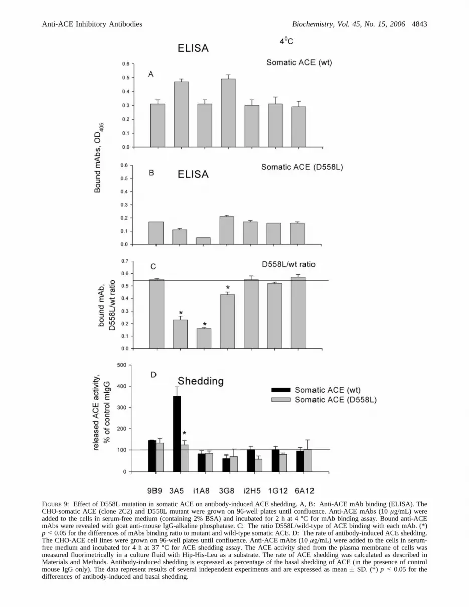
FIGURE 9: Effect of D558L mutation in somatic ACE on antibody-induced ACE shedding. A, B: Anti-ACE mAb binding (ELISA). TheCHO-somatic ACE (clone 2C2) and D558L mutant were grown on 96-well plates until confluence. Anti-ACE mAbs (10µg/mL) wereadded to the cells in serum-free medium (containing 2% BSA) and incubated for 2 h at 4°C for mAb binding assay. Bound anti-ACEmAbs were revealed with goat anti-mouse IgG-alkaline phosphatase. C: The ratio D558L/wild-type of ACE binding with each mAb. (*)p < 0.05 for the differences of mAbs binding ratio to mutant and wild-type somatic ACE. D: The rate of antibody-induced ACE shedding.The CHO-ACE cell lines were grown on 96-well plates until confluence. Anti-ACE mAbs (10µg/mL) were added to the cells in serum-free medium and incubated for 4 h at 37°C for ACE shedding assay. The ACE activity shed from the plasma membrane of cells wasmeasured fluorimetrically in a culture fluid with Hip-His-Leu as a substrate. The rate of ACE shedding was calculated as described inMaterials and Methods. Antibody-induced shedding is expressed as percentage of the basal shedding of ACE (in the presence of controlmouse IgG only). The data represent results of several independent experiments and are expressed as mean( SD. (*) p < 0.05 for thedifferences of antibody-induced and basal shedding.
Anti-ACE Inhibitory Antibodies Biochemistry, Vol. 45, No. 15, 20064843

obviating penetration of substrate into the active site channel.This physical mechanism of action would correspond to anoncompetitive kinetic mechanism of inhibition of ACEactivity by this mAb, i.e., the mAb would be equally able tobind to the free enzyme and the enzyme-substrate complex.However, in kinetic experiments we observed amixedtypeof inhibition, close touncompetitiVe (a mechanism in whichinhibitor binds only to an enzyme-substrate complex, butnot to free enzyme), with much better binding of antibodyto the enzyme-substrate complex. We obtained similarresults when using lisinopril as a ligand and measuring thebinding of mAb by ELISA. In this case, the presence oflisinopril caused an increase in binding of mAb i2H5 by 40%(Figure 3). The results of both kinetic experiments in solutionand ELISA on the microtiter plates indicate probableconformational changes in the N-domain globule caused bya ligand binding in the active site of the enzyme and resultedin better binding of mAb i2H5 with its surface.
Epitope mapping for mAb 3A5 demonstrated that this areais quite far from both entrances to the active site channel(Figure 7). Nevertheless, in this case, we also observed amixedtype of inhibition of the activity of the N-domain bythis mAb in solution with better binding of the mAb to theenzyme-substrate complex. The presence of inhibitor lisi-nopril led to an increase in binding of antibody to theN-domain by up to 30% as well (ELISA).
An investigation of the recently found structural homo-logue of ACE, carboxypeptidase ACE2, demonstrated thatbinding of a ligand to the ACE2 active site is accompaniedby a so-called “hinge-bending” movement (48). The structureof the free enzyme represents an “open” conformation ofthe protein with the active site on the bottom of the “openedmouth”. Binding of a ligand to the active site causesstructural movements in the enzyme, which, in turn, cause“closing” of the “mouth” with the active site inside. Theauthors suggested that this mechanism could be common forboth ACE2 and ACE (48). This suggestion is supported bythe fact that while the 3D structure of the C-domain of humanACE was reported to be identical with inhibitor-boundstructure (11), the first structure included two ligands, acetateand carboxyalanine, in the active site. Therefore, the structureof the C-domain cannot be considered as, in fact, free.Besides, the same hinge-bending movement has been ob-served for two other structural homologues of ACE andACE2, namely, neurolysin and human thimet oligopeptidase(69, 70). Most probably, this mechanism pertains to two otherACE homologues as well:Escherichia coli dipeptidylcarboxypeptidase Dcp has been presented in a “closed”conformation in complex with two dipeptides (71), andcarboxypeptidase fromPyrococcus furiosushas been pre-sented in an “open” conformation (72). On the other hand,investigation of the structure of AnCE, theDrosophila
FIGURE 10: Shifting regions in the ACE N-domain upon putative hinge-bending movement (ribbon presentation). The ribbon presentationof the N-domain of human ACE was modeled on the basis of the 3D structures of human C-domain (PDB 108A) and ACE2 (PDB 1R42).The domain movement between the “open” and “closed” conformations was analyzed using the DynDom program. Amino acid residueschanging spatial angles and their position concerning other elements of the molecule structure upon the hinge-bending movement aremarked by red. TheR-helices changing relative spatial positions are numbered. The regions of the epitopes for mAbs i2H5 and 3A5 are inblue and green colors, respectively.
4844 Biochemistry, Vol. 45, No. 15, 2006 Skirgello et al.

homologue of ACE (73), showed that the structures of thefree and inhibitor-bound enzyme are identical. However, theauthors also pointed out that the diameters of the entrancesinto the active site channel (3 Å) are too small for allowinga substrate to pass through, which, in turn, suggests thatconformational movements must occur on binding a ligand.Also, the 2.4 Å refinement used in this work may be notenough for discovery of a small ligand in the active site(similar to ligands in the ACE structure).
Thus, conformational changes connected with a ligandbinding in an enzyme active site, or a hinge-bendingmovement, could be a common mechanism for all thesestructurally related zinc-dependent enzymes. Moreover, thepossibility exists that these enzymes have a common ancestorprotein (74).
This suggestion allows us to explain the results obtainedwith the N-domain of ACE and anticatalytic mAbs i2H5 and3A5. Epitope mapping for mAb i2H5 shows that the aminoacid residues included in this epitope are located on the sidesof the “mouth” with the active site inside (Figure 8), and,therefore, the “closed” conformation appears preferable forthis mAb binding. If the N-domain of ACE could exist inboth “closed” and “open” conformations, binding of a ligand(substrate or inhibitor) in the active site would cause the“jaws” closing and increase mAb i2H5 binding to theN-domain. The inhibiting action of mAb i2H5 itself couldconsist of fixing of one of the conformations of the ACEmolecule, thus preventing movement of the enzyme and,therefore, catalysis.
The mechanism of inhibition of ACE activity by mAb 3A5can be also explained by fixing one of the ACE conforma-tions upon binding. The epitope for mAb 3A5 is located onthree R-helixes, which move relative to each other uponchanging from the “open” to the “closed” conformation(Figure 7). Thus, binding of this mAb can fix the relativeposition of these structural elements and, therefore, preventcatalysis. This suggestion is supported by the fact thatmutation of amino acid residues, N203, R550, K557, andD558, positioned on these threeR-helixes (Figure 7) doesnot disturb binding of mAb 3A5 due to multipoint interac-tions of the mAb with ACE, but abolishes its inhibitory actiontoward ACE.
Moreover, both epitopes for mAbs i2H5 and 3A5 containamino acid residues and structural elements that could bedirectly involved in the hinge-bending movement, as can beseen in Figure 10. Thus, the epitope for mAb i2H5 containsP272, D273, and amino acid residues in positions 412-416;the epitope for 3A5 includes R381, G382, A383, R550, P551,G561, L562, and amino acid residues in positions 529-533;both epitopes contain the amino acid residues 520-523.These amino acid residues are marked by red in Figure 10.During the movement of ACE from an “open” conformationto a “closed” conformation, the positions of these residuesrelative to other elements of the molecular structure arechanging. The torsion angles of these residues, as revealedby the DynDom program, are different in the two N-domainconformations as well. Moreover, relative spatial positionsof some secondary structural elements also appeared to bedifferent in the “closed” and “open” conformations of theN-domain: (i) the positions ofR-helicesR14 andR19 inthe mAb i2H5 epitope differ in the two N-domain structures;(ii) the positions ofR-helicesR11 andR27 in the mAb 3A5
epitope in the N-domain structure with the C-domain as atemplate and the positions of correspondingR-helicesR10andR31 in the N-domain structure with ACE2 as a templatediffer as well (Figure 10).
The results of the present study also demonstrate that theamino acid residue D558 (and, likely, N203, R550, andK557) in the epitope for mAb 3A5, crucial for its inhibitoryaction, is also crucial for the significant increase in ACEshedding after binding with mAb 3A5 (Figure 9), thusconfirming our previous suggestion (36, 37) that the con-formational changes in the ACE molecule upon mAb 3A5binding are responsible for both effects of this mAb:anticatalytic activity and antibody-induced shedding from thecell surface.
Thus, fine epitope mapping of anticatalytic mAbs inconjunction with a homology model of ACE N-domainstructure has allowed us to reveal putative mechanisms ofinhibitory action of mAbs 3A5 and i2H5 and shed some lighton the mechanism of angiotensin-converting enzyme func-tion.
ACKNOWLEDGMENT
We are grateful to Prof. J. Riordan (Harvard MedicalSchool) for the critical reading of the manuscript and to PierreRedelinghuys (University of Cape Town, South Africa) fortechnical advice and providing two mutants of the N-domain: N289Q and N416Q.
REFERENCES
1. Ehlers, M. R. W., and Riordan, J. F. (1989) Angiotensin-convertingenzyme: new concepts concerning its biological role,Biochemistry28, 5311-5318.
2. Skidgel, R. A., and Erdos, E. G. (1993) Biochemistry of angio-tensin I- converting enzyme, inThe Renin-Angiotensin System(Robertson, J. I. S., Nichols, M. G., Eds.) pp 10.1-10.10, GlowerMedical Publishing, New York.
3. Corvol, P., Eyries, M., and Soubrier, F. (2004) Peptidyl-dipeptidaseA/Angiotensin I-converting enzyme, in:Handbook of ProteolyticEnzymes(Barret, A. A., Rawlings, N. D., Woessner, J. F, Eds.)pp 332-349, Elsevier Academic Press, New York.
4. Dzau, V. J., Bernstein, K., Celermajer, D., Cohen, J., Dahlof, B.,Deanfield, J., Diez, J., Drexler, H., Ferrari, R., van Gilst, W.,Hansson, L., Hornig, B., Husain, A., Johnston, C., Lazar, H., Lonn,E., Luscher, T., Mancini, J., Mimran, A., Pepine, C., Rabelink,T., Remme, W., Ruilope, L., Ruzicka, M., Schunkert, H.,Swedberg, K., Unger, T., Vaughan, D., and Weber, M. (2001)The relevance of tissue angiotensin-converting enzyme: mani-festations in mechanistic and endpoint data,Am. J. Cardiol. 88(Suppl.), 1L-20L.
5. Danilov, S. M., Franke, F. E., and Erdos, E. G. (1997) Angiotensin-Converting Enzyme (CD143), inLeucocyte Typing VI: White CellDifferentiation Antigens(Kishimoto, T., et al., Eds.) pp 746-749, Garland Publishing Inc., New York.
6. Franke, F. E., Metzger, R., Bohle, R.-M., Kerkman, L., Alhenc-Gelas, F., and Danilov, S. M. (1997). Angiotensin I-ConvertingEnzyme (CD 143) on endothelial cells in normal and in pathologi-cal conditions, inLeucocyte Typing VI: White Cell DifferentiationAntigens (Kishimoto, T., et al., Eds.) pp 749-751, GarlandPublishing Inc., New York.
7. Hagaman, J. R., Moyer, J. S., Bachman, E. S., Sibony, M., Magyar,P. L., Welch, J. E., Smithies, O., Krege, J. H., and O’Brien, D.A. (1998) Angiotensin-converting enzyme and male fertility,ProcNatl. Acad. Sci. U.S.A. 95, 2552-2557.
8. Soubrier, F., Alhenc-Gelas, F., Hubert, C., Allegrini, J., John, M.,Tregear, G., and Corvol, P. (1988) Two putative active centers inhuman angiotensin I-converting enzyme revealed by molecularcloning,Proc. Natl. Acad. Sci. U.S.A. 85, 9386-9390.
9. Hubert, C., Houot, A.-M., Corvol, P., and Soubrier, F. (1991)Structure of the angiotensin I-converting enzyme gene. Two
Anti-ACE Inhibitory Antibodies Biochemistry, Vol. 45, No. 15, 20064845

alternate promoters correspond to evolutionary steps of a dupli-cated gene,J. Biol. Chem. 266, 15377-15383.
10. Ehlers, M. R. W., Fox, E. A., Strydom, D. J., and Riordan, J. F.(1989) Molecular cloning of human testicular angiotensin-convert-ing enzyme: the testis isoenzyme is identical to the C-terminalhalf of endothelial angiotensin-converting enzyme,Proc. Natl.Acad. Sci. U.S.A. 86, 7741-7745.
11. Natesh, R., Schwager, S. L., Sturrock, E. D., and Acharya, K. R.(2003) Crystal structure of the human angiotensin-convertingenzyme-lisinopril complex,Nature 421, 551-554.
12. Hooper, N. M., Keen, J., Pappin, D. J. C., and Turner, A. J. (1987)Pig kidney angiotensin converting enzyme. Purification andcharacterization of amphipatic and hydrophilic forms of theenzyme establishes C-terminal anchorage to the plasma membrane,Biochem. J. 247, 85-93.
13. Wei, L., Alhenc Gelas, F., Soubrier, F., Michaud, A., Corvol, P.,and Clauser, E. (1991) Expression and characterization ofrecombinant human angiotensin I-converting enzyme. Evidencefor a C-terminal transmembrane anchor and for a proteolyticprocessing of the secreted recombinant and plasma enzymes,J.Biol. Chem. 266, 5540-5546.
14. Ramchandran, R., Kasturi, S., Douglas, J. G., and Sen, I. (1996)Metalloprotease-mediated cleavage secretion of pulmonary ACEby vasculary endothelial and kidney epithelial cells,Am. J. Physiol.271, H744-H751.
15. Hooper, N. M., Karran, E. H., and Turner, A. J. (1997) Membraneprotein secretases,Biochem. J. 321, 265-279.
16. Woodman, Z. L., Oppong, S. Y., Cook, S., Hooper, N. M.,Schwager, S. L., Brandt, W. F., Ehlers, M. R., and Sturrock, E.D. (2000) Shedding of somatic angiotensin-converting enzyme(ACE) is inefficient compared with testis ACE despite cleavageat identical stalk sites,Biochem. J. 347, 711-718.
17. Deddish, P. A., Wang, J., Michel, B., Morris, P. W., Davidson,N. O., Skidgel, R. A., and Erdos, E. G. (1994) Naturally occurringactive N-domain of human angiotensin I-converting enzyme,Proc.Natl. Acad. Sci. U.S.A. 91, 7807-7811.
18. Deddish, P. A., Wang, L.-X., Jackman, H. L., Michel, B., Wang,J., Skidgel, R. A., Erdo¨s, E. G. (1996) Single-domain angiotensinI converting enzyme (kininase II): characterization and properties,J. Pharmacol. Exp. Ther. 279, 1582-1589.
19. Sturrock, E. D., Danilov, S. M., and Riordan, J. F. (1997) Limitedproteolysis of human kidney angiotensin-converting enzyme andgeneration of catalytically active N- and C-terminal domains,Biochem. Biophys. Res. Commun. 236, 16-19.
20. Binevski, P. V., Sizova, E. A., Pozdnev, V. F. and Kost, O. A.(2003) Evidence for the negative cooperativity of the two activesites within bovine somatic angiotensin-converting enzyme,FEBSLett. 550, 84-88.
21. Voronov, S., Zueva, N., Orlov, V., Arutyunyan, A., and Kost, O.(2002) Temperature-induced selective death of the C-domainwithin angiotensin-converting enzyme molecule,FEBS Lett. 522,77-82.
22. Wei, L., Alhenc-Gelas, F., Corvol, P., and Clauser, E. (1991) Thetwo homologous domains of human angiotensin I-convertingenzyme are both catalytically active,J. Biol. Chem. 266, 9002-9008.
23. Wei, L., Clauser, E., Alhenc-Gelas, F., and Corvol, P. (1992) Thetwo homologous domains of human angiotensin I-convertingenzyme interact differently with competitive inhibitors,J. Biol.Chem. 267, 13398-13405.
24. Jaspard, E., Wei, L., and Alhenc-Gelas, F. (1993) Differences inproperties and enzymatic specificities between the two active sitesof human angiotensin I-converting enzyme: studies with brady-kinin and other natural peptides,J. Biol. Chem. 268, 9496-9503.
25. Danilov, S., Jaspard, E., Churakova, T., Towbin, H., Savoie, F.,Lei, W., and Alhenc-Gelas, F. (1994) Structure-function analysisof angiotensin I-converting enzyme using monoclonal antibodies,J Biol Chem. 269, 26806-26814.
26. Tzakos, A. G., Galanis, A. S., Spyroulias, G. A., Cordopatis, P.,Manessi-Zoupa, E., and Gerothanassis, I. P. (2003) Structure-function discrimination of the N- and C- catalytic domains ofhuman angiotensin-converting enzyme: implications for Cl-activation and peptide hydrolysis,Protein Eng. 16, 993-1003.
27. Skirgello, O. E., Binevski, P. V., Pozdnev, V. F., and Kost, O. A.(2005) Kinetic probes for inter-domain cooperation in humansomatic angiotensin-converting enzyme,Biochem. J. 391, 641-647.
28. Dive, V., Cotton, J., Yiotakis, A., Michaud, A., Vassiliou, S.,Jiracek, J., Vazeux, G., Chauvet, M. T., Cuniasse, P., and Corvol,
P. (1999) RXP 407, a phosphinic peptide, is a potent inhibitor ofangiotensin I converting enzyme able to differentiate between itstwo active sites,Proc. Natl. Acad. Sci. U.S.A. 96, 4330-4335.
29. Cotton, J., Hayaashi, M. A. F., Cuniasse, Ph., Vazeux, G., Ianzer,D., De Camargo, A. C. M., and Dive, V. (2002) Selectiveinhibition of the C-domain of angiotensin I converting enzymeby bradykinin potentiating peptides,Biochemistry 41, 6065-6071.
30. Georgiadis, D., Beau, F., Czarny, B., Cotton, J., Yiotakis, A., andDive, V. (2003) Roles of the two active sites of somaticangiotensin-converting enzyme in the cleavage of angiotensin Iand bradykinin: insights from selective inhibitors,Circ Res. 93,148-154.
31. Sturrock, E. D., Natesh, R., van Rooyen, J. M., and Acharya, K.R. (2004) Structure of angiotensin I-converting enzyme,Cell. Mol.Life. Sci. 61, 2677-2686.
32. Tzakos, A. G., and Gerothanassis, I. P. (2005) Domain-SelectiveLigand-Binding Modes and Atomic Level Pharmacophore Refine-ment in Angiotensin I Converting Enzyme (ACE) Inhibitors,ChemBioChem 6, 1089-1103.
33. Fernandez, J. H., Hayashi, M. A. F., Camargo, A. C. M., andNeshich, G. (2003) Structural basis of the lisinopril-bindingspecificity in N- and C-domains of human somatic ACE,Biochem.Biophys. Res. Commun. 308, 219-226.
34. Perich, R. B., Jackson, B., Rogerson, F., Mendelson, F. A., Paxton,D., and Johnston, C. I. (1992) Two binding sites on angiotensin-converting enzyme: evidence from radioligand binding studies,Mol. Pharmacol. 42, 286-293.
35. Balyasnikova, I. V., Karran, E. H., Albrecht, R. F., II, and Danilov,S. M. (2002) Epitope-specific antibody-induced cleavage ofangiotensin-converting enzyme from the cell surface, Biochem.J. 362, 585-595.
36. Kost, O. A., Balyasnikova, I. V., Chemodanova, E. E., Nikolskaya,I. I., Albrecht, R. F., II, and Danilov, S. M. (2003) Epitope-dependent blocking of the angiotensin-converting enzyme dimer-ization by monoclonal antibodies to N-terminal domain of ACE:Possible Link of ACE dimerization and shedding from the cellsurface,Biochemistry 42, 6965-6976.
37. Balyasnikova, I. V., Woodman, Z. L., Albrecht, R. F., II, Natesh,R., Acharya, K. R., Sturrock, E. D., and Danilov, S. M. (2005)Localization of an N domain region of angiotensin-convertingenzyme involved in the regulation of ectodomain shedding usingmonoclonal antibodies,J. Proteome Res. 4, 258-267.
38. Stanislav, M. L., Balabanova, R. M., Alekperov, R. T., Miagkova,M. A., Abramenko, T. V., Kiselev, I. P., Kost, O. A., Nikolskaya,I. I., and Garats, E. V. (2001) Autoantibodies to vasoactivepeptides and angiotensin converting enzyme in patients withsystemic diseases of the connective tissue,Ter Arkh. (Russian)73, 20-25.
39. Balyasnikova, I. V., Gavriljuk, V. D., McDonald T. D., Berkowitz,R., Miletich, D. J., and Danilov, S. M. (1999) Antibody-mediatedlung endothelium targeting:In Vitro model using a cell lineexpressing angiotensin-converting enzyme,Tumor Targeting 4,70-83.
40. Balyasnikova, I. V., Metzger, R., Franke, F. E., and Danilov, S.M. (2003) Monoclonal antibodies to denatured human ACE(CD143): broad species specificity, reactivity on paraffin-section,and detection of subtle conformational changes in the C-terminaldomain of ACE,Tissue Antigens 61, 49-62.
41. Binevski, P. V., Nikolskaya, I. I., Pozdnev, V. F., and Kost, O.A. (2000) Isolation and characterization of the N-domain of bovineangiotensin-converting enzyme,Biochemistry (Moscow) 65, 651-658.
42. Pilliquod, Y., Reinharz, A., and Roth, M. (1970) Studies on theangiotensin-converting enzyme with different substrates,Biochim.Biophys. Acta 206, 136-142.
43. Friedland, J., and Silverstein, E. (1976) A sensitive fluorometricassay for serum angiotensin-converting enzyme, Am. J. Clin. Path.66, 416-424.
44. Dixon, M., and Webb, E. C. (1979)Enzymes, Longman, London.45. Cornish-Bowden, A. (1999) pp 306-307,Fundamentals of Enzyme
Kinetics, Portland Press Ltd, London.46. Danilov, S. M., Savoie, F., Lenoir, B., Jeunemaitre, X., Azizi,
M., Tarnow, L., and Alhenc-Gelas, F. (1996) Development ofenzyme-linked immunoassays for human angiotensin I convertingenzyme suitable for large-scale studies,J. Hypertens. 14, 719-727.
47. Scatchard, G. (1949) The attractions of proteins for small moleculesand ions,Ann. N.Y. Acad. Sci. 51, 660-672.
4846 Biochemistry, Vol. 45, No. 15, 2006 Skirgello et al.

48. Towler, P., Staker, B., Prasad, S. G., Menon, S., Tang, J., Parsons,T., Ryan, D., Fisher, M., Williams, D., Dales, N. A., Patane, M.A., and Pantoliano, M. W. (2004) ACE2 X-ray structures reveala large hinge-bending motion important for inhibitor binding andcatalysis,J. Biol. Chem. 279, 17996-18007.
49. Thompson, J. D., Higgins, D. G., and Gibson, T. J. (1994)CLUSTAL W: improving the sensitivity of progressive multiplesequence alignment through sequence weighting, positions, spe-cific gap penalties and weight matrix choice,Nucleic Acids Res.22, 4673-4680.
50. Berman, H. M., Westbrook, J., Feng, Z., Gilliland, G., Bhat, T.N, Weissig, H., Shindyalov, I. N., and Bourne, P. E. (2000) TheProtein Data Bank,Nucleic Acids Res. 28, 235-242.
51. Laskowski, R. A., MacArthur, M. W., Moss, D. S., and Thornton,J. M. (1993) PROCHECK: a program to check the stereochemicalquality of protein structures,J. Appl. Crystallogr. 26, 283-291.
52. Balyasnikova, I. V., Yeomans, D. C., McDonald, T. B., andDanilov, S. M. (2002) Antibody-mediated lung endotheliumtargeting: in vivo model on primates, Gene Ther. 9, 282-290.
53. Dufour, C., Casane, D., Denton, D., Wickings, J., Corvol, P., andJeunemaitre, X. (2000) Human-chimpanzee DNA sequence varia-tion in the four major genes of the renin-angiotensin system,Genomics 69, 14-26.
54. Mylvaganam, S. E., Paterson, Y., and Gertzoff, E. D. (1998)Structural basis for the binding of an anti-cytochrome antibodyto its antigen: crystal structures of Fab E8-cytochromec complexto 1.8Å resolution and FabE8 to 2.26Å resolution,J. Mol. Biol.281, 301-322.
55. Huang, M., Syed, R., Stura, E. A., Sone, M. J., Stefanko, R. S.,Ruf, W., Edgington, T. S., and Wilson, I. A. (1998) Themechanism of an inhibitory antibody on TF-initiated bloodcoagulation revealed by the crystal structures of human tissuefactor, Fab 5G9 and TF-5G9 complex,J. Mol. Biol. 275, 873-894.
56. Danilov, S. M., Sadovnikova, E., Scharenborg, N., Balyasnikova,I. V., Svinareva, D. A., Semikina, E. L., Parovichnikova, E. N.,Savchenko, V. G., and Adema, G. J. (2003) Angiotensin-converting enzyme (CD143) is abundantly expressed by dendriticcells and discriminates human monocyte-derived dendritic cellsfrom acute myeloid leukemia-derived dendritic cells,Exp. He-matol. 31, 1301-1309.
57. Muzykantov, V. R., Atochina, E. N., Ischiropoulos, H., Danilov,S. M., and Fisher, A. B. (1996) Immunotargeting of antioxidantenzymes to the pulmonary endothelium, Proc. Natl. Acad. Sci.U.S.A. 93, 5213-5218.
58. Atochina, E. N., Balyasnikova, I. V., Danilov, S. M., Granger, D.N., Fisher, A. B., and Muzykantov, V. R. (1998) Catalase targetingto the surface endothelial antigens protects pulmonary vasculatureagainst oxidative insult,Am. J. Physiol. 275, L806-L817.
59. Reynolds, P. N., Zinn, K. R., Gavrilyuk, V. D., Balyasnikova, I.V., Rogers, B. E., Buchsbaum, D. J., Wang, M. H., Miletich, D.J., Crizzle, W. E., Douglas, J. T., Danilov, S. M., and Curiel, D.T. (2000) A targetable, injectable adenoviral vector for selectivegene delivery to pulmonary endothelium in vivo, Mol. Ther. 2,562-578.
60. Muller, W. H., Brosnan, M. J., Graham, D., Nicol, C. G.,Morecroft, I., Channon, K. M., Danilov, S. M., Reynolds, P. N.,Baker, A. H., and Dominiczak, A. F. (2005) Targeting endothelialcells with adenovirus expressing nitric oxide synthase preventselevation of blood pressure in stroke-prone spontaneously hyper-tensive rats,Mol. Ther. 12, 321-327.
61. Danilov, S. M., Martynov, A., Klibanov, A. L., Slinkin, M. A.,Sakharov, I. Y., Malov, A. G., Sergienko, V. B., Vedernikov, A.Y., Muzykantov, V. R., and Torchilin, V. P. (1989) Radioimmu-noimaging of lung vessels: an approach using 111-In-labeledmonoclonal antibody to angiotensin-converting enzyme,J. Nucl.Med. 30, 1688-1692.
62. Muzykantov, V. R., and Danilov, S. M. (1995) Targeting ofradiolabelled monoclonal antibody against angiotensin-convertingenzyme to the pulmonary vasculature, inHandbook of TargetingDeliVery of Imaging Agents(Torchilin, V. P., Ed.) pp 465-486,CRC, Boca Raton, FL.
63. Danilov, S. M., Faerman, A. I., Printseva, O. Y., Martynov, A.V., Sakharov, I. Y., and Trakht, I. N. (1987) Immunohistochemicalstudy of angiotensin-converting enzyme in human tissues usingmonoclonal antibodies,Histochemistry 87, 487-490.
64. Metzger, R., Bohle, R.-M., Kerkman, L., Eichner, G., Alhenc-Gelas, F., Danilov, S. M., and Franke, F. E. (1999) Distributionof angiotensin I-converting enzyme (CD 143) in the normal humankidney and in non-neoplastic kidney diseases,Kidney Int. 56,1442-1454.
65. Metzger, R., Bohle, R.-M., Chumachenko, P., Danilov, S. M., andFranke, F. E. (2000) CD 143 in the development of atherosclerosis,Atherosclerosis 150, 21-31.
66. Franke, F. E., Fink, L., Kerkman, L., Steger, K., Klonisch, T.,Metzger, R., Alhenc-Gelas, F., Burkhard, E., Bergmann, M., andDanilov, S. M. (2000) Somatic isoform of angiotensin-convertingenzyme in the pathology of testicular germ cell tumors,HumanPathol. 31, 1466-1476.
67. Pauls, K., Metzger, R., Steger, K., Klonisch, T., Danilov, S., andFranke, F. E. (2003) Isoforms of angiotensin I-converting enzymein the development and differentiation of human testis andepididymis,Andrologia 35, 32-43.
68. Acharya, K. R., Sturrock, E. D., Riordan, J. F., and Ehlers, M. R.(2004) Ace revisited: a new target for structure-based drug design,Nat. ReV. Drug DiscoVery 11, 891-902.
69. Brown, C. K., Madauss, K., Lian, W., Beck, M. R., Tolbert, W.D., and Rodgers, D. W. Structure of neurolysin reveals a deepchannel that limits substrate access, (2001)Proc. Natl. Acad. Sci.U.S.A. 98, 3127-3132.
70. Ray, K., Hines, C. S., Coll-Rodriguez, J., and Rodgers, D. W.(2004) Crystal structure of human thimet oligopeptidase providesinsight into substrate recognition, regulation, and localization,J.Biol. Chem. 279, 20480-20489.
71. Comellas-Bigler, M., Lang, R., Bode, W., and Maskos, K. (2005)Crystal structure of the E. coli dipeptidyl carboxypeptidase Dcp:further indication of a ligand-dependent hinge movement mech-anism,J. Mol. Biol. 349, 99-112.
72. Arndt, J. W., Hao, B., Ramakrishnan, V., Cheng, T., Chan, S. I.,and Chan, M. K. (2002) Crystal structure of a novel carboxypep-tidase from the hyperthermophilic archaeon Pyrococcus furiosus,Structure (Cambridge) 10, 215-224.
73. Kim, H. M., Shin, D. R., Yoo, O. J., Lee, H., and Lee, J. (2003)Crystal structure of Drosophila angiotensin I-converting enzymebound to captopril and lisinopril,FEBS Lett. 538, 65-70.
74. Fernandez, J. H., Neshich, G., and Camargo, A. C. M. (2004)Using bradykinin-potentiating peptide structures to develop newantihypertensive drugs,Genet. Mol. Res. 3, 554-563.
BI052591H
Anti-ACE Inhibitory Antibodies Biochemistry, Vol. 45, No. 15, 20064847


