Embed Size (px)
Citation preview

JOURNAL OF VIROLOGY, Apr. 2010, p. 3666–3681 Vol. 84, No. 70022-538X/10/$12.00 doi:10.1128/JVI.01340-09Copyright © 2010, American Society for Microbiology. All Rights Reserved.
Internalization of Coxsackievirus A9 Is Mediated by �2-Microglobulin,Dynamin, and Arf6 but Not by Caveolin-1 or Clathrin�†Outi Heikkila,1 Petri Susi,1 Tuire Tevaluoto,1 Heidi Harma,1‡ Varpu Marjomaki,2
Timo Hyypia,1 and Saija Kiljunen1,3*Department of Virology, University of Turku, Kiinamyllynkatu 13, 20520 Turku,1 Department of Biological andEnvironmental Science/Nanoscience Center, University of Jyvaskyla, Jyvaskyla,2 and Division of Genetics and
Physiology, Department of Biology, University of Turku, Turku,3 Finland
Received 30 June 2009/Accepted 11 January 2010
Coxsackievirus A9 (CAV9) is a member of the human enterovirus B species within the Enterovirus genus ofthe family Picornaviridae. It has been shown to utilize �V integrins, particularly �V�6, as its receptors. Theendocytic pathway by which CAV9 enters human cells after the initial attachment to the cell surface has so farbeen unknown. Here, we present a systematic study concerning the internalization mechanism of CAV9 to A549human lung carcinoma cells. The small interfering RNA (siRNA) silencing of integrin �6 subunit inhibitedvirus proliferation, confirming that �V�6 mediates the CAV9 infection. However, siRNAs against integrin-linked signaling molecules, such as Src, Fyn, RhoA, phosphatidylinositol 3-kinase, and Akt1, did not reduceCAV9 proliferation, suggesting that the internalization of the virus does not involve integrin-linked signalingevents. CAV9 endocytosis was independent of clathrin or caveolin-1 but was restrained by dynasore, aninhibitor of dynamin. The RNA interference silencing of �2-microglobulin efficiently inhibited virus infectionand caused CAV9 to accumulate on the cell surface. Furthermore, CAV9 infection was found to depend on Arf6as both silencing of this molecule by siRNA and the expression of a dominant negative construct resulted indecreased virus infection. In conclusion, the internalization of CAV9 to A549 cells follows an endocytic pathwaythat is dependent on integrin �V�6, �2-microglobulin, dynamin, and Arf6 but independent of clathrin andcaveolin-1.
Coxsackievirus A9 (CAV9), a member of the human entero-virus B species in the family Picornaviridae, is a significanthuman pathogen. It causes infections of the central nervoussystem, myocarditis, and respiratory diseases and may occa-sionally cause fatal generalized infections in newborns (6, 22,26). The CAV9 particle is about 30 nm in diameter and con-sists of a naked capsid with an icosahedral symmetry, sur-rounding a positive-sense RNA genome of approximately7,400 nucleotides (30). The capsid is made up of 60 copies ofeach of the four proteins VP1 to VP4 and interacts with cellsurface integrins during the early stages of infection via argi-nine-glycine-aspartic acid (RGD) motif that resides in the Cterminus of the VP1 protein (11). While CAV9 binds to bothintegrin �V�3 and �V�6 in vitro (53, 61), our recent data showthat integrin �V�6 is the primary receptor of the virus (29).
Viruses can utilize several endocytic pathways to enter mam-malian cells: macropinocytosis and clathrin-mediated, caveo-lin-mediated, and clathrin- and caveolin-independent routes(14, 40–41, 50). Recent studies have shown that some of thesepathways differ only slightly from each other, and certain en-docytic components can participate in more than just one path-
way (35, 41, 55). Most of the research carried out on entero-virus endocytosis has been done with echovirus 1 (EV1),coxsackievirus B3 (CBV3), and poliovirus (PV). Recently, Kar-jalainen et al. showed that EV1 enters SAOS cells via tubu-lovesicular structures in a dynamin-independent manner thatresembles fluid-phase endocytosis and macropinocytosis andthat at later stages of infection is targeted to caveosomes (33).EV1 entry to CV-1 cells, on the other hand, was shown to bestrictly dynamin dependent (49). PV is endocytosed to HeLacells by a rapid clathrin- and caveolin-independent pathway,whereas in brain microvascular endothelial cells it uses slower,caveolin- and dynamin-dependent endocytosis (4, 7, 17). CBV3enters HeLa cells by clathrin-mediated endocytosis (13) andpolarized epithelial CaCo-2 cells by a process that combinesfeatures of caveolar endocytosis and macropinocytosis (16, 18).Foot-and-mouth-disease virus (FMDV), a member of the Aph-thovirus genus of the family Picornaviridae, binds to several�V-integrins, including �V�6, and is internalized through theclathrin-mediated pathway (5, 19, 31). In the light of theseexamples, it is evident that enterovirus internalization to hu-man cells is a complex phenomenon wherein a virus may usedifferent mechanisms to enter different cell types.
In the CAV9 infection cycle, the steps that follow the initialattachment of the virus to the cell surface integrins are stillpoorly characterized. An early electron microscopic work byHecker et al. has shown that single CAV9 particles enter mon-key kidney cells in vesicles, which then occasionally fuse andform larger structures (28). Interestingly, they found that mostinternalized virus particles became eventually trapped in largevacuoles, presumably lysosomes, where they were confined
* Corresponding author. Present address: Division of Genetics andPhysiology, Department of Biology, University of Turku, Vesilinnantie5, 20014 Turun Yliopisto, Finland. Phone: 358 2 333 5576. Fax: 358 2333 6680. E-mail: [email protected].
‡ Present address: Orion Corporation Orion Pharma, Espoo, Fin-land.
† Supplemental material for this article may be found at http://jvi.asm.org/.
� Published ahead of print on 20 January 2010.
3666
at Univ of T
urku on March 8, 2010
jvi.asm.org
Dow
nloaded from

without proceeding to capsid uncoating and RNA release.More recently, a number of cell surface molecules have beenproposed to contribute to CAV9 internalization. A subunit ofmajor histocompatibility complex class I (MHC-I) complex,�2-microglobulin (�2M), has been shown to be essential forthe infection of several picornaviruses, including CAV9, and itis supposed to have a postattachment role (12, 59, 61). Inaddition, heat shock 70-kDa protein 5 (HSPA5 protein, alsoknown as glucose-regulated protein 78-kDa, or GRP78) hasbeen suggested to function as a coreceptor for the virus and tomediate CAV9 infection by its interaction with �2M on the cellsurface (57). CAV9 entry has been proposed to occur throughlipid microdomains, where a number of signaling events takesplace (58).
The aim of this study was to elucidate the internalizationmechanism of CAV9 in A549 human lung carcinoma cells. Weused chemical inhibitors, RNA interference (RNAi) silencing,and the expression of dominant negative constructs combinedto virus infectivity assays and confocal imaging to examinewhich cellular molecules are involved in the entry process. Theresults indicate that CAV9 internalization is dependent onintegrin �V�6, �2M, dynamin 2, and Arf6 (ADP-ribosylationfactor 6) but not clathrin or caveolin-1.
MATERIALS AND METHODS
Cells, viruses, and antibodies. The human lung carcinoma A549 cell line wasobtained from the American Type Culture Collection (ATCC), and the caveolin-1-negative human hepatocellular carcinoma cell line HuH7 (60) was from ElinaIkonen (University of Helsinki, Finland). The Phoenix Gag-Pol packaging cellline (http://www.stanford.edu/group/nolan/index.html) was obtained from AkiManninen (University of Oulu, Finland) with authorization by Garry Nolan(School of Medicine, Stanford, University, Stanford, CA). A549 cells were main-tained in Ham’s F12 medium supplemented with 7% fetal calf serum (FCS) andgentamicin. HuH7 and Phoenix cells were grown in Dulbecco’s modified Eagle’smedium (DMEM) containing 10% FCS and gentamicin.
CAV9 (Griggs strain) (11, 30) was propagated in A549 cells and purified insucrose gradient as described previously (1). Culture medium for virus infectionswas supplemented with 1% FCS.
Polyclonal rabbit antiserum against CAV9 was produced as described earlier(51), and mouse monoclonal antibody ([MAb] K6) against CAV9 (9) was ob-tained from Lucia Fiore (Instituto Superiore di Sanita, Rome, Italy). MAbagainst �2-microglobulin and Arf6 and rabbit polyclonal antiserum specific tocaveolin-1 were from Santa Cruz Biotechnology (catalog items sc-51509, sc-7971,and sc-894, respectively). Rabbit polyclonal hemagglutinin (HA) antiserum wasfrom Zymed Laboratories Inc. (catalog item 71-5500), and integrin �V�6 MAb(MAB2074Z), which does not block function, was from Chemicon. Rabbit poly-clonal antiserum specific to Erk1 (sc-94) was from Santa Cruz Biotechnology.Alexa Fluor (AF) 488-, 546-, and 568-labeled anti-mouse and anti-rabbit sec-ondary antibodies were from Molecular Probes, and horseradish peroxidase(HRP)-labeled anti-rabbit secondary antibody was from Pierce. The function-inhib-iting antibodies used in the blocking assays were against integrin �V (L230; ATCC),integrin �V�3 (MAB1976Z; Chemicon), and integrin �V�6 (MAB2077Z; Chemi-con).
Chemical inhibitors. A549 cells were incubated at 37°C in medium supple-mented with 100 �M 5-(N-ethyl-N-isopropyl)amiloride ([EIPA] catalog no.A3085; Sigma), 25 �M chlorpromazine (C8138; Sigma), 1 mM methyl-�-cyclo-dextrin ([M�C] C4555; Sigma), 33.2 �M nocodazole (M1404; Sigma), 5 �g/mlcytochalasin D (C8273; Sigma), 2 �M jasplakinolide (420127; Calbiochem), 1�M latrunculin A (L5163; Sigma), 100 nM wortmannin (W1628; Sigma), or withthe combination of 25 �g/ml nystatin (N3503; Sigma) and 10 �g/ml progesterone(P8783; Sigma). The inhibitors were present for 30 min prior to and throughoutthe CAV9 infection assay. The importance of endosome acidification was studiedby incubating the cells with 0.5 mM, 2 mM, 5 mM, or 25 mM NH4Cl at 37°C for30 min and performing the virus infection assay in the presence of the salt. Cellviability after the treatment with NH4Cl was determined by the cell viability assay(see below).
The function of chlorpromazine, M�C, and cytochalasin D was confirmed by
following the uptake of specific endocytic markers. AF 488-conjugated choleratoxin B (0.2 �g/ml; Molecular Probes) and AF 546-conjugated transferrin (10�g/ml; Molecular Probes) were used to control the function of chlorpromazineand M�C, while AF 546-conjugated dextran (Molecular Probes) was used tocontrol the function of EIPA (at 1 �M, 50 �M, 100 �M, and 150 �M) andcytochalasin D (at 250 �g/ml). A549 cells were incubated with the inhibitor for30 min at 37°C, after which the marker was added. The incubation was continuedfor 15 min, the cells were fixed and stained with Hoechst 33342 (Sigma-Aldrich),and the data were analyzed by confocal imaging.
To analyze the effect of dynasore (D7693; Sigma) on CAV9 infection, A549cells were incubated at 37°C for 30 min with 80 �M dynasore or 0.4% dimethylsulfoxide (DMSO) diluted in serum-free cell medium. The CAV9 infection assaywas then performed in the presence of the drug. The effective inhibitory con-centration of dynamin was determined by incubating the cells with variableconcentrations of dynasore (10 �M, 30 �M, 50 �M, and 80 �M), followed byfeeding the cells with AF 546-conjugated transferrin (10 �g/ml) for 2 min atroom temperature room temperature. Unbound transferrin was removed bywashing the cells with serum-free medium containing dynasore or DMSO, andthe transferrin uptake was followed for 15 min at 37°C. The cell plate was thentransferred onto ice, and transferrin bound to the cell surface was removed byrinsing with an acidic solution (100 mM glycine, pH 2.5, and 150 mM NaCl). Theinternalization of virus and transferrin was visualized by confocal microscopy.
To study the simultaneous internalization of CAV9 and dextran, A549 cellswere washed with ice-cold serum-free medium, and CAV9 was allowed to attachto cell surfaces on ice for 1 h. Cells were then washed with ice-cold serum-freemedium before the addition of AF 546-conjugated dextran (250 �g/ml) dilutedin prewarmed serum-free medium. Cells were transferred to 37°C, where theywere incubated for the desired times (5 min, 15 min, or 30 min), followed by cellfixation, permeabilization, staining, and visualization by confocal microscopy.
Plasmid constructs, transfection, and transduction with adenoviral vectors.HA-tagged caveolin-3 and caveolin-3DGV (an amino-terminal truncation mu-tant) expression plasmids for wild-type (wt) and dominant negative (DN) caveo-lin-3, respectively (54), were obtained from Robert Parton (The University ofQueensland, Brisbane, Australia). Green fluorescent protein (GFP)-dynamin2aa and GFP-dynamin 2aaK44A (where 2aaK44A is the dynamin 2aa isoform witha K44A mutation) expression plasmids (10, 47) for wt and DN dynamin 2,respectively, were from Mark McNiven (Mayo Clinic, Rochester, MN); Eps15-GFP and Eps15E�95/295-GFP (where Eps15E�95/295 is Eps15E with a deletionof residues 95 to 295) constructs (2) for wt and DN Eps15, respectively, werefrom Alice Dautry-Varsat (Pasteur Institute, Paris, France); and the DN AP180Cconstruct (23) was from Dieter Blaas (University of Vienna, Austria). PlasmidspcDNA3 HA Arf6 and pcDNA3 HA Arf6 DN T27N (25) for the expression ofwt and DN Arf6, respectively, were from Addgene (10834 and 10831, respec-tively; www.addgene.org). Recombinant GFP-tagged Eps15-adenoviral vectorsrAd:DIII and rAd:DIII�2 (3, 36) for wt and DN Eps15, respectively, wereprovided by Yves Rouille (Pasteur Institute, Lille, France).
For transient transfection, A549 cells were cultured up to 50% confluence andtransfected with expression plasmids using FuGENE 6 or FuGENE HD(Roche), according to the protocol recommended by the manufacturer. Trans-fection efficiencies were approximately 5 to 10%. The adenovirus vectors wereproduced in HEK293T cells and used to transduce semiconfluent A549 cells. TheCAV9 infection assay was done at 48 h after plasmid transfection or adenovirustransduction.
TABLE 1. Pharmacological inhibitors used in this work
Inhibitor Effect Concn
EIPA Inhibits Na�/H� exchange 100 �MChlorpromazine Inhibits clathrin-dependent
endocytosis25 ��
Methyl-�-cyclodextrin Extracts cholesterol from lipidmembranes
1 mM
Nocodazole Depolymerizes microtubules 33.2 ��Cytochalasin D Disrupts actin polymerization 5 �g/mlLatrunculin A Disrupts actin polymerization 1 �MJasplakinolide Stabilizes actin microfilaments 2 �MWortmannin Inhibits phosphoinositide-3-
kinase100 nM
Nystatin �progesterone
Disrupts caveolae/lipid rafts 25 �g/ml �10 �g/ml
VOL. 84, 2010 INTERNALIZATION OF CAV9 3667
at Univ of T
urku on March 8, 2010
jvi.asm.org
Dow
nloaded from

FIG. 1. Effects of endocytosis inhibitors on CAV9 infection on A549 cells. (A) A549 cells were preincubated at 37°C for 30 min with EIPA (100�M), chlorpromazine (25 �M), methyl-�-cyclodextrin (M�C; 1 mM), nocodazole (33.2 �M), cytochalasin D (5 �g/ml), jasplakinolide (2 �M),latrunculin A (1 �M), wortmannin (100 nM), or with a combination of nystatin (25 �g/ml) and progesterone (10 �g/ml). The cells were infectedwith CAV9 at 60% efficiency of infection and incubated on ice for 1 h. The unbound virus was removed, and cells were transferred to 37°C andincubated for 6 h. The inhibitors were present throughout the experiment. Cells were fixed, permeabilized, and stained with Hoechst and
3668 HEIKKILA ET AL. J. VIROL.
at Univ of T
urku on March 8, 2010
jvi.asm.org
Dow
nloaded from

siRNA transfections. Two individual small interfering RNA (siRNA) mole-cules for each gene (Qiagen) (see Table S1 in the supplemental material) wereused. To transfect A549 cells in 96-well plates, 0.5 pmol of siRNA in 25 �l ofH2O was mixed with 0.2 �l of siLentFect (Bio-Rad) diluted in 25 �l of serum-free medium and incubated for 30 min at room temperature. A total of 13,000cells were then added in 150 �l of serum-supplemented medium and cultured at37°C in 5% CO2 for 48 h. The transfection conditions were optimized by trans-fecting the cells with siRNA targeting glyceraldehyde-3-phosphate dehydroge-nase (GAPDH) and measuring the GAPDH enzyme activity with a KDalertGAPDH assay kit (Applied Biosystems). The optimal conditions that producedan approximately 70% reduction in the GAPDH activity without being toxic tothe cells were then used throughout the work. To transfect cells on 24-well plates,the method was scaled up accordingly.
Determination of silencing efficiencies. The silencing efficiencies were mea-sured by quantitative reverse transcription-PCR (qRT-PCR) or Western analy-sis. For qRT-PCR, the mRNA was isolated at 48 h after siRNA transfection withPolyATract System 1000 (Promega) according to the manufacturer’s instruc-tions. The cDNA was synthesized using Moloney murine leukemia virus (M-MLV) reverse transcriptase (Promega) and oligo(dT)15 primer (Finnzymes)according to the protocol recommended for M-MLV by the manufacturer. Thereal-time PCR was done with Maxima SYBR green qPCR Master Mix (Fermen-tas) using a QuantiTect Primer Assay and primer set (Qiagen) for each studiedgene. The run was performed with a RotorGene 6000 real-time PCR cycler(Corbett). The expression levels of �2M or GAPDH were used to standardizethe qRT-PCR results.
For Western analysis, the cells were suspended in a lysis buffer (1% TritonX-100 in Tris-buffered saline [TBS]), supplemented with protease inhibitor cock-tail and benzonase (both from Sigma-Aldrich), incubated at room temperaturefor 15 min, and centrifuged at 5,000 rpm for 5 min in an Eppendorf 5810 Rcentrifuge. The soluble fraction was recovered, and the precipitated membranefraction was dissolved in one volume of denaturing buffer (8 M urea–10 mM�-mercaptoethanol in TBS). The total protein concentration of the solublefraction was measured with a bicinchoninic acid (BCA) protein assay kit (Pierce).Protein samples corresponding to 30 �g of total protein were separated in a 15%SDS-PAGE gel (Bio-Rad) and transferred onto a Hybond-P membrane (Amer-sham). The membrane was treated with blocking buffer (5% skim milk in phos-phate-buffered saline [PBS]) at room temperature for 3 h and incubated over-night at 4°C with a primary antibody diluted in blocking buffer. The membranewas washed and incubated with HRP-labeled secondary antibody at room tem-perature for 1.5 h. After the membrane was washed, it was soaked in enhancedchemiluminescence (ECL) reagent (Pierce) and exposed to Biomax XAR films(Kodak). The films were scanned, and band intensities were analyzed with UVPVisionWorksLS Image Acquisition and Analysis Software.
Cell viability assay. The cell viability at 48 h after siRNA transfection andNH4Cl treatment was tested by double staining the cells with the dead-cellmarker Sytox Orange nucleic acid stain (Molecular Probes) and Hoechst. Cellsincubated with 10 mM M�C for 40 min at 37°C were used as negative controls,and nontransfected, mock-transfected, and scramble-transfected cells were usedas positive controls. Transfected and M�C-treated cells were incubated in asolution containing Sytox Orange (1.7 �M) and Hoechst (1 �g/ml) in TBS for 30min at room temperature. The stain was removed, and fluorescence intensitieswere measured with a Victor3 multilabel reader (Perkin Elmer). The proportionof Hoechst signal to Sytox Orange signal was calculated, and the value of eachsample was given as a percentage of the mean value of positive controls. Theexperiment was repeated three times, and the mean was considered the finalresult. Cutoff values were calculated as the positive-control mean � 2 standarddeviations (SDs).
Generation of caveolin-1-silenced cell line by retrovirus-mediated RNAi. Aretroviral vector, RVH1-cav1-KD-puro, a derivative of pQCXIH (Clontech),bearing a short hairpin RNA (shRNA) target sequence against canine caveolin-1coding sequence (gi:50979109) (37, 56), and plasmid pMD.G, expressing the
vesicular stomatitis virus G protein (VSV-G), were obtained from Aki Manninen(University of Oulu, Finland). Recombinant viruses were generated by transfect-ing Phoenix cells at 70% confluence with 2 �g of pRVH1-cav1-KD-puro or thecontrol vector pRVH1-puro, 0.2 �g of pMD.G, and 6 �l of FuGENE 6 per wellof a 24-well plate according to the manufacturer’s instructions. The cells werecultured at 37°C for 24 h, after which the medium was changed, and incubationwas continued at 37°C. The medium was collected at 2, 3, 4, and 5 days post-transfection; centrifuged for 2 min at 1,000 � g to remove cell debris; and passedthrough a 0.2-�m-pore-size syringe filter. The aliquots were combined and fro-zen in liquid nitrogen.
To generate caveolin-1-silenced A549 cell line, the cells were seeded onto24-well plates and cultured overnight in DMEM supplemented with 7% FCS andgentamicin. After 24 h, the medium was removed from the subconfluent (50 to70%) cells, and 300 �l of RVH-cav1-KD-puro or RVH-puro retrovirus-contain-ing supernatant from Phoenix cells was added. Polybrene (hexadimethrine bro-mide; Sigma-Aldrich) was added to virus preparations prior to use at 4 �g/ml.One hour later, 1 ml of DMEM–7% FCS was added, and incubation was con-tinued overnight. The transduction procedure was repeated, after which the cellswere trypsinized and cultured in DMEM containing 2 �g/ml of puromycin (BDBiosciences) to select retrovirus-transduced cells. Single-cell clones were selectedon 96-well plates from the cell population that survived puromycin treatment.Silencing efficiencies were determined by Western analysis and confocal micros-copy.
Virus infectivity assays. In experiments where the efficiency of virus infectionwas analyzed by microscopy, the cells were infected with a virus dilution aimingat 60% efficiency of infection in untreated cells. After 1 h of incubation on ice,unbound virus was removed by washing with cold medium. Warm medium wasadded, and the cells were transferred to 37°C. In all virus infectivity assays, theaddition of warm medium marked 0-min time point. The infection was allowedto proceed for different time periods, after which the cells were fixed with 4%formalin and permeabilized with 0.2% Triton X-100 (the 0-min samples were notpermeabilized). The cells were then stained with the desired antibodies andHoechst. For the inhibitor assays, the efficiency of infection was determined asthe ratio of infected cells to the total cell number, and the values were calculatedfrom at least three independent experiments. In the assays concerning transfec-tion of expression plasmids or adenovirus transduction, the infection efficiencywas determined by enumerating transfected or transduced cells in 5 to 10 images.
In the siRNA screen, transfected cells were inoculated with a CAV9 dilutionthat infected approximately 10% of the untreated cells. Noninfected cells wereused as negative controls, and nontransfected, mock-transfected, and scramble-transfected cells were used as positive controls. After 6 h of incubation at 37°C,the cells were fixed and permeabilized as above and stained with rabbit poly-clonal CAV9 antiserum combined with AF 488-labeled secondary antibody andHoechst. Fluorescence intensities were measured with a Victor3 apparatus, andthe ratio of virus-specific signal to Hoechst signal was determined and used as ameasure of the infection efficiency. The assay was repeated for a total of fivetimes, and the results of the individual experiments were standardized accordingto the mean value of positive controls. For each siRNA, the mean of thestandardized values was considered the final result. The cutoff values werespecified as the positive-control mean � 3 SDs.
For the Arf6 silencing assay, the virus infection assay was performed as in thesiRNA screen. Altogether, 33 wells for each sample were analyzed in threeseparate assays. The statistical analysis between treated and control cells wasdone by a paired-sample t test, and a P value of 0.05 was considered significant.For the NH4Cl test, the infection assay was done as above. The values werecalculated from five wells for each NH4Cl concentration. The test was done twotimes, and the mean indicated the final result.
The antibody blocking assays were performed as described elsewhere (29).Confocal microscopy and image analysis. Fixed, permeabilized, and immuno-
stained cells were mounted on microscope slides in Mowiol 4-88 (Calbiochem-Novabiochem), 25% glycerol, and 0.1 M Tris-HCl, pH 8.5, with 25 mg/ml (diazabi-
virus-specific antibody, and the efficiency of infection was calculated from confocal images as the ratio of infected cells to the total cell number.The experiment was performed three times, and the mean is shown. Statistical significance was calculated with a paired-sample t test, in which aP of 0.05 was considered significant. Inhibitors showing a statistically significant effect are shown with an asterisk. Effects of chlorpromazine (25�M) and M�C (1 mM) on the internalization of cholera toxin B and transferrin (B) and of cytochalasin D (5 �g/ml) on the internalization ofdextran (C) are shown. A549 cells were incubated with the inhibitors for 30 min at 37°C, after which AF 488-conjugated cholera toxin B (0.2 �g/ml),AF 546-conjugated transferrin (10 �g/ml), or AF 546-conjugated dextran (250 �g/ml) was added. The incubation was continued for 15 min, thecells were fixed and stained with Hoechst, and confocal images were taken. Cholera toxin B is shown in green, transferrin and dextran are in red,and nuclei are in blue. Bar, 10 �m.
VOL. 84, 2010 INTERNALIZATION OF CAV9 3669
at Univ of T
urku on March 8, 2010
jvi.asm.org
Dow
nloaded from
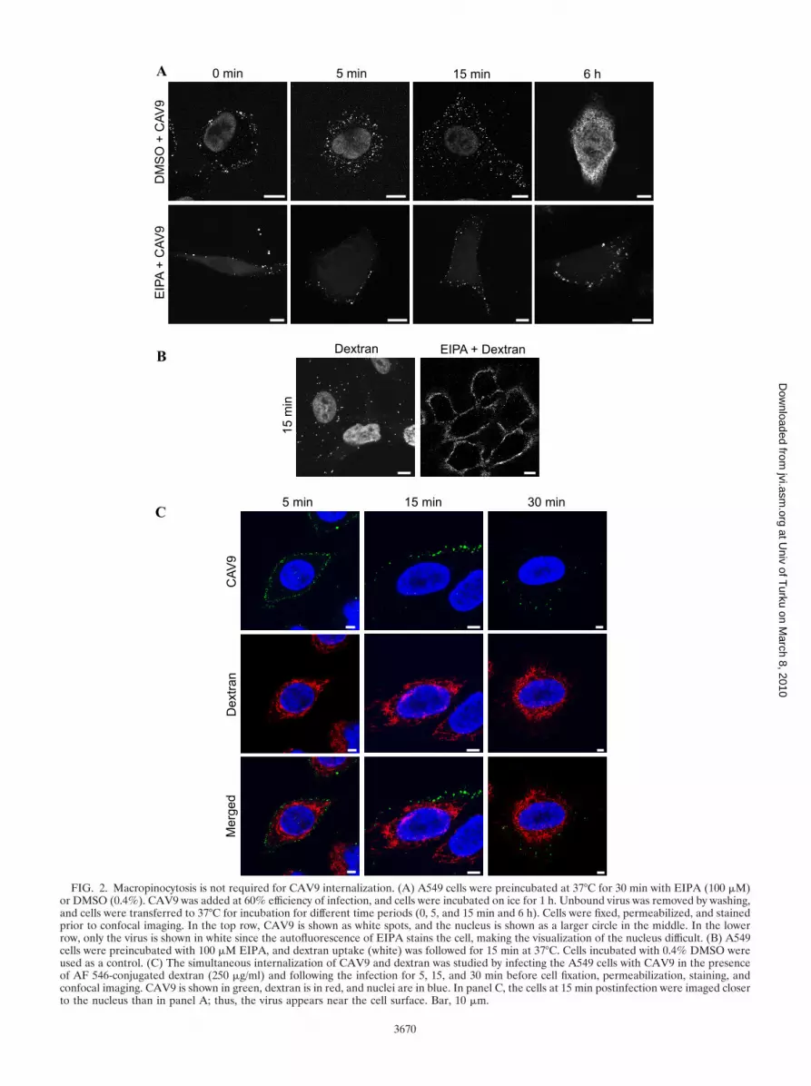
FIG. 2. Macropinocytosis is not required for CAV9 internalization. (A) A549 cells were preincubated at 37°C for 30 min with EIPA (100 �M)or DMSO (0.4%). CAV9 was added at 60% efficiency of infection, and cells were incubated on ice for 1 h. Unbound virus was removed by washing,and cells were transferred to 37°C for incubation for different time periods (0, 5, and 15 min and 6 h). Cells were fixed, permeabilized, and stainedprior to confocal imaging. In the top row, CAV9 is shown as white spots, and the nucleus is shown as a larger circle in the middle. In the lowerrow, only the virus is shown in white since the autofluorescence of EIPA stains the cell, making the visualization of the nucleus difficult. (B) A549cells were preincubated with 100 �M EIPA, and dextran uptake (white) was followed for 15 min at 37°C. Cells incubated with 0.4% DMSO wereused as a control. (C) The simultaneous internalization of CAV9 and dextran was studied by infecting the A549 cells with CAV9 in the presenceof AF 546-conjugated dextran (250 �g/ml) and following the infection for 5, 15, and 30 min before cell fixation, permeabilization, staining, andconfocal imaging. CAV9 is shown in green, dextran is in red, and nuclei are in blue. In panel C, the cells at 15 min postinfection were imaged closerto the nucleus than in panel A; thus, the virus appears near the cell surface. Bar, 10 �m.
3670
at Univ of T
urku on March 8, 2010
jvi.asm.org
Dow
nloaded from
cyclooctane) ([DABCO] Sigma-Aldrich) and examined with a Zeiss LSM510META confocal microscope using a Plan-Apochromat objective (63� oil objective;1.4 numerical aperture). The colocalization analysis was performed from three tofour images by quantifying the proportion of colocalizing channel intensities withBioImageXD software (32). The statistical significance of the colocalization wasevaluated using the algorithm of Costes et al.(15) embedded in the software. Theimages were converted to TIFF format for the adjustment of brightness and contrastin the ImageJ (http://rsb.info.nih.gov/ij) or Photoshop CS3 program.
RESULTS
The role of macropinocytosis in CAV9 internalization. Inorder to elucidate the endocytic pathway utilized by CAV9 inA549 cells, the effects of chemical inhibitors of endocytosiswere studied. The inhibitors used and their mode of action aredescribed in Table 1. As shown in Fig. 1A, EIPA and jas-
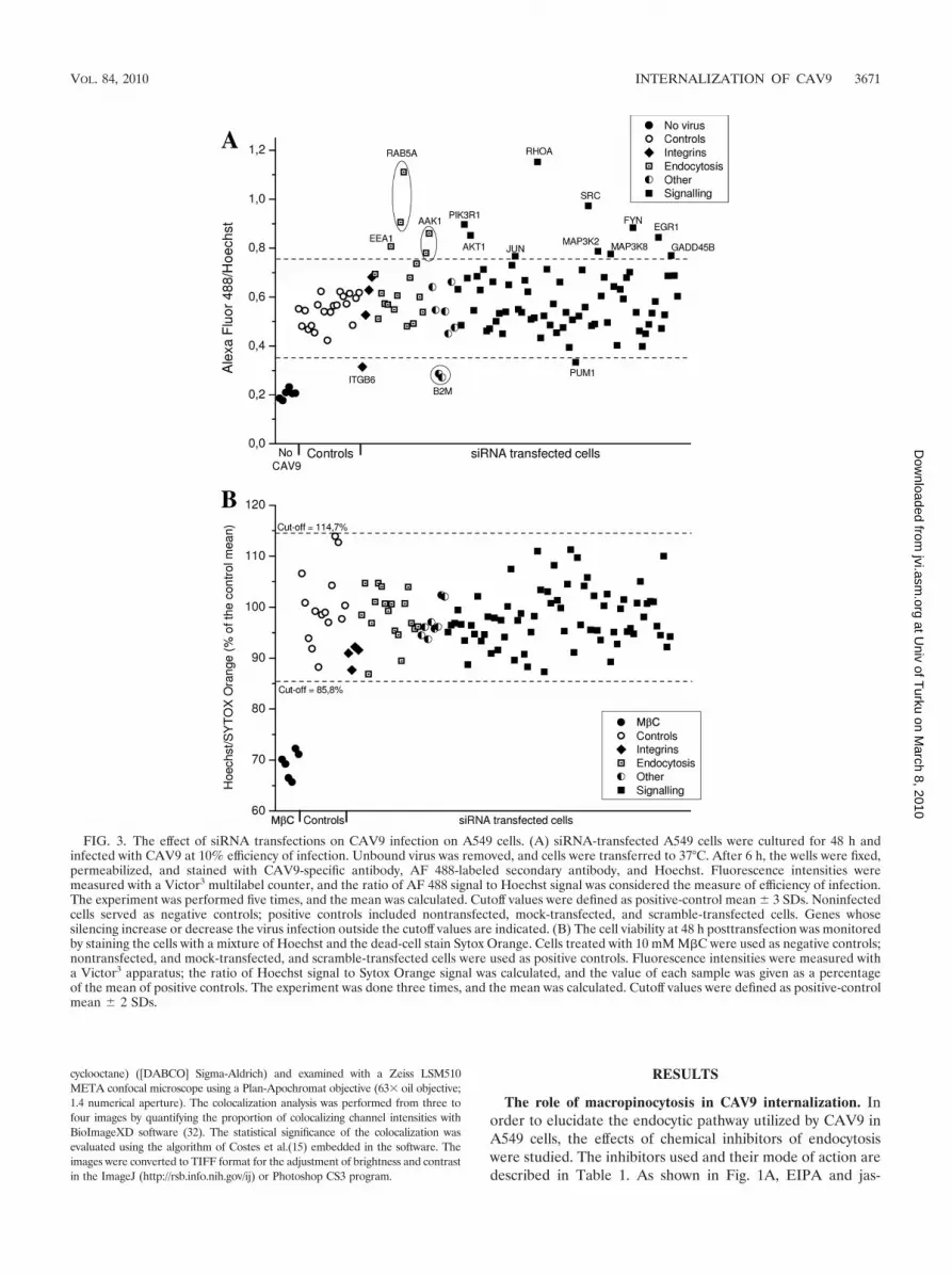
FIG. 3. The effect of siRNA transfections on CAV9 infection on A549 cells. (A) siRNA-transfected A549 cells were cultured for 48 h andinfected with CAV9 at 10% efficiency of infection. Unbound virus was removed, and cells were transferred to 37°C. After 6 h, the wells were fixed,permeabilized, and stained with CAV9-specific antibody, AF 488-labeled secondary antibody, and Hoechst. Fluorescence intensities weremeasured with a Victor3 multilabel counter, and the ratio of AF 488 signal to Hoechst signal was considered the measure of efficiency of infection.The experiment was performed five times, and the mean was calculated. Cutoff values were defined as positive-control mean � 3 SDs. Noninfectedcells served as negative controls; positive controls included nontransfected, mock-transfected, and scramble-transfected cells. Genes whosesilencing increase or decrease the virus infection outside the cutoff values are indicated. (B) The cell viability at 48 h posttransfection was monitoredby staining the cells with a mixture of Hoechst and the dead-cell stain Sytox Orange. Cells treated with 10 mM M�C were used as negative controls;nontransfected, and mock-transfected, and scramble-transfected cells were used as positive controls. Fluorescence intensities were measured witha Victor3 apparatus; the ratio of Hoechst signal to Sytox Orange signal was calculated, and the value of each sample was given as a percentageof the mean of positive controls. The experiment was done three times, and the mean was calculated. Cutoff values were defined as positive-controlmean � 2 SDs.
VOL. 84, 2010 INTERNALIZATION OF CAV9 3671
at Univ of T
urku on March 8, 2010
jvi.asm.org
Dow
nloaded from

plakinolide caused a statistically significant reduction in CAV9proliferation (P 1.6 � 10�5 and 0.013, respectively), whereaschlorpromazine, methyl-�-cyclodextrin (M�C), nocodazole,cytochalasin D, latrunculin A, wortmannin, and a mixture ofnystatin-progesterone had no statistically significant effect. Thefunctionality of chlorpromazine and M�C was verified bystudying their effects on the internalization of transferrin andcholera toxin B, and dextran was used to monitor the effect ofcytochalasin D. As expected, chlorpromazine inhibited the in-ternalization of transferrin but had no effect on cholera toxinB, whereas M�C inhibited the internalization of cholera toxinB but did not affect transferrin (Fig. 1B). Cytochalasin D to-tally blocked the internalization of dextran (Fig. 1C).
The inhibitory effect of EIPA was studied further by confo-cal imaging of virus infection at different time points. As shownin Fig. 2A, the virus infection was arrested in EIPA-treatedcells, and virus particles were seen in clusters or in vesicle-likestructures at or close to the cell periphery. This effect is similarto that found earlier with EV1 (33). To demonstrate that theconcentration of EIPA was effective, it was used to inhibit theinternalization of a fluid-phase marker, AF 568-conjugateddextran (Fig. 2B).
EIPA is inhibits macropinocytosis and is sometimes consid-ered the main diagnostic test for this pathway (42). Yet itsfunction has been shown to be cell type dependent and in some
cases unspecific (24). We found that even though actin-stabi-lizing jasplakinolide inhibited CAV9 infection in a statisticallysignificant manner, the actin-disrupting agents cytochalasin Dand latrunculin (Fig. 1A) did not cause statistically significantinhibition on CAV9 infection. This contradicts the involve-ment of macropinocytosis in CAV9 internalization. To furtherstudy the role of macropinocytosis in CAV9 entry, a cointer-nalization assay with CAV9 and dextran was performed. Dex-tran entered A549 cells clearly faster than CAV9 (Fig. 2C) asit had entered the cytoplasm by 5 min postinfection whenCAV9 was still closer to the cell surface. Furthermore, siRNAsagainst macropinocytosis-regulatory molecules (phosphatidyl-inositol 3-kinase [PI(3)K], Rac1, PacI, Cdc42, and Rab5) didnot reduce CAV9 proliferation (see below). In conclusion, ourresults suggest that the endocytosis of CAV9 to A549 cells doesnot depend on macropinocytosis.
siRNA screen. In order to obtain more information aboutthe cell entry mechanism of CAV9 and cellular moleculesinvolved in its infectious cycle, an siRNA panel was designed.The panel included siRNAs targeting central endocytosis ef-fectors and regulators as well as a selected set of signalingmolecules. Two individual siRNA molecules for each genewere used (see Table S1 in the supplemental material), andsiRNAs against reported CAV9 receptor genes, integrin sub-units �3 and �6, were chosen as controls. The effects of the
FIG. 4. CAV9 is not internalized into A549 cells in association with �V�6 integrin. A549 cells were grown on coverslips for 24 h and infectedwith CAV9 at 60% efficiency of infection. Unbound virus was removed, warm medium was added (0 min), and cells were transferred to 37°C. Thecells were incubated for 5 or 20 min before they were fixed and permeabilized. Cells were stained with virus-specific polyclonal antiserum and AF488-labeled secondary antibody (green) and with integrin �v�6-specific monoclonal antibody and AF 568-labeled secondary antibody (red) priorto confocal imaging. Bar, 10 �m.
3672 HEIKKILA ET AL. J. VIROL.
at Univ of T
urku on March 8, 2010
jvi.asm.org
Dow
nloaded from
siRNA panel on CAV9 infection of A549 cells are shown inFig. 3A and Table S1 in supplemental material. The siRNAsincreasing CAV9 proliferation are shown as values exceedingthe cutoff (positive-control mean � 3 SDs), and the siRNAsinhibiting the virus are shown as values below the cutoff. Thestrict cutoff was used to avoid the identification of false siRNAs, even at the expense of missing weak effects. The silencingefficiencies of some of the siRNAs were measured by qRT-PCR (Table S1 in supplemental material), but as exact dataabout protein knockdown levels are missing, the siRNA resultsneed to be considered preliminary. To evaluate cell viabilityafter siRNA transfections, the cells were stained with the dead-cell indicator Sytox Orange. As shown in Fig. 2B, the transfec-tions did not cause significant cell death, indicating that theobserved changes in virus proliferation were due to the silenc-ing of the specific gene and not to the overall effects on cellviability.
One out of two siRNAs against integrin �6 inhibited CAV9infection. The reason for the different outcome of the �6
siRNAs was most probably their different silencing efficiency(89% versus 57%). Neither of the �3 siRNAs suppressedCAV9 infection even though one of them had very good si-lencing efficiency (�99%). The other genes whose silencinginhibited virus proliferation were �2M and Pumilio homolog 1(PUM1). For �2M, both siRNAs silenced their target effi-ciently (91% and 80%) and showed a similar effect on virusinfection. The dependence on �2M will be discussed below.For the siRNAs targeting PUM1, the one showing higher si-lencing efficiency (85% versus 70%) inhibited CAV9 infection.However, as PUM1 was recently shown to be a central regu-lator that regulates the expression of over 10% of human genes(43), it is very difficult to deduce the reason for this suppres-sion. To our surprise, there were more siRNAs that enhancedCAV9 infection than inhibited it. The siRNAs that promotedvirus proliferation targeted genes involved in endocytosis andcell signaling and will be discussed below.
Role of integrins in CAV9 cell entry. CAV9 has previouslybeen shown to utilize �V�6 and �V�3 as its receptors (53, 61).However, our recent results demonstrate that the virus binds tointegrin �V�6 with higher affinity than to �V�3 (29). In oursiRNA screen, the silencing of integrin subunit �6 but not of�3 inhibited the virus infection (Fig. 3A), which strengthensthe hypothesis that �V�6 is the main receptor for CAV9 inA549 cells. A further support to this result came from anexperiment showing that function-blocking antibody against�V�6 could inhibit CAV9 infection by 60%, whereas �V�3antibody inhibited the virus by only 30%. Function-blocking�V antibody inhibited CAV9 by 80% (not shown).
To our surprise, double labeling of CAV9 and integrin �V�6in CAV9-infected A549 cells (Fig. 4) revealed that there is nosignificant colocalization or clustering between the virus andintegrin at the 0-min, 5-min, or 20-min time points (2%, 3%,and 6% of the virus colocalized with �V�6, respectively). Thecolocalization was calculated as the mean of four images ateach time point (data not shown). This finding is in clearcontrast to that with echovirus 1, which has been shown tocluster and internalize into the cells in association with itsreceptor integrin �2�1 (39), and with FMDV, which colocal-izes with �V�6 both on the cell surface and inside the cells at5 min postinfection (5). These results thus indicate that eventhough the integrin �V�6 is crucial for the CAV9 infection, thevirus and �V�6 are not internalized in association with eachother. Interestingly, the siRNAs targeting integrin-linked sig-naling molecules either did not alter CAV9 infection or in-creased it, with the enhancing effect being seen with one out oftwo siRNAs against Src, Fyn, RhoA, PI(3)K, and Akt1 (Fig.3A; see also Table S1 in the supplemental material). ThesiRNA results are supported by the finding that wortmannin, aPI(3)K inhibitor, did not inhibit CAV9 infection (Fig. 1A).Based on these observations, it seems that integrin-linked sig-naling is not required for CAV9 entry to A549 cells.
Collectively, the above results indicate that integrin �V�6and, to a lesser extent, �V�3 mediate CAV9 infection in A549cells. However, the virus is not internalized to the cell togetherwith the integrin, and integrin-linked signaling does not appearto have a role in CAV9 infection.
Clathrin-mediated endocytosis is not involved in CAV9 cellentry. Since the internalization of CAV9 into A549 cells didnot seem to follow the macropinocytic pathway, it was essential
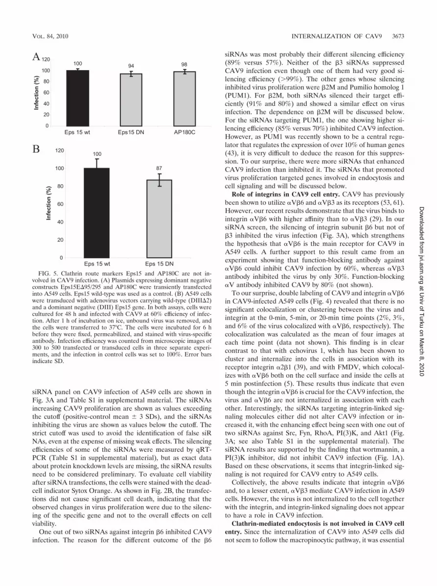
FIG. 5. Clathrin route markers Eps15 and AP180C are not in-volved in CAV9 infection. (A) Plasmids expressing dominant negativeconstructs Eps15E�95/295 and AP180C were transiently transfectedinto A549 cells. Eps15 wild-type was used as a control. (B) A549 cellswere transduced with adenovirus vectors carrying wild-type (DIII�2)and a dominant negative (DIII) Eps15 gene. In both assays, cells werecultured for 48 h and infected with CAV9 at 60% efficiency of infec-tion. After 1 h of incubation on ice, unbound virus was removed, andthe cells were transferred to 37°C. The cells were incubated for 6 hbefore they were fixed, permeabilized, and stained with virus-specificantibody. Infection efficiency was counted from microscopic images of300 to 500 transfected or transduced cells in three separate experi-ments, and the infection in control cells was set to 100%. Error barsindicate SD.
VOL. 84, 2010 INTERNALIZATION OF CAV9 3673
at Univ of T
urku on March 8, 2010
jvi.asm.org
Dow
nloaded from

3674 HEIKKILA ET AL. J. VIROL.
at Univ of T
urku on March 8, 2010
jvi.asm.org
Dow
nloaded from

to examine whether the central endocytic molecules, clathrin,caveolin, and dynamin, participate in the cell entry of the virus.Chlorpromazine did not inhibit viral infection (Fig. 1A), sug-gesting that clathrin-dependent endocytosis does not play arole in CAV9 internalization. To confirm this observation, theability of two DN constructs of clathrin route markers,Eps15E�95/295 and AP180C, to inhibit virus infection wasstudied. As shown in Fig. 5A, cells transfected with these con-structs were infected with CAV9 as efficiently as control cellstransfected with wt Eps15-GFP plasmid. A similar result wasobtained with cells transduced with adenovirus vectors carryingwt and DN Eps15 (Fig. 5B).
Silencing of genes involved in clathrin-mediated endocytosiseither increased CAV9 infection or did not have any effect onit (Fig. 3A; see also Table S1 in the supplemental material).siRNAs against EEA1 (1/2), Rab5 (2/2), and AAK1 (2/2) en-hanced CAV9 infection, whereas siRNAs against clathrinheavy chain showed no effect. The silencing efficiencies of thesiRNAs, however, were variable (Rab5, 98% and 52%; EEA1,67% and 17%; and AAK1, 35% and 0%); thus, the resultsneed to be interpreted with some caution. The silencing effi-ciencies of clathrin siRNAs were not measured.
Endosomal acidification is often linked to clathrin-mediatedendocytosis (48), and the dependence of an acidic pH mayindicate that a virus utilizes clathrin-mediated endocytosis. Theincubation of cells with 0.5 mM, 2 mM, 5 mM, and 25 mMNH4Cl did not inhibit CAV9 proliferation (data not shown).This indicates that CAV9 infection is not sensitive to inhibitionof endosomal acidification, giving further support to the as-sumption that CAV9 entry does not rely on clathrin-mediatedendocytosis. Altogether, the results obtained with chlorprom-azine, dominant negative Eps15 and AP180 constructs, andsiRNA transfections indicate that clathrin-mediated endocyto-sis does not play a role in CAV9 internalization.
Caveolae-mediated endocytosis is not required for CAV9internalization. Enteroviruses have been shown to be internal-ized through caveolin-mediated endocytosis in some cell lines(16–17, 39). Thus, we wanted to find out whether this is alsothe case for CAV9. As shown in Fig. 1A, a mixture of nystatin-progesterone, an inhibitor of caveolar endocytosis, did notsignificantly affect CAV9 infection. However, the overexpres-sion of the DN form of caveolin-3 inhibited the virus in astatistically significant way (P 0.0026), with inhibition ofapproximately 50% compared to the corresponding wt con-struct (Fig. 6A). Caveolin-3 is considered a muscle-specific
isoform, but the DN caveolin-3 construct used in this study hasearlier been shown to efficiently suppress caveolar endocytosisof simian virus 40 (SV40) infection in different cell types (54).
In the siRNA screen, caveolin-1 siRNAs did not affectCAV9 proliferation (Fig. 2A and Table S1 in the supplementalmaterial). The silencing efficiencies of the siRNAs were 83%and 75% by Western analysis (data not shown). Since theresults with the caveolin-3 DN construct and caveolin-1 siRNAs were rather contradictory, a caveolin-1-silenced cell line,A549-C9, was generated by using a retroviral RNAi vector. Acaveolin-1 silencing efficiency of about 69% in A549-C9 wasverified by confocal imaging and Western analysis (Fig. 6B). Asshown in Fig. 6C, CAV9 was endocytosed into the caveolin-1-silenced cell line as efficiently as into the vector control cells(A549-RVH1). To confirm that this result was not due to theresidual caveolin-1 expression, the ability of CAV9 to infectHuH7 cells, shown to be devoid of caveolae (60), was tested.The CAV9 infection in HuH7 cells was very efficient (Fig. 6D),demonstrating that caveolin-1 is not required for CAV9 inter-nalization.
Dynamin 2 is needed for CAV9 endocytosis. Dynamin is alarge GTPase that promotes the fission of endocytic mem-branes. It was originally considered to participate in clathrin-dependent endocytosis only but has recently been implicated inseveral other endocytic pathways (20). Since dynamin 2 hasbeen shown to be required for the internalization of numerousviruses, we analyzed its role in CAV9 infection. For this, theeffect of the dominant negative form of dynamin 2 was tested.The expression of DN dynamin 2aaK44A resulted in approxi-mately 50% inhibition compared to the corresponding wt con-struct (Fig. 7A), but the inhibition was not statistically signif-icant (P 0.082). In the siRNA panel, dynamin 2 siRNAs didnot exhibit any effect on virus proliferation (Fig. 3A and TableS1 in the supplemental material). The silencing efficiencies ofthe siRNAs were relatively good (80% and 82%), but it is stillpossible that the remaining 20% dynamin 2 expression is suf-ficient to promote the virus entry. Therefore, the role of dy-namin 2 in CAV9 infection was further studied by dynasore, acell-permeable, noncompetitive dynamin GTPase activity in-hibitor. Dynasore effectively blocked the virus infection andcaused CAV9 accumulation at the cell periphery (Fig. 7B).The function of dynasore was verified by its ability to inhibittransferrin uptake (Fig. 7C). The result with dynasore thusconfirmed that dynamin 2 is needed for CAV9 endocytosis.
FIG. 6. Caveolin-1 is not needed for CAV9 infection. (A) Plasmid constructs expressing wild-type and dominant negative caveolin-3 weretransiently transfected to A549 cells. After 48 h, the cells were infected with CAV9 at 60% efficiency of infection, incubated on ice for 1 h, andwashed. Warm medium was added, and the cells were transferred to 37°C. The cells were incubated for 6 h prior to fixation and staining. Infectionefficiency was counted from microscopic images of 100 to 200 transfected cells in three separate experiments, and the infection in wild-type controlcells was set to 100%. Error bar indicates SD. (B) The caveolin-1-silenced cell line A549-C9 was generated by infecting A549 cells with retrovirusvector carrying caveolin-1-silencing shRNA. The vector RVH1 was used as a control. The silencing efficiency was analyzed by confocal imagingand Western analysis. For confocal imaging, the cells were cultured for 24 h on coverslips, fixed, and permeabilized, after which caveolin-1 wasstained with rabbit polyclonal antiserum and AF 546-labeled secondary antibody (red). For Western analysis, protein samples (30 �g) wereseparated in a 15% SDS-PAGE gel and transferred to a Hybond-P membrane. Caveolin-1-specific antibody combined to HRP-labeled secondaryantibody was used for detection, and Erk1-specific antibody was used as a loading control. (C) Cell lines A549-RVH1 and A549-C9 were infectedwith CAV9 as above. The infection was allowed to proceed for 30 or 90 min before fixation, permeabilization, and immunostaining. CAV9 is shownin green, caveolin-1 is in red, and nuclei are in blue. Bar, 10 �m. (D) A549, A549-C9, and HuH-7 cells were infected with CAV9 as above. After6 h of incubation at 37°C, cells were fixed, permeabilized, and stained with CAV9-specific antibody. Infection efficiency was counted frommicroscopic images of 100 to 200 cells, and the infection in A549 cells was set to 100%.
VOL. 84, 2010 INTERNALIZATION OF CAV9 3675
at Univ of T
urku on March 8, 2010
jvi.asm.org
Dow
nloaded from

CAV9 internalization is dependent on �2-microglobulin andArf6. The dependence of CAV9 on �2-microglobulin has ear-lier been shown by antibody blocking (59). The role of �2M invirus infection has remained speculative, with indications thatit is not required for virus attachment to the cell surface but isinvolved in some postattachment step(s) (59). In the siRNAscreen, both �2M siRNAs efficiently inhibited CAV9 infectionin A549 cells (Fig. 3A and Table S1 in the supplemental ma-terial). In the antibody blocking assay, �2M antibody inhibitedCAV9 proliferation by 80% (data not shown).
Confocal imaging of cells transfected with �2M and nega-tive-control siRNAs showed that CAV9 was attached effi-ciently to the surface of cells lacking �2-microglobulin, but theendocytosis was arrested, and the virus still remained on the cellperiphery at 20 min postinfection. In comparison, in the controlsiRNA-transfected cells, the virus had already begun to enterthe cytoplasm after 5 min (Fig. 8). Interestingly, at 1 h postin-fection, the CAV9 endocytosis also continued in the �2M-silenced cells, but this delayed entry never resulted in theproduction of a new virus progeny (data not shown). Thismight indicate that CAV9 is directed to an unproductive en-docytic pathway in the absence of �2M. The proportion of thevirus that was colocalized with �2M at 0 min, 5 min, and 20 minpostinfection was quantified from three confocal images ateach time point, and the mean colocalization values were 40%,16%, and 18%, respectively (data not shown).
Arf6 (ADP-ribosylation factor 6) is a small GTPase involvedin the regulation of endosomal membrane traffic and structuralorganization of the cell (21). It is known to mediate the inter-nalization of several ligands, including major histocompatibil-ity class I (MHC-I) proteins and �1 integrin (8, 45, 52). Arf6was recently shown to be needed for baculovirus entry into 293cells (34) and to direct CBV3 into an unproductive internal-ization pathway (38). We studied the involvement of Arf6 inCAV9 endocytosis by measuring virus proliferation in cellstransfected with two individual Arf6 siRNAs. Figure 9A showsthat one of the siRNAs inhibited CAV9 infection in a statisti-
FIG. 7. CAV9 infection is dependent on dynamin 2. (A) A549 cellstransiently transfected with plasmids expressing wild-type and domi-nant negative (K44A) forms of dynamin-2 were infected with CAV9 at60% efficiency of infection and incubated on ice for 1 h. Unbound viruswas removed by washing, and cells were transferred to 37°C, wherethey were incubated for 6 h. Cells were fixed, permeabilized, andstained with CAV9-specific antibody, and confocal images were taken.Infection efficiency was counted from microscopic images of 100 to 200transfected cells in three separate experiments, and the infection inwild-type control cells was set to 100%. Error bar indicates SD.(B) A549 cells were preincubated with 80 �M dynasore or 0.4%DMSO for 30 min before CAV9 infection. The infection was per-formed as above and incubated at 37°C for 0 min, 5 min, 15 min, and6 h before fixation, permeabilization, staining with CAV9-specific an-tibody, and confocal imaging. Dynasore was present throughout theassay. The virus is shown in green, and nuclei are in blue. (C) To verifythe function of dynasore, A549 cells were preincubated with 80 �Mdynasore, and the control cells were preincubated with DMSO. AF546-conjugated transferrin was added and incubated for 2 min at roomtemperature. The cells were washed with medium containing dynasoreor DMSO, and the cells were incubated for 15 min at 37°C. The cellplate was then transferred onto ice, and transferrin bound to the cellsurface was removed by acidic washing. The internalization of trans-ferrin was visualized by confocal microscopy. Bar, 10 �m.
3676 HEIKKILA ET AL. J. VIROL.
at Univ of T
urku on March 8, 2010
jvi.asm.org
Dow
nloaded from

cally significant manner (P 2.4 � 10�4), whereas the otherhad no effect. The silencing efficiencies of the siRNAs were89% and 91% by qRT-PCR. In order to verify the siRNAresult, A549 cells were transfected with plasmid constructsoverexpressing wt and DN forms of Arf6. As shown in Fig. 9B,the expression of DN Arf6 resulted in an 85% decrease inCAV9 infection efficiency compared to cells expressing wtArf6. Confocal imaging and colocalization analysis of CAV9and Arf6 at 0 min, 5 min, and 20 min postinfection showed asimilar pattern of staining (Fig. 9C). The colocalization (cal-culated as the mean of four images at each time point [data notshown]) was modest, with 7%, 9%, and 9% of CAV9 colocal-izing with Arf6 at the indicated time points, respectively. Toconclude, the results obtained with Arf6 siRNA and overex-pression of DNA Arf6 indicate that CAV9 internalization inA549 cells is dependent on Arf6.
DISCUSSION
Enteroviruses utilize several mechanisms to enter their hostcells. The early steps of CAV9 infection have earlier beenreported to be dependent on �V integrins, �2-microglobulin,and lipid microdomains (29, 58–59, 61). In this work, we con-ducted a systematic study in order to identify the internaliza-tion mechanism of CAV9 in A549 lung carcinoma cells.
Integrins �V�3 and �V�6 have previously been shown to
function as receptors for CAV9 (53, 61). A recent work fromour group illustrated that soluble �V�6 but not �V�3 bound toimmobilized CAV9 and that soluble �V�6 but not �V�3blocked virus infectivity (29). The results presented in thisstudy show that siRNA silencing of integrin subunit �6 but notof �3 inhibited CAV9 infection, and a similar effect was seenwith antibody blocking. It thus seems that �V�6 and, to alesser extent, �V�3 mediate CAV9 infection in this cell model.The role of �V�6 as an internalizing agent was, however,complicated by the observation that no clustering or colocal-ization between integrin �V�6 and the virus was seen at theearly time points of infection. The situation seems thus sub-stantially different from FMDV, where �V�6 serves to deliverthe virus to acidic endosomes via clathrin-dependent endocy-tosis (5). The siRNA silencing of a number of integrin-linkedsignaling molecules did not inhibit CAV9 infection; on thecontrary, several of these siRNA molecules enhanced virusproliferation. This implies that integrin-linked signaling is notrequired for CAV9 infection in A549 cells. It has earlier beenshown by Triantafilou and Triantafilou that CAV9 infectionactivates the Raf/mitogen-activated protein kinase (MAPK)pathway in GMK cells (58). However, these investigators didnot examine whether this activation was essential for the in-fection. It is thus possible that signaling cascades become ac-tivated upon the contact of CAV9 and integrin, but this acti-vation does not promote virus entry and proliferation. In
FIG. 8. �2M is required for CAV9 infection. A549 cells were transfected with control siRNA or �2M siRNA (Hs_�2M_3) and cultured for48 h. The cells were infected with CAV9 at 60% efficiency of infection, incubated on ice for 1 h, and washed. Warm medium was added at the 0-mintime point; the cells were transferred to 37°C and incubated for 5 min, 20 min, and 6 h before fixing, permeabilization (except for the 0-min sample),staining with CAV9- and �2M-specific antibodies, and confocal imaging. CAV9 is shown in green, �2M is in red, and nuclei are in blue. A silencedcell in the 0-min images is indicated by an arrow. Bar, 10 �m.
VOL. 84, 2010 INTERNALIZATION OF CAV9 3677
at Univ of T
urku on March 8, 2010
jvi.asm.org
Dow
nloaded from

conclusion, integrin �V�6 is essential for CAV9 infection inA549 cells, but the virus is not internalized in association withit. In addition, integrin-linked signaling does not seem to beinvolved in the process. The exact role of integrin �V�6 inCAV9 infection is not yet understood and still needs furtherstudy. One explanation might be that it mediates the primarybinding and concentration of CAV9 on the cell surface. Sinceour protocol included an incubation of the infected cells for 1 hon ice before cells were fixed and stained, it is possible that aweak and transient association was missed.
The finding that EIPA inhibited CAV9 endocytosis suggeststhat CAV9 might enter A549 cells through the macropinocyticpathway. However, several lines of evidence illustrate that thisis not the case: siRNAs targeting regulators of macropinocy-tosis [Cdc42, Rac1, Pak1, and PI(3)K] did not reduce CAV9infection, and a similar result was obtained with wortmannin, aPI(3)K inhibitor. Chemicals inhibiting actin dynamics eitherhad no effect on virus proliferation or caused only partialinhibition. Furthermore, the kinetics of virus internalizationwas clearly different from that of dextran, a generally usedfluid-phase marker. The criteria for macropinocytic entry ofviruses, proposed by Mercer and Helenius, include the sensi-tivity to actin inhibitors and to inhibition of the above-men-tioned regulators (42). Thus, we conclude that CAV9 is notinternalized in A549 cells through macropinocytosis. Eventhough EIPA is generally considered an inhibitor of macropi-nocytosis, it may have a number of other effects, such as thealteration of the morphology and subcellular localization ofearly and late endosomes and lysosomes (24). In a recent work,EIPA was shown to inhibit CBV3 replication, most probably byinhibiting the virus RNA polymerase (27). Since CAV9 andCBV3 RNA polymerases are highly similar, having 95% aminoacid identity (11), it is possible that the inhibitory effect ofEIPA on CAV9 proliferation is due to its effect on CAV9replication.
In order to identify the internalization route of CAV9, weanalyzed whether clathrin- and caveolin-mediated pathwaysare involved. We found that chlorpromazine, endosomal acid-ification with NH4Cl, the expression of Eps15 and AP180 dom-inant negative constructs, and siRNA silencing of genes par-ticipating in the clathrin route all allowed efficient CAV9infection. This implies that clathrin-mediated endocytosis isnot involved in CAV9 cell entry.
Methyl-�-cyclodextrin has earlier been shown to inhibitCAV9 infection, which has been considered to indicate thedependence of the virus on lipid microdomains (58). In addi-tion, it has been suggested that caveolin-mediated entry isinvolved in the endocytosis of other enteroviruses (16–17, 39).In our work, silencing of caveolin-1 either by siRNA or retro-viral RNAi vector did not prevent virus proliferation. Further-more, CAV9 was able to efficiently infect the caveolin-1-neg-ative cell line HuH7. On the other hand, the overexpression ofDN caveolin-3 caused approximately 50% inhibition in CAV9infection. The reason for this inhibition is not totally clear, butit might resemble the situation with EV1, where DN caveolin-3does not prevent endocytosis but inhibits infection at laterstages (33). In conclusion, our results indicate that caveolin-1 isnot essential for CAV9 entry.
Dynamin is a high-molecular-weight GTPase involved inmembrane fission during endocytic events. Its function is nec-essary for clathrin-mediated endocytosis, but it has also beenimplicated in other internalization pathways, such as phagocy-tosis and caveolin-1-, ILR�2-, and flotillin-dependent path-ways (20). Since dynamin is a central regulator of endocytosisand is needed for the internalization of numerous viruses, wewanted to analyze whether it is involved in the entry process ofCAV9. The DN form of dynamin-2 inhibited virus infection byapproximately 50%. Even though this inhibition was not sta-tistically significant, it might suggest that dynamin 2 has a rolein CAV9 endocytosis. The dynamin 2 siRNAs had no effect onvirus proliferation, but this may be explained by the function-ality of residual dynamin. To further elucidate the role ofdynamin, we used dynasore, a recently introduced chemicalinhibitor. Dynasore completely blocked CAV9 infection andresulted in the trapping of the virus close to the cell surface.This finding thus confirmed that dynamin-2 is required for theinternalization of CAV9.
In our siRNA screen, the siRNAs targeting �2-microglobu-lin effectively inhibited CAV9 infection. The dependence ofCAV9 infection on �2M has earlier been shown by antibodyblocking (59, 61), but the role of �2M in virus infection has notbeen identified. Although CAV9 is not directly bound to �2Mon the cell surface (59), our confocal imaging revealed that�2M and the virus are in close association at early time pointsof infection. In cells depleted of �2M, endocytosis was ar-rested, and the virus remained close to the cell surface even at
FIG. 9. Arf6 mediates CAV9 endocytosis. (A) A549 cells transfected with negative-control siRNA and two individual Arf6 siRNAs wereinfected with a CAV9 at 10% efficiency of infection, incubated on ice for 1 h, and washed. Warm medium was added, and the cells were transferredto 37°C and incubated for 6 h, after which they were fixed and permeabilized. The cells were stained with CAV9-specific antibody, AF 488-labeledsecondary antibody, and Hoechst. Fluorescence intensities were measured with a Victor3 multilabel counter, and the ratio of AF 488 signal toHoechst signal was considered the measure of efficiency of infection. For each sample, 33 wells in three separate assays were analyzed. Statisticalsignificance between control siRNA- and Hs_ARF6_5 and Hs_ARF6_7 siRNA-silenced cells was calculated with a paired-sample t test, in whicha P value of 0.05 was considered significant. In the box plot, mean, median, upper and lower quartiles, upper and lower 95% values, and maximaland minimal values are indicated by square, horizontal line, box boundaries, vertical lines, and a cross, respectively. Statistically significantdifference is shown with an asterisk. (B) Plasmids expressing HA-conjugated wild-type and dominant negative Arf6 were transfected into A549 cellsand cultured for 48 h. The cells were infected with CAV9 at 60% efficiency of infection by a procedure described above. Cells were stained withCAV9-specific antibody combined with AF 488-labeled secondary antibody and with HA-specific antibody combined with AF 568–labeledsecondary antibody and Hoechst. The efficiency of CAV9 infection was calculated by counting the proportion of infected cells in cells transfectedwith wt and DN Arf6 (total values of six and seven images, respectively). HA tag is shown in red, CAV9 is in green, and nuclei are in blue. (C) A549cells were infected with CAV9 as above and incubated at 37°C for 0 min, 5 min, and 20 min prior to fixation, permeabilization (not in the 0-minsample), and staining with antibodies specific to CAV9 and Arf6 and with Hoechst. Arf6 is shown in red, CAV9 is in green, and nuclei are in blue.Bar, 10 �m.
VOL. 84, 2010 INTERNALIZATION OF CAV9 3679
at Univ of T
urku on March 8, 2010
jvi.asm.org
Dow
nloaded from

20 min postinfection. After 1 h, the virus also began to enterthe �2M-silenced cells, but this delayed entry did not result inthe production of new virus progeny. Thus, without �2M,CAV9 seems to be directed to an unproductive endocytic path-way The existence of such an unproductive pathway is sup-ported by the early finding of Hecker et al., who showed thatmost endocytosed CAV9 particles became accumulated in ly-sosomes without causing an infection (28).
Arf6 is a small GTPase that has multiple roles in the regu-lation of endosomal membrane traffic and other cellular func-tions (19). The dependence of CAV9 infection on �2M and thefinding that Arf6 regulates the endocytosis of MHC-I proteins(44) directed us to study whether Arf6 is involved in the entryprocess of the virus. One of two Arf6 siRNAs inhibited CAV9infection in a statistically significant manner, and overexpres-sion of DN Arf6 resulted in a clear decrease in the infectionefficiency compared to the corresponding wt construct. Theresults thus indicate that Arf6 controls the endocytosis ofCAV9 to A549 cells.
In conclusion, we propose a model for CAV9 entry intoA549 cells in which the virus is first attached to the cell surfaceby a process requiring integrin �V�6 and then rapidly trans-ferred forward to form an association with �2M It is possiblethat other molecules are also involved. The virus is then inter-nalized by an Arf6-controlled route, perhaps still in associationwith �2M. The function of dynamin 2 is needed for the path-way, but its exact role and relation to Arf6 are still unclear.Although dynamin is not generally thought to be involved inArf6-dependent endocytosis, there is a report by Nishi andSaigo showing that herpes simplex virus structural proteinVP22 is internalized in HeLa cells in an Arf6- and dynamin-dependent manner (46). Interestingly, the endocytoses ofCAV9 and VP22 appear very similar in being independent ofclathrin, caveolin, and the Rho family GTPases RhoA, Rac1,and Cdc42 while requiring dynamin and Arf6. We thus presentthe first demonstration of an infectious virus entering the cellvia a dynamin/Arf6-dependent endocytosis pathway.
ACKNOWLEDGMENTS
Elina Ikonen (University of Helsinki, Finland), Aki Manninen (Uni-versity of Oulu, Finland), Lucia Fiore (Instituto Superiore di Sanita,Rome, Italy), Robert Parton (University of Queensland, Brisbane,Australia), Mark McNiven (Mayo Clinic, Rochester, MN), Alice Dau-try-Varsat (Institute Pasteur, Paris, France), Dieter Blaas (Universityof Vienna, Austria), and Yves Rouille (Institute Pasteur de Lille,France) are warmly thanked for providing cell lines, antibodies, andexpression constructs. We acknowledge Harri Savilahti and Ari Hele-nius for their comments concerning the manuscript and Ritva Ka-jander for her assistance in the laboratory work.
This work was supported by research grants from the Academy ofFinland (number 122215 to T.H., 114721 to V.M., and 128539 to P.S.),the Sigrid Juselius Foundation, and Turku University GraduateSchool.
REFERENCES
1. Abraham, G., and R. J. Colonno. 1984. Many rhinovirus serotypes share thesame cellular receptor. J. Virol. 51:340–345.
2. Benmerah, A., M. Bayrou, N. Cerf-Bensussan, and A. Dautry-Varsat. 1999.Inhibition of clathrin-coated pit assembly by an Eps15 mutant. J. Cell Sci.112:1303–1311.
3. Benmerah, A., C. Lamaze, B. Begue, S. L. Schmid, A. Dautry-Varsat, and N.Cerf-Bensussan. 1998. AP-2/Eps15 interaction is required for receptor-me-diated endocytosis. J. Cell Biol. 140:1055–1062.
4. Bergelson, J. M. 2008. New (fluorescent) light on poliovirus entry. TrendsMicrobiol. 16:44–47.
5. Berryman, S., S. Clark, P. Monaghan, and T. Jackson. 2005. Early events inintegrin �v�6-mediated cell entry of foot-and-mouth disease virus. J. Virol.79:8519–8534.
6. Blomqvist, S., A. Paananen, C. Savolainen-Kopra, T. Hovi, and M.Roivainen. 2008. Eight years of experience with molecular identification ofhuman enteroviruses. J. Clin. Microbiol. 46:2410–2413.
7. Brandenburg, B., L. Y. Lee, M. Lakadamyali, M. J. Rust, X. Zhuang, andJ. M. Hogle. 2007. Imaging poliovirus entry in live cells. PLoS Biol. 5:e183.
8. Brown, F. D., A. L. Rozelle, H. L. Yin, T. Balla, and J. G. Donaldson. 2001.Phosphatidylinositol 4,5-bisphosphate and Arf6-regulated membrane traffic.J. Cell Biol. 154:1007–1017.
9. Buttinelli, G., V. Donati, F. M. Ruggeri, P. Joki-Korpela, T. Hyypia, and L.Fiore. 2003. Antigenic sites of coxsackie A9 virus inducing neutralizingmonoclonal antibodies protective in mice. Virology 312:74–83.
10. Cao, H., F. Garcia, and M. A. McNiven. 1998. Differential distribution ofdynamin isoforms in mammalian cells. Mol. Biol. Cell 9:2595–2609.
11. Chang, K. H., P. Auvinen, T. Hyypia, and G. Stanway. 1989. The nucleotidesequence of coxsackievirus A9; implications for receptor binding and entero-virus classification. J. Gen. Virol. 70:3269–3280.
12. Chevaliez, S., J. Balanant, P. Maillard, Y. C. Lone, F. A. Lemonnier, and F.Delpeyroux. 2008. Role of class I human leukocyte antigen molecules in earlysteps of echovirus infection of rhabdomyosarcoma cells. Virology 381:203–214.
13. Chung, S. K., J. Y. Kim, I. B. Kim, S. I. Park, K. H. Paek, and J. H. Nam.2005. Internalization and trafficking mechanisms of coxsackievirus B3 inHeLa cells. Virology 333:31–40.
14. Conner, S. D., and S. L. Schmid. 2003. Regulated portals of entry into thecell. Nature 422:37–44.
15. Costes, S. V., D. Daelemans, E. H. Cho, Z. Dobbin, G. Pavlakis, and S.Lockett. 2004. Automatic and quantitative measurement of protein-proteincolocalization in live cells. Biophys. J. 86:3993–4003.
16. Coyne, C. B., and J. M. Bergelson. 2006. Virus-induced Abl and Fyn kinasesignals permit coxsackievirus entry through epithelial tight junctions. Cell124:119–131.
17. Coyne, C. B., K. S. Kim, and J. M. Bergelson. 2007. Poliovirus entry intohuman brain microvascular cells requires receptor-induced activation ofSHP-2. EMBO J. 26:4016–4028.
18. Coyne, C. B., L. Shen, J. R. Turner, and J. M. Bergelson. 2007. Coxsack-ievirus entry across epithelial tight junctions requires occludin and the smallGTPases Rab34 and Rab5. Cell Host Microbe 2:181–192.
19. Dicara, D., A. Burman, S. Clark, S. Berryman, M. J. Howard, I. R. Hart, J. F.Marshall, and T. Jackson. 2008. Foot-and-mouth disease virus forms ahighly stable, EDTA-resistant complex with its principal receptor, integrin�v�6: implications for infectiousness. J. Virol. 82:1537–1546.
20. Doherty, G. J., and H. T. McMahon. 2009. Mechanisms of endocytosis.Annu. Rev. Biochem. 78:857–902.
21. D’Souza-Schorey, C., and P. Chavrier. 2006. ARF proteins: roles in mem-brane traffic and beyond. Nat. Rev. Mol. Cell Biol. 7:347–358.
22. Eisenhut, M., B. Algawi, T. Wreghitt, J. Foweraker, T. McKee, R. Miles, andJ. Challener. 2000. Fatal coxsackie A9 virus infection during an outbreak ina neonatal unit. J. Infect. 40:297–298.
23. Ford, M. G., B. M. Pearse, M. K. Higgins, Y. Vallis, D. J. Owen, A. Gibson,C. R. Hopkins, P. R. Evans, and H. T. McMahon. 2001. Simultaneousbinding of PtdIns(4,5)P2 and clathrin by AP180 in the nucleation of clathrinlattices on membranes. Science 291:1051–1055.
24. Fretz, M., J. Jin, R. Conibere, N. A. Penning, S. Al-Taei, G. Storm, S. Futaki,T. Takeuchi, I. Nakase, and A. T. Jones. 2006. Effects of Na�/H� exchangerinhibitors on subcellular localisation of endocytic organelles and intracellulardynamics of protein transduction domains HIV-TAT peptide and octaargi-nine. J. Control. Release 116:247–254.
25. Furman, C., S. M. Short, R. R. Subramanian, B. R. Zetter, and T. M.Roberts. 2002. DEF-1/ASAP1 is a GTPase-activating protein (GAP) forARF1 that enhances cell motility through a GAP-dependent mechanism.J. Biol. Chem. 277:7962–7969.
26. Grist, N. R., E. J. Bell, and F. Assaad. 1978. Enteroviruses in human disease.Prog. Med. Virol. 24:114–157.
27. Harrison, D. N., E. V. Gazina, D. F. Purcell, D. A. Anderson, and S. Petrou.2008. Amiloride derivatives inhibit coxsackievirus B3 RNA replication. J. Vi-rol. 82:1465–1473.
28. Hecker, W., J. Meyer, R. Boeni, and K. Bienz. 1974. Pinocytotic uptake andintralysosomal crystal formation of coxsackievirus A9 in monkey kidney cells.An electron microscopic autoradiographic study. Arch. Gesamte Virusfor-sch. 46:167–174.
29. Heikkila, O., P. Susi, G. Stanway, and T. Hyypia. 2009. Integrin �V�6 is ahigh-affinity receptor for coxsackievirus A9. J. Gen. Virol. 90:197–204.
30. Hendry, E., H. Hatanaka, E. Fry, M. Smyth, J. Tate, G. Stanway, J. Santti,M. Maaronen, T. Hyypia, and D. Stuart. 1999. The crystal structure ofcoxsackievirus A9: new insights into the uncoating mechanisms of enterovi-ruses. Structure 7:1527–1538.
31. Johns, H. L., S. Berryman, P. Monaghan, G. J. Belsham, and T. Jackson.2009. A dominant-negative mutant of rab5 inhibits infection of cells by
3680 HEIKKILA ET AL. J. VIROL.
at Univ of T
urku on March 8, 2010
jvi.asm.org
Dow
nloaded from

foot-and-mouth disease virus: implications for virus entry. J. Virol. 83:6247–6256.
32. Kankaanpaa, P., K. Pahajoki, V. Marjomaki, J. Heino, D. White, and X. D.BioImage. 2006. BioImageXD. University of Jyvaskyla, Jyvaskyla, Finland,and University of Turku, Turku, Finland. http://www.bioimagexd.net.
33. Karjalainen, M., E. Kakkonen, P. Upla, H. Paloranta, P. Kankaanpaa, P.Liberali, G. H. Renkema, T. Hyypia, J. Heino, and V. Marjomaki. 2008. ARaft-derived, Pak1-regulated entry participates in �2�1 integrin-dependentsorting to caveosomes. Mol. Biol. Cell 19:2857–2869.
34. Laakkonen, J. P., A. R. Makela, E. Kakkonen, P. Turkki, S. Kukkonen, J.Peranen, S. Yla-Herttuala, K. J. Airenne, C. Oker-Blom, M. Vihinen-Ranta,and V. Marjomaki. 2009. Clathrin-independent entry of baculovirus triggersuptake of E. coli in non-phagocytic human cells. PLoS One 4:e5093.
35. Lau, A. W., and M. M. Chou. 2008. The adaptor complex AP-2 regulatespost-endocytic trafficking through the non-clathrin Arf6-dependent endo-cytic pathway. J. Cell Sci. 121:4008–4017.
36. Lecot, S., S. Belouzard, J. Dubuisson, and Y. Rouille. 2005. Bovine viraldiarrhea virus entry is dependent on clathrin-mediated endocytosis. J. Virol.79:10826–10829.
37. Manninen, A., P. Verkade, S. Le Lay, J. Torkko, M. Kasper, J. Fullekrug,and K. Simons. 2005. Caveolin-1 is not essential for biosynthetic apicalmembrane transport. Mol. Cell. Biol. 25:10087–10096.
38. Marchant, D., A. Sall, X. Si, T. Abraham, W. Wu, Z. Luo, T. Petersen, R. G.Hegele, and B. M. McManus. 2009. ERK MAP kinase-activated Arf6 traf-ficking directs coxsackievirus type B3 into an unproductive compartmentduring virus host-cell entry. J. Gen. Virol. 90:854–862.
39. Marjomaki, V., V. Pietiainen, H. Matilainen, P. Upla, J. Ivaska, L. Nissinen,H. Reunanen, P. Huttunen, T. Hyypia, and J. Heino. 2002. Internalization ofechovirus 1 in caveolae. J. Virol. 76:1856–1865.
40. Marsh, M., and A. Helenius. 2006. Virus entry: open sesame. Cell 124:729–740.
41. Mayor, S., and R. E. Pagano. 2007. Pathways of clathrin-independent endo-cytosis. Nat. Rev. Mol. Cell Biol. 8:603–612.
42. Mercer, J., and A. Helenius. 2009. Virus entry by macropinocytosis. Nat. CellBiol. 11:510–520.
43. Morris, A. R., N. Mukherjee, and J. D. Keene. 2008. Ribonomic analysis ofhuman Pum1 reveals cis-trans conservation across species despite evolutionof diverse mRNA target sets. Mol. Cell. Biol. 28:4093–4103.
44. Naslavsky, N., R. Weigert, and J. G. Donaldson. 2004. Characterization of anonclathrin endocytic pathway: membrane cargo and lipid requirements.Mol. Biol. Cell 15:3542–3552.
45. Naslavsky, N., R. Weigert, and J. G. Donaldson. 2003. Convergence ofnon-clathrin- and clathrin-derived endosomes involves Arf6 inactivation andchanges in phosphoinositides. Mol. Biol. Cell 14:417–431.
46. Nishi, K., and K. Saigo. 2007. Cellular internalization of green fluorescentprotein fused with herpes simplex virus protein VP22 via a lipid raft-medi-
ated endocytic pathway independent of caveolae and Rho family GTPasesbut dependent on dynamin and Arf6. J. Cell Biol. 282:27503–27517.
47. Ochoa, G. C., V. I. Slepnev, L. Neff, N. Ringstad, K. Takei, L. Daniell, W.Kim, H. Cao, M. McNiven, R. Baron, and P. De Camilli. 2000. A functionallink between dynamin and the actin cytoskeleton at podosomes. J. Cell Biol.150:377–389.
48. Pelkmans, L., and A. Helenius. 2003. Insider information: what viruses tell usabout endocytosis. Curr. Opin. Cell Biol. 15:414–422.
49. Pietiainen, V., V. Marjomaki, P. Upla, L. Pelkmans, A. Helenius, and T.Hyypia. 2004. Echovirus 1 endocytosis into caveosomes requires lipid rafts,dynamin II, and signaling events. Mol. Biol. Cell 15:4911–4925.
50. Pietiainen, V. M., V. Marjomaki, J. Heino, and T. Hyypia. 2005. Viral entry,lipid rafts and caveosomes. Ann. Med. 37:394–403.
51. Pulli, T., H. Lankinen, M. Roivainen, and T. Hyypia. 1998. Antigenic sites ofcoxsackievirus A9. Virology 240:202–212.
52. Radhakrishna, H., and J. G. Donaldson. 1997. ADP-ribosylation factor 6 reg-ulates a novel plasma membrane recycling pathway. J. Cell Biol. 139:49–61.
53. Roivainen, M., L. Piirainen, T. Hovi, I. Virtanen, T. Riikonen, J. Heino, andT. Hyypia. 1994. Entry of coxsackievirus A9 into host cells: specific interac-tions with �v�3 integrin, the vitronectin receptor. Virology 203:357–365.
54. Roy, S., R. Luetterforst, A. Harding, A. Apolloni, M. Etheridge, E. Stang, B.Rolls, J. F. Hancock, and R. G. Parton. 1999. Dominant-negative caveolininhibits H-Ras function by disrupting cholesterol-rich plasma membranedomains. Nat. Cell Biol. 1:98–105.
55. Sandvig, K., M. L. Torgersen, H. A. Raa, and B. van Deurs. 2008. Clathrin-independent endocytosis: from nonexisting to an extreme degree of com-plexity. Histochem. Cell Biol. 129:267–276.
56. Schuck, S., A. Manninen, M. Honsho, J. Fullekrug, and K. Simons. 2004.Generation of single and double knockdowns in polarized epithelial cells byretrovirus-mediated RNA interference. Proc. Natl. Acad. Sci. U. S. A. 101:4912–4917.
57. Triantafilou, K., D. Fradelizi, K. Wilson, and M. Triantafilou. 2002. GRP78,a coreceptor for coxsackievirus A9, interacts with major histocompatibilitycomplex class I molecules which mediate virus internalization. J. Virol.76:633–643.
58. Triantafilou, K., and M. Triantafilou. 2003. Lipid raft microdomains: keysites for coxsackievirus A9 infectious cycle. Virology 317:128–135.
59. Triantafilou, M., K. Triantafilou, K. M. Wilson, Y. Takada, N. Fernandez,and G. Stanway. 1999. Involvement of �2-microglobulin and integrin �v�3molecules in the coxsackievirus A9 infectious cycle. J. Gen. Virol. 80:2591–2600.
60. Vainio, S., S. Heino, J. E. Mansson, P. Fredman, E. Kuismanen, O. Vaarala,and E. Ikonen. 2002. Dynamic association of human insulin receptor withlipid rafts in cells lacking caveolae. EMBO Rep. 3:95–100.
61. Williams, C. H., T. Kajander, T. Hyypia, T. Jackson, D. Sheppard, and G.Stanway. 2004. Integrin �v�6 is an RGD-dependent receptor for coxsack-ievirus A9. J. Virol. 78:6967–6973.
VOL. 84, 2010 INTERNALIZATION OF CAV9 3681
at Univ of T
urku on March 8, 2010
jvi.asm.org
Dow
nloaded from


