Embed Size (px)
Citation preview

European Journal of Cancer (2014) 50, 2695– 2704
A v a i l a b l e a t w w w . s c i e n c e d i r e c t . c o m
ScienceDirect
journa l homepag e : www.e j cancer . com
Original Research
International variation in management of screen-detectedductal carcinoma in situ of the breast
http://dx.doi.org/10.1016/j.ejca.2014.07.019
0959-8049/� 2014 Elsevier Ltd. All rights reserved.
⇑ Corresponding author: Address: Unit of Cancer Epidemiology, CPO Piemonte, AOU Citta della Salute e della Scienza, via San FrancPaola 31, 10123 Torino, Italy. Tel.: +39 011 6333866; fax: +39 011 6333861.
E-mail address: [email protected] (A. Ponti).1 See Appendix A.
Antonio Ponti a,⇑, Elsebeth Lynge b, Ted James c, Ondrej Majek d,My von Euler-Chelpin b, Ahti Anttila e, Patricia Fitzpatrick f, Maria Piera Mano a,Masaaki Kawai g, Astrid Scharpantgen h, Jacques Fracheboud i, Solveig Hofvind j,Carmen Vidal k, Nieves Ascunce l, Dolores Salas m, Jean-Luc Bulliard n, Nereo Segnan a,Karla Kerlikowske o,p, Stephen Taplin q, the ICSN DCIS Working group,1
a CPO Piemonte, AOU Citta della Salute e della Scienza, Torino, Italyb Department of Public Health, University of Copenhagen, Copenhagen, Denmarkc Department of Surgery, University of Vermont, Burlington, VT, USAd Institute of Biostatistics and Analyses, Masaryk University, Brno, Czech Republice Mass Screening Registry, Finnish Cancer Registry, Helsinki, Finlandf National Cancer Screening Service, Dublin, Irelandg Department of Surgical Oncology, Tohoku University Graduate School of Medicine, Sendai, Miyagi, Japanh Programme Mammographie, Direction de la Sante, Luxembourgi Erasmus Medical Centre, Rotterdam, The Netherlandsj The Cancer Registry of Norway, Oslo, Norwayk Cancer Detection and Control Programme, Catalan Institute of Oncology, Barcelona, Spainl Breast Cancer Screening Programme, Instituto de Salud Publica, Navarra, Spainm General Directorate Research and Public Health and Centre for Public Health Research, Valencia, Spainn Lausanne University Hospital, Lausanne, Switzerlando Department of Medicine, University of California San Francisco, San Francisco, CA, USAp Department of Epidemiology and Biostatistics, University of California San Francisco, San Francisco, CA, USAq Behavioral Research Program, Division of Cancer Control and Population Sciences, National Cancer Institute, Bethesda, MD, USA
Received 22 May 2014; received in revised form 16 July 2014; accepted 18 July 2014Available online 19 August 2014
KEYWORDS
Breast cancerDuctal carcinoma in situ(DCIS)Screening mammography
Abstract Background: Ductal carcinoma in situ (DCIS) incidence has grown with theimplementation of screening and its detection varies across International Cancer ScreeningNetwork (ICSN) countries. The aim of this survey is to describe the management of screen-detected DCIS in ICSN countries and to evaluate the potential for treatment related morbidity.
esco da

2696 A. Ponti et al. / European Journal of Cancer 50 (2014) 2695–2704
OvertreatmentAxillary stagingCancer registration
Methods: We sought screen-detected DCIS data from the ICSN countries identified during2004–2008. We adopted standardised data collection forms and analysis and explored DCISdiagnosis and treatment processes ranging from pre-operative diagnosis to type of surgeryand radiotherapy.Results: Twelve countries contributed data from a total of 15 screening programmes, all fromEurope except the United States of America and Japan. Among women aged 50–69 years,7,176,050 screening tests and 5324 screen-detected DCIS were reported. From 21% to 93% ofDCIS had a pre-operative diagnosis (PO); 67–90% of DCIS received breast conservation surgery(BCS), and in 41–100% of the cases this was followed by radiotherapy; 6.4–59% received sentinellymph node biopsy (SLNB) only and 0.8–49% axillary dissection (ALND) with 0.6% (range byprogrammes 0–8.1%) being node positive. Among BCS patients 35% received SLNB only and4.8% received ALND. Starting in 2006, PO and SLNB use increased while ALND remained sta-ble. SLNB and ALND were associated with larger size and higher grade DCIS lesions.Conclusions: Variation in DCIS management among screened women is wide and includeslymph node surgery beyond what is currently recommended. This indicates the presence of vary-ing levels of overtreatment and the potential for its reduction.
� 2014 Elsevier Ltd. All rights reserved.
1. Introduction
Ductal carcinoma in situ (DCIS) has become arelatively common disease after the introduction ofscreening mammography, representing up to 20–25%of all incident breast malignancies in industrialisedcountries [1–4]. The natural history of screen-detectedDCIS is not yet completely understood [5] and we aretherefore in large part unable to distinguish differentconditions that are likely to exist under the same labelof DCIS [6,7].
Management guidelines increasingly take this uncer-tainty into account by trying both to provide adequatecare and to avoid unnecessary treatment. For example,axillary lymph node dissection (ALND) is not recom-mended for women with DCIS [8–10]. The InternationalCancer Screening Network (ICSN) oversees organisedprogrammes that include quality monitoring of the pro-cess of screening and care. The purpose of the report isto assess practice variation in the management of screen-detected DCIS and the potential morbidity associatedwith detection of DCIS among participants in the ICSN.
2. Patients and methods
A survey was launched within the ICSN. All of thescreening settings covered were population-based,organised screening programmes, with the exception ofCzech Republic, which at the time did not adopt per-sonal invitations, and of the United States, whose data,provided by the Breast Cancer Surveillance Consortium,derived from opportunistic screening in well definedpopulations.
Selected characteristics of participating programmeswere collated from the ICSN web site (http://appliedre-search.cancer.gov/icsn) and reported in Table 1. Atten-dance rates exceeded 60% in all programmes for which
this information was available with the exceptions ofSwitzerland and Japan.
A previous paper [4] on DCIS detection reports indetail the design of this survey. In brief, we sought datafrom the 33 ICSN member countries regarding the pureDCIS cases they identified within their screened popula-tion between January 1, 2004 and December 31, 2008.We asked sites to complete, based on individual datarecords from their screening and clinical databases oftenobtained by linkage with population-based cancer regis-tries, a structured questionnaire that summarised dataon DCIS detection, diagnosis and treatment. The ques-tionnaire was piloted in a regional screening programmebefore distribution. Internal data consistency waschecked routinely and outlying data were verified withdata providers. All data were stratified by calendar yearand age in decades, both referred to the date of the screen-ing test. The following data stratifications were alsoincluded in the questionnaire: type of breast surgery byDCIS size; nodal surgery by DCIS size; nodal surgeryby nuclear grade; nodal surgery by type of breast surgery;and radiotherapy by type of breast surgery. As size byclinical imaging was often unavailable, all sites wereasked to provide pathological size (610 mm, 11–20 mm,>20 mm).
For the analysis of DCIS management process weselected a number of measures encompassing issuesranging from diagnosis to surgical and adjuvant treat-ment, namely: pre-operative diagnosis (PO); time fromabnormal screen to surgery; use of breast conservingsurgery (BCS) as definitive intervention; use of ALNDand sentinel lymph nodes biopsy (SLNB); radiotherapyafter BCS. Indicators were identified, following a sys-tematic literature review, from two main sources [9,10],by selecting measures believed to be collectable retro-spectively from participating screening programmes.A pre-operative diagnosis was defined as the presence

Table 1International cancer screening network survey on the management of ductal carcinoma in situ (DCIS). Description of the screening programmesincluded in the analysis, number of reported tests and number of screen-detected DCIS.
Country/region Yearprogrammestarted
Target agegroup
Attendancerate (2010)
Datacollectionyears
No. of reported tests(age 50–69)
No. of screen- detectedDCIS (age 50–69)
Czech Republic 2002 45–69 Not available 2007–2008 699,726 359Denmark
Copenhagen1991 50–69 73% 2004–2007 47,249 73
Denmark Fyn 1993 50–69 2004–2007 97,176 63Finland 1987 50–69a 85% 2004–2007 862,908 361Ireland 2000 50–64 Not available 2004–2008 331,854 393Italyb 1990 50–69 61% 2006–2008 1,453,292 1,066Japanc 2000 50–69 19% 2004–2008 106,898 72Luxembourg 1992 50–69 64% 2006–2008 45,586 48Netherlands 1990 50–74d 81% 2007 718,202 576Norway 1996 50–69 76% 2004–2008 963,424 899Spain Barcelona 2001 50–69 Not available 2004–2008 184,748 90Spain Navarra 1989 45–69 87% 2004–2008 131,948 95Spain Valencia 1992 45–69 Not available 2004–2008 739,829 422Switzerlande 1999 50–69 48% 2004–2008 176,318 190United States of
America (USA)f1991 40–74 67% 2004–2007 616,892 617
Total – – – 2004–2008 7,176,050 5,324
a Targeted women aged 50–59 until 2006.b Data from five regional programmes: Piemonte, Valle d’Aosta, Emilia Romagna, Toscana, and Lazio.c Data from the Miyagi Prefecture, source The Miyagi Cancer Society.d Targeted women aged 50–69 until 1999.e Data from four Swiss regional programmes: Vaud, Valais and Fribourg (2004–2008), and Jura-Neuchatel (2005–2008).f Data from the Breast Screening Surveillance Consortium.
A. Ponti et al. / European Journal of Cancer 50 (2014) 2695–2704 2697
prior to open surgery of a definitive diagnosis of malig-nancy based on either fine needle aspiration cytology(FNAB) or core biopsy. Waiting time applied topatients with surgery as first treatment only. SLNB ratesrefer to patients who received this procedure as the onlyaxillary procedure.
For all parameters, project documentation instructedsites to indicate the number of missing values. All anal-yses reported in this paper were restricted to ages 50–69,as this was the age range covered by most participatingprogrammes, and in order to minimise confounding byage. As not all programmes were able to provide datafor the entire time period, time trend analysis wasrestricted to the years 2004–2007.
All files provided by participating centres wereincluded in a flat file and the resulting database analysedby using the R environment (v. 3.0.0). All measures wereexpressed as proportions, where the numerator was thenumber of cases managed as described in the measuredefinition and the denominator the number of eligiblecases, after subtraction of missing values. The v2 testwas used for studying differences between pairs ofparameters or trends.
3. Results
Screening co-ordination centres in 12 countries vol-unteered to participate and contributed data from a
total of 15 screening programmes, all from Europeexcept United States of America (USA) and Japan.Denmark and Spain provided separate regional data.In the age group 50–69 years 7,176,050 screening testsand 5324 screen-detected DCIS were reported, rangingfrom 48 from Luxembourg to 1066 from Italy (Table 1).
Results of process of care measures are illustrated inTable 2. Not all programmes were able to provide infor-mation for all items. In total, a pre-operative diagnosiswas reported for 73% of the DCIS cases ranging from21% to 93% across areas, surgical-waiting-time-within-60-days was 47% ranging from 25% to 85%, BCS wasperformed for 78% of cases ranging from 67% to 90%,radiotherapy (RT) after BCS for 66% of cases rangingfrom 41% to 100%, ALND for 7.9% ranging from0.8% to 49%, and SLNB (with no ALND) for 35% rang-ing from 6.4% to 59%. Any nodal surgery was performedfor 43% of all DCIS, ranging from 19% in The Nether-lands to 63% in Ireland. Most centres reported to usemore frequently SLNB only than ALND, with the excep-tions of Japan, Luxembourg and the USA (Table 2).
Results for each indicator stratified by time periodare shown in Table 3. Use of pre-operative diagnosisand SLNB increased over time. There was a slightdecrease in the proportion of DCIS cases operatedwithin 60 days of diagnosis.
Both ALND and SLNB were more frequent at mas-tectomy (Table 4) and in high grade and larger tumours

Table 2Ductal carcinoma in situ (DCIS): process of care indicators and lymph node status by country/region, age 50–69. Results are expressed as proportion of cases with known information(PO = pre-operative diagnosis; BCS = breast conserving surgery; RT = radiotherapy; ALND = axillary lymph node dissection; SLNB = sentinel lymph node biopsy, NA = not available).
Area No.DCIS
%PO
%missing
% surgery660 days
%missing
%BCS %missing
% RT inBCS
%missing
%ALND
%SLNBonly
% any nodalsurgery
%missing
No. DCIS withALND or SLNB
%N+
% N statusmissing
Czech Republic 359 81 0 53 17 NA 100 NA NA NA NA NA 100 NA NA 100Denmark
Copenhagen73 NA 100 25 8.2 NA 100 NA NA NA NA NA 100 NA NA 100
Denmark Fyn 63 NA 100 60 4.8 NA 100 NA NA NA NA NA 100 NA NA 100Finland 361 60 0.3 NA 100 67 11 NA 100 11 31 42 0 151 2.3 12Ireland 393 76 0 85 0.3 78 0 NA 100 3.3 59 63 0.3 245 0 0.8Italy 1066 73 3.8 29 13 86 1.4 83 74 4.4 53 57 8.2 562 0.2 8.2Japan 72 21 0 54 0 71 0 41 0 49 7.0 56 0 40 0 43Luxembourg 48 77 0 50 4.2 75 2.1 NA 100 30 6.4 36 2.1 17 0 11Netherlands 576 74 14 NA 100 70 43 NA 100 0.8 19 19 14 95 0 47Norway 899 NA 100 55 3.2 72 0 73 25 7.3 43 51 0 454 0 0Spain Barcelona 90 89 12 NA 100 78 0 78 17 7.5 51 59 11 47 8.1 35Spain Navarra 95 93 0 30 1.1 90 1.1 100 0 1.1 38 39 0 37 0 0Spain Valencia 422 63 22 50 4.0 84 5.9 53 60 14 24 38 7.3 147 1.4 17Switzerland 190 76 0 65 3.2 86 0 54 0 2.6 23 25 0 48 4.2 0United States of
America (USA)617 68 38 71 78 79 4.7 59 3.9 14 9.1 23 1.8 137 0 0
All Areasa 5324 73 11 47b 5.7 78 7.4 66c 13 7.9 35 43 4.6 1980 0.6 27
a Excluding countries for which information is not available.b Excluding USA, in addition to countries for which information is not available, due to the high proportion of missing values.c Excluding Italy and Valencia, in addition to countries for which information is not available, due to the high proportion of missing values.
2698A
.P
on
tiet
al./E
uro
pea
nJ
ou
rna
lo
fC
an
cer5
0(
20
14
)2
69
5–
27
04

Tab
le3
Du
ctal
carc
ino
ma
insi
tu(D
CIS
):p
roce
sso
fca
rein
dic
ato
rsb
yti
me
per
iod
,ag
e50
–69.
Cas
esre
po
rted
for
year
2008
and
cou
ntr
ies
no
tre
po
rtin
gca
ses
for
the
wh
ole
per
iod
2004
–200
7w
ere
excl
ud
ed.
Res
ult
sar
eex
pre
ssed
asp
rop
ort
ion
of
case
sw
ith
kn
ow
nin
form
atio
n.
2004
–200
520
06–2
007
To
tal
No
.o
fD
CIS
%m
issi
ng
Res
ult
%N
o.
of
DC
IS%
mis
sin
gR
esu
lt%
No
.o
fD
CIS
%m
issi
ng
Res
ult
%p-v
alu
ee
Pre
-op
erat
ive
dia
gno
sisa
977
206
491
413
74
1891
176
9<
0.00
1S
urg
ery
wit
hin
60d
ays
fro
mab
no
rmal
scre
enin
gte
stb
790
1.0
62
888
3.4
56
1678
2.3
59
0.01
Bre
ast
con
serv
atio
nsu
rger
yc13
164.
67
612
831.
87
725
993.
27
70.
74R
adio
ther
apy
afte
rb
reas
tco
nse
rvat
ion
surg
eryd
678
9.0
66
597
166
512
7512
66
0.94
Axi
llar
yd
isse
ctio
nc
1316
1.7
11
1283
1.2
11
2599
1.5
11
0.86
Sen
tin
elL
ymp
hN
od
eB
iop
syo
nly
c13
161.
72
612
831.
23
525
991.
53
1<
0.00
1
aIn
clu
din
gF
inla
nd
,Ir
elan
d,
Jap
an,
Sp
ain
,S
wit
zerl
and
and
Un
ited
Sta
tes
of
Am
eric
a(U
SA
).b
Incl
ud
ing
Den
mar
k,
Irel
and
,Ja
pan
,N
orw
ay,
Sp
ain
(exc
l.B
arce
lon
a),
and
Sw
itze
rlan
d.
cIn
clu
din
gF
inla
nd
,Ir
elan
d,
Jap
an,
No
rway
,S
pai
n,
Sw
itze
rlan
dan
dU
SA
.d
Incl
ud
ing
Jap
an,
No
rway
,S
pai
n(e
xcl.
Val
enci
a),
Sw
itze
rlan
dan
dU
SA
.e
v2te
stb
etw
een
2004
–200
5an
d20
06–2
007.
A. Ponti et al. / European Journal of Cancer 50 (2014) 2695–2704 2699
(Table 4 and Figs. 1, 2). ALND and SLNB were per-formed in about 20% and more than 50% of mastecto-mies, respectively, and in 5% and 35% of BCS. Theirusage approximately doubles from low to high nucleargrade and from small (610 mm) to large (>20 mm)pathological size. Of cases with any type of nodal sur-gery (1980/4607or 43%), only 0.6% were node positive(range by programmes 0–8.1%, Table 2).
4. Discussion
We evaluated six measures of DCIS managementacross 15 active screening programmes in Europe, Japanand the USA. As reported by us elsewhere [4], age-standardised detection rates of DCIS varied from 0.41to 1.38/1000 women. In this report we observed thatpre-operative evaluation, surgical wait times, use ofnodal surgery, and radiation therapy also varied sub-stantially across programmes. The implications are thatwomen with potentially detectable DCIS may experi-ence very different morbidity depending upon wherethey are screened and seek care because both their like-lihood of a diagnosis and how it is treated vary acrosscountries. Despite this wide variation, practices overallseem to be moving towards the consensus recommenda-tions on DCIS treatment except SLNB has increasedover time also in low and intermediate grade and smallDCIS treated with BCS.
Cytological or histological pre-operative diagnosis isrecommended in order to limit the need for open surgi-cal biopsies, to allow for surgical planning, and to avoidunder or overtreatment. Our overall result of 73%(Table 2), though slightly increasing over time (Table 3),is short of the target of 90% suggested by some guide-lines [9,10] and the range among programmes is verywide, with only two Spanish programmes coming closeto or above the stated standard. Even though FNABand core biopsy are both accepted modalities for preop-erative diagnosis, the latter allows discriminating inva-sive from in situ lesions and, in most settings, it islikely to provide a higher proportion of preoperativediagnosis being more sensitive and specific [11]. How-ever, this distinction is not available in our data. Centreswith low level of preoperative diagnosis reported that, atthe time under study, cases received exclusively or pre-dominantly FNAB.
Women also face a wide variation in the range ofwaiting times for the definitive operation. Although itis recognised that two or three months delay fromscreening to treatment is not likely to affect prognosis(especially in the case of slowly growing lesions suchas most DCIS), relatively long waiting times may causeanxiety and affect quality of life [12].
Using BCS for the surgical treatment of DCIS is usu-ally considered good practice, even if it is recognisedthat patient preference plays a role [13]. The proportion
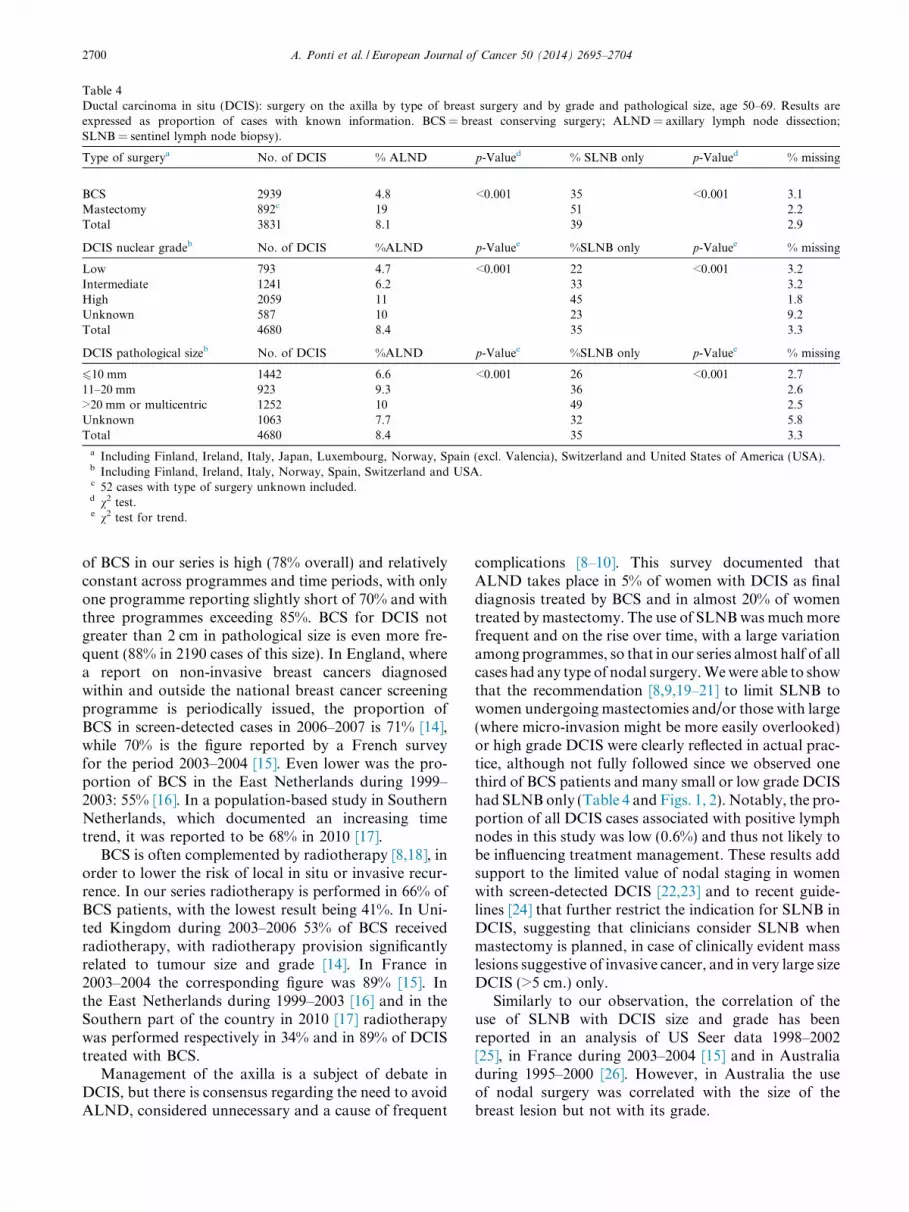
Table 4Ductal carcinoma in situ (DCIS): surgery on the axilla by type of breast surgery and by grade and pathological size, age 50–69. Results areexpressed as proportion of cases with known information. BCS = breast conserving surgery; ALND = axillary lymph node dissection;SLNB = sentinel lymph node biopsy).
Type of surgerya No. of DCIS % ALND p-Valued % SLNB only p-Valued % missing
BCS 2939 4.8 <0.001 35 <0.001 3.1Mastectomy 892c 19 51 2.2Total 3831 8.1 39 2.9
DCIS nuclear gradeb No. of DCIS %ALND p-Valuee %SLNB only p-Valuee % missing
Low 793 4.7 <0.001 22 <0.001 3.2Intermediate 1241 6.2 33 3.2High 2059 11 45 1.8Unknown 587 10 23 9.2Total 4680 8.4 35 3.3
DCIS pathological sizeb No. of DCIS %ALND p-Valuee %SLNB only p-Valuee % missing
610 mm 1442 6.6 <0.001 26 <0.001 2.711–20 mm 923 9.3 36 2.6>20 mm or multicentric 1252 10 49 2.5Unknown 1063 7.7 32 5.8Total 4680 8.4 35 3.3
a Including Finland, Ireland, Italy, Japan, Luxembourg, Norway, Spain (excl. Valencia), Switzerland and United States of America (USA).b Including Finland, Ireland, Italy, Norway, Spain, Switzerland and USA.c 52 cases with type of surgery unknown included.d v2 test.e v2 test for trend.
2700 A. Ponti et al. / European Journal of Cancer 50 (2014) 2695–2704
of BCS in our series is high (78% overall) and relativelyconstant across programmes and time periods, with onlyone programme reporting slightly short of 70% and withthree programmes exceeding 85%. BCS for DCIS notgreater than 2 cm in pathological size is even more fre-quent (88% in 2190 cases of this size). In England, wherea report on non-invasive breast cancers diagnosedwithin and outside the national breast cancer screeningprogramme is periodically issued, the proportion ofBCS in screen-detected cases in 2006–2007 is 71% [14],while 70% is the figure reported by a French surveyfor the period 2003–2004 [15]. Even lower was the pro-portion of BCS in the East Netherlands during 1999–2003: 55% [16]. In a population-based study in SouthernNetherlands, which documented an increasing timetrend, it was reported to be 68% in 2010 [17].
BCS is often complemented by radiotherapy [8,18], inorder to lower the risk of local in situ or invasive recur-rence. In our series radiotherapy is performed in 66% ofBCS patients, with the lowest result being 41%. In Uni-ted Kingdom during 2003–2006 53% of BCS receivedradiotherapy, with radiotherapy provision significantlyrelated to tumour size and grade [14]. In France in2003–2004 the corresponding figure was 89% [15]. Inthe East Netherlands during 1999–2003 [16] and in theSouthern part of the country in 2010 [17] radiotherapywas performed respectively in 34% and in 89% of DCIStreated with BCS.
Management of the axilla is a subject of debate inDCIS, but there is consensus regarding the need to avoidALND, considered unnecessary and a cause of frequent
complications [8–10]. This survey documented thatALND takes place in 5% of women with DCIS as finaldiagnosis treated by BCS and in almost 20% of womentreated by mastectomy. The use of SLNB was much morefrequent and on the rise over time, with a large variationamong programmes, so that in our series almost half of allcases had any type of nodal surgery. We were able to showthat the recommendation [8,9,19–21] to limit SLNB towomen undergoing mastectomies and/or those with large(where micro-invasion might be more easily overlooked)or high grade DCIS were clearly reflected in actual prac-tice, although not fully followed since we observed onethird of BCS patients and many small or low grade DCIShad SLNB only (Table 4 and Figs. 1, 2). Notably, the pro-portion of all DCIS cases associated with positive lymphnodes in this study was low (0.6%) and thus not likely tobe influencing treatment management. These results addsupport to the limited value of nodal staging in womenwith screen-detected DCIS [22,23] and to recent guide-lines [24] that further restrict the indication for SLNB inDCIS, suggesting that clinicians consider SLNB whenmastectomy is planned, in case of clinically evident masslesions suggestive of invasive cancer, and in very large sizeDCIS (>5 cm.) only.
Similarly to our observation, the correlation of theuse of SLNB with DCIS size and grade has beenreported in an analysis of US Seer data 1998–2002[25], in France during 2003–2004 [15] and in Australiaduring 1995–2000 [26]. However, in Australia the useof nodal surgery was correlated with the size of thebreast lesion but not with its grade.

●
●
●
●
● ●
●
●
●●
●
●
2004 2005 2006 2007
0%20
%40
%60
%80
%
<10 mm.11−20 mm.>20 mm. + Multicentric
●
●
●
●●
●
●
●
●
●
●
●
2004 2005 2006 2007
0%20
%40
%60
%80
%
LowIntermediateHigh
(a) SLNB only by size
(b) SLNB only by grade
Fig. 1. Ductal carcinoma in situ: performance of sentinel lymph node biopsy (SLNB) only by pathological size and time period (a), and by nucleargrade and time period (b). Any type of breast surgery included. Cases reported for year 2008 and countries not reporting cases for the whole period2004–2007 or lacking the stratification by size and grade were excluded from this analysis. Data are included for Finland, Ireland, Norway, Spain,Switzerland and United States of America (USA).
A. Ponti et al. / European Journal of Cancer 50 (2014) 2695–2704 2701
In England in 2006–2007 the use of SLNB in screen-detected non-invasive breast cancers having breast con-serving surgery was 4.0% [14], a figure lower than in anyof the programmes included in our survey. In France in2003-2004 SLNB was performed in 21% of patients andthe proportion of ALND was 10% [15]. In the EastNetherlands during 1999–2003 any axillary staging pro-cedure was performed in 25% of DCIS [16] while inSouthern Netherlands use of SLNB was reported being65% in 2010 [17]. In Italy the use of SLNB in screen-detected DCIS increased from 20% to slightly over50% from 2001 to 2007 and then remained virtually sta-ble through 2010 [27].
Limitations of this study are those specific to aggregatedata surveys: limited detail in available data, possible useof different definitions of study parameters in the differentsites, need to restrict overall data analyses to data stratifi-cations being planned in advance. Not all programmescould contribute all required data and the number ofmissing values for some of the parameters was high. How-ever, we minimised these limitations by providing strictly
structured data collection forms, with several pre-specified stratification tables, detailed documentationon definitions used, and internal consistency checks. Itmust be also acknowledged that this paper provides apicture of DCIS management during 2004–2008, andpractice is likely to have evolved since then, both in detec-tion, with the gradual introduction of digital mammogra-phy [4], and in treatment. ICSN will consider updatingthese results seeking data from an even larger numberof programmes.
This survey covered screen-detected DCIS cases only.Few countries have yet similar information availablefrom the in situ carcinoma diagnosed at all ages outsideorganised screening programmes, which have beenquantified as 51% of all cases in Southern Netherlands[17], 43% in Finland [28], and 38% in United Kingdom[14]. Projects conducted in co-operation between clinicalCentres and population Cancer Registries [17] couldcover this gap.
This study is, to our knowledge, the first large (morethan 5000 cases) international survey of DCIS

●
●
●
●
●● ●
●
●
● ●
●
2004 2005 2006 2007
0%5%
10%
15%
20%
25%
<10 mm.11−20 mm.>20 mm. + Multicentric
●●
●●
● ●
●
●
●●
●
●
2004 2005 2006 2007
0%5%
10%
15%
20%
25%
LowIntermediateHigh
(a) ALND by size
(b) ALND by grade
Fig. 2. Ductal carcinoma in situ: axillary lymph node dissection (ALND) by pathological size and time period (a), and by nuclear grade and timeperiod (b). Any type of breast surgery included. Cases reported for year 2008 and countries not reporting cases for the whole period 2004–2007 orlacking the stratification by size and grade were excluded from this analysis. Data are included for Finland, Ireland, Norway, Spain, Switzerlandand United States of America (USA).
2702 A. Ponti et al. / European Journal of Cancer 50 (2014) 2695–2704
management practices. We found wide variation in clin-ical management for all of the parameters studied. Whileawaiting progress from research enabling to differentiateindolent lesions amenable of follow up only from thoseat high risk of subsequent invasive cancer [29–31], effortsshould be made to optimise diagnostic assessment andmanagement of screen-detected cases to mitigate overdi-agnosis and overtreatment [32]. Specifically, we foundthat axillary surgery, although used more often in highgrade and large size lesions, showed an increasing timetrend and was performed, with large variation betweencentres, beyond what is recommended by guidelines.This indicates the presence of varying levels of overtreat-ment and the potential for the reduction of treatment-related morbidity. In fact, although less frequently harm-ful than ALND, SLNB is not exempt from complica-tions. According to the update of the SLNB AmericanSociety of Clinical Oncology Clinical Practice Guidelines[24], which includes a literature review of adverse events,important morbidity of node surgery includes lymphoe-dema, infections, seroma and neurologic complications.
These were found to be more frequent in patients receiv-ing ALND as opposed to SLNB only, but they are stillnot negligible even in the latter. For example, in theALMANAC trial [33] at 12 months after operation lym-phoedema occurred in 5% of patients having receivedSLNB only versus 13% of patients having receivedALND, and sensory loss 11% and 31% respectively.
Specialised multidisciplinary care for breast cancerhas proved to improve process of care [34] anddecrease mortality [35]. Screening programmes shouldlink to specialised clinical Units and Cancer Registriesand jointly set up or expand multidisciplinary teams incharge of quality assurance of diagnosis and treatmentof screen-detected lesions, including DCIS, so to assurethat current guidelines are applied and opportunitiesfor research in the heterogeneity of these lesions aretaken.
Conflict of interest statement
None declared.

A. Ponti et al. / European Journal of Cancer 50 (2014) 2695–2704 2703
Acknowledgements
The ICSN is an activity funded by the US NationalCancer Institute, Bethesda, Maryland. No specific fundingwas made available for this study, but the National CancerInstitute provided co-ordination of the project and secre-tarial support. Data management and analysis, for whichthe authors acknowledge Mariano Tomatis and DeniseCasella, were provided by CPO Piemonte, Torino, Italy.
The collection of US data was supported by theNational Cancer Institute-funded Breast CancerSurveillance Consortium (BCSC) co-operative agree-ment (U01CA63740, U01CA86076, U01CA86082,U01CA63736, U01CA70013, U01CA69976, U01CA63731, U01CA70040). A list of the BCSC investigatorsand procedures for requesting BCSC data for researchpurposes are provided at: http://breastscreening.cancer.gov. The collection of cancer data used in this study wassupported in part by several state public healthdepartments and cancer registries throughout the US.For a full description of these sources, please see: http://breastscreening.cancer.gov/work/acknowledgement.html.
The content is solely the responsibility of the authorsand does not necessarily represent the official views ofthe National Cancer Institute or the National Institutesof Health.
We thank national and regional screening pro-gramme co-ordinators, and all professionals involvedin breast cancer screening and treatment for their contri-bution to this study.
Appendix A. Additional members of the ICSN DCIS
Working Group
Mireille Broeders, National Expert and TrainingCentre for Breast Cancer Screening, Nijmegen, TheNetherlands;
Jan Danes, First Faculty of Medicine, Charles Uni-versity in Prague, Czech Republic;
Maria Ederra, Instituto de Salud Publica, Navarra,Spain;
Bernard Filliez, Valais breast cancer screening pro-gramme, Switzerland;
Matti Hakama, Finnish Cancer Registry, Helsinki,Finland;
Carlos Munoz, Jura and Neuchatel breast cancerscreening programme, Switzerland;
Montse Garcia Martinez, Catalan Institute of Oncol-ogy, Barcelona, Spain;
Paola Mantellini, U.O. Epidemiologia Clinica e Des-crittiva, Istituto per lo Studio e la Prevenzione Oncolog-ica, Firenze, Italy;
Josefa Miranda, General Directorate Research andPublic Health and Centre for Public Health Research,Valencia, Spain;
Therese Mooney, National Cancer Screening Service,Dublin, Ireland;
Noriaki Ohuchi, Tohoku University GraduateSchool of Medicine, Japan;
Isabelle Robert, Programme Mammographie, Direc-tion de la Sante, Luxembourg;
Hiroshi Saito, National Cancer Centre, Japan;Ragnhild Sørum Falk, The Cancer Registry of Nor-
way, Oslo, Norway;Asta Taskinen, Finnish Cancer Registry, Helsinki,
Finland;Janine Timmers, National Expert and Training Cen-
tre for Breast Cancer Screening, Nijmegen, TheNetherlands;
Leonardo Ventura, U.O. Epidemiologia Clinica eDescrittiva, Istituto per lo Studio e la PrevenzioneOncologica, Firenze, Italy;
Marie-Christine Wagnon, Programme Mammogra-phie, Direction de la Sante, Luxembourg.
References
[1] Virnig BA, Tuttle TM, Shamliyan T, Kane RL. Ductal carcinomain situ of the breast: a systematic review of incidence, treatment,and outcomes. J Natl Cancer Inst 2010;102:170–8.
[2] Ernster V, Ballard-Barbash R, Barlow W, et al. Detection ofDCIS in women undergoing screening mammography. J NatlCancer Inst 2002;94:1546–54.
[3] Broeders MJM, Scharpantgen A, Ascunce N, et al. Comparisonof early performance indicators for screening projects within theEuropean Breast Cancer Network: 1989–2000. Eur J Cancer Prev2005;14:107–16.
[4] Lynge E, Ponti A, James T, et al. Variation in detection of ductalcarcinoma in situ (DCIS) in screening mammography. A surveywithin the International Cancer Screening Network (ICSN). Eur JCancer 2014;50:185–92.
[5] Erbas B, Provenzano E, Armes J, Gertig D. The natural history ofductal carcinoma in situ of the breast: a review. Breast Cancer ResTreat 2006;97:135–44.
[6] Kuerer HM, Albarracin CT, Yang WT, et al. Ductal carcinomain situ: state of the science and roadmap to advance the field. JClin Oncol 2009;27:279–88.
[7] Mokbel K, Cutuli B. Heterogeneity of ductal carcinoma in situand its effects on management. Lancet Oncol 2006;7:756–65.
[8] Moran MS, Bai HX, Harris EE, et al. ACR appropriatenesscriteria for ductal carcinoma in situ. Breast J 2012;18:8–15.
[9] British Association of Surgical Oncology. Surgical guidelines forthe management of breast cancer. Eur J Surg Oncol 2009;35(Sup-pl. 1):1–22.
[10] Del Turco MR, Ponti A, Bick U, et al. Quality indicators in breastcancer care. Eur J Cancer 2010;46:2344–56.
[11] Willems SM, van Deurzen CHM, van Diest PJ. Diagnosis ofbreast lesions: fine-needle aspiration cytology or core needlebiopsy? A review. J Clin Pathol 2012;65:287–92.
[12] Brazda A, Estroff J, Euhus D, et al. Delays in time to treatmentand survival impact in breast cancer. Ann Surg Oncol2010;17(Suppl. 3):291–6.
[13] Caldon LJ, Collins KA, Wilde DJ, et al. Why do hospitalmastectomy rates vary? Differences in the decision-makingexperiences of women with breast cancer. Br J Cancer2011;104:1551–7.
[14] NCIN (National Cancer Intelligence Network). The non-invasivebreast cancer report. Birmingham: West Midlands Cancer Intel-ligence Unit; 2011.
[15] Cutuli B, Lemanski C, Fourquet A, et al. Breast-conservingsurgery with or without radiotherapy vs mastectomy for ductal

2704 A. Ponti et al. / European Journal of Cancer 50 (2014) 2695–2704
carcinoma in situ: French Survey experience. Br J Cancer2009;100:1048–54.
[16] Schouten van der Velden AP, Van Dijck JA, Wobbes T.Variations in treatment of ductal carcinoma in situ of the breast:a population-based study in the East Netherlands. Eur J SurgOncol 2007;33:424–9.
[17] Van Steenbergen LN, Voogd AC, Roukema JA, et al. Time trendsand inter-hospital variation in treatment and axillary staging ofpatients with ductal carcinoma in situ of the breast in the era ofscreening in Southern Netherlands. The Breast 2014;23:63–8.
[18] Dodwell D, Clements K, Lawrence G, et al. Radiotherapyfollowing breast–conserving surgery for screen-detected ductalcarcinoma in situ: indications and utilisation in the UK. Interimfindings from the Sloane Project. Br J Cancer 2007;97:725–9.
[19] NICE (National Institute for Clinical Excellence). Guidelines onearly and locally advanced breast cancer. Cardiff: National Col-laborating Centre for Cancer; 2009.
[20] Van Deurzen CH, Hobbelink MG, van Hillegersberg R, van DiestPJ. Is there an indication for sentinel node biopsy in patients withductal carcinoma in situ of the breast? A review. Eur J Cancer2007;43:993–1001.
[21] Silverstein MJ, Recht A, Lagios MD, et al. Consensus conferenceIII: Image-detected breast cancer. State of the art diagnosis andtreatment. J Am Coll Surg 2009;209:504–20.
[22] Silverstein MJ, Rosser RJ, Gierson ED, et al. Axillary lymphnode dissection for intraductal breast carcinoma—is it indicated?Cancer 1987;59:1819–24.
[23] Morrow M. Axillary surgery in DCIS: is less more? Ann SurgOncol 2008;15:2641–2.
[24] Lyman GH, Temin S, Edge SB, et al. Sentinel lymph node biopsyfor patients with early-stage breast cancer: American Society ofClinical Oncology Clinical Practice Guideline Update. J ClinOncol 2014;32. http://dx.doi.org/10.1200/JCO.2013.54.1177.
[25] Porembka MR, Abraham RL, Sefko JA, Deshpande AD, JeffeDB, Margenthaler JA. Factors associated with lymph nodeassessment in ductal carcinoma in situ: analysis of 1988–2002 Seerdata. Ann Surg Oncol 2008;15:2709–19.
[26] Kricker A, Armstrong B. Surgery and outcomes of ductalcarcinoma in situ of the breast: a population-based study inAustralia. Eur J Cancer 2004;40:396–402.
[27] Ponti A, Mano MP, Tomatis M, et al. Audit of breast cancerdiagnosis and treatment in Italy, 2010. Epidemiol Prev2012;36(Suppl. 1):87–95.
[28] Finnish Cancer Registry – Institute for Statistical and Epidemi-ological Cancer Research: Cancer in Finland 2008 and 2009.Cancer Statistics of the National Institute for Health and Welfare.Cancer Society of Finland. Publication No. 84, Helsinki; 2011.
[29] Berson JR, Wishart GC. Predictors of recurrence for ductalcarcinoma in situ after breast-conserving surgery. Lancet Oncol2013;14:348–57.
[30] Punglia RS, Stuart JS, Weeks JC. Treatment of ductal carcinomain situ after excision. Would a prophylactic paradigm be moreappropriate? J Natl Cancer Inst 2013;105:1527–33.
[31] Allegra CJ, Aberle DR, Ganschow P, et al. National Institutes ofHealth State-of-the-Science Conference statement: Diagnosis andmanagement of ductal carcinoma in situ September 22–24, 2009. JNatl Cancer Inst 2010;102:161–9.
[32] Esserman LJ, Thompson IM, Reid B. Overdiagnosis and over-treatment in cancer: an opportunity for improvement. JAMA2013;310:797–8.
[33] Mansel RE, Fallowfield L, Kissin M, et al. Randomized multi-center trial of sentinel node biopsy versus standard axillarytreatment in operable breast cancer: the ALMANAC trial. J NatlCancer Inst 2006;98:599–609.
[34] Vrijens F, Stordeur S, Beirens K, et al. Effect of hospital volumeon processes of car and 5-year survival after breast cancer: apopulation-based study on 25000 women. The Breast2012;21:261–6.
[35] Kesson EM, Allardice GM, George WD, et al. Effects ofmultidisciplinary team working on breast cancer survival: retro-spective, comparative, interventional cohort study of 13722women. BMJ 2012;344. http://dx.doi.org/10.1136/bmj.e2718.


