Embed Size (px)
Citation preview
STUDY PROTOCOL Open Access
Intrauterine resuscitation during the secondstage of term labour by maternalhyperoxygenation versus conventionalcare: study protocol for a randomisedcontrolled trial (INTEREST O2)Lauren M. Bullens1,2* , Alexandra D. J. Hulsenboom1, Suzanne Moors1, Rohan Joshi3,4, Pieter J. van Runnard Heimel1,M. Beatrijs van der Hout-van der Jagt1,2, Edwin R. van den Heuvel5 and S. Guid Oei1,2
Abstract
Background: Perinatal asphyxia is, even in developed countries, one the major causes of neonatal morbidity andmortality. Therefore, if foetal distress during labour is suspected, one should try to restore foetal oxygen levels oraim for immediate delivery. However, studies on the effect of intrauterine resuscitation during labour are scarce.We designed a randomised controlled trial to investigate the effect of maternal hyperoxygenation on the foetalcondition. In this study, maternal hyperoxygenation is induced for the treatment of foetal distress during thesecond stage of term labour.
Methods/design: This study is a single-centre randomised controlled trial being performed in a tertiary hospital in TheNetherlands. From among cases of a suboptimal or abnormal foetal heart rate pattern during the second stage of termlabour, a total of 116 patients will be randomised to the control group, where normal care is provided, or to theintervention group, where before normal care 100% oxygen is supplied to the mother by a non-rebreathing mask untildelivery. The primary outcome is change in foetal heart rate pattern. Secondary outcomes are Apgar score, modeof delivery, admission to the neonatal intensive care unit and maternal side effects. In addition, blood gas valuesand malondialdehyde are determined in umbilical cord blood.
Discussion: This study will be the first randomised controlled trial to investigate the effect of maternal hyperoxygenationfor foetal distress during labour. This intervention should be recommended only as a treatment for intrapartumfoetal distress, when improvement of the foetal condition is likely and outweighs maternal and neonatal side effects.
Trial registration: EudraCT, 2015-001654-15; registered on 3 April 2015. Dutch Trial Register, NTR5461; registered on 20October 2015.
Keywords: Foetal distress, Foetal heart rate, Cardiotocogram, Intrauterine resuscitation, Maternal hyperoxygenation,Neonatal outcome, Free oxygen radicals, Randomised controlled trial
* Correspondence: [email protected] of Obstetrics and Gynaecology, Máxima Medical Centre, POBox 7777, 5500, MB, Veldhoven, The Netherlands2Department of Electrical Engineering, Eindhoven University of Technology,PO Box 513, 5600, MB, Eindhoven, The NetherlandsFull list of author information is available at the end of the article
© The Author(s). 2018 Open Access This article is distributed under the terms of the Creative Commons Attribution 4.0International License (http://creativecommons.org/licenses/by/4.0/), which permits unrestricted use, distribution, andreproduction in any medium, provided you give appropriate credit to the original author(s) and the source, provide a link tothe Creative Commons license, and indicate if changes were made. The Creative Commons Public Domain Dedication waiver(http://creativecommons.org/publicdomain/zero/1.0/) applies to the data made available in this article, unless otherwise stated.
Bullens et al. Trials (2018) 19:195 https://doi.org/10.1186/s13063-018-2567-x
BackgroundLabour contractions cause alterations in intrauterinepressure and can thereby affect uterine and umbilicalblood flow [1–5]. These fluctuations in blood flowtowards the foetus can negatively influence oxygen flowand blood pressure [1–5]. Through chemo- and barorecep-tor responses, these changes in foetal oxygenation andblood pressure affect foetal heart rate (FHR) [1, 2, 6, 7].Hence, non-reassuring FHR patterns such as FHR decelera-tions may be a sign of foetal hypoxia [8–10]. Prolongedfoetal hypoxia may lead to an increased risk of foetalmorbidity, including renal insufficiency, pulmonary hyper-tension, necrotising enterocolitis and hypoxic–ischemicencephalopathy and foetal death [11, 12]. A prospectivecohort study of term neonates in 2010 showed that 48% ofadmissions of these neonates to neonatal intensive careunits (NICUs) were related to perinatal asphyxia (defined bythe authors as a 5-minute Apgar score < 7). The neonatalmortality rate was 8% in this study, the largest proportion ofwhich (71%, n = 12 of 17) was related to asphyxia [13].Methods to directly measure foetal oxygenation during
labour are unavailable, whereas methods for the continuousintrapartum monitoring of pH, oxygen saturation (SpO2),partial carbon dioxide pressure (pCO2) and partial oxygenpressure (pO2) are not yet suitable for clinical practice [14–16]. Therefore, the cardiotocogram (CTG), with occasionalfoetal scalp blood sampling (FSBS), is still the method offirst choice to estimate foetal well-being during labour. TheCTG has very good specificity but poor sensitivity for foetalwell-being [17]. In other words, if the FHR pattern isreassuring, the foetus is very likely to be well-oxygenated.However, when FHR patterns are non-reassuring, the foetalcondition is unclear, and foetal distress cannot be ruled out.Instead of aiming for immediate delivery in the presence
of suspected foetal distress, one may try to improve foetaloxygenation to avoid an invasive intervention. Severalintrauterine resuscitation techniques are used in clinicalpractice and have been described in the literature [18, 19].However, robust evidence regarding their effect on neo-natal outcome is limited [18]. One of the interventionsthat still raises discussion is the administration ofadditional oxygen to the mother to treat foetal distressduring labour [18, 20–23].
Summary of findings from clinical studiesIn the past five decades, researchers in several studieshave investigated the effect of maternal hyperoxygena-tion on maternal and foetal oxygenation. Indeed, theyfound increasing maternal pO2 [24] and foetal SpO2 andpO2 levels, but unfortunately these studies were mainlyperformed in the non-compromised foetus [25–27].Furthermore, only a few non-randomised studies of poorquality have been performed in the distressed foetus[28–32]. These studies suggest an improvement in FHR
patterns and foetal scalp pH when 100% oxygen is appliedto the mother. Based on these publications, authors of aCochrane review published in 2012 concluded that ‘thereis not enough evidence to support the use of prophylacticoxygen therapy for women in labour, nor to evaluate itseffectiveness for fetal distress’, owing to the lack of rando-mised controlled trials (RCTs) [33].An important concern in the use of maternal hyperoxy-
genation for foetal distress is the potential negative effecton umbilical cord pH. In a study by Thorp et al. [34], 86term parturients were randomised to receive additionaloxygen or normal care during the second stage of labour.The main outcome measures were cord blood gas and co-oximetry values. The mean cord blood gas values did notsignificantly differ between the intervention and controlgroups. However, Thorp et al. found significantly morearterial pH values < 7.20 in the group receiving extraoxygen. The lowest pH in arterial blood gas (pHa) valuethat they found was 7.09. They also found that the durationof oxygen therapy was inversely related to arterial cord pH,whereas Apgar scores and hospital admission rates did notdiffer between the groups. They concluded that prolongedoxygen treatment during the second stage of labour leadsto a deterioration of cord blood gas values at birth. Animportant fact is that only patients with reassuring FHRpatterns were included in their study. Therefore, (ominous)foetal hypoxia at the start of oxygen delivery was very un-likely. Thus, their study did not address the effect of mater-nal hyperoxygenation in cases of suspected foetal distress.Another frequently stated argument to discourage mater-
nal hyperoxygenation as standard care is the potential in-crease in free oxygen radicals in both mother and foetus[35, 36]. An increase in the markers for free oxygen radicalproduction has been seen for the use of high fractions of in-spired oxygen and in the presence of non-reassuring FHRpatterns [35–38]. Also, lipid peroxide concentrations inarterial cord blood are higher after uncomplicated vaginaldelivery than after elective caesarean section [39].To a certain degree, free oxygen radicals are physio-
logical and known to be higher in the presence of severalmaternal and foetal conditions, such as pre-eclampsia,diabetes, smoking, intrauterine growth restriction andfoetal distress [37, 39–42]. The effect of maternal hyperoxy-genation on free oxygen radical release, in response to non-reassuring foetal status, has not yet been investigated.What we do know is that neonatal resuscitation with
100% oxygen may lead to an increase in neonatal mortalityand morbidity, including bronchopulmonary disease andretinopathy, mainly in premature infants [43–46]. How-ever, the increase in foetal pO2 due to maternal hyperoxy-genation will never reach the levels obtained by the directapplication of 100% oxygen directly to the foetus [23]. Toour knowledge, the clinical implication of increased freeradical production due to maternal hyperoxygenation has
Bullens et al. Trials (2018) 19:195 Page 2 of 11
not been investigated. Researchers in studies using maternalhyperoxygenation as a treatment for the growth restrictedfoetus did not report any harmful effects [47, 48].With regard to the mother, some potential side effects
have to be taken into account. The use of high fractions ofinspired oxygen in the absence of tissue hypoxia may causetoxic effects as a result of oxidative stress [49, 50]. This maylead to, for example, mucosal inflammation, hypoperfusionand pneumonitis [51]. A reversible vasoconstriction ofapproximately 10% in the maternal brain has been described[52]. However, this is not expected to cause any harm [53,54]. Administration of 100% oxygen during labour is notinvestigated. However, it has been well investigated for thetreatment of cluster headaches, and no severe side effects(e.g., hypoventilation and fainting) have been reported [54].Inhaling high fractions of inspired oxygen will increase
the concentration of free oxygen radicals in maternalblood [35]. Despite the adverse effects of free oxygenradicals that have been described [55], it is unlikely thesewill cause clinically relevant tissue damage, owing to themature anti-oxidant system in the adult [35, 36]. Also,the Dutch Pharmacovigilance Centre Lareb has not beeninformed of any side effects of oxygen therapy [56].
Current recommendations on the use of maternalhyperoxygenationOn the basis of current knowledge, it is difficult todetermine whether the beneficial effects of maternal hyper-oxygenation outweigh the potential side effects. As a conse-quence, recommendations in international guidelines anduse in clinical practice are non-uniform [20]. Maternalhyperoxygenation during labour is often used in the UnitedStates to increase oxygen transport towards the foetus [21].The American College of Obstetricians and Gynecologistsguideline on foetal resuscitation recommends the adminis-tration of oxygen to the mother in cases of foetal distress[57]. In contrast, the Royal College of Obstetricians andGynaecologists explicitly states in their Green Top Guidelinenot to apply maternal oxygenation for reasons other thanmaternal hypoxia, until the beneficial effect is proven [58]. Arecent discussion on benefit and harm of maternal hyperoxy-genation in the American Journal of Obstetrics andGynecology emphasised the current lack of evidence [21–23].In fact, several reviews underline an urgent need for an RCTinvestigating the effect of maternal hyperoxygenation on thefoetal condition [21–23, 33].
Methods/designAimThe aim of this study is to investigate the effect ofmaternal hyperoxygenation with 100% oxygen on thefoetal condition during the second stage of labour in thepresence of suspected foetal distress during term labour.Also, we will investigate the potential side effects to
formulate recommendations for international clinicalpractice and future research.
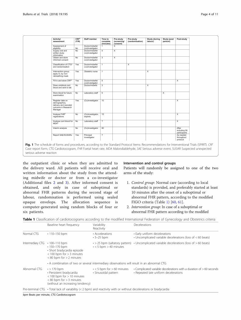
Study designThis study will be a single-centre RCT performed in atertiary hospital in The Netherlands. We are comparingmaternal hyperoxygenation for the treatment of foetaldistress during the second stage of labour with conven-tional care. All procedures and time frames are displayedin Fig. 1 according to the Standard Protocol Items:Recommendations for Interventional Trials (SPIRIT) [59].Additional file 1 contains the complete SPIRIT checklist.
ParticipantsThe study population will be drawn from among parturientsadmitted to the labour ward of a tertiary hospital (MáximaMedical Centre, Veldhoven, The Netherlands), whereapproximately 2200 deliveries occur annually, of whichapproximately 1900 are term births. CTG and, if necessary,FSBS are generally used for foetal monitoring during labour.Maternal repositioning, discontinuation of administration ofoxytocin, use of tocolytic drugs and intermittent pushingare common interventions used to achieve intrauterineresuscitation, whereas amnioinfusion and maternal hyperoxy-genation are never applied as standard care in our centre.
Inclusion criteriaPregnant women ≥ 18 years old in term labour and withan intended vaginal delivery of a singleton in cephalicpresentation can participate in this study.
Exclusion criteriaExclusion criteria are determined with a focus on therisk of excessive production of free oxygen radicals andreducing the influence of other factors affecting FHRpattern. These are recent use of any of the followingmedications: corticosteroids, anti-hypertensives, magnesiumsulphate, amiodarone, opioids, adriamycin, bleomycin, acti-nomycin, menadione, promazine, thioridazine or chloro-quine. Other exclusion criteria are the use of tobacco,recreational drugs or alcohol during pregnancy. Parturientswith pre-existing cardiac disease, pulmonary disease withthe use of medication, diabetes, hyperthyroidism oranaemia (haemoglobin < 6.5 mmol/L or 10.5 g/dl) will alsobe excluded. Foetal factors leading to exclusion aresuspected infection during labour (need for antibiotics),congenital malformations and normal or pre-terminal FHRpattern, or prolonged bradycardia (according to themodified International Federation of Gynecology andObstetrics [FIGO] classification; seeTable 1) [60, 61].
Patient recruitment and randomisationAll patients eligible to be included in this study willbe asked antepartum to participate when they visit
Bullens et al. Trials (2018) 19:195 Page 3 of 11
the outpatient clinic or when they are admitted tothe delivery ward. All patients will receive oral andwritten information about the study from the attend-ing midwife or doctor or from a co-investigator(Additional files 2 and 3). After informed consent isobtained, and only in case of suboptimal orabnormal FHR patterns during the second stage oflabour, randomisation is performed using sealedopaque envelops. The allocation sequence iscomputer-generated using random blocks of four orsix patients.
Intervention and control groupsPatients will randomly be assigned to one of the twoarms of the study:
1. Control group: Normal care (according to localstandards) is provided, and preferably started at least10 minutes after the onset of a suboptimal orabnormal FHR pattern, according to the modifiedFIGO criteria (Table 1) [60, 61].
2. Intervention group: In case of a suboptimal orabnormal FHR pattern according to the modified
Fig. 1 The schedule of forms and procedures, according to the Standard Protocol Items: Recommendations for Interventional Trials (SPIRIT). CRFCase report form, CTG Cardiotocogram, FHR Foetal heart rate, MDA Malondialdehyde, SAE Serious adverse event, SUSAR Suspected unexpectedserious adverse reaction
Table 1 Classification of cardiotocograms according to the modified International Federation of Gynecology and Obstetrics criteria
Baseline heart frequency VariabilityReactivity
Decelerations
Normal CTG • 110–150 bpm • Accelerations• 5–25 bpm
• Early uniform decelerations• Uncomplicated variable decelerations (loss of < 60 beats)
Intermediary CTG • 100–110 bpm• 150–170 bpm• Short bradycardia episode< 100 bpm for > 3 minutes< 80 bpm for > 2 minutes
• > 25 bpm (saltatory pattern)• < 5 bpm > 40 minutes
• Uncomplicated variable decelerations (loss of > 60 beats)
• A combination of two or several intermediary observations will result in an abnormal CTG
Abnormal CTG • > 170 bpm• Persistent bradycardia< 100 bpm for > 10 minutes< 80 bpm for > 3 minutes(without an increasing tendency)
• < 5 bpm for > 60 minutes• Sinusoidal pattern
• Complicated variable decelerations with a duration of > 60 seconds• Repeated late uniform decelerations
Pre-terminal CTG • Total lack of variability (< 2 bpm) and reactivity with or without decelerations or bradycardia
bpm Beats per minute, CTG Cardiotocogram
Bullens et al. Trials (2018) 19:195 Page 4 of 11
FIGO criteria, 100% oxygen is applied to the mother at10 L/minute via a non-rebreathing mask and continueduntil delivery. Co-interventions (normal care) may beinitiated after 10 minutes of oxygen administrationwithout a satisfactory effect on FHR to investigate theeffect of only maternal hyperoxygenation on FHR,without risking prolonged foetal hypoxia. In case apatient needs to undergo a caesarean section, oxygenadministration will be continued until the baby is born.
Obviously, in any case where the delivery room staffbelieve additional interventions should be applied for safetyreasons, the study protocol can be overruled at any time.
Study outcomes and data analysisThe primary outcome is the percentage reduction in thedepth and duration of FHR deceleration in the interven-tion group compared with the control group. Secondaryoutcomes include foetal, neonatal and maternal outcomes.
Foetal outcome
1. FHR changes: Changes in specific features of theCTG, including the following:a. Decelerations with loss of internal variability (beat-
to-beat variability of < 5 beats per minute [bpm])b. Decelerations in combination with tachycardia of
bradycardia (> 160 or < 110 bpm)c. Unassignable baselined. Phase-rectified signal averaging (PRSA), a
relatively new technique used to determine FHRvariability by estimating the accelerative capacity(ACPRSA) and decelerative capacity (DCPRSA) ofthe foetal heart. This technique is explained inarticles by Bauer and Huhn [62, 63].
2. Change in modified FIGO classification (Table 1) [60, 61].
In the next subsection, methodology regarding thecomparison of FHR tracings and timeframes is describedin more detail.
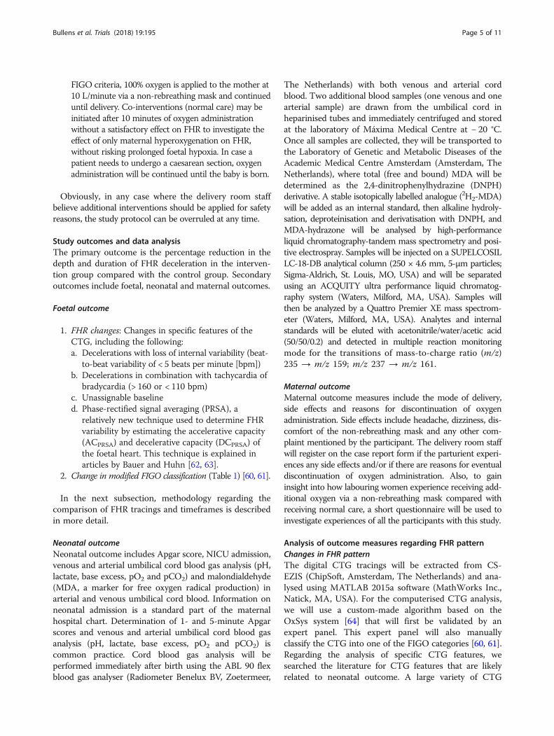
Neonatal outcomeNeonatal outcome includes Apgar score, NICU admission,venous and arterial umbilical cord blood gas analysis (pH,lactate, base excess, pO2 and pCO2) and malondialdehyde(MDA, a marker for free oxygen radical production) inarterial and venous umbilical cord blood. Information onneonatal admission is a standard part of the maternalhospital chart. Determination of 1- and 5-minute Apgarscores and venous and arterial umbilical cord blood gasanalysis (pH, lactate, base excess, pO2 and pCO2) iscommon practice. Cord blood gas analysis will beperformed immediately after birth using the ABL 90 flexblood gas analyser (Radiometer Benelux BV, Zoetermeer,
The Netherlands) with both venous and arterial cordblood. Two additional blood samples (one venous and onearterial sample) are drawn from the umbilical cord inheparinised tubes and immediately centrifuged and storedat the laboratory of Máxima Medical Centre at − 20 °C.Once all samples are collected, they will be transported tothe Laboratory of Genetic and Metabolic Diseases of theAcademic Medical Centre Amsterdam (Amsterdam, TheNetherlands), where total (free and bound) MDA will bedetermined as the 2,4-dinitrophenylhydrazine (DNPH)derivative. A stable isotopically labelled analogue (2H2-MDA)will be added as an internal standard, then alkaline hydroly-sation, deproteinisation and derivatisation with DNPH, andMDA-hydrazone will be analysed by high-performanceliquid chromatography-tandem mass spectrometry and posi-tive electrospray. Samples will be injected on a SUPELCOSILLC-18-DB analytical column (250 × 4.6 mm, 5-μm particles;Sigma-Aldrich, St. Louis, MO, USA) and will be separatedusing an ACQUITY ultra performance liquid chromatog-raphy system (Waters, Milford, MA, USA). Samples willthen be analyzed by a Quattro Premier XE mass spectrom-eter (Waters, Milford, MA, USA). Analytes and internalstandards will be eluted with acetonitrile/water/acetic acid(50/50/0.2) and detected in multiple reaction monitoringmode for the transitions of mass-to-charge ratio (m/z)235 → m/z 159; m/z 237 → m/z 161.
Maternal outcomeMaternal outcome measures include the mode of delivery,side effects and reasons for discontinuation of oxygenadministration. Side effects include headache, dizziness, dis-comfort of the non-rebreathing mask and any other com-plaint mentioned by the participant. The delivery room staffwill register on the case report form if the parturient experi-ences any side effects and/or if there are reasons for eventualdiscontinuation of oxygen administration. Also, to gaininsight into how labouring women experience receiving add-itional oxygen via a non-rebreathing mask compared withreceiving normal care, a short questionnaire will be used toinvestigate experiences of all the participants with this study.
Analysis of outcome measures regarding FHR patternChanges in FHR patternThe digital CTG tracings will be extracted from CS-EZIS (ChipSoft, Amsterdam, The Netherlands) and ana-lysed using MATLAB 2015a software (MathWorks Inc.,Natick, MA, USA). For the computerised CTG analysis,we will use a custom-made algorithm based on theOxSys system [64] that will first be validated by anexpert panel. This expert panel will also manuallyclassify the CTG into one of the FIGO categories [60, 61].Regarding the analysis of specific CTG features, wesearched the literature for CTG features that are likelyrelated to neonatal outcome. A large variety of CTG
Bullens et al. Trials (2018) 19:195 Page 5 of 11
features have been investigated in relation to neonataloutcome, with varying results. However, three features areconsistently mentioned as being related to neonataloutcome: decelerations with loss of internal variability,decelerations in combination with tachycardia or brady-cardia and periods with unassignable baseline [3, 60, 64–71]. Besides, ACPRSA and DCPRSA turned out to predictacidaemia better than short-term variation [62, 72, 73].We therefore include this parameter as an outcome measure.
What is the time frame of interest?All patients serve as their own control, with changes inFHR being compared before and after the start of the studyprotocol, regardless of whether the patients belong to thecontrol or the intervention group. Results in the interven-tion group and the control group will also be compared.For the analysis where patients serve as their own
control, the time frames of interest for outcomes relatedto changes in FHR are as follows:
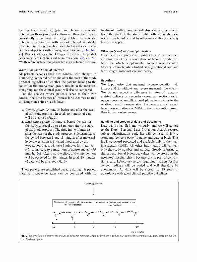
1. Control group: 10 minutes before and after the startof the study protocol. In total, 20 minutes of datawill be analysed (Fig. 2).
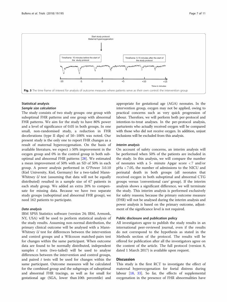
2. Intervention group: 10 minutes before the start ofthe study protocol up to 15 minutes after the startof the study protocol. The time frame of interestafter the start of the study protocol is determined asthe period between 5 and 15 minutes after maternalhyperoxygenation is initiated, motivated by theexpectation that it will take 5 minutes for maternalpO2 to increase to a maximum of approximately 475mmHg [24]. After that, the effect of the interventionwill be observed for 10 minutes. In total, 20 minutesof data will be analysed (Fig. 3).
These periods are established because during this period,maternal hyperoxygenation can be compared with no
treatment. Furthermore, we will also compare the periodsfrom the start of the study until birth, although theseresults may be influenced by other interventions that mayhave been applied.
Other study endpoints and parametersOther study endpoints and parameters to be recordedare duration of the second stage of labour, duration oftime for which supplemental oxygen was received,baseline characteristics (infant sex, gestational age andbirth weight, maternal age and parity).
HypothesisWe hypothesise that maternal hyperoxygenation willimprove FHR, without any severe maternal side effects.We do not expect a difference in rates of vacuum-assisted delivery or secondary caesarean sections or inApgar scores or umbilical cord pH values, owing to therelatively small sample size. Furthermore, we expectlarger concentrations of MDA in the intervention groupthan in the control group.
Handling and storage of data and documentsData will be handled anonymously, and we will adhereto the Dutch Personal Data Protection Act. A securedsubject identification code list will be used to link astudy number to a patient’s name and date of birth. Thisfile is password-protected and available only to the maininvestigator (LMB). All other information will containonly the study number and no data directly referring tothe patient. Foetal blood gas values will be stored in theneonates’ hospital charts because this is part of conven-tional care. Laboratory results regarding markers for freeoxygen radicals will be coded and will therefore beanonymous. All data will be stored for 15 years inaccordance with good clinical practice guidelines.
Fig. 2 The time frame of interest for analysis of outcome measures where patients serve as their own control: the control group. bpm, Beats per minute;CTG, Cardiotocogram
Bullens et al. Trials (2018) 19:195 Page 6 of 11
Statistical analysisSample size calculationThe study consists of two study groups: one group withsuboptimal FHR patterns and one group with abnormalFHR patterns. We aim for the study to have 80% powerand a level of significance of 0.05 in both groups. In onesmall, non-randomised study, a reduction in FHRdecelerations (type II dips) of 50–100% was noted. Ourpresent study is the only one to report FHR changes as aresult of maternal hyperoxygenation. On the basis ofavailable literature, we expect ≥ 50% improvement in theoxygen group and 0% in the control group in both sub-optimal and abnormal FHR patterns [28]. We estimateda mean improvement of 50% with an SD of 50% in eachgroup. A power analysis performed in G*Power 3.0.10(Kiel University, Kiel, Germany) for a two-tailed Mann-Whitney U test (assuming that data will not be equallydistributed) resulted in a sample size of 67 patients ineach study group. We added an extra 20% to compen-sate for missing data. Because we have two separatestudy groups (suboptimal and abnormal FHR group), weneed 162 patients to participate.
Data analysisIBM SPSS Statistics software (version 24; IBM, Armonk,NY, USA) will be used to perform statistical analysis ofthe study results. Assuming non-normal distribution, theprimary clinical outcome will be analysed with a Mann-Whitney U test for differences between the interventionand control groups and a Wilcoxon matched-pairs testfor changes within the same participant. When outcomedata are found to be normally distributed, independentsamples t tests (two-tailed) will be used to analysedifferences between the intervention and control groups,and paired t tests will be used for changes within thesame participant. Outcome measures will be calculatedfor the combined group and the subgroups of suboptimaland abnormal FHR tracings, as well as for small forgestational age (SGA, lower than 10th percentile) and
appropriate for gestational age (AGA) neonates. In theintervention group, oxygen may not be applied, owing topractical concerns such as very quick progression oflabour. Therefore, we will perform both per-protocol andintention-to-treat analyses. In the per-protocol analysis,parturients who actually received oxygen will be comparedwith those who did not receive oxygen. In addition, unjustinclusions will be excluded from this analysis.
Interim analysisOn account of safety concerns, an interim analysis willbe performed when 50% of the patients are included inthe study. In this analysis, we will compare the numberof neonates with a 5- minute Apgar score < 7 and/orpHa < 7.05, the number of admissions to the NICU andperinatal death in both groups (all neonates thatreceived oxygen in both suboptimal and abnormal CTGgroups versus ‘conventional care’ group). If the interimanalysis shows a significant difference, we will terminatethe study. This interim analysis is performed exclusivelyfor safety reasons; because the primary outcome measure(FHR) will not be analysed during the interim analysis andpower analysis is based on the primary outcome, adjust-ment of the significance level is not required.
Public disclosure and publication policyAll investigators agree to publish the study results in aninternational peer-reviewed journal, even if the resultsdo not correspond to the hypothesis as stated in theMethods section of the protocol. The results will beoffered for publication after all the investigators agree onthe content of the article. The full protocol (version 8,dated 1 March 2017) is available upon request.
DiscussionThis study is the first RCT to investigate the effect ofmaternal hyperoxygenation for foetal distress duringlabour [18, 33]. So far, the effects of supplementaloxygenation in the presence of FHR abnormalities have
Fig. 3 The time frame of interest for analysis of outcome measures where patients serve as their own control: the intervention group
Bullens et al. Trials (2018) 19:195 Page 7 of 11
been investigated only in small, non-randomised studies.Because of the lack of concrete results from clinical trials,it is hard to compare the beneficial effects of maternalhyperoxygenation with the potential side effects. As aresult, recommendations on the use of this interventionfor foetal distress in international guidelines are non-uniform [20]. Thus, the results of this study will help tofilling an internationally recognised ‘research gap’.We believe patient safety is carefully addressed in this
study, and ethical concerns are limited. One of the majorconcerns of administering high fractions of oxygen is theincrease in free oxygen radicals. Whether this has a clinicaleffect remains unclear. We excluded from this study allpatients with a higher a priori risk of exposure to increasedfree oxygen radical levels.Both practical and safety issues led to limitations of
this study. An important limitation is the primaryoutcome measure. We recognise that changes in FHR asa primary outcome measure are not optimal, becauseFHR does not accurately reflect foetal oxygenation andacid-base balance [60, 74, 75]. However, we believe thisis the best available method to record changes in thefoetal condition during labour. Furthermore, we assumethat if no beneficial effect on FHR can be shown, animprovement in neonatal outcome is unlikely. Ideally,neonatal outcome measures such as Apgar score andumbilical cord pH are the outcome measures of firstchoice. However, a study with appropriate power toaddress these outcome measures would need a very largesample size. Because the potentially harmful effects havenot been investigated properly yet, we chose not toexpose a large group of women and their foetuses to thisintervention. If a positive effect on FHR pattern withoutsevere side effects can be confirmed by this study, wewill perform a larger multicentre RCT to investigate theeffect on Apgar score and cord blood gas values.In this study, we focus on the foetal condition during
the second stage of labour and short-term neonataloutcome. This implies that abnormalities in FHRpatterns during the first stage of labour are not takeninto account. We believe that the randomisation processwill limit its influence. With regard to the neonatalperiod, we did not arrange long-term follow-up, becausewe do not expect any clinically relevant side effects thatcan be attributed to maternal hyperoxygenation. Besides,the sample size is too small to draw firm conclusions onlong-term neonatal effects in this study.Power analysis of the present study is based on the
expected effect on the primary outcome measure, and thestudy is not powered to find any significant differences inApgar score and umbilical cord blood gas values. In thepower analysis, we used an expected improvement indeceleration depth and duration of 50%. On one hand, thisvalue is based on small non-randomised studies and may
be overestimated. On the other hand, those studies providethe only available data. Also, we believe it is unlikely that alimited improvement in deceleration depth and durationhas clinical relevance. The sample size is calculated for eachof the subgroups of suboptimal and abnormal FHRtracings. We believe it is important to assess the effect ofthe intervention in these subgroups because foetuses withlower initial pO2 levels may profit more from maternalhyperoxygenation [29].Regarding the subgroups of AGA and SGA infants, we
did not increase our sample size to reach an adequatenumber of participants in the SGA group. Nevertheless,we find it interesting to investigate whether there is adifferent effect of maternal hyperoxygenation in SGAinfants compared with AGA infants.Because of organisational challenges, it is not possible
to conduct a double-blind trial. Hence patients anddelivery room staff are not blinded to the patient’sallocation to a study group, which may lead to observerbias. However, analysis of FHR tracings will be doneusing a computerised algorithm, to minimise bias, andthe investigators judging the CTGs and secondaryoutcome measures are blinded to the study arm.To investigate the effect of maternal hyperoxygenation
in the presence of foetal distress on the release of freeoxygen radicals, MDA is estimated in umbilical cordblood. MDA is the peroxidation product of membranepolyunsaturated fatty acids. We chose to measure thismarker for oxidative stress because it was used in priorstudies performed during labour and it is related tovaginal birth, non-reassuring FHR tracings, maternalhyperoxygenation and acidaemia in arterial cord blood[36, 37, 39, 41]. We realise that differences in values inumbilical cord blood may be confounded by mode andduration of delivery; therefore, we will correct the resultsfor the mode of delivery. A practical ground on which tochoose this marker is that it is the only marker foroxidative stress that can be estimated accurately inDutch laboratories. In the intervention group, oxygenadministration will be continued until delivery to enableanalysis of its effect on cord blood gas values and MDA.Despite some important limitations of this study, we
believe this is the best possible way to perform a studywhile restricting safety issues. If the results do not showany improvement in FHR, we believe that maternalhyperoxygenation should not be used as a treatment forfoetal distress. However, if a beneficial effect is demon-strated, we will design a multicentre RCT to investigatethe effect on neonatal outcome.
Trial statusPatient recruitment started on 1 March 2016 and wasestimated to be fulfilled on 31 March 2018.
Bullens et al. Trials (2018) 19:195 Page 8 of 11
Additional files
Additional file 1: SPIRIT 2013 checklist: recommended items to addressin a clinical trial protocol and related documents. (PDF 23602 kb)
Additional file 2: Information and informed consent in English. (DOCX23 kb)
Additional file 3: Information and informed consent in Dutch. (DOCX28 kb)
AbbreviationsAGA: Appropriate for gestational age; bpm: Beats per minute;CTG: Cardiotocogram; DCPRSA: Phase-rectified signal-averaging decelerativecapacity; DNPH: 2,4-Dinitrophenylhydrazine; FHR: Foetal heart rate;FIGO: International Federation of Gynecology and Obstetrics; FSBS: Foetalscalp blood sampling; MDA: Malondialdehyde; m/z: Mass-to-charge ratio;NICU: Neonatal intensive care unit; pCO2: Partial carbon dioxide pressure;pHa: pH in arterial blood gas; pO2: Partial oxygen pressure; PRSA: Phase-rectified signal averaging; RCT: Randomised controlled trial; SAE: Seriousadverse event; SGA: Small for gestational age; SPIRIT: Standard ProtocolItems: Recommendations for Interventional Trials; SpO2: Oxygen saturation;SUSAR: Suspected unexpected serious adverse reaction
AcknowledgementsThis research was performed within the framework of the IMPULS Perinatology,in collaboration with Philips Healthcare, Eindhoven, The Netherlands.
FundingNot applicable.
Availability of data and materialsThe full study protocol is available upon request.
Authors’ contributionsLMB is the main investigator and is, as a doctoral student, leading thisproject. ADJH, SM, RJ, MBvdHvdJ, PJvRH and SGO helped in the design ofthe study, writing of the study protocol and obtaining ethical approval. RJand MBvdHvdJ advised on data analysis and the choice of features for FHR.PJvRH and MBvdHvdJ carefully checked and improved the study protocol,and they advised on several medical and technical topics of the studyprotocol. ERvdH advised on the statistical analysis (power analysis,description of the statistical tests that will be used to analyse the studyresults and the interim analysis). SGO is the initiator of this project. SGOchecked and improved the study protocol. The study sponsor (MáximaMedical Centre Board of Management) was not involved in the design,implementation, data analysis or any reporting of this study. All authors readand approved the final manuscript.
Ethics approval and consent to participateThis study (including amendments) was approved by the Central Committeeon Research Involving Human Subjects (protocol number NL53018.000.15).The study was registered in the EudraCT database (2015-001654-15) on 3April 2015 and in the Dutch Trial Register (NTR5461) on 20 October 2015.This published study protocol corresponds to the seventh version of theoriginal protocol, dated 2 November 2016. The sponsor (Máxima MedicalCentre Board of Management, Veldhoven, The Netherlands) approved theconduct of this study at their hospital. The study will be conductedaccording to the principles of the Declaration of Helsinki (64th version,October 2013) and in accordance with the Medical Research InvolvingHuman Subjects Act (WMO). All patients will receive oral and writteninformation about the study and must give their written informed consentbefore randomisation. When handling personal data, we adhere to theDutch Personal Data Protection Act.
Consent for publicationNot applicable.
Competing interestsThe authors declare that they have no competing interests.
Publisher’s NoteSpringer Nature remains neutral with regard to jurisdictional claims inpublished maps and institutional affiliations.
Author details1Department of Obstetrics and Gynaecology, Máxima Medical Centre, POBox 7777, 5500, MB, Veldhoven, The Netherlands. 2Department of ElectricalEngineering, Eindhoven University of Technology, PO Box 513, 5600, MB,Eindhoven, The Netherlands. 3Department of Clinical Physics, MáximaMedical Centre, PO Box 7777, 5500, MB, Veldhoven, The Netherlands.4Department of Industrial Design, Eindhoven University of Technology, POBox 513, 5600, MB, Eindhoven, The Netherlands. 5Department ofMathematics and Computer Science, Eindhoven University of Technology,PO Box 513, 5600, MB, Eindhoven, The Netherlands.
Received: 17 June 2017 Accepted: 1 March 2018
References1. Caldeyro-Barcia R, Mendez-Bauer C, Poseiro J, Escarena L, Pose S, Bieniarz A.
Control of the human fetal heart rate during labor. In: Cassels DE, editor.The heart and circulation in the newborn and infant: an internationalsymposium presented by the Chicago Heart Association. New York: Grune &Stratton; 1966. p. 7–36.
2. Murray ML. Antepartal and intrapartal fetal monitoring. 3rd ed. New York:Springer; 2007.
3. Westgate JA, Wibbens B, Bennet L, Wassink G, Parer JT, Gunn AJ. Theintrapartum deceleration in center stage: a physiologic approach to theinterpretation of fetal heart rate changes in labor. Am J Obstet Gynecol.2007;197. 236:e1–11.
4. Ball RH, Parer JT. The physiologic mechanisms of variable decelerations. AmJ Obstet Gynecol. 1992;166:1683–8.
5. Bennet L, Gunn AJ. The fetal heart rate response to hypoxia: insights fromanimal models. Clin Perinatol. 2009;36:655–72.
6. Freeman RK, Garite TJ, Nageotte MP. Fetal heart rate monitoring. 3rd ed.Philadelphia: Lippincott Williams & Wilkins; 2000.
7. Hanson MA. Do we now understand the control of the fetal circulation? EurJ Obstet Gynecol Reprod Biol. 1997;75:55–61.
8. Mendez-Bauer C, Arnt IC, Gulin L, Escarcena L, Caldeyro-Barcia R.Relationship between blood pH and heart rate in the human fetus duringlabor. Am J Obstet Gynecol. 1967;97:530–45.
9. Elliott C, Warrick PA, Graham E, Hamilton EF. Graded classification of fetalheart rate tracings: association with neonatal metabolic acidosis andneurologic morbidity. Am J Obstet Gynecol. 2010;202:258 e1–8.
10. Kubli FW, Hon EH, Khazin AF, Takemura H. Observations on heart rate and pHin the human fetus during labor. Am J Obstet Gynecol. 1969;104:1190–206.
11. Graham A, Ruis KA, Hartman A, Northington F, Fox H. A systematic reviewof the role of intrapartum hypoxia-ischemia in the causation of neonatalencephalopathy. Am J Obstet Gynecol. 2008;199:587–95.
12. Martin-Ancel A, Garcia-Alix A, Gaya F, Cabanas F, Burgueros M, Quero J.Multiple organ involvement in perinatal asphyxia. J Pediatr. 1995;127:786–93.
13. Evers AC, Brouwers HA, Hukkelhoven CW, Nikkels PG, Boon J, van Egmond-Linden A, et al. Perinatal mortality and severe morbidity in low and high riskterm pregnancies in the Netherlands: prospective cohort study. BMJ. 2010;341:c5639.
14. McNamara HM, Dildy GA 3rd. Continuous intrapartum pH, pO2, pCO2, andSpO2 monitoring. Obstet Gynecol Clin North Am. 1999;26:671–93.
15. East CE, Begg L, Colditz PB, Lau R. Fetal pulse oximetry for fetal assessmentin labour. Cochrane Database Syst Rev. 2014;10:CD0004075.
16. Dildy GA, van den Berg PP, Katz M, Clark SL, Jongsma HW, Nijhuis JG, et al.Intrapartum fetal pulse oximetry: fetal oxygen saturation trends during laborand relation to delivery outcome. Am J Obstet Gynecol. 1994;171:679–84.
17. Schiermeier S, Pidner von Steinburg S, Thieme A, Reinhard J, DaumerM, Scholz M, Hatzmann W, Schneider KT. Sensitivity and specificity ofintrapartum computerised FIGO criteria for cardiotocography and fetalscalp pH during labour: multicentre, observational study. BJOG. 2008;115:1557–63.
18. Bullens LM, van Runnard Heimel PJ, van der Hout-van der Jagt MB, Oei SG.Interventions for intrauterine resuscitation in suspected fetal distress duringterm labor: a systematic review. Obstet Gynecol Surv. 2015;70:524–39.
Bullens et al. Trials (2018) 19:195 Page 9 of 11
19. Simpson KR. Intrauterine resuscitation during labor: review of current methodsand supportive evidence. J Midwifery Womens Health. 2007;52:229–37.
20. Bullens LM, Moors S, van Runnard Heimel PJ, van der Hout-van der Jagt MB,Oei SG. Practice variation in the management of intrapartum fetal distress inThe Netherlands and the Western world. Eur J Obstet Gynecol Reprod Biol.2016;205:48–53.
21. Hamel MS, Anderson BL, Rouse DJ. Oxygen for intrauterine resuscitation: ofunproved benefit and potentially harmful. Am J Obstet Gynecol. 2014;211:124–7.
22. Hamel MS, Hughes BL, Rouse DJ. Whither oxygen for intrauterineresuscitation? Am J Obstet Gynecol. 2015;212:461–2.
23. Garite TJ, Nageotte MP, Parer JT. Should we really avoid giving oxygen tomothers with concerning fetal heart rate patterns? Am J Obstet Gynecol.2015;212:459–60.
24. Vasicka A, Quilligan EJ, Aznar R, Lipsitz PJ, Bloor BM. Oxygen tension inmaternal and fetal blood, amniotic fluid, and cerebrospinal fluid of themother and the baby. Am J Obstet Gynecol. 1960;79:1041–7.
25. Aldrich CJ, Wyatt JS, Spencer JA, Reynolds EO, Delpy DT. The effect ofmaternal oxygen administration on human fetal cerebral oxygenationmeasured during labour by near infrared spectroscopy. Br J ObstetGynaecol. 1994;101:509–13.
26. McNamara H, Johnson N, Lilford R. The effect on fetal arteriolar oxygensaturation resulting from giving oxygen to the mother measured by pulseoximetry. Br J Obstet Gynaecol. 1993;100:446–9.
27. Khazin AF, Hon EH, Hehre FW. Effects of maternal hyperoxia on the fetus. I.Oxygen tension. Am J Obstet Gynecol. 1971;109:628–37.
28. Althabe O, Schwarcz R, Pose S, Escarcena L, Caldeyro-Barcia R. Effects onfetal heart rate and fetal pO2 of oxygen administration to the mother. Am JObstet Gynecol. 1967;98:858–70.
29. Haydon ML, Gorenberg DM, Nageotte MP, Ghamsary M, Rumney PJ, PatilloC, et al. The effect of maternal oxygen administration on fetal pulseoximetry during labor in fetuses with nonreassuring fetal heart ratepatterns. Am J Obstet Gynecol. 2006;195:735–8.
30. Gare DJ, Shime J, Paul WM, Hoskins M. Oxygen administration during labor.Am J Obstet Gynecol. 1969;105:954–61.
31. Hidaka A, Komatani M, Ikeda H, Kitanaka T, Okada K, Sugawa T. Acomparative study of intrauterine fetal resuscitation by β-stimulant and O2
inhalation. Asia Oceania J Obstet Gynaecol. 1987;13:195–200.32. Dildy GA, Clark SL, Loucks CA. Intrapartum fetal pulse oximetry: the effects
of maternal hyperoxia on fetal arterial oxygen saturation. Am J ObstetGynecol. 1994;171:1120–4.
33. Fawole B, Hofmeyr GJ. Maternal oxygen administration for fetal distress.Cochrane Database Syst Rev. 2012;12:CD000136.
34. Thorp JA, Trobough T, Evans R, Hedrick J, Yeast JD. The effect of maternaloxygen administration during the second stage of labor on umbilical cordblood gas values: a randomized controlled prospective trial. Am J ObstetGynecol. 1995;172:465–74.
35. Nesterenko TH, Acun C, Mohamed MA, Mohamed AN, Karcher D, Larsen JrJ, et al. Is it a safe practice to administer oxygen during uncomplicateddelivery: a randomized controlled trial? Early Hum Dev. 2012;88:677–81.
36. Khaw KS, Wang CC, Ngan Kee WD, Pang CP, Rogers MS. Effects of highinspired oxygen fraction during elective caesarean section under spinalanaesthesia on maternal and fetal oxygenation and lipid peroxidation. Br JAnaesth. 2002;88:18–23.
37. Dede FS, Guney Y, Dede H, Koca C, Dilbaz B, Bilgihan A. Lipid peroxidationand antioxidant activity in patients in labor with nonreassuring fetal status.Eur J Obstet Gynecol Reprod Biol. 2006;124:27–31.
38. Yalcin S, Aydoğan H, Kucuk A, Yuce HH, Altay N, Karahan MA, et al.Supplemental oxygen in elective cesarean section under spinal anesthesia:handle the sword with care. Braz J Anesthesiol. 2013;63:393–7.
39. Rogers MS, Mongelli JM, Tsang KH, Wang CC, Law KP. Lipid peroxidation in cordblood after birth: the effect of labor. Br J Obstet Gynaecol. 1998;105:739–44.
40. Blackburn S. Free radicals in perinatal and neonatal care, part 2: oxidativestress during the perinatal and neonatal period. J Perinat Neonatal Nurs.2006;20:125–7.
41. Wang W, Pang CC, Rogers MS, Chang AM. Lipid peroxidation in cord bloodat birth. Am J Obstet Gynecol. 1996;174:62–5.
42. Nordström L, Arulkumaran S. Intrapartum fetal hypoxia and biochemicalmarkers: a review. Obstet Gynecol Surv. 1998;53:645–57.
43. Saugstad OD, Ramji S, Soll RF, Vento M. Resuscitation of newborn infantswith 21% or 100% oxygen: an updated systematic review and meta-analysis.Neonatology. 2008;94:176–82.
44. SUPPORT Study Group of the Eunice Kennedy Shriver NICHD NeonatalResearch Network. Target ranges of oxygen saturation in extremely preterminfants. N Engl J Med. 2010;362:1959–69.
45. Davis PG, Tan A, O’Donnell CP, Schulze A. Resuscitation of newborn infantswith 100% oxygen or air: a systematic review and meta-analysis. Lancet.2004;364:1329–33.
46. Rabi Y, Rabi D, Yee W. Room air resuscitation of the depressed newborn: asystematic review and meta-analysis. Resuscitation. 2007;72:353–63.
47. Brantberg A, Sonesson SE. Central arterial hemodynamics in small-for-gestational-age fetuses before and during maternal hyperoxygenation: a Doppler velocimetricstudy with particular attention to the aortic isthmus. Ultrasound Obstet Gynecol.1999;14:237–43.
48. Bartnicki J, Saling E. Influence of maternal oxygen administration on thecomputer-analysed fetal heart rate patterns in small-for-gestational-agefetuses. Gynecol Obstet Investig. 1994;37:172–5.
49. Duling BR. Microvascular responses to alterations in oxygen tension. CircRes. 1972;31:481–9.
50. Cornet AD, Kooter AJ, Peters MJ, Smulders YM. The potential harm of oxygentherapy in medical emergencies. Crit Care. 2013;17:313.
51. Sjöberg F, Singer M. The medical use of oxygen: a time for critical reappraisal. JIntern Med. 2013;274:505–28.
52. Fitch W. Cerebral blood flow: physiological principles and methods ofmeasurement. In: Sebel PS, Fitch W, editors. Monitoring the central nervoussystem. Oxford, UK: Blackwell Science; 1994. p. 78–117.
53. Watson NA, Beards SC, Altaf N, Kassner A, Jackson A. The effect of hyperoxiaon cerebral blood flow: a study in healthy volunteers using magneticresonance phase-contrast angiography. Eur J Anaesthesiol. 2000;17:152–9.
54. Bennett MH, French C, Schnabel A, Wasiak J, Kranke P. Normobaric andhyperbaric oxygen therapy for migraine and cluster headache. CochraneDatabase Syst Rev. 2008;3:CD005219.
55. Kehrer JP, Klotz LO. Free radicals and related reactive species as mediators oftissue injury and disease: implications for health. Crit Rev Toxicol. 2015;45:765–98.
56. Lareb. ’s-Hertogenbosch, The Netherlands: Netherlands PharmacovigilanceCentre Lareb; https://www.lareb.nl/nl/databank/Result?formGroup=&atc=V03AN01&drug=ZUURSTOF+MEDICINAAL+%28ZUURSTOF%29. Accessed 21Mar 2017.
57. American Congress of Obstetricians and Gynecologists. Practice bulletin no.116: management of intrapartum fetal heart rate tracings. Obstet Gynecol.2010;116:1232–40.
58. National Institute for Health and Care Excellence (NICE). Intrapartum care forhealthy women and babies. NICE clinical guideline [CG190]. London: NICE;2014. [updated February 2017]. https://www.nice.org.uk/guidance/cg190/chapter/Recommendations. Accessed 16 May 2017.
59. Chan AW, Tetzlaff JM, Gøtzsche PC, Altman DG, Mann H, Berlin JA, et al.SPIRIT 2013 explanation and elaboration: guidance for protocols of clinicaltrials. BMJ. 2013;346:e7586.
60. Ayres-de-Campos D, Spong CY, Chandraharan E. FIGO Intrapartum FetalMonitoring Expert Consensus Panel. FIGO consensus guidelines on intrapartumfetal monitoring: cardiotocography. Int J Gynecol Obstet. 2015;131:13–24.
61. Yli B, Hahn T, Kessler J, Lie HK, Martinussen M. Bigger is not always better…the validity of the US randomized trial of STAN for Norway. Mölndal,Sweden: Neoventa Medical. http://www.neoventa.com/2015/11/bigger-is-not-always-better/. Accessed 6 Nov 2017.
62. Bauer A, Kantelhardt JW, Barthel P, Schneider R, Mäkikallio T, Ulm K,Hnatkova K, et al. Deceleration capacity of heart rate as a predictor ofmortality after myocardial infarction: cohort study. Lancet. 2006;367:1674–81.
63. Huhn EA, Lobmaier S, Fischer T, Schneider R, Bauer A, Schneider KT,Schmidt G. New computerized fetal heart rate analysis for surveillance ofintrauterine growth restriction. Prenat Diagn. 2011;31:509–14.
64. Georgieva A, Payne SJ, Moulden M, Redman CWG. Computerized fetal heartrate analysis in labor: detection of intervals with un-assignable baseline.Physiol Meas. 2011;32:1549–60.
65. Ozden S, Demirci F. Significance for fetal outcome of poor prognosticfeatures in fetal heart rate traces with variable decelerations. Arch GynecolObstet. 1999;262:141–9.
66. Gaziano EP. A study of variable decelerations in association with other heartrate patterns during monitored labor. Am J Obstet Gynecol. 1979;135:360–3.
67. Krebs HB, Petres RE, Dunn LJ. Intrapartum fetal heart rate monitoring. VIII.Atypical variable decelerations. Am J Obstet Gynecol. 1983;145:297–305.
68. Hamilton E, Warrick P, O’Keeffe D. Variable decelerations: do size and shapematter? J Matern Fetal Neonatal Med. 2012;25:648–53.
Bullens et al. Trials (2018) 19:195 Page 10 of 11

69. Kazandi M, Sendag F, Akercan F, Terek MC, Gundem G. Different types ofvariable decelerations and their effects to neonatal outcome. SingaporeMed J. 2003;44:243–7.
70. Holzmann M, Wretler S, Cnattingius S, Nordström L. Cardiotocography patternsand risk of intrapartum fetal acidemia. J Perinat Med. 2015;43:473–9.
71. Georgieva A, Payne SJ, Moulden M, Redman CW. Relation of fetal heart ratesignals with unassignable baseline to poor neonatal state at birth. Med BiolEng Comput. 2012;50:717–25.
72. Georgieva A, Papageroghiou AT, Payne SJ, Moulden M, Redman CW. Phase-rectified signal averaging for intrapartum electronic fetal heart ratemonitoring is related to acidaemia at birth. BJOG. 2014;121:889–94.
73. Lobmaier SM, Mensing van Charante N, Ferrazzi E, Giussani DA, Shaw CJ,Müller A, et al. TRUFFLE investigators. Phase-rectified signal averagingmethod to predict perinatal outcome in infants with very preterm fetalgrowth restriction—a secondary analysis of TRUFFLE-trial. Am J ObstetGynecol. 2016;215:630.e1–7.
74. James LS, Morishima HO, Daniel SS, Bowe ET, Cohen H, Niemann WH.Mechanism of late deceleration of the fetal heart rate. Am J Obstet Gynecol.1972;113:578–82.
75. Morishima HO, Daniel SS, Richards RT, James LS. The effect of increased maternalPaO2 upon the fetus during labor. Am J Obstet Gynecol. 1975;123:257–64.
• We accept pre-submission inquiries
• Our selector tool helps you to find the most relevant journal
• We provide round the clock customer support
• Convenient online submission
• Thorough peer review
• Inclusion in PubMed and all major indexing services
• Maximum visibility for your research
Submit your manuscript atwww.biomedcentral.com/submit
Submit your next manuscript to BioMed Central and we will help you at every step:
Bullens et al. Trials (2018) 19:195 Page 11 of 11


