Embed Size (px)
Citation preview
\et' ì.ûG
AoneToMEDULLARY RCCUIATION DUNIruC ITINNUTERINE
Srness rN THE Fernl SHeep
Michael Brenton Adams B.Sc. (Hons)
Department of Physiology
The University of Adelaide
A thesis submitted for the degree of Doctor of Philosophy
to
The University of Adelaide
October 1999
" 'Forty-two!'yelled Loonquwal. 'ls that all you've got to show for seven and a half
million years'work?'
'l checked it very thoroughly,' said the computer, 'and that quite definitely is the
answer. I think the problem, to be quite honest with you, is that you've never
actually known what the question is.'
'But it was the Great Question! The Ultimate Question of Life, the Universe and
Everything,' howled Loonquwal.
'Yes,' said Deep Thought with the air of one who suffers fools gladly, 'but what
actually is it?'"
Douglas Adams
The Hitch Hiker's Guide to the Galaxy (1979)
TABLE OF CONTENTS
1. ADRENOMEDULLARY CATECHOLAMINE SECRETION, SYNTHESIS, ANDACTIONS lN THE FETUS.... ............ 1
1.1 TNTRODUCTION ........ 1
1.2 ANATOMY AND EMBRYOLOGY OF THE ADRENAL GLAND ................. 1
1.2.1 The Adrenal Medulla ........'....'...1
1.2.2 The Adrenal Cortex.,. ......".""".4
1.2.3 lnnervation of the Adrenal G1and........ '."".7
1.2.3.1 Adrenomedullary innervation .".'...-.."'.."'.7
1 .2.3.2 Adrenocortical inneruation............ .......".'.7
1.2.3.3 Postganglionic sympathetic innervation............ ........8
1.2.3.4 Sensory innervation ........."..."8
1.2.3.5 lntrinsic innervation "...'....'..'."9
1.2.3.6 Ontogeny of adrenal innervation ."'..""' 10
1.2.4 Vasculature....,..,....... ..............11
1 .3 THE ADULT ADRENAL MEDULLA.......... ................ 13
1.3.1 Adrenomedullary Catecholamine Synthesis and Storage.... ......13
1.3.1.1 Synthesis '..."'...".13
1.3.1.2 Storage '.."'...""...16
1.3.1.2.1 Chromaffin granules ..........16
1.3.1.2.2 Chromaffin granule synthesis.... ............. 16
1.3.1.2.3 Catecholamine transport and storage '.'-..-.-"'.."-'-..-.17
1.3.1.2.4 Chromaffin granules and catecholamine synthesis.... ..................18
1.3.1.2.5 Chromaffin granules and calcium homeostasis '......18
1.3.2 Adrenomedullary Neuropeptides........... ..................'. 19
1.3.2.1 Enkephatins ....,......... .........."19
1.3.2.2 Neuropeptide tyrosine ..."'.".20
TABLE OF CONTENTS
1
1
1
1.3
1
3.2.3 Vasoactive intestinal polypeptide ..........,..........
3.2.4 Nitric oxide
3.2.5 Other neuropeptides .........
3 Regulation of Adrenal Catecholamine Secretion
3.3. 1 Neurogenic catecholamine secretion ...............
1.3.3.1.1 Secretorypathway......
1.3.3.1.2 Nicotinic receptor stimulation
1.3.3.1.2.1 Calcium dependence
1.3.3.1.2.2 lonic events
...20
...21
...22
',,22
...22
...22
...24
,...24
,...24
...26
....27
....27
....27
...28
....29
....30
...31
....32
....33
....33
....34
....35
....36
....37
...38
...39
....39
....39
...41
....41
....43
....45
....47
....47
....49
1.3.3.1.2.3 Adenylate cyclase activation
1 .3.3.1 .3 Muscarinic receptor stimulation..
1.3.3.1.3.1 Calcium dependence
1.3.3.1.3.2 Calcium mobilisation
1.3.3.1.3.3 Muscarinic modulation of nicotinic stimulation
1.3.3.1.4 Cellularmechanisms
1.3.3.1.5 Exocytosis
1 .3.3.2 Non-cholinergic regulation of adrenal catecholamine secretion..
1.3.3.2.1 Experimentalevidence
1.3.3.2.2 Potential non-cholinergic transmitters ...........
1.3.3.2.2.1 VIP and PACAP
1.3.3.2.2.2 Substance P.................
1.3.3.2.2.3 NPY and the opioid peptides.....
1 .3. 3.2.2.4 Angiotensin I 1.................
1.3.3.2.2.5 Nitric oxide.
1.3.4 Regulation of Adrenomedullary Catecholamine Biosynthesis.......
1.3.4.1 Short term regulation of catecholamine biosynthesl's.................
1.3.4.1.1 Negativefeedbackinhibition
1.3.4.1.2 Tyrosine hydroxylase phosphorylation ............
1.3.4.2 Long term regulation of catecholamine biosynthesls..................
1.3.4.2.1 Trans-synaptic enzyme induction
1.3.4.2.1.1 c\MP.........
1.3.4.2.1.2 Protein kinase C.................
1.3.4.2.2 Hormonal regulation of catecholamine biosynthesis
1.3.4.2.2.1 Glucocorticoids...........
1.3.4.2.2.2 Angiotensin 11.................
TABLE OF CONTENTS
1.4 THE FETAL ADRENAL MEDU114...........
1.4.1 Physiological Role of Adrenomedullary Catecholamines in the Fetus ....
1.4.1.1 Catecholamine secretion in response fo sfress in utero
1.4.1.1.1 Acute stress
1.4.1.1.1.1 Hypoxaemia
1.4.1.1.1.2 Hypoglycaemia......
1.4.1.1.1.3 Labour and parturition
1.4.1.1.2 Chronic stress
1 .4. 1. 1.2. 1 Clinical observations .................
1.4.1.1.2.2 Experimental observations,...........,.....
1.4.1.2 Physiological actions of catecholamines in the fetus and neonate ........
1.4.1.2.1 Cardiovascularactions
1 .4. 1 .2. 1 . 1 Fetal cardiovascular responses to hypoxaemia ................
1.4.1.2.1.2 Catecholamines and haemodynamic responses to hypoxaemia...........
1.4.1.2.1.3 Catecholamines and cardiac responses to hypoxaemia........................
1.4.1.2.1.4 Catecholamines and cardiovascular adaptations at birth...
1.4.1.2.2 Effect of catecholamines on metabolism and growth.
1.4.1.2.2.1 Fetal and neonatal metabolism
1.4.1.2.2.2 Fetal growth
1.4.1.2.3 Respiratory actions........
1.4.1.2.4 Thermogenicactions
1.4.2 Regulation of Catecholamine Secretion and Biosynthesis in the FetalAdrenal Medulla
1.4.2.1 Ontogeny of adrenomedullary catecholamines and enkephalins...........
1.4.2.1.1 Catecholamines.............
1.4.2.1.2 Enkephalins
1.4.2.2 Neuropeptide regulation of adrenomedullary catecholamine secretion..
1 .4.2.3 Non-neurogenic catecholamine secretion ..............
1.4.2.3.1 Experimental evidence
1.4.2.3.2 Cellularmechanisms
1.4.2.3.2.1 Mitochondrial oxygen sensing......
1 .4.2. 3. 2.2 Oxygen-sensitive potassium channe\s................
1 .4. 2.4 Regu lation of fetal adrenomedullary catecholamine biosynthesis..........
1.4.2.4.1 Oxygen regulation of catecholamine biosynthesis
1.4.2.4.1.1 Hypoxia and catecholamine synthesis.....
1.4.2.4.1.2 Oxygen regulation of gene expression.....
50
..50
..50
... 50
...50
...51
...52
...53
...53
...54
...56
...57
...57
...58
...59
...60
...61
...61
...63
...64
...65
.67
,.67
..67
..70
.72
..73
..74
..76
..78
..78
,.82
..82
..83
..84
TABLE OF CONTENTS
1.4.2.4.1.3 Oxygen regulation of catecholamine biosynthetic enzyme genes ............. 85
1.4.2.4.1.4 Mechanisms of hypoxia induced TH gene transcription ..."".. 87
1.4.2.4.1.5 Mechanisms of hypoxia induced TH nRNA stabilisation .."." Bg
1.4.2.4.1.6 Oxygen sensing and TH gene transcription '.."'.' 90
1.4.2.4.2 Hormonal regulation of adrenomedullary development and catecholaminebiosynthesis ......................... 92
1.4.2.4.2.1 Glucocorticoids and adrenomedullary development ...................'.'."'."'....92
1.4.2.4.2.2 Glucocorticoids and fetal catecholamine biosynthesis ...........95
1.5 AIMS AND HYPOTHESES ..........97
1.5.1 Ontogeny of TH, PNMT, and PEnk A mRNA Expression in the Fetal SheepAdrenal . ................97
1.5.2 lmpact of Acute Hypoxaemia on the Expression of TH and PNMT mRNA in
the Adrenal Gland of the Fetal Sheep Prior to and After the Development ofFunctional Splanchnic lnnervation ...........98
1.5.3 The lmpact of a Chronic Physiological lncrease of Plasma Glucocorticoidson the Expression of TH, PNMT, and PEnk A mRNA in the Adrenal Gland ofthe Fetal Sheep....... ................99
1.5.4 Effect of Placental Restriction on TH, PNMT, and PEnk A mRNA Expressionin the Adrenal Gland of the Late Gestation Sheep Fetus ..........99
2. ONTOGENY OF TYROSINE HYDROXYLASE, PHENYLETHANOLAMINE N.METHYLTRANSFERASE AND PROENKEPHALIN A MRNA EXPRESSIONIN THE ADRENAL GLAND OF THE FETAL SHEEP 100
1002.1 INTRODUCTION
2.2 METHODS.............
2.2.1 Ontogeny Study
2.2.2 RNA 1soIation..................
2.2.3 Oligonucleotide Probes.,.
2.2.4 TH cDNA Probe
2.2.5 Probe Labelling
2.2.6 Northern Blotting
2.2.7 MembraneHybridisation.
2.2.8 DataAnalysis...,....
2.3 RESULTS
2.3.1 ProbeValidation....
2.3.2 Fetal Outcomes and Adrenal Growth With Development
2.3.3 Ontogeny of Adrenal TH, PNMT, and PEnk A mRNA Expression..
2.4 DISCUSSION ..
........... 101
............. 1 01
.............101
.............102
.............102
.............104
.............104
.............1Q7
......,...... 1 08
...........108
............. 1 08
109
109
112
TABLE OF CONTENTS
3. IMPACT OF ACUTE HYPOXAEMIA ON THE EXPRESSION OF TH ANDPNMT mRNA lN THE ADRENAL GLAND OF THE FETAL SHEEP........... 115
3.1 INTRODUCTION 115
3.2 MATERIALS AND METHODS.............. ..116
3.2.1 Animals and Sur9ery............... ......,........ 1 16
3.2.2 Post Operative Care... ...........117
3.2.3 Fetal Hypoxia ............ ............... 1 18
3.2.4 Hexamethonium......... ........... 1 18
3.2.5 Blood Sampling ...118
3.2.6 Post Mortem and Tissue Collection .......120
3.2.7 Plasma Catecholamine Extraction............. ..............120
3.2.8 Catecholamine Measurement ................121
3.2.9 Total RNA Extraction.. ...........121
3.2.10 Probes and Probe Labelling ...................122
3.2.11 Northern Blot 4na1ysis............... .............124
3.2.12 StatisticalAnalysis ................124
3.3 RESULTS .....125
3.3.1 Fetal Arterial Blood Gases and Experimental Hypoxia ............125
3.3.1.1 Arterial PO2.......... ...............125
3.3.1.2 Arterial PCO2........ ...............126
3.3.1.3 Arterial pH............ ...............126
3.3.2 Fetal Plasma Catecholamines..,,.. ........,.127
3.3.2.1 Basal plasma catecholamine levels... ...127
3.3.2.2 lmpact of experimental hypoxia on plasma catecholamine \eve\s............127
3.3.3 Hypoxia and Catecholamine Synthetic Enzyme Gene Expression ............128
3.3.3.1 TH nRNA expression .........128
3.3.3.2 PNMT nRNA expression ....128
3.3.3.3 Relationships of adrenalTH and PNMT nRNA expression withexperimentally induced changes in fetal arterial PO2 ..............................128
3.3.4 Effect of Hexamethonium on Catecholamine Synthetic Enzyme GeneExpression in Response to Hypoxia
3.3.4.1 TH nRNA expression
3.3.4.2 PNMT nRNA expression
3.4 D|SCUSSION.........
......129
......129
......129
....137
TABLE OF CONTENTS
4. GLUCOCORTICOID REGULATION OF TH, PNMT AND PEnk A mRNAEXPRESSION IN THE FETAL SHEEP ADRENAL 146
4.1 INTRODUCTION
4.2 MATERIALS AND METHODS.............
4.2.1 Animals and Sur9ery...............
4.2.2 Post Operative Care...
4.2.3 Blood Sampling and Cortisol lnfusion..........
4.2.4 Post Mortem and Tissue Collection
4.2.5 CortisolAssay
4.2.6 ACTH Assay
4.2.7 Plasma Glucose and Lactate Determination
4.2.8 Total RNA Extraction..
4.2.9 Probes and Probe Labelling
4.2.10 Northern Blot 4na1ysis...............
4.2.11 Antibodies
4.2.12 lmmunohistochemistry...............
4.2.13 Adrenal Morphometry.............
4.2.14 Statistical Analysis
4.3 RESULTS ............... 153
4.3.1 Fetal Outcomes........... ..........153
4.3.2 Fetal Endocrine and Metabolic Status ... 153
4.3.3 Cortisol lnfusion and Adrenal TH, PNMT and PEnk A mRNA Expression . 154
4.3.4 Cortisol lnfusion and Adrenal Morphometry........... ..................154
4.4 D|SCUSSION., ............... 160
5. EFFECT OF PLACENTAL RESTRICTION ON TH, PNMT, AND PEnk AmRNA EXPRESSION IN THE ADRENAL GLAND OF THE LATEGESTATION SHEEP FETUS 165
165
166
166
167
167
167
168
168
................. 146
................. 147
................... 1 47
,................... 1 47
..............,.... 148
.................... 148
.................... 148
.................... 149
................... 149
,................... 149
.................... 1 50
.................... 1 50
....,.,............ 151
.................... 1 51
.................... 152
.................... 153
5.1 INTRODUCTION
5.2 METHODS ........
5.2.1
5.2.2
5.2.3
5.2.4
5.2.5
5.2.6
Animals and Sur9ery......................
Post Operative Care...
Post Mortem and Tissue Collection
Blood Sampling Protocol
Plasma Glucose Determination....,,
Plasma Catecholamine Extraction..
TABLE OF CONTENTS
5.2.7 Catecholamine Measurement.......
5.2.8 RNA lsolation and Analysis
5.2.9 Probes and Probe Labelling
5.2.10 Northern Blot 4na|ysis...................
5.2.11 lmmunohistochemistry
5.2.12 Adrenal Morphometry....................
5.2.13 Data Ana|ysis............
5.3 RESULTS
Localisation
5.4 DISCUSSION .,
6. SUMMARY AND CONCLUSIONS
5.3.3 Placental Restriction and Adrenal TH, PNMT, and PEnk A mRNAExpression
5.3.4 Placental Restriction and Adrenal Catecholamine Synthetic Enzyme
174
.., 169
...169
...170
...170
...171
...172
...172
..173
179
181
186
192BIBLIOGRAPHY
ABSTRACT
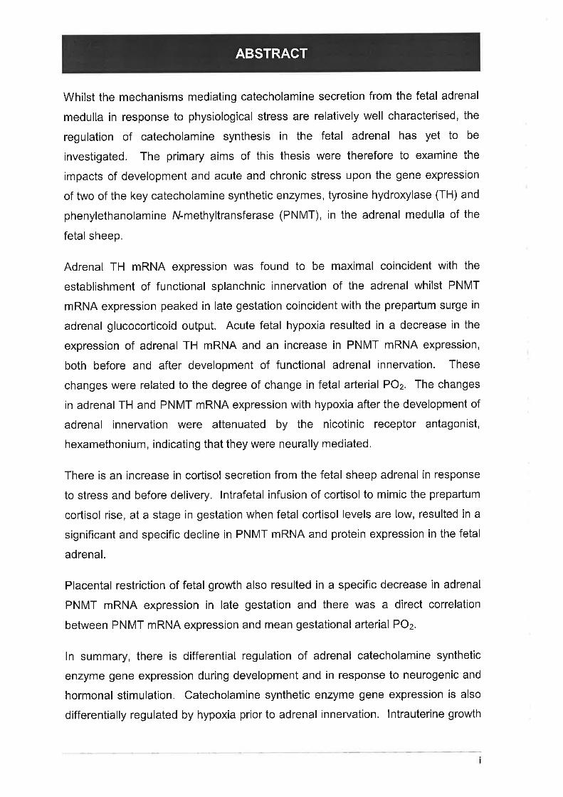
Whilst the mechanisms mediating catecholamine secretion from the fetal adrenal
medulla in response to physiological stress are relatively well characterised, the
regulation of catecholamine synthesis in the fetal adrenal has yet to be
investigated. The primary aims of this thesis were therefore to examine the
impacts of development and acute and chronic stress upon the gene expression
of two of the key catecholamine synthetic enzymes, tyrosine hydroxylase (TH) and
phenylethanolamine N-methyltransferase (PNMT), in the adrenal medulla of the
fetal sheep.
Adrenal TH mRNA expression was found to be maximal coincident with the
establishment of functional splanchnic innervation of the adrenal whilst PNMT
mRNA expression peaked in late gestation coincident with the prepartum surge in
adrenal glucocorticoid output. Acute fetal hypoxia resulted in a decrease in the
expression of adrenal TH mRNA and an increase in PNMT mRNA expression,
both before and after development of functional adrenal innervation. These
changes were related to the degree of change in fetal arterial POz. The changes
in adrenal TH and PNMT mRNA expression with hypoxia after the development of
adrenal innervation were attenuated by the nicotinic receptor antagonist,
hexamethonium, indicating that they were neurally mediated.
There is an increase in cortisol secretion from the fetal sheep adrenal in response
to stress and before delivery. lntrafetal infusion of cortisol to mimic the prepartum
cortisol rise, at a stage in gestation when fetal cortisol levels are low, resulted in a
significant and specific decline in PNMT mRNA and protein expression in the fetal
adrenal.
Placental restriction of fetal growth also resulted in a specific decrease in adrenal
PNMT mRNA expression in late gestation and there was a direct correlation
between PNMT mRNA expression and mean gestational arterial POz.
ln summary, there is differential regulation of adrenal catecholamine synthetic
enzyme gene expression during development and in response to neurogenic and
hormonal stimulation. Catecholamine synthetic enzyme gene expression is also
differentially regulated by hypoxia prior to adrenal innervation. lntrauterine growth
ABSTRACT
retardation and inappropriate fetal exposure to excess glucocorticoids exert
specific suppressive effects upon adrenal PNMT mRNA expression which may
manifest themselves as impaired responses to physiological stress in the fetus
and neonate.

ACKNOWLEDGMENTS
I would firstly like to acknowledge my supervisor, Professor Caroline McMillen, for
providing me both with the opportunity to undertake these studies, and the
encouragement and advice which made this thesis possible.
Thank you to those members, past and present, of the Fetal Physiology Group in
the Department of Physiology at the University of Adelaide, who provided
guidance, help, and above all, friendship.
I would like to extend my gratitude to a number of people who have facilitated
these studies with their expertise and gifts of reagents. I would particularly like to
thank Professor Peter Howe (Department of Biochemistry, University of
Wollongong) for supplying PNMT antibody; and Dr Barry Kaplan (Department of
Psychiatry, University of Pittsburgh) for providing the bovine TH cDNA. Thanks
also to the Laboratory Animal Services Staff at the University of Adelaide for their
professionalism and care with regards to the maintenance of the animals used in
these studies.
Thanks to all members, past and present, of the Department of Physiology at the
University of Adelaide, for providing the environment and services which allowed
me to complete these studies.
I would especially like to acknowledge my family, especially my parents and
grandmother, whose love, encouragement, and sacrifice, allowed me to strive for
this goal.
Finally, I would like to thank my beautiful wife, Narelle, who has shared all of the
emotional highs and lows of this period with ffie, always with a love and
compassion that enabled me to believe in myself.
tv
PUBLICATIONS ARISING FROM THIS THESIS
AoRwrS, M.8., PHltupS, LD., Sln¡o¡lETTA, G., McMtlLrru, l.C. (1998) The differential
effects of increasing gestational age and placental restriction on tyrosine
hydroxylase, phenylethanolamine N-methyltransferase, and proenkephalin A
mRNA levels in the fetal sheep adrenal. Journal of Neurochemistry,71,394-401.
AoRtvtS, M.B., ROsS, J.T., ButlER, T.G., MCMllleru, l.C. (1999) Glucocorticoids
decrease phenylethanolamine N-methyltransferase mRNA expression in the
immature foetal sheep adrenal. Journal of Neuroendocrinology,ll, 569-575.
AoRwrs, M.8., SlvoruertR, G.,McMlt-l-Elrl, l.C. (1996) The non-neurogenic
catecholamine response of the fetal adrenal to hypoxia is dependent on voltage
sensitive Ca2* channels. Developmental Brain Research,94, 182-189.
McMlt-lrx, LC., SIH¡ollEttR, G., RogeRls, M.L., Aoaus, M.B. (1997)
Catecholamines, enkephalins and the response of the fetal adrenal medulla to
hypoxaemia. Equine Veterinary Journal,Supplement 24, 68-73.
RycHxov, G.Y., AoRwts, M.8., McMlt-1eru, 1.C., Roatnts, M.L. (1998) Oxygen-
sensing mechanisms are present in the chromaffin cells of the sheep adrenal
medulla before birth. Journal of Physiology (London), 509,887-893.
RELATED PUBLICATIONS
v
COM MON LY USED ABBREVIATIONS
A
AADC
ABC
ACE
ACh
ACTH
Ad
Ang ll
ANOVA
AP-1
ATFlATP
aromatic amino acid decarboxylase
avidin-biotin-peroxidase com plex
angiotensi n converting enzyme
acetylcholine
adrenocorticotrophic hormone
adrenaline
angiotensin ll
analysis of variance
activator protein 1
activating transcription factor 1
adenosine triphosphate
BC
b
bp
BPt
BAT
BSA
cAMP
CAT
cDNA
cGMP
CGRP
CRE
CREB
CRF
CTP
CV
bovine
base pair(s)
tetrahydrobiopterine
brown adipose tissue
bovine serum albumin
cyclic adenosine 3',5'-monophosphate
chloramphenicol acetyltransferase
complementary deoxyribonucleic acid
cyclic guanosine 3',5'-monophosphate
calcitonin gene related peptide
cyclic AMP response element
cAMP response element binding protein
corticotrophin releasing factor
cytidine triphosphate
coefficient of variation
DE
d
DAB
DAG
DBH
DHBA
DMPP
DNA
DOPA
days
3, 3-diaminobenzadine tetrahydrochloride
diacylglycerol
dopamine B hydroxylase
3,4 dihydroxybenzylamine
dimethylphenylpiperazinium
deoxyribonucleic acid
dihydroxyphenylalani ne
vt
CO MMO N LY U SED ABB REVIATI ON S vil
APOz exper¡mentally induced changearterial POz
embryological day
electrochemical detection
ethylenediamine tetraacetic acid
enkephalin
erythropoietin
tn fetal
E
ECD
EDTA
Enk
EPO
FGH
F¡Oz
FGF
g
GR
GRE
GSH
GSSG
h
Hex
HIF-1
HIP
HIPBS
HPA
HPLC
HPX
fractional inspired oxygen
fibroblast growth factor
g ravitational acceleration
glucocorticoid type ll receptor
glucocorticoid response element
reduced glutathione
oxidised glutathione
hours
hexamethonium
hypoxia inducible factor 1
hypoxia induced proteins
hypoxia induced protein binding sequence
hypothalam ic-pitu itary-ad renal
high performance liquid chromatography
hypophysectomised
IKLMN
ICER
lPe
ir
IUGR
iv
kb
K,
K^
L
LB
Leu
mAChR
Met
min
mRNA
nAChR
inducible cAMP early represser
inositol trisphosphate
immunoreactive
intrauterine g rowth retardation
intravenous
kilobases
lnhibitory constant
Michaelis constant
lumbar
Luria-Bertani
Leucine
muscarinic acetylcholine receptor
Methionine
minutes
messenger ribonucleic acid
nicotinic acetylcholine receptor
CO MMO N LY U S ED AB B REVIATIO N S vlll
NAd
NADPH
NGF
NGS
NO
NOS
NPY
NSF
OPR
noradrenaline
nicotinamide adenine dinucleotide phosphate
nerve growth factor
normal goat serum
nitric oxide
nitric oxide synthase
neuropeptide tyrosine
N-ethylmaleimide sensitive fusion protein
OD
ODS
P-450at
P-45O"zt
P-45Oscc
PACAP
P"COz
P^Oz
PBS
PCA
PEnk A
PlP2
PKA
PKC
PMA
PNMT
POMC
PR
RAS
ROt
rpm
rRNA
RT
optical density
octadecylsilylsilica
P-450 1 7cr-hydroxylase / C17 -C20 lyase
P-450 21-hydroxylase
P-450 cholesterol side chain cleavage
pituitary adenylate cyclase activating peptide
partial pressure of COz in arterial blood
partial pressure of Oz in arterial blood
phosphate buffered saline
perchloric acid
proenkephalin Aphosphatidylinositol 4, 5-bisphosphate
protein kinase Aprotein kinase C
phorbol 12-myristate 13-acetate
phenylethanolamine N-methyltransferase
proopiomelanocortin
placental restriction
renin angiotensin system
reactive oxygen intermediate
revolutions per minute
ribosomal ribonucleic acid
room temperature
srSA
SAM
S"Oz
SCG
SDS
SEM
SNAP
sympathoadrenal
S-adenosylmethionine
oxygen saturation of arterial blood
superior cervical ganglion
sodium dodecyl sulphate
standard error of the mean
soluble NSF attachment proteins
CO MMO N LY U S ED AB B REVIATION S tx
SNARE
SSC
SSPE
T
TBE
TH
TPA
TTX
SNAP receptors
saline sodium citrate
saline sodium phosphate EDTA
thoracic
tris-borate EDTA
tyrosine hydroxylase
1 2- O-tetr adeca noyl p horba I 1 3-acetate
tetrodotoxin
UV
UCP 1
VAMP
VEGF
VIP
V^^
uncoupling protein 1
vesicle membrane associated protein
vascular associated growth factor
vasoactive intestinal polypeptide
maximal velocity
LIST OF FIGURES AND TABLES
Figure 1.1 Biosynthetic pathway of adrenomedullary catecholamines 15
Figure 2.1 Target sequences of the bovine PNMT, PEnk A, and rat 18 S oligonucleotide
probes ...............105
Figure 2.2 lsolation and purification of the TH cDNA probe from pBluescript ll SK-
phagemid vector ................ 106
Table 2.1 Fetal weight, adrenal weight, and adrenal total RNA yield with development
............109
Figure 2.3 Validation of TH cDNA, PNMT, and PEnk A oligodeoxynucleotide probes..110
Figure 2.4 Ontogeny of TH, PNMT, and PEnk A mRNA expression in the fetal sheep
adrenal 111
Figure 3.1 Experimental set up for the regulation maternal F¡Oz 119
Figure 3.2 Example of catecholamine calibration curves generated using reverse phase
HPLC coupled with ECD ...123
Figure 3.3 Effects of experimental hypoxia and normoxia upon fetal arterial blood gas
variables... .........130
Figure 3.4 HPLC chromatograms of plasma catecholamine levels in a 141 d fetal sheep
exposed to hypoxia 131
Figure 3.5 lmpact of acute hypoxia on plasma catecholamine levels in the fetal sheep
..132
Figure 3.6 Autoradiographs of Northern blots of total adrenal RNA from fetal sheep
exposed to acute hypoxia 133
Figure 3.7 Time course of acute hypoxia induced changes in adrenal TH and PNMT
mRNA expression in the fetal sheep 134
Figure 3.8 Relationships of adrenal TH and PNMT mRNA expression with experimentally
induced changes of arterial POz in fetal sheep ..... 135
Figure 3.9 The effect of ganglionic blockade upon the hypoxia induced changes in
adrenal TH and PNMT mRNA expression in the 129-144 d fetal sheep 136
Table 4.1 Fetal outcomes for saline and cortisol infused fetuses at 1 16 d gestation .... 153
Figure 4.1 Effect of cortisol infusion on plasma cortisol and ACTH concentrations in the
fetal sheep ....... 155
x
LIST OF FIGURES AND TABLES
Figure 4.2 Representative autoradiographs of Northern blots of total adrenal RNA from
cortisol and saline infused fetal sheep hybridised with TH cDNA, PNMT, PEnk A, and
18 S rRNA oligodeoxynucleotide probes .............. 156
Figure 4.3 lmpact of cortisol infusion on adrenal catecholamine synthetic enzyme and
PEnk A mRNA expression in the fetal sheep .......157
Figure 4.4 lmmunohistochemical staining of adrenal sections from saline and cortisol
infused fetal sheep with antibodies to TH and PNMT ............ 158
Figure 4.5 Relationship between adrenal PNMT mRNA expression and PNMT
immunoreactivity within the adrenal medulla in saline infused and cortisol infused fetal
sheep .........,......159
Table 4.2 Adrenal morphometry data for saline and cortisol infused fetuses at 1 16 d
gestation 160
Table 5.1 Fetal, placental, and adrenal weights for control and placentally restricted fetal
sheep at 140 d gestation 173
Table 5.2 Mean gestational values for arterial blood gas variables and plasma glucose in
control and placentally restricted fetal sheep....... ..................174
xt
Figure 5.1 The effect of placental restriction on basal plasma catecholamine
concentrations in the fetal sheep....... 175
Figure 5.2 Representative autoradiographs of Northern blots of total adrenal RNA from
control and placentally restricted fetuses, hybridised with TH cDNA, PNMT, PEnk A,
and 18 S oligodeoxynucleotide probes .................176
Figure 5.3 The effect of placental restriction on adrenal TH, PNMT, and PEnk A mRNA
expression in the fetal sheep ...............177
Figure 5.4 The relationship of adrenal PNMT mRNA expression with mean gestational
arterial POz and plasma glucose concentration in the fetal sheep .........178
Table 5.3 Adrenal morphometry data for control and placentally restricted fetal sheep at
140 d gestation 179
Figure 5.5 lmmunohistochemical staining of adrenal sections from placentally restricted
and control fetal sheep with PNMT antibody ........ 180
1. ADRENOMEDULLARY CATECHOLAMINE SECRETIOhI,SYNTHESIS, AND ACTIONS IN THE FETUS
It is well established that catecholamines synthesised in the adrenal medulla of
the fetus are secreted in response to an arcay of intrauterine stressors. The
subsequent increase in circulating catecholamine concentrations initiate and
coordinate a number of physiological responses in the fetus which are essential
for survival. The fetal adrenal glands, which are proportionally larger than those of
the adult, are a key source of catecholamines during physiological stress due to
the functional immaturity of the peripheral sympathetic nervous system.
Despite the documented importance of adrenomedullary catecholamines in the
response to stress in utero, our current understanding of how catecholamine
secretion and biosynthesis in the fetal adrenal medulla are regulated during acute
and chronic stress remains poor. The current review briefly summarises the
structural and functional development of the adrenal gland and then discusses the
regulation of catecholamine biosynthesis and secretion in the adult adrenal
medulla. The crucial role that the adrenal medulla and catecholamines play in the
fetal response to physiological stress and the transition to extrauterine life is then
considered in detail. The main focus of this review is the concluding examination
of the current state of knowledge on the regulation of adrenomedullary
catecholamine synthesis and secretion in the adrenal medulla of the fetus.
1.2.1 THE ADRENAL MEDULLA
The embryological origins of the adrenal medulla lie in ectodermal neural crest
cells which migrate ventrally from the apex of the neural tube to the dorsal aorta
where they aggregate and differentiate to form sympathetic neurones, or to the
adrenal gland primordia where they differentiate to form chromaffin cells
(Coupland, 1980; Hammond & Yntema, 1947; LeDouarin & Dupin, 1993; Turkel &
Itabashi, 1974). The migratory primitive chromaffin cells invade the medial side of
the developing adrenal cortical anlage and pass between the cortical cells and as
the medulla occupy the centre of the gland (Boshier et al., 1989a; Coupland,
1
1.'I ¡NTROÐUCTION
1.2 ANATOMY AND EMBRYOLOGY OF THE ADRENAL GLAND
ADRENOMEDULLARY STRUCTURE, FUNCTION, AND REGULATION 2
1980; Coupland & Tomlinson, 19B9; Robinson et al., 1979b)
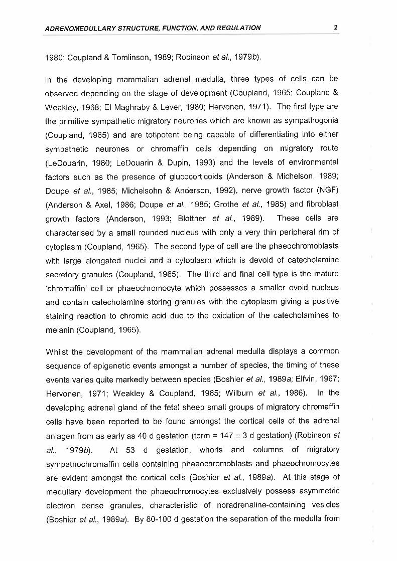
ln the developing mammalian adrenal medulla, three types of cells can be
observed depending on the stage of development (Coupland, 1965; Coupland &
Weakley, 1968; El Maghraby & Lever, 1980; Heryonen, 1971). The first type are
the primitive sympathetic migratory neurones which are known as sympathogonia
(Coupland, 1965) and are totipotent being capable of differentiating into either
sympathetic neurones or chromaffin cells depending on migratory route
(LeDouarin, 1980; LeDouarin & Dupin, 1993) and the levels of environmental
factors such as the presence of glucocorticoids (Anderson & Michelson, 1989;
Doupe et al., 1985; Michelsohn & Anderson, 1992), nerye growth factor (NGF)
(Anderson & Axel, 1986; Doupe et al., 1985; Grothe et al., 1985) and fibroblast
growth factors (Anderson, 1993; Blottner et al., 1989). These cells are
characterised by a small rounded nucleus with only a very thin peripheral rim of
cytoplasm (Coupland, 1965). The second type of cell are the phaeochromoblasts
with large elongated nuclei and a cytoplasm which is devoid of catecholamine
secretory granules (Coupland, 1965). The third and final cell type is the mature
'chromaffin' cell or phaeochromocyte which possesses a smaller ovoid nucleus
and contain catecholamine storing granules with the cytoplasm giving a positive
staining reaction to chromic acid due to the oxidation of the catecholamines to
melanin (Coupland, 1965).
Whilst the development of the mammalian adrenal medulla displays a common
sequence of epigenetic events amongst a number of species, the timing of these
events varies quite markedly between species (Boshier et a\.,1989a; Elfvin, 1967
Hervonen, 1971; Weakley & Coupland, 1965; Wilburn et al., 1986). ln the
developing adrenal gland of the fetal sheep small groups of migratory chromaffin
cells have been reported to be found amongst the cortical cells of the adrenal
anlagen from as early as 40 d gestation (term = 147 + 3 d gestation) (Robinson ef
al., 1979b). At 53 d gestation, whorls and columns of migratory
sympathochromaffin cells containing phaeochromoblasts and phaeochromocytes
are evident amongst the cortical cells (Boshier ef al., 1989a). At this stage of
medullary development the phaeochromocytes exclusively possess asymmetric
electron dense granules, characteristic of noradrenaline-containing vesicles
(Boshier et a1.,1989a). By B0-100 d gestation the separation of the medulla from
CHAPTER 1
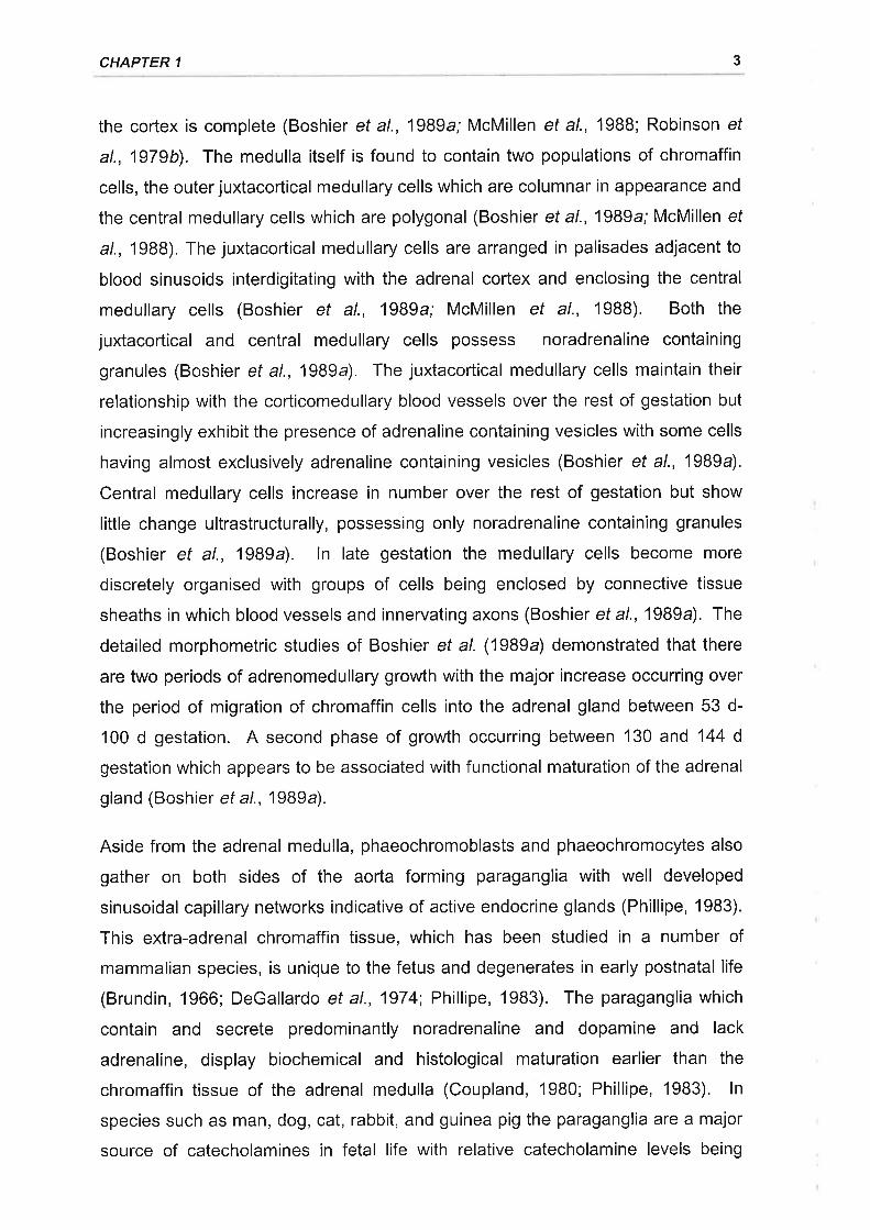
the cortex is complete (Boshier et al., 1989a; McMillen et al., 1988; Robinson ef
al., 1979b). The medulla itself is found to contain two populations of chromaffin
cells, the outer juxtacortical medullary cells which are columnar in appearance and
the central medullary cells which are polygonal (Boshier et al., 1989a; McMillen ef
a/., 1988), The juxtacortical medullary cells are arranged in palisades adjacent to
blood sinusoids interdigitating with the adrenal cortex and enclosing the central
medullary cells (Boshier et al., 1989a; McMillen et al., 1988). Both the
juxtacortical and central medullary cells possess noradrenaline containing
granules (Boshier et al., 1989a). The juxtacortical medullary cells maintain their
relationship with the corticomedullary blood vessels over the rest of gestation but
increasingly exhibit the presence of adrenaline containing vesicles with some cells
having almost exclusively adrenaline containing vesicles (Boshier et al., 1989a).
Central medullary cells increase in number over the rest of gestation but show
little change ultrastructurally, possessing only noradrenaline containing granules
(Boshier et al., 1989a). ln late gestation the medullary cells become more
discretely organised with groups of cells being enclosed by connective tissue
sheaths in which blood vessels and innervating axons (Boshier etal.,19B9a). The
detailed morphometric studies of Boshier ef al. (1989a) demonstrated that there
are two periods of adrenomedullary growth with the major increase occurring over
the period of migration of chromaffin cells into the adrenal gland between 53 d-
100 d gestation. A second phase of growth occurring between 130 and 144 d
gestation which appears to be associated with functional maturation of the adrenal
gland (Boshier et al.,1989a).
Aside from the adrenal medulla, phaeochromoblasts and phaeochromocytes also
gather on both sides of the aorta forming paraganglia with well developed
sinusoidal capillary networks indicative of active endocrine glands (Phillipe, 1983).
This extra-adrenal chromaffin tissue, which has been studied in a number of
mammalian species, is unique to the fetus and degenerates in early postnatal life
(Brundin, 1966; DeGallardo et al., 1974 Phillipe, 1983). The paraganglia which
contain and secrete predominantly noradrenaline and dopamine and lack
adrenaline, display biochemical and histological maturation earlier than the
chromaffin tissue of the adrenal medulla (Coupland, 1980; Phillipe, 1983). ln
species such as man, dog, cat, rabbit, and guinea pig the paraganglia are a major
source of catecholamines in fetal life with relative catecholamine levels being
3
ADRENOMEDULLARY STRUCTURE, FUNCTION, AND REGULATION
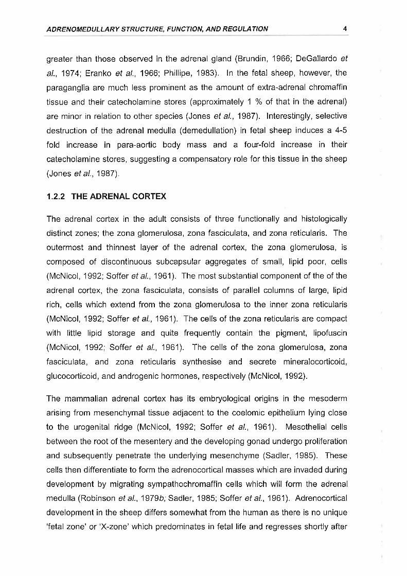
greater than those observed in the adrenal gland (Brundin, 1966; DeGallardo ef
al., 1974; Eranko et al., 1966; Phillipe, 1983). ln the fetal sheep, however, the
paraganglia are much less prominent as the amount of extra-adrenal chromaffin
tissue and their catecholamine stores (approximately 1 % of that in the adrenal)
are minor in relation to other species (Jones et al., 1987). lnterestingly, selective
destruction of the adrenal medulla (demedullation) in fetal sheep induces a 4-5
fold increase in para-aortic body mass and a four-fold increase in their
catecholamine stores, suggesting a compensatory role for this tissue in the sheep
(Jones et al., 1987).
1.2.2 THE ADRENAL CORTEX
The adrenal cortex in the adult consists of three functionally and histologically
distinct zones; the zona glomerulosa, zona fasciculata, and zona reticularis. The
outermost and thinnest layer of the adrenal cortex, the zona glomerulosa, is
composed of discontinuous subcapsular aggregates of small, lipid poor, cells
(McNicol, 1992; Soffer et a\.,1961). The most substantial component of the of the
adrenal cortex, the zona fasciculata, consists of parallel columns of large, lipid
rich, cells which extend from the zona glomerulosa to the inner zona reticularis
(McNicol, 1992; Soffer et al., 1961). The cells of the zona reticularis are compact
with little lipid storage and quite frequently contain the pigment, lipofuscin
(McNicol, 1992; Soffer et al., 1961). The cells of the zona glomerulosa, zona
fasciculata, and zona reticularis synthesise and secrete mineralocorticoid,
glucocorticoid, and androgenic hormones, respectively (McNicol, 1992).
The mammalian adrenal cortex has its embryological origins in the mesoderm
arising from mesenchymal tissue adjacent to the coelomic epithelium lying close
to the urogenital ridge (McNicol, 1992; Soffer et al., 1961). Mesothelial cells
between the root of the mesentery and the developing gonad undergo proliferation
and subsequently penetrate the underlying mesenchyme (Sadler, 1985). These
cells then differentiate to form the adrenocortical masses which are invaded during
development by migrating sympathochromaffin cells which will form the adrenal
medulla (Robinson et al., 1979b;Sadler, 1985; Soffer et al., 1961). Adrenocortical
development in the sheep differs somewhat from the human as there is no unique
'fetal zone' or'X-zone' which predominates in fetal life and regresses shortly after
4
CHAPTER 1
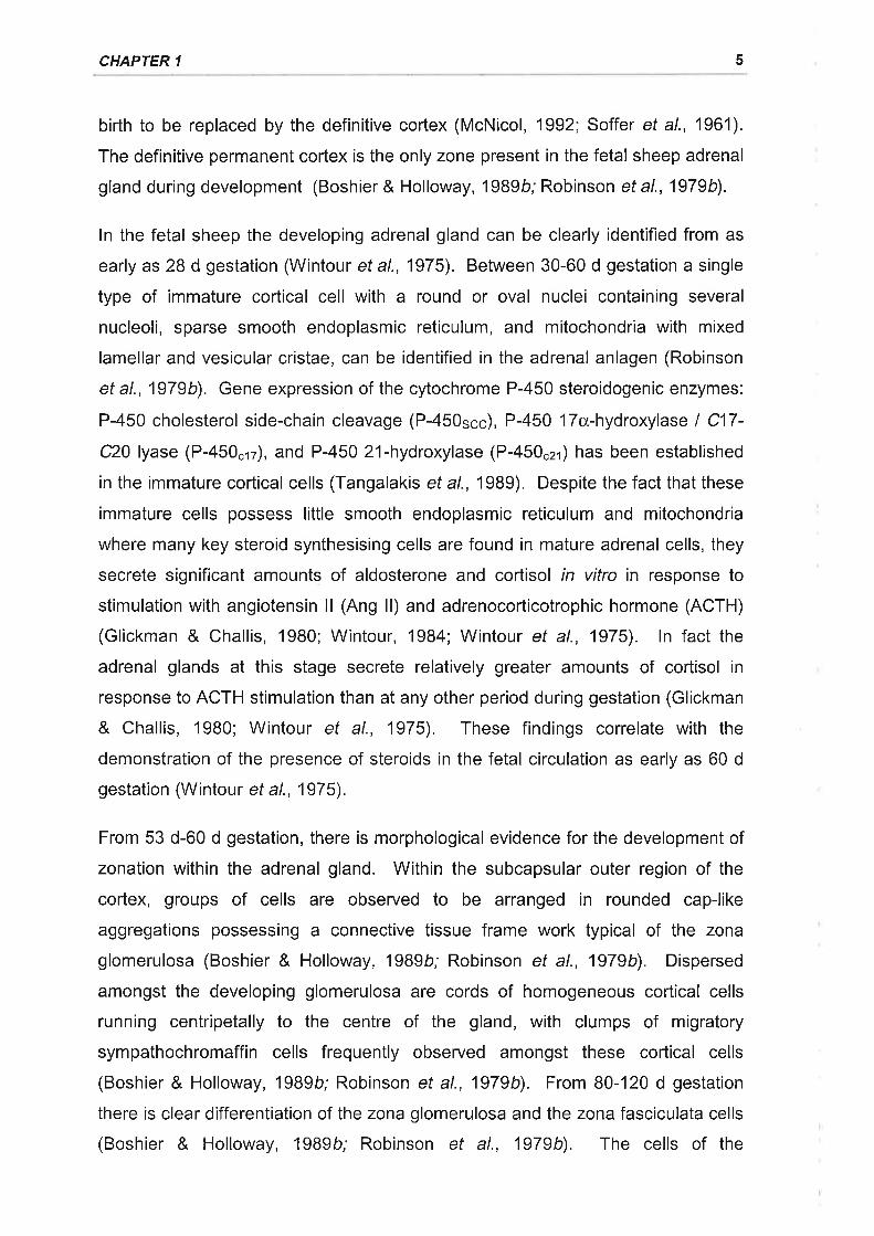
birth to be replaced by the definitive cortex (McNicol, 1992; Soffer et al., 1961).
The definitive permanent cortex is the only zone present in the fetal sheep adrenal
gland during development (Boshier & Holloway,l989b; Robinson et al., 1979b).
ln the fetal sheep the developing adrenal gland can be clearly identified from as
early as 28 d gestation (Wintour ef al., 1975). Between 30-60 d gestation a single
type of immature cortical cell with a round or oval nuclei containing several
nucleoli, sparse smooth endoplasmic reticulum, and mitochondria with mixed
lamellar and vesicular cristae, can be identified in the adrenal anlagen (Robinson
et al., 1979b). Gene expression of the cytochrome P-450 steroidogenic enzymes:
P-450 cholesterol side-chain cleavage (P-450sçç), P-450 17cx,-hydroxylase I C17-
C20 lyase (P-450crz), and P-450 21-hydroxylase (P-450czr) has been established
in the immature cortical cells (Tangalakis et a|.,1989). Despite the fact that these
immature cells possess little smooth endoplasmic reticulum and mitochondria
where many key steroid synthesising cells are found in mature adrenal cells, they
secrete significant amounts of aldosterone and cortisol in vitro in response to
stimulation with angiotensin ll (Ang ll) and adrenocorticotrophic hormone (ACTH)
(Glickman & Challis, 1980; Wintour, 1984; Wintour et al., 1975). ln fact the
adrenal glands at this stage secrete relatively greater amounts of cortisol in
response to ACTH stimulation than at any other period during gestation (Glickman
& Challis, 1980; Wintour et al., 1975). These findings correlate with the
demonstration of the presence of steroids in the fetal circulation as early as 60 d
gestation (Wintour et al., 1975).
From 53 d-60 d gestation, there is morphological evidence for the development of
zonation within the adrenal gland. Within the subcapsular outer region of the
cortex, groups of cells are observed to be arranged in rounded cap-like
aggregations possessing a connective tissue frame work typical of the zona
glomerulosa (Boshier & Holloway, 1989b; Robinson et al., 1979b). Dispersed
amongst the developing glomerulosa are cords of homogeneous cortical cells
running centripetally to the centre of the gland, with clumps of migratory
sympathochromaffin cells frequently observed amongst these cortical cells
(Boshier & Holloway, 1989b; Robinson et al., 1979b). From 80-120 d gestation
there is clear differentiation of the zona glomerulosa and the zona fasciculata cells
(Boshier & Holloway, 1989b; Robinson et al., 1979b). The cells of the
5
ADRENOMEDULLARY STRUCTURE, FUNCTION, AND REGULATION
glomerulosa are arranged into well organised glomus-like aggregations
surrounded by subcapsular loose connective tissue (Boshier & Holloway, 1989b).
This connective tissue marked the site of entry of the cortical blood vessels which
either ramified in the zona glomerulosa or rapidly branched into anastomosing
sinusoids within the zona fasciculata (Boshier & Holloway, 1989b). The
glomerulosa cells at this stage exhibit features consistent with mature cells in that
they are found to possess large amounts of smooth endoplasmic reticulum and
mitochondria with mainly lamellar cristae (Robinson et al., 1979b). ln contrast to
the glomerulosa cells, the cells in the inner zone of the cortex are still immature
displaying sparse amounts of smooth endoplasmic reticulum, few ribosomes, and
mitochondria with mixed lamellar and vesicular cristae (Robinson ef al., 1979b;
Webb, 1980). Most of the migratory chromaffin cells have accumulated in the
centre of the adrenal gland at this stage and there is a well defined cortico-
medullary junction (Boshier & Holloway, 1989b).
The establishment of morphological zonation of the adrenal cortex is also
associated with the development of a functional zonation. From 80 d gestation P-
450czt and P-450... mRNA expression is found in both the zona fasciculata and
zona glomerulosa. Labelling for P-450"r2 ffiRNA, which is exclusively involved in
the synthesis of cortisol, is restricted to the cortical cells within the zona
fasciculata (Tangalakis ef a/., 19Bg). The expression of these steroidogenic
enzymes, most notably P-450scc âhd P-450.r2, is relatively low during this period
when compared to earlier and later periods in gestation (Tangalakis ef a/., 1989).
This decline in steroidogenic enzyme gene expression coincides with a functional
quiescence of the cortex; ACTH stimulation of fetal cortisol secretion is relatively
inefficient and Ang ll and K* are unable to stimulate aldosterone secretion (Challis
& Brooks, 1989; Glickman & Challis, 1980; Siegel & Fischer, 1980; Wintour etal.,
1979', Wintour et al., 1975).
ln late gestation from 120 d onwards there is morphological maturation of the cells
in the inner cortex, with the appearance of more spherical mitochondria with
vesicular cristae and well developed smooth endoplasmic reticulum (Robinson ef
al., 1979b). Over the later stages of gestation from 130 d onwards there is a
dramatic increase in adrenal cortical size which is primarily due to rapid growth of
the zona fasciculata (Boshier & Holloway, 1989b; Robinson et al., 1979b). The
6
CHAPTER 1
growth of the fasciculata region is due to both hypertrophy and hyperplasia of the
secretory cells (Boshier & Holloway, 1989b). Gene expression of the
steroidogenic enzymes P-450.21, P-450r.", and P-450.r2 within the adrenal cortex
also increases dramatically in late gestation (Tangalakis et al., 1989). The
increase in growth of the zona fasciculata coincides with increased ACTH output
from the fetal pituitary, increased responsiveness of the fetal cortex to ACTH
stimulation, and a dramatic surge in cortisol output from the adrenal gland (Challis
& Brooks, 1989; Wintour, 1984).
There is no evidence for the development of the zona reticularis in the sheep up to
early postnatal life (Boshier & Holloway, 1989b; Robinson et al., 1979b).
1.2.3 INNERVATION OF THE ADRENAL GLAND
1.2.3.'l Adrenomedullary innervation
Cholinesterase staining and nerye degeneration studies confirm that the majority
of the nerve fibres which project to the mammalian adrenal gland are pre-
ganglionic cholinergic sympathetic fibres arising from the splanchnic nerye
(MacFarland & Davenport, 1941; Robinson et al., 1977b). Retrograde tracer
studies reveal that the pre-ganglionic sympathetic fibres which innervate the
adrenal gland arise ipsilaterally from the intermediolateral horn of the spinal cord
between thoracic level 3 (T3) and lumbar level 2 (L2) with the majority arising from
T8-T11 (Kesse et al., 1988; Parker et al., 1993; Pyner & Coote, 1994). The
pattern of pre-ganglionic sympathetic innervation within the mammalian adrenal
gland appears to demonstrate a common pattern between species. Cholinergic
fibres from the capsular network run radially through the cortex with very little
branching until reaching the adrenal medulla whereupon there is extensive
ramification to form a dense medullary plexus with numerous axons running
between chromaffin secretory cells with frequent synapses (MacFarland &
Davenport , 1941; Parker et al., 1993; Robinson et al., 1977 b).
1.2.3.2 Adrenocortical innervation
Despite the fact that the adrenal cortex is poorly innervated in relation to the
adrenal medulla, ultrastructural studies identify axon terminals adjacent to cortical
cells (Robinson et al., 1977b; Unsicker, 1971). There is a body of evidence to
7
ADRENOMEDIJLLARY STRUCTURE, FUNCTION, AND REGULATION
suggest that splanchnic nerve stimulation can enhance ACTH induced
glucocorticoid output from the adrenal cortex (Edwards & Jones, 1987', Edwards ef
al., 1986). The peptide neurotransmitter, corticotrophin releasing factor (CRF)
modulates steroidogenesis in the adrenal gland of the calf (Jones & Edwards,
1990) and is present along with its receptors in the adrenal glands of a number of
species (Aguilera et a\.,1991; Bruhn et al., 1987; Hashimoto et al., 1984; Rundle
et al., 1988). lmmunocytochemical and retrograde tracer techniques reveal that
the splanchnic nerve contains CRF-positive fibres which project from the
intermediolateral cell column of the spinal cord at levels T5-T13 (Li & McDonald,
1997; Pomerantz et al., 1996) and stimulation of the splanchnic nerye elicits an
increase in the concentration of CRF in adrenal venous blood (Edwards & Jones,
1988; Plotsky et a\.,1990).
1.2.3.3 Postganglionic sympathetic innervation
The vast majority of the nerve fibres projecting to the adrenal gland have cell
bodies in the spinal cord confirming their pre-ganglionic nature, although small
numbers of post ganglionic sympathetic nerves are evident synapsing in the
sympathetic ganglia chain or in the suprarenal ganglion (Celler & Schram, 1981;
Kesse et al., 19BB; Parker et al., 1993). Post-ganglionic sympathetic fibres
containing catecholamines have been found using fluorescence histochemistry to
run in close association with vascular supply of the gland (Robinson et al., 1977b).
Post-ganglionic sympathetic fibres do not degenerate following transection of the
splanchnic nerye but stripping the adventitia of the arteries supplying the adrenal
gland does result in a reduction in the number of these fibres (Robinson et al.,
1977b). lt therefore appears that the post-ganglionic sympathetic fibres innervate
blood vessels within the gland (Carlson et al., 1992; Carlson et al., 1990;
Robinson et al., 1977 b; U nsicker, 197 1).
1.2.3.4 Sensory innervation
There is also a significant sensory innervation of the mammalian adrenal gland
with afferent nerves being identified in a number of species (Mohamed et al.,
1988; Parker et a\.,1993; Zhou et al., 1991b). The majority of the afferent nerve
supply to the adrenal gland via the splanchnic nerve has cell bodies lying
ipsilaterally in the dorsal root ganglion of the spinal cord with the highest
I
CHAPTER 1
proportions of ganglia between T9-T10 (Mohamed et al., 19BB; Parker et al.,
1993; Zhou et al., 1991b). Most sensory nerve fibre endings are found in the
capsular region or in association with capillaries in both the adrenal cortex and
medulla (Parker et a1.,1993). The role of sensory innervation in the adrenal gland
is unclear but is likely to involve regulation of adrenal blood flow and modulation of
secretory output (Khalil et al., 1986; Livett et al., 1990; Niijima & Winter, 1968;
Parker et al., 1993).
1.2.3.5 lntrinsic innervation
The adrenal gland also appears to have an intrinsic innervation with ganglion cells
having been identified within the gland with their numbers being species specific
(Coupland & Holmes, 1958; Migally, 1979; Mikhail & Mahran, 1965; Parker et al.,
1993; Unsicker et al., 1978b;Watanabe et a1.,1990). ln the most studied species,
the rat, two populations of ganglion cells have been identified. One population
which has been termed Type lganglion cells by Holgert and colleagues (1996a)
are relatively large and exhibit properties consistent with a post-ganglionic,
noradrenergic phenotype (Dagerlind et al., 1990; Oomori et al., 1994; Pelto-
Huikko, 1989). These cells are probably derived from neural crest cells which
invaded the cortical anlage but unlike the chromaffin cells were exposed to a
different set of environmental factors (e.9. NGF) (Anderson, 1993; Doupe et al.,
1985) which led to their differentiation into neurones. Type ll ganglion cells,
whose origins have yet to be determined, are smaller, have a non-classical
peptidergic transmitter phenotype, and express nitric oxide synthase (NOS) (Dun
et al., 1993; Holgert et al., 1995a; Holzwarth, 1984). lntra-adrenal ganglion cells
have been demonstrated to have fibres projecting to cortical, medullary, and
capsular regions of the gland with a number appearing in close approximation with
blood vessels and chromaffin cells (Afework et al., 1994; Holgert et al., 1996a;
Kondo et al., 1986; Pelto-Huikko, 1989). Type I ganglion cells also have
projections which have been described to leave the adrenal gland via the
splanchnic nerve (Dagerlind et al., 1995). Little is known about the physiological
function of these intrinsic ganglion cells but they may act to regulate adrenal blood
flow and also secretory output from the gland and in the case of the type I
ganglion cells may act as a feedback system.
I
ADRENOMEDULLARY STRUCTURE, FUNCTION, AND REGULATION 10
1.2.3.6 Ontogeny of adrenal innervation
Functional innervation of the rat adrenal gland, and in particular the medulla, by
the preganglionic, cholinergic, splanchnic nerve does not occur until after the first
week of postnatal life. Ultrastructural studies reveal a dramatic increase in the
number of synapses from splanchnic nerve fibres on chromaffin cells occurring at
10 d postnatally (Lau ef a/., 1988) which correlates with an increase in the number
of acetylcholinesterase immunoreactive fibres within the gland at this stage
(Holgerl et al., 1994). These structural observations are corroborated by
functional studies which demonstrate that insulin induced hyperglycaemia, which
requires functional splanchnic innervation of the adrenal gland to stimulate
catecholamine secretion, does not elicit a full catecholamine response in the rat
until postnatal day 10 (Slotkin & Seidler, 19BB). ln contrast to pre-ganglionic
sympathetic innervation, sensory innervation of the rat adrenal gland already
appears quite mature at postnatal day 2 stage (Holgert et al., 1994) as do type ll
ganglion neurones (Holgert et al., 1996a), whereas type I ganglion neurones
display a developmental pattern similar to that of the extrinsic innervation (Holgert
et al., 1996a; Holgert et al., 1994).
Unlike the rat, functional splanchnic innervation to the sheep adrenal medulla
occurs prior to birth. Ultrastructural studies reveal the absence of sympathetic
innervation to the adrenal medulla at 53 d gestation, however, by 100 d gestation
axonal profiles are present amongst the chromaffin cells but synapses are absent
(Boshier et al., 1989a). At 130 d, synapses between the axons and chromaffin
cells of the medulla are present, increasing in frequency over late gestation and
the early post-partum period (Boshier et al., 1989a). Comline and Silver (1961)
found significant increases in adrenal venous catecholamine concentrations of
anaesthetised, fetal lambs could only be elicited by electrical stimulation of the
splanchnic nerye from 125 d gestation, concurring with the structural observations
of Boshier et al. (1989). Currently there is no available information on the
development of sensory and intra-adrenal ganglion cells in the fetal sheep
adrenal.
CHAPTER 1 11
1.2.4 VASCULATURE
The adrenal gland exhibits an unusually high degree of vascularity, with the
arterial blood supply to the mammalian adrenal gland arising from a number of
sources (Harrison & McDonald, 1966; Harrison & Hoey, 1960; Vinson & Hinson,
1992). ln the sheep the sources of arterial blood supply exhibit variability amongst
breeds but primarily arise from the lumbar arteries and renal arteries, with
contributions from the coeliac axis, and anterior mesenteric artery (Harrison &
McDonald, 1966).
The arterial supply to the adrenal divides repeatedly into smaller arterioles in and
just below the connective tissue of the capsule, forming a plexus which consists of
an extensive capillary network. The capillaries of this subcapsular network then
form an extensively cross connecting network of thin-walled blood vessels, termed
sinusoids, which supply the zona glomerulosa. Continuous with this network is a
straighter centripetal network of sinusoids which pass through and supply the zona
fasciculata. The latter network as it reaches the inner parts of the cortex
converges and forms numerous cross connecting channels in the deeper regions
of the cortex, giving rise to the extensive reticular network in the zona reticularis
(Harrison, 1951; Harrison & Asling, 1955; Vinson et al., 1985). The continuous
networks of vessels which supply the cortex converge and empty into the larger
sinusoids present within the corticomedullary and medullary regions, such that the
medulla is directly exposed to blood which has passed through the adrenal cortex
(Harrison & Hoey, 1960; Vinson & Hinson,1992; Vinson et al., 1985). The
medullary sinusoids then drain into medullary veins (Vinson et al., 1985).
Aside from the cortical effluent which drains into the medullary sinusoids, the
medulla also receives blood directly from arteries known as arteriae medullae
(Coupland & Selby, 1976; Merklin, 1962; Vinson & Hinson, 1992; Vinson et al.,
1985). The presence of arteriae medullae within the adrenal gland is species
specific with relatively few being observed in the rat adrenal gland (Vinson et al.,
1985) whilst substantially greater numbers are found in cat and bovine adrenals
(Coupland & Selby, 1976). The arteriae medullae derive from branches of the
large capsular arteries which pass directly through the adrenal cortex before
entering the medulla and branching into arterioles and capillaries. These vessels
ADRENOMEDULLARY STRUCTURE, FUNCTION, AND REGULATION 12
then empty into the medullary veins which in turn empty into the central adrenal
vein, where all venous effluent drains from the adrenal gland (Vinson et a1.,1985).
The adrenal venous effluent subsequently discharges into either the renal vein
(left adrenal) or directly into the inferior vena cava (right adrenal) (Soffer et al.,
1961; Vinson & Hinson, 1992).
There is evidence to suggest that the blood flow to the adrenal cortex and the
adrenal medulla can be independently regulated. Under basal conditions the
majority of blood flow to the adrenal gland has been found to be to the medulla by
the use of microsphere and microradiographic techniques (Carter et al., 1993;
Harrison & Hoey, 1960). Cortical and medullary blood flow have been found in the
adult dog to be independently regulated during haemorrhagic hypotension
(Breslow et al., 1987), hypoxic hypoxia (Nishijima et al., 19Bg), and splanchnic
nerye stimulation (Breslow et al., 1987). ln the fetal sheep radiolabelled
microsphere studies have revealed that ACTH administration elicits a preferential
increase in adrenal cortical blood flow (Carter et al., 1993). The mechanisms
underlying the independent regulation of cortical and medullary blood flow within
the adrenal gland have yet to be elucidated, however, it has been speculated that
alterations in the tone of the muscular arlerioles in the sub capsular plexus control
the blood flow to the thin walled sinusoids (Vinson et al., 1985).
ln the fetal sheep, wide anastomosing sinusoids arising from thin walled vessels in
the adrenal capsule and leading to the centre of the gland are observed as early
as 53 d gestation where the vasculature occupies a relatively large proportion of
the cortex (Boshier & Holloway, 1989b). By 100 d gestation there is ramification
of a number of the cortical blood vessels within the zona glomerulosa to form the
glomerulosa network, however, the majority of the vessels branch within the zona
fasciculata to form somewhat narrower anastomosing sinusoids which recombine
into larger vessels at the corticomedullary junction (Boshier & Holloway, 1989b).
At this stage of development, the volume density of the vasculature within the
zona fasciculata falls significantly due to the re-organisation of the vasculature,
cellular proliferation, and functional zonation within the cortex (Boshier &
Holloway, 1989b). Separation of the medulla from the cortex is complete at 100 d
gestation with the relative volume of the medulla occupied by blood vessels
CHAPTER 1 t3
increasing over mid to late gestation as vascularisation of the medulla continues
(Boshier et al., 1989a).
1.3.1 ADRENOMEDULLARY CATECHOLAMINE SYNTHESIS AND STORAGE
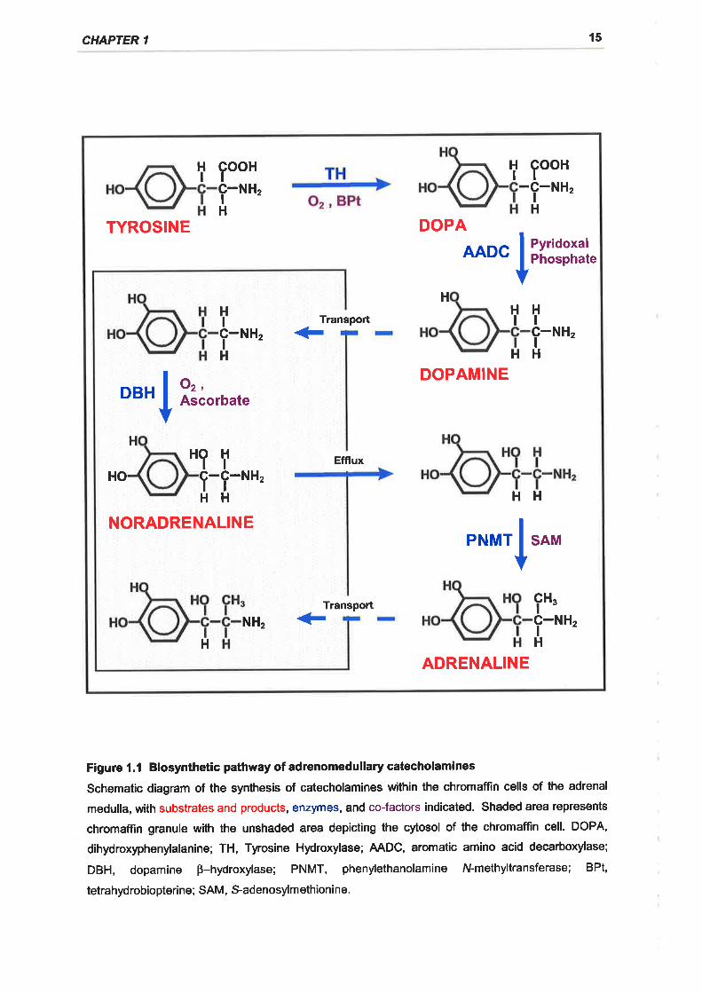
1.3.1.1 Synthesis
Catecholamines are synthesised in the chromaffin cells of the adrenal gland from
the dietary amino acid, tyrosine, as illustrated in Figure 1.1 (for reviews see
Kirshner, 1975; Ungar and Phillips, 1983). The initial step in the synthesis of
adrenomedullary catecholamines is the oxidation of tyrosine to
dihydroxyphenylalanine (DOPA) by the enzyme tyrosine hydroxylase ITH; tyrosine
3-monooxygenase; tyrosine, tetrahydropteridine:oxygen oxidoreductase (3-
hydroxylating), EC 1.14.16.21 (Levitt et al., 1965; Nagatsu et al., 1964). TH is a
mixed function oxidase which requires molecular oxygen and utilises a
tetrahydrobiopterine as a co-substrate (Brenneman & Kaufman, 1964; Fitzpatrick,
1993; Joh et al., 1969). The tetrahydropteridine co-substrate is oxidised to
dihydrobiopteridine in the conversion of tyrosine to DOPA (Fitzpatrick, 1993; Joh
et al., 1969), with dihydropteredine reductase (EC 1.6.99.7) and nicotinamide
adenine dinucleotide phosphate (NADPH) being required to regenerate the pool of
the reduced form of the biopterine (Brenneman & Kaufman, 1964; Musacchio,
1969; Shiman et al., 1970). Tyrosine hydroxylase is the rate limiting enzyme in
the catecholamine synthetic pathway (Levitt et al., 1965), as TH has a
substantially lower specific activity than any of the other catecholamine synthetic
enzymes and the pool of tyrosine is greater than that of any of the other
catecholamine synthetic enzyme substrates (Nagatsu et al., 1964). Tyrosine
hydroxylase is located in the soluble fractions of adrenal medullary homogenates
indicating its presence in the cytoplasm of the chromaffin cells (Laudron &
Belpaire, 1968; Sabban & Goldstein, 1984; Wurzburger & Musacchio, 1971).
DOPA is subsequently decarboxylated by DOPA decarboxylase, also known as
aromatic L-amino acid decarboxylase (AADC; EC 4.1.1.28), to form dopamine
(Kirshner, 1975). AADC is not specific for catecholamine synthesis as it is
involved in the decarboxylation of a number of aromatic t-amino acids (Lovenberg
ADRENOMEDULLARY STRUCTURE, FUNCTION, AND REGULATION 14
et al., 1962) and appears to have a wide distribution in a number of tissues
(Christenson ef al., 1970; Kirshner, 1975; Sourkes, 1966). Like TH, AADC activity
in the chromaffin cell is found in the water-soluble fraction of adrenal
homogenates indicating that its located in the cytosol of chromaffin cells (Laudron
& Belpaire, 1968; Sabban & Goldstein, 1984).
Dopamine is actively transported into the chromaffin granules and converted to
noradrenaline by dopamine B-hydroxylase [DBH; 3,4-dihydroxyphenylethylamine,
ascorbateroxygen oxidoreductase (B-hydroxylating), EC 1.14.17 .11 (Kirshner,
1975; Kirshner, 1962; Levin et al., 1960). DBH is the only enzyme in the
catecholamine biosynthetic pathway located within the chromaffin granules with all
of the other enzymes situated in the cytoplasm of the chromaffin cells (Laudron,
1975; Sabban & Goldstein, 1984), DBH is present in the chromaffin granules in
one of two similar forms, either bound to the internal membrane or free in the
internal matrix of the chromaffin granule (Helle et al., 1984; Joh & Hwang, 1987).
The relative proportions of the enzyme which are free and membrane bound vary
between species (Ciaranello et al., 1975; Gagnon et al., 1976b) with the free form
probably being derived from a membrane bound precursor which undergoes
proteolysis (Helle et al., 1984; Joh & Hwang, 1987). Like TH, DBH is a mixed
function oxidase and it catalyses the oxidation of dopamine to noradrenaline and
requires molecular oxygen and utilises ascorbate as a co-factor (Dilberto & Allen,
'1981 ; Levin et al., 1960).
The final step in the catecholamine biosynthetic pathway is the N-methylation of
noradrenaline which passively permeates into the cytosol from chromaffin
granules, to adrenaline (Kirshner, 1975; Ungar & Phillips, 1983). This is achieved
by transfer of the S-methyl group from S-adenosylmethionine to the primary
nitrogen group of noradrenaline by the enzyme phenylethanolamine N-
methyltransferase IPNMT; S-adenosyl-l-methionine:phenylethanolamine N-
methyltransferase, EC 2.1 .1 .281.
CHAPTER '
t5
i looH I looH1-*r,H
-1-*r,H
TYROSINE DOPA
AADC IPyridoxalPhosphate
HHIc-IH
TransportHHtlNHz + 1-*t,
HH
Ior,Ascorbate
DOPAMINEDBH
HHOHltHo 1-1-*t,HH
Efflux
HH
NORADRENALINE
IPNMT SAM
TranspoÉcH"t-1-*r,H
NHz <-H
ADRENALINE
Figure l.l Biosynthetic pathway of adrenomedullary catecholamines
Schematic diagram of the synthesis of catecholamines within the chromaffin cells of the adrenal
medulla, with substrates and products, enzymes, and co-factors indicated. Shaded area represents
chromaffin granule with the unshaded area depicting the cytosol of the chromaffin cell. DOPA,
dihydroxyphenylalanine; TH, Tyrosine Hydroxylase; AADC, aromatic amino acid decarboxylase;
DBH, dopamine p-hydroxylase; PNMT, phenylethanolamine N-methyltransferase; BPt,
tetrahydrobiopteri ne; SAM, S-adenosylm ethioni ne.
ADRENOMEDIJLLARY STRUCTURE, FUNCTION, AND REGULATION l6
1.3.1.2 Storage
1.3.1.2.1 Chromaffin granules
The adrenomedullary catecholamines are stored in the chromaffin cells within
specialised vesicles known as chromaffin granules. Chromaffin granules are
membrane bound spheres which have been identified to contain a number of
components including, catecholamines, nucleotides (predominantly ATP),
ascorbic acid, calcium, a family of acidic glycoproteins known as the
chromogranins, a number of neuropeptides predominantly consisting of the
enkephalins and their precursors and neuropeptide tyrosine, and DBH (soluble
form) (Winkler et al., 1986; Winkler & Westhead, 1980). The chromaffin granule
membrane exhibits a characteristically high lipid to protein ratio with large amounts
of cholesterol, and the phospholipid, lysolecithin, present in the membrane
(Frischenschlager et al., 1983; Winkler, 1976). The lipids are arranged in a
bimolecular leaflet to form the membrane of the granule (Winkler & Westhead,
1980) and within the membrane are a number of enzymes some of which have
been identified using 2D gel electrophoresis, whilst others are known to be
present due to their functional identification. Amongst these membrane bound
proteins are DBH (membrane bound form), cytochrome bsor, phosphotidylinositol
kinase, NADH dehydrogenase, and a Mg2*-ATPase (H* translocating) as well as
transporters for catecholamines, adenine nucleotides, and a Ca2* / Na* exchanger
(Winkler et al., 1986; Winkler & Westhead, 1980).
1.3.1.2.2 Chromaffin granule synthesis
The chromaffin granules are synthesised by the Golgi bodies and when the newly
formed granules are pinched off from the Golgi stacks they already contain
chromogranins, and neuropeptides which have been incorporated into the granule
within the Golgi region (Winkler et al., 1987). These immature granules are less
dense than mature granules and they gradually accumulate catecholamines, ATP,
and calcium (Winkler,1977; Winkler et a\.,1987). The synthesis rate of secretory
proteins in the chromaffin granules is significantly greater than membrane bound
proteins suggesting that there is recycling of the chromaffin granule membranes
following exocytosis (Ungar & Phillips, 1983; Winkler, 1977). The use of specific
antigens against chromaffin granule membrane components which can be
CHAPTER 1 17
visualised by immunofluorescence has revealed that during stimulation these
antigens appear on the surface of the cell membrane consistent with exocytosis
(Lingg et a\.,1983; Phillips et a\.,1983). After stimulation, the chromaffin granule
membrane antigens are retrieved into the cell and are initially found in close
proximity to the Golgi region and finally in newly formed chromaffin granules
consistent with recycling of the chromaffin granule membranes (Lingg et a\.,1983;
Phillips et a\.,1983).
1.3.1.2.3 Catecholamine transport and storage
The concentration of catecholamines within chromaffin granules has been
estimated to be around 20 000 times greater than the cytosolic concentration
(Phillips & Apps, 1980; Ungar & Phillips, 1983). The large concentration between
granular and cytosolic catecholamines is maintained by a secondary active
transport mechanism (Njus et al., 1981; Phillips, 1982). The interior of isolated
chromaffin granules is acidic and has been estimated to be pH 5.7 with the
membrane potential of the granule being around +60 mV in respect to that across
the cytosol (Johnson & Scarpa, 1976; Johnson & Scarpa, 1979; Njus ef al., 1978;
Ungar & Phillips, 1983). The acidic nature of the granule and the positive
membrane potential arise from the translocation of H* across the chromaffin
granule membrane by the membrane bound H*-ATPase which utilises the
hydrolysis of cytoplasmic Mg-ATP to drive electrogenic proton transport (Johnson
et al., 1982; Njus et al., 1981; Winkler et al., 1986). Transport of the
catecholamines into the granule is directly coupled to the proton gradient by a
translocator which exchanges a proton for one uncharged catecholamine
molecule (Marron et al., 1983; Ramu et al., '1983; Winkler et al., 1986).
Catecholamines are able to passively leak out of the chromaffin granules down
their concentration gradient into the cytosol which allows noradrenaline to be
methylated to adrenaline by the enzyme PNMT (Corcoran et a\.,1984) as outlined
previously. A steady state equilibrium exists between the levels of cytosolic and
granular catecholamines such that an increase in granular catecholamine content
results in an increased efflux from the granule, whereas an increased cytoplasmic
catecholamine level stimulates uptake into the chromaffin granule such that the
granules act as buffers of cytoplasmic catecholamine content in the resting cell
ADRENOMEDIJLLARY STRUCTURE, FUNCTION, AND REGULATION 18
(Ungar & Phillips, 1983). Adrenaline and noradrenaline secreting chromaffin cells
are identifiable by ultrastructural differences and immunohistochemical
techniques. The presence or absence of PNMT in chromaffin cells determines
their adrenaline synthesising capacity. ln the sheep adrenal the expression of
PNMT is confined to the peripheral juxtacortical region of the medulla which
interdigitate with the cortex and also to a degree around the central adrenal vein
and sinusoids (McMillen et al., 19BB). This organisation of adrenaline and
noradrenaline synthesising chromaffin cells is similar to that observed in the
bovine adrenal gland (Livett et a\.,1982).
1.3.1.2.4 Chromaffin granules and catecholamine synthesis
As outlined previously the chromaffin granules contain both a soluble form of DBH
and a membrane bound form. The presence of relatively large amounts of the
DBH cofactor ascorbic acid within the chromaffin granule allows the conversion of
dopamine to noradrenaline to occur (Dilberto & Allen, 1981; lngebretsen et al.,
1980). During the conversion of dopamine to noradrenaline the co-factor,
ascorbic acid is oxidised to semidehydroasorbate (Dilberto & Allen, 1981). ln
order to maintain the pool of the co-factor for noradrenaline synthesis,
semidehydroascorbate must be reduced to regenerate ascorbic acid (Dilberto &
Allen, 1981;Winkler et al., 1986). This reduction process requires electrons and
the presence of the heme-containing cytochrome b561 as the most abundant
membrane protein within the chromaffin granule seems to provide the most
obvious means for the transportation of electrons across the chromaffin granule
membrane (Njus et al., 1983; Srivastava et al., 1984) to allow this process to
occur (Tsubaki et al., 1997; Winkler & Westhead, 1980). The midpoint redox
potential of cytochrome bsor at the acidic pH of the chromaffin granule is
consistent with a role in the reduction of oxidised forms of ascorbate (Winkler ef
a/., 1986).
1.3.1.2.5 Chromaffin granules and calcium homeosfasis
The chromaffin granules also store an unusually high amount of calcium with
estimates of 20-40 mM having been reported (Bulenda & Gratzl, 1985; Phillips ef
al., 1977; Reiffen & Gratzl, 1986). lndeed the majority of calcium in the bovine
adrenal medulla is reported to be located within the chromaffin granules (Phillips
CHAPTER 1 19
et al., 1977). Whilst the granules may have a high calcium content the free
calcium concentrations are only in the low micromolar range (= 4 p@, with this
anomaly being attributed to the binding of calcium by the acidic granule matrix
proteins, the chromogranins (Bulenda & Gratzl, 1985; Reiffen & Gratzl, 1986;
Ungar & Phillips, 1983). Acetylcholine (ACh) induced secretion from perfused
bovine adrenal glands results in an increase in calcium content within the
chromaffin granules (Serck-Hanssen & Christiansen, 1973) suggesting that they
play an important role in scavenging cytosolic Caz* following stimulus induced
entry into the cell (Ungar & Phillips, 1983). Calcium appears to enterthe vesicle
via an electroneutral Na* I Ca2* exchanger present within the membrane of the
chromaffin granule (Krieger-Bauer & Gratzl, 1982; Phillips, 1981; Winkler, 1987;
Yoon & Sharp, 1985).
1.3.2 ADRENOMEDULLARY NEUROPEPTIDES
A number of neuropeptides and non-classical neurotransmitters are also present
within the chromaffin cells, nerve fibres, and intra-adrenal ganglion cells of the
adrenal medulla.
1.3.2.1 Enkephalins
The most abundant neuropeptides and the first to be positively identified, within
the adrenal medulla, are the enkephalin-containing opioid peptides (Schultzberg ef
al., 1978b; Winkler et al., 1986). Enkephalin (Enk) containing peptides are
established to be present within the adrenaline containing chromaffin cells and
preganglionic splanchnic nerve fibres in the adrenal glands of several species
(Dagerlind et al.,'1993; Kondo & Yui, 1984; Livett et al., 1982; McMillen et al.,
19BB; Pelto-Huikko ef a/., 1985; Schultzberg et al., 1978a). Enkephalin containing
peptides within the adrenal medulla are derived from a 263 amino acid, 30 kDa
precursor molecule, the gene for which was first cloned from bovine adrenal
medulla by Noda and co-workers (1982). Subsequent studies derived the peptide
sequence of the enkephalin-containing precursor molecule, termed Proenkephalin
A (PEnk A), which contains four copies of [Met]enkephalin, one copy of
[Leu]enkephalin, and one copy each of [Met]enkephalyl-Argu-Phet (MERF) and
[Met]enkephalyl-Argu-Glyt-Leuu lfrlfnCt) (Comb et al., 1982; Gubler et al., 1982).
Proenkephalin A is cleaved by trypsin-like and carboxypeptidase B-like proteases
AD RE N OM ED IJ LLARY ST R U CT U RE, F U N CTI O N, AN D REG U LAT IO N 20
(Evangelisla et al., 1982; Hook, 1984; Wallace et al., 1gB2) within the chromaffin
granule to form a number of smaller enkephalin-containing peptides ranging in
size from > 20 kDa to < 1 kDa, as well as free enkephalin peptides 5-B amino
acids in length (Coulter et al., 1990a; Fleminger et al., 1983; Fleminger et al.,
1984; Kojima et al., 1982; Patey et al., 1985; Patey et al., 1984).
',.3.2.2 Neu ropeptide tyrosine
Neuropeptide tyrosine (NPY) is a 36 amino acid regulatory peptide which has a
wide distribution in the central and peripheral nervous system being found in sites
such as the hypothalamus, cerebral cortex, hippocampus, brainstem, and in
particular within noradrenergic sympathetic neurones (Hokfelt et al., 1983;
Jhanwar-Uniyal ef al., 1993; Lundberg et al., 1983; Lundberg et al., 1984; Warnes
et al., 1998; Woodhams et al., 1985). lt is therefore not surprising that NPY has
also been identified in the adrenal gland within both the chromaffin cells and the
type I intra-adrenal ganglion cells and their processes (Allen et al., 1983;
Dagerlind et al., 1990; Fried et al., 1991; Kondo, '1985; Kuramoto et al., 1986;
Oomori et al., 1994; Pelto-Huikko, 1989; Varndell et al., 1984), which are
considered to be analogous to post-ganglionic sympathetic neurones. Next to the
enkephalin-containing peptides, NPY is the second most abundant neuropeptide
found within the chromaffin cells of the adrenal medulla (Winkler et al., 1986).
1.3.2.3 Vasoactive intestinal polypeptide
Vasoactive lntestinal Polypeptide (VlP) was one of the first neuropeptides to have
been established to be secreted from the adrenal medulla (Said, 1976).
lmmunohistochemical localisation of VIP within the adrenal gland reveals that it is
predominantly located in type ll intra-adrenal ganglion cells as well as nerve fibres
which are distributed in networks around medullary chromaffin cells and also in
the cortex and capsule usually in association with vascular structures (Holgert ef
al., 1996a; Holzwarth,1984; Kondo et al., 1986; Linnoila et al., 1980; Pelto-
Huikko, 1989; Yoshikawa etal.,'1990). The VIP immunopositive fibres within the
adrenal gland consist of fibres which project into the splanchnic nerve and are
presumably sensory in origin, as well as projections from type ll intra-adrenal
ganglion cells (Holgert ef al., 1996a; Holzwarth, 1984; Linnoila et a\.,1980). VIP is
not generally co-stored with catecholamines in bovine or rat adrenomedullary
CHAPTER 1 21
chromaffin cells (Holzwarth, 1984; Kondo, 1985; Pelto-Huikko, 1989; Yoshikawa
et al., 1990) although there does appear to be species differences with VIP
immunoreactivity being detected within the chromaffin cells of the frog adrenal
gland (Leboulenger ef a/., 1983). VIP immunoreactivity which has been reported
to exist in human phaeochromocytoma cells and cultured bovine chromaffin cells
(Eiden et al., 1982; Gozes et al., 1983; Siegel et al., 1985) and is found to be
associated with the transformation of the chromaffin cell phenotype to the
neurone-like phenotype and culture conditions (Eiden et al., 1983; Siegel et al.,
1985; Tischler et a\.,1985; Tischler et a\.,1984).
1.3.2.4 Nitric oxide
Nitric oxide (NO) is a novel neurotransmitter as it is a highly labile, free radical gas
which diffuses to its target sites after synthesis rather than being released from
synaptic vesicles by exocytosis (Dawson & Snyder, 1994). Nitric oxide is
synthesised by the calmodulin dependent enzyme, nitric oxide synthase (NOS),
which oxidises t--arginine utilising NADPH and Oz to form t--citrulline and NO
(Stuehr et al., 1991;White & Marletta,1992). Due to its highly labile nature, the
localisation of NO in tissue is demonstrated indirectly by the presence of NOS
immunoreactivity or by NADPH-diaphorase staining which is found to be co-
localised with NOS in neurones. lndeed, it is believed that NOS is responsible for
the NADPH diaphorase staining reaction within neurones (Dawson et al., 1991;
Hope et al., 1991). There are isoforms of NOS that are structurally, functionally,
and immunologically distinct, which include inducible isoforms found inmacrophages (iNOS) and neurones (nNOS) as well as the constitutively
expressed NOS in vascular endothelial cells (eNOS) (Marletta, 1993). nNOS
immunoreactivity and NADPH diaphorase staining has been established to be
present in nerve fibres amongst adrenomedullary chromaffin cells and also in
fibres running in the subcapsular region and zona glomerulosa quite often in
association with vascular structures (Afework et al., 1994; Dun et al., 1993;
Holgert et al., 1995a; Holgert et al., 1996a; Marley et al., 1995a). These fibres
arise from preganglionic sympathetic nerye fibres originating from the
intermediolateral column of the spinal cord and the type ll intra-adrenal ganglion
cells where NOS is co-localised with VIP (Blottner & Baumgarten, 1992; Holgert ef
al., 1995a; Holgert et al., 1996a).
ADRENOMEDULLARY STRUCTURE, FUNCTION, AND REGULATION 22
1.3.2.5 Other neuropeptides
Other neuropeptides present within the adrenal medulla include Substance P,
which is present in capsaicin-sensitive sensory nerye fibres which can be
retrogradely traced back through the splanchnic nerve synapsing in the dorsal root
ganglion (Parker et a1.,1993; Zhou et a1.,1991b). Calcitonin gene related peptide
(CGRP) is also observed in sensory nerye fibres within the mature adrenal gland
(Holgert et al., 1994; Pelto-Huikko, 1989). Pituitary Adenylate Cyclase Activating
Peptide (PACAP), a novel peptide molecule which has similar actions to VIP but
with greater potency, is also found within the adrenal gland predominantly in nerve
fibres which are probably sensory in origin (Arimura & Shioda, 1995; Dun ef a/.,
1996; Tabarin et a\.,1994).
1.3.3 REGULATION OF ADRENAL CATECHOLAMINE SECRETION
1.3,3.1 Neurogenic catecholamine secretion
1.3.3.1.1 Secretory pathway
Physiological stressors in the mature animal elicit catecholamine secretion from
the adrenal medulla via reflex stimulation of the pre-ganglionic, cholinergic
splanchnic nerve which innervates the chromaffin cells. Electrical stimulation of
the splanchnic nerves of a number of species including the sheep (Comline &
Silver, 1961), cat (Marley & Prout, 1965), dog (Malmejac, 1964), cow (Bloom ef
al., 1988; Edwards et al., 1980), and rat (Wakade, 1988; Wakade, 1981) evokes
substantial increases in the output of adrenal catecholamines. The reflex nature
of adrenomedullary catecholamine secretion in response to physiological
stressors is demonstrated further by the observations that adrenomedullary
catecholamine secretion in response to insulin induced hypoglycaemia can be
abolished by transection of the splanchnic nerve (Seidler & Slotkin, 1986b; Slotkin
& Seidler, 19BS). ln the case of acute hypoxaemia, pharmacological blockade of
cholinergic transmission or sectioning of the spinal cord at the level of the fifth and
sixth cervical spines, prevents the catecholamine secretory response of the
adrenal gland (Lee ef a\.,1987; Seidler & Slotkin, 1985; Seidler & Slotkin, 1986b).
Furthermore, chemoreceptor stimulation by perfusion of vascularly isolated carotid
bifurcations with hypoxic blood and baroreceptor stimulation by the selective
CHAPTER 1 23
reduction of carotid sinus pressure stimulates adrenomedullary catecholamine
secretion in both cats and dogs (Critchley et a\.,1980).
Splanchnic output to the adrenal medulla is regulated by distinct regions within the
hypothalamus and brainstem (Goadsby, 1985; Robinson et al.,19B3b) which have
been established by trans-neuronal labelling studies to have inputs into the
intermediolateral neurones from which the splanchnic nerve arises (Strack et al.,
1989; Wesselingh et al., 19Bg). Carotid body chemoreceptor and carotid sinus
baroreceptor sensory afferent fibres project to the brainstem via the carotid sinus
nerye (Donoghue et al., 1984', Eyzaguirre &Zapata, 1984; Fidone & Gonzalez,
1986; Finley & Katz, 1992). Sectioning of the afferent sensory carotid sinus nerve
results in an attenuation of the increase in splanchnic nerye activity and
catecholamine secretion which occurs in response to asphyxia and acute
hypoxaemia (Biesold et al., 1989; Jensen & Hanson, 1995; Stein et al., 1998). lt
is interesting to note that during long term hypoxia, however, sectioning of the
carotid sinus nerve does not affect the adrenomedullary catecholamine response
(Dalmaz et al., 1994; Soulier et al., 1997). This finding raises the possibility that
other peripheral chemosensory paraganglia (Martin-Body et al., 1986; Sinclair,
1987) may become involved in reflex adrenomedullary activation in the absence of
input from the primary oxygen sensors in the carotid body (Dalmaz et al., 1994).
Central mechanisms may also be involved as neurones within the ventrolateral
medulla are found to be oxygen sensitive, although their ability to regulate
sympathoadrenal output is not clear (Soulier et a1.,1997', Sun & Reis, 1994).
Acetylcholine (ACh) secreted from splanchnic nerve terminals binds to nicotinic
receptors (nAChR) and / or muscarinic receptors (mAChR) on the chromaffin cell
membrane to elicit catecholamine secretion. The classic studies of Feldberg and
colleagues (1934) first demonstrated the predominantly cholinergic nature of
neural transmission within the adrenal medulla, via the splanchnic nerve to
stimulate catecholamine secretion. The relative proportions of nAChR and
mAChR present on chromaffin cells varies between species. For instance,
catecholamine secretion from the bovine adrenal medulla is essentially mediated
by nicotinic stimulation (Fisher et al., 1981; Livett, 1984), whereas both nAChR
and mAChR activation stimulate adrenomedullary catecholamine secretion in the
cat (Rubin & Miele, 1968), dog (Critchley et al., 1986), and rat (Wakade &
AD RE N OM ED U LLARY ST R U CTU RE, F U N CTIO N, AN D REG U LAT I O N 24
Wakade, 1983). ln the chicken , catecholamine secretion is under the exclusive
control of mAChR (Knight & Baker, 1986). ln the perfused sheep adrenal gland
the majority of the catecholamine secretory response to ACh is abolished by the
nicotinic antagonist hexamethonium (Butler et al., 1995), indicating that
catecholamine secretion in this species is predominantly under nAChR control.
1.3.3.1.2 Nicotinic receptor stimulation
1.3.3.1.2.1 Calcium dependence
Nicotinic receptor activation appears to stimulate catecholamine secretion by a
mechanism which is dependent on the presence of extracellular Caz* (Harish ef
al., 1987; Holz et al., 1982; Kao & Schneider, 1986; Knight & Kesteven, 1983;
Malhotra et al., 19BBa). Nicotinic stimulation of catecholamine secretion markedly
facilitates uptake of the radioactive calcium isotope 45Ca2+ by the adrenal medulla
(Douglas & Poisner, 1962; Malhotra et al., 1988b; Wakade et al., 1986).
Burgoyne and colleagues have examined the temporal and spatial changes of
intracellula r Ca2* flux in isolated bovine chromaffin cells loaded with the Ca2*-
sensitive fluorescent dye, Fura-2, in response to a number of agonists (Cheek ef
at., 1989; O'Sullivan et a\.,1989). Both depolarisation with high K* or exposure to
nicotine results in a large and transient rise in cytosolic Ca2* levels in the
subplasmalemmal region of the chromaffin cell which then sweeps throughout the
cell in a manner compatible with the influx of extracellular Ca2* through membrane
channels (Cheek et al., 1989; O'Sullivan et al., 1989).
1.3.3.1.2.2 lonic events
Electrophysiological data concerning the effects of nicotinic receptor stimulation
on chromaffin cells was first provided by Douglas and colleagues (1967). These
investigators found that ACh and nicotine depolarised isolated gerbil chromaffin
cells in which transmembrane potential was measured by intracellular micro-
electrodes (Douglas et al., 1967). More detailed electrophysiological studies on
isolated bovine and guinea pig chromaffin cells using the patch clamp technique
(Hamill et al., 1981) established the presence of ACh- and nicotine-activated ion
channels which are similar in many respects to the nAChR-activated ion channels
observed in ganglion cells (Fenwick ef al., 1982a; lnoue & Kuriyama, 1991). Both
CHAPTER 1 25
the depolarisation of the chromaffin cell membrane and inward depolarising
currents associated with the opening of ACh-activated channels occur in a dose-
dependent manner over a range of ACh concentrations (Douglas ef al., 1967
Fenwick et al., 1982a). The ACh induced depolarisation of chromaffin cells is
reliant on the presence of extracellular Na* (Brandt et a\.,1976) and stimulation of
catecholamine secretion by carbachol stimulates 22Na* uptake into chromaffin
cells, an action which is antagonised by the nicotinic receptor antagonist,
hexamethonium (Wada et al., 1984). Furthermore, neurotoxins which specifically
block ion flux via nAChR-activated channels abolish nicotinic stimulation of both
Ca2* uptake and catecholamine secretion from chromaffin cells by inhibiting Na.
influx through nAChR channels (Wada et al., 1989a; Wada et al., 1g8gb).
ACh is able to induce the formation of action potentials in chromaffin cells (Biales
et al., 1976; Brandt et al., 1976; Fenwick et al., 1982a; Kidokoro & Ritchie, 1980).
There is a significant correlation between the opening of ACh-sensitive channels
in chromaffin cells and the formation of action potentials (Fenwick et al., 1982a).
ln fact, the opening of a single ACh-sensitive channel appears to be sufficient to
trigger the formation of an action potential in chromaffin cells, a phenomenon
which is probably related to the high input resistance of the cell (Fenwick et al.,
1982a). Administration of the voltage-sensitive Na* channel antagonist,
tetrodotoxin (TTX), to chromaffin cells blocks action potential formation and
attenuates ACh induced catecholamine release (Biales et al., 1976; Brandt et al.,
1976; Holman et al., 1994; Kidokoro & Ritchie, 1980). Patch clamp studies
directly demonstrate voltage-sensitive Na+ channels, which display similar
properties to those of nerve cells, to be present within chromaffin cells (Fenwick ef
al., 1982b). lt therefore appears that nAChR activation depolarises the chromaffin
cell by the entrance of cations, predominantly Na*, through receptor-linked
channels which can then stimulate action potential formation by the opening of
voltage-sensitive Na* channels.
The depolarisation of the chromaffin cell by Na* influx stimulated by the activation
of nAChR, facilitates the movement of extracellular Ca2* across the membrane of
the chromaffin cell via the opening of voltage-sensitive Ca2* channels (Wada ef
a/., 1985). Voltage-sensitive Ca2* channels are established in patch clamp studies
to be present in isolated bovine chromaffin cells (Bossu ef al., 1991; Fenwick ef
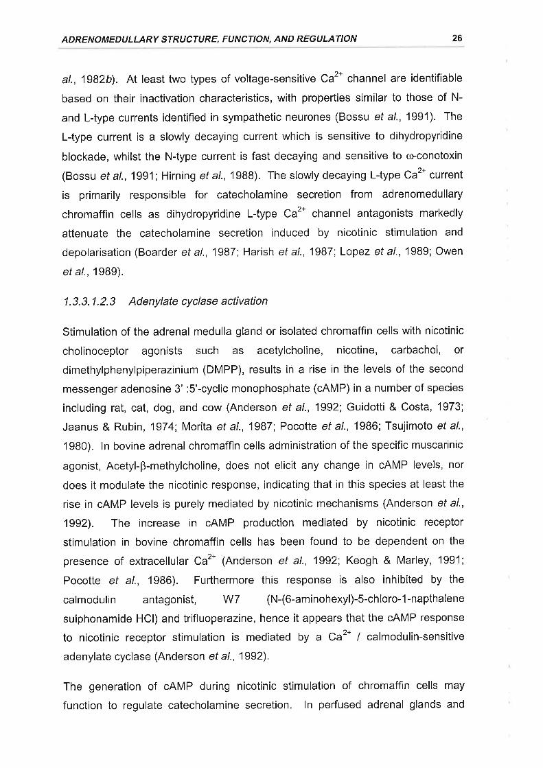
ADRENOMEDULLARY STRUCTURE, FUNCTION, AND REGULATION 26
at., 1g82b). At least two types of voltage-sensitive Ca2* channel are identifiable
based on their inactivation characteristics, with properties similar to those of N-
and L-type currents identified in sympathetic neurones (Bossu ef a/., 1991). The
L-type current is a slowly decaying current which is sensitive to dihydropyridine
blockade, whilst the N-type current is fast decaying and sensitive to o>conotoxin
(Bossu et al.,1991 ; Hirning et a|.,1988). The slowly decaying L-type Ca2* current
is primarily responsible for catecholamine secretion from adrenomedullary
chromaffin cells as dihydropyridine L-type Ca2* channel antagonists markedly
attenuate the catecholamine secretion induced by nicotinic stimulation and
depolarisation (Boarder ef al., 1987; Harish et al., 1987', Lopez et al., 1989; Owen
et a\.,1989).
1.3.3.1.2.3 Adenylate cyclase activation
Stimulation of the adrenal medulla gland or isolated chromaffin cells with nicotinic
cholinoceptor agonists such as acetylcholine, nicotine, carbachol, or
dimethylphenylpiperazinium (DMPP), results in a rise in the levels of the second
messenger adenosine 3' :5'-cyclic monophosphate (cAMP) in a number of species
including rat, cat, dog, and cow (Anderson ef al., 1992; Guidotti & Costa, 1973;
Jaanus & Rubin, 1974; Morita et al., 1987; Pocotte et al., 1986; Tsujimoto et al.,
1980). ln bovine adrenal chromaffin cells administration of the specific muscarinic
agonist, Acetyl-B-methylcholine, does not elicit any change in cAMP levels, nor
does it modulate the nicotinic response, indicating that in this species at least the
rise in cAMP levels is purely mediated by nicotinic mechanisms (Anderson et al.,
1992). The increase in cAMP production mediated by nicotinic receptor
stimulation in bovine chromaffin cells has been found to be dependent on the
presence of extracellular Ca2* (Anderson et al., 1992; Keogh & Marley, 1991;
Pocotte et al., 1986). Furthermore this response is also inhibited by the
calmodulin antagonist, W7 (N-(6-aminohexyl)-5-chloro-1-napthalene
sulphonamide HCI) and trifluoperazine, hence it appears that the cAMP response
to nicotinic receptor stimulation is mediated by a Ca2* / calmodulin-sensitive
adenylate cyclase (Anderson et al., 1992).
The generation of cAMP during nicotinic stimulation of chromaffin cells may
function to regulate catecholamine secretion. ln perfused adrenal glands and
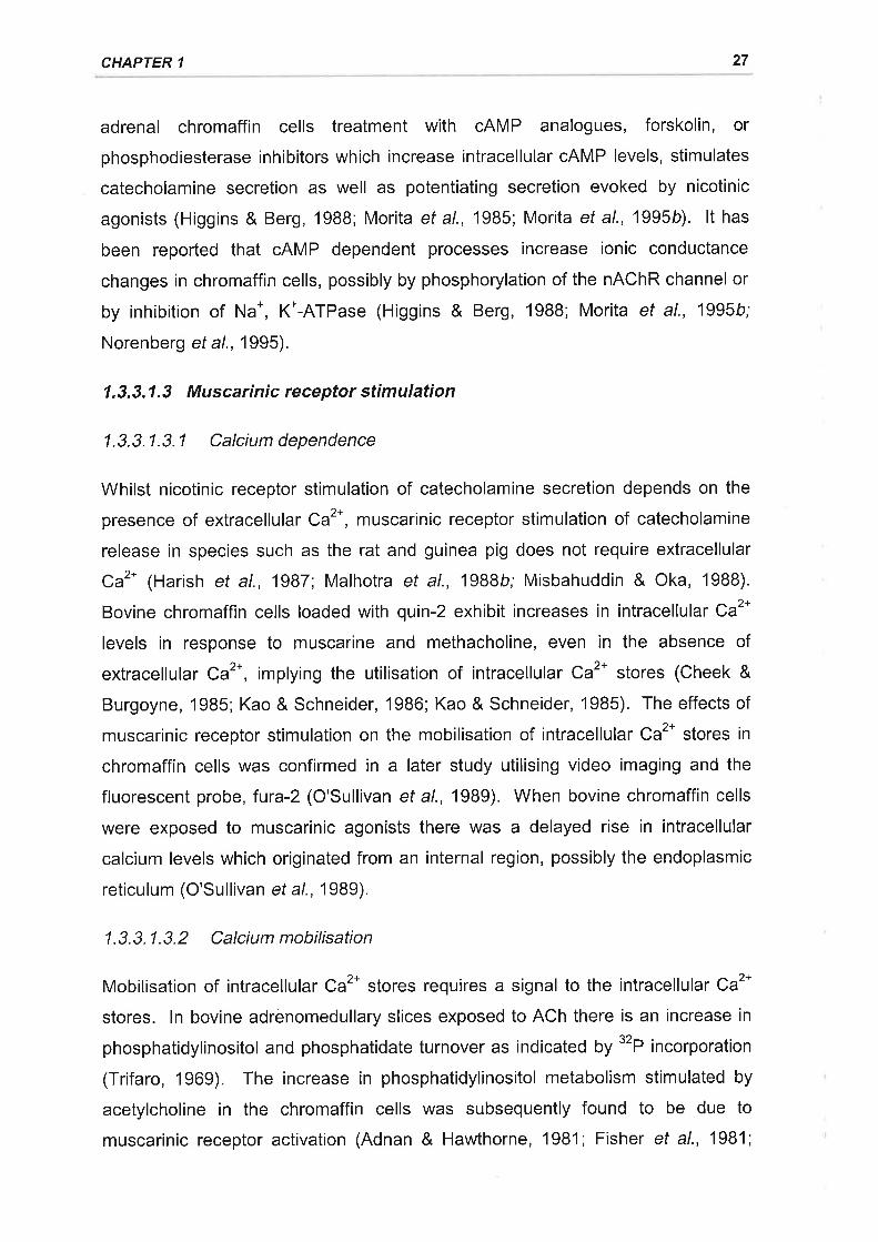
CHAPTER 1 27
adrenal chromaffin cells treatment with cAMP analogues, forskolin, or
phosphodiesterase inhibitors which increase intracellular cAMP levels, stimulates
catecholamine secretion as well as potentiating secretion evoked by nicotinic
agonists (Higgins & Berg, 1988; Morita et al., 1985; Morita et al., 1995b). lt has
been reported that cAMP dependent processes increase ionic conductance
changes in chromaffin cells, possibly by phosphorylation of the nAChR channel or
by inhibition of Na*, K*-ATPase (Higgins & Berg, 1988; Morita et al., 1995b;
Norenberg et al., 1995).
1.3.3.1.3 Muscarinic receptor stimulation
1.3.3.1.3.1 Calcium dependence
Whilst nicotinic receptor stimulation of catecholamine secretion depends on the
presence of extracellular Ca2*, muscarinic receptor stimulation of catecholamine
release in species such as the rat and guinea pig does not require extracellular
Ca2* lHarish ef a/., 1987; Malhotra et al., 1988b; Misbahuddin & Oka, 1988).
Bovine chromaffin cells loaded with quin-2 exhibit increases in intracellular Ca2*
levels in response to muscarine and methacholine, even in the absence of
extracellular Ca2*, implying the utilisation of intracellular Ca2* stores (Cheek &
Burgoyne,'1985; Kao & Schneider, 1986; Kao & Schneider, 1985). The effects of
muscarinic receptor stimulation on the mobilisation of intracellular Ca2* stores in
chromaffin cells was confirmed in a later study utilising video imaging and the
fluorescent probe, fura-2 (O'Sullivan et al., 1989). When bovine chromaffin cells
were exposed to muscarinic agonists there was a delayed rise in intracellular
calcium levels which originated from an internal region, possibly the endoplasmic
reticulum (O'Sullivan et al., 1989).
1.3.3.1.3.2 Calcium mobilisation
Mobilisation of intracellular Ca2* stores requires a signal to the intracellular Ca2*
stores. ln bovine adrenomedullary slices exposed to ACh there is an increase in
phosphatidylinositol and phosphatidate turnover as indicated by "P incorporation
(Trifaro, 1969). The increase in phosphatidylinositol metabolism stimulated by
acetylcholine in the chromaffin cells was subsequently found to be due to
muscarinic receptor activation (Adnan & Hawthorne, 1981; Fisher ef al., 1981;
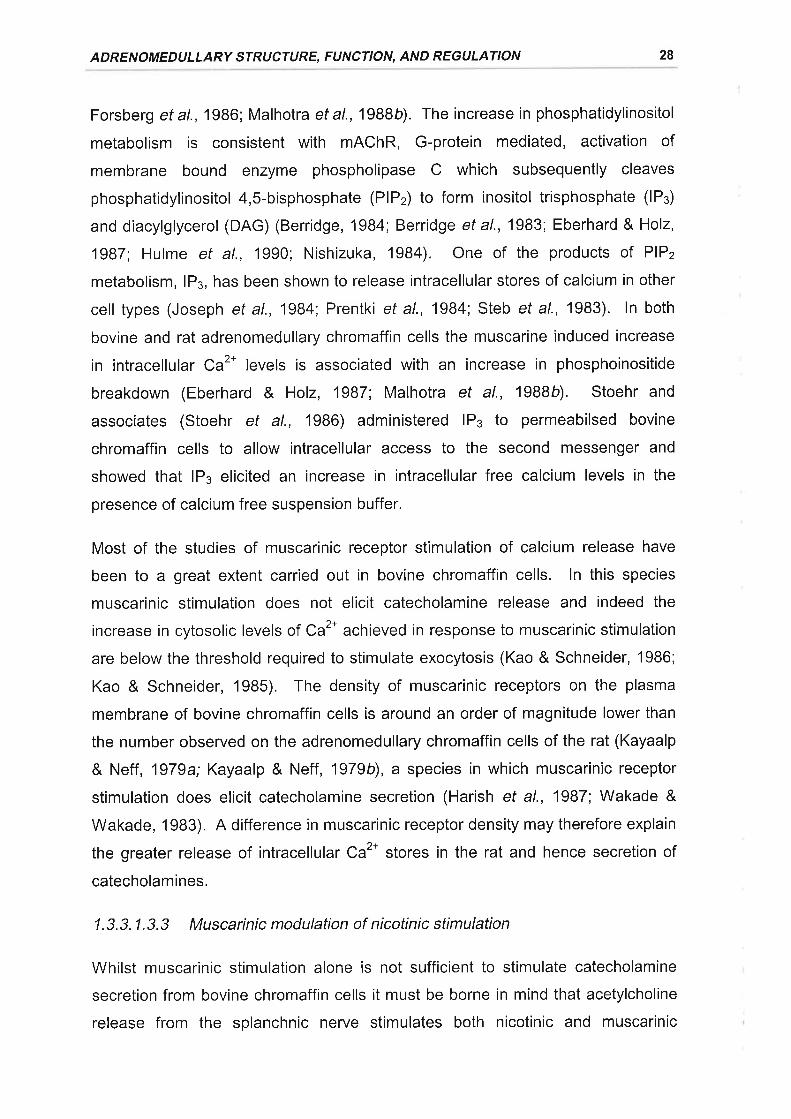
ADRENOMEDULLARY STRUCTURE, FUNCTION, AND REGULATION 28
Forsberg et al., 1986; Malhotra et al., 1988b). The increase in phosphatidylinositol
metabolism is consistent with mAChR, G-protein mediated, activation of
membrane bound enzyme phospholipase C which subsequently cleaves
phosphatidylinositol 4,S-bisphosphate (PlP2) to form inositol trisphosphate (lPs)
and diacylglycerol (DAG) (Berridge, 1984; Berridge et al., 1983; Eberhard & Holz,
1987; Hulme et al., 1990; Nishizuka, 1984). One of the products of PlPz
metabolism, lP3, has been shown to release intracellular stores of calcium in other
cell types (Joseph et al., 1984; Prentki et al., 1984; Steb ef a/., '1983). ln both
bovine and rat adrenomedullary chromaffin cells the muscarine induced increase
in intracellular Ca2* levels is associated with an increase in phosphoinositide
breakdown (Eberhard & Holz, 1987; Malhotra et al., 1gBBb). Stoehr and
associates (Stoehr et al., 1986) administered lP¡ to permeabilsed bovine
chromaffin cells to allow intracellular access to the second messenger and
showed that lP¡ elicited an increase in intracellular free calcium levels in the
presence of calcium free suspension buffer.
Most of the studies of muscarinic receptor stimulation of calcium release have
been to a great extent carried out in bovine chromaffin cells. ln this species
muscarinic stimulation does not elicit catecholamine release and indeed the
increase in cytosolic levels of Caz* achieved in response to muscarinic stimulation
are belowthe threshold required to stimulate exocytosis (Kao & Schneider, 1986;
Kao & Schneider, 1985). The density of muscarinic receptors on the plasma
membrane of bovine chromaffin cells is around an order of magnitude lower than
the number observed on the adrenomedullary chromaffin cells of the rat (Kayaalp
& Neff, 1979a; Kayaalp & Neff, 1979b), a species in which muscarinic receptor
stimulation does elicit catecholamine secretion (Harish et al., 1987; Wakade &
Wakade, 1983). A difference in muscarinic receptor density may therefore explain
the greater release of intracellular Ca2* stores in the rat and hence secretion of
catecholamines.
1.3.3.1.3.3 Muscarinic modulation of nicotinic stimulation
Whilst muscarinic stimulation alone is not sufficient to stimulate catecholamine
secretion from bovine chromaffin cells it must be borne in mind that acetylcholine
release from the splanchnic nerve stimulates both nicotinic and muscarinic
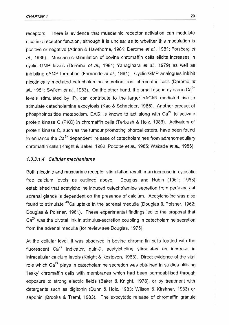
CHAPTER 1 29
receptors. There is evidence that muscarinic receptor activation can modulate
nicotinic receptor function, although it is unclear as to whether this modulation is
positive ornegative (Adnan & Hawthorne, 1981;Derome ef al., 1981; Forsberg ef
a/., 1986). Muscarinic stimulation of bovine chromaffin cells elicits increases in
cyclic GMP levels (Derome et al., 1981; Yanagihara et al., 1979) as well as
inhibiting cAMP formation (Fernando et al., 1991). Cyclic GMP analogues inhibit
nicotinically mediated catecholamine secretion from chromaffin cells (Derome ef
at.,1981; Swilem et a|.,1983). On the other hand, the small rise in cytosolic Ca2*
levels stimulated by lP3 can contribute to the larger nAChR mediated rise to
stimulate catecholamine exocytosis (Kao & Schneider, 1985). Another product of
phosphoinositide metabolism, DAG, is known to act along with Ca2* to activate
protein kinase C (PKC) in chromaffin cells (Terbush & Holz, 1986). Activators of
protein kinase C, such as the tumour promoting phorbal esters, have been found
to enhance the Ca2* dependent release of catecholamines from adrenomedullary
chromaffin cells (Knight & Baker, 1983; Pocotte et al., 1985; Wakade et al., 1986).
1.3.3.1.4 Cellular mechanisms
Both nicotinic and muscarinic receptor stimulation result in an increase in cytosolic
free calcium levels as outlined above. Douglas and Rubin (1961; 1963)
established that acetylcholine induced catecholamine secretion from perfused cat
adrenal glands is dependent on the presence of calcium. Acetylcholine was also
found to stimulate asca uptake in the adrenal medulla (Douglas & Poisner, 1962;
Douglas & Poisner, 1961). These experimental findings led to the proposal that
Ca2* was the pivotal link in stimulus-secretion coupling in catecholamine secretion
from the adrenal medulla (for review see Douglas, 1975).
At the cellular level, it was observed in bovine chromaffin cells loaded with the
fluorescenT Ca2* indicator, quin-2, acetylcholine stimulates an increase in
intracellular calcium levels (Knight & Kesteven, 1983). Direct evidence of the vital
role which Ca2* plays in catecholamine secretion was obtained in studies utilising
'leaky' chromaffin cells with membranes which had been permeabilised through
exposure to strong electric fields (Baker & Knight, 1978), or by treatment with
detergents such as digitonin (Dunn & Holz, 1983; Wilson & Kirshner, 1983) or
saponin (Brooks & Treml, 1983). The exocytotic release of chromaffin granule
ADRENOMEDULLARY STRUCTURE, FUNCTION, AND REGULATION 30
contents from permeabilised chromaffin cells, as evidenced by parallel increases
in the secretion of high and low molecular weight chromaffin granule contents, is
stimulated by simply raising Ca2* in the suspension buffer to micromolar levels
(Baker & Knight, 1978; Brooks & Treml, 1983; Dunn & Holz, '1983; Wilson &
Kirshner, 1983). Whilst the stimulatory effect of Caz* on chromaffin granule
exocytosis is independent of cholinergic stimuli it does require the presence of
Mg-ATP (Baker & Knight, 1978), implicating the possible involvement of protein
phosphorylation in the secretory process (Burgoyne, 1984).
1.3.3.1.5 Exocytosis
The mechanisms by which an increase in intracellular Ca2* levels stimulates
exocytosis of the chromaffin granules remains to be fully elucidated. One of the
key events in exocytosis is the docking of secretory vesicles to the plasma
membrane of the cell to allow membrane fusion and the release of vesicle
contents to occur (Burgoyne,1984; Smith et al., 1973). This docking has been
established in rat and bovine neurones to be due to the formation of a fusion
particle consisting of a number of proteins which combine to join the secretory
vesicletotheacceptormembrane(Bennett etal., 1992 Sollner etal., 1993). N-
ethylmaleimide-sensitive fusion protein (NSF) is an important component of the
fusion particle and in its absence, transport vesicles merely accumulate at the
acceptor membrane (Malhotra et a\.,1988c). NSF requires the presence of other
cytoplasmic factors known as soluble NSF attachment proteins (SNAPS) to allow
vesicle fusion to proceed (Clary et a|.,1990; Sollner et al., 1993). There is also a
further requirement for membrane bound co-factors on both the secretory vesicle
and the plasma membrane to allow for targeted docking of the synaptic vesicle to
specific areas of the plasma membrane (Robinson et al., 1996). Two such
membrane bound synaptic proteins which have been identified to function as
SNAP receptors (SNAREs) are the vesicle associated membrane protein (VAMP)
also known as synaptobrevin, and syntaxin, a protein which is found in the plasma
membrane (Sollner et al., 1993). NSF, SNAP, and the SNAREs bind to form a
characteristic 20 S fusion particle (Sollner et a\.,1993) which results in the docking
of the secretory vesicle with the plasma membrane. NSF is established to be a
functional ATP-ase (Tagaya et al., 1993) and the hydrolysis of Mg-ATP by this
component of the fusion engine leads to a disassembly of the complex which
CHAPTER 1 31
appears to be an intrinsic step in membrane fusion (Sollner et al., 1993; Wilson ef
al., 1992).
All of the synaptic components which are required for the formation of the fusion
engine in brain tissue have also been identified in chromaffin cells including
synaptobrevin / VAMP, syntaxin, cx-SNAP, SNAP-2S, and NSF (Burgoyne &
Williams, 1997; Hodel et al., 1994; Roth & Burgoyne, 1994). lndeed, this fusion
engine is functional in chromaffin cells as administration of exogenous cr-SNAP to
chromaffin cells stimulates exocytosis while the administration of tetanus toxin
which cleaves synaptobrevin / VAMP, blocks the calcium induced exocytosis from
chromaffin cells (Hohne-Zell et al., 1994; Livett, 1993). As all of the components
required for formation of the fusion engine are already present within chromaffin
cells it then follows that resting chromaffin granules must be prevented from
reaching the docking zone with the plasma membrane. lt has been demonstrated
using electron microscopy that chromaffin granules are restrained within a three
dimensional filamentous network within intact chromaffin cells (Kondo et al.,
1982). Substances which are found to disrupt the cortical actin network within
chromaffin cells stimulate the exocytosis of chromaffin granules (Roth &
Burgoyne, 1995). Furthermore, it is interesting that the chromaffin granule
membrane contains on its cytoplasmic side the protein cr-actinin which binds the
cytoskeletal component, F-actin (Aunis et al., 1980; Bader & Aunis, 1983;
Burgoyne, 1984). The binding of chromaffin granule cr-actinin to F-actin is Ca2*-
dependent and increasing the levels of Ca2* to levels commensurate with those
observed to stimulate exocytosis results in dissociation of the two components
(Fowler & Pollard, 1982a;Fowler & Pollard, 1982b).
1.3.3.2 Non-cholinergic regulation of adrenal catecholamine secretion
As previously described, the adrenal gland contains a number of neuropeptides
which are expressed in chromaffin cells, splanchnic nerve fibres, intra-adrenal
ganglion cells, or a combination of the three. The exact function of these
neuropeptides within the adrenal gland has yet to be fully determined but it is
thought that they, along with other peptidergic hormones, may act as non-
cholinergic transmitters to regulate catecholamine secretion and adrenal blood
ADRENOMEDULLARY STRUCTURE, FUNCTION, AND REGULATION 32
flow. Non-cholinergic transmitters may influence catecholamine secretion either
directly, or indirectly by the modulation of cholinergic transmitter actions.
1.3.3.2.1 Experimental evidence
Convincing evidence for a major physiological role of non-cholinergic transmitters
in regulating adrenomedullary function comes from studies of retrogradely
perfused rat (Malhotra & Wakade, 1987a; Malhotra et al., 19BBb; Wakade, 19BB;
Wakade et al., 1991) and bovine adrenal glands (Marley et al., 1993). Wakade
and colleagues reported that catecholamine secretion from the pedused ral
adrenal increased in a linear fashion in response to transmural electrical
stimulation at frequencies between 0.5 and 3 Hz, as cholinergic fibres were
recruited, reaching a plateau at 10 Hz (Wakade, 1981). Similarly, Marley and
colleagues established that catecholamine secretion from the perfused bovine
adrenal gland increased linearly in response to continuous field stimulation from 2
to 10 Hz, and then subsequently plateaued (Marley et al., 1993). The profile of
adrenomedullary catecholamine secretion in response to field stimulation of the
perfused bovine gland in vitro (\Aarley et al., 1993) closely resembled that
previously reported for stimulation of the peripheral end of the splanchnic nerve rn
vivo for the conscious calf (Edwards, 1982; Edwards et al., 1980).
The cholinergic contribution to the catecholamine secretory response of the
perfused adrenal gland to electrical stimulation was examined by using a
combination of nicotinic and muscarinic receptor antagonists (Malhotra & Wakade,
1987a;Marley et a\.,1993; Wakade, 1988). At high frequency stimulation (10 Hz)
nicotinic and muscarinic receptor blockade combined reduced catecholamine
secretion from perfused rat and bovine adrenals by 80 o/o and 75 Vo, respectively.
At low frequency stimulation (0.5-2 Hz) the catecholamine secretory response in
the presence of nicotinic and muscarinic blockade is only reduced by 35 % in the
rat adrenal, yet it is abolished in the bovine adrenal (Malhotra & Wakade, 1987 a;
Marley et al., 1993; Wakade, 1988). The residual catecholamine secretion which
occurred in response to electrical stimulation despite nicotinic and muscarinic
receptor blockade was attributed to a non-cholinergic component of adrenal nerve
stimulation (Malhotra & Wakade, 1987a; Marley et al., 1993). Therefore the non
cholinergic component predominates at low frequency stimulation in the rat,
CHAPTER 1 33
representing about 65 % of secretion at 0.5 Hz, and 20 % of secretion at 10 Hz
(Malhotra & Wakade, 1987a; Wakade, 1988). The non-cholinergic component
associated with low frequency electrical stimulation of the rat adrenal gland is
capable of maintaining catecholamine secretion for up to several hours, whereas
more transient responses are observed at the higher frequencies associated with
cholinergic stimulation (Wakade, 1988). ln contrast to the rat, non-cholinergic
secretion is more prominent at high frequency stimulation in the bovine adrenal
gland, representing 25 % of the total secretion at 10 Hz, whilst being absent at 2
Hz (Marley et al., 1993). Bursts of high frequency electrical stimulation of the
peripheral end of the splanchnic nerye have been reported to stimulate the
release of a number of peptidergic transmitters in the calf (Bloom et al., 1988;
Edwards & Jones, 1993b).
1.3.3.2.2 Potential non-cholinergic transmitters
Of all the neuropeptides and non-classical transmitters known to be present within
the adrenal medulla the roles of VlP, PACAP, Substance P, NPY, Enk-containing
peptides, Angiotensin ll, and NO in the regulation of adrenomedullary function are
probably the best characterised.
1.3.3.2.2,1 VIP and PACAP
Both VIP and the VIP-like secretin peptide, PACAP, which shares a 70 %
sequence homology with VIP (Arimura, 1998), are released from the adrenal
gland in response to splanchnic nerve stimulation (Bloom et al., 1988; Wakade ef
al., 1992i Wakade et al., 1991). VIP and PACAP directly stimulate or facilitate
cholinergic catecholamine secretion in a dose dependent manner from the
adrenomedullary chromaffin cells of a number of species, both in vitro and in vivo
(Cheung & Holzwarth, 1986; Chowdhury et al., 1994; Edwards & Jones, 1993a;
Edwards & Jones, 1994; Gaspo et al., 1997; Guo & Wakade, 1994; lsobe et al.,
1993; Lamouce et al., 1999; Malhotra & Wakade, 1987b; Perrin et al., 1995;
Watanabe et al., 1995; Wilson, 1988). Receptors for VIP and PACAP belong to
the VIP receptor family and have been demonstrated to be present on the
chromaffin cells of the adrenal medulla by immunohistochemical, radioligand
binding, in situ hybridisation, and pharmacological studies (Arimura, 1998; Arimura
& Shioda, 1995; Gaspo et al., 1997; Hashimoto et al., 1993; Magistretti et al.,
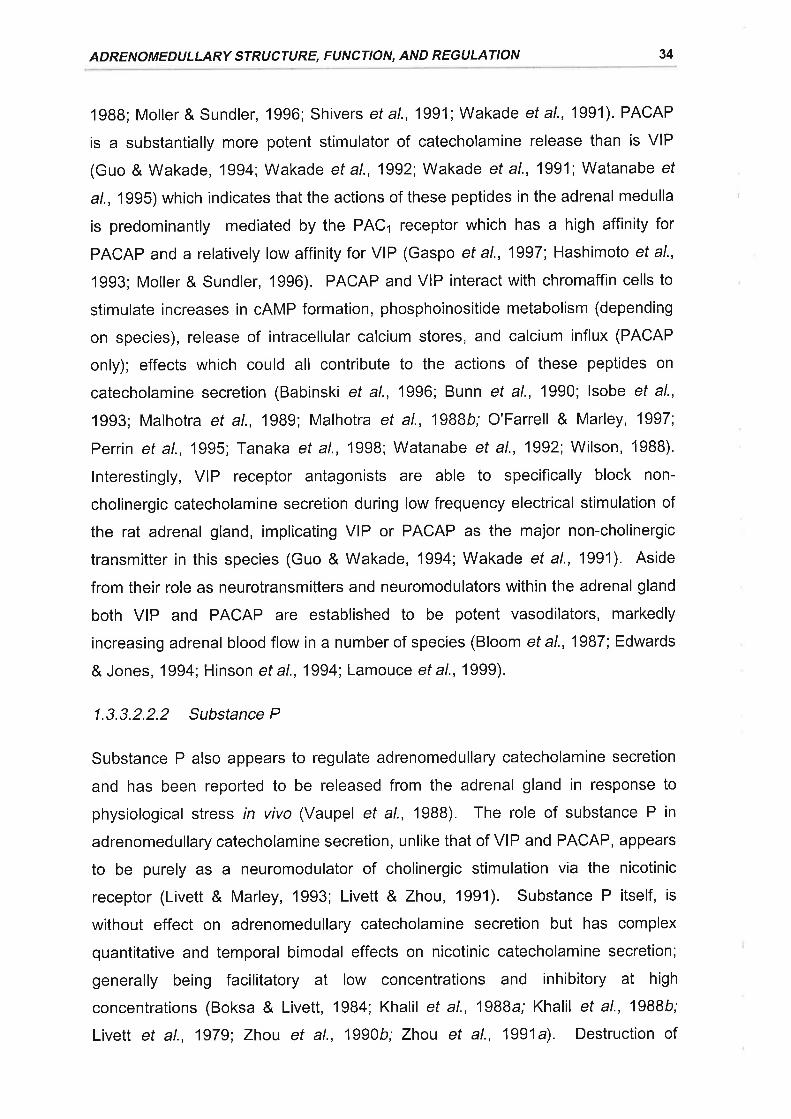
ADRENOMEDULLARY STRUCTURE, FUNCTION, AND REGULATION 34
1988; Moller & Sundler, 1996; Shivers et al., 1991 ; Wakade et al., 1991). PACAP
is a substantially more potent stimulator of catecholamine release than is VIP
(Guo & Wakade, 1994; Wakade et al., 1992; Wakade et al., 1991; Watanabe ef
a/., 1995) which indicates that the actions of these peptides in the adrenal medulla
is predominantly mediated by the PACr receptor which has a high affinity for
PACAP and a relatively low affinity for VIP (Gaspo et al., 1997; Hashimoto et al.,
1993; Moller & Sundler, 1996). PACAP and VIP interact with chromaffin cells to
stimulate increases in cAMP formation, phosphoinositide metabolism (depending
on species), release of intracellular calcium stores, and calcium influx (PACAP
only); effects which could all contribute to the actions of these peptides on
catecholamine secretion (Babinski ef al., 1996; Bunn et al., 1990; lsobe et al.,
1993; Malhotra et al., 1989; Malhotra et al., 1988b; O'Farrell & Marley, 1997;
Perrin et al., 1995; Tanaka et al., 1998; Watanabe et al., 1992; Wilson, 1988).
lnterestingly, VIP receptor antagonists are able to specifically block non-
cholinergic catecholamine secretion during low frequency electrical stimulation of
the rat adrenal gland, implicating VIP or PACAP as the major non-cholinergic
transmitter in this species (Guo & Wakade, 1994; Wakade et al., 1991). Aside
from their role as neurotransmitters and neuromodulators within the adrenal gland
both VIP and PACAP are established to be potent vasodilators, markedly
increasing adrenal blood flow in a number of species (Bloom et al., 1987; Edwards
& Jones, 1994; Hinson et al., 1994; Lamouce et al., 1999).
1.3.3.2.2.2 SubstanceP
Substance P also appears to regulate adrenomedullary catecholamine secretion
and has been reported to be released from the adrenal gland in response to
physiological stress in vivo (Vaupel et al., 19BB). The role of substance P in
adrenomedullary catecholamine secretion, unlike that of VIP and PACAP, appears
to be purely as a neuromodulator of cholinergic stimulation via the nicotinic
receptor (Livett & Marley, 1993; Livett & Zhou, 1991). Substance P itself, is
without effect on adrenomedullary catecholamine secretion but has complex
quantitative and temporal bimodal effects on nicotinic catecholamine secretion;
generally being facilitatory at low concentrations and inhibitory at high
concentrations (Boksa & Livett, 1984; Khalil et al., 1988a; Khalil et al., 1988b;
Livett et al., 1979; Zhou et al., 1990b; Zhou et al., 1991a). Destruction of
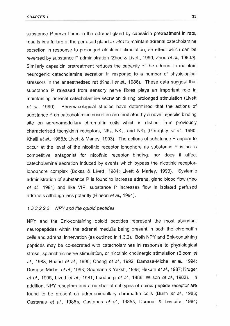
CHAPTER 1 35
substance P nerve fibres in the adrenal gland by capsaicin pretreatment in rats,
results in a failure of the perfused gland in vitro to maintain adrenal catecholamine
secretion in response to prolonged electrical stimulation, an effect which can be
reversed by substance P administration (Zhou & Livett, 1990; Zhou et a\.,1990a).
Similarly capsaicin pretreatment reduces the capacity of the adrenal to maintain
neurogenic catecholamine secretion in response to a number of physiological
stressors in the anaesthetised rat (Khalil et al., 1986). These data suggest that
substance P released from sensory nerve fibres plays an important role in
maintaining adrenal catecholamine secretion during prolonged stimulation (Livett
et al., 1990). Pharmacological studies have determined that the actions of
substance P on catecholamine secretion are mediated by a novel, specific binding
site on adrenomedullary chromaffin cells which is distinct from previously
characterised tachykinin receptors, NKr, NKz, and NKe (Geraghty et al., 1990;
Khalil et al.,19B8b; Livett & Marley, 1993). The actions of substance P appear to
occur at the level of the nicotinic receptor ionophore as substance P is not a
competitive antagonist for nicotinic receptor binding, nor does it affect
catecholamine secretion induced by events which bypass the nicotinic receptor-
ionophore complex (Boksa & Livett, 1984; Livett & Marley, 1993). Systemic
administration of substance P is found to increase adrenal gland blood flow (Yeo
et al., 1984) and like VlP, substance P increases flow in isolated perfused
adrenals although less potently (Hinson et a\.,1994).
1.3.3.2.2.3 NPY and the opioid peptides
NPY and the Enk-containing opioid peptides represent the most abundant
neuropeptides within the adrenal medulla being present in both the chromaffin
cells and adrenal innervation (as outlined in 1.3.2). Both NPY and Enk-containing
peptides may be co-secreted with catecholamines in response to physiological
stress, splanchnic nerve stimulation, or nicotinic cholinergic stimulation (Bloom ef
al., 19BB; Briand et al., 1990; Cheng et al., 1992; Damase-Michel ef al., 1994;
Damase-Michel ef a1.,1993; Gaumann & Yaksh, 19BB; Hexum et al., 1987; Kruger
et al., 1995; Livett et al., 1981; Lundberg et al., 1986; Wilson et al., 1982). ln
addition, NPY receptors and a number of subtypes of opioid peptide receptor are
found to be present on adrenomedullary chromaffin cells (Bunn et al., 1988;
Castanas et al., 1985a; Castanas et al., 1985b; Dumont & Lemaire, 1984;
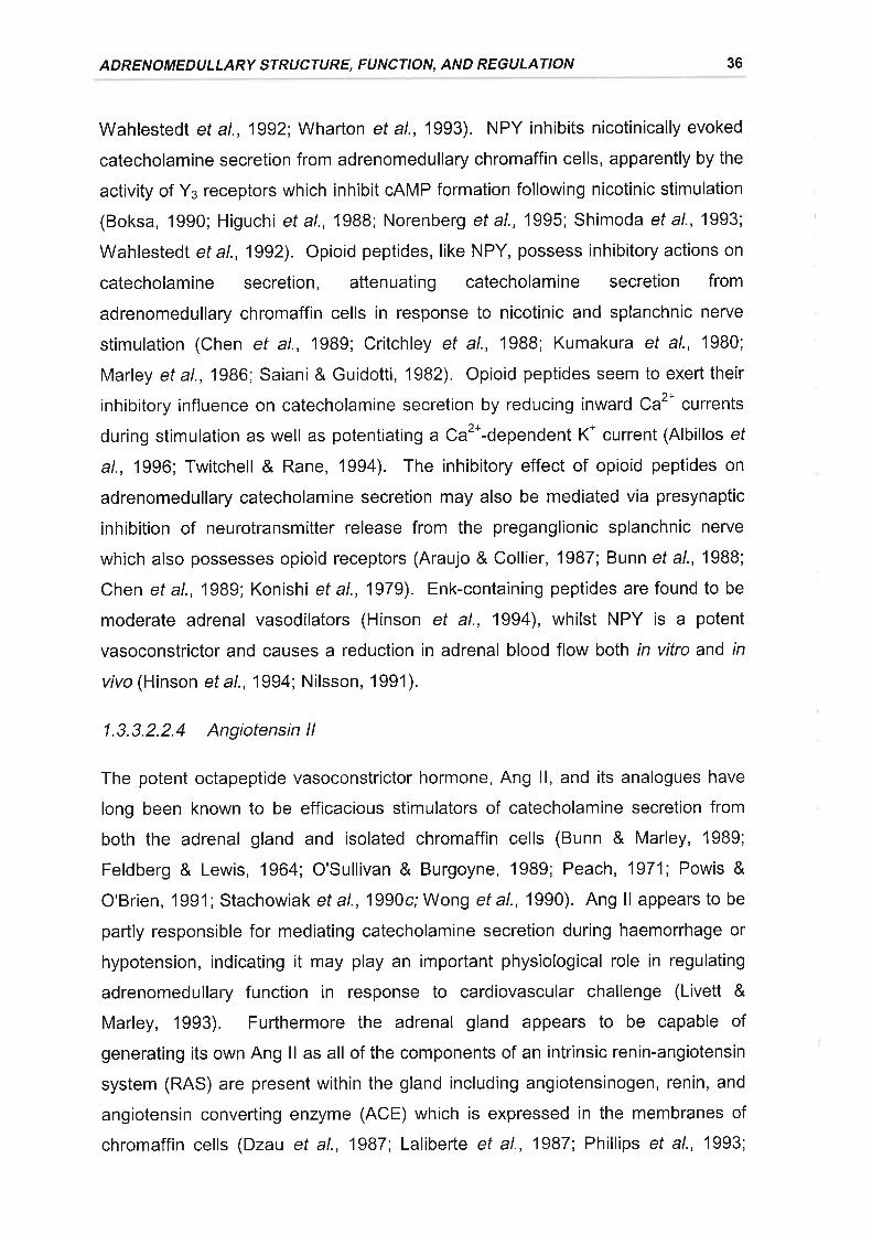
ADRENOMEDULLARY STRUCTURE, FUNCTION, AND REGULATION 36
Wahlestedt et al., 1992; Wharton et al., 1993). NPY inhibits nicotinically evoked
catecholamine secretion from adrenomedullary chromaffin cells, apparently by the
activity of Yo receptors which inhibit cAMP formation following nicotinic stimulation
(Boksa, 1990; Higuchi et al., 1988; Norenberg et a\.,1995; Shimoda et al., 1993;
Wahlestedt et al., 1992). Opioid peptides, like NPY, possess inhibitory actions on
catecholamine secretion, attenuating catecholamine secretion from
adrenomedullary chromaffin cells in response to nicotinic and splanchnic nerye
stimulation (Chen et al., 1989; Critchley et al., 1988; Kumakura et al., 1980;
Marley et a\.,1986; Saiani & Guidotti,1982). Opioid peptides seem to exert their
inhibitory influence on catecholamine secretion by reducing inward Ca2* currents
during stimulation as well as potentiating a Ca2*-dependent K* current (Albillos ef
al., 1996; Twitchell & Rane, 1994). The inhibitory effect of opioid peptides on
adrenomedullary catecholamine secretion may also be mediated via presynaptic
inhibition of neurotransmitter release from the preganglionic splanchnic nerve
which also possesses opioid receptors (Araujo & Collier, 1987; Bunn et al., 1988;
Chen et al., 1989; Konishi et al., 1979). Enk-containing peptides are found to be
moderate adrenal vasodilators (Hinson et al., 1994), whilst NPY is a potent
vasoconstrictor and causes a reduction in adrenal blood flow both in vitro and in
vivo (Hinson ef al., 1994; Nilsson, 1991).
1.3.3.2.2.4 Angiotensin ll
The potent octapeptide vasoconstrictor hormone, Ang ll, and its analogues have
long been known to be efficacious stimulators of catecholamine secretion from
both the adrenal gland and isolated chromaffin cells (Bunn & Marley, 1989;
Feldberg & Lewis, 1964; O'Sullivan & Burgoyne, 1989; Peach, 1971; Powis &
O'Brien, 1991; Stachowiak et a\.,1990c; Wong et a\.,1990). Ang ll appears to be
partly responsible for mediating catecholamine secretion during haemorrhage or
hypotension, indicating it may play an important physiological role in regulating
adrenomedullary function in response to cardiovascular challenge (Livett &
Marley, 1993). Furthermore the adrenal gland appears to be capable of
generating its own Ang ll as all of the components of an intrinsic renin-angiotensin
system (RAS) are present within the gland including angiotensinogen, renin, and
angiotensin converting enzyme (ACE) which is expressed in the membranes of
chromaffin cells (Dzau et al., 1987; Laliberte et al., 1987; Phillips et al., 1993;
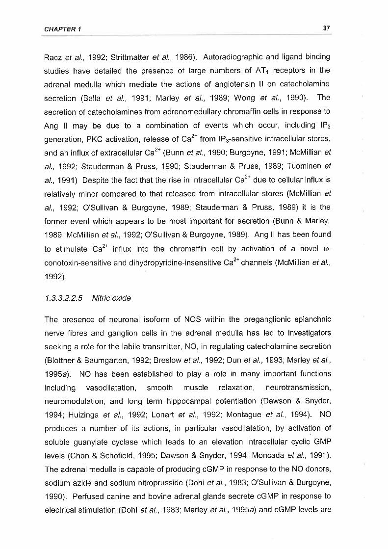
CHAPTER 1 37
Racz et al., 1992; Strittmatter et al., 1986). Autoradiographic and ligand binding
studies have detailed the presence of large numbers of ATr receptors in the
adrenal medulla which mediate the actions of angiotensin ll on catecholamine
secretion (Balla et al., 1991; Marley et al., 1989; Wong et al., 1990). The
secretion of catecholamines from adrenomedullary chromaffin cells in response to
Ang ll may be due to a combination of events which occur, including lPs
generation, PKC activation, release of Ca2* from lP¡-sensitive intracellular stores,
and an influx of extracellular Ca2* (Bunn et a\.,1990; Burgoyne, 1991; McMillian ef
al., 1992; Stauderman & Pruss, 1990; Stauderman & Pruss, 1989; Tuominen ef
at., 1991) Despite the fact that the rise in intracellular Ca2t due to cellular influx is
relatively minor compared to that released from intracellular stores (McMillian ef
al., 1992; O'Sullivan & Burgoyne, 1989; Stauderman & Pruss, 1989) it is the
former event which appears to be most important for secretion (Bunn & Marley,
1989; McMillian et a\.,1992; O'Sullivan & Burgoyne, 1989). Ang ll has been found
to stimulate Ca2* influx into the chromaffin cell by activation of a novel ú>
conotoxin-sensitive and dihydropyridine-insensitive Ca2t channels (McMillian et al.,
1ee2).
1.3.3.2.2.5 Nitricoxide
The presence of neuronal isoform of NOS within the preganglionic splanchnic
nerve fibres and ganglion cells in the adrenal medulla has led to investigators
seeking a role for the labile transmitter, NO, in regulating catecholamine secretion
(Blottner & Baumgarten, 1992; Breslow ef al., 1992; Dun ef al., 1993; Marley et al.,
1995a). NO has been established to play a role in many important functions
including vasodilatation, smooth muscle relaxation, neurotransmission,
neuromodulation, and long term hippocampal potentiation (Dawson & Snyder,
1994; Huizinga et al., 1992; Lonart et al., 1992i Montague et al., 1994). NO
produces a number of its actions, in particular vasodilatation, by activation of
soluble guanylate cyclase which leads to an elevation intracellular cyclic GMP
levels (Chen & Schofield, 1995; Dawson & Snyder, 1994; Moncada et al., 1991).
The adrenal medulla is capable of producing cGMP in response to the NO donors,
sodium azide and sodium nitroprusside (Dohi et al., 1983; O'Sullivan & Burgoyne,
1990). Perfused canine and bovine adrenal glands secrete cGMP in responseto
electrical stimulation (Dohi et a\.,1983; Marley et a\.,1995a) and cGMP levels are
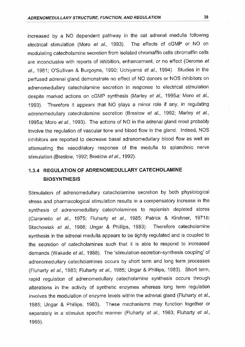
ADRENOMEDULLARY STRUCTURE, FUNCTION, AND REGULATION 38
increased by a NO dependent pathway in the cat adrenal medulla following
electrical stimulation (Moro et al., 1993). The effects of cGMP or NO on
modulating catecholamine secretion from isolated chromaffin cells chromaffin cells
are inconclusive with reports of inhibition, enhancement, or no effect (Derome ef
al., 1981; O'Sullivan & Burgoyne, 1990; Uchiyama et al., 1994). Studies in the
perfused adrenal gland demonstrate no effect of NO donors or NOS inhibitors on
adrenomedullary catecholamine secretion in response to electrical stimulation
despite marked actions on cGMP synthesis (Marley et al., 1995a; Moro et al.,
1993). Therefore it appears that NO plays a minor role if any, in regulating
adrenomedullary catecholamine secretion (Breslow et al., 1992; Marley et al.,
1995a; Moro eta\.,1993). The actions of NO in the adrenal gland most probably
involve the regulation of vascular tone and blood flow in the gland. lndeed, NOS
inhibitors are reported to decrease basal adrenomedullary blood flow as well as
attenuating the vasodilatory response of the medulla to splanchnic nerye
stimulation (Breslow, 1992; Breslow et a\.,1992).
1.3.4 REGULATION OF ADRENOMEDULLARY CATECHOLAMINE
BIOSYNTHESIS
Stimulation of adrenomedullary catecholamine secretion by both physiological
stress and pharmacological stimulation results in a compensatory increase in the
synthesis of adrenomedullary catecholamines to replenish depleted stores
(Ciaranello et al., 1975; Fluharty et al., 1985; Patrick & Kirshner, 1971b;
Stachowiak et al., 1986; Ungar & Phillips, 1983). Therefore catecholamine
synthesis in the adrenal medulla appears to be tightly regulated and is coupled to
the secretion of catecholamines such that it is able to respond to increased
demands (Wakade et a\.,1988). The'stimulation-secretion-synthesis coupling'of
adrenomedullary catecholamines occurs by short term and long term processes
(Fluharty et al., 1983; Fluharty et al., 1985; Ungar & Phillips, 1983). Short term,
rapid regulation of adrenomedullary catecholamine synthesis occurs through
alterations in the activity of synthetic enzymes whereas long term regulation
involves the modulation of enzyme levels within the adrenal gland (Fluharty et al.,
1985; Ungar & Phillips, 1983). These mechanisms may function together or
separately in a stimulus specific manner (Fluharty et al., 1983; Fluharty et al.,
1e8s).
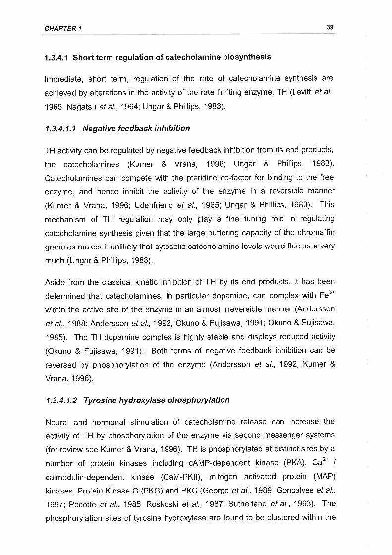
CHAPTER 1 39
1.3.4.1 Short term regulation of catecholamine biosynthesis
lmmediate, short term, regulation of the rate of catecholamine synthesis are
achieved by alterations in the activity of the rate limiting enzyme, TH (Levitt et al.,
1965; Nagatsu et a1.,1964; Ungar & Phillips, 1983).
1.3.4.1.1 Negative feedback inhibition
TH activity can be regulated by negative feedback inhibition from its end products,
the catecholamines (Kumer & Vrana, 1996; Ungar & Phillips, 1983).
Catecholamines can compete with the pteridine co-factor for binding to the free
enzyme, and hence inhibit the activity of the enzyme in a reversible manner
(Kumer & Vrana, 1996; Udenfriend et al., 1965; Ungar & Phillips, 1983). This
mechanism of TH regulation may only play a fine tuning role in regulating
catecholamine synthesis given that the large buffering capacity of the chromaffin
granules makes it unlikely that cytosolic catecholamine levels would fluctuate very
much (Ungar & Phillips, 1983).
Aside from the classical kinetic inhibition of TH by its end products, it has been
determined that catecholamines, in particular dopamine, can complex with Fe3*
within the active site of the enzyme in an almost irreversible manner (Andersson
et al., 1988; Andersson et al., 1992; Okuno & Fujisawa, 1991 ; Okuno & Fujisawa,
1985). The TH-dopamine complex is highly stable and displays reduced activity
(Okuno & Fujisawa, 1991). Both forms of negative feedback inhibition can be
reversed by phosphorylation of the enzyme (Andersson et al., 1992; Kumer &
Vrana, 1996).
1.3.4.1.2 Tyrosine hydroxylase phosphorylation
Neural and hormonal stimulation of catecholamine release can increase the
activity of TH by phosphorylation of the enzyme via second messenger systems
(for review see Kumer & Vrana, 1996). TH is phosphorylated at distinct sites by a
number of protein kinases including cAMP-dependent kinase (PKA), Ca2* |
calmodulin-dependent kinase (CaM-PKll), mitogen activated protein (MAP)
kinases, Protein Kinase G (PKG) and PKC (George eta\.,1989; Goncalves etal.,
1997; Pocotte et al., 1985; Roskoski et al., 1987; Sutherland et al., 1993). The
phosphorylation sites of tyrosine hydroxylase are found to be clustered within the
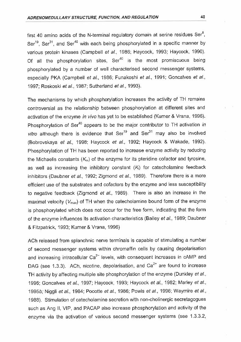
ADRENOMEDULLARY STRUCTURE, FUNCTION, AND REGULATION 40
first 40 amino acids of the N-terminal regulatory domain at serine residues Ser8,
Ser1e, Ser31, and Serao with each being phosphorylated in a specific manner by
various protein kinases (Campbell et al., 1986; Haycock, 1993; Haycock, 1990).
Of all the phosphorylation sites, Sera0 is the most promiscuous being
phosphorylated by a number of well characterised second messenger systems,
especially PKA (Campbell ef al., 1986; Funakoshi et al., 1991; Goncalves et al.,
1997; Roskoski et a\.,1987; Sutherland et a\.,1993).
The mechanisms by which phosphorylation increases the activity of TH remains
controversial as the relationship between phosphorylation at different sites and
activation of the enzyme in vivo has yet to be established (Kumer & Vrana, 1996).
Phosphorylation of Serao appears to be the major contributor to TH activation rn
vitro although there is evidence that Serle and Ser31 may also be involved
(Bobrovskaya et al., 1998; Haycock et al., 1992; Haycock & Wakade, 1992)'
Phosphorylation of TH has been reported to increase enzyme activity by reducing
the Michaelis constants (Kr) of the enzyme for its pteridine cofactor and tyrosine,
as well as increasing the inhibitory constant (K) for catecholamine feedback
inhibitors (Daubner et al., 1992; Zigmond et al., 1989). Therefore there is a more
efficient use of the substrates and cofactors by the enzyme and less susceptibility
to negative feedback (Zigmond et al., 1989). There is also an increase in the
maximal velocity (Vru*) of TH when the catecholamine bound form of the enzyme
is phosphorylated which does not occur for the free form, indicating that the form
of the enzyme influences its activation characteristics (Bailey et al.,'1989; Daubner
& Fitzpatrick, 1993; Kumer & Vrana, 1996)
ACh released from splanchnic nerye terminals is capable of stimulating a number
of second messenger systems within chromaffin cells by causing depolarisation
and increasing intracellular Ca2* levels, with consequent increases in cAMP and
DAG (see 1.3.3). ACh, nicotine, depolarisation, and Ca2* are found to increase
TH activity by affecting multiple site phosphorylation of the enzyme (Dunkley et al.,
1996; Goncalves et al., 1997 Haycock, 1993; Haycock et al., 1982; Marley et al',
1995b; Niggli et al., 1984; Pocotte et al., 1986; Powis et al., 1996; Waymire et al',
19BB). Stimulation of catecholamine secretion with non-cholinergic secretagogues
such as Ang ll, VlP, and PACAP also increase phosphorylation and activity of the
enzyme via the activation of various second messenger systems (see 1 .3.3.2,
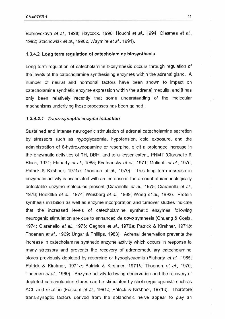
CHAPTER 1 41
Bobrovskaya et al., 1998; Haycock, 1996; Houchi et al., 1994; Olasmaa et al.,
1992; Stachowiak et al., 1990c; Waymire et al., 1991).
1.3.4.2 Long term regulation of catecholamine biosynthesis
Long term regulation of catecholamine biosynthesis occurs through regulation of
the levels of the catecholamine synthesising enzymes within the adrenal gland. A
number of neural and hormonal factors have been shown to impact on
catecholamine synthetic enzyme expression within the adrenal medulla, and it has
only been relatively recently that some understanding of the molecular
mechanisms underlying these processes has been gained.
1.3.4.2.1 Trans-synaptic enzyme induction
Sustained and intense neurogenic stimulation of adrenal catecholamine secretion
by stressors such as hypoglycaemia, hypotension, cold exposure, and the
administration of 6-hydroxydopamine or reserpine, elicit a prolonged increase in
the enzymatic activities of TH, DBH, and to a lesser extent, PNMT (Ciaranello &
Black, 1971; Fluharty et al., 1985; Kvetnansky et al., 1971; Molinoff et al., 1970;
Patrick & Kirshner, 1971b; Thoenen et al., 1970). This long term increase in
enzymatic activity is associated with an increase in the amount of immunologically
detectable enzyme molecules present (Ciaranello et al., 1975; Ciaranello et al.,
1976; Hoeldtke et al., 1974; Weisberg et al., 1989; Wong et al., 1993). Protein
synthesis inhibition as well as enzyme incorporation and turnover studies indicate
that the increased levels of catecholamine synthetic enzymes following
neurogenic stimulation are due to enhanced de novo synthesis (Chuang & Costa,
1974; Ciaranello et al., 1975; Gagnon et al., 1976a; Patrick & Kirshner, 1971b;
Thoenen et al., 1969; Ungar & Phillips, 1983). Adrenal denervation prevents the
increase in catecholamine synthetic enzyme activity which occurs in response to
many stressors and prevents the recovery of adrenomedullary catecholamine
stores previously depleted by reserpine or hypoglycaemia (Fluharty et a/., 1985;
Patrick & Kirshner, 1971a; Patrick & Kirshner, 1971b; Thoenen et al., 1970;
Thoenen et al., 1969). Enzyme activity following denervation and the recovery of
depleted catecholamine stores can be stimulated by cholinergic agonists such as
ACh and nicotine (Fossom et al., 1991a; Patrick & Kirshner, 1971a). Therefore
trans-synaptic factors derived from the splanchnic nerve appear to play an
ADRENOMEDULLARY STRUCTURE, FUNCTION, AND REGULATION 42
important role in the long term regulation of catecholamine synthetic enzyme
activity.
The enhanced synthesis of adrenomedullary catecholamine synthetic enzymes in
response to prolonged neural stimulation appears to be due to an upregulation of
gene expression (DeCristofaro & LaGamma, 1994; Faucon Biguet et al., 1991;
Schalling et a\.,1988; Stachowiak et al., 1985; Stachowiak et al., 1986; Weisberg
et al., '1989; Wessel & Joh, 1992; Wong & Wang, 1994). Furthermore, cholinergic
stimulation of adrenomedullary and immortalised PC12 chromaffin cells elicits
increases in the expression of TH, DBH, and PNMT mRNAs (Craviso et al., 1992;
Evinger et al., 1994; Hiremagalur ef al., 1993; Jahng et al., 1997; Stachowiak ef
al., 1988a; Stachowiak et al., 1990b). Chromaffin cells from many species
possess both nicotinic and muscarinic cholinergic receptors and both types of
receptor have been reported to differentially regulate the gene expression of a
number of catecholamine synthetic enzymes (Chen et al., 1996; Craviso et al.,
1992', Evinger et al., 1994', Hiremagalur et a\.,1993). Nuclear run-off assays and
promoter region-reporter gene construct studies reveal that cholinergic stimulation
of gene expression is attributable in large part to an augmented transcription of
catecholamine synthetic enzyme genes (Craviso et al., 1992; Craviso et al., 1995;
Evinger et al., 1994; Fossom et al., 1991b; Hemmick et al., 1995; Tonshoff et al.,
1997). Transcriptional regulation of gene expression requires the interaction
between regulatory proteins (trans-acting transcription factors) and sequences
within the gene (cis-acting elements) (Maniatis et al., 1987). Trans-synaptic
stimulation of catecholamine synthetic enzyme gene transcription is likely to be
mediated by the interaction of second messenger systems with frans-acting
regulatory proteins, Events which occur downstream from cholinergic receptor
activation that contribute to second messenger system activation, such as
depolarisation and increased intracellular calcium levels, are in themselves
sufficient to bring about selective changes in TH, DBH, and PNMT mRNA
expression (Hwang & Lee, 1995; Kilbourne et al., 1992; Kilbourne & Sabban,
1990; Livett et al., 1990; Stachowiak et al., 1994; Stachowiak et al., 1990c;
Tschernitz et al., 1995; Wan ef al., 1991b). Two second messenger systems
likely to play a key role in the cholinergic stimulation of catecholamine synthetic
enzyme gene expression are cAMP dependent PKA, and PKC (see 1.3.3).
CHAPTER 1 43
1.3.4.2.1 .1 oAMP
A distinct feature of nicotinic stimulation of the adrenal gland in vivo (Guidotti &
Costa, 1973) and both adrenomedullary and PC12 chromaffin cells in vitro (Baizer
& Weiner, 1985; Eiden et al., 1984; Olasmaa et al., 1992) is an increase in cAMP
formation. Cyclic AMP binds cooperatively to two sites on the regulatory sub-unit
of PKA releasing the active catalytic sub-unit (Sassone-Corsi, 1995). Protein
kinase A activators, such as forskolin, and stable cAMP analogues are capable of
increasing the expression of TH and DBH mRNAs in both adrenomedullary and
PC12 chromaffin cells (Hwang & Joh, 1993; Hwang ef al., 1994; Hwang et al.,
1997; Kim ef al., 1994; Kim ef al., 1993b; McMahon & Sabban, 1992; Stachowiak
et al., 1990b). There are conflicting reports as to whether or not forskolin or cAMP
analogues can induce PNMT mRNA expression in chromaffin cells (Hwang et al.,
1994; Hwang et al., 1997; Livett et al., 1990; Stachowiak et al., 1994; Stachowiak
et al., 1990b; Wan ef al., 1991a). ln studies which report upregulated PNMT
mRNA expression in response to forskolin or cAMP analogues, the effect was
relatively minor when compared to the induction of DBH and TH mRNAs (Hwang
et al., 1994; Hwang et al., 1997; Stachowiak et al., 1994; Stachowiak et al.,
1990b). The effects of forskolin and cAMP analogues are specifically mediated by
PKA activation as the specific inhibitor, N-[2-(ebromocinnamylamine)ethyl]-S-
isoquinolinesulfonamide (H-89), prevents the induction of catecholamine synthetic
enzyme mRNA expression by forskolin (Hwang ef al., 1997) and TH and DBH
mRNA induction by cyclic AMP is abolished in PKA-deficient PC12 cell lines (Kim
et al., 1994; Kim ef al., 1993b). Both nicotinic stimulation and PKA activation
demonstrate similar non-additive patterns of catecholamine synthetic enzyme
mRNA induction, implicating a common transduction pathway for both stimuli
(Craviso et a\.,1995; Hiremagalur et al.,'1993; Hwang et al., 1994; Stachowiak ef
al., 1990c). ln addition, the induction of TH mRNA expression and gene
transcription by nicotine is not observed in PKA-deficient PC12 cells (Hiremagalur
et a\.,1993). Non-cholinergic secretagogues such as PACAP and VlP, which also
stimulate cAMP formation and hence PKA activation in chromaffin cells also have
stimulatory effects on catecholamine synthetic enzyme mRNA expression (lsobe
et al., 1996; Olasmaa et al., 1992; Park et al., 1999; Rius ef al., 1994; Tonshoff ef
al., 1997).
ADRENOMEDIJLLARY STRUCTURE, FUNCTION, AND REGULATION 44
Aside from their role in nicotinic induction of gene expression, cAMP dependent
mechanisms also appear to be important in the maintenance of the basal
expression of adrenomedullary TH, DBH, and PNMT mRNA. PKA-deficient PC12
cell lines exhibit reduced basal levels of TH and DBH gene transcription when
compared with wild type PC12 cells (Kim ef al., 1994; Kim ef al., 1993a; Kim et al.,
1993b). ln isolated adrenomedullary chromaffin cells the inhibition of PKA activity
with H-89 profoundly inhibits the basal expression of TH, DBH, and PNMT mRNAs
(Hwang et a1.,1997).
Analysis of the 5'-promoter regions of the TH (Best et al., 1995; Coker et al., 1988;
D'Mello et al., 1989; Harrington et al., 1987; Piech-Dumas & Tank, 1999) and DBH
genes (Afar et al., 1996; Kobayashi et al., 1989; McMahon & Sabban,1992;
Shaskus et al., 1992) has established the presence of functional cis-acting cAMP
response element (CRE) motifs. A family of trans-acting CRE binding proteins
including CRE binding protein (CREB) and activating transcription factor 1 (ATF1)
are phosphorylated by PKA, whereupon they dimerise and bind to the CRE motifs
to stimulate transcription (Sassone-Corsi, 1995). Transfection assays using PC12
and human neuroblastoma SK-N-BE(2) cell lines have established that mutations
and deletions of the CRE motif in both the rat TH gene and human DBH gene 5'
promoter regions effectively impair transcriptional activity of the promoter in
response to nicotine and cAMP (Hiremagalur ef al., 1993; Kim ef al., 1993a; Kim
et al., 1994). Western blot analysis of nuclear protein extracts from PC12 cells
reveals an enhanced expression of the frans-acting CREB protein following PKA
activation byforskolin (Tinti et a\.,1996). Gel supershift assays of nuclear protein
extracts from PC12 cells using oligonucleotides containing the consensus CRE
sequence of the TH promoter show that antiserum for both CREB and ATFI are
able to produce supershifted bands indicating an interaction between these
transcription factors and the TH-CRE (Tinti ef a/., 1996). Furthermore, PC12 cells
stably transfected with CREB antisense mRNA expression vectors display
reduced levels of CREB protein and mRNA expression and there is a failure of
cAMP to induce TH promoter-reporter gene construct expression (Piech-Dumas &
Tank, 1999). DNase'1 footprinting analysis and DNA binding assays also implicate
the involvements of CREB and ATFl in stimulating DBH gene transcription via the
CREs in the 5'proximal promoter region of the DBH gene (Afar et al., 1996; Seo
et a\.,1996).
CHAPTER 1 45
The 5'-promoter regions of the PNMT genes of a number of species have not
been reported to contain a consensus CRE sequence motif (Morita et al., 1992;
Ross et al., 1990; Sasaoka et al., 1989), which may explain the relatively poor
inducibility of PNMT mRNA expression in response to PKA activation and nicotine
administration (Hwang et al., 1994; Hwang et al., 1997; Livett et al., 1990;
Stachowiak et al., 1990b; Wan et al., 1991a). PKA may regulate PNMT
expression by a novel CRE sequence motif, or via the activation of a CRE
independent pathway (Hwang et al., 1997). There is increasing evidence of
functional "cross-talk" between second messenger systems (Cambier et al., 1987
Hoeffler et al., 1989; Kim et al., 1997; Masquilier & Sassone-Corsi, 1992;
Narindrasorasak et al., 1987; Yoshimasa et al., 1987) such that activated PKA
might indirectly regulate PNMT gene expression via interaction at a number of
possible levels with a separate second messenger system.
1.3.4.2.1.2 Protein kinase C
Nicotinic and muscarinic receptor activation stimulate protein kinase C activity in
chromaffin cells by enhanced phosphoinositide metabolism mediated by calcium
influx and G-protein activation, respectively (Akaike et al., 1993; Eberhard & Holz,
1987; Malhotra et a\.,1989; Terbush et al., 1988). Phorbol esters, such as 12-O
tetradecanoylphorbal 13-acetate (TPA), which are also potent stimulators of PKC
activity are found to stimulate the expression of TH and PNMT mRNA expression
in adrenomedullary and PC12 chromaffin cells (Stachowiak et al., 1990c; Terbush
et al., 1988; Vyas et al., 1990; Wan ef al., 1991a). Thus PKC may play a role in
the trans-synaptic induction of catecholamine synthetic enzyme genes.
The S'-promoter region of the TH gene contains an AP-1 genomic regulatory
elements (lcard-Liepkalns et al., 1992; Stachowiak et al., 1990a). The regulation
of gene expression by the AP-1 element involves interactions with frans-acting
homodimers or heterodimers formed between fos and jun leucine zipper DNA-
binding proteins (Chiu et al., 1988; Ransone & Verma, 1990). The expression of
fos and jun is induced in catecholaminergic cells both in vivo and rn vitroby a wide
variety of neural stimuli including hypoglycaemia, reserpine treatment, nicotinic
and muscarinic stimulation, and protein kinase C activation (Craviso et al., 1995;
Faucon Biguet et al., 1991; Goc et al., 1992; lcard-Liepkalns et al., 1992;
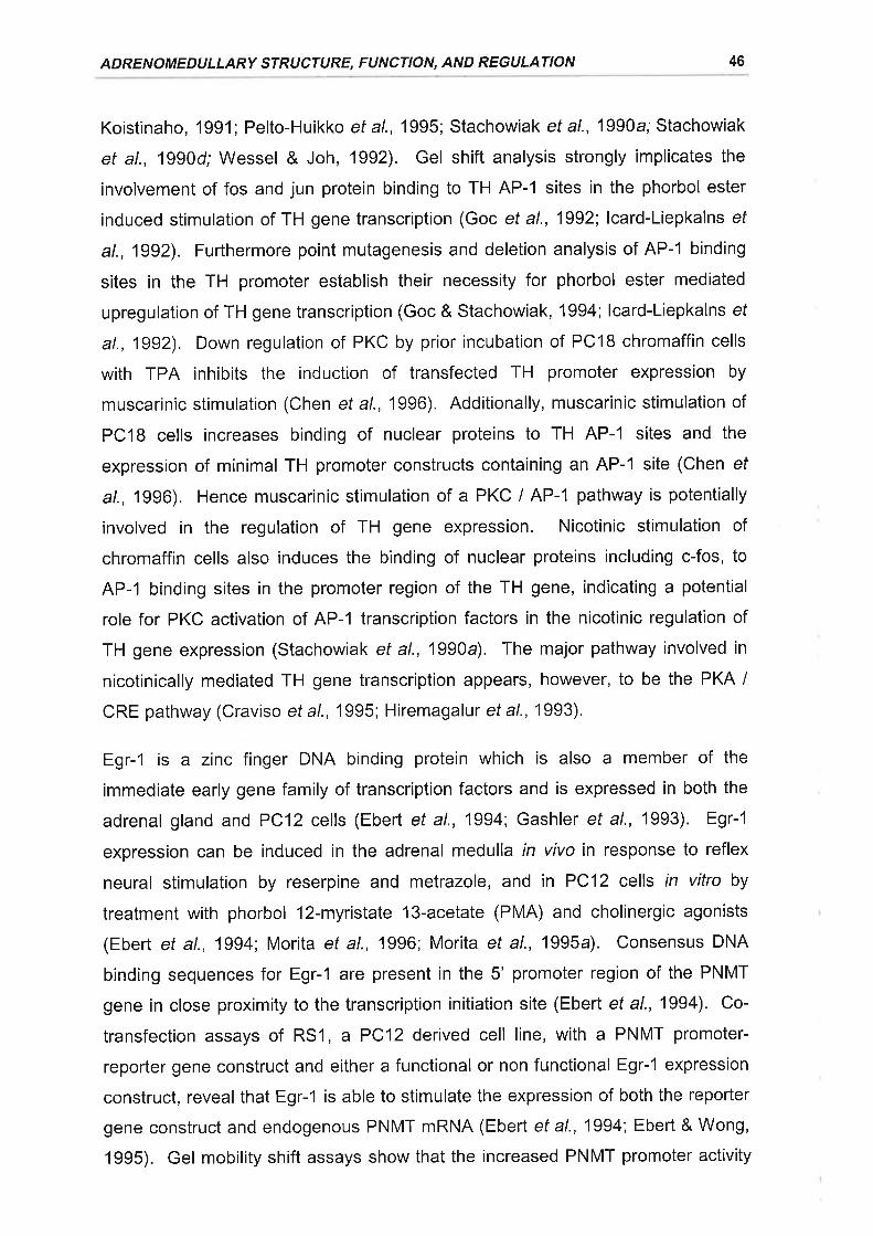
ADRENOMEDULLARY STRUCTURE, FUNCTION, AND REGULATION 46
Koistinaho, 1991 ; Pelto-Huikko ef a/., 1995; Stachowiak et al., 1990a; Stachowiak
et al., 1990d; Wessel & Joh, 1992). Gel shift analysis strongly implicates the
involvement of fos and jun protein binding to TH AP-1 sites in the phorbol ester
induced stimulation of TH gene transcription (Goc et al., 1992; lcard-Liepkalns ef
at., 1992). Furthermore point mutagenesis and deletion analysis of AP-1 binding
sites in the TH promoter establish their necessity for phorbol ester mediated
upregulation of TH gene transcription (Goc & Stachowiak, 1994; lcard-Liepkalns ef
at., 1992). Down regulation of PKC by prior incubation of PClB chromaffin cells
with TPA inhibits the induction of transfected TH promoter expression by
muscarinic stimulation (Chen et a\.,1996). Additionally, muscarinic stimulation of
PC18 cells increases binding of nuclear proteins to TH AP-1 sites and the
expression of minimal TH promoter constructs containing an AP-1 site (Chen ef
a/., 1996). Hence muscarinic stimulation of a PKC I AP-1 pathway is potentially
involved in the regulation of TH gene expression. Nicotinic stimulation of
chromaffin cells also induces the binding of nuclear proteins including c-fos, to
AP-1 binding sites in the promoter region of the TH gene, indicating a potential
role for PKC activation of AP-1 transcription factors in the nicotinic regulation of
TH gene expression (Stachowiak ef al., 1990a). The major pathway involved in
nicotinically mediated TH gene transcription appears, however, to be the PKA /
CRE pathway (Craviso ef a/., 1995; Hiremagalur et a1.,1993).
Egr-1 is a zinc finger DNA binding protein which is also a member of the
immediate early gene family of transcription factors and is expressed in both the
adrenal gland and PC12 cells (Ebert et al., 1994; Gashler et al., 1993). Egr-1
expression can be induced in the adrenal medulla in vivo in response to reflex
neural stimulation by reserpine and metrazole, and in PC12 cells in vitro by
treatment with phorbol 12-myristate 13-acetate (PMA) and cholinergic agonists
(Ebert et al., 1994; Morita et al., 1996; Morita et al., 1995a). Consensus DNA
binding sequences for Egr-1 are present in the 5' promoter region of the PNMT
gene in close proximitytothetranscription initiation site (Ebert etal., 1994). Co-
transfection assays of RSl , a PC12 derived cell line, with a PNMT promoter-
reporter gene construct and either a functional or non functional Egr-1 expression
construct, reveal that Egr-1 is able to stimulate the expression of both the reporter
gene construct and endogenous PNMT mRNA (Ebert et al., 1994; Ebert & Wong,
1995). Gel mobility shift assays show that the increased PNMT promoter activity
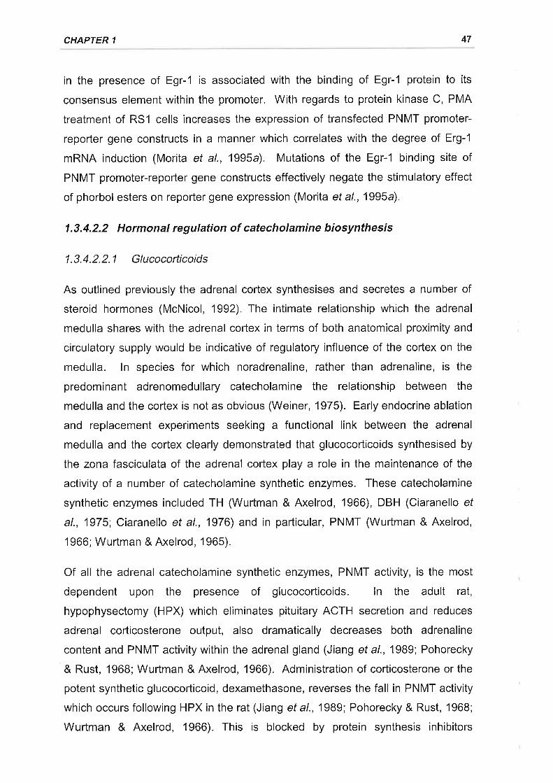
CHAPTER 1 47
in the presence of Egr-1 is associated with the binding of Egr-1 protein to its
consensus element within the promoter. With regards to protein kinase C, PMA
treatment of RS1 cells increases the expression of transfected PNMT promoter-
reporter gene constructs in a manner which correlates with the degree of Erg-1
mRNA induction (Morita et al., 1995a). Mutations of the Egr-1 binding site of
PNMT promoter-reporter gene constructs effectively negate the stimulatory effect
of phorbol esters on reporter gene expression (Morita et a\.,1995a).
1.3.4.2.2 Hormonal regulation of catecholamine biosynthesis
1.3.4.2.2.1 Glucocorticoids
As outlined previously the adrenal cortex synthesises and secretes a number of
steroid hormones (McNicol, 1992). The intimate relationship which the adrenal
medulla shares with the adrenal cortex in terms of both anatomical proximity and
circulatory supply would be indicative of regulatory influence of the cortex on the
medulla. ln species for which noradrenaline, rather than adrenaline, is the
predominant adrenomedullary catecholamine the relationship between the
medulla and the cortex is not as obvious (Weiner, 1975). Early endocrine ablation
and replacement experiments seeking a functional link between the adrenal
medulla and the cortex clearly demonstrated that glucocorticoids synthesised by
the zona fasciculata of the adrenal cortex play a role in the maintenance of the
activity of a number of catecholamine synthetic enzymes. These catecholamine
synthetic enzymes included TH (Wurtman & Axelrod, 1966), DBH (Ciaranello ef
al., 1975; Ciaranello et al., 1976) and in particular, PNMT (Wurtman & Axelrod,
1966; Wurtman & Axelrod, 1965).
Of all the adrenal catecholamine synthetic enzymes, PNMT activity, is the most
dependent upon the presence of glucocorticoids. ln the adult rat,
hypophysectomy (HPX) which eliminates pituitary ACTH secretion and reduces
adrenal corticosterone output, also dramatically decreases both adrenaline
content and PNMT activity within the adrenal gland (Jiang et al., 1989; Pohorecky
& Rust, 1968; Wurtman & Axelrod, 1966). Administration of corticosterone or the
potent synthetic glucocorticoid, dexamethasone, reverses the fall in PNMT activity
which occurs following HPX in the rat (Jiang et a\.,1989; Pohorecky & Rust, 1968;
Wurtman & Axelrod, 1966). This is blocked by protein synthesis inhibitors
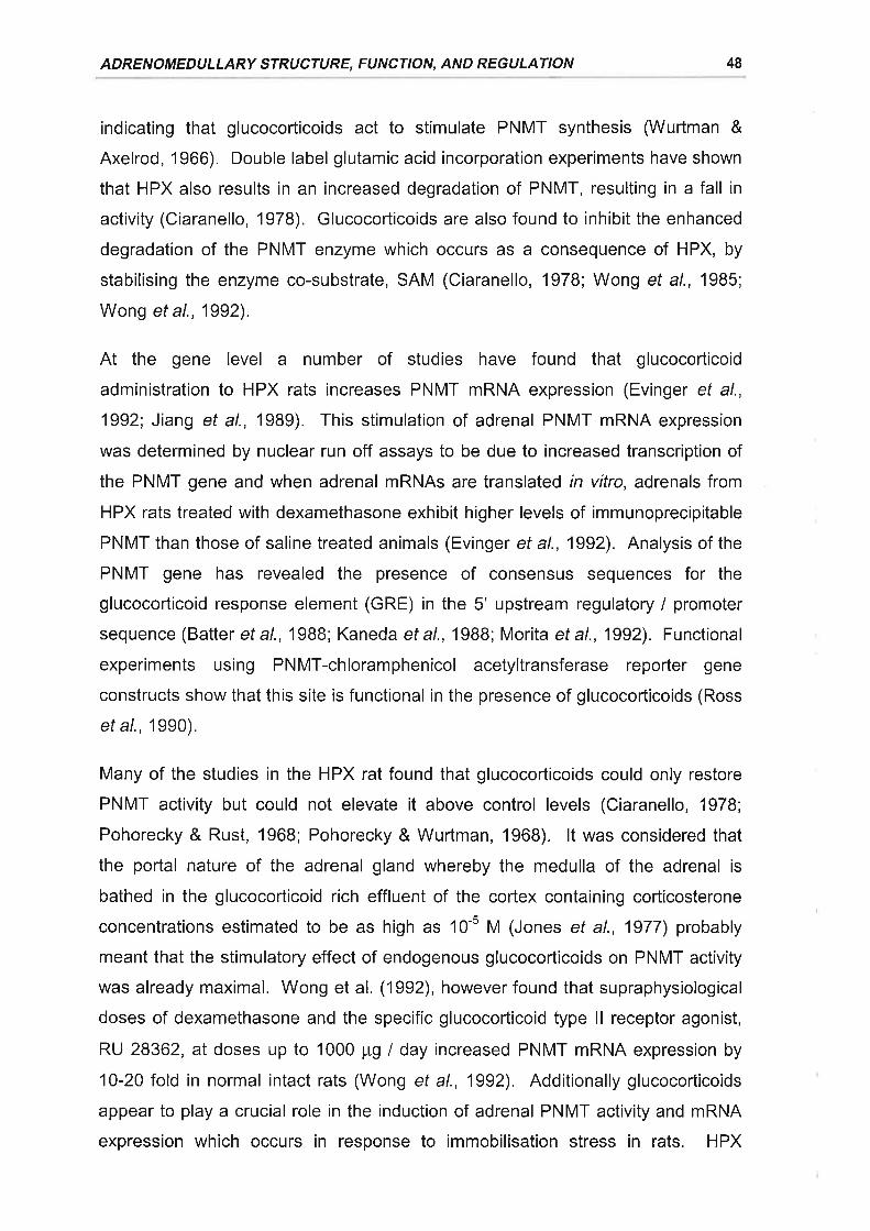
ADRENOMEDULLARY STRUCTURE, FUNCTION, AND REGULATION 48
indicating that glucocorticoids act to stimulate PNMT synthesis (Wurtman &
Axelrod, 1966). Double label glutamic acid incorporation experiments have shown
that HPX also results in an increased degradation of PNMT, resulting in a fall in
activity (Ciaranello, 1978). Glucocorticoids are also found to inhibit the enhanced
degradation of the PNMT enzyme which occurs as a consequence of HPX, by
stabilising the enzyme co-substrate, SAM (Ciaranello, 1978; Wong et al., 1985;
Wong et a\.,1992).
At the gene level a number of studies have found that glucocorticoid
administration to HPX rats increases PNMT mRNA expression (Evinger et al.,
1992; Jiang et al., 1989). This stimulation of adrenal PNMT mRNA expression
was determined by nuclear run off assays to be due to increased transcription of
the PNMT gene and when adrenal mRNAs are translated rn vitro, adrenals from
HPX rats treated with dexamethasone exhibit higher levels of immunoprecipitable
PNMT than those of saline treated animals (Evinger et al., 1992). Analysis of the
PNMT gene has revealed the presence of consensus sequences for the
glucocorticoid response element (GRE) in the 5' upstream regulatory / promoter
sequence (Batter et al., 19BB; Kaneda et al., 1988; Morita et al., 1992). Functional
experiments using PNMT-chloramphenicol acetyltransferase reporter gene
constructs show that this site is functional in the presence of glucocorticoids (Ross
et al., 1990).
Many of the studies in the HPX rat found that glucocorticoids could only restore
PNMT activity but could not elevate it above control levels (Ciaranello, 1978;
Pohorecky & Rust, 1968; Pohorecky & Wurlman, 1968). lt was considered that
the portal nature of the adrenal gland whereby the medulla of the adrenal is
bathed in the glucocorticoid rich effluent of the cortex containing corticosterone
concentrations estimated to be as high as 10-5 M (Jones et al., 1977) probably
meant that the stimulatory effect of endogenous glucocorticoids on PNMT activity
was already maximal. Wong et al. (1992), however found that supraphysiological
doses of dexamethasone and the specific glucocorticoid type ll receptor agonist,
RU 28362, at doses up to 1000 pg I day increased PNMT mRNA expression by
10-20 fold in normal intact rats (Wong et al., 1992). Additionally glucocorticoids
appear to play a crucial role in the induction of adrenal PNMT activity and mRNA
expression which occurs in response to immobilisation stress in rats. HPX
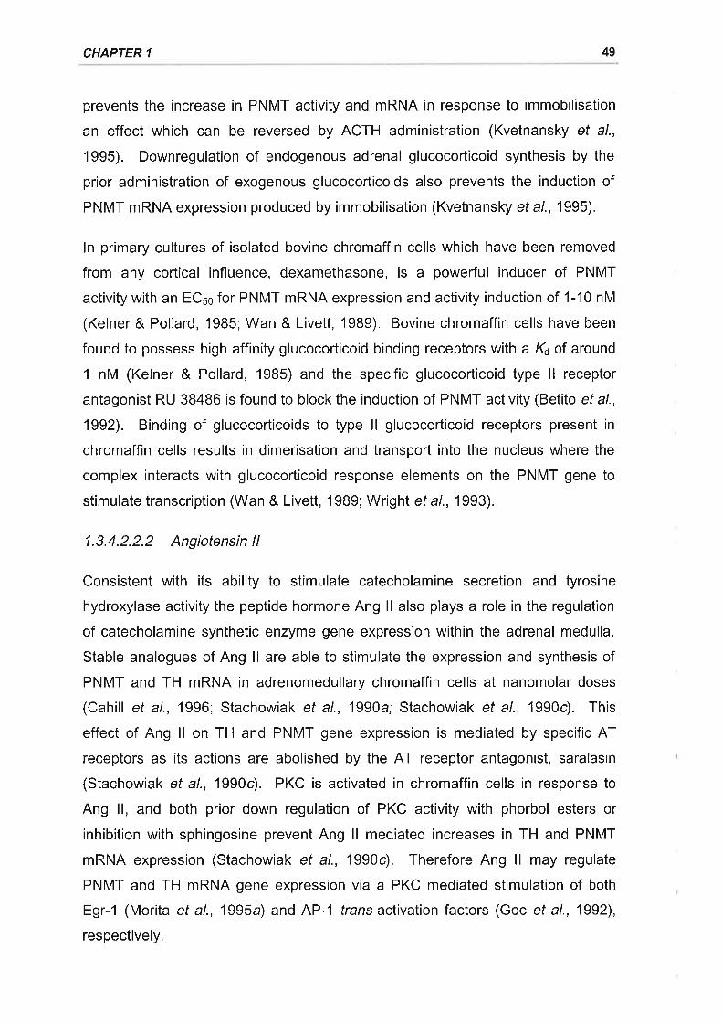
CHAPTER 1 49
prevents the increase in PNMT activity and mRNA in response to immobilisation
an effect which can be reversed by ACTH administration (Kvetnansky et al.,
1995). Downregulation of endogenous adrenal glucocorticoid synthesis by the
prior administration of exogenous glucocorticoids also prevents the induction of
PNMT mRNA expression produced by immobilisation (Kvetnansky et a\.,1995).
ln primary cultures of isolated bovine chromaffin cells which have been removed
from any cortical influence, dexamethasone, is a powerful inducer of PNMT
activity with an ECso for PNMT mRNA expression and activity induction of 1-10 nM
(Kelner & Pollard, 1985; Wan & Livett, 1989). Bovine chromaffin cells have been
found to possess high affinity glucocorticoid binding receptors with a K¿ of around
1 nM (Kelner & Pollard, 1985) and the specific glucocorticoid type ll receptor
antagonist RU 38486 is found to block the induction of PNMT activity (Betito et al.,
1992). Binding of glucocorticoids to type ll glucocorticoid receptors present in
chromaffin cells results in dimerisation and transport into the nucleus where the
complex interacts with glucocorticoid response elements on the PNMT gene to
stimulate transcription (Wan & Livett, 1989; Wright et a|.,1993).
1.3.4.2.2.2 Angiotensin ll
Consistent with its ability to stimulate catecholamine secretion and tyrosine
hydroxylase activity the peptide hormone Ang ll also plays a role in the regulation
of catecholamine synthetic enzyme gene expression within the adrenal medulla.
Stable analogues of Ang ll are able to stimulate the expression and synthesis of
PNMT and TH mRNA in adrenomedullary chromaffin cells at nanomolar doses
(Cahill et al., 1996; Stachowiak et al., 1990a; Stachowiak et al., 1990c). This
effect of Ang ll on TH and PNMT gene expression is mediated by specific AT
receptors as its actions are abolished by the AT receptor antagonist, saralasin
(Stachowiak et al., 1990c). PKC is activated in chromaffin cells in response to
Ang ll, and both prior down regulation of PKC activity with phorbol esters or
inhibition with sphingosine prevent Ang ll mediated increases in TH and PNMT
mRNA expression (Stachowiak et al., 1990c). Therefore Ang ll may regulate
PNMT and TH mRNA gene expression via a PKC mediated stimulation of both
Egr-1 (Morita et al., 1995a) and AP-1 trans-activation factors (Goc et al., 1992),
respectively.
ADRENOMEDULLARY STRUCTURE, FUNCTION, AND REGULATION 50
ln the case of TH, treatment of adrenomedullary chromaffin cells with Ang ll
induces the protein and mRNA expression of a number of AP-1 factors including
c-fos and c-jun via a PKC mediated mechanism (Stachowiak et al., 1990a;
Stachowiak et al., 1990d). Concomitant with the elevation of AP-1 factors in
chromaffin cells by Ang ll there is also enhanced binding of nuclear proteins,
including c-fos, from chromaffin cells to AP-1 sites on the TH gene promoter
(Stachowiak ef al., 1990a). Deletion of an AP-1 element between base pairs -269
and -194 of the bovine TH promoter substantially reduces Ang ll induction of the
promoter-reporter gene construct (Goc & Stachowiak, 1994). The mechanisms of
Ang ll induction of PNMT mRNA expression are yet to be determined, however
the inductive effect of Ang ll can be blocked by the protein synthesis inhibitor,
cycloheximide (Cahill et al., 1996). Therefore PNMT mRNA induction by Ang ll
requires ongoing protein synthesis indicating that the de novo synthesis of
transcriptional factors is required. This finding would be consistent with Ang ll
induction of Egr-1 to stimulate PNMT gene expression
1.4.'I PHYSIOLOGICAL ROLE OF ADRENOMEDULLARY CATECHOLAMINES
IN THE FETUS
1.4.1.1 Gatecholamine secretion in response to stress in utero
ln mature animals the activation of sympathetic neurones is a key factor in the
physiological response to stressful stimuli. The role of the adrenal medulla as a
source of catecholamines during fetal life is particularly important, as in a number
of mammals peripheral sympathetic innervation of end organs does not become
fully competent until late fetal or early neonatal life (Comline & Silver, 1966; Erath
et al., 1982; Gootman et al., 1979; Lebowitz et al., 1972; Pappano, 1977; Smith ef
al., 1982).
1.4.1.1.1 Acute sfress
1.4.1.1.1.1 Hypoxaemia
ln the fetal lamb and neonatal rat, hypoxaemia stimulates an increase in both
plasma noradrenaline and adrenaline levels (Cheung, 1990; lwamoto et al., 1989;
1.4 THE FETAL ADRENAL MEDULLA
CHAPTER 1 51
Jones & Robinson, 1983; Robinson et al., 1977a;Shaul et al., 1989; Widmark ef
a/., 1989). ln fetal sheep in which adrenal venous effluentwas collected directly
during asphyxia or hypoxaemia, there are marked increases in catecholamine
output (Cohen et al., 1991 ; Cohen et al., 1984; Comline & Silver, 1966; Comline &
Silver, 1961). The changes in peripheral plasma catecholamine levels during
hypoxaemia tend to be qualitative reflections of changes which occur in the
adrenal venous plasma with the latter displaying greater sensitivity to hypoxaemia
(Cohen eta\.,1991). ln neonatal rats exposed to hypoxia (7-9% F¡Oz) there is a
significant depletion of adrenomedullary catecholamine stores (Seidler & Slotkin,
1985; Seidler & Slotkin, 1986b; Shaul et a\.,1989). Furthermore, selective adrenal
demedullation in the fetal sheep by the injection of acid formalin into the adrenal
medulla, or bilateral adrenalectomy, totally abolishes the hypoxia (30-50%
decrease in fetal arterial pOz) induced rise in plasma adrenaline levels and
markedly attenuates the noradrenaline secretory response (Jones et al., 1988;
Simonetta et al., 1996a). lt has clearly been demonstrated that the fetal and
neonatal adrenal medulla secretes catecholamines in response to hypoxaemia
even prior to the establishment of functional innervation to the gland by a direct,
non-neurogenic mechanism (Adams et a\.,1996; Cheung, 1990; Comline & Silver,
1966; Comline & Silver, 1961; Seidler & Slotkin, 1985; Seidler & Slotkin, 1986a;
Seidler & Slotkin, 1986b; Slotkin & Seidler, 1988). With the advent of functional
innervation to the adrenal gland the non-neurogenic response to hypoxaemia is
replaced by the reflex neurogenic stimulation observed in the mature animal
(Cheung, 1990; Comline & Silver, 1966; Slotkin & Seidler, 1988). Therefore the
adrenal medulla is clearly a vital source of catecholamines in the fetus and
neonate during episodes of acute hypoxaemia.
1.4.1.1.1.2 Hypoglycaemia
ln the fetal sheep and neonatal rat, hypoglycaemia is also able to stimulate
adrenomedullary catecholamine release. ln the late gestation sheep fetus insulin-
induced hypoglycaemia selectively increases adrenaline levels in adrenal venous
outflow (Cohen et al., 1991). The selective nature of hypoglycaemia stimulated
adrenomedullary catecholamine secretion is also observed in rats where insulin-
induced hypoglycaemia preferentially depletes adrenaline content in the adrenal
gland (Lau ef al., 1987). Whilst insulin itself under euglycaemic conditions is
ADRENOMEDULLARY STRUCTURE, FUNCTION, AND REGULATION
unable to stimulate adrenomedullary catecholamine secretion (Cohen et a\.,1991;
Young et al., 1984), it has been found to stimulate the peripheral sympathetic
nervous system (Liang et al., 1982; Morgan et a\.,1993; Rowe et al., 1981). This
observation may explain the observed increases in plasma noradrenaline levels
which occur in response to insulin-induced hypoglycaemia in the fetal sheep
(Papparella et al., 1994). Adrenomedullary catecholamine secretion in response
to hypoglycaemia is reliant on the presence of functional splanchnic innervation to
the adrenal gland. ln the neonatal rat catecholamine secretion from the adrenal
medulla does not occur in response to hypoglycaemia until after the first week of
life (Seidler & Slotkin, 1986b; Slotkin & Seidler, 1988), a time which coincides with
the establishment of functional splanchnic synapses within the adrenal medulla
(Lau et a/., 1988). The selective secretion of adrenaline from the adrenal medulla
in response to hypoglycaemia in late (127-140 d) gestation fetal sheep and
neonatal rats is also characteristic of reflex neurogenic stimulation (Cohen et al.,
1991; Lau ef al., 1987).
1.4.1.1.1.3 Labour and parturition
A profound increase in fetal catecholamine levels is found to occur in umbilical
arterial blood and neonatal blood at birth in humans (Eliot etal.,19B0; Lagercrantz
& Bistoletti, 1973; Nakai & Yamada, 1978', Nylund et al., 1979; Padbury et al.,
1982). Circulating catecholamine levels far in excess of the levels observed in
adults during heavy exercise have been reported for newborn infants following an
uneventful delivery (Lagercrantz & Slotkin, 1986). The large umbilical artery :
umbilical vein gradient in catecholamine concentrations which exists in newborn
infants (Lagercrantz & Bistoletti, 1973) demonstrates that the catecholamine surge
is of fetal not maternal origin. ln agreement with the fetal origin of the
catecholamine surge at birth is the observation that there is very little placental
transfer of catecholamines with the placenta being a major site of catecholamine
clearance (Bzoskie et al., 1995; Jones & Robinson, 1975; Parvez & Parvez,
1974). A similar catecholamine response to parturition has also been established
to occur in the fetal sheep (Eliot et al., 198'1; Pad'bury et al., 1987 a;Padbury et al.,
1985). The elevation in plasma catecholamine levels occurs during the last few
hours of spontaneous labour prior to delivery in catheterised fetal sheep (Eliot ef
al., 1981). On top of the catecholamine surge which occurs during labour in the
52
CHAPTER 1 53
fetal sheep there are further increases in noradrenaline and adrenaline levels in
response to both delivery and cutting of the umbilical cord (Padbury ef a/., 1985).
Bilateral adrenalectomy in the fetal sheep with compensatory glucocorticoid
replacement resulted in a blunted noradrenaline surge and an abolition of the
adrenaline surge in response to birth (Padbury et al., 1987a). Therefore it
appears that the fetal adrenal glands are a major source of circulating
catecholamines during labour and birth.
1.4.1.1.2 Chronic sfress
1.4.1.1.2.1 Clinical observations
lntrauterine growth retardation (IUGR) is a relatively common fetal condition (de
Onis ef al., 1998; Villar & Belizan, 1982) which results in disproportionately high
levels of perinatal morbidity and mortality (Divon et al., 1998; Knutzen & Sher,
1982; Minior & Divon, 1998; Newton et al., 1987). IUGR is associated with a
number of factors including pre-eclampsia and maternal smoking (Peters et al.,
1983). Ultrasonographic measurements of midgestational placental volume reveal
that reduced placental volumes are highly predictive of IUGR at term (Wolf et al.,
1989). Furthermore, the functional sudace area of the placenta available for
oxygen and nutrient exchange is significantly reduced in pre-eclamptic
pregnancies and in cases of idiopathic fetal IUGR (Boyd & Scott, 1985). Maternal
smoking is linked to smaller placentas, which display reduced capillary number,
and increased diffusional distances across villi membranes (Robinson et al.,
1995). Hence IUGR is generally considered to arise from a chronic deficiency of
maternal oxygen and nutrient supply to the fetus via the placenta.
The high incidence of perinatal asphyxia observed amongst IUGR infants is
suggestive of pre-existing hypoxia (Cassady, 1981; Cetrulo & Freeman, 1977).
Chronic intra-uterine hypoxaemia is also inferred by increased packed cell volume,
red blood cell numbers, and erythropoietin concentrations observed in IUGR
infants (Robinson, 1979; Robinson et al., 1985). Cordocentesis studies which
allow for ultrasound guided blood sampling from the umbilical cord of undisturbed
human fetuses in utero reveal that IUGR fetuses are frequently hypoxaemic
(Economides ef al., 1991; Soothill et al., 1987). Depending on the severity of the
hypoxaemia, IUGR fetuses may also exhibit hypercapnia, respiratory and
ADRENOMEDULLARY STRUCTURE, FUNCTION, AND REGULATION 54
metabolic acidosis, and increased lactate levels due to decreased oxidative
metabolism (Economides et al., 1991 ; Soothill et al., 1987). Erythrocyte numbers
increase in hypoxaemic IUGR fetuses due to elevated fetal erythropoietin
concentrations, a compensatory response which occurs as early as 24-26 weeks
gestation (Snijders et al., 1993; Soothill et al., 1987; Thomas et al., 1983).
The IUGR infant is also at significantly greater risk of hypoglycaemia than
appropriately grown infants at birth (Minior & Divon, 1998; Robinson, 1979).
Cordocentesis of IUGR fetuses shows that these fetuses are often hypoglycaemic,
with the degree of hypoglycaemia closely correlating with the severity of
hypoxaemia present (Economides ef al., 1991; Soothill et al., 1987). Attendant
with a reduction in blood glucose levels, IUGR fetuses are also hypoinsulinaemic
(Economides ef al., 1991). Corlisol levels are elevated in the cord blood of IUGR
fetuses (Economides ef al., 1991; Economides ef a/., 1988) which would serve to
counter the decrease in blood glucose levels by stimulating fetal gluconeogenesis
in late gestation (Fowden et al., 1993; Townsend et al., 1991).
As previously outlined, both acute hypoxaemia and hypoglycaemia stimulate
catecholamine secretion from the fetal adrenal medulla. The clinical evidence
presented above demonstrates that the IUGR fetus is frequently both chronically
hypoxaemic and hypoglycaemic. Levels of catecholamines and their metabolites
in amniotic fluid are increased in IUGR pregnancies, which is consistent with
increased fetal sympathoadrenal tone (Divers et al., 1981; Lagercrantz et al.,
1980; Puolakka et a1.,1984).
1.4.1.1.2.2 Experimental observations
There are several models of IUGR in experimental animals which induce growth
retardation by restricting oxygen and nutrient supply to the fetus. A number of
these models involve impeding the utero-placental or umbilico-placental circulation
by a variety of methods including uterine artery and terminal aorta occlusion
(Boyle et al., 1996; Kollee et al., 1979; Lafeber et al., 1985), and placental
embolisation with microspheres (Block et al., 1990; Clapp et al., 1981; Murotsuki
et al., 1997). Another approach is to restrict placental implantation. This is
achieved in sheep by the surgical removal of the majority of visible endometrial
caruncles from the uterus of the ewe prior to pregnancy. (Alexander, 1964). The
CHAPTER 1 55
excision of these placental implantation sites, which are the anlagen of the
maternal cotyledons, limits the number of individual placentomes formed
restricting both placental size and fetal growth (Alexander, 1964 Robinson et al.,
1979a). Fetuses in experimental models of growth retardation also demonstrate
hypoxaemia, hypercapnia, hyperlacticaemia, hypoglycaemia, hypoinsulinaemia,
and increased cortisol levels (Clapp et a\.,1980; Gagnon et al., 1994; Robinson et
at., 1985; Simonetta et al., 1997; Sug Tang et al., 1992; Unterman et a\.,1993).
Elevated fetal circulating catecholamine levels have been reported in a number of
paradigms of experimental IUGR (Gagnon et al., 1994; Gu ef al., 1985; Hooper ef
a/., 1990; Jones & Robinson, 1983; Simonetta et a\.,1997). Basal levels of both
noradrenaline and adrenaline are elevated in the placentally restricted (PR) fetal
sheep (Jones & Robinson, 1983; Simonetta et al., 1997). Basal plasma
noradrenaline levels in the both the normal and PR ovine fetus are inversely
related to arterial POz and pH (Simonetta ef al., 1997). Basal plasma adrenaline
levels which are also inversely related to arterial POz in the normal fetal sheep,
display a paradoxical positive relationship to arterial POz in hypoxaemic PR
fetuses (Simonetta et al., 1997). This finding may imply that adrenaline secretion
despite being greater under basal conditions in the PR fetal sheep may be
compromised in response to further stressful stimuli. lndeed, the adrenaline
secretory response to acute hypoxaemia (9% Maternal F¡Oz for 60 min) in PR fetal
sheep is attenuated compared to the response which occurs in normal fetuses,
whilst the noradrenaline response is enhanced (Jones & Robinson, 1983).
Furthermore, the effects of IUGR on adrenaline secretion may persist postnatally,
as adrenaline secretion from the adrenal gland in response to acute hypoxia is
abolished in growth retarded neonatal rat pups (Shaul et al., 1989). lt therefore
appears that adrenaline secretion is impaired with IUGR despite an elevation in
plasma cortisol levels which as previously noted has a stimulatory effect of PNMT
activity and synthesis in the adult.
The source of the increase in basal circulating catecholamine levels in response to
IUGR has yet to be fully determined. ln the normal fetal sheep basal
noradrenaline levels are not significantly affected by adrenal demedullation but are
substantially reduced following peripheral sympathectomy by guanethidine
administration (Jones et al., 1987). Adrenal demedullation alone is sufficient to
AD RE N OMED U LLARY ST RU CTU RE, F U N CT IO N, AN D REG U LATIO N 56
eliminate adrenaline from the circulation under basal conditions (Jones et al.,
1987). Therefore under basal conditions the adrenal medulla is the major source
of adrenaline whilst "overspill" from sympathetic terminals appears to be primarily
responsible for basal noradrenaline levels in the fetal sheep. lnfusion of tyramine,
which acts to selectively displace catecholamines from sympathetic nerve
terminals (Jones et al., 1987; Lewis & Sischo, 1985; Schuijers et al., 1986),
stimulates a greater overall noradrenaline increase in PR fetal sheep than in their
respective controls (Simonetta et al., 1997). The proportional increase in
noradrenaline secretion in response to tyramine infusion, however, is not different
between PR and control fetuses (Simonetta et al., 1997). This finding would be
consistent with the increased basal plasma noradrenaline levels associated with
PR being derived from increased sympathetic "overspill" (Simonetta et al., 1997).
Further support of this hypothesis can be derived from the observation that IUGR
neonatal rat pups also have elevated cardiac sympathetic neuronal activities
under basal conditions (Shaul et a\.,1989).
Chronic stress associated with IUGR is therefore associated with long term
changes in basal circulating catecholamine levels and responsiveness to
subsequent acute stressors. These changes may reflect long term alterations in
fetal catecholamine secretion, synthesis, and metabolism. lt is unknown to what
degree any of these determinants of peripheral catecholamine levels is altered in
the fetal adrenal gland in response to chronic stress.
1.4.1.2 Physiological actions of catecholamines in the fetus and neonate
The fetus grows and matures in an intrauterine environment in which temperature
is maintained and oxygen as well as other metabolic substrates required for
growth and survival are continuously supplied via the placenta. As detailed above,
this intrauterine environment, however, can be disrupted during pregnancy by
conditions such as maternal anaemia, maternal undernutrition, placental
insufficiency, placental abruption, pre-eclampsia and cord occlusions, such that
the fetus may experience potentially threatening shortages of oxygen and / or
nutrient supply. Furthermore, at the time of parturition the fetus must prepare for
the transition to extrauterine life which entails obtaining its own oxygen via air
breathing, generating its own body heat, and utilising its own energy stores. The
CHAPTER 1 57
consequent increase in plasma catecholamine levels due to birth and shortages of
metabolic substrates, particularly oxygen, elicit a number of cardiovascular,
metabolic, respiratory, and thermogenic responses which act to promote fetal and
neonatal survival (Lagercrantz & Slotkin, 1986; Padbury, 1989; Phillipe, 1983;
Slotkin & Seidler, 1988).
1.4.1.2.1 Cardiovascular actions
1.4.1.2.1.1 Fetal cardiovascular responses to hypoxaemia
The cardiovascular response of the fetus and neonate to acute hypoxia and
asphyxia have been extensively examined due to the array of physiological and
pathophysiological situations which can lead to reduced fetal oxygenation (for
review see Giussani et al., 1994). The cardiovascular responses of the fetal
sheep to hypoxaemia are dependent upon gestational age. Prior to 102 d
gestation there is a sustained increase in heart rate, and little change in arterial
pressure (Boddy et al., 1974; lwamoto et al., 1989). ln older fetuses however,
there is a progressive increase in arterial pressure, and transient bradycardia
(Boddy et al., 1974; Cohn et al., 1974; Giussani et al., 1993; Jones & Robinson,
1975; Robinson et al., 1977 a).
Microsphere studies reveal that hypoxaemia in fetal sheep stimulates a strategic
redistribution of blood flow to vital organs, including the brain, heart, and adrenal
glands (Cohn et al., 1974; lwamoto et al., 1989; Peeters et al., 1979; Rudolph,
1985; Yaffe et al., 1987). The redistribution of blood flow in older sheep fetuses
occurs at the expense of blood flow to the skeletal muscle, pulmonary, cutaneous,
gastrointestinal, and renal circulations (Cohn et al., 1974; Peeters et al., 1979;
Rudolph, 1985; Yaffe et a1.,1987). ln fetuses younger than 100 d gestation, blood
flow is only compromised in the pulmonary and placental circulations, suggesting
along with a lack of change in arterial pressure, that hypoxaemia elicits its effects
on blood flow distribution in the very young fetus by direct local vascular actions
(lwamoto et al., 1989). The circulatory redistribution which occurs during acute
hypoxia is found to be maintained during chronic hypoxaemia (Bocking et al.,
19BB). A strategic redistribution of blood flow similar to that which occurs in the
fetal sheep has also been reported during asphyxia in the primate fetus (Behrman
et a\.,1970).
AD RE N OM E D U LLARY ST RU CTU RE, F U N CT IO N, AN D REG U LAT IO N
1.4.1.2.1.2 Catecholamines and haemodynamic responses to hypoxaemia
Like hypoxaemia, intrafetal infusions of physiological levels of noradrenaline and
adrenaline stimulate an increase of arterial blood pressure in the fetal sheep
(Cheung & Brace, 1988; Jones & Ritchie, 1978a; Miyake etal., 1991;Padbury ef
al., 1987c). cr-adrenoreceptor stimulation by catecholamines appears to be
primarily responsible for the increase in blood pressure (Jones & Ritchie, 1978a)
and causes a redistribution of cardiac output which mirrors that observed during
hypoxaemia (Barrett et al., 1972).
The hypoxia-induced increase in arterial pressure and reduction in peripheral
blood flow due to vasoconstriction, is sensitive to blockade with the o(-
adrenoreceptor antagonist, phentolamine (Brace & Cheung, 1987; Giussani et al.,
1993; Lewis et al., 1980; Paulick et al., 1991; Reuss et al., 1982). The u-
adrenergic vasoconstriction induced by hypoxia is stimulated by catecholamines
derived from both sympathetic vasomotor innervation and the adrenal medulla.
Abolition of post-ganglionic sympathetic input to the femoral artery by sectioning of
the sciatic nerve depresses the reduction in femoral blood flow which occurs in
response to asphyxia, whilst noradrenaline infusion is able to reproduce the
response (Dawes et al., 1968). lnterruption of the chemoreceptor reflex arc, which
stimulates sympathoadrenal outflow during hypoxia, by carotid body and vagal
denervation blunts the increases in circulating catecholamine levels and femoral
artery resistance as well as the decrease in peripheral blood flow (Giussani ef a/.,
1994; Giussani et al., 1993; ltskovitz et al., 1991;Jansen et al., 1989; Jensen &
Hanson, 1995; Stein et a1.,1998). A slower increase in femoral resistance during
hypoxaemia which persists despite chemoreceptor denervation may be due to
direct stimulation of catecholamine release from the adrenal medulla (Giussani ef
a/., 1993).
Chemical destruction of the peripheral sympathetic nervous system in the fetal
sheep by the infusion of 6-hydroxydopamine or guanethidine, does not
substantially alter the blood pressure or catecholamine secretory responses to
hypoxaemia (Jones et al., 19BB; Lewis & Sischo, 1985). Hence circulating
catecholamines derived from the adrenal medulla during hypoxia play a key role in
the circulatory responses which occur during hypoxia. ln further support of this
58
CHAPTER 1 59
conclusion, adrenal demedullation of 127-138 d fetal sheep abolishes both the
increase in peripheral catecholamine levels and mean arterial pressure which
occurs in response to hypoxaemia (Jones et a\.,1988).
1.4.1.2.1.3 Catecholamines and cardiac responses fo hypoxaemia
The initial slowing of heart rate in late gestation fetal sheep is a vagal reflex as it is
antagonised by atropine and abolished by vagotomy (Boddy et al., 1974; Giussani
et a\.,1993; Jansen et al.,19Bg). The reflex is not due to baroreceptor activation
stimulated by the increase in arterial pressure as it precedes the rise in pressure
and can be stimulated independently of it (Giussani ef al., 1993; Lewis et al.,
1980; Parer et a\.,1980). Chemoreceptor denervation by sectioning of the carotid
sinus nerve eliminates the bradycardic response to hypoxia indicating that it is a
carotid body reflex (Giussani et al., 1993; ltskovitz et al., 1991). ln addition to
eliminating the transient bradycardia, atropine infusion during hypoxia elicits an
increase in heart rate analogous to that observed in fetuses < 102 d gestation
(Boddy et al., 1974; Giussani et al., 1994; Giussani et al., 1993; lwamoto et al.,
1e8e).
Despite the initial bradycardia which occurs in response to fetal hypoxaemia heart
rate gradually increases back to baseline levels during hypoxaemia (Boddy et al.,
1974; Giussani et a1.,1993; Jones & Robinson, 1975). Both the recovery of heart
rate from the initial bradycardia and an increase in cardiac contractility which occur
during hypoxaemia are blocked by the B-adrenoreceptor antagonist, propranolol
(Court et al., 1984; Evers et al., 1981; Robinson et al., 1977a). Furthermore
infusion of physiological levels of adrenaline stimulate an increase in heart rate in
the fetal sheep which is also antagonised by propranolol (Jones & Ritchie, 1978a;
Jones & Robinson, 1975; Robinson et al., 1977a). Demedullation of the fetal
sheep also prevents the recovery of heart rate from the initial vagal inhibition
during hypoxaemia (Jones et al., 1988). Therefore circulating catecholamines
released during hypoxaemia act at cardiac B-adrenoreceptors to overcome the
vagal inhibition of heart rate and maintain cardiac output (Court et al., 1984; Evers
et al., 1981 ; Jones & Robinson, 1975; Robinson et al., 1977 a).
The neonatal rat which lacks a functionally mature peripheral sympathetic at birth
and is reliant on adrenomedullary catecholamines, possesses specialised cardiac
ADRENOMEDULLARY STRUCTURE, FUNCTION, AND REGULATION 60
ü,-adrenoreceptors (Slotkin & Seidler, 1988). These receptors are unique to the
immature animal and are gradually replaced by the cardiac B-adrenoreceptors
which are associated with mature animals (Slotkin et al., 1986). Catecholamines
released from the adrenal medulla during the exposure of the neonatal rat to
hypoxia prevents any significant changes in cardiac conduction characteristics
from occurring (Seidler ef al., 1987; Slotkin & Seidler, 1988). The presence of the
o-adrenoreceptor antagonist, phenoxybenzamine, is associated with the
development of marked atrio-ventricular conduction defects and a progressive
decline in sinus rhythm during hypoxia leading to cardiac arrest (Seidler et al.,
1987). Elevated levels of the cardiac enzyme ornithine decarboxylase, indicative
of cardiac tissue damage, are also associated with ø-adrenergic blockade during
hypoxaemia (Slotkin et a\.,1987; Slotkin & Seidler, 1988).
1.4.1.2.1.4 Catecholamines and cardiovascular adaptations at birth
At birth the fetus must undergo remarkable circulatory changes to adapt to
neonatal life. Birth, umbilical cord cutting, inflation of the lungs, and increased
pulmonary oxygenation results in the closure of fetal shunts as well as increases
in blood pressure, peripheral vascular resistance, heart rate, cardiac output, and
left ventricular dominance (Padbury, 1989; Rudolph ef a/., 1988). Catecholamines
released from the adrenal medulla during labour and birth facilitate a number of
these events (Padbury & Martinez, 1988).
A decrease in pulmonary resistance occurs at delivery, predominantly due to
inflation of the lungs and a local dilator effect of oxygenation (Rudolph ef a/.,
1988). As a consequence of increased pulmonary blood flow there is a shift to left
ventricular dominance with a greater return of blood to the left atrium and
increased atrial pressure which leads to increased left ventricular output due to the
Frank-Starling mechanism (Rudolph et al., 1988). The consequent increase in
cardiac output is further augmented by an increase in heart rate (Padbury et al.,
1987a). Large increases in the isovolemic measure of left ventricular contractility,
peak derivative of pressure with respect to time (dP I dt), are established to occur
at delivery (Padbury et al.,1987a). Although the left ventricular dP I dt is directly
related to heart rate, the increase in dP / df is unable to be accounted for by heart
rate alone, indicating that there is enhanced left ventricular contractility also
CHAPTER 1 61
contributing to increased cardiac output at birth (Padbury et al., 1987 a). Abolition
of the catecholamine surge which occurs at birth by adrenalectomy eliminates the
rapid increase in heart rate following delivery and also markedly attenuates the
rise in dP I dt and left ventricular output (Padbury et al., 1987a). Peripheral
sympathectomy on the other hand does not significantly affect these variables of
cardiac function at birth (Agata et al., 1986). Adrenomedullary catecholamines,
particularly adrenaline, appear to contribute to the increase in cardiac output at
birth by stimulating heart rate and myocardial contractility through p-adrenergic
mechanisms.
There is a marked redistribution of blood flow which occurs at birth with a
diversion of blood flow from the periphery to central organs to protect the neonate
from episodes of hypoxaemia during delivery and in the event of postnatal
respiratory difficulties (Faxelius et al., 1984; Lagercrantz & Slotkin, 1986). The
elevation in peripheral vascular resistance which has been noted to occur in
animal models of simulated birth would contribute to the redistribution of blood
flow and the increase in blood pressure at birth (Rudolph et al., 1988). Studies in
the human infant comparing vaginal delivery and Caesarean section delivery have
found that Caesarean section is associated with lower plasma catecholamine
levels at birth and an increased peripheral blood flow (Faxelius ef a/., 1984).
Peripheral vascular resistance in the neonate in the first few hours of life was
found to correlate with plasma noradrenaline levels (Faxelius et a\.,1984). Arterial
pressure is depressed following delivery in lambs which are adrenalectomised
(Padbury et al., 1987 a) but not after chemical sympathectomy (Agata et al., 1986).
Adrenalectomised newborn puppies are also found to be hypotensive and
bradycardic (Geis et al., 1975). These findings indicate that the sympathoadrenal
system plays a key role in peripheral vasoconstriction and blood pressure
regulation at birth and early neonatal life (Padbury & Martinez, 19BB).
1.4.1.2.2 Effect of catecholamines on metabolism and growth
1.4.1.2.2.1 Fetal and neonatal metabolism
The fetus is primarily reliant on glucose as the main metabolic fuel although
lactate, amino acids, free fatty acids, and ketones are utilised to varying degrees
(Battaglia & Meschia,1978; Jones & Rolph, 1985). There is an absence of active
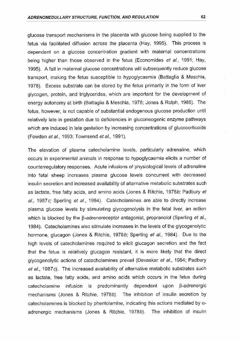
ADRENOMEDULLARY STRUCTURE, FUNCTION, AND REGULATION 62
glucose transporl mechanisms in the placenta with glucose being supplied to the
fetus via facilitated diffusion across the placenta (Hay, 1995). This process is
dependent on a glucose concentration gradient with maternal concentrations
being higher than those observed in the fetus (Economides ef al., 1991; Hay,
1995). A fall in maternal glucose concentrations will subsequently reduce glucose
transport, making the fetus susceptible to hypoglycaemia (Battaglia & Meschia,
1978). Excess substrate can be stored by the fetus primarily in the form of liver
glycogen, protein, and triglycerides, which are important for the development of
energy autonomy at birth (Battaglia & Meschia, 1978; Jones & Rolph, 1985). The
fetus, however, is not capable of substantial endogenous glucose production until
relatively late in gestation due to deficiencies in gluconeogenic enzyme pathways
which are induced in late gestation by increasing concentrations of glucocorticoids
(Fowden et al., 1993; Townsend et al., 1991).
The elevation of plasma catecholamine levels, parlicularly adrenaline, which
occurs in experimental animals in response to hypoglycaemia elicits a number of
counterregulatory responses. Acute infusions of physiological levels of adrenaline
into fetal sheep increases plasma glucose levels concurrent with decreased
insulin secretion and increased availability of alternative metabolic substrates such
as lactate, free fatty acids, and amino acids (Jones & Ritchie, 1978b; Padbury ef
al., 1987c; Sperling et al., 1984). Catecholamines are able to directly increase
plasma glucose levels by stimulating glycogenolysis in the fetal liver, an action
which is blocked by the B-adrenoreceptor antagonist, propranolol (Sperling et a/.,
1984). Catecholamines also stimulate increases in the levels of the glycogenolytic
hormone, glucagon (Jones & Ritchie, 1978b; Sperling et al., 1984). Due to the
high levels of catecholamines required to elicit glucagon secretion and the fact
that the fetus is relatively glucagon resistant, it is more likely that the direct
glycogenolytic actions of catecholamines prevail (Devaskar et al., 1984; Padbury
et al., 1987c). The increased availability of alternative metabolic substrates such
as lactate, free fatty acids, and amino acids which occurs in the fetus during
catecholamine infusion is predominantly dependent upon B-adrenergic
mechanisms (Jones & Ritchie, 1978b). The inhibition of insulin secretion by
catecholamines is blocked by phentolamine, indicating this actions mediated by cx-
adrenergic mechanisms (Jones & Ritchie, 1978b). The inhibition of insulin
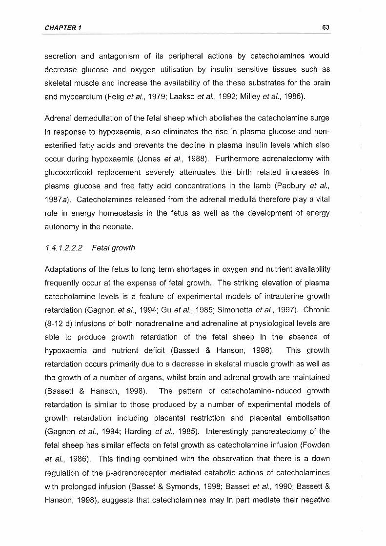
CHAPTER 1 63
secretion and antagonism of its peripheral actions by catecholamines would
decrease glucose and oxygen utilisation by insulin sensitive tissues such as
skeletal muscle and increase the availability of the these substrates for the brain
and myocardium (Felig et al., 1979; Laakso et al., 1992; Milley et al., 1986).
Adrenal demedullation of the fetal sheep which abolishes the catecholamine surge
in response to hypoxaemia, also eliminates the rise in plasma glucose and non-
esterified fatty acids and prevents the decline in plasma insulin levels which also
occur during hypoxaemia (Jones et al., 19BB). Furthermore adrenalectomy with
glucocorticoid replacement severely attenuates the birth related increases in
plasma glucose and free fatty acid concentrations in the lamb (Padbury et al.,
1987a). Catecholamines released from the adrenal medulla therefore play a vital
role in energy homeostasis in the fetus as well as the development of energy
autonomy in the neonate.
1.4.1.2.2.2 Fetal growth
Adaptations of the fetus to long term shortages in oxygen and nutrient availability
frequently occur at the expense of fetal growth. The striking elevation of plasma
catecholamine levels is a feature of experimental models of intrauterine growth
retardation (Gagnon et al., 1994; Gu ef al., 1985; Simonetta et al., 1997). Chronic
(8-12 d) infusions of both noradrenaline and adrenaline at physiological levels are
able to produce growth retardation of the fetal sheep in the absence of
hypoxaemia and nutrient deficit (Bassett & Hanson, 1998). This growth
retardation occurs primarily due to a decrease in skeletal muscle growth as well as
the growth of a number of organs, whilst brain and adrenal growth are maintained
(Bassett & Hanson, 1998). The pattern of catecholamine-induced growth
retardation is similar to those produced by a number of experimental models of
growth retardation including placental restriction and placental embolisation
(Gagnon et al., 1994; Harding et al., 1985). lnterestingly pancreatectomy of the
fetal sheep has similar effects on fetal growth as catecholamine infusion (Fowden
et al., 1986). This finding combined with the observation that there is a down
regulation of the B-adrenoreceptor mediated catabolic actions of catecholamines
with prolonged infusion (Basset & Symonds, 1998; Basset et al., 1990; Bassett &
Hanson, 1998), suggests that catecholamines may in part mediate their negative
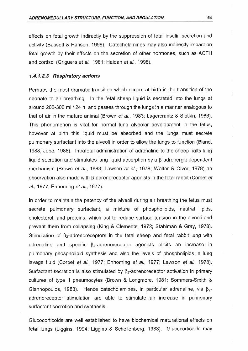
ADRENOMEDULLARY STRUCTURE, FUNCTION, AND REGULATION 64
effects on fetal growth indirectly by the suppression of fetal insulin secretion and
activity (Bassett & Hanson, 1998). Catecholamines may also indirectly impact on
fetal growth by their effects on the secretion of other hormones, such as ACTH
and cortisol (Griguere et al., 1981 ; Haidan et al., 1998).
1.4.1.2.3 Respiratory actions
Perhaps the most dramatic transition which occurs at birth is the transition of the
neonate to air breathing. ln the fetal sheep liquid is secreted into the lungs at
around 200-300 ml I 24 h and passes through the lungs in a manner analogous to
that of air in the mature animal (Brown et al., 1983; Lagercrantz & Slotkin, 1986).
This phenomenon is vital for normal lung alveolar development in the fetus,
however at birth this liquid must be absorbed and the lungs must secrete
pulmonary surfactant into the alveoli in order to allow the lungs to function (Bland,
19BB; Jobe, 1988). lntrafetal administration of adrenaline to the sheep halts lung
liquid secretion and stimulates lung liquid absorption by a B-adrenergic dependent
mechanism (Brown et al., 1983; Lawson et al., 1978; Walter & Olver, 1978) an
observation also made with p-adrenoreceptor agonists in the fetal rabbit (Corbet ef
al., 1977; Enhorning et al., 1977).
ln order to maintain the patency of the alveoli during air breathing the fetus must
secrete pulmonary surfactant, a mixture of phospholipids, neutral lipids,
cholesterol, and proteins, which act to reduce surface tension in the alveoli and
prevent them from collapsing (King & Clements, 1972; Stahlman & Gray, 1978).
Stimulation of B2-adrenoreceptors in the fetal sheep and fetal rabbit lung with
adrenaline and specific Þz-adrenoreceptor agonists elicits an increase in
pulmonary phospholipid synthesis and also the levels of phospholipids in lung
lavage fluid (Corbet ef al., 1977; Enhorning ef al., 1977 Lawson et al., 1978).
Sudactant secretion is also stimulated by B2-adrenoreceptor activation in primary
cultures of type ll pneumocytes (Brown & Longmore, 1981; Sommers-Smith &
Giannopoulos, 1983). Hence catecholamines, in particular adrenaline, via B2-
adrenoreceptor stimulation are able to stimulate an increase in pulmonary
surfactant secretion and synthesis.
Glucocorticoids are well established to have biochemical maturational effects on
fetal lungs (Liggins, 1994; Liggins & Schellenberg, 1988). Glucocorticoids may
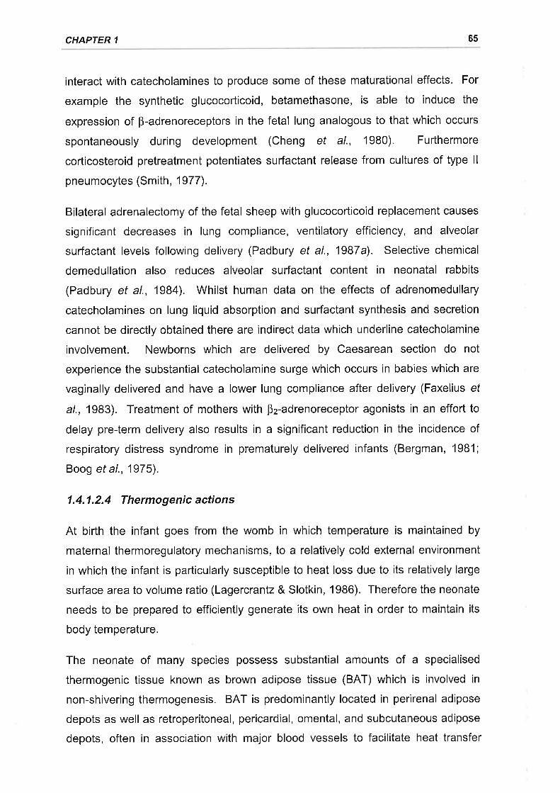
CHAPTER 1 65
interact with catecholamines to produce some of these maturational effects. For
example the synthetic glucocorticoid, betamethasone, is able to induce the
expression of B-adrenoreceptors in the fetal lung analogous to that which occurs
spontaneously during development (Cheng et al., 1980). Furthermore
corticosteroid pretreatment potentiates surfactant release from cultures of type ll
pneumocytes (Smith, 1977).
Bilateral adrenalectomy of the fetal sheep with glucocorticoid replacement causes
significant decreases in lung compliance, ventilatory efficiency, and alveolar
sudactant levels following delivery (Padbury et al., 1987a). Selective chemical
demedullation also reduces alveolar surfactant content in neonatal rabbits
(Padbury et al., 1984). Whilst human data on the effects of adrenomedullary
catecholamines on lung liquid absorption and surfactant synthesis and secretion
cannot be directly obtained there are indirect data which underline catecholamine
involvement. Newborns which are delivered by Caesarean section do not
experience the substantial catecholamine surge which occurs in babies which are
vaginally delivered and have a lower lung compliance after delivery (Faxelius ef
a/., 1983). Treatment of mothers with Þz-adrenoreceptor agonists in an effort to
delay pre-term delivery also results in a significant reduction in the incidence of
respiratory distress syndrome in prematurely delivered infants (Bergman, 1981;
Boog et al., 1975).
1.4.1.2.4 Thermogenic actions
At birth the infant goes from the womb in which temperature is maintained by
maternal thermoregulatory mechanisms, to a relatively cold external environment
in which the infant is particularly susceptible to heat loss due to its relatively large
sufface area to volume ratio (Lagercranlz & Slotkin, 1986). Therefore the neonate
needs to be prepared to efficiently generate its own heat in order to maintain its
body temperature.
The neonate of many species possess substantial amounts of a specialised
thermogenic tissue known as brown adipose tissue (BAT) which is involved in
non-shivering thermogenesis. BAT is predominantly located in perirenal adipose
depots as well as retroperitoneal, pericardial, omental, and subcutaneous adipose
depots, often in association with major blood vessels to facilitate heat transfer
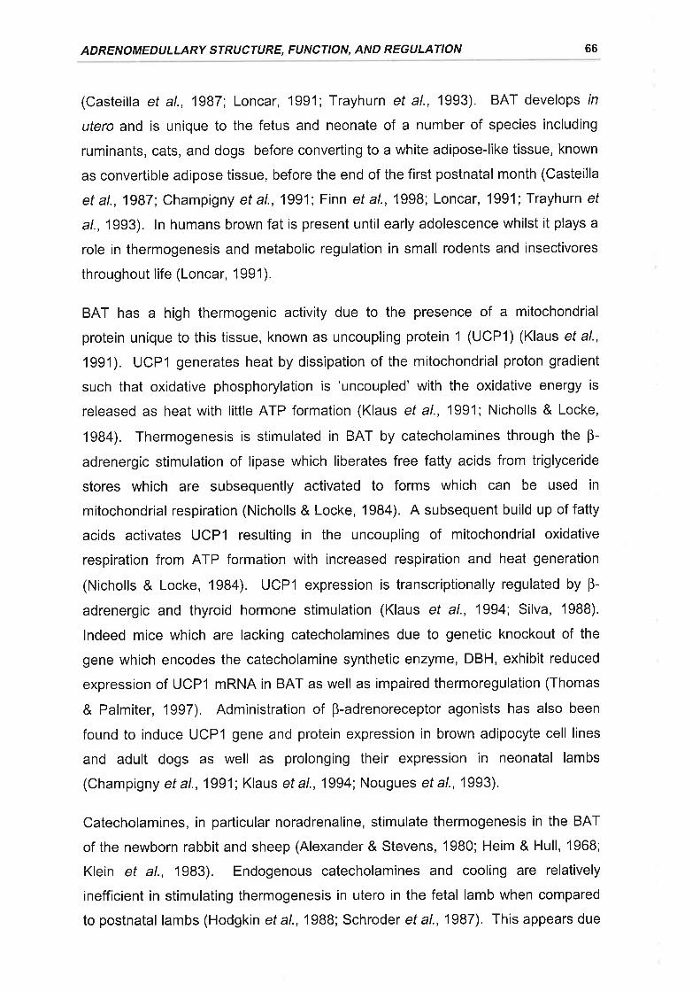
ADRENOMEDULLARY STRUCTURE, FUNCTION, AND REGULATION 66
(Casteilla et al., 1987; Loncar, 1991; Trayhurn et al., 1993). BAT develops tn
utero and is unique to the fetus and neonate of a number of species including
ruminants, cats, and dogs before converting to a white adipose-like tissue, known
as convertible adipose tissue, before the end of the first postnatal month (Casteilla
et al., 1987; Champigny et al., 1991; Finn ef a/., 1998; Loncar, 1991; Trayhurn ef
a/., 1993). ln humans brown fat is present until early adolescence whilst it plays a
role in thermogenesis and metabolic regulation in small rodents and insectivores
throughout life (Loncar, 1991).
BAT has a high thermogenic activity due to the presence of a mitochondrial
protein unique to this tissue, known as uncoupling protein 1 (UCP1) (Klaus et al.,
1991). UCPI generates heat by dissipation of the mitochondrial proton gradient
such that oxidative phosphorylation is 'uncoupled' with the oxidative energy is
released as heat with little ATP formation (Klaus ef al., 1991; Nicholls & Locke,
1984). Thermogenesis is stimulated in BAT by catecholamines through the B-
adrenergic stimulation of lipase which liberates free fatty acids from triglyceride
stores which are subsequently activated to forms which can be used in
mitochondrial respiration (Nicholls & Locke, 1984). A subsequent build up of fatty
acids activates UCPl resulting in the uncoupling of mitochondrial oxidative
respiration from ATP formation with increased respiration and heat generation
(Nicholls & Locke, 1984). UCPl expression is transcriptionally regulated by Þ-
adrenergic and thyroid hormone stimulation (Klaus et al., 1994; Silva, 1988).
lndeed mice which are lacking catecholamines due to genetic knockout of the
gene which encodes the catecholamine synthetic enzyme, DBH, exhibit reduced
expression of UCPl mRNA in BAT as well as impaired thermoregulation (Thomas
& Palmiter, 1997), Administration of B-adrenoreceptor agonists has also been
found to induce UCPl gene and protein expression in brown adipocyte cell lines
and adult dogs as well as prolonging their expression in neonatal lambs
(Champigny ef al., 1991; Klaus et al., 1994; Nougues et al., 1993).
Catecholamines, in particular noradrenaline, stimulate thermogenesis in the BAT
of the newborn rabbit and sheep (Alexander& Stevens, 1980; Heim & Hull, 1968;
Klein et al., 1983). Endogenous catecholamines and cooling are relatively
inefficient in stimulating thermogenesis in utero in the fetal lamb when compared
to postnatal lambs (Hodgkin et a1.,1988; Schroder et a1.,1987). This appears due
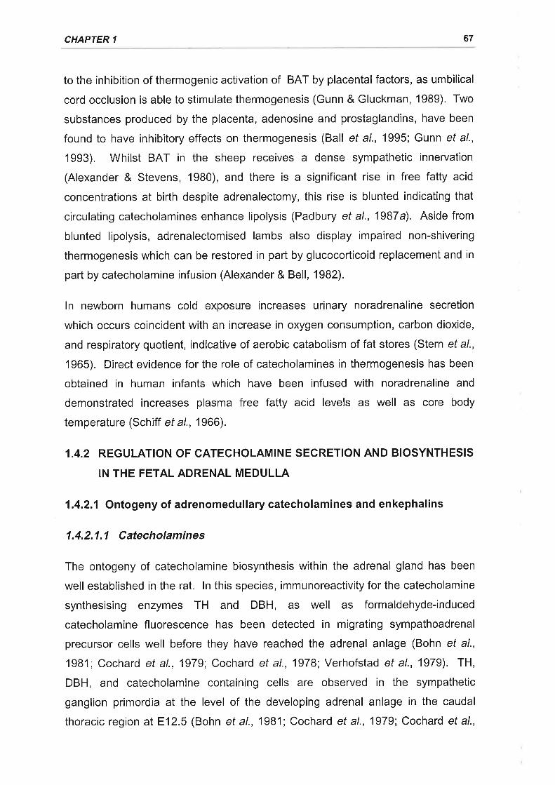
CHAPTER 1 67
to the inhibition of thermogenic activation of BAT by placental factors, as umbilical
cord occlusion is able to stimulate thermogenesis (Gunn & Gluckman, 1989). Two
substances produced by the placenta, adenosine and prostaglandins, have been
found to have inhibitory effects on thermogenesis (Ball ef al., 1995; Gunn et al.,
1993). Whilst BAT in the sheep receives a dense sympathetic innervation
(Alexander & Stevens, 1980), and there is a significant rise in free fatty acid
concentrations at birth despite adrenalectomy, this rise is blunted indicating that
circulating catecholamines enhance lipolysis (Padbury et al., 1987a). Aside from
blunted lipolysis, adrenalectomised lambs also display impaired non-shivering
thermogenesis which can be restored in part by glucocorticoid replacement and in
part by catecholamine infusion (Alexander & Bell, 1982).
ln newborn humans cold exposure increases urinary noradrenaline secretion
which occurs coincident with an increase in oxygen consumption, carbon dioxide,
and respiratory quotient, indicative of aerobic catabolism of fat stores (Stern et al.,
1965). Direct evidence for the role of catecholamines in thermogenesis has been
obtained in human infants which have been infused with noradrenaline and
demonstrated increases plasma free fatty acid levels as well as core body
temperature (Schiff et al., 1966).
1.4.2 REGULATION OF CATECHOLAMINE SECRETION AND BIOSYNTHESIS
IN THE FETAL ADRENAL MEDULLA
1.4.2.1 Ontogeny of adrenomedullary catecholamines and enkephalins
1.4.2.1.1 Catecholamines
The ontogeny of catecholamine biosynthesis within the adrenal gland has been
well established in the rat. ln this species, immunoreactivity for the catecholamine
synthesising enzymes TH and DBH, as well as formaldehyde-induced
catecholamine fluorescence has been detected in migrating sympathoadrenal
precursor cells well before they have reached the adrenal anlage (Bohn et al.,
'1981; Cochard et al., 1979; Cochard et al., 1978; Verhofstad et al., 1979). TH,
DBH, and catecholamine containing cells are observed in the sympathetic
ganglion primordia at the level of the developing adrenal anlage in the caudal
thoracic region at E12.5 (Bohn et al., 1981; Cochard et al., 1979; Cochard et al.,
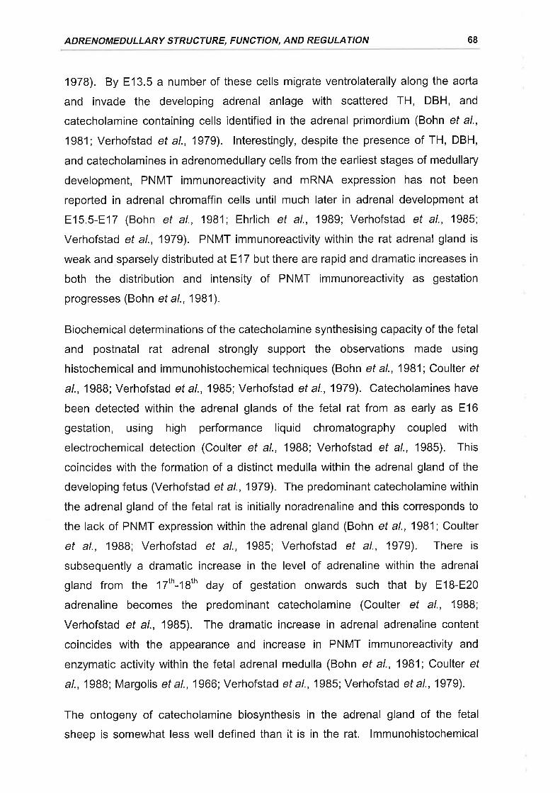
ADRENOMEDULLARY STRUCTURE, FUNCTION, AND REGULATION 68
1978). By E13.5 a number of these cells migrate ventrolaterally along the aorta
and invade the developing adrenal anlage with scattered TH, DBH, and
catecholamine containing cells identified in the adrenal primordium (Bohn et al.,
1981; Verhofstad et al., 1979). lnterestingly, despite the presence of TH, DBH,
and catecholamines in adrenomedullary cells from the earliest stages of medullary
development, PNMT immunoreactivity and mRNA expression has not been
reported in adrenal chromaffin cells until much later in adrenal development at
E15.5-E17 (Bohn et al., 1981; Ehrlich et al., 1989; Verhofstad et al., 1985;
Verhofstad et al., 1979). PNMT immunoreactivity within the rat adrenal gland is
weak and sparsely distributed at E17 but there are rapid and dramatic increases in
both the distribution and intensity of PNMT immunoreactivity as gestation
progresses (Bohn et a\.,1981).
Biochemical determinations of the catecholamine synthesising capacity of the fetal
and postnatal rat adrenal strongly support the observations made using
histochemical and immunohistochemical techniques (Bohn et al., 1981; Coulter ef
al., 1988; Verhofstad et a|.,1985; Verhofstad et al., 1979). Catecholamines have
been detected within the adrenal glands of the fetal rat from as early as E16
gestation, using high performance liquid chromatography coupled with
electrochemical detection (Coulter et al., 1988; Verhofstad et al., 1985). This
coincides with the formation of a distinct medulla within the adrenal gland of the
developing fetus (Verhofstad et al., 1979). The predominant catecholamine within
the adrenal gland of the fetal rat is initially noradrenaline and this corresponds to
the lack of PNMT expression within the adrenal gland (Bohn et al., 1981;Coulter
et al., 1988; Verhofstad et al., 1985; Verhofstad et al., 1979). There is
subsequently a dramatic increase in the level of adrenaline within the adrenal
gland from the 17th-18th day of gestation onwards such that by E18-E2O
adrenaline becomes the predominant catecholamine (Coulter et al., 1988;
Verhofstad et al., 1985). The dramatic increase in adrenal adrenaline content
coincides with the appearance and increase in PNMT immunoreactivity and
enzymatic activity within the fetal adrenal medulla (Bohn et al., 1981; Coulter ef
al., 1988; Margolis et a\.,1966; Verhofstad et a\.,1985; Verhofstad et a\.,1979).
The ontogeny of catecholamine biosynthesis in the adrenal gland of the fetal
sheep is somewhat less well defined than it is in the rat. lmmunohistochemical
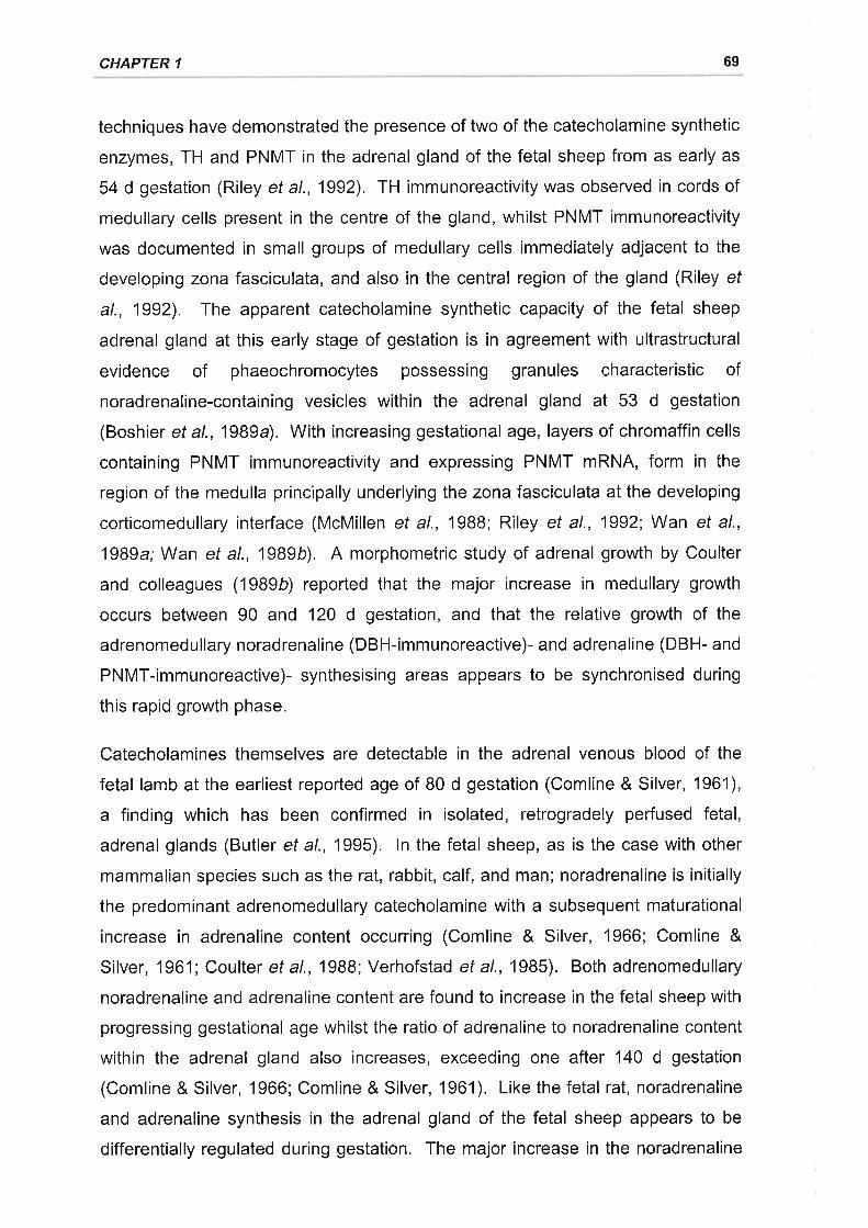
CHAPTER 1 69
techniques have demonstrated the presence of two of the catecholamine synthetic
enzymes, TH and PNMT in the adrenal gland of the fetal sheep from as early as
54 d gestation (Riley et al., 1992). TH immunoreactivity was observed in cords of
medullary cells present in the centre of the gland, whilst PNMT immunoreactivity
was documented in small groups of medullary cells immediately adjacent to the
developing zona fasciculata, and also in the central region of the gland (Riley ef
al., 1992). The apparent catecholamine synthetic capacity of the fetal sheep
adrenal gland at this early stage of gestation is in agreement with ultrastructural
evidence of phaeochromocytes possessing granules characteristic of
noradrenaline-containing vesicles within the adrenal gland at 53 d gestation
(Boshier et al.,19B9a). With increasing gestational age, layers of chromaffin cells
containing PNMT immunoreactivity and expressing PNMT mRNA, form in the
region of the medulla principally underlying the zona fasciculata at the developing
corticomedullary interface (McMillen et al., 19BB; Riley et al., 1992;Wan et al.,
1989a; Wan ef al., 1989b). A morphometric study of adrenal growth by Coulter
and colleagues ('1989b) reported that the major increase in medullary growth
occurs between 90 and 120 d gestation, and that the relative growth of the
adrenomedullary noradrenaline (DBH-immunoreactive)- and adrenaline (DBH- and
PNMT-immunoreactive)- synthesising areas appears to be synchronised during
this rapid growth phase.
Catecholamines themselves are detectable in the adrenal venous blood of the
fetal lamb at the earliest reported age of 80 d gestation (Comline & Silver, 1961),
a finding which has been confirmed in isolated, retrogradely perfused fetal,
adrenal glands (Butler et al., 1995). ln the fetal sheep, as is the case with other
mammalian species such as the rat, rabbit, calf, and man; noradrenaline is initially
the predominant adrenomedullary catecholamine with a subsequent maturational
increase in adrenaline content occurring (Comline & Silver, 1966; Comline &
Silver, 1961; Coulter et al., 1988; Verhofstad et al., 1985). Both adrenomedullary
noradrenaline and adrenaline content are found to increase in the fetal sheep with
progressing gestational age whilst the ratio of adrenaline to noradrenaline content
within the adrenal gland also increases, exceeding one after 140 d gestation
(Comline & Silver, 1966; Comline & Silver, 1961). Like the fetal rat, noradrenaline
and adrenaline synthesis in the adrenal gland of the fetal sheep appears to be
differentially regulated during gestation. The major increase in the noradrenaline
ADRENOMEDULLARY STRUCTURE, FUNCTION, AND REGULATION 70
content of the fetal sheep adrenal occurs between 80 and 110 d gestation (ie.
during the major phase of adrenomedullary growth) whereas the predominant
increase in the adrenaline content of the adrenal occurs somewhat later, after
130d gestation (Boshier & Holloway, 1989b; Comline & Silver, 1961;Coulter ef
a\.,1989b).
1.4.2,1.2 Enkephalins
Enk-containing peptides have been identified within the prenatal adrenal medulla
of several species including human, primate, sheep, and rat (Dunlap et al., 1985;
Henion & Landis, 1990; Wilburn et al., 1986). The ontogeny of Enk peptide
expression has been characterised most extensively in the rat (Henion, 1992;
Henion & Landis, 1990; Holgert et al., 1995a; Holgert et al., 1994; LaGamma ef
a/., 1989) and sheep (Coulter et al., 1990a; Dunlap et al., 1985; McMillen et al.,
1988; Wan ef a\.,1989a). Both immunohistochemical and biochemical techniques
have established that Enk peptide expression appears in the fetal rat adrenal
medulla at E16 (Henion & Landis, 1990; LaGamma et al., 1989). ln the fetal
sheep Enk-containing peptides have been reported within the adrenal medulla
from 68-80 d, the earliest gestational ages studied (Coulter et al., 1990a; Dunlap
et a\.,1985; McMillen et a|.,1988).
The appearance of Enk-containing peptides within the chromaffin cells of the fetal
rat adrenal at E16 coincides with the appearance of PNMT, and it is found that
Enk expression is colocalised within PNMT expressing chromaffin cells (Henion &
Landis, 1990). There is a subsequent dramatic increase in Enk peptide
expression within the adrenal medulla between E16 and E20, which is followed by
a marked decline in the first postnatal week of life (Henion & Landis, 1990; Holgert
et al., 1994). ln the fetal sheep, Enk peptide and PEnk A mRNA expression is
initially distributed homogeneously throughout the adrenal medulla from 80-110 d
gestation (McMillen et al., 1988; Wan ef al., 1989a). After 110 d, however, Enk-
containing peptides and PEnk A mRNA expression is predominantly confined to
the peripheral adrenaline-containing cells of the fetal sheep adrenal medulla
(McMillen et al., 1988; Wan ef al., 1989a). Along with the alterations in Enk-
peptide localisation there is also a large increase in the total enkephalin content of
the fetal adrenal between 70 and 120 d gestation which tends to decrease in later
CHAPTER 1 71
gestation (Coulter et al., 1990a). The ratio of high molecularweightforms to low
molecular weight forms significantly decreases between 70-120 d gestation,
indicative of increased precursor molecule processing, and this decrease is
maintained over gestation (Coulter et a\.,1990a).
The appearance of Enk peptide expression in adrenal chromaffin cells of the fetal
rat only after they have entered the developing adrenal anlage suggests that
adrenocortical glucocorticoids may regulate Enk peptide expression in the
developing adrenal (Henion & Landis, 1990). Glucocorticoids appear to be
involved in the regulation of Enk peptide synthesis in mature adrenal chromaffin
cells (LaGamma & Alder, 1987; Naranjo et al., 1986; Stachowiak et al., 1988b;
Yoburn et al., 1987). lt has been found that the synthetic glucocorticoid,
dexamethasone, increases Leu-Enk levels in cultures of embryonic rat adrenal
chromaffin cells (Henion, 1992). ln the sheep, fetal hypophysectomy at 105 and
118 d reduced both plasma cortisol and total adrenal Enk content in the 140-141 d
fetal sheep (Coulter et al., 1992; Simonetta et al., 1996b). Furthermore, ACTH
replacement in late gestation following fetal HPX significantly increased both
plasma cortisol and total adrenal Enk content in the fetal sheep above the levels
observed in saline infused fetuses (Coulter et a\.,1992).
A number of the changes in the localisation, synthesis, and post-translational
processing of adrenal Enk-containing peptides occur in parallel with the
development of functional splanchnic innervation (McMillen et al., 1997). ln the
rat, a number of factors associated with cholinergic innervation are found to
increase the levels of Enk-peptides in chromaffin cells. These factors include
depolarisation (Henion, '1992) and cholinergic receptor stimulation (LaGamma ef
a/., 1989). Paradoxically, there is a decrease in adrenal Enk-containing peptide
expression in vivo with the onset of splanchnic innervation to the gland (Henion &
Landis, 1990; Holgert et al., 1994). This decrease coincides with the appearance
of Enk-peptide containing cholinergic nerye fibres within the adrenal medulla
(Henion & Landis, 1990; Holgert et al., 1994; Holgert et al., 1996b). Adrenal
denervation by the transection of the preganglionic cholinergic splanchnic nerve or
by the administration of acetylcholinesterase antibodies results in an upregulation
of enkephalin peptide expression in the chromaffin cells of the medulla (Henion,
1992; Holgert et al., 1996b; Lewis et al., 1981). Administration of the opioid
ADRENOMEDULLARY STRUCTURE, FUNCTION, AND REGULATION 72
receptor antagonist, naloxone, mimics the effects of denervation whilst the
administration of synthetic enkephalin analogues mimics the effect of innervation
(Henion, 1992). Hence the developmental suppression of Enk expression in the
adrenal gland of the rat with the onset of functional innervation appears to be due
to the actions of opioid peptides within the splanchnic nerve.
A similar suppressive effect of adrenal innervation on Enk-containing peptide
expression during development may also occur in the fetal sheep adrenal gland.
lnterestingly, HPX of the fetal sheep results in the continued expression of Enk-
immunoreactivity in the central noradrenaline-containing region of the adrenal
gland in late gestation (Coulter et al., 1991; Coulter et a\.,1989b). Administration
of ACTH to HPX fetuses whilst increasing adrenal Enk-containing peptide content,
is unable to suppress this central medullary expression which normally disappears
at around the time of innervation of the gland, with expression restricted to the
outer adrenaline-containing region of the medulla (Coulter et al., 1991; McMillen ef
a/., 1988). HPXof thefetal sheep results in a functional athyroidism (Mesiano ef
al., 1987). Thyroid hormones play a crucial role in the development of functional
splanchnic innervation to the adrenal (Lau ef a/., 19BB). Simonetta et al. (1996c)
reported that continuous thyroxine replacement after fetal hypophysectomy
resulted in a relative increase in the density of enkephalin staining in the
peripheral adrenaline-containing region and a decrease in the central
noradrenaline-containing region of the late gestation adrenal. This effect may be
either a direct effect of thyroid hormones or more likely an indirect effect mediated
by their actions on the development of splanchnic innervation (Lau et al., 1988;
Simonetta et al., 1996c). Thyroxine replacement did not, however, restore either
the total Enk content or free Enk content in the adrenals of HPX fetal sheep
(Simonetta et al., 1996b).
1.4.2.2 Neuropeptide regulation of adrenomedullary catecholamine
secretion
There is currently very little information available on the role of adrenomedullary
neuropeptides in the regulation of catecholamine secretion during stress in the
fetus. The best characterised neuropeptides in this regard are the opioid peptides
(McMillen et al., 1997). Adrenalectomy does not alter the basal levels of Enk-
CHAPTER 1 73
containing peptides in the circulation of the fetal sheep, indicating that under basal
conditions the adrenal medulla is not a major source of circulating Enk-containing
peptides (Simonetta et al., 1993). During fetal hypoxia and asphyxia there are
increases in the levels of circulating Enk-containing peptides (Coulter et al.,
1990b; Jones, 1992; Martinez et al., 1990). These Enk-containing peptides may
be derived from both the adrenal medulla and sympathetic nervous system as
both adrenalectomy and chemical sympathectomy substantially blunt the
response to hypoxia (Jones, 1992). ln addition, moderate fetal hypoxaemia over
48 h has also been reported to increase PEnk A mRNA expression within the fetal
sheep adrenal gland (Fraser et al., 1997) indicating an increased synthesis of
adrenal Enk-containing peptides in response to hypoxia.
The non-selective opioid receptor antagonist, naloxone, when administered to
fetal sheep augments the elevations in plasma catecholamine levels which occur
in response to both hypoxia and delivery (Martinez et a/., 1988; Padbury et al.,
1987 b). The adrenal medulla is the predominant source of circulating
catecholamines in response to these stressors (Jones et al., 1988; Padbury et al.,
1987 a), which indicates that intra-adrenal opioid peptides co-secreted with
catecholamines during fetal stress act to modulate catecholamine secretion by
suppressing their release (Martinez et al., 1988; Padbury et al., 1987 b; Padbury ef
a/., 1989). Furthermore, naloxone augments the increases in heart rate, blood
pressure, and blood flow redistribution which occur in response to asphyxia and
delivery in the fetal sheep (Espinoza et al., 1989; Padbury et al., 1987b). These
findings indicate that at the physiological level opioid peptides act to oppose the
actions of catecholamines on the cardiovascular system, possibly by central
mechanisms or by peripheral mechanisms via modulation of adrenomedullary
catecholamine secretion or antagonism of their peripheral actions (Espinoza et al.,
1989; Padbury et al., 1987 b; Padbury et al., 1989) .
1 .4.2.3 Non-neu rogen ic catecholami ne secretion
The adrenal medulla of the fetus and in some species, the neonate, possess the
unique capacity to secrete catecholamines in direct response to hypoxia prior to
the development of functional splanchnic innervation by a direct non-neurogenic
mechanism (McMillen et al., 1997).
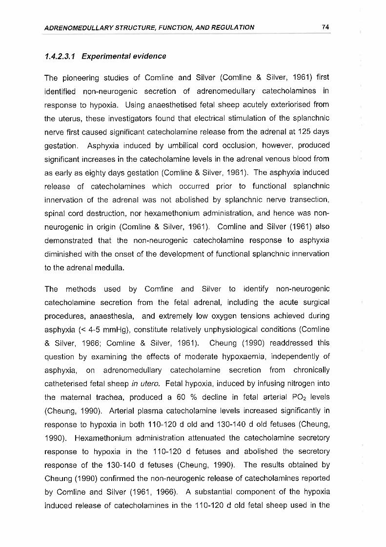
ADRENOMEDULLARY STRUCTURE, FUNCTION, AND REGULATION 74
1.4.2.3.1 Experimental evidence
The pioneering studies of Comline and Silver (Comline & Silver, 1961) first
identified non-neurogenic secretion of adrenomedullary catecholamines in
response to hypoxia. Using anaesthetised fetal sheep acutely exteriorised from
the uterus, these investigators found that electrical stimulation of the splanchnic
nerve first caused significant catecholamine release from the adrenal at 125 days
gestation. Asphyxia induced by umbilical cord occlusion, however, produced
significant increases in the catecholamine levels in the adrenal venous blood from
as early as eighty days gestation (Comline & Silver, 1961). The asphyxia induced
release of catecholamines which occurred prior to functional splanchnic
innervation of the adrenal was not abolished by splanchnic nerve transection,
spinal cord destruction, nor hexamethonium administration, and hence was non-
neurogenic in origin (Comline & Silver, 1961). Comline and Silver (1961) also
demonstrated that the non-neurogenic catecholamine response to asphyxia
diminished with the onset of the development of functional splanchnic innervation
to the adrenal medulla.
The methods used by Comline and Silver to identify non-neurogenic
catecholamine secretion from the fetal adrenal, including the acute surgical
procedures, anaesthesia, and extremely low oxygen tensions achieved during
asphyxia (. 4-5 mmHg), constitute relatively unphysiological conditions (Comline
& Silver, 1966; Comline & Silver, 1961). Cheung (1990) readdressed this
question by examining the effects of moderate hypoxaemia, independently of
asphyxia, on adrenomedullary catecholamine secretion from chronically
catheterised fetal sheep in utero. Fetal hypoxia, induced by infusing nitrogen into
the maternal trachea, produced a 60 % decline in fetal arterial POz levels
(Cheung, 1990), Arterial plasma catecholamine levels increased significantly in
response to hypoxia in both 110-120 d old and 130-140 d old fetuses (Cheung,
1990). Hexamethonium administration attenuated the catecholamine secretory
response to hypoxia in the 110-120 d fetuses and abolished the secretory
response of the 130-140 d fetuses (Cheung, 1990). The results obtained by
Cheung (1990) confirmed the non-neurogenic release of catecholamines reported
by Comline and Silver (1961, 1966). A substantial component of the hypoxia
induced release of catecholamines in the 110-120 d old fetal sheep used in the
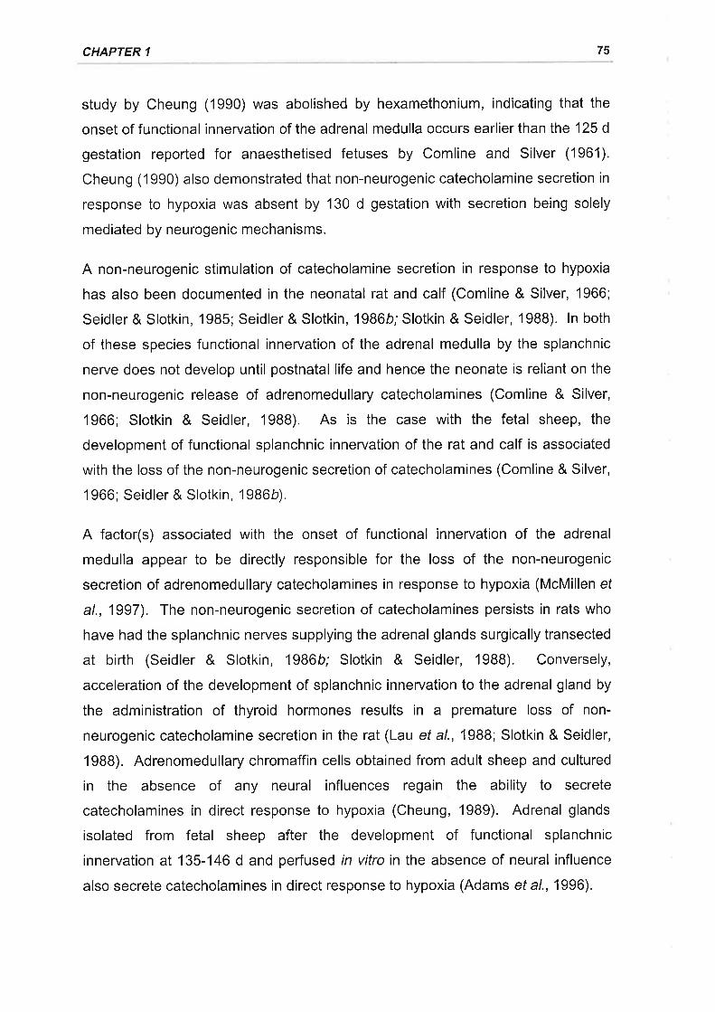
CHAPTER 1 75
study by Cheung (1990) was abolished by hexamethonium, indicating that the
onset of functional innervation of the adrenal medulla occurs earlier than the 125 d
gestation reported for anaesthetised fetuses by Comline and Silver (1961).
Cheung (1990) also demonstrated that non-neurogenic catecholamine secretion in
response to hypoxia was absent by 130 d gestation with secretion being solely
mediated by neurogenic mechanisms.
A non-neurogenic stimulation of catecholamine secretion in response to hypoxia
has also been documented in the neonatal rat and calf (Comline & Silver, 1966;
Seidler & Slotkin, 1985; Seidler & Slotkin, 1986b; Slotkin & Seidler, 1988). ln both
of these species functional innervation of the adrenal medulla by the splanchnic
nerve does not develop until postnatal life and hence the neonate is reliant on the
non-neurogenic release of adrenomedullary catecholamines (Comline & Silver,
1966; Slotkin & Seidler, 1988). As is the case with the fetal sheep, the
development of functional splanchnic innervation of the rat and calf is associated
with the loss of the non-neurogenic secretion of catecholamines (Comline & Silver,
1966; Seidler & Slotkin, 1986b).
A factor(s) associated with the onset of functional innervation of the adrenal
medulla appear to be directly responsible for the loss of the non-neurogenic
secretion of adrenomedullary catecholamines in response to hypoxia (McMillen ef
al., 1997). The non-neurogenic secretion of catecholamines persists in rats who
have had the splanchnic nerves supplying the adrenal glands surgically transected
at birth (Seidler & Slotkin, 1986b; Slotkin & Seidler, 1988). Conversely,
acceleration of the development of splanchnic innervation to the adrenal gland by
the administration of thyroid hormones results in a premature loss of non-
neurogenic catecholamine secretion in the rat (Lau et al., 1988; Slotkin & Seidler,
1988). Adrenomedullary chromaffin cells obtained from adult sheep and cultured
in the absence of any neural influences regain the ability to secrete
catecholamines in direct response to hypoxia (Cheung, 19Bg). Adrenal glands
isolated from fetal sheep after the development of functional splanchnic
innervation at 135-146 d and perfused in vitro in the absence of neural influence
also secrete catecholamines in direct response to hypoxia (Adams et al., 1996).
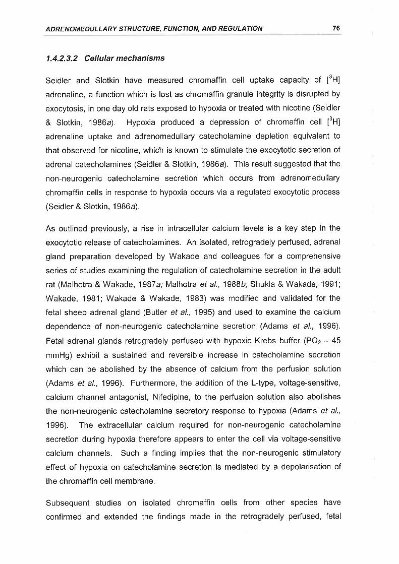
ADRENOMEDULLARY STRUCTURE, FUNCTION, AND REGULATION 76
1,4.2,3.2 Cellular mechanisms
Seidler and Slotkin have measured chromaffin cell uptake capacity of ¡3H1
adrenaline, a function which is lost as chromaffin granule integrity is disrupted by
exocytosis, in one day old rats exposed to hypoxia or treated with nicotine (Seidler
& Slotkin, 1986a). Hypoxia produced a depression of chromaffin cell [tH]
adrenaline uptake and adrenomedullary catecholamine depletion equivalent to
that observed for nicotine, which is known to stimulate the exocytotic secretion of
adrenal catecholamines (Seidler & Slotkin, 1986a). This result suggested that the
non-neurogenic catecholamine secretion which occurs from adrenomedullary
chromaffin cells in response to hypoxia occurs via a regulated exocytotic process
(Seidler & Slotkin, 1986a).
As outlined previously, a rise in intracellular calcium levels is a key step in the
exocytotic release of catecholamines. An isolated, retrogradely pedused, adrenal
gland preparation developed by Wakade and colleagues for a comprehensive
series of studies examining the regulation of catecholamine secretion in the adult
rat (Malhotra & Wakade, 1987 a; Malhotra et al., 19BBb; Shukla & Wakade, 1991 ;
Wakade, 198'1; Wakade & Wakade, 1983) was modified and validated for the
fetal sheep adrenal gland (Butler et al., 1995) and used to examine the calcium
dependence of non-neurogenic catecholamine secretion (Adams et al., 1996).
Fetal adrenal glands retrogradely perfused with hypoxic Krebs buffer (POz - 45
mmHg) exhibit a sustained and reversible increase in catecholamine secretion
which can be abolished by the absence of calcium from the perfusion solution
(Adams et al., 1996). Furthermore, the addition of the L-type, voltage-sensitive,
calcium channel antagonist, Nifedipine, to the perfusion solution also abolishes
the non-neurogenic catecholamine secretory response to hypoxia (Adams et al.,
1996). The extracellular calcium required for non-neurogenic catecholamine
secretion during hypoxia therefore appears to enter the cell via voltage-sensitive
calcium channels. Such a finding implies that the non-neurogenic stimulatory
effect of hypoxia on catecholamine secretion is mediated by a depolarisation of
the chromaffin cell membrane.
Subsequent studies on isolated chromaffin cells from other species have
confirmed and extended the findings made in the retrogradely perfused, fetal
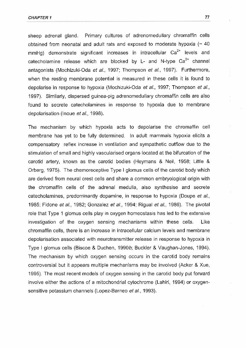
CHAPTER 1 77
sheep adrenal gland. Primary cultures of adrenomedullary chromaffin cells
obtained from neonatal and adult rats and exposed to moderate hypoxia (- 40
mmHg) demonstrate significant increases in intracellular Ca2* levels and
catecholamine release which are blocked by L- and N-type Ca2* channel
antagonists (Mochizuki-Oda et al., 1997; Thompson et al., 1997). Furthermore,
when the resting membrane potential is measured in these cells it is found to
depolarise in response to hypoxia (Mochizuki-Oda et al., 1997; Thompson et al.,
1997). Similarly, dispersed guinea-pig adrenomedullary chromaffin cells are also
found to secrete catecholamines in response to hypoxia due to membrane
depolarisation (lnoue et al., 1998).
The mechanism by which hypoxia acts to depolarise the chromaffin cell
membrane has yet to be fully determined. ln adult mammals hypoxia elicits a
compensatory reflex increase in ventilation and sympathetic outflow due to the
stimulation of small and highly vascularised organs located at the bifurcation of the
carotid artery, known as the carotid bodies (Heymans & Neil, 1958; Little &
Orberg, 1975). The chemoreceptive Type I glomus cells of the carotid body which
are derived from neural crest cells and share a common embryological origin with
the chromaffin cells of the adrenal medulla, also synthesise and secrete
catecholamines, predominantly dopamine, in response to hypoxia (Doupe et al.,
1985; Fidone et al., 1982; Gonzalez et al., 1994; Rigual et al., 1986). The pivotal
role that Type 1 glomus cells play in oxygen homeostasis has led to the extensive
investigation of the oxygen sensing mechanisms within these cells. Like
chromaffin cells, there is an increase in intracellular calcium levels and membrane
depolarisation associated with neurotransmitter release in response to hypoxia in
Type lglomus cells (Biscoe & Duchen, 1990b; Buckler & Vaughan-Jones, 1994).
The mechanism by which oxygen sensing occurs in the carotid body remains
controversial but it appears multiple mechanisms may be involved (Acker & Xue,
1995). The most recent models of oxygen sensing in the carotid body putforward
involve either the actions of a mitochondrial cytochrome (Lahiri, 1994) or oxygen-
sensitive potassium channels (Lopez-Barneo ef al., 1993).
ADRENOMEDULLARY STRUCTURE, FUNCTION, AND REGULATION 78
1.4.2.3.2.1 Mitochondrial oxygen sensing
The first model involves mitochondrial cytochrome aas which has a relatively low
affinity for oxygen (Acker & Xue, 1995). During hypoxia this cytochrome is unable
to reduce oxygen leading to a diminished energy state and an efflux of calcium
from mitochondrial stores, presumably through a mitochondrial Ca2*-ATPase, with
subsequent neurotransmitter release (Biscoe & Duchen, 1990a; Bunn & Poyton,
1996; Mulligan etal., 1981). Spectral analysis of the carotid body during anoxia
supports a functional role of the mitochondrial cytochrome aa3 (Acker & Xue,
1995). Furthermore inhibition of mitochondrial respiration by the administration of
cyanide or carbon monoxide are able to stimulate carotid body discharge (Biscoe
& Duchen, 1990a; Lahiri, 1994; Wilson et al., 1994). Photochemical action
spectrum reveal that the cytochrome ââs is the key mitochondrial heme protein
involved in the CO mediated increase in discharge (Lahiri, 1994; Wilson et al.,
1994). As mitochondrial POz is extremely low and subject to metabolic
fluctuations (Bunn & Poyton, 1996) it is unlikely that this mechanism is likely to
function under physiological conditions but more so during anoxia or extreme
hypoxia and also with cyanide and carbon monoxide poisoning. The release of
catecholamines which has been reported to occur from bovine and guinea pig
chromaffin cells independently of extracellular calcium during anoxia (ltoh ef a/.,
1994) and during metabolic inhibition with cyanide (Dry et al., 1991; lnoue et al.,
1998) is also probably due to the release of calcium from mitochondrial stores.
1.4.2.3.2.2 Oxygen-sensitive potassium channels
Oxygen sensitivity may also be mediated by ion conductance (Lopez-Barneo,
1996). Oxygen-sensitive potassium channels have been identified in the Oz
sensing type lglomus cells of the carotid body (Lopez-Barneo et al., 1988).
These cells display voltage-sensitive, outward potassium currents which are
reversibly inhibited by reductions in POz.(Lopez-Lopez et al., 1989). Single
channel analysis in excised membrane patches of Type I glomus cell membranes
reveal a specific type of calcium-independent, potassium channel is responsible
for the change in outward potassium current (Ganfornina & Lopez-Barneo, 1992).
These channels which are voltage dependent, possess a slope conductance of 20
pS, have relatively fast activation and inactivation kinetics and are transient
CHAPTER 1 79
voltage-dependent K* channels (Ganfornina & Lopez-Barneo, 1991). The Oz-
sensitive K* channels of the Type 1 glomus cells display a reduced opening
probability in response to hypoxia without any alteration in their conductance
(Ganfornina & Lopez-Barneo, 1992). This effect due to alterations in the kinetic
properties of the channel in response to reductions in oxygen tension therefore
leads to a reduction in outward potassium flux, membrane depolarisation
(Delpiano & Hescheler, 1989), enhanced calcium entry through voltage-sensitive
calcium channels (Buckler & Vaughan-Jones, 1994), and finally neurotransmitter
secretion (Fidone et al., 1982; Rigual et al., 1986). Oz-sensitive K* channels
appear to have a relatively ubiquitous distribution (Lopez-Barneo, 1994) and have
also been identified in a number of other tissues including pulmonary
neuroepithelial bodies (Youngson ef a/., 1993), pulmonary artery myocytes (Archer
et a\.,1996; Post ef at.,1992), and central neurones (Jiang & Haddad, 1994).
The extremely small size of the carotid body (= 1 x 10⁴ cells) make the
mechanisms of oxygen sensing difficult to study in this tissue (Beitner-Johnson ef
al., 1997). The immortalised rat PC12 cell line which is derived from an adrenal
medullary tumour shares morphological and phenotypical properties with Type I
glomus cells with both cell types synthesising and secreting the neurotransmitter
dopamine (Fidone & Gonzalez, 1986; Green & Tischler, 1987). Examination of
this cell line as a potential model for oxygen sensing has revealed an oxygen
dependent regulation of gene expression analogous to that observed in the
glomus cells (see following sections for further detail) (Beitner-Johnson et al.,
1997; Czyzyk-Krzeska, 1997). Furthermore, it was subsequently established that
PC12 cells also depolarise in response to hypoxia with a subsequent influx of
calcium through voltage-sensitive calcium channels stimulating the secretion of
dopamine (Beitner-Johnson et al., 1997; Zhu et a/., 1996). Patch clamp studies
also show that membrane depolarisation during hypoxia is due to the reduction of
an oxygen sensitive, outward potassium current (Zhu et al., 1996). The Oz-
sensitive K* channels responsible for this current appear similar to those reported
for the carotid body in that they are voltage dependent, calcium independent, and
possess a unitary conductance of 20 pS (Conforti & Millhorn, 1997). The Oz-
sensitive K* channels observed in PC12 cells however show somewhat slower
inactivation kinetics than those of the type I glomus cells and are more
ADRENOMEDULLARY STRUCTURE, FUNCTION, AND REGULATION 80
characteristic of delayed rectifiers (Conforti & Millhorn , 1997; Ganfornina & Lopez-
Barneo, 1991).
Subsequent studies seeking to elaborate on the mechanisms of non-neurogenic
catecholamine secretion from the adrenal medulla have also revealed the
presence of Oz-sensitive K* currents in the chromaffin cells of the adult rat,
neonatal rat, and the fetal sheep (Mochizuki-Oda et al., 1997; Rychkov et al.,
1998; Thompson et al., 1997). Oz-sensitive K* currents which display slow
inactivation characteristic of delayed rectifier type potassium channels and are
similar to those observed in PC12 cells were identified in both rat and fetal sheep
adrenal chromaffin cells (Mochizuki-Oda et al., 1997; Rychkov et al., 1998;
Thompson et al., 1997). However, the slow inactivating outward potassium
current observed in fetal sheep chromaffin cells was found to be a calcium-
dependent (Rychkov et al., 1998) whilst one study in the rat showed that it was
calcium-independent (Mochizuki-Oda et al., 1997) and the other failed to test the
calcium dependency (Thompson ef a/., 1997). A unique fast inactivating 02-
sensitive K* current which displays pharmacological and functional similarities to
an /a-type K* current was also identified in the chromaffin cells of the fetal sheep
(Rychkov et al., 1998). lnterestingly the delayed rectifier type and the /n type 02-
sensitive K* currents appeared separately in different chromaffin cell types
(Rychkov et al., 1998). The delayed rectifier type current was associated with
small capacitance chromaffin cells (cf. 4.8 pF) whilst the /n-type current was
associated with large capacitance cells (cf. 12.3 pF) (Rychkov ef a/., 1998). The
functional significance of the two populations of chromaffin cells possessing two
different types of Oz-sensitive K* current is not clear, although it is interesting to
note that there is a specific distribution of the current types to large high
capacitance and smaller low capacitance chromaffin cells which may reflect
adrenaline storing and noradrenaline storing chromaffin cells, respectively
(Rychkov et al., 1998). The non neurogenic release of catecholamines in
response to hypoxia though is characterised by a non-selective release of the
catecholamines noradrenaline and adrenaline indicating a similar mechanism is
operating in both cell types (Adams et a\.,1996; Slotkin & Seidler, 1988). lt is also
not clear whether the unique observation of the /a-type current in fetal sheep
adrenal chromaffin cells reflects a species difference or differences in cell
CHAPTER 1 8l
preparation and culture conditions which appear to influence the expression of this
current (Rychkov et al., 1998).
lnterestingly the study of Thompson et al. (1997) reported that there was a loss of
the Oz-sensitive K* current in the adrenomedullary chromaffin cells of the rat when
taken after the onset of functional innervation of the gland. This was interpreted
as a possible reason for the loss of the non-neurogenic response to hypoxia
following innervation of the adrenal gland. Mochizuki-Oda (1997) and co-workers,
however, reported the presence of the Oz-sensitive K* current in adult rats. Oz-
sensitive K* currents also persist in isolated adrenal chromaffin cells from the adult
sheep (G. Rychkov and M.L. Roberts, personal communication) as does non-
neurogenic secretion in response to hypoxia (Cheung, 19Bg). As stated above the
functional evidence suggests that it is a factor(s) transmitted via the splanchnic
nerve which is responsible for the loss of non-neurogenic catecholamine secretion
rather than a loss of potassium channels (Adams et al., 1996).
The mechanism by which Oz-sensitive K* channels sense oxygen availability is
uncertain, but appears to occur via membrane delimited mechanisms as
reversible inhibition of potassium currents by hypoxia occurs in excised membrane
patches of glomus cells and central neurones (Ganfornina & Lopez-Barneo, 1992;
Ganfornina & Lopez-Barneo, 1991;Jiang & Haddad,1994). There is evidence to
suggest that the redox status of specific amino acid residues within the protein
structure of potassium channels alters their conformation and activity (Lopez-
Barneo, 1994). Specifically the formation of disulfide linkages between cystine
residues in the reduced state has been found to reduce the activity of a cloned
neuronal fast inactivating K* channel, RCK4 (Ruppersberg et al., 1991). Aputative membrane bound, multisubunit cytochrome þlike oxidase is speculated
to function as the oxygen sensor, influencing protein redox status by the
production of hydrogen peroxide (see below, Acker & Xue, 1995; Bunn & Poyton,
1996; Czyzyk-Krzeska, 1997; Kummer & Acker, 1995). An alternative proposal is
the formation of co-ordination complexes between oxygen and amino acids via
metal containing sites in the main channel subunits, regulating channel
conformation (Lopez-Barneo, 1996). The presence of oxygen sensitive potassium
channels in adrenomedullary chromaffin cells could confer upon them the oxygen
sensing capacity required for non-neurogenic catecholamine secretion.
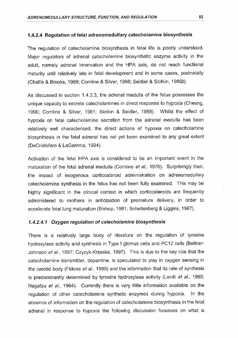
ADRENOMEDULLARY STRUCTURE, FUNCTION, AND REGULATION
1 .4.2.4 Reg u lation of fetal ad renomed u I lary catecholami ne biosynthesis
The regulation of catecholamine biosynthesis in fetal life is poorly understood.
Major regulators of adrenal catecholamine biosynthetic enzyme activity in the
adult, namely adrenal innervation and the HPA axis, do not reach functional
maturity until relatively late in fetal development and in some cases, postnatally
(Challis & Brooks, 1989; Comline & Silver, 1966; Seidler & Slotkin, 1986b).
As discussed in section 1.4.2.3, the adrenal medulla of the fetus possesses the
unique capacity to secrete catecholamines in direct response to hypoxia (Cheung,
'1990; Comline & Silver, 1961; Slotkin & Seidler, 1988). Whilst the effect of
hypoxia on fetal catecholamine secretion from the adrenal medulla has been
relatively well characterised, the direct actions of hypoxia on catecholamine
biosynthesis in the fetal adrenal has not yet been examined to any great extent
(DeCristofaro & LaGamma, 1994).
Activation of the fetal HPA axis is considered to be an important event in the
maturation of the fetal adrenal medulla (Comline et al., 1970). Surprisingly then,
the impact of exogenous corticosteroid administration on adrenomedullary
catecholamine synthesis in the fetus has not been fully examined. This may be
highly significant in the clinical context in which corticosteroids are frequently
administered to mothers in anticipation of premature delivery, in order to
accelerate fetal lung maturation (Bishop, 1981; Schellenberg & Liggins, 1987).
1.4.2.4.1 Oxygen regulation of catecholamine biosynthesis
There is a relatively large body of literature on the regulation of tyrosine
hydroxylase activity and synthesis in Type I glomus cells and PC12 cells (Beitner-
Johnson et al., 1997; Czyzyk-Krzeska, 1997). This is due to the key role that the
catecholamine transmitter, dopamine, is speculated to play in oxygen sensing in
the carotid body (Fidone ef a/., 1990) and the information that its rate of synthesis
is predominantly determined by tyrosine hydroxylase activity (Levitt et al., 1965;
Nagatsu et al., 1964). Currently there is very little information available on the
regulation of other catecholamine synthetic enzymes during hypoxia. ln the
absence of information on the regulation of catecholamine biosynthesis in the fetal
adrenal in response to hypoxia the following discussion focusses on what is
82
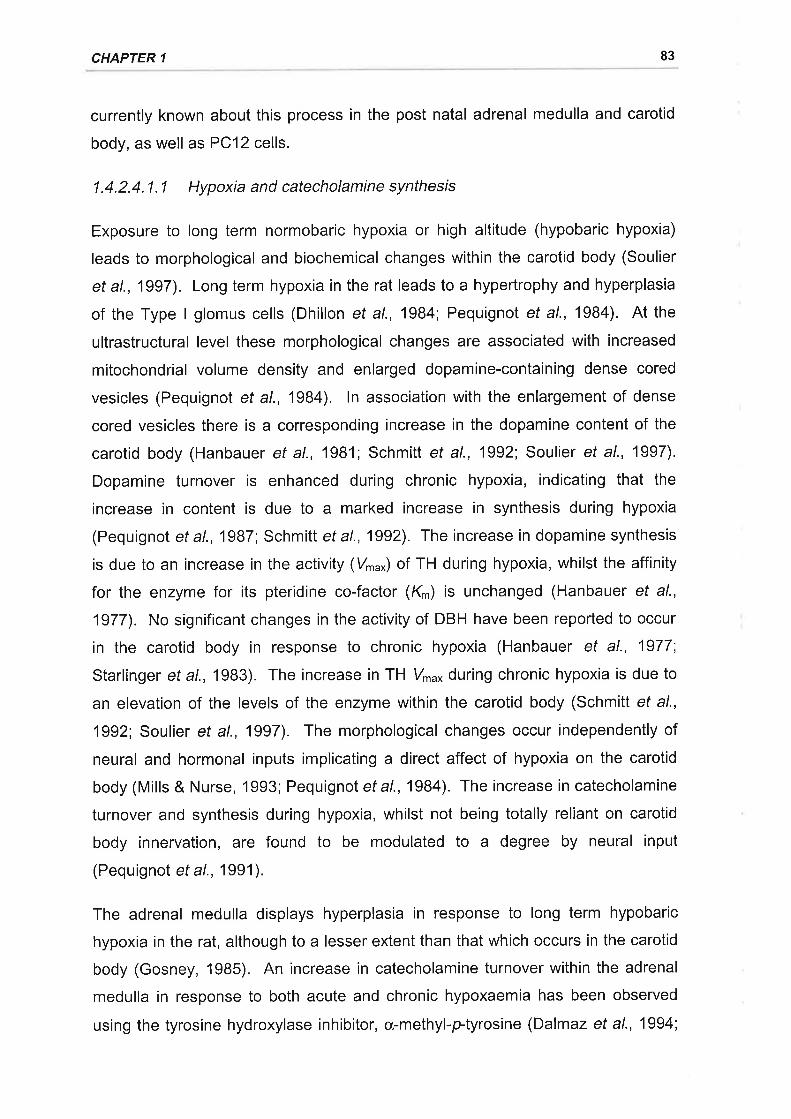
CHAPTER 1 83
currently known about this process in the post natal adrenal medulla and carotid
body, as well as PC12 cells.
1.4.2.4.1.1 Hypoxia and catecholamine synthesis
Exposure to long term normobaric hypoxia or high altitude (hypobaric hypoxia)
leads to morphological and biochemical changes within the carotid body (Soulier
eta1.,1997). Long term hypoxia in the rat leads to a hypertrophy and hyperplasia
of the Type I glomus cells (Dhillon ef al., 1984; Pequignot et al., 1984). At the
ultrastructural level these morphological changes are associated with increased
mitochondrial volume density and enlarged dopamine-containing dense cored
vesicles (Pequigno| et al., 1984). ln association with the enlargement of dense
cored vesicles there is a corresponding increase in the dopamine content of the
carotid body (Hanbauer et al., 1981; Schmitt et al., 1992; Soulier et al., 1997).
Dopamine turnover is enhanced during chronic hypoxia, indicating that the
increase in content is due to a marked increase in synthesis during hypoxia
(Pequignot et al., 1987 Schmitt et al., 1992). The increase in dopamine synthesis
is due to an increase in the activity (Vr.*) of TH during hypoxia, whilst the affinity
for the enzyme for its pteridine co-factor (K'n) is unchanged (Hanbauer et al.,
1977). No significant changes in the activity of DBH have been reported to occur
in the carotid body in response to chronic hypoxia (Hanbauer et al., 1977;
Starlinger et al., 1983). The increase in TH Vr"* during chronic hypoxia is due to
an elevation of the levels of the enzyme within the carotid body (Schmitt et al.,
1992; Soulier et al., 1997). The morphological changes occur independently of
neural and hormonal inputs implicating a direct affect of hypoxia on the carotid
body (Mills & Nurse, 1993; Pequignot et a1.,1984). The increase in catecholamine
turnover and synthesis during hypoxia, whilst not being totally reliant on carotid
body innervation, are found to be modulated to a degree by neural input
(Pequignot et al., 1 991 ).
The adrenal medulla displays hyperplasia in response to long term hypobaric
hypoxia in the rat, although to a lesser extent than that which occurs in the carotid
body (Gosney, 1985). An increase in catecholamine turnover within the adrenal
medulla in response to both acute and chronic hypoxaemia has been observed
using the tyrosine hydroxylase inhibitor, cx-methyl-ptyrosine (Dalmaz et al., 1994;
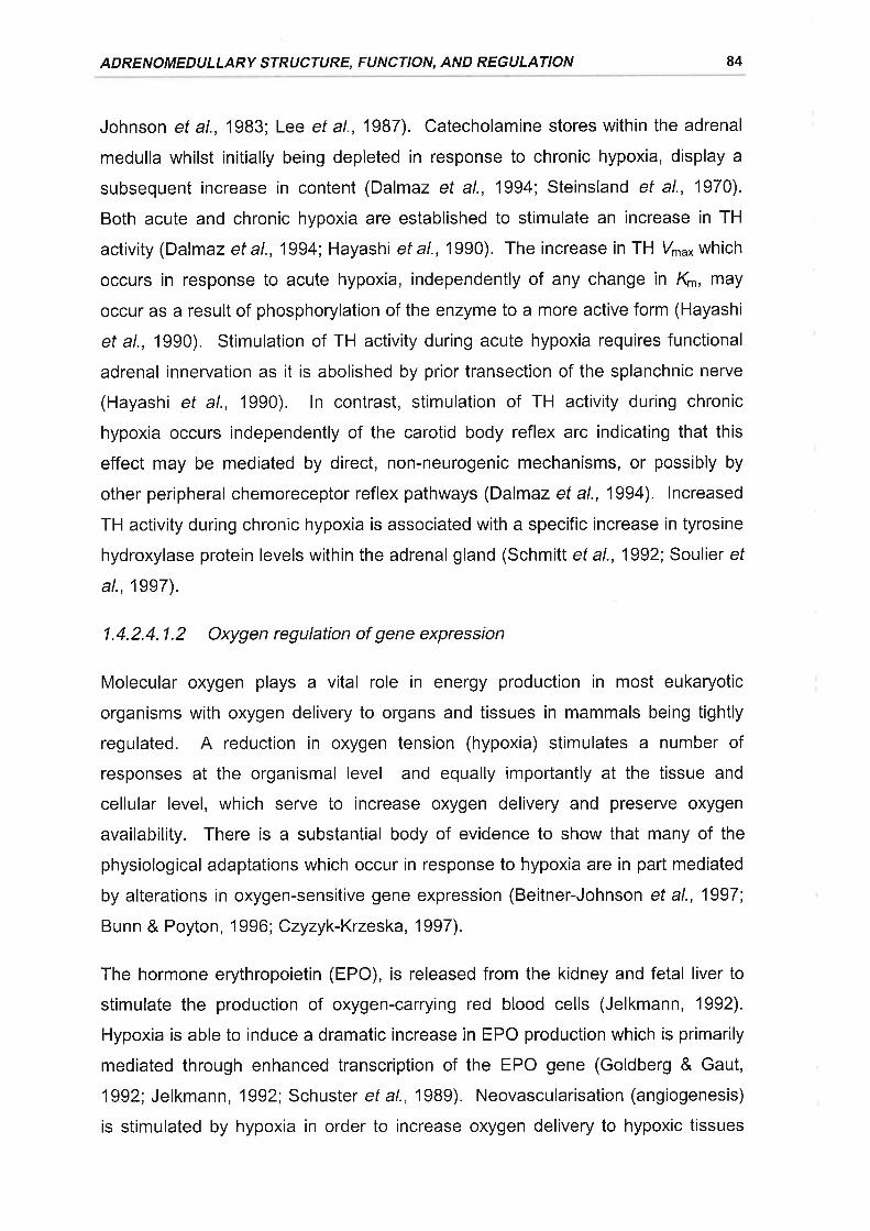
ADRENOMEDULLARY STRUCTURE, FUNCTION, AND REGULATION 84
Johnson et al., 1983; Lee ef al., 1987). Catecholamine stores within the adrenal
medulla whilst initially being depleted in response to chronic hypoxia, display a
subsequent increase in content (Dalmaz et al., 1994; Steinsland et al., 1970).
Both acute and chronic hypoxia are established to stimulate an increase in TH
activity (Dalmaz et al., 1994; Hayashi et al., 1990). The increase in TH V'u* which
occurs in response to acute hypoxia, independently of any change in Kn,', may
occur as a result of phosphorylation of the enzyme to a more active form (Hayashi
et al., 1990). Stimulation of TH activity during acute hypoxia requires functional
adrenal innervation as it is abolished by prior transection of the splanchnic nerve
(Hayashi et al., 1990). ln contrast, stimulation of TH activity during chronic
hypoxia occurs independently of the carotid body reflex arc indicating that this
effect may be mediated by direct, non-neurogenic mechanisms, or possibly by
other peripheral chemoreceptor reflex pathways (Dalmaz et al., 1994). lncreased
TH activity during chronic hypoxia is associated with a specific increase in tyrosine
hydroxylase protein levels within the adrenal gland (Schmitt et al., 1992; Soulier ef
al., 1997).
1.4.2.4.1.2 Oxygen regulation of gene expression
Molecular oxygen plays a vital role in energy production in most eukaryotic
organisms with oxygen delivery to organs and tissues in mammals being tightly
regulated. A reduction in oxygen tension (hypoxia) stimulates a number of
responses at the organismal level and equally importantly at the tissue and
cellular level, which serve to increase oxygen delivery and preserve oxygen
availability. There is a substantial body of evidence to show that many of the
physiological adaptations which occur in response to hypoxia are in part mediated
by alterations in oxygen-sensitive gene expression (Beitner-Johnson et al., 1997;
Bunn & Poyton, 1996; Czyzyk-Krzeska, 1997).
The hormone erythropoietin (EPO), is released from the kidney and fetal liver to
stimulate the production of oxygen-carrying red blood cells (Jelkmann, 1992).
Hypoxia is able to induce a dramatic increase in EPO production which is primarily
mediated through enhanced transcription of the EPO gene (Goldberg & Gaut,
1992; Jelkmann , 1992; Schuster et al., 19Bg). Neovascularisation (angiogenesis)
is stimulated by hypoxia in order to increase oxygen delivery to hypoxic tissues
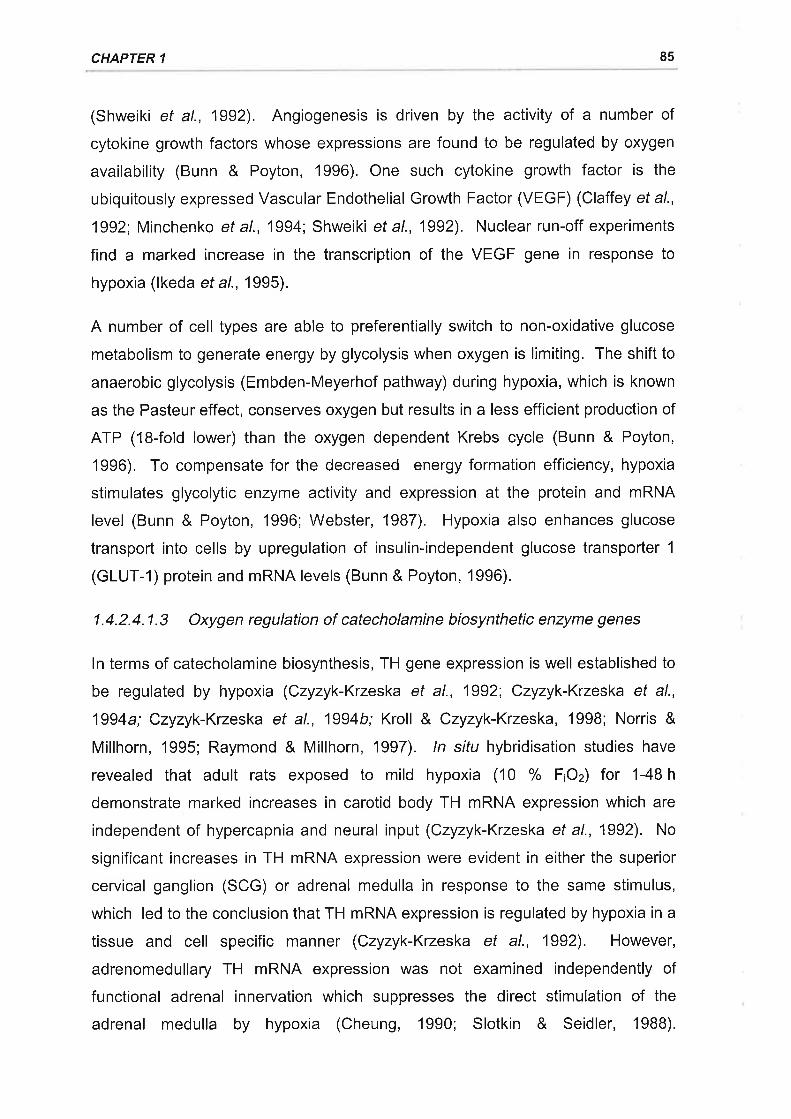
CHAPTER 1 85
(Shweiki et al., 1992). Angiogenesis is driven by the activity of a number of
cytokine growth factors whose expressions are found to be regulated by oxygen
availability (Bunn & Poyton, 1996), One such cytokine growth factor is the
ubiquitously expressed Vascular Endothelial Growth Factor (VEGF) (Claffey et al.,
1992; Minchenko et al., 1994; Shweiki et al., 1992). Nuclear run-off experiments
find a marked increase in the transcription of the VEGF gene in response to
hypoxia (lkeda et al., 1995).
A number of cell types are able to preferentially switch to non-oxidative glucose
metabolism to generate energy by glycolysis when oxygen is limiting. The shift to
anaerobic glycolysis (Embden-Meyerhof pathway) during hypoxia, which is known
as the Pasteur effect, conserves oxygen but results in a less efficient production of
ATP (18-fold lower) than the oxygen dependent Krebs cycle (Bunn & Poyton,
1996). To compensate for the decreased energy formation efficiency, hypoxia
stimulates glycolytic enzyme activity and expression at the protein and mRNA
level (Bunn & Poyton, 1996; Webster, 1987). Hypoxia also enhances glucose
transport into cells by upregulation of insulin-independent glucose transporter 1
(GLUT-1)protein and mRNA levels (Bunn & Poyton, 1996).
1.4.2.4.1.3 Oxygen regulation of catecholamine biosynthetic enzyme genes
ln terms of catecholamine biosynthesis, TH gene expression is well established to
be regulated by hypoxia (Czyzyk-Krzeska et al., 1992; Czyzyk-Krzeska et al.,
1994a; Czyzyk-Krzeska et al., 1994b; Kroll & Czyzyk-Krzeska, 1998; Norris &
Millhorn, 1995; Raymond & Millhorn, 1997). ln situ hybridisation studies have
revealed that adult rats exposed to mild hypoxia (10 % F¡Oz) for 1-48h
demonstrate marked increases in carotid body TH mRNA expression which are
independent of hypercapnia and neural input (Czyzyk-Krzeska et al., 1992). No
significant increases in TH mRNA expression were evident in either the superior
cervical ganglion (SCG) or adrenal medulla in response to the same stimulus,
which led to the conclusion that TH mRNA expression is regulated by hypoxia in a
tissue and cell specific manner (Czyzyk-Krzeska et al., 1992). However,
adrenomedullary TH mRNA expression was not examined independently of
functional adrenal innervation which suppresses the direct stimulation of the
adrenal medulla by hypoxia (Cheung, 1990; Slotkin & Seidler, 1988).
ADRENOMEDULLARY STRUCTURE, FUNCTION, AND REGULATION 86
Furthermore, the modest level of hypoxia used in these studies may have been
insufficient to result in a trans-synaptic activation of TH mRNA expression in the
adrenal.
DeCristofaro and LaGamma (1994) examined the hypothesis that non-neurogenic
stimulation of catecholamine secretion could stimulate catecholamine biosynthetic
enzyme synthesis by acutely subjecting 4 day old rat pups to modest hypoxia (7-
8 % FiOz for 2 h) and measuring adrenal TH mRNA expression 24 h later
(DeCristofaro & LaGamma, 1994). These investigators found that non-neurogenic
stimulation of the adrenal medulla did not significantly increase TH mRNA
expression following hypoxia (DeCristofaro & LaGamma, 1994). Examination of
the timecourse of adrenal TH mRNA expression following the exposure of
neonatal rats to hypoxia was not carried out raising the possibility that a change in
TH mRNA expression following hypoxia may have been missed.
TH mRNA expression in the adrenal medulla of the adult and neonatal rat in vivo
appears relatively unresponsive to modest hypoxia (Czyzyk-Krzeska et al., 1992;
DeCristofaro & LaGamma, 1994). lt is interesting then that TH mRNA expression
in the rat PC12 cell line exhibits an oxygen sensitivity which is analogous to that
demonstrated in the type I glomus cells of the carotid body, with marked
elevations of TH mRNA expression occurring in as little as '1 h in response to a
75% reduction in oxygen availability (Czyzyk-Krzeska et al., 1994a). As outlined
in the previous section the type I glomus cells of the carotid body, PC12 cells, and
fetal adrenomedullary chromaffin cells posses functionally similar chemoreceptive
mechanisms (Adams et al.,'1996; Buckler & Vaughan-Jones, 1994; Lopez-Barneo
et al., 1988; Mochizuki-Oda et al., 1997; Rychkov et al., 1998; Thompson et al.,
1997;Zhu et a/., 1996). The fetus lives at a much lower arterial POz level than
either the neonate or the adult and hence the modest levels of hypoxia used by
previous investigators may have been insufficient to uncover any direct effect on
hypoxia on adrenomedullary TH mRNA expression (Czyzyk-Krzeska et al., 1992;
DeCristofaro & LaGamma, 1994). lt has yet to be established if hypoxia is able to
directly regulate catecholamine synthetic enzyme mRNA expression in the fetus rn
utero or in adrenomedullary chromaffin cells in vitro which are not subject to
neural influences.
CHAPTER 1 87
The establishment of the PC12 cell line as a suitable rn vitro model in which to
study oxygen regulation of TH gene expression has greatly facilitated the
elucidation of a number of potential mechanisms behind this process (Beitner-
Johnson et al., 1997). Nuclear run-off assays reveal that hypoxia increases
transcription of the TH gene in PC12 cells (Czyzyk-Krzeska et al., 1994b).
Furthermore, studies using both transcriptional inhibitors and pulse chase
techniques find that there is also an increase in the stability of TH mRNA in PC12
cells in response to hypoxia (Czyzyk-Krzeska et al., 1994b). The two forms of
mRNA expression regulation come into effect at different points during exposure
to hypoxia with the increase in TH gene transcription prevailing early (< 5 h) during
hypoxia. With longerexposure to hypoxia (. 10 h) there is an attenuation of gene
transcription and stabilisation of the mRNA becomes the primary contributor to the
overall increase in expression (Czyzyk-Krzeska, 1997; Czyzyk-Krzeska et al.,
1994b). Stabilisation of mRNA during hypoxia is advantageous as it provides
greater energy efficiency, with cells utilising existing mRNA rather than
tra n scri b i n g n ew m R NA (Czyzyk-Krzes ka, 1 997).
1.4.2.4.1.4 Mechanisms of hypoxia induced TH gene transcription
The novel transcriptional factor, hypoxia-inducible factor 1 (HlF-1), interacts with
specific crs-elements within the promoter and enhancer regions of numerous
genes, including those for EPO (Semenza & Wang, 1992), VEGF (Forsythe et al.,
1996), GLUT-1 (Ebert et a\.,1995), and many glycolytic enzymes (Semenza et al.,
1994), to regulate transcription during hypoxia. HIF-1, which was first identified
and characterised by Wang and Semenza, is induced by low cellular Oz tension
and consists of cr and p subunits which must heterodimerise to bind to their
consensus site and influence gene transcription (Jiang et al., 1996; Semenza &
Wang, 1992; Wang & Semenza, 1993a; Wang & Semenza, 1993c; Wang &
Semenza, 1995; Wood et al., 1996). HIF-1o and -1p belong to the basic helix-
loop-helix family of transcription factors and whilst the Hif-1o subunit is a novel
protein, the Hif-18 subunit has previously been identified as the Aryl Hydrocarbon
Receptor Nuclear Translocator (ARNT) functional in the transcriptional response
to xenobiotics (Hoffman et al., 1991; Wang et al., 1995a).
ADRENOMEDULLARY STRUCTURE, FUNCTION, AND REGULATION 88
Hypoxia induced upregulation of TH gene transcription in PC12 cells has been
determined by TH-CAT reporter gene construct experiments to be dependent
upon a short sequence of the 5'-proximal promoter region between bases -284
and -1 50 (Czyzyk-Krzeska et al., 1994b; Norris & Millhorn, 1995). Analysis of this
sequence reveals the presence of AP-1, AP-2, and HIF-1 regulatory crs-elements
(Norris & Millhorn, 1995). Gel shift assays using a TH DNA fragment
corresponding to bases -284 Ío -190 of the 5' promoter region show that there is
increased PC12 cell nuclear protein binding to this fragment following prior
exposure of these cells to hypoxia (Norris & Millhorn, 1995). lncreased nuclear
protein binding to the native TH DNA fragment following hypoxia can be padially
competed for by the addition of excess TH HIF-1 element oligonucleotide (Norris
& Millhorn, 1995). HIF-1 may, therefore, playa role in hypoxia induced regulation
of TH gene transcription (Beitner-Johnson et al., 1997; Czyzyk-Krzeska, 1997;
Norris & Millhorn, 1995).
Aside from the HIF-1 regulatory element, the -284 to -190 base TH DNA fragment
also contains the AP-1 regulatory element (Beitner-Johnson et al., 1997; Norris &
Millhorn, 1995). The importance of the AP-1 regulatory element in the TH gene
transcriptional response to hypoxia is highlighted by a number of findings. First,
the addition of unlabelled TH AP-1 element oligonucleotide attenuates the
increase in hypoxia induced protein binding to the native ÎH -284 to -190 base
DNA fragment (Norris & Millhorn, 1995). Secondly, the binding of PC12 cell
nuclear protein extracts to double stranded oligonucleotides containing the TH AP-
1 element sequence remains enhanced during prolonged hypoxia (Norris &
Millhorn, 1995). Finally, mutation of the AP-1 element abolishes hypoxia induced
TH (-284 to -150 base)-CAT reporter gene expression in PC'12 cells (Norris &
Millhorn, 1995).
UV cross linking experiments of PC12 nuclear protein extracts have found that the
proteins which bind to the AP-1 site during hypoxia range in size from 20-85 kDa
(Norris&Millhorn, 1995). Thesizerangeof theseproteinsfall withintherangeof
several members of the frans-acting Fos and Jun family of proteins.
lmmunological gel shift assays revealed that antibodies against c-fos and jun-B
produced supershifted bands resulting from their interaction with labelled TH AP-1
element I PC12 cell nuclear protein complexes (Norris & Millhorn, 1995). An
CHAPTER 1 89
antibody against c-jun was unable to produce a supershifted band, indicating a
lack of involvement of this protein in the hypoxia induced binding to the AP-1
element (Norris & Millhorn, 1995). The binding of both c-fos and jun-B to the AP-1
element were induced by hypoxia (Norris & Millhorn, 1995).
lnterestingly, increases in intracellular calcium are required for hypoxia to
stimulate an increase in TH mRNA expression in PC12 cells. The hypoxia
induced increase in TH mRNA expression is abolished by the omission of calcium
from the external medium or by the addition of calcium chelators (Raymond &
Millhorn, 1997). The major signal transduction pathways that are activated by an
increase in cytosolic Ca2* levels are Ca2* / calmodulin-dependent protein kinases
(CamK), Ca2* | calmodulin-dependent phosphatases and various isoforms of
protein kinase C (PKC). Specific inhibitors of protein kinase C are able to block
the hypoxia induced up-regulation of TH mRNA indicating that the Ca2* lprotein
kinase C may play a critical role in hypoxia induced TH mRNA expression
(Beitner-Johnson et al., 1997; Raymond & Millhorn, 1997). Phosphorylation is a
necessary requirement for the activation of a number of transcription factors,
although HIF-1 phosphorylation occurs independently of PKC activation (Bunn &
Poyton, 1996; Czyzyk-Krzeska, 1997; Wang et al., 1995c). The mechanisms by
which hypoxia stimulates phosphorylation have yet to be determined but in the
case of HIF-1 induction, pharmacological studies implicate the involvement of
both serine / threonine kinases and phosphatases (Bunn & Poyton, 1996; Czyzyk-
Krzeska, 1997; Wang et al., 1995c).
1.4.2.4.1.5 Mechanisms of hypoxia induced TH nRNA stabilisation
TH mRNA in PC12 cells has a half life of around 10 h and this is stabilised a
further 2-4 fold during prolonged hypoxia (Czyzyk-Krzeska, 1997; Czyzyk-Krzeska
et al., 1994b). The increased stability of TH mRNA during hypoxia in PC12 cells
appears to result from an increased binding of cytoplasmic protein to a pyrimidine-
rich ci+acting sequence in the 3' untranslated region of the TH mRNA (Czyzyk-
Krzeska et al., 1994a). Gel retardation assays and RNase T1 mapping using
fragments of rat TH mRNA incubated with cytoplasmic protein extract from PC12
cells, revealed that a sequence located between bases 1551-1579 in the 3'
untranslated region exhibited increased protein binding following prior exposure of
AD RE N OM E D U LLARY ST RU CT U RE, F U N CTIO N, AN D REG U LAT I O N
the cells to hypoxia (Czyzyk-Krzeska & Beresh, 1996; Czyzyk-Krzeska et al.,
1994a). This sequence has been termed the Hypoxia-lnducible Protein Binding
Sequence (HIPBS; Czyzyk-Krzeska, 1997). Mutation of this sequence abolishes
protein binding, resulting in a decrease in the constitutive half life of TH mRNA as
well as the abolition of the hypoxia induced increase in TH mRNA stability
(Czyzyk-Krzeska & Beresh, 1996; Czyzyk-Krzeska ef al., 1997). Furthermore the
mRNA of chloramphenicol acetyltransferase (CAT) reporter gene constructs
containing the TH HIPBS sequence exhibit 02 dependent regulation of stability
which is not normally observed in the absence of this sequence (Czyzyk-Krzeska
et al., 1997). RNA affinity purification of hypoxia inducible proteins (HlP) which
bind to the HIPBS yielded two protein factors with approximate molecular weights
of 45 and 55-60 kD (Czyzyk-Krzeska et al., 1997). Hypoxia inducible binding
activity to HIPBS has also been identified in other catecholaminergic tissues such
as the carotid body and the superior cervical ganglion (Czyzyk-Krzeska et al.,
1997). Post-transcriptional regulation of gene expression by the binding of
proteins to stability elements within the 3'-untranslated region of the mRNA has
also been described for other oxygen regulated genes including EPO and VEGF
(Bunn & Poyton, 1996).
1.4.2.4.1.6 Oxygen sensing and TH gene transcription
The actions of molecular oxygen at the cellular level are in great part mediated by
heme proteins. Heme proteins bind molecular Oz via a central Fe2* in the
molecule which results in a conformational change from the deoxy to the oxy form.
Conformational changes in the heme molecule affect their function and make
them potential candidates for an "O2 seÍìsor" (for review see Bunn, 1996). There
is evidence to suggest that heme proteins may be involved in the regulation of a
number of oxygen sensitive genes. The transition metals cobalt and nickel which
are able to substitute for iron in the heme moiety, locking it into the deoxy
configuration due to their relatively low affinity for oxygen, are found to induce the
expression of many oxygen regulated genes including EPO and VEGF, amongst
others (Bunn & Poyton, 1996; Czyzyk-Krzeska, 1997; Goldberg & Schneider,
1994; Ho & Bunn, 1996; Sinclair et al., 1979; Sinclair et al., 1982). Conversely,
carbon monoxide which binds non-covalently to the ferrous heme group in a
manner analogous to that of oxygen, locking it into the oxy-formation, antagonises
90
CHAPTER 1 91
the hypoxia induced upregulation of EPO, VEGF, and other genes (Bunn &
Poyton, 1996; Goldberg et al., 1988; Goldberg & Schneider, 1994).
The oxygen sensor appears to be coupled to HzOz production within the cell and
hence is speculated to be a multisubunit oxidase (Bunn & Poyton, 1996). ln PC12
cells which have been loaded with the HzOz-sensitive fluorescent dye 2',7'-
dichlorofluorescein (DCF) and exposed to 5 o/o Oz, HzOz levels are found to
decrease as early as t h after exposure with the decrease reaching a maximum of
40 % after 6 h (Kroll &Czyzyk-Krzeska, 1998). Peroxide production is also found
to be inhibited in a graded manner in EPO producing Hep 38 cells in response to
decreasing oxygen tensions (Fandrey et al., 1994). Therefore HzOz production
appears to be a plausible intracellular signal for oxygen regulated gene expression
(Bunn & Poyton, 1996; Czyzyk-Krzeska, 1997). ln support of this theory, the
hypoxia induced increase in TH mRNA expression in PC12 cells is found to be
inversely related to intracellular HzO2 formation (Kroll & Czyzyk-Krzeska, 1998).
Pretreatment of PC12 cells and Hep 38 cells with HzOz prevents the induction of
TH mRNA and EPO mRNA expression, respectively, in response to hypoxia
(Fandrey et al., 1994; Kroll & Czyzyk-Krzeska, 1998). Conversely, the
administration of the HzOz decomposing enzyme, catalase, or the reducing
antioxidant N-(2-mercaptopropionyl)-glycine which decrease HzOz formation are
able to increase TH mRNA expression (Kroll &Czyzyk-Krzeska, 1998).
HzOz is known as a reactive oxygen intermediate (ROl) and may mediate its
effects on intracellular signal transduction pathways by regulation of intracellular
redox status. Glutathione peroxidase decomposes HzOz using reduced glutathione
(GSH) as the hydrogen donor (Sies et al., 1972). The reaction oxidises
glutathione (GSSG), shifting the subcellular ratio of GSH:GSSG towards the
oxidised form (Acker et a1.,1992). HzOz can also generate more reactive oxygen
compounds, such as hydroxyl radicals (OH ) and singlet oxygen, via the Fenton
reaction which is catalysed by intracellular iron (Bunn & Poyton, 1996; Czyzyk-
Krzeska, 1997 lsaacs & Binkley, 1977). These next-step ROls interact with and
oxidise glutathione and thiol groups (Czyzyk-Krzeska, 1997). Thus as a result of
the decreased generation of HzOz during hypoxia, decreased concentrations of
ROls and increased concentrations of reduced glutathione are expected (Acker,
1994; Acker & Xue, 1995; Czyzyk-Krzeska, 1997). Consequently the redox
ADRENOMEDULLARY STRUCTURE, FUNCTION, AND REGULATION 92
equilibrium of protein thiols would shift towards the reduced form; affecting the
nucleic acid-binding properties of regulatory proteins and influencing gene
expression (Bunn & Poyton, 1996; Czyzyk-Krzeska, 1997). Reduction of next step
ROI formation by the chelation of free intracellular iron using deferoxamine
induces TH mRNA expression in PC12 cells (Kroll & Czyzyk-Krzeska, 1998), a
phenomenon also observed in other cell lines for VEGF mRNA (Gleadle et al.,
1995) and EPO mRNA expression (Wang & Semenza, 1993b). lndeed AP-1
complexes require the reduction of specific cysteine residues in their DNA-binding
domains for association with DNA /n vifro (Abate et al., 1990; Toledano &
Leonard, 1991). HIF-1 binding to DNA is also redox sensitive and this is primarily
due to redox dependent stabilisation of the HIF-1cx subunit in the reduced form
(Huang et al., 1996; Wang et al., 1995b). Furthermore HIBPs binding to TH
mRNA is also dependent on these proteins being in the reduced form (Czyzyk-
Krzeska, 1997).
1.4,2,4.2 Hormonal regulation of adrenomedullary development and
catech ol am i ne bi osynthesi s
As in the adult animal there is substantial evidence to suggest that glucocorticoids
derived from the adrenal cortex play an important role in the regulation of
catecholamine synthesis, particularly adrenaline synthesis, in the fetal adrenal
gland. Aside from a regulatory role of glucocorticoids in fetal catecholamine
biosynthesis, they also play an important role in development of the fetal adrenal
medulla.
1.4.2.4.2.1 Glucocorticoids and adrenomedullary development
The neural crest is a transient embryonic structure consisting of precursor cells
which migrate to specific locations and give rise to a diverse affay of derivatives
including sensory neurones, autonomic neurones, and glial cells (Anderson, 1993;
Cochard et al., 1978; Coulombre et al., 1974; LeDouarin et al., 1991 ; LeDouarin &
Dupin, 1993). The fates of the multipotent neural precursor cells appear to be
determined in large part by environmental factors encountered along the migratory
route to their ultimate position (LeDouarin & Dupin, 1993; Stern et al., 1991;
Unsicker, 1985). One such neural crest derived cell lineage is the
sympathoadrenal lineage which develops from sympathoadrenal progenitor cells
CHAPTER 1
which migrate ventrolaterally from the apex of the neural tube to the dorsal aorta
where they aggregate and differentiate to form sympathetic neurones or continue
migration into the adrenal anlage where they differentiate to form chromaffin cells
(Anderson, 1 993; Patterson, 1 990).
Previous studies have shown that when dissociated chromaffin cells from
immature adrenals are cultured in the absence of glucocorticoids they exhibit poor
longtermsurvival inculture(Doupe etal., 1985). Additionof nervegrowthfactor
(NGF) to these cultures is able to rescue many of the chromaffin cells however
they subsequently undergo a phenotypical change exhibiting extensive neurite
outgrowth (Doupe et al., 1985; Naujoks et al., 1982; Unsicker et al., 1978a).
Glucocorticoid administration is able to block or delay NGF-induced neurite
outgrowth (Doupe et al., 1985; Unsicker et al., 1978a). Long term culture of rat
adrenal chromaffin cells in the presence of NGF and the absence of
glucocorticoids results in a complete transition to a sympathetic neuron phenotype
as assessed by a number of morphological and biochemical criteria (Doupe et al.,
1985). Furthermore in vivo studies showthat injection of pregnant rats with NGF
results in the replacement of fetal chromaffin cells with sympathetic ganglion cells
(Aloe & Levi-Montalcini, 1979). Hence glucocorticoids appearto act as a survival
factor for chromaffin cells and repress neuronal transdifferentiation of these cells
(Doupe et al., 1985; Unsicker et al., 1978a). These data suggest that the
sympathetic progenitor cells which migrate from the sympathetic ganglion
primordium through the developing adrenal anlage develop into chromaffin cells in
the adrenal medulla under the influence of the glucocorticoid rich environment.
More definitive proof of the role of glucocorticoids in the developmental regulation
of the adrenal medulla arose with the development of antigenic markers which
allowed for the identification of fetal sympathoadrenal (SA) progenitor cells
(Anderson & Axel, 1986; Carnahan & Patterson, 1991 a; Carnahan & Patterson,
1991b). Subsequently fetal SA progenitors were isolated and rn vifro studies
demonstrated that these cells are indeed bipotential and can develop into either
sympathetic neurones or chromaffin cells depending on the hormonal milieu and
culture conditions (Anderson & Axel, 1986; Carnahan & Patterson, 1991b;
Michelsohn & Anderson, 1992). The development of a sympathetic neuron
phenotype is initially dependent upon the presence of fibroblast growth factor and
93
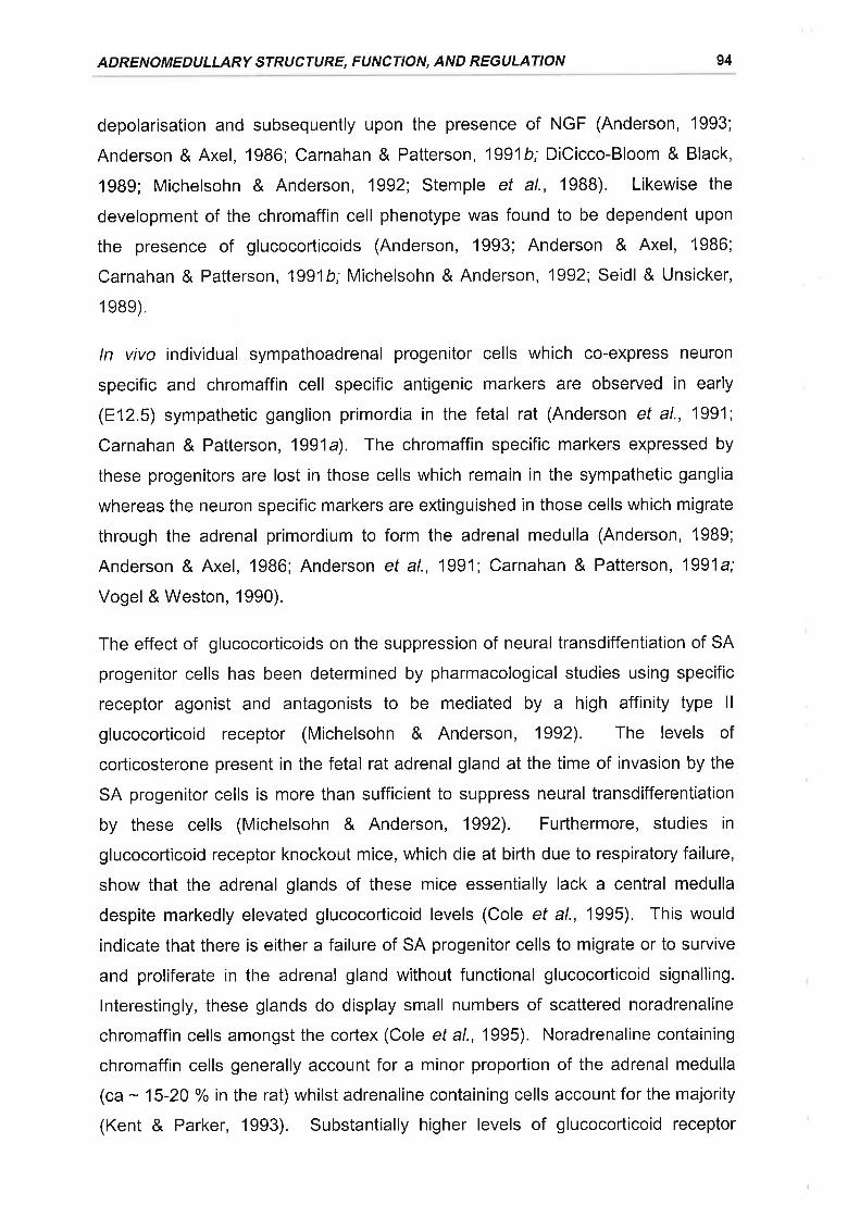
ADRENOMEDULLARY STRUCTURE, FUNCTION, AND REGULATION 94
depolarisation and subsequently upon the presence of NGF (Anderson, 1993;
Anderson & Axel, 1986; Carnahan & Patterson, 1991b; DiCicco-Bloom & Black,
1989; Michelsohn & Anderson, 1992; Stemple et al., 1988). Likewise the
development of the chromaffin cell phenotype was found to be dependent upon
the presence of glucocorticoids (Anderson, 1993; Anderson & Axel, 1986;
Carnahan & Patterson, 1991b; Michelsohn & Anderson, 1992; Seidl & Unsicker,
1g8e).
ln vivo individual sympathoadrenal progenitor cells which co-express neuron
specific and chromaffin cell specific antigenic markers are observed in early
(812.5) sympathetic ganglion primordia in the fetal rat (Anderson et al., 1991;
Carnahan & Patterson, 1991a). The chromaffin specific markers expressed by
these progenitors are lost in those cells which remain in the sympathetic ganglia
whereas the neuron specific markers are extinguished in those cells which migrate
through the adrenal primordium to form the adrenal medulla (Anderson, 1989;
Anderson & Axel, 1986; Anderson et al., 1991; Carnahan & Patterson, 1991a;
Vogel & Weston, 1990).
The effect of glucocorticoids on the suppression of neural transdiffentiation of SA
progenitor cells has been determined by pharmacological studies using specific
receptor agonist and antagonists to be mediated by a high affinity type ll
glucocorticoid receptor (Michelsohn & Anderson, 1992). The levels of
corticosterone present in the fetal rat adrenal gland at the time of invasion by the
SA progenitor cells is more than sufficient to suppress neural transdifferentiation
by these cells (Michelsohn & Anderson, 1992). Furthermore, studies in
glucocorticoid receptor knockout mice, which die at birth due to respiratory failure,
show that the adrenal glands of these mice essentially lack a central medulla
despite markedly elevated glucocorticoid levels (Cole et al., 1995). This would
indicate that there is either a failure of SA progenitor cells to migrate or to survive
and proliferate in the adrenal gland without functional glucocorticoid signalling.
lnterestingly, these glands do display small numbers of scattered noradrenaline
chromaffin cells amongst the cortex (Cole et al., 1995). Noradrenaline containing
chromaffin cells generally account for a minor proportion of the adrenal medulla
(ca - 15-20 % in the rat) whilst adrenaline containing cells account for the majority
(Kent & Parker, 1993). Substantially higher levels of glucocorticoid receptor
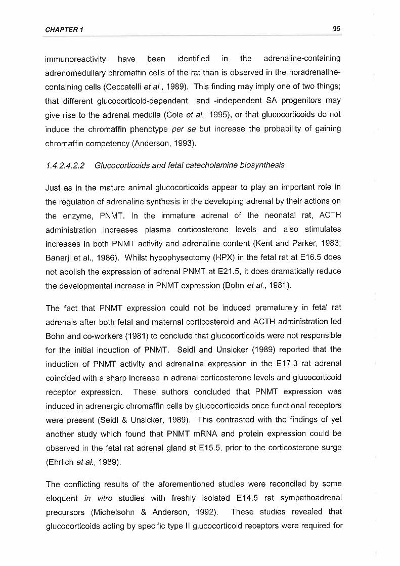
CHAPTER 1 95
immunoreactivity have been identified in the adrenaline-containing
adrenomedullary chromaffin cells of the rat than is observed in the noradrenaline-
containing cells (Ceccatelli et a\.,1989). This finding may imply one of two things;
that different glucocorticoid-dependent and -independent SA progenitors may
give rise to the adrenal medulla (Cole etal., 1995), orthat glucocorticoids do not
induce the chromaffin phenotype per se but increase the probability of gaining
chromaffin competency (Anderson, 1993).
1.4.2.4.2.2 Glucocorticoids and fetal catecholamine biosynthesis
Just as in the mature animal glucocorticoids appear to play an important role in
the regulation of adrenaline synthesis in the developing adrenal by their actions on
the enzyme, PNMT. ln the immature adrenal of the neonatal rat, ACTH
administration increases plasma corticosterone levels and also stimulates
increases in both PNMT activity and adrenaline content (Kent and Parker, 1983;
Banerji et al., 1986). Whilst hypophysectomy (HPX) in the fetal rat at E16.5 does
not abolish the expression of adrenal PNMT atE21.5, it does dramatically reduce
the developmental increase in PNMT expression (Bohn et al., 1981).
The fact that PNMT expression could not be induced prematurely in fetal rat
adrenals after both fetal and maternal corticosteroid and ACTH administration led
Bohn and co-workers (1981)to conclude that glucocorticoids were not responsible
for the initial induction of PNMT. Seidl and Unsicker (1989) reported that the
induction of PNMT activity and adrenaline expression in the E17.3 rat adrenal
coincided with a sharp increase in adrenal corticosterone levels and glucocorticoid
receptor expression, These authors concluded that PNMT expression was
induced in adrenergic chromaffin cells by glucocorticoids once functional receptors
were present (Seidl & Unsicker, 1989). This contrasted with the findings of yet
another study which found that PNMT mRNA and protein expression could be
observed in the fetal rat adrenal gland at E15.5, prior to the corticosterone surge
(Ehrlich et al., 19Bg).
The conflicting results of the aforementioned studies were reconciled by some
eloquent in vitro studies with freshly isolated E14.5 rat sympathoadrenal
precursors (Michelsohn & Anderson, 1992). These studies revealed that
glucocorticoids acting by specific type ll glucocorticoid receptors were required for
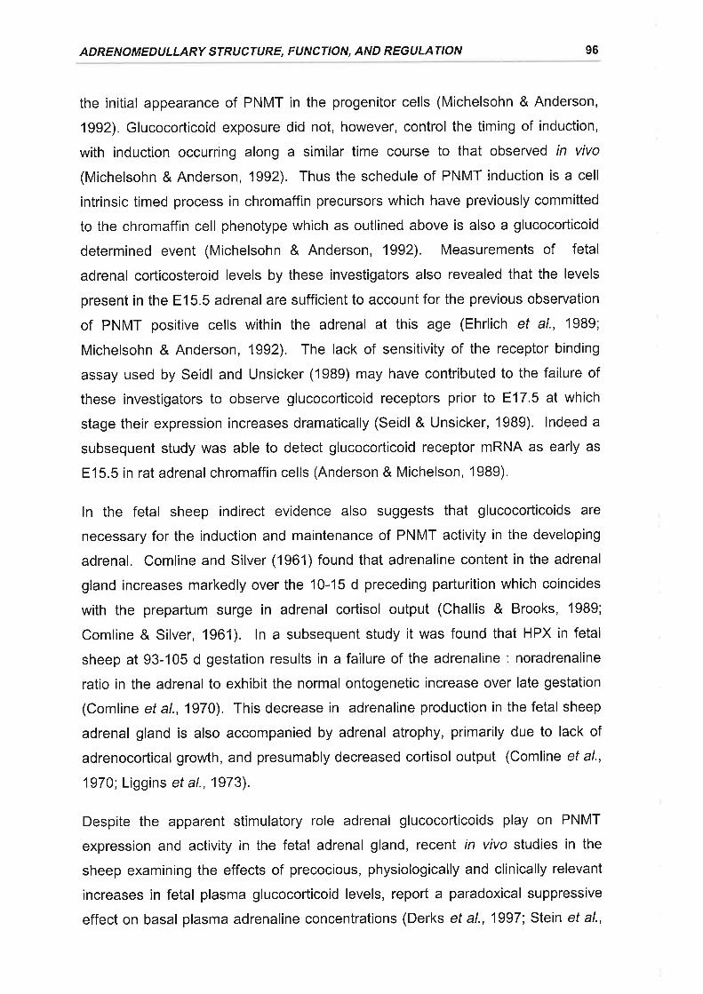
ADRENOMEDULLARY STRUCTURE, FUNCTION, AND REGULATION 96
the initial appearance of PNMT in the progenitor cells (Michelsohn & Anderson,
1992). Glucocorticoid exposure did not, however, control the timing of induction,
with induction occurring along a similar time course to that observed in vivo
(Michelsohn & Anderson, 1992). Thus the schedule of PNMT induction is a cell
intrinsic timed process in chromaffin precursors which have previously committed
to the chromaffin cell phenotype which as outlined above is also a glucocorticoid
determined event (Michelsohn & Anderson, 1992). Measurements of fetal
adrenal corticosteroid levels by these investigators also revealed that the levels
present in the E15.5 adrenal are sufficient to account for the previous observation
of PNMT positive cells within the adrenal at this age (Ehrlich et al., 1989;
Michelsohn & Anderson, 1992). The lack of sensitivity of the receptor binding
assay used by Seidl and Unsicker (1989) may have contributed to the failure of
these investigators to observe glucocorticoid receptors prior to 817.5 at which
stage their expression increases dramatically (Seidl & Unsicker, 1989). lndeed a
subsequent study was able to detect glucocorticoid receptor mRNA as early as
E15.5 in rat adrenal chromaffin cells (Anderson & Michelson, 1989).
ln the fetal sheep indirect evidence also suggests that glucocorticoids are
necessary for the induction and maintenance of PNMT activity in the developing
adrenal. Comline and Silver (1961)found that adrenaline content in the adrenal
gland increases markedly over the 10-15 d preceding parturition which coincides
with the prepartum surge in adrenal cortisol output (Challis & Brooks, 1989;
Comline & Silver, 1961). ln a subsequent study it was found that HPX in fetal
sheep at 93-105 d gestation results in a failure of the adrenaline : noradrenaline
ratio in the adrenal to exhibit the normal ontogenetic increase over late gestation
(Comline et a1.,1970). This decrease in adrenaline production in the fetal sheep
adrenal gland is also accompanied by adrenal atrophy, primarily due to lack of
adrenocortical growth, and presumably decreased cortisol output (Comline et al.,
1970; Liggins et al., 1973).
Despite the apparent stimulatory role adrenal glucocorticoids play on PNMT
expression and activity in the fetal adrenal gland, recent in vivo studies in the
sheep examining the effects of precocious, physiologically and clinically relevant
increases in fetal plasma glucocorticoid levels, report a paradoxical suppressive
effect on basal plasma adrenaline concentrations (Derks ef al., 1997; Stein et al.,
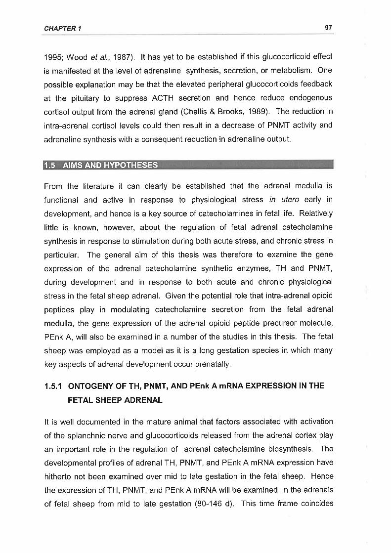
CHAPTER 1 97
1995; Wood et a\.,1987). lt has yet to be established if this glucocorticoid effect
is manifested at the level of adrenaline synthesis, secretion, or metabolism. One
possible explanation may be that the elevated peripheral glucocorticoids feedback
at the pituitary to suppress ACTH secretion and hence reduce endogenous
cortisol outputfrom the adrenal gland (Challis & Brooks, 1989). The reduction in
intra-adrenal cortisol levels could then result in a decrease of PNMT activity and
adrenaline synthesis with a consequent reduction in adrenaline output.
From the literature it can clearly be established that the adrenal medulla is
functional and active in response to physiological stress in utero early in
development, and hence is a key source of catecholamines in fetal life. Relatively
little is known, however, about the regulation of fetal adrenal catecholamine
synthesis in response to stimulation during both acute stress, and chronic stress in
particular. The general aim of this thesis was therefore to examine the gene
expression of the adrenal catecholamine synthetic enzymes, TH and PNMT,
during development and in response to both acute and chronic physiological
stress in the fetal sheep adrenal. Given the potential role that intra-adrenal opioid
peptides play in modulating catecholamine secretion from the fetal adrenal
medulla, the gene expression of the adrenal opioid peptide precursor molecule,
PEnk A, will also be examined in a number of the studies in this thesis. The fetal
sheep was employed as a model as it is a long gestation species in which many
key aspects of adrenal development occur prenatally.
1.5.1 ONTOGENY OF TH, PNMT, AND PEnk A mRNA EXPRESSION lN THE
FETAL SHEEP ADRENAL
It is well documented in the mature animal that factors associated with activation
of the splanchnic nerye and glucocorticoids released from the adrenal cortex play
an important role in the regulation of adrenal catecholamine biosynthesis. The
developmental profiles of adrenal TH, PNMT, and PEnk A mRNA expression have
hitherto not been examined over mid to late gestation in the fetal sheep. Hence
the expression of TH, PNMT, and PEnk A mRNA will be examined in the adrenals
of fetal sheep from mid to late gestation (80-146 d). This time frame coincides
I.5 .AIMS AND HYPOTHESES
ADRENOMEDULLARY STRUCTURE, FUNCTION, AND REGULATION 98
with the development of functional splanchnic innervation and the prepartum
surge in corticosteroid output from the cortex.
Hypothesis
That the developmental expression of adrenal TH, PNMT, and PEnk A are altered
over mid to late gestation with the development of adrenal innervation and
adrenocortical maturation.
1.5.2 IMPACT OF ACUTE HYPOXAEMIA ON THE EXPRESSION OF TH AND
PNMT MRNA IN THE ADRENAL GLAND OF THE FETAL SHEEP PRIOR
TO AND AFTER THE DEVELOPMENT OF FUNCTIONAL SPLANCHNIC
INNERVATION
The fetal adrenal medulla is capable of secreting catecholamines in direct
response to hypoxaemia prior to innervation. This is in contrast to the mature
adrenal which requires reflex stimulation of the splanchnic nerve to stimulate
catecholamine secretion. Neural stimulation of catecholamine release is
associated with a compensatory increase in catecholamine synthesis known as
stimulation-synthesis-coupling. lt is not known whether the non-neurogenic
release of catecholamines from the fetal adrenal gland during hypoxia is also
associated with alterations in catecholamine synthesis. Hence the effect of acute
fetal hypoxaemia on adrenal TH and PNMT mRNA expression before and after
the development of functional splanchnic innervation will be examined in the fetal
sheep.
Hypothesis
That acute hypoxia will act before the development of adrenal innervation to
stimulate catecholamine synthetic enzyme mRNA expression. lt is also
hypothesised that the effect of hypoxia on catecholamine synthetic enzyme mRNA
expression in the fetal adrenal will become dependent on reflex neural stimulation
with the development of functional cholinergic innervation.

CHAPTER 1 99
1.5.3 THE IMPACT OF A CHRONIC PHYSIOLOGICAL INCREASE OF
PLASMA GLUCOCORTIGOIDS ON THE EXPRESSION OF TH, PNMT,
AND PENK A MRNA IN THE ADRENAL GLAND OF THE FETAL SHEEP
Fetal plasma glucocorticoids are elevated in response to intrauterine stressors or
as a consequence of therapeutic administration in anticipation of premature
delivery. Despite well documented stimulatory effects of glucocorticoids on adult
catecholamine synthesis and PNMT induction in sympathoadrenal progenitor
cells, recent studies have found a fall in basal adrenaline levels in the late
gestation sheep fetus in response to exogenous glucocorticoid administration. ln
an attempt to clarify whether the actions of exogenously administered
glucocorticoids in the fetal sheep occur at the level of adrenal catecholamine
synthesis, the impact of chronic intrafetal infusions of physiological levels of
cortisol on adrenal TH, PNMT, and PEnk A mRNA expression will be determined.
Hypothesis
That exogenous cortisol administration to the fetal sheep acts via negative
feedback on the HPA axis to cause a reduction in PNMT mRNA expression.
1.5.4 EFFECT OF PLACENTAL RESTRICTION ON TH, PNMT, AND PEnk A
mRNA EXPRESSION IN THE ADRENAL GLAND OF THE LATE
GESTAT¡ON SHEEP FETUS
Placental restriction is associated with chronic fetal hypoxaemia and
hypoglycaemia. As a consequence basal plasma catecholamine levels are found
to be elevated in the PR fetus. There is evidence to suggest that the capacity of
the fetal and neonatal adrenal to secrete adrenaline in response to further stress
is diminished with PR, however the impact of PR on catecholamine synthesis has
yet to be determined. Adrenal expression of TH, PNMT, and PEnk A mRNA will
be compared between normally grown and placentally restricted fetal sheep in late
gestation.
Hypothesis
That the expression of TH mRNA will increase and that of PNMT mRNA will
decrease in the chronically hypoxaemic, growth restricted sheep fetus.
2. ONTOGENY OF TYROSINE HYDROXYLASE,PHENYLETHANOLAMINE N-METHYLTRANSFERASE AND
PROENKEPHALIN A MRNA EXPRESSION IN THE ADRENALGLAND OF THE FETAL SHEEP
Studies in a number of species have shown that the fetal adrenal medulla
secretes noradrenaline and adrenaline in response to a range of perinatal stimuli
including acute hypoxaemia, asphyxia, labour and vaginal delivery, and that these
catecholamines play a key role in the initiation and coordination of cardiovascular
and metabolic adaptations of the immature animal to these stimuli (Comline &
Silver, 196'1;Jones et al., 19BB; Lagercrantz & Bistoletti, 1973; Padbury et al.,
1982; Slotkin & Seidler, 1988). lt is clear that noradrenaline, adrenaline, and Enk-
containing peptides are present in the adrenal medulla and in the fetal circulation
before birth, and that the adrenal medulla is the predominant source of circulating
catecholamines during acute fetal hypoxaemia (Cohen et al., 1991; Cohen et al.,
1984; Coulter et al., 1990a; Coulter et al., 1990b; Jones et al., 1988). The
regulation of the synthesis of catecholamines and enkephalins in the fetal adrenal
before birth is, however, not well understood.
ln the human and sheep, the adrenal medulla synthesises and secretes
catecholamines throughout late gestation (Comline & Silver, 1961;Jones et al.,
1988; Padbury et al., 1982; Wilburn et al., 1986). ln the adrenal of the fetal
sheep, noradrenaline containing cells are predominantly localised in the centre of
the adrenal medulla whereas adrenaline is present in peripheral columnar cells
adjacent to, and interdigitating with, the inner zone of the adrenal cortex (McMillen
et al., 1988). Met-Enk containing peptides are also present in the fetal sheep
adrenal (Coulter et al., 1990a; McMillen et al., 1988) from as early as 80 d
gestation (term = 147 + 3 d gestation) and it has been demonstrated that the
pattern of localisation of PEnk A mRNA and Met-Enk containing peptides changes
in the fetal adrenal during late gestation (McMillen ef al., 1988; Wan ef a/., 1989b).
Whilst there is only limited evidence for co-secretion of the enkephalin peptides
with the catecholamines from the fetal adrenal (Coulter ef al., 1990b; Martinez ef
al., 1990; Simonetta et al., 1996a), there is evidence that intra-adrenal opioids
may play an important role in modulating catecholamine secretion from the fetal
100
CHAPTER 2
adrenal (Padbury et al., 1987b). ln the present study we have investigated the
impact of increasing gestational age on the adrenal mRNA levels for TH, PNMT,
and PEnk A in the fetal sheep.
All procedures used in this study were approved by the University of Adelaide
Standing Committee on Ethics in Animal Experimentation
2.2.1 ONTOGENY STUDY
Eighteen dated pregnant Border Leicester x Merino ewes with gestational ages
calculated from the date of mating, were electively killed by an intravenous
overdose of sodium pentobarbitone (25 ml at 325 mg.ml-1; Lethabarb, Virbac,
Peakhurst, NSW, Australia) at 80-100 d (n = 6); 125 d (n = 6), and 140-146 d
gestation (n = 6). Fetal sheep were delivered by laparotomy, weighed, and then
killed by decapitation. Fetal adrenal glands were rapidly dissected, weighed, snap
frozen in liquid Nz and stored at -70'C prior to total RNA extraction.
2.2.2 RNA ISOLATION
Total RNA was extracted from fetal adrenal glands using modifications of the
method previously described by Chirgwin ef al. (1979). lndividual fetal adrenal
glands were homogenised using a Polytron PT3000 (Kinematica, Littau,
Switzerland) at 30 000 rpm in 3 ml of guanidine isothiocyante (GlT) buffer, which
consisted of : 4 M guanidine thiocyanate, 25 mM sodium citrate (pH 7.0), 0.5 %
(w / v) sodium laurylsarcosine, and 0.3 % (v / v) Antifoam A (Sigma Chemicals, St.
Louis, MO, USA). Caesium chloride (1.2 g) was added to the adrenal
homogenate which was then carefully layered onto a 5.7 M CsCl cushion (1 .2 ml)
in an 11 x 60 mm polyallomer centrifuge tube (Beckman lnstruments, Palo Alto,
CA, USA). The samples were ultracentrifuged for 16 h at 130 000 gusing a LE-
BOK Optima ultracentrifuge equipped with a SW60 rotor (Beckman lnstruments).
Following ultracentrifugation the supernatant was decanted and the insides of the
tubes were wiped dry with a tissue with care taken not to disturb the pellets. The
pellets were then briefly air dried by storing the tubes upside down at room
2.2 METHODS
ONTOGENY OF ADRENAL CATECHOLAMINE SYNTHETIC ENZYME mRNA 102
temperature (RT) for 5-10 min. The RNA pellets were subsequently resuspended
in 100 pl of sterile water and transferred to sterile 1.5 ml eppendorf tubes. The
RNA was further purified by the addition of 0.1 x volumes of 3 M sodium acetate
and 2.5 x volumes of cold absolute ethanol, and reprecipitation on dry ice for 30
min. The RNA was pelleted by centrifuging the samples at 12 000 g for 30 min at
4"C. The supernatant was decanted from the pellet, which was air dried for 5 min
and resuspended in a volume of sterile water estimated to give a final
concentration of 4-5pg total RNA.¡.rl-1. Maximum absorbance at 260 nm and 280
nm was used to quantify nucleic acid purity, yield, and concentration. RNA
solutions were kept at -70'C until use.
2.2.3 OLIGONUCLEOTIDE PROBES
Oligonucleotide antisense probes complementary to mRNA nucleotide sequences
for bovine PNMT (Baetge et al., 1986; Wan ef al., 1989b, Figure 2.1A), bovine
PEnk A (Noda et al., 1982; Wan ef al., 1987, Figure 2.18), and rat 18 S rRNA
(Chan et al., 1984, Figure 2.1C) were synthesised by a commercial manufacturer
(Bresatec, Thebarton, SA, Australia) and supplied as desalted, lyophilised stocks.
The oligonucleotide probes were resuspended in sterile water and further purified
by elution from NICK columns containing Sephadex G-50 (Pharmacia, North
Ryde, NSW, Australia) with Tris-EDTA buffer (10 mM Tris-HCl, 1 mM EDTA; pH
B0)
2.2.4 TH cDNA PROBE
A 1.727 kb bovine TH cDNA (bTH) containing the full length coding sequence
(D'Mello et al., 1988) was used to detect TH mRNA. The TH cDNA which had
been subcloned into the EcoRl site of pBluescript ll SK- phagemid vector (Figure
2.2A) was a generous gift of Dr. B.B. Kaplan, University of Pittsburgh. There is
extensive homology (- 93 %) between bovine and ovine TH cDNA (Tillet et al.,
1ee7).
E. colihost strain DHSo (Department of Microbiology, The University of Adelaide)
were made competent by the CaClz protocol outlined by Sambrook ef a/. (1989).
Plasmid DNA containing bTH (50 ng in 10 pl of sterile water) was added to 200 pl
CHAPTER 2 103
of 0.1 M CaClz containing competent DH5cr E. coliand allowed to stand on ice for
30 min. The bacteria were subsequently heat shocked at42C for exactly 90 sec
and then placed back on ice for a further 1-2 min. 800 pl of SOC medium (bacto-
tryptone, 20 g.l-1; bacto-yeast extract, 5 g.l-1; NaCl, 0.5 g.l-r; MgClz, 0.01 M ;
Glucose, 20 mM; pH 7.0) was added to the bacteria which were then incubated at
37"C for 45 min with gentle shaking (150 cycles.min-1¡. The transformed bacteria
were then plated out onto a LB agar plate (bacto-tryptone, 10 g.l-1; bacto-yeast
extract, 5 g.l-t; NaCl, 10 g.l-1; pH 7.0, with 1.5 % w/v bactoagar) containing
ampicillin (60 pg.ml-1; Boehringer Mannheim, Mannheim, Germany) to select
bacteria expressing the ampicillin resistance marker, and incubated at 37'C
overnight. A single bacterial colony was selected from the plate, inoculated into
10 ml of LB broth (bacto-tryptone, 10 g.l-1; bacto-yeast extract, S g.l-1; NaCl, 10 g.l-
t; pH 7.0) containing ampicillin (60 pg.ml-1) and grown to late log phase (ODooo -0,4-0,6) at 37"C with vigorous shaking (300 cycles.min-1). Upon reaching late log
phase I ml of the 10ml starter culture was inoculated into a further 100 ml of
ampicillin containing-LB broth and grown to saturation (12-16 h) at 37"C with
vigorous shaking (300 cycles.min-r). The bacteria were pelleted by centrifugation
at 4000 g and the plasmid DNA was obtained and purified by alkaline lysis using a
Maxiprep plasmid purification kit (Qiagen, Clifton H¡ll, VlC, Australia) as per
manufacturer's instructions. The purified plasmid DNA was resuspended in Tris-
EDTA buffer and the yield was quantitated by specrophotometric determination of
ODzoo.
The bTH cDNA insert was isolated from plasmid DNA by EcoRl digestion.
Plasmid DNA (20 pg) was incubated at 37"C for 16 h in a solution consisting of 1 x
EcoRl reaction buffer (MBl Fermentas, Vilnius, Lithuania), EcoRl enzyme (30 U,
MBI Fermentas) and sterile waterto a total reaction volume of 100 pl. The cDNA
was separated from the digested plasmid DNA by electrophoresis on a 1 o/o low
melting point agarose gel (BDH, Poole, England) using 0.5 x TBE running buffer
(5 x TBE: 0.045 M Tris-borate, 0.01 M EDTA, pH 8.0). The DNA was visualised
by ethidium bromide staining of the gel and viewing under UV light. The cDNA
insert was identified on the basis of size by comparison with ¡. DNA I
Eco1301(Sfy l) DNA size markers (MBl Fermentas) and the appropriate fragment
ONTOGENY OF ADRENAL CATECHOLAMINE SYNTHETIC ENZYME '¡.RNA 104
was excised from the gel using a sterile scalpel blade (Figure 2.28). The cDNA
was purified from the gel slice using a Bresa-Clean nucleic acid purification kit
(Bresatec, Thebarton, SA, Australia) according to the manufacturer's instructions
(Figure 2.28).
2.2.5 PROBE LABELLING
The oligonucleotides were labelled using T4 polynucleotide kinase (7.9 U.pl-1;
Pharmacia, North Ryde, NSW, Australia) and y-¡32e1nte (4000 Ci.mmol-1,
Bresatec) according to the end labelling protocol for oligonucleotides outlined by
Sambrook et at. (1989). Briefly, 3 pl of oligonucleotide (2 pmoles.pl-1) was added
to a sterile eppendorf tube, followed by 2 pl of 10 x One-Phor-All buffer
(Pharmacia), 0.5 pl of sterile water, 13.5 pl of y-¡32P¡ATP, and 1 pl of T4
polynucleotide kinase (Pharmacia). The ingredients were mixed and allowed to
incubate at37"C for t h.
The cDNA probe was labelled by the random priming method with cx-[32P]dCTP
(3000 Ci.mmol-1, Bresatec) and Klenow fragment (6.4 U.pl-1 Pharmacia) using an
oligolabelling kit (Pharmacia) as per manufacturer's instructions. The labelling
reaction was incubated for 60 min at 37'C.
For both the cDNA and oligoprobes, the labelled probes were separated from
unincorporated radionucleotide by eluting them from NICK columns containing
Sephadex G50 (Pharmacia) with Tris-EDTA buffer. The activities of the probes
were determined by adding 2 ¡tl of the labelled probe to 4 ml of aqueous counting
scintillant (ACS, Amersham lnternational, Buckinghamshire, UK) and counting the
activity using a liquid scintillation B-counter (LS-3801, Beckman lnstruments, Palo
Alto, CA, USA).
2.2.6 NORTHERN BLOTTING
Total RNA samples (20 pg in 5.5 pl of sterile water) were denatured at 55'C for 10
min in 2.2 M formaldehyde, 50 % (v I v) formamide, and 1 x formaldehyde gel
running buffer (5 x formaldehyde gel running buffer: 0.1 M 3-(N-
morpholino)propane sulphonic acid, 40 mM sodium acetate, 5 mM EDTA, pH 7.0).
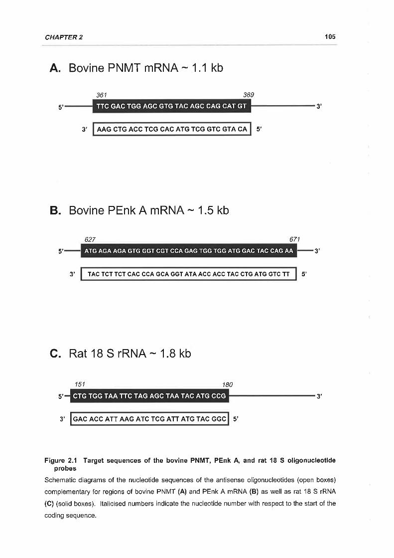
CHAPTER 2 10s
A. Bovine PNMT mRNA - 1.1 kb
361 389
TTC GAC TGG AGC GTG TAC AGC CAG CAT GT5' 3'
3',
B. Bovine PEnk A mRNA - 1.5 kb
627
AAG CTG ACC TCG CAC ATG TCG GTC GTA CA 5',
671
5', 3'
TAC TCT TCT CAC CCA GCA GGT ATA ACC ACC TAC CTG ATG GTC TT
C. Rat 18 S rRNA - 1.8 kb
151 180
3' 5',
ATG AGA AGA GTG GGT CGT CCA GAG TGG TGG ATG GAC TAC CAG AA
CTG TGG TAA TTC TAG AGC TAA TAC ATG CCG 3'
3' GAC ACC ATT AAG ATC TCG ATT ATG TAC GGC 5'
Figure 2.1 Target sequences of the bovine PNMT, PEnk A, and rat 18 S oligonucleotideprobes
Schematic diagrams of the nucleotide sequences of the antisense oligonucleotides (open boxes)
complementary for regions of bovine PNMT (A) and PEnk A mRNA (B) as well as rat '18 S rRNA
(C) (solid boxes). ltalicised numbers indicate the nucleotide number with respect to the start of the
coding sequence.
5'
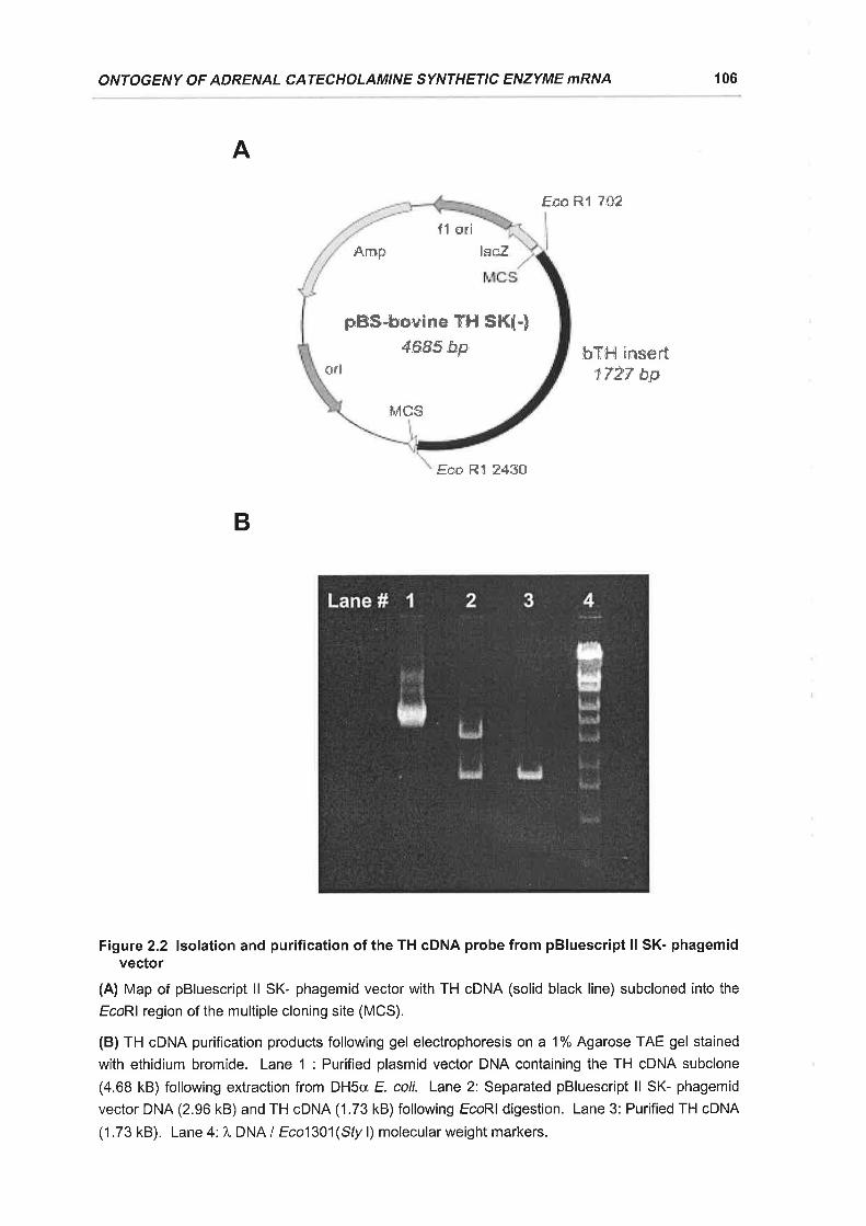
ONTOGENY OF ADRENAL CATECHOLAMINE SYNTHETIC ENZYME mRNA 106
A
Fcn R1 7*?
bY{-il år"¡sert"rTÅ'f bp
Eso,Rl 8430
Figure 2.2 lsolation and purification of the TH cDNA probe from pBluescript ll SK- phagemidvector
(A) Map of pBluescript ll SK- phagemid vector with TH cDNA (solid black line) subcloned into the
EcoRl region of the multiple cloning site (MCS).
(B) TH cDNA purification products following gel electrophoresis on a 1% Agarose TAE gel stained
with ethidium bromide. Lane 1 : Purified plasmid vector DNA containing the TH cDNA subclone
(4.68 kB) following extraction from DHSa E. coli. Lane 2: Separated pBluescript ll SK- phagemid
vector DNA (2.96 kB)and TH cDNA (1.73 kB) following EcoRl digestion. Lane 3: Purified TH cDNA
(1 .73 kB). Lane 4: 1, DNA I Eco1301(Sfy l) molecular weight markers.
B
pEfS'lro,uine TH SK{+4685 åp'
fl ori
lscZ
ildcs
s,mF
o*i
CHAPTER 2 107
After denaturation, 2 pl of loading buffer (50 % glycerol, 1 mM EDTA, 0.25 %
bromophenol blue, and 0.25 % xylene cyanol) was added to the samples and total
RNA was separated by electrophoresis on a denaturing gel containing 1 %
agarose (BDH, Poole, England) and2.2 Mformaldehyde, using 1 x formaldehyde
gel running buffer. Following gel electrophoresis, total RNA was transferred to
Zetaprobe nylon membranes by capillary blotting (Biorad, Richmond, CA, USA) as
described by Sambrook ef a/. (1989), using 10 x SSC (20 x SSC: 175.3 g.l-1 NaCl,
88.2 g.l-1 sodium citrate, pH 7.0) as the transfer buffer. Lanes containing RNA
size markers (Millennium Markers; Ambion, Austin, TX, USA) were removed from
the gels prior to total RNA transfer whereupon they were stained with ethidium
bromide and photographed under UV light to allow for the verification of RNA
transcript sizes. Membranes were washed in'10 x SSC for 10 min at RT before
being baked for t h at B0"C and stored flat at -20"C until hybridisation.
2.2.7 MEMBRANE HYBRIDISATION
For oligonucleotide probes, membranes were prehybridised at 50'C for 18 h in a
solution consisting of 5 x SSC, 20 mM NaH2POa, pH 7 .2,7 % SDS, 5 x Denhardt's
(50 x Denhardt's: 5 g Pharmacia Ficoll 400, 5 g polyvinylpyrollidone, 5 g bovine
serum albumin) and 100 pg.ml-1 of heat denatured salmon sperm DNA
(Boheringer Mannheim). Membranes were hybridised with 1 -2 x 10⁶ cpm.ml-r of
labelled oligonucleotide in 30 ml of fresh hybridisation solution for 20 h at 50'C.
The same protocol was used for the TH cDNA probe, however the hybridisation
solution consisted of 50 % (v I v) deionised formamide, 5 x SSPE (20 x SSPE:
175.3 g.l-1 Nacl, 27.6 g.l-1 NaHzPo¿.H2o,7.4 g.t-1 EDTA, pH 7.4),7 % sDS, and
100 pg.ml-1 of heat denatured salmon sperm DNA (Boehringer Mannheim), with
hybridisation being carried out at 42C. Following hybridisation the membranes
were washed in 1 x SSC and 0.1 % SDS at their hybridisation temperatures (1 x
30 min). The membranes were then washed at their hybridisation temperature in
0.1 x SSC and 0.1 % SDS (2 x 30 min), briefly air dried, and exposed to a Fuji
BAS-Ills phosphorimager plate (Fuji Photo Film Co., Tokyo, Japan) for 24 - 48 h.
Autoradiographs were visualised using a Fuji BAS 1000 phosphorimager and
quantitated using Fuji MacBAS software (Fuji Photo Film Co.).
ONTOGENY OF ADRENAL CATECHOLAMINE SYNTHETIC ENZYME 'zl,RNA
108
2.2.8 DATA ANALYSIS
All data are presented as the mean + standard error of the mean (SEM). The
expressions of TH, PNMT, and PEnk A mRNA are presented as ratios of 18 S
rRNA expression to correct for lane loading errors. Fetal growth, adrenal growth,
total adrenal RNA yields and the ratios of TH, PNMT, and PEnk A mRNA: 18 S
rRNA were compared amongst the different gestational age groups using one way
ANOVA. A Fisher's post hoc test was subsequently used when significance was
identified by ANOVA.
A probability of less than 5 % (ie. P < 0.05) was taken to be significant for all
statistical analyses.
2.3.1 PROBE VALIDATION
Hybridisation of Northern blot membranes with the TH cDNA probe yielded a
prominent band which migrated at around 1.8 kb (Figure 2.34), consistent with the
established size of bovine TH mRNA (D'Mello et al., 1988). The PNMT and
PEnk A antisense oligonucleotide probes hybridised to bands which migrated at
approximately 1.1 and 1.5 kb respectively (Figures 238 & C), which is in
agreement with the previously reported sizes for both bovine and ovine PNMT
(Baetge et al., 1986; Wan ef al., 1989b) and PEnk A mRNA transcripts (Jingami ef
al., 1984; Wan ef al., 1987). The PNMT oligonucleotide probe also specifically
labelled a second band of around 4.5 kb (not shown) which has also been
reported by Wan and colleagues (Wan et al., 1991b), and may represent a
precursor RNA transcript for the mature PNMT mRNA. Only the signal intensity of
the 1.1 kb band was measured in these studies to estimate PNMT mRNA
expression. The 18 S antisense oligonucleotide probe hybridised to a single
prominent band at 1.9 kb, which correlates well with the predicted size of rat 18 S
rRNA (Chan et al., 1984, Figure 2.3D). Autoradiographs of a Northern blot
membrane onto which dilutions of fractionated total RNA from a 140 d fetal sheep
adrenal had been transferred, demonstrated an increasing signal intensity for
.2.3 RESULTS
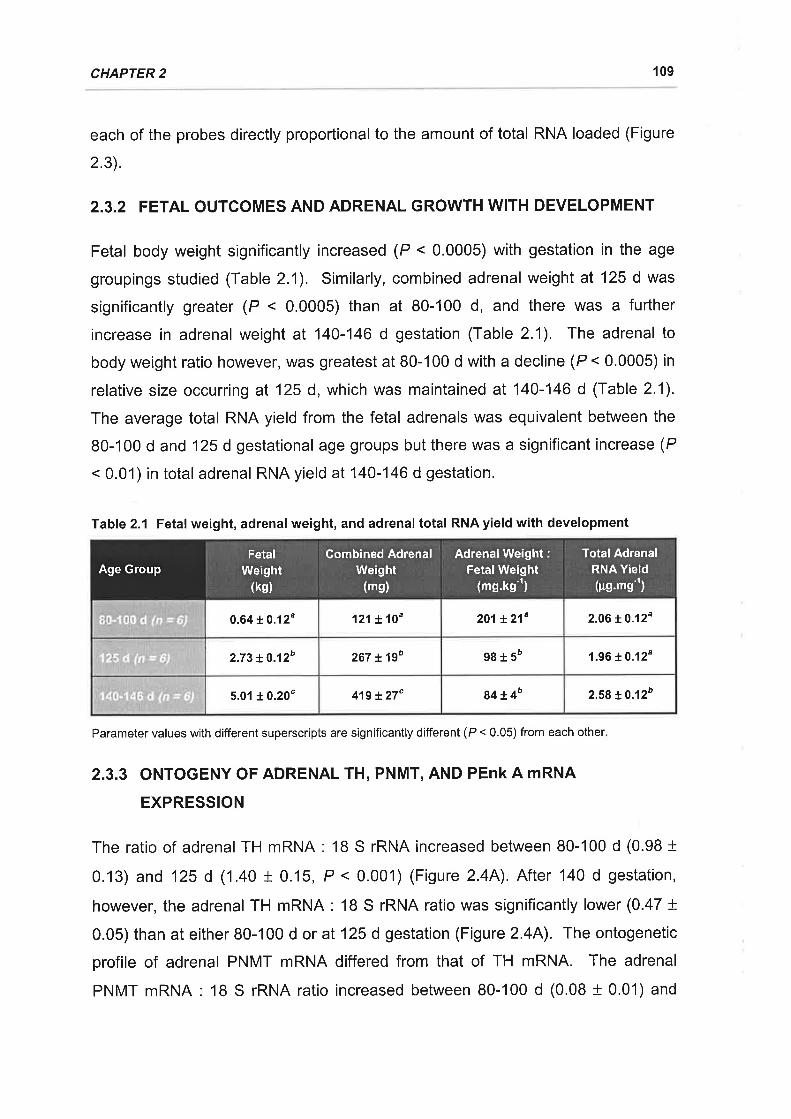
CHAPTER 2 109
each of the probes directly proportional to the amount of total RNA loaded (Figure
2.3).
2.3.2 FETAL OUTCOMES AND ADRENAL GROWTH WITH DEVELOPMENT
Fetal body weight significantly increased (P < 0.0005) with gestation in the age
groupings studied (Table 2.1). Similarly, combined adrenal weight at 125 d was
significantly greater (P < 0.0005) than at 80-100 d, and there was a further
increase in adrenal weight at 140-146 d gestation (Table 2.1). The adrenal to
body weight ratio however, was greatest at B0-100 d with a decline (P < 0.0005) in
relative size occurring at 125 d, which was maintained at140-146 d (Table 2.1).
The average total RNA yield from the fetal adrenals was equivalent between the
80-100 d and 125 d gestational age groups but there was a significant increase (P
< 0.01) in total adrenal RNA yield at 140-146 d gestation.
Table 2.1 Fetal weight, adrenalweight, and adrenaltotal RNAyield with development
Parameter values with different superscripts are significantly different (P < 0.05) from each other
2.3.3 ONTOGENY OF ADRENAL TH, PNMT, AND PEnk A mRNA
EXPRESSION
The ratio of adrenal TH mRNA: 18 S rRNA increased between 80-100 d (0.98 I0.13) and 125 d (1.40 t 0.15, P < 0.001) (Figure 2.4A). After 140 d gestation,
however, the adrenal TH mRNA: 18 S rRNA ratio was significantly lower (0.47 t0.05) than at either 80-100 d or at 125 d gestation (Figure 2.4A). The ontogenetic
profile of adrenal PNMT mRNA differed from that of TH mRNA. The adrenal
PNMT mRNA: 1B S rRNA ratio increased between 80-100 d (0.08 + 0.01) and
121 t 10" 201t21" 2.06 t 0.12"0.64 to.12u
zal t lgo 98r5' 1.96 + 0.12"z.z3 +o.tzo
419 t27' 84 t4o z.sg +0.120s.ol + o.zo"
FetalWeight
(ks)
Combined AdrenalWeight
(ms)
Adrenal Weight:FetalWeight
(mg.kg-t)
Total AdrenalRNA Yield(pg.,ng'')
Age Group
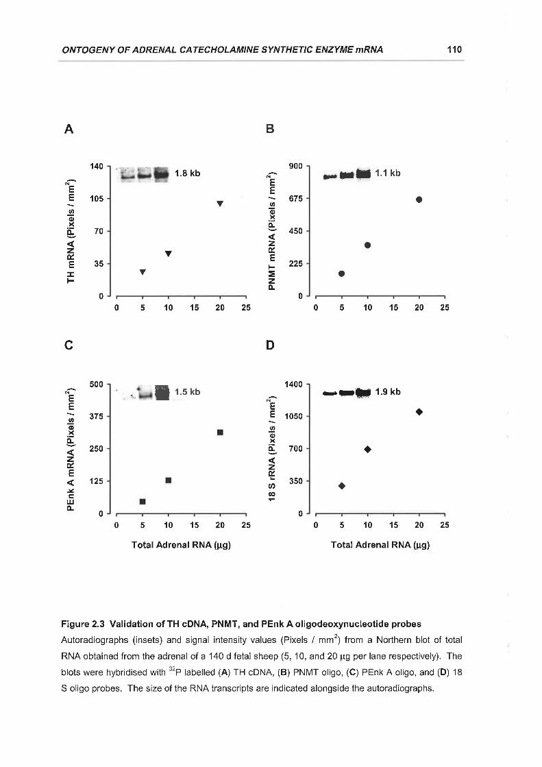
ONTOGENY OF ADRENAL CATECHOLAMINE SYNTHETIC ENZYME mRNA 110
Y
V
140
105
70
35
A
EE
Øo.xo-
zú.EI
o
o
900N
EE\ 675Io.xo-v 450
zÉ.tr; 225
=zÀ
00
a
400
050
700
350
1
1
D
NEE
Io.xo-
zÉ,
U'@
500
375
250
125
c
N
EE
Io.xÀ
zú.E
.Ytrl¡lÈ
B
00
00
1.8 kb
a¿ f r's rto
510152025
TotalAdrenal RNA (pg)
x5l 1.1 kb
Y
0510152025
o
0510152025
--ffi 1.9 kb
o
a
510152025
Total Adrenal RNA (pg)
Figure 2.3 Validation of TH cDNA, PNMT, and PEnk A oligodeoxynucleotide probes
Autoradiographs (insets) and signal intensity values (Pixels / mm2¡ from a No¡1hern blot of total
RNA obtained from the adrenal of a 140 d fetal sheep (5, '10, and 20 pg per lane respectively). The
blots were hybridised with 32P labelled (A) TH GDNA, (B) PNMT oligo, (C) PEnk A oligo, and (D) 18
S oligo probes. ïhe size of the RNA transcripts are indicated alongside the autoradiographs.

CHAPTER 2 111
A1.6
1.2
0.8
0.4
0.0
2.4
1.8
'1.2
0.6
0.0
c
a
a
80-100 d 125 d 140-146 d
GestationalAge
Figure 2.4 Ontogeny of TH, PNMT, and PEnk A mRNA expression in the fetal sheep adrenal
Gestational age comparison of adrenal TH mRNA (A), PNMT mRNA (B), and PEnk A mRNA (C)
relative to 18 S rRNA (mean t SEM) in fetal sheep aged 80-100 d (n = 6, white histograms),125 d
(n = 6, grey histograms), and 140-146 d (n = 6, black histograms). Significant differences [P < 0.05]
between the mean ratios at different gestational ages are denoted by different superscript letters.
B
b
zÉ.
ØG'
zúEI
zÉ.
(¡)@
..b(-zxÉ-E
=zÈ
a
b1 1o.oÉ.
Ø 7.s@
.-ä þ 5.0t¿E
llcH o.o
ab
a
c

ONTOGENY OF ADRENAL CATECHOLAMTNE SYNTHETIC ENZYME mRNA 112
140-146 d gestation (0.17 + 0.03, P < 0.05) (Figure 2.48). The ratio of adrenal
PEnk A mRNA : 18 S rRNA was higher (P < 0.01), however, at 125 d (0.085 t0.005) than at either 80-100 d (0.038 t 0.007) or at140-146 d gestation (0.055 t0.013) (Figure 2.4C).
The present study has found that there is a differential pattern of expression of
catecholamine synthetic enzyme and PEnk A mRNA levels in the fetal sheep
adrenal during late gestation. lt was also established that the adrenal gland is
proportionally larger in the 80-100 d gestation fetuses than it is in later gestation.
This may reflect the importance of the fetal adrenal gland as a source of
catecholamines when the peripheral sympathetic innervation is quite immature
(Comline & Silver, 1966; Lebowitz et al., 1972). Total adrenal RNA yield was
significantly elevated in the 140-146 d gestation fetal sheep, coincident with the
major phase of adrenocortical growth which is associated with both cellular
hypertrophy and hyperplasia (Boshier & Holloway, 1989b).
ln the present study adrenal TH mRNA levels increased between 100 and 125 d
gestation and then significantly decreased after 140 d gestation. The increase in
TH mRNA expression at 125 d is coincident with the onset of splanchnic
innervation of the sheep adrenal. Ultrastructural studies have shown that
synapses between splanchnic nerve terminals and adrenal chromaffin cells
develop in the fetal sheep adrenal between 100 and 130 days gestation (Boshier
et al., 1989a) and there is evidence that functional splanchnic innervation of the
adrenal develops around 120-130 days gestation (Cheung, 1990; Comline &
Silver, 1961). lt is possible that the onset of adrenal innervation is associated with
a related stimulation of TH mRNA expression. There is evidence from studies in
the adult rat and in bovine adrenomedullary cells that TH mRNA levels are
increased after trans-synaptic or cholinergic stimulation and membrane
depolarisation (Biguet ef al., 1986; Faucon Biguet et al., 1991; Fossom et al.,
1991b; Hiremagalur ef a/., 1993; Schalling et al., 1988; Stachowiak et al., 1994;
Stachowiak et al., 1990b). lt is interesting that the increase in adrenomedullary
TH mRNA expression at 125 d gestation is relatively transient in that there is a
2.4 DISCUSSTON
CHAPTER 2 113
subsequent decrease in adrenal TH mRNA levels in the week before delivery. We
have previously shown that there is a decrease in the noradrenaline secretory
response of the fetal sheep adrenal to sub-maximal doses of Acetylcholine (ACh)
alr. 144 d compared with earlier in gestation and that this is not associated with a
decrease in the size of the releasable pool of noradrenaline in the fetal adrenal
(Butler et al., 1995). One possible explanation for the parallel decrease in TH
mRNA and adrenal sensitivity to ACh may be that the full establishment of
functional innervation of the fetal adrenal in late gestation is associated with an
absolute increase in the resting membrane potential, ie a relative
hyperpolarisation of the noradrenaline containing cells.
Adrenal PEnk A mRNA levels also significantly increased al 125 d gestation,
although there was no significant decline in the mRNA levels for the opioid peptide
precursor after 140 d gestation. A range of studies in bovine adrenomedullary
cells in culture have shown that PEnk A mRNA levels are increased in response to
nicotinic stimulation and membrane depolarisation (Kley et al., 1986; Kley et al.,
1987; Stachowiak et al., 1990c; Wan ef al., 1991b) and it is therefore possible that
the onset of splanchnic innervation stimulates a coordinated increase in PEnk A
and TH mRNA levels. lt is interesting that the pattern of PEnk A mRNA and Enk
peptide localisation changes in the fetal sheep adrenal at around 125 d gestation,
from localisation within both noradrenaline and adrenaline cells to the adult
pattern of localisation of Enk-containing peptides predominantly within adrenaline
cells (McMillen et al., 1988; Wan et al., 1989b). We have previously
demonstrated that the change in Enk localisation in the sheep adrenal is
dependent on an intact fetal pituitary-thyroid axis and have suggested that
splanchnic innervation of the fetal adrenal may also be dependent on fetal thyroid
status (Coulter et al., 1989a; Simonetta et al., 1996c). lt appears that neural or
hormonal events at around 125 d gestation produce both transient and long term
changes in Enk gene expression and localisation within the sheep adrenal.
ln contrast to TH mRNA, adrenal PNMT mRNA levels were highest after 140 days
gestation than at any earlier gestational age studied. This gestational increase in
PNMT mRNA levels is coincident with an increase in adrenal adrenaline content
after 130 days gestation (Comline & Silver, 196'1), an increase in the basal output
ONTOGENY OF ADRENAL CATECHOLAMINE SYNTHETIC ENZYME mRNA 114
of adrenaline from the perfused adrenal preparation and in circulating adrenaline
concentrations in the sheep fetus after 135 days gestation (Butler et al., 1995;
Simonetta et a\.,1993). lt is likely that the prepartum increases in adrenal PNMT
mRNA levels and adrenaline synthesis are directly related to the increase in
adrenal corticosteroidogenesis which occurs in the last 10-15 d of gestation in the
sheep (Challis & Brooks, 1989). lt has previously been established in the fetal
and neonatal rat that whilst the emergence of the adrenergic cell phenotype is not
dependent on glucocorticoids (Bohn et al., 1981) the subsequent maintenance of
adrenaline synthesis is glucocorticoid dependent. Glucocorticoids have been
shown to stimulate an increase in PNMT mRNA levels in bovine adrenomedullary
cells in culture (Stachowiak ef al., 1990d;Wan & Livett, 1989), probably through
an action at the glucocorticoid response element present in the 5' regulatory
region of the PNMT gene (Ross et al., 1990).
It is clear, that whatever the precise neural and hormonal factors involved in the
control of catecholamine and opioid peptide biosynthesis, there is a differential
regulation of the gene expression of TH, PNMT and PEnk A within the adrenal in
late gestation in sheep.
3. IMPACT OF ACUTE HYPOXAEMIA ON THE EXPRESSION OFTH AND PNMT MRNA IN THE ADRENAL GLAND OF THE
FETAL SHEEP
3.1 INTRODUGTION
ln Chapter 2 it was found that the ontogenetic changes in adrenal TH and PNMT
mRNA expression coincide with the development of functional splanchnic
innervation and activation of the HPA axis. ln the adult, both splanchnic
innervation and the HPA axis play major roles in the regulation of catecholamine
biosynthesis during stress (Fluharty et al., 1985; Kvetnansky et al., 1995; Ungar &
Phillips, 1983). The secretion of catecholamines from the mature adrenal medulla
in response to stress is predominantly stimulated by reflex activation of the
splanchnic nerve (Biesold et a1.,1989; Lee ef al., 1987; Seidler & Slotkin, 1986b).
It is well established that there is a functional coupling of catecholamine secretion
and synthesis in the mature adrenal gland which aids in the maintenance of
catecholamine stores in the face of increased secretory demands (Bygdeman &
von Euler, '1958; Wakade et al., 1988; Weiner, 1975). The stimulation-secretion-
synthesis coupling of catecholamines within the chromaffin cells of the mature
adrenal medulla is mediated by trans-synaptic events which occur in response to
splanchnic nerve activation. Trans-synaptic stimulation of the adrenal medulla
increases catecholamine synthetic enzyme levels, activity, and gene expression
(Fluharty et al., 1983; Fluharty et al., 1985; Wong et al., 1993; Wong & Wang,
1ee4).
Fully functional innervation of the fetal sheep adrenal gland by the preganglionic,
cholinergic, splanchnic nerve does not develop until around 120 d gestation
(Cheung, 1990; Comline & Silver, 1961). lt is clear however, that the immature
adrenal medulla of the sheep and a number of other species is capable of
secreting catecholamines in response to hypoxia by a direct or non-neurogenic
mechanism, prior to functional splanchnic innervation of the adrenal gland
(Cheung, 1990; Comline & Silver, 1966; Seidler & Slotkin, 1986b; Slotkin &
Seidler, 1988). lt has yet to be determined if there is also an associated
catecholamine synthesis-secretion coupling present in the immature fetal adrenal
for non-neurogenic stimulation. The cellular mechanisms underlying the non-
neurogenic secretion of catecholamines from the fetal and neonatal adrenal in
115
CHAPTER 3 116
response to hypoxia have only recently been examined (Adams et al., 1996;
Rychkov et al., 1998; Thompson et al., 1997). Both neurogenic and non-
neurogenic catecholamine secretion appear to share common mechanistic
elements including membrane depolarisation and the influx of intracellular calcium
into the cell via voltage sensitive Ca2* channels (Adams ef a/., 1996; Mochizuki-
Oda et al., 1997; Thompson et al., 1997). ln chromaffin cells obtained from
mature bovine adrenal glands, cellular depolarisation and increased intracellular
Ca2* levels have each been demonstrated to stimulate catecholamine synthetic
enzyme activity and the expression of TH and PNMT mRNA (Stachowiak et al.,
1994; Stachowiak et al., 1990c; Wan ef al., 1991b).
Given the apparent similarities between the stimulation of catecholamine secretion
by both neurogenic and non-neurogenic pathways it is hypothesised that there will
also be changes in catecholamine synthesis associated with non-neurogenic
catecholamine secretion. ln order to examine this hypothesis the current study
examined the effect of acute hypoxaemia upon the time course of expression of
TH and PNMT mRNA in the adrenals of the fetal sheep both before and after the
development of functional splanchnic innervation. ln order to determine the
relative contribution of the splanchnic nerve to catecholamine synthetic enzyme
gene expression, adrenal TH and PNMT mRNA expression following acute
hypoxia was also determined in fetal sheep in which cholinergic transmission was
blocked by the nicotinic receptor antagonist, hexamethonium.
All procedures were approved by the University of Adelaide Standing Committee
on Ethics in Animal Experimentation.
3.2.1 ANIMALS AND SURGERY
Fifty two fetal sheep from timed pregnant Border Leicester x Merino ewes were
used in this study. Surgerywas performed on the ewes at either 88-92 d or 116-
120 d gestation (calculated according to date of mating) under general
anaesthesia induced by intravenous injection of sodium thiopentone (1.25 g;
Pentothal, Rhone Merieux, Pinkenba, QLD, Australia) and maintained with
halothane (3-4 % in oxygen; Fluothane, lCl, Melbourne, VlC, Australia). Ewes
ACUTE HYPOXIA AND ADRENOMEDULLARY REGULATION 117
were administered 2 ml of antibiotics (250 mg.ml-1 procaine penicillin,2SO mg.ml-r
dihydrostreptomycin sulphate, 20 mg.ml-1 Procaine Hydrochloride; Penstrep llium,
Troy Laboratories, Smithfield, NSW, Australia) intramuscularly, prior to surgery.
During surgery the uterus was exposed by a midline laparotomy incision and the
fetal head was palpated. The fetal head and neck were then delivered through an
incision in the uterine wall and the fetal membranes. Single lumen polyvinyl
catheters (Critchley Electrical Products, Silverwater, NSW, Australia) were
inserted into the fetal carotid artery and external jugular vein (outer diameter, 1 .52
mm; inner diameter, 0.86 mm) and the amniotic sac (outer diameter, 2.70 mm;
inner diameter, 1.50 mm) essentially as described by Dawes et al. (1972). The
fetus was also administered 2 ml of antibiotics (Penstrep llium, Troy Laboratories)
intramuscularly during surgery. The fetal head was then replaced and the uterus
and fetal membranes were closed in a single layer using catgut (2 I 0; Ethicon,
Johnson and Johnson, Sydney, NSW, Australia). A layer of inverting sutures (2 /
0 catgut, Ethicon, Johnson and Johnson) was subsequently placed in the
myometrium. The maternal abdomen was closed in two layers, the peritoneum
and rectus sheet followed by the skin and subcutaneous tissue, using catgut (3 /
0; Ethicon, Johnson and Johnson) and vetafil synthetic suture (0.3 mm; WDT,
Garbsen, Germany) respectively. A catheter (outer diameter, 2.70 mm; inner
diameter, ',l.50 mm) was also inserted into the maternal external jugular vein. The
catheters were flushed and filled with heparinised saline (500 lU.ml-1; Multiparin,
Fisons Pharmaceuticals, Sydney, NSW, Australia) and the fetal catheters were
exteriorised through an incision in the ewe's flank.
3.2.2 POST OPERATIVE GARE
Ewes were housed in individual metabolic crates in animal holding rooms under a
12 h light-dark cycle and fed lucerne chaff once daily between 0900 and 1300 h
with water ad libitum. Sodium ampicillin (500 mg, Austrapen, Commonwealth
Serum Laboratories, Melbourne, VlC, Australia) was administered intra-
amniotically to all fetal sheep for 4 consecutive days post operatively. A
post operative recovery of at least a week was allowed before any experiment was
conducted.
CHAPTER 3 118
3.2.3 FETAL HYPOXIA
Separate groups of fetuses at 96-105 d (n = 15) and 129-144 d (n = 15) were
subjected to 30 min of hypoxia. Fetal hypoxia was achieved in this study by
altering the maternal F¡Oz. Following a 60 min baseline period a large clear
polypropylene bag was tied loosely over the ewe's head and a mixture of air
(39 %), N2 (58 %), and CO2 (3 %) was infused into the bag at a flow rate of
58 Lmin-1 (Figure 3.1). The gas mixture was titrated over the 30 min experimental
period by adjusting the balance of air and nitrogen to maintain fetal SuOz at
approximately 50 % of baseline values. The normoxia controls for the hypoxia
experiment consisted of 96-105 d (n = 5) and 129-144 d (n = 5) fetuses for which
the pregnant ewes were administered a gas mixture which consisted of air (97 o/o)
and COz (3 %) at a flow rate of 58 l.min-1 as outlined for the hypoxia experiment.
3.2.4 HEXAMETHONIUM
ln a separate group of 129-144 d (n = 12) fetuses hexamethonium
(hexamethonium bromide dihydrate; ICN Biomedicals, Cleveland, OH, USA) was
administered by a 3 ml intravenous bolus (25 mg.kg-t in 0.9 % saline). The dose
of hexamethonium used in this study has previously been demonstrated to
produce a blockade of sympathetic function which persists for several hours in the
fetal sheep (Brace & Brittingham, 1986). Hexamethonium was administered at -
30 min relative to fetal exposure to either hypoxia (n = 6) or normoxia (n = 6).
3.2.5 BLOOD SAMPLING
Fetal arterial blood samples (0.5 ml) were collected once daily on the first four
days following surgery and every second day thereafter for measurement of fetal
blood gases and pH using an ABL 520 blood gas analyser (Radiometer,
Copenhagen, Denmark) to assess fetal well being. During an experiment, 3.5 ml
samples of fetal arterial blood were obtained at the following time points relative to
the onsetof maternal F¡O2 regulation; -60, -35, -25, -5,5, 10,30, and 60 min. An
equivalent volume of isotonic saline was infused into the fetus following the
withdrawal of each sample. A fetal arterial blood sample (0.5 ml) was used for
blood gas analysis as outlined above, whilst the remaining 3 ml was placed into
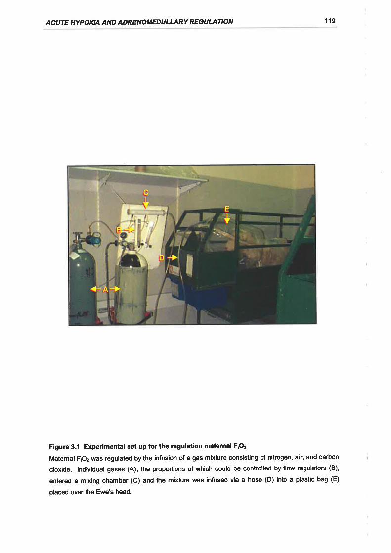
ACUTE HYPOXIA AND ADRENOTTEDUL¿,,RY REGULATION l19
Figure 3.1 Experimental set up for the regulation maternal F¡Oz
Maternal F¡O2 was regulated by the infusion of a gas mixture consisting of nitrogen, air, and carbon
dioxide. Individual gases (A), the proportions of which could be controlled by flow regulators (B),
entered a mixing chamber (C) and the mixture was infused via a hose (D) into a plastic bag (E)
placed over the Ewe's head.
CHAPTER 3 120
ice chilled heparinised tubes (125 U, Disposable Products, Adelaide, SA,
Australia). The tubes were spun at 1800 g for 10 min at 4'C and the plasma was
subsequently separated and transferred to polypropylene tubes whereupon they
were snap frozen in liquid N2. Plasma samples were stored at -20'C until assayed
for catecholamine content.
3.2.6 POST MORTEM AND TISSUE COLLECTION
The ewes were sacrificed with an overdose of sodium pentobarbitone (8.125 g;
Lethabarb, Virbac, Peakhurst, NSW, Australia) and the fetal sheep were delivered
via laparotomy and killed by decapitation. The adrenal glands were dissected
from each fetus, weighed, snap frozen in liquid Nz, ârd stored at -70'C until total
RNA was extracted.
Adrenals were collected from fetuses sacrificed at 3-5 h (96-105 d, n = 5;129'
144 d,n = 5), 12 h (96-105 d, n = 5; 129-144d, n = 5), and 20 h (96-105 d, n = 5;
129-144 d, n = 5) after the onset of hypoxia. Adrenals were collected from the
fetal sheep in the control group (96-105 d, n = 5; 129-144 d, n = 5) following the
onset of experimental normoxia over the range of time points described for the
hypoxia group. For the hexamethonium treated fetuses adrenals were collected
from fetuses 3-5 h after the onset of either experimental normoxia (n = 6) or
hypoxia (n = 6).
3.2.7 PLASMA CATECHOLAMINE EXTRACTION
Prior to extraction, plasma samples collected at -25 and -5 min (basal) as well as
the 5 and 10 min (hypoxia) samples were combined in equivalent portions for
catecholamine measurement. The maximal rise in plasma catecholamines in the
fetal sheep in response to acute hypoxia has been established within our
laboratory to occur within the first 10 min of hypoxia (Simonetta et al., 1996a).
With the hexamethonium treated fetuses, the plasma samples collected at -60
and -35 min were also combined before extraction for the determination of basal
catecholamine levels prior to the addition of hexamethonium.
Plasma catecholamines were partially purified by alumina absorption using a BAS
plasma catecholamine kit (MF-9017; Bioanalytical Systems, West Lafayette, lN,
USA) essentially as per the manufacturer's instructions with minor modifications.
ACUTE HYPOXIA AND ADRENOMEDULLARY REGULATION 121
Briefly 500 pl of fetal plasma was used and made up to a volume of 2 ml with
distilled water and the sample pre treatment step was omitted. 3,4-
dihydroxybenzylamine (DHBA; Bioanalytical Systems) was added to each sample
(50 pl at 80 pmol.l-1 in 0.1 Mperchloric acid) as an internal standard to monitor the
recovery of catecholamines from plasma. The efficiency of catecholamine
recovery averaged 67.4 ! 1.8 To.
3.2.8 CATEGHOLAMINE MEASUREMENT
50 ¡rl aliquots of the extracted samples were injected onto a BAS 2004 HPLC
system (Bioanalytical Systems) using a refrigerated microsampler (CMA I
Microdialysis, Stockholm, Sweden) at 4'C. Separation of the catecholamines was
achieved using a 3 pm ODS, 100 x 3.2mm, Brownlee Velosep RP-18 cartridge
column (Perkin Elmer, Non¡ualk, CT, USA) and MP-2 catecholamine mobile phase
(Bioanalytical Systems). The column temperature was maintained at 40'C and
the mobile phase flow rate was 1 ml.min-1. The potential of the glassy carbon
electrode was set at + 700 mV vs. a Ag / AgCl reference electrode whilst the gain
was set to 0.5 nA. Catecholamines were automatically quantified by peak height
integration using an IBM compatible personal computer equipped with Barspec
Data System chromatographic software (Barspec, Rehovot, lsrael). Peak heights
referenced to standard curves generated for authentic catecholamine standards
(norepinephrine bitartrate, epinephrine bitartrate, and DHBA hydrobromide;
Bioanalytical Systems; 0.3125-20 nmol.l-1 in 0.1 M PCA; Figure 3.2) to determine
the catecholamine content of the samples. The sensitivity of the assay was
approximately 0.05 pmol.ml-1 and the intra- and inter-assay , coefficients of
variation (CVs) were less than 10 Yo for both noradrenaline and adrenaline.
3.2.9 TOTAL RNA EXTRACTION
Total adrenal RNA was extracted from each adrenal using Tri-reagent (Sigma
Chemical Co., St. Louis, MO, USA), a proprietary version of the single-step
method of RNA isolation by acid guanidinium thiocyanate-phenol chloroform
extraction (Chomczynski, 1993; Chomczynski & Sacchi, 1987). Briefly, adrenals
were homogenised separately in 1 ml of Tri-reagent using a Polytron PT3000
homogeniser (Kinematica, Littau, Switzerland) at 30 000 rpm. The homogenate
CHAPTER 3 122
was transferred to a sterile 1.5 ml eppendorf tube and bromo-3-chloropropane
(0.1 ml) was added to the homogenate. The samples were then mixed and
allowed to stand at room temperature (RT) for 5 min. The samples were
centrifuged at 12 000 g for 10 min aI 4"C and the upper aqueous phase was
transferred to a separate sterile eppendorf tube. Total RNA was precipitated from
the aqueous phase by adding isopropanol (0.5 ml) and allowing the samples to
stand at RT. After 10 min, the samples were centrifuged (12 000 gfor 10 min at
4'C) and the supernatant was subsequently removed. The RNA pellets were
washed by adding 1 ml of 75 % ethanol and centrifuging at 12 000 g for 10 min at
4"C. Following the removal of the ethanol solution the RNA pellets were air dried
and resuspended in an appropriate volume of sterile water. Nucleic acid
concentration, yield, and purity were quantified by spectrophotometric
measurement at 260 and 280 nm. Total RNA solutions were stored at -70"C prior
to Northern blot analysis.
3.2.10 PROBES AND PROBE LABELLING
The probes utilised in this study and the radiolabelling methods for the probes are
described in detail in chapter 2. To summarise, oligonucleotide antisense probes
complementary to nucleotides 361-389 of the peptide coding region of bovine
PNMT mRNA (29 mer, Wan ef al., 1989b), nucleotides 627-671 of the peptide
coding region of bovine PEnk A mRNA (44 mer, Wan et al., 1987), and
nucleotides 151-180 of rat 18 S rRNA (30 mer, Chan et al., 1984) sequence were
synthesised (Bresatec, Thebarton, SA, Australia). A full length (1.73 kb) bovine
TH cDNA (D'Mello et al., 1988) was used to detect TH mRNA. The
oligonucleotides were end labelled using T4 polynucleotide kinase 7.9 U.pl-1,
Pharmacia, North Ryde, NSW, Australia) and y-¡32e1nte (4000 Ci.mmol-1,
Bresatec) in accordance with the method described for the end labelling of
oligonucleotides by Sambrook ef a/. (1989). The cDNA probe was labelled by the
random priming method with o-[32P]dCTP (3000 Ci.mmol-1, Bresatec) and Klenow
fragment (6.4 U.pl-r) using an oligolabelling kit (Pharmacia) as per manufacturer's
instructions.
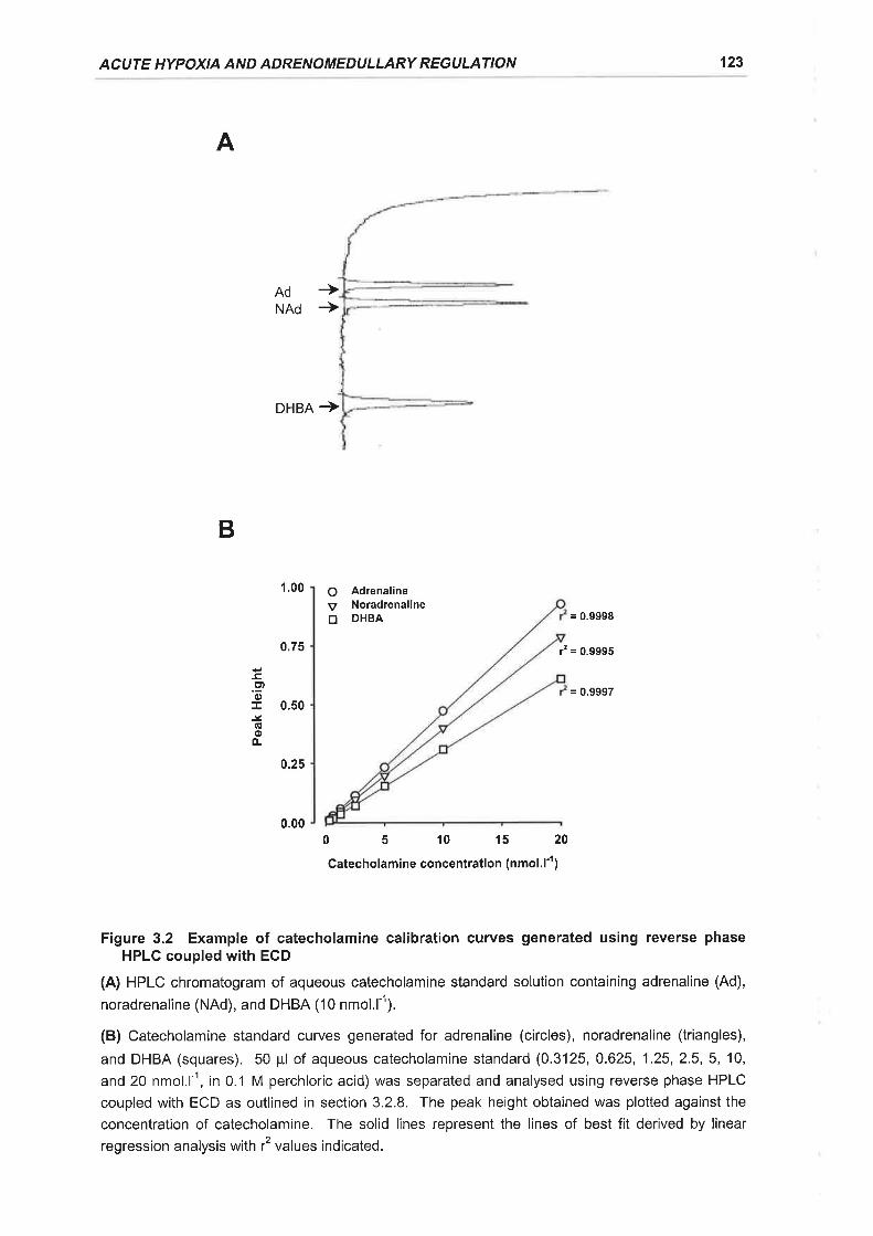
ACUTE HYPOXIA AND ADRENOMEDULLARY REGULATION 123
A
+Ad
B
NAd +
DHBA +
1.00 O AdrenalineV Noradrenalinetr DHBA = 0.9998
0.75 É = 0.9995
= 0.99970.50
0.25
0.0005101520Gatecholamine concentration (nmol.l'r)
Figure 3.2 Example of catecholamine calibration curves generated using reverse phaseHPLC coupled w¡th ECD
(A) HPLC chromatogram of aqueous catecholamine standard solution containing adrenaline (Ad),
noradrenaline (NAd), and DHBA (10 nmol.ll).
(B) Catecholamine standard curves generated for adrenaline (circles), noradrenal¡ne (triangles),
and DHBA (squares). 50 pl of aqueous catecholamine standard (0.3125, 0.625, 1.25,2.5,5, 10,
and 20 nmol.l-1, in 0.1 M perchloric acid) was separated and analysed using reverse phase HPLC
coupled with ECD as outlined in section 3.2.8. The peak height obtained was plotted against the
concentration of catecholamine. The solid lines represent the lines of best fit derived by linear
regression analysis with r2 values indicated.
ct0)J-lÉ(!oo-
CHAPTER 3 124
3.2.11 NORTHERN BLOT ANALYSIS
For each fetus, 20 pg of total adrenal RNA was denatured in 2.2 M formaldehyde
and 50 % (v lv) formamide at 55'C for 10 min. Separation of the total adrenal
RNA was achieved by electrophoresis on a 1 % agarose gel containing 2.2 M
formaldehyde as described in detail in chapter 2. The separated total adrenal
RNA was subsequently transferred to zetaprobe nylon membranes (Biorad,
Richmond, CA, USA) via downward capillary transfer as described by
Chomczynski & Mackey (1994) using 10 x SSC as the transfer buffer. Following
transfer the membranes were washed in 10 x SSC for 10 min at room temperature
and baked for t h at 80"C to fix the RNA to the membrane.
Prior to hybridisation with oligonucleotide probes the membranes were initially
prehybridised at 50"C for 18 h in a solution consisting of 5 x SSC,20 mM
NaHzPO¿, pH 7.2,7% SDS, 5 x Denhardt's, and 100 pg.ml-1 of heat denatured
Salmon sperm DNA. Membraneswere then hybridised for20 h at 50 C in 15 ml
of fresh hybridisation solution containing 1-2 x 10⁶ cpm.ml-1 of labelled
oligonucleotide. For the TH cDNA probe equivalent prehybridisation and
hybridisation times were used, however, the hybridisation solution consisted of 50
% (v lv) deionised formamide, 5 x SSPE,7 % SDS, pH7.2, and 100 pg.ml-1 of
heat denatured salmon sperm DNA, with hybridisation being carried out at 42C in
the presence of 1-2 x 10⁶ cpm.ml-1 of labelled probe.
Following hybridisation the membranes were washed in 1 x SSC and 0.1 % SDS
(1 x 30 min) and then subsequently in 0.1 x SSC and 0.1 % SDS (1 x 30 min) at
their hybridisation temperatures before being briefly air dried and exposed to a
Fuji BAS-Ills Phosphorimager Plate (Fuji Photo Co., Tokyo, Japan) for 24-48 h.
Autoradiographs were obtained using a Fuji BAS 1000 Phosphorimager (Fuji
Photo Co.) and quantitated using Fuji MacBAS software (Fuji Photo Co.)
3.2.12 STATI STI CAL ANALYS I S
All data are presented as the mean t SEM. Levene's tests for homogeneity of
variance were conducted and logarithmic transformation of data carried out if
appropriate. Blood gas variables over the time course of an experiment were
examined within treatment groups using one way ANOVA with repeated
ACUTE HYPOXIA AND ADRENOMEDULLARY REGULATION 125
measures. Fisher's Post Hoc tests were used to identify significant differences
between mean values. Unpaired one tailed Student's t-tests were used to
compare the percentage changes of fetal plasma catecholamines from baseline
levels in both the normoxia and hypoxia experiments. One way ANOVAS with
Fisher's Post Hoc tests were used to compare the ratios of adrenal PNMT and TH
mRNA : 18 S rRNA expression at different time points and in the hexamethonium
experiments. Polynomial regression analyses were used to determine
relationships of fetal adrenal PNMT mRNA and TH mRNA : 18 S rRNA expression
with the experimentally induced changes in fetal arterial POz during normoxia and
hypoxia. Data from the hypoxia groups at the time points for which a significant
change in adrenal mRNA expression first occurred were used for the regression
analyses.
A probability of less than 5 % (ie. P < 0.05) was taken to be significant.
3.3.1 FETAL ARTERIAL BLOOD GASES AND EXPERIMENTAL HYPOXIA
3.3.1.1 Arterial POz
With experimental normoxia there were no significant changes in fetal arterial POz
from baseline values for either the 96-105 d (Figure 3.3A) or 129-144 d (Figure
3.38) fetal sheep. With experimental hypoxia there were immediate and
sustained reductions (P < 0.0005) in fetal arterial POz from basal values for both
the 96-105 d (41 .9+2.1 o/o,n = 15, Figure 3.3A) and 129-144 d (37.5 + 1.8 Yo,î=
15, Figure 3.38) fetal sheep. Fetal arterial POz recovered to baseline values by
30 min after the cessation of hypoxia in both age groups (Figures 3.34, B).
Hexamethonium administration at -30 min had no significant impact upon basal
arterial POz values in the 129-144 d fetuses (Figure 3.38). There was no change
in arterial POz values with experimental normoxia in the hexamethonium treated
fetuses (Figure 3.38). The reduction in arterial POz which occurred in response to
experimental hypoxia was similar in the hexamethonium treated fetuses (41.5 t1.3 o/o, n = 6) to that observed in the absence of hexamethonium (Figure 3.38).
3.3 RESULTS
CHAPTER 3 126
There was also a recovery of arterial POz levels by 30 min after the end of the
hypoxia period in the hexamethonium treated fetuses (Figure 3.38).
3.3.1.2 Arterial PCOz
Fetal arterial PCOz remained constant with experimental normoxia in the 96-105 d
fetuses (Figure 3.3C) in this age group there was a moderate but significant (P <
0.0005) decline in fetal arterial PCOz from baseline levels (46.0 + 0.7 mmHg) at
10 min (42.0 + 1.1 mmHg) and 30 min (41.0 + 1.0 mmHg) of hypoxia, which
returned to baseline levels after 30 min of recovery (Figure 3.3C). ln the 129-
144 d fetuses there were no significant alterations in fetal arterial PCOz from
baseline values with either experimental normoxia or hypoxia (Figure 3.3D).
Arterial PCOz levels in 129-144 d fetuses was unchanged by ganglionic blockade
with hexamethonium at -30 min (Figure 3.3D). Furthermore, there were no
significant effects of either experimental normoxia or hypoxia upon arterial PCOz
levels in the hexamethonium treated fetuses (Figure 3.3D).
3.3.1.3 Arterial pH
For the 96-105 d fetuses, arterial pH values did not significantly change from their
baseline levels during either experimental hypoxia or normoxia (Figure 3.3E). ln
the 129-144 d fetuses there was also no change in arterial pH values during
experimental normoxia but with hypoxia there was a moderate but significant (P <
0.0005) fall from basal levels (7.389 t 0.006) at 30 min (7.369 + 0.004) which
persisted into the recovery period (60 min, 7.360 t 0.004) (Figure 3.3F).
Basal arterial pH values were unchanged in the 129-144 d fetuses by the
administration of hexamethonium. Hexamethonium treated fetuses displayed a
small reduction (P < 0.0005) in arterial pH over the duration of experimental
normoxia (baseline, 7.393 + 0.003; minimum, 5 min, 7.372 t 0.006), which
recovered after the experimental period. There was no significant effect of
experimental hypoxia on arterial pH in hexamethonium treated fetuses (Figure
3.3F).
ACUTE HYPOXIA AND ADRENOMEDULLARY REGULATION 127
3,3.2 FETAL PLASMA CATECHOLAMINES
3.3.2.1 Basal plasma catecholamine levels
ln the 96-105 d fetal sheep, plasma adrenaline concentrations frequently fell
below the sensitivity of the assay. Basal (-25 and -5 min) noradrenaline levels and
adrenaline levels, when they were detectable, in the 96-105 d fetal sheep (NAd,
0.32t0.09 nmol.l-1, n = 14; Ad,0.13 t 0.04 nmol.l-t; n = 6) were significantly less
(P < 0.05) than those observed in the 129-144 d felal sheep (NAd, 0.78 + 0.11
nmol.l-1; 4d,0.36 + 0.04 nmol.l-1; n = 19). The ratios of the basal concentrations
of plasma noradrenaline to adrenaline were highly variable between individual
fetuses and no significant differences were established between the 96-105 d
(4.37 + 2.49, n = 5) and 129-144 d (2.47 + 0.38, n = 19) fetuses.
ln the 129-144 d fetuses hexamethonium administration at -30 min appeared to
lower basal plasma noradrenaline (-60 and -35 min, L24 + 0.23 nmol .l-1; -25 and -
5 min, 0.86 + 0.18 nmol.l-1) and adrenaline (-60 and -35 min, 2.97 + 2.38 nmol.l-1; -
25 and -5 min, 0.15 t 0.02 nmol.l-1) levels, although this effect was not significant.
3.3.2.2 lmpact of experimental hypoxia on plasma catecholamine levels
For 96-105 d fetuses the net change in plasma noradrenaline levels from baseline
(-25 and -5 min) during hypoxia (5 and 10 min) was significantly greater (P< 0.05)
than that which occurred during normoxia (normoxia, 0.09 + 0.05 nmol.l-1 , n =4;
hypoxia,0.43 t 0.11 nmol-1, n = 10). ln those experiments in which plasma
adrenaline was measurable, the net change in plasma adrenaline levels from
baseline during normoxia was 0.01 nmol.l-1 (n = 1), and 0.09 t 0.03 nmol.l-1 (n = 5¡
during hypoxia.
At 129-144 d gestation, hypoxia (5 and 10 min) stimulated increases in both
plasma noradrenaline and adrenaline levels above baseline (-25 and -5 min)
which were significantly greater (P < 0.05) than that observed during experimental
normoxia (Figures 3.4, 3.5A). ln the presence of hexamethonium the increase in
plasma noradrenaline levels during hypoxia above those observed during
normoxia was abolished, whilst a significant (P < 0.05) plasma adrenaline
response to hypoxia was still maintained (Figure 3.58).
CHAPTER 3 128
3.3.3 HYPOXIA AND CATECHOLAMINE SYNTHETIC ENZYME GENE
EXPRESSION
3.3.3.1 TH mRNA expression
ln the 96-105 d fetal sheep, hypoxia resulted in a significant decline (P< 0.05) in
adrenal TH mRNA: 18 S rRNA expression at20 h following exposure to hypoxia
(control, î = 5,0.58 t 0.12;20 h, î = 4,0.23 t 0.06) (Figure 3.7A). Forthe 129-
144 d fetuses there was also a significant decrease (P < 0.005) in TH mRNA
expression following exposure to acute hypoxia however, this decrease in
expression was evident as early as 3-5 h (control, fl = 5, 0.42+ 0.09; 3-5 h, h = 5,
0.13 + 0.03) and maintained at 12 h (n = 5, 0.10 + 0.01) with a recovery to control
levels by 20 h following exposure (Figures 3.6, 3.7B).
3.3.3.2 PNMT mRNA expression
ln the 96-105 d fetal sheep, adrenal PNMT mRNA expression increased
significantly (P< 0.01)at 3-5 h (control, 0.12+ 0.05, n = 5; 3-5 h,0.24 * 0.03, n =
5), and 12 h (0.24 + 0.03, n =5) following exposure to hypoxia with a recovery to
basal levels at 20 h (Figure 3.7C). At 129-144 d gestation adrenal PNMT mRNA
expression also increased significantly (P < 0.005) 3-5 h following exposure to
hypoxia (control, 0.23 + 0.05, n = 5; 3-5 h, 0.53 + 0.07, n = 5). PNMT mRNA
expression in the older fetuses however, recovered to control values by 12 h
(Figures 3.6, 3.7D).
3.3.3.3 Relationships of adrenal TH and PNMT mRNA expression with
experimentally induced changes in fetal arterial POz
Adrenal TH mRNA expression for the 96-105 d fetuses in the control and 20 h
post hypoxia groups displayed a significant, linear relationship with the
experimentally induced changes in arterial POz (APO2) from baseline values (TH
mRNA = 3.1O*APOz+ 54.12,f = 0.55, P< 0.05) (Figure 3.84). ln the 129-144 d
fetuses the relationship of adrenal TH mRNA expression with APOz in the control
and 3-5 h post hypoxia groups was more complex (Figure 3.BA), displaying a
cubic relationship (TH mRNA = -O.4B*APOz3 -4.10*LPOz2 + 8.25xAPOz+ 117.92,
r2 = 0.82, P < 0.05). TH mRNA expression in the 129-144 d fetuses appeared to
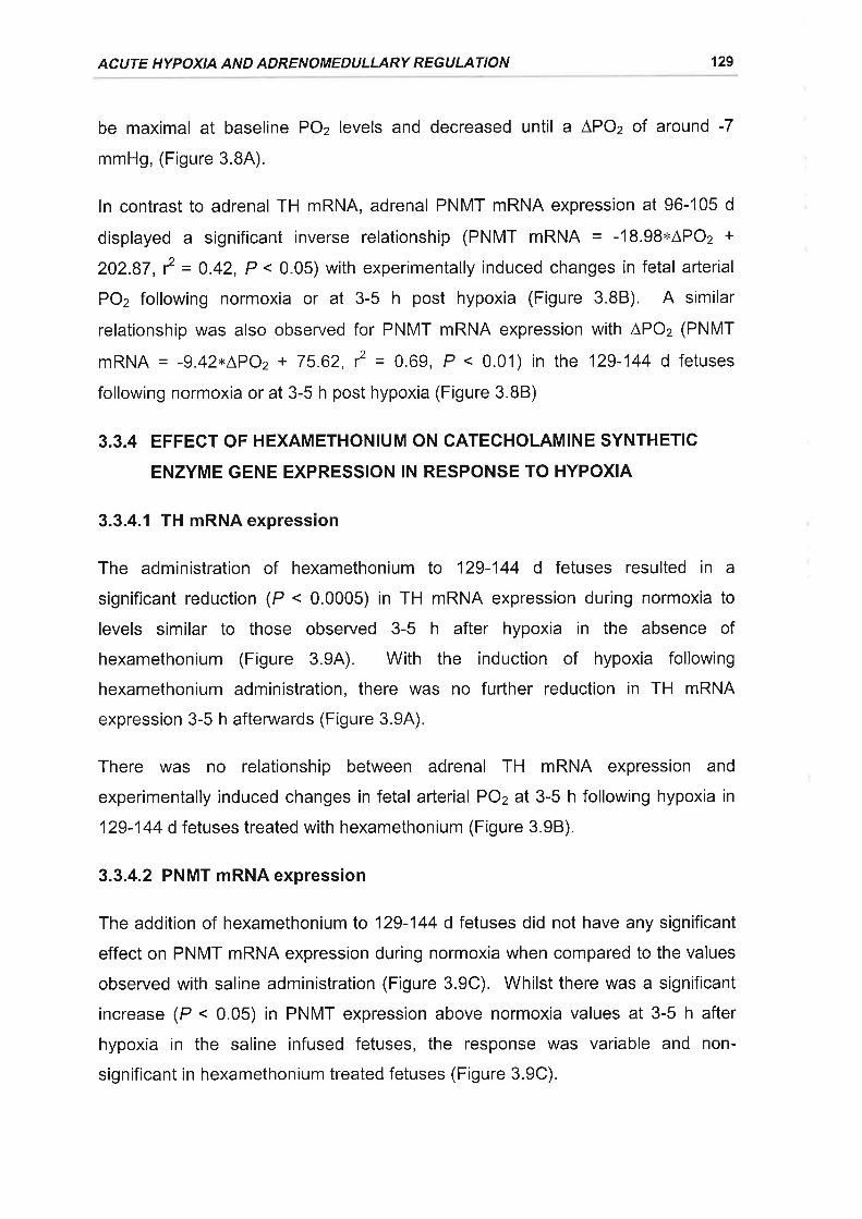
ACUTE HYPOXIA AND ADRENOMEDULLARY REGULATION 129
be maximal at baseline POz levels and decreased until a ÂPOz of around -7
mmHg, (Figure 3.84).
ln contrast to adrenal TH mRNA, adrenal PNMT mRNA expression at 96-105 d
displayed a significant inverse relationship (PNMT mRNA = -18.98xAPOz +
202.87, r2 = 0.42, P < 0.05) with experimentally induced changes in fetal arterial
PO2 following normoxia or at 3-5 h post hypoxia (Figure 3.88). A similar
relationship was also observed for PNMT mRNA expression with APOz (PNMT
mRNA = -9.42rLPO2 + 75.62, r2 = 0.69, P < 0.01) in the 129-144 d fetuses
following normoxia or at 3-5 h post hypoxia (Figure 3.BB)
3.3.4 EFFECT OF HEX,AMETHONIUM ON CATECHOLAMINE SYNTHETIC
ENZYME GENE EXPRESSION IN RESPONSE TO HYPOXIA
3.3.4.1 TH mRNA expression
The administration of hexamethonium to 129-144 d fetuses resulted in a
significant reduction (P < 0.0005) in TH mRNA expression during normoxia to
levels similar to those observed 3-5 h after hypoxia in the absence of
hexamethonium (Figure 3.9A). With the induction of hypoxia following
hexamethonium administration, there was no further reduction in TH mRNA
expression 3-5 h afterwards (Figure 3.94).
There was no relationship between adrenal TH mRNA expression and
experimentally induced changes in fetal arterial POz at 3-5 h following hypoxia in
129-144 d fetuses treated with hexamethonium (Figure 3.98).
3.3.4.2 PNMT mRNA expression
The addition of hexamethonium to 129-144 d fetuses did not have any significant
effect on PNMT mRNA expression during normoxia when compared to the values
observed with saline administration (Figure 3.9C). Whilst there was a significant
increase (P < 0.05) in PNMT expression above normoxia values at 3-5 h after
hypoxia in the saline infused fetuses, the response was variable and non-
significant in hexamethonium treated fetuses (Figure 3.9C).
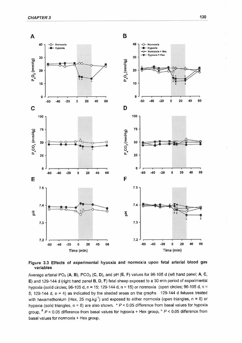
CHAPTER 3 130
A
c
E
TçL
0
100
0
7.5
7.4
7.3
7.2
D
F
40
0
100
7.5
7.4
7.3
7.2
B
40 -O- Normoxla-G Hypoxla
-60 -40 -20 0 20 40 60
-60 -40 -20 0 20 40 60
-60 -40 -20 0 20 40 60
Time (min)
4 Normoxla-+ Hypoxla
+ Normoxla + Hex
-,F Hypoxla + Hex
-60 -40 -20 0 20 40 60
-60 -40 -20 0 20 40 60
30c,tTE820
floo
o-10
30El
IE
E20o
Eo-
't0
75C')
IE
E50oo
oo- 2s
675IEÈ
50qoo
FfL
25
0
Ict
*
-60 .40 -20 0 20 40 60
Time (min)
Figure 3.3 Effects of experimental hypox¡a and normoxia upon fetal arterial blood gasvar¡ables
Average arterial PO2 (4, B), PCO2 (C, D), and pH (E, F) values for 96-105 d (left hand panel; A, C,
E) and 129-144 d (right hand panel B, D, F) fetal sheep exposed to a 30 min period of experimental
hypoxia (solid circles;96-105 d, n = 15; 129-144 d, n = 15) or normoxia (open circles; 96-105 d, n =
5; 129-144 d, n = 4) as indicated by the shaded areas on the graphs. 129-144 d fetuses treated
with hexamethonium (Hex, 25 mg.kg-1) and exposed to either normoxia (open triangles, n = 6) or
hypoxia (solid triangles, n = 6) are also shown. * P < 0.05 difference from basal values for hypoxia
group, # P. 0.05 difference from basal values for hypoxia + Hex group, o P < 0.05 difference from
basalvalues for normoxia + Hex group.
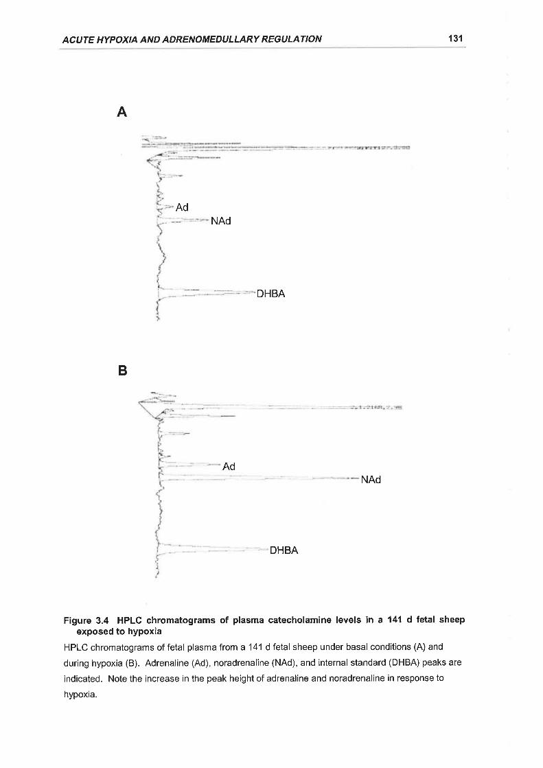
ACUTE HYPOXIA AND ADRENOMEDU LLARY REGULATION 131
A
NAd
DHBA
NAd
DHBA
Figure 3.4 HPLC chromatograms of plasma catecholamine levels in a 141 d fetal sheepexposed to hypoxia
HPLC chromatograms of fetal plasma from a 141 d fetal sheep under basal conditions (A) and
during hypoxia (B). Adrenaline (Ad), noradrenaline (NAd), and internalstandard (DHBA) peaks are
indicated. Note the increase in the peak height of adrenaline and noradrenaline in response to
hypoxia.
Ad
B
Ad
CHAPTER 3 132
ASaline
r-r Normoxia + SalineI Hypoxia + Saline
*
0
Adrenaline Noradrenaline
Hexamethon¡um
: Normoxia + HexI Hypoxia + Hex
*
Adrenaline Noradrenaline
Gatecholamine
Figure 3.5 lmpact of acute hypoxia on plasma catecholamine levels in the fetal sheep
Percentage change in fetal plasma catecholamine levels from average baseline values (-25, -5 min)
in response to hypoxia (solid histograms; 5, 10 min) or normoxia (open histograms; 5, 10 min) for
129-144 d fetal sheep administered saline (A; normoxia, n = 4; hypoxia, n = '15), and 129-144 d
fetal sheep treated with 25 mg.kg-1 hexamethonium (B; normoxia, n = 6; hypoxia, n = 6). .P < 0.05
difference from normoxia value.
B
*300
200
100
â(!th(E
dtso'Ë
sooo(!oGE|t,G'
À
300
200
100
0
-(õ.n(!os(¡)
.E
ftlõoo(úoG'
Ett,so
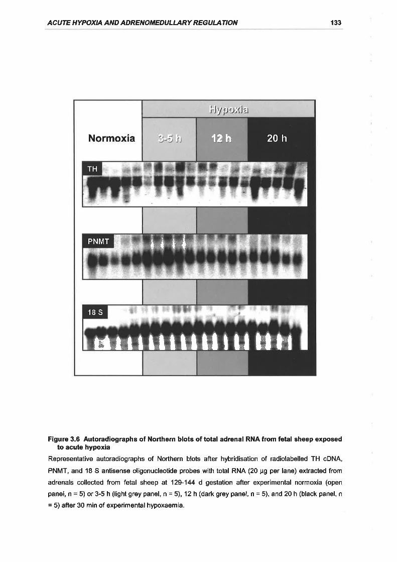
AC UTE HYPOXIA AN D ADRENOTiíEDU L'ARY REG U ATION 133
Figure 3.6 Autoradiographs of Northern blots of total adrenal RNA from fetal sheep exposedto acute hypoxia
Representative autoradiographs of Northern blots after hþridisation of radiolabelled TH cDNA,
PNMT, and 18 S antisense oligonucleotide probes with total RNA (20 pg per lane) extracted from
adrenals collected from fetal sheep al 129-144 d gestation after experimental normoxia (open
panel,n=5)or3-5h(lightgreypanel,î=5), 12h(darkgreypanel,n=5),and20h(blackpanel,n
= 5) after 30 min of experimental hypoxaemia.
Normoxia
ÉtrIilrllllIu Htr
20h12h'
PNMT *t ++
18Sf
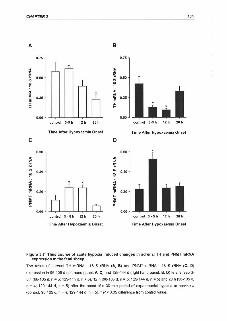
CHAPTER 3 134
**
zÉU'€
zÉE.L
A
c
0.75
0.50
0.25
0.00
0.60
0.40
0.20
0.00
B
zÉ.
U'c,
zÉ,EI
D
0.75
0.50
0.25
0.00
0.60
0.40
0.20
0.00
*
control 3-5 h 12h 20h
Time After Hypoxaemia Onset
* *
control 3-5h 12h 20h
Time After Hypoxaemia Onset
control 3-5 h 12h 20h
Time After Hypoxaemia Onset
control 3-5h 12h 20h
Time After Hypoxaemia Onset
*
zÉ.
U)€
zÉ.Et-=zo-
zÉ.
Ø@
zÉ.Et-=zÀ
Figure 3.7 Time course of acute hypoxia induced changes in adrenal TH and PNMT mRNAexpression in the fetal sheep
The ratios of adrenal TH mRNA : '18 S rRNA (4, B) and PNMT mRNA : 18 S rRNA (C, D)
expression in 96-105 d (left hand panel;4, C) and 129-144 d (right hand panel; B, D) fetalsheep 3-
5h(96-105d,n=5;129-144d,n=5),12h(96-105d,n=5;129-144d,n=5)and20h(96-105d,n = 4; 129-144 d, n = 5) after the onset of a 30 min period of experimental hypoxia or normoxia
(control; 96-105 d, n = 4; 129-144 d, n = 5). * P< 0.05 differencefrom controlvalue.
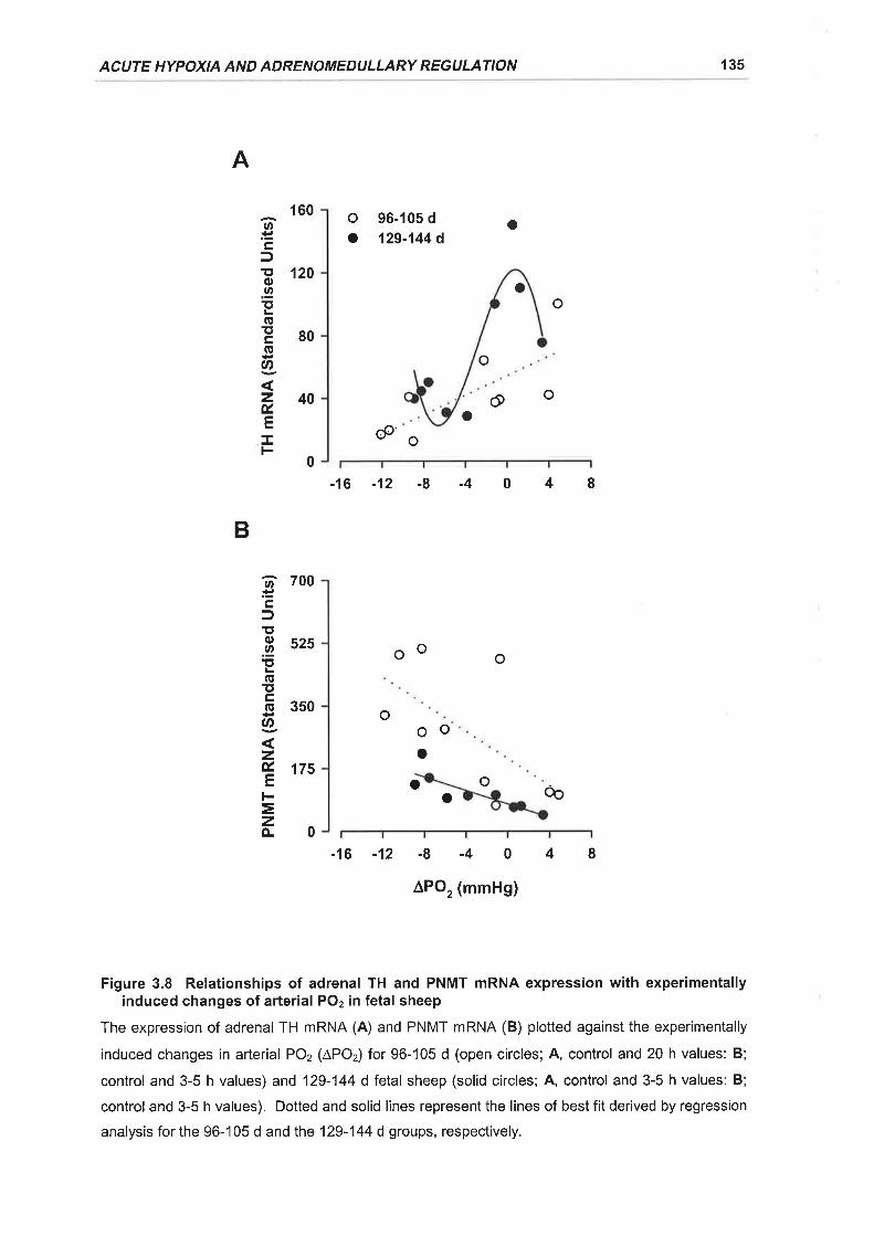
ACUTE HYPOXIA AND ADRENOMEDULLARY REGULATION 135
A
o 96-105 d
o 129-144 do
oo
o
@oo
o
-16-12-8-4048
B
oo
o
o o
-16-12-8-4048
^PO2 (mmHg)
Figure 3.8 Relationships of adrenal TH and PNMT mRNA expression with experimentallyinduced changes of arterial POz in fetal sheep
The expression of adrenal TH mRNA (A) and PNMT mRNA (B) plotted against the experimentally
induced changes in arterial PO2 (APO2) for 96-105 d (open circles; A, control and 20 h values: B;
control and 3-5 h values) and 129-144 d fetal sheep (solid circles; A, control and 3-5 h values: B;
control and 3-5 h values). Dotted and solid lines represent the lines of best fit derived by regression
analysis for the 96-'105 d and the 129-144 d groups, respectively.
@
160
120
80
40
.nËçlE'o.9EIEEcõU'
zú.E
F
OO
o
OOo
o
6 700.ËÊft,.ü 525ï,(ET'E 3sogzryflstsF=zo-0
0
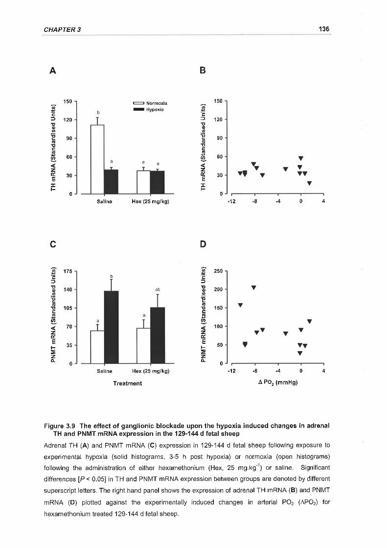
CHAPTER 3 136
A B
oËcJEo.2Ìt(!îtco|t,
zÉ.E
!
oËcf!o.9Þc,!tcG
Ø
zÉ.EI
c
U'ËtrfÎto.9!tfit!cfilU'
zú.tr
=zfL
1s0
120
90
60
30
175
140
105
70
35
- Normoxia
- Hypoxia
150
'120
90
60
30
b
a aa Y
YYYY
YYYYY
00
D
Saline Hex (25 mg/kg)
ab
Saline Hex (25 mg/kg)
Treatment
-'t2 -8
v
-4 0 4
Y
Yv Y V
-4 4
 Po, (mmHg)
b
YYv
9- 250É,l
i 2oo.2!ta!îr 150c(lt
o
zÉ.
.- 50F
=zo-0
Y
Y
a
V
0
-12 -8 0
Figure 3.9 The effect of ganglionic blockade upon the hypoxia induced changes in adrenalTH and PNMT mRNA expression in the 129-144 d fetal sheep
Adrenal TH (A) and PNMT mRNA (C) expression in'129-'144 dfelal sheep following exposure to
experimental hypoxia (solid histograms, 3-5 h post hypoxia) or normoxia (open histograms)
following the administration of either hexamethonium (Hex, 25 mg.kg-1) or saline. Significant
differences [P < 0.05] in TH and PNMT mRNA expression between groups are denoted by different
superscript letters. The right hand panel shows the expression of adrenal TH mRNA (B) and PNMT
mRNA (D) plotted against the experimentally induced changes in arterial PO2 (APO2) for
hexamethonium treated 129-144 d fetal sheep.
ACUTE HYPOXIA AND ADRENOMEDULLARY REGULATION 137
No relationship was observed between adrenal PNMT mRNA expression and
experimentally induced changes in fetal arterial POz in 129-144 d fetal sheep
treated with hexamethonium (Figure 3.9D).
ln the present study we found that basal circulating concentrations of
noradrenaline and adrenaline increased with increasing gestational age in the fetal
sheep. This agrees with the findings of Cheung (1990), who established that
basal noradrenaline levels were positively correlated with gestational age and that
adrenaline concentrations also tended to increase with gestational age. ln the
fetus, circulating noradrenaline is primarily derived from sympathetic neurones
under basal conditions, as destruction of the peripheral sympathetic nerves after
guanethidine administration markedly reduces plasma noradrenaline levels whilst
they do not decrease after adrenal demedullation (Jones et a\.,1987). Hence the
increase in circulating noradrenaline in late gestation may reflect maturational
changes in the synthetic and secretory capacity the peripheral sympathetic
nervous system (Simonetta et al., 1993). ln contrast plasma concentrations of
adrenaline decrease significantly after adrenal demedullation (Jones et a\.,1987).
There is also an increase in adrenaline output from the isolated perfused adrenal
gland in late gestation (Butler et al., 1995) which coincides with the increased
expression of adrenal PNMT mRNA measured in the studies described in
Chapter 2. lt appears likely therefore that the increase in circulating adrenaline
levels in the late gestation sheep fetus is derived in large part from an increased
output from the fetal adrenal medulla.
A number of studies have previously established that hypoxia and asphyxia
stimulate the secretion of catecholamines from the adrenal gland into the
circulation in both anaesthetised fetal sheep during acute studies and in
chronically catheterised conscious fetal sheep in utero (Cheung, 1990; Cohen ef
al., 1991; Cohen et al., 1984; Comline & Silver, 1966; Comline & Silver, 1961;
Jones et al., 1988; Widmark et al., 1989). Plasma catecholamine levels and
adrenal catecholamine output exhibit an inverse exponential relationship with the
degree of fetal hypoxia (Cheung, 1990; Cohen et al., 1984). Whilst a number of
anatomical and functional studies in the fetal sheep have repoded that
3.4 DTSCUSSION
CHAPTER 3 138
preganglionic cholinergic innervation of the adrenal gland does not occur until
about 120 d gestation (Boshier et al., 1989a; Comline & Silver, 1966; Comline &
Silver,'1961), it is has been demonstrated that hypoxia can stimulate
adrenomedullary catecholamine secretion before 120 d by a 'direct' or non-
neurogenic mechanism (Adams et al., '1996; Cheung, 1989; Cheung, 1990;
Comline & Silver, 1966; Comline & Silver, 1961). ln agreement with these
previous findings the current study also found that acute hypoxia stimulated
increases in plasma catecholamine levels in chronically catheterised fetal sheep tn
utero both before (96-105 d) and after (129-144 d) the development of functional
splanchnic innervation.
The majority of nerye fibres projecting to the adrenal medulla via the splanchnic
nerve are preganglionic, sympathetic, cholinergic nerves (Parker et al., 1993;
Robinson et al., 1977b). Acetylcholine released from splanchnic nerye terminals
acts at nicotinic and muscarinic receptors on chromaffin cells to stimulate
catecholamine secretion from the adrenal medulla (Burgoyne, 1984; Ungar &
Phillips, 1983). ln the present study, administration of the nicotinic cholinoreceptor
antagonist, hexamethonium, did not cause a decrease in the basal concentrations
of plasma noradrenaline and adrenaline in fetal sheep between 129 and 144 days
gestation. Hexamethonium administration did however, abolish the increase in
plasma noradrenaline concentrations in response to acute hypoxaemia in these
fetuses, supporting the previous observations of Cheung (1990) in chronically
catheterised fetal sheep. ln contrast, there was still a significant adrenaline
secretory response to hypoxia in the older fetuses in the presence of
hexamethonium. lt appears therefore that after the development of splanchnic
innervation, hypoxia stimulates noradrenaline secretion by reflex activation of
nicotinic receptors. Adrenaline secretion, however did not appear to be
completely dependent upon nicotinic stimulation.
It is possible that a component of the adrenaline secretory response to hypoxia in
the older fetal sheep may be due to muscarinic stimulation. Previous studies in
our laboratory using the retrogradely per{used adrenal gland preparation,
demonstrated that acetylcholine evoked catecholamine secretion is not completely
abolished by hexamethonium (Butler et al., 1995). Furthermore Comline and
Silver (1961) reported that hexamethonium treatment did not completely abolish
ACUTE HYPOXIA AND ADRENOMEDULLARY REGULATION 139
the catecholamine secretory response of the adrenal gland to electrical stimulation
of the splanchnic nerye. Alternatively the rise in plasma adrenaline levels may be
derived from sources other than the adrenal medulla and post ganglionic
sympathetic nerves, such as the C1 neurones which project from the ventrolateral-
medulla of the brainstem (Walker & Schuijers, 1989). This would appear to be
unlikely as numerous studies which have examined the source of the plasma
catecholamine rise in response to acute hypoxia have found that both the
noradrenaline and adrenaline response to acute hypoxia are virtually abolished
with adrenal demedullation or adrenalectomy (Jones et a\.,1988; Simonetta et al.,
1996a).
It has been demonstrated in a number of species that reflex neural stimulation of
the adrenal medulla results in an increase in intracellular Ca2* levels due to the
activation of nicotinic and muscarinic receptors by acetylcholine (Cheek et al.,
1989; O'Sullivan et al., 1989). Consequently a number of second messenger
systems such as cAMP / PKA and PKC, which interact with transcription factors
involved in the regulation of catecholamine synthetic enzyme gene expression, are
also activated (Anderson et al., 1992; Hwang et al., 1997; lcard-Liepkalns et al.,
1992; Morita et al., 1995a; Terbush et al., 1988; Terbush & Holz, 1986). Thus
there is a coupling of catecholamine secretion with catecholamine synthesis. The
non-neurogenic catecholamine secretory response of fetal sheep and neonatal rat
adrenomedullary chromaffin cells and PC12 cells to hypoxaemia is established to
be mediated by oxygen-sensitive potassium channels (Rychkov et al., 1998;
Thompson et al., 1997; Zhu et al., 1996). The closing of these channels in
response to hypoxia results in a depolarisation of the chromaffin cells and opening
of voltage-sensitive calcium channels (Mochizuki-Oda et al., 1997; Thompson ef
al., 1997). lt has also been shown in studies using the retrogradely perfused fetal
sheep adrenal gland, that hypoxia stimulates catecholamine secretion directly by a
mechanism dependent on voltage-sensitive Ca2* channels (Adams ef a/., 1996).
The subsequent influx of Caz* into the cell with non-neurogenic stimulation could
therefore potentially activate a number of second messenger systems including
isoforms of PKC, Ca2* lcalmodulin dependent protein kinases, and PKA viaCa2* |
calmodulin sensitive adenylate cyclase (Anderson et al., 1992; Beitner-Johnson ef
at., 1997). lt is probable therefore that there is also a coupling of non-neurogenic
stimulated secretion with catecholamine synthesis via the interaction of second
CHAPTER 3 140
messenger systems with transcriptional factors which regulate catecholamine
synthetic enzyme gene expression.
Surprisingly, acute hypoxaemia elicited a significant reduction in TH mRNA
expression within the adrenal glands of fetal sheep both before and after the
development of functional splanchnic innervation. Therefore acute hypoxia in the
fetal sheep appears to be able to suppress TH mRNA expression by a non-
neurogenic mechanism prior to functional splanchnic innervation of the adrenal
gland. The non-neurogenic repression of TH mRNA expression in the fetal sheep
adrenal was observed 20 h after the acute hypoxic and was directly related to the
experimentally induced change in fetal PuOz. ln contrast to the findings of the
current study, Holgert and colleagues ('1995) reported that adrenal TH mRNA
expression increased in fetal rats at E21 following 48 h of maternal hypoxia.
Therefore in the rat in which functional cholinergic innervation of the adrenal
medulla does not develop until the first postnatal week, chronic hypoxia is able to
stimulate TH gene expression by a direct non-neurogenic mechanism (Holgert ef
al., 1995b). The fact that TH mRNA expression was decreased with fetal
hypoxaemia in the current study may be attributable to species difference, degree
of fetal hypoxaemia achieved, or the timing of the hypoxic insult, ie. acute vs
chronic. lt may also reflect that a rise in TH mRNA expression may have occurred
earlier than 3-5 h after the hypoxic insult and was subsequently missed, although
this would be highly unlikely as TH mRNA in most catecholaminergic tissues is
relatively stable with a half life of around 10 h (Czyzyk-Krzeska, 1997; Czyzyk-
Krzeska et al., 1994b). Studies in the 1 d old neonatal rat and adult rat have
found that moderate hypoxaemia does not significantly affect adrenal TH mRNA
expression (Czyzyk-Krzeska et al., 1992; DeCristofaro & LaGamma, 1994).
Arterial POz, however, is much higher in the neonate and the adult than it is in the
fetus and the lack of change in adrenal TH mRNA expression may indicate that
the reductions in arterial POz were insufficient to reach a response threshold
(Czyzyk-Krzeska et al., 1992; DeCristofaro & LaGamma, 1994).
With the development of functional adrenal innervation after 129 d gestation
however, the suppression of adrenal TH mRNA expression was observed as early
as 3-5 h after exposure to hypoxia. The relationship of TH mRNA expression with
the change in arterialPOzwas also more complex after 129 d gestation. There
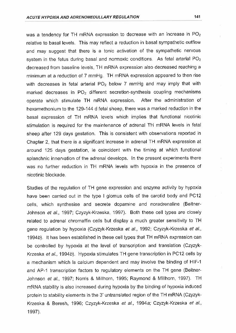
ACIJTE HYPOXIA AND ADRENOMEDULLARY REGULATION 141
was a tendency for TH mRNA expression to decrease with an increase in POz
relative to basal levels. This may reflect a reduction in basal sympathetic outflow
and may suggest that there is a tonic activation of the sympathetic nervous
system in the fetus during basal and normoxic conditions. As fetal arterial POz
decreased from baseline levels, TH mRNA expression also decreased reaching a
minimum at a reduction of 7 mmHg. TH mRNA expression appeared to then rise
with decreases in fetal arterial POz below 7 mmHg and may imply that with
marked decreases in POz different secretion-synthesis coupling mechanisms
operate which stimulate TH mRNA expression. After the administration of
hexamethonium to the 129-144 d fetal sheep, there was a marked reduction in the
basal expression of TH mRNA levels which implies that functional nicotinic
stimulation is required for the maintenance of adrenal TH mRNA levels in fetal
sheep after 129 days gestation. This is consistent with observations reported in
Chapter 2, that there is a significant increase in adrenal TH mRNA expression at
around 125 days gestation, ie coincident with the timing at which functional
splanchnic innervation of the adrenal develops. ln the present experiments there
was no further reduction in TH mRNA levels with hypoxia in the presence of
nicotinic blockade.
Studies of the regulation of TH gene expression and enzyme activity by hypoxia
have been carried out in the type I glomus cells of the carotid body and PC12
cells, which synthesise and secrete dopamine and noradrenaline (Beitner-
Johnson et al., 1997; Czyzyk-Krzeska, 1997). Both these cell types are closely
related to adrenal chromaffin cells but display a much greater sensitivity to TH
gene regulation by hypoxia (Czyzyk-Krzeska et al., 1992; Czyzyk-Krzeska et al.,
1994b). lt has been established in these cell types that TH mRNA expression can
be controlled by hypoxia at the level of transcription and translation (Czyzyk-
Krzeska et al., 1994b). Hypoxia stimulates TH gene transcription in PC12 cells by
a mechanism which is calcium dependent and may involve the binding of HIF-1
and AP-1 transcription factors to regulatory elements on the TH gene (Beitner-
Johnson et al., 1997; Norris & Millhorn, 1995; Raymond & Millhorn, 1997). TH
mRNA stability is also increased during hypoxia by the binding of hypoxia induced
protein to stability elements in the 3' untranslated region of the TH mRNA (Czyzyk-
Krzeska & Beresh, 1996; Czyzyk-Krzeska et al., 1994a; Czyzyk-Krzeska et al.,
1ee7).
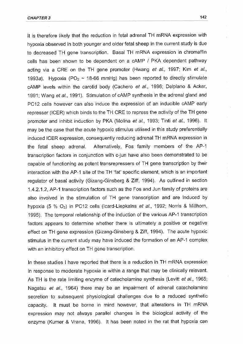
CHAPTER 3 142
It is therefore likely that the reduction in fetal adrenal TH mRNA expression with
hypoxia observed in both younger and older fetal sheep in the current study is due
to decreased TH gene transcription. Basal TH mRNA expression in chromaffin
cells has been shown to be dependent on a cAMP / PKA dependent pathway
acting via a CRE on the TH gene promoter (Hwang et al., 1997; Kim et al.,
1993a). Hypoxia (POz - 18-66 mmHg) has been reported to directly stimulate
cAMP levels within the carotid body (Cachero et al., 1996; Delpiano & Acker,
1991 ; Wang et al., 1991). Stimulation of cAMP synthesis in the adrenal gland and
PC12 cells however can also induce the expression of an inducible cAMP early
represser (ICER) which binds to the TH CRE to repress the activity of the TH gene
promoter and inhibit induction by PKA (Molina et al., 1993; Tinti ef a/., 1996). lt
may be the case that the acute hypoxic stimulus utilised in this study preferentially
induced ICER expression, consequently reducing adrenal TH mRNA expression in
the fetal sheep adrenal. Alternatively, Fos family members of the AP-1
transcription factors in conjunction with c-jun have also been demonstrated to be
capable of functioning as potent transrepressers of TH gene transcription by their
interaction with the AP-1 site of the TH 'fat' specific element, which is an important
regulator of basal activity (Gizang-Ginsberg & Ziff , 1994). As outlined in section
1.4.2.1.2, AP-l transcription factors such as the Fos and Jun family of proteins are
also involved in the stimulation of TH gene transcription and are induced by
hypoxia (5 % Oz) in PC12 cells (lcard-Liepkalns et al., 1992; Norris & Millhorn,
1995). The temporal relationship of the induction of the various AP-1 transcription
factors appears to determine whether there is ultimately a positive or negative
effect on TH gene expression (Gizang-Ginsberg &Ziff ,1994). The acute hypoxic
stimulus in the current study may have induced the formation of an AP-1 complex
with an inhibitory effect on TH gene transcription.
ln these studies I have reported that there is a reduction in TH mRNA expression
in response to moderate hypoxia ie within a range that may be clinically relevant.
As TH is the rate limiting enzyme of catecholamine synthesis (Levitt et al., 1965;
Nagatsu et al., 1964) there may be an impairment of adrenal catecholamine
secretion to subsequent physiological challenges due to a reduced synthetic
capacity. lt must be borne in mind however, that alterations in TH mRNA
expression may not always parallel changes in the biological activity of the
enzyme (Kumer & Vrana, 1996). lt has been noted in the rat that hypoxia can
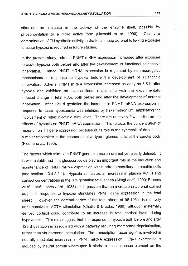
ACUTE HYPOXIA AND ADRENOMEDULLARY REGULATION 143
stimulate an increase in the activity of the enzyme itself, possibly by
phosphorylation to a more active form (Hayashi et al., 1990). Clearly a
determination of TH synthetic activity in the fetal sheep adrenal following exposure
to acute hypoxia is required in future studies.
ln the present study, adrenal PNMT mRNA expression increased after exposure
to acute hypoxia both before and after the development of functional splanchnic
innervation. Hence PNMT mRNA expression is regulated by non-neurogenic
mechanisms in response to hypoxia before the development of splanchnic
innervation. Adrenal PNMT mRNA expression increased as early as 3-5 h after
hypoxia and exhibited an inverse linear relationship with the experimentally
induced change in fetal P^Oz, both before and after the development of adrenal
innervation. After 129 d gestation the increase in PNMT mRNA expression in
response to acute hypoxaemia was inhibited by hexamethonium, implicating the
involvement of reflex nicotinic stimulation. There are relatively few studies on the
effects of hypoxia on PNMT mRNA expression. This reflects the concentration of
research on TH gene expression because of its role in the synthesis of dopamine,
a major transmitter in the chemoreceptive type I glomus cells of the carotid body
(Fidone et a\.,1990).
The factors which stimulate PNMT gene expression are not yet clearly defined. lt
is well established that glucocorticoids play an important role in the induction and
maintenance of PNMT mRNA expression within adrenomedullary chromaffin cells
(see section 1.3.4.2.2.1). Hypoxia stimulates an increase in plasma ACTH and
cortisol concentrations in the late gestation fetal sheep (Akagi et al., 1990; Braems
et a|.,1996; Jones et a\.,1988). lt is possible that an increase in adrenal cortisol
output in response to hypoxia stimulates PNMT gene expression in the fetal
sheep. However, the adrenal cortex of the fetal sheep at 96-105 d is relatively
unresponsive to ACTH stimulation (Challis & Brooks, 19Bg), although maternally
derived cortisol could contribute to an increase in fetal cortisol levels during
hypoxaemia. This may suggest that the response to hypoxia both before and after
129 d gestation is associated with a pathway requiring membrane depolarisation,
rather than via hormonal stimulation. The transcription factor Egr-1 is involved in
neurally mediated increases in PNMT mRNA expression. Egr-1 expression is
induced by neural stimuli whereupon it binds to its consensus element on the
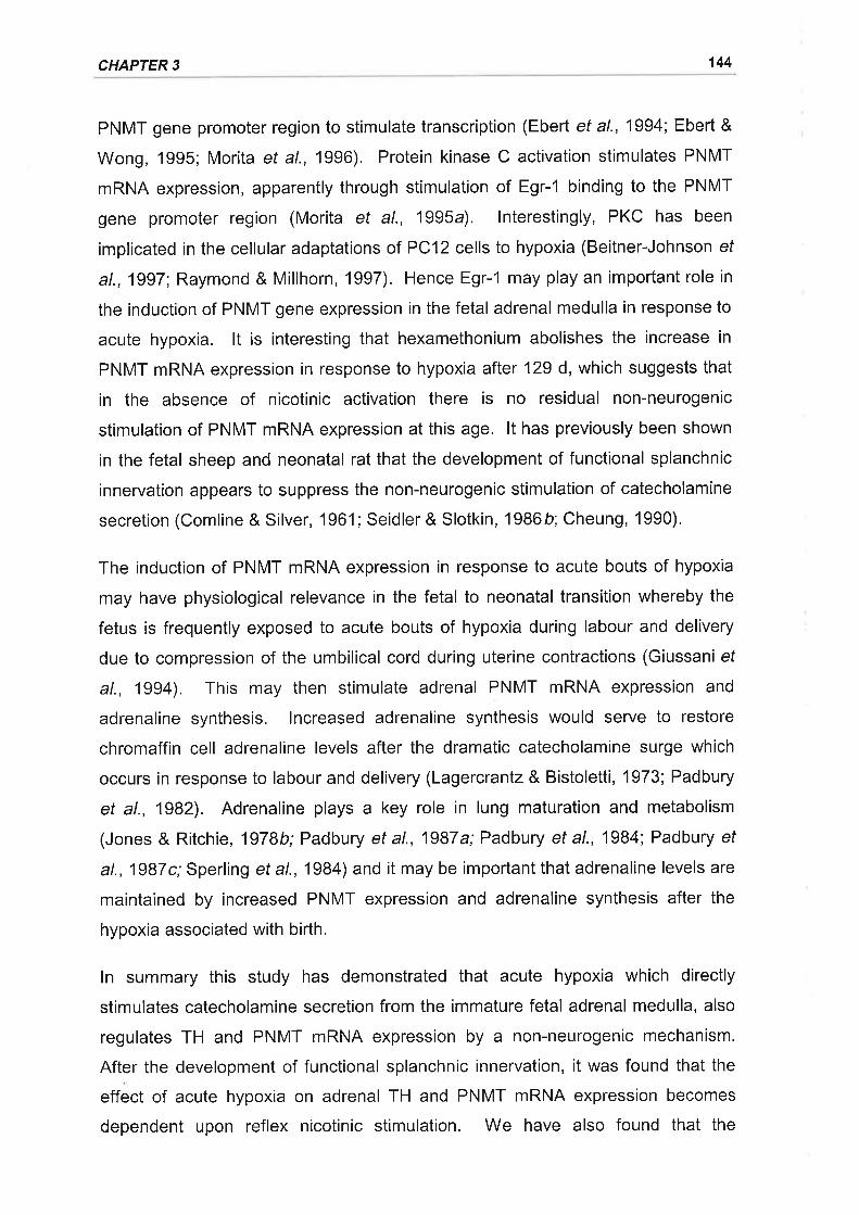
CHAPTER 3 144
PNMT gene promoter region to stimulate transcription (Ebert et al., 1994; Ebert &
Wong, 1995; Morita et al., 1996). Protein kinase C activation stimulates PNMT
mRNA expression, apparently through stimulation of Egr-'l binding to the PNMT
gene promoter region (Morita et al., 1995a). lnterestingly, PKC has been
implicated in the cellular adaptations of PC12 cells to hypoxia (Beitner-Johnson ef
a\.,1997 Raymond & Millhorn, 1997). Hence Egr-1 may play an important role in
the induction of PNMT gene expression in the fetal adrenal medulla in response to
acute hypoxia. lt is interesting that hexamethonium abolishes the increase in
PNMT mRNA expression in response to hypoxia after 129 d, which suggests that
in the absence of nicotinic activation there is no residual non-neurogenic
stimulation of PNMT mRNA expression at this age. lt has previously been shown
in the fetal sheep and neonatal rat that the development of functional splanchnic
innervation appears to suppress the non-neurogenic stimulation of catecholamine
secretion (Comline & Silver, '1961 ; Seidler & Slotkin, 1986þ; Cheung, 1990).
The induction of PNMT mRNA expression in response to acute bouts of hypoxia
may have physiological relevance in the fetal to neonatal transition whereby the
fetus is frequently exposed to acute bouts of hypoxia during labour and delivery
due to compression of the umbilical cord during uterine contractions (Giussani ef
al., 1994). This may then stimulate adrenal PNMT mRNA expression and
adrenaline synthesis. lncreased adrenaline synthesis would serve to restore
chromaffin cell adrenaline levels after the dramatic catecholamine surge which
occurs in response to labour and delivery (Lagercrantz & Bistoletti, 1973; Padbury
et al., 1982). Adrenaline plays a key role in lung maturation and metabolism
(Jones & Ritchie, 1978b;Padbury et al., 1987a; Padbury et al., 1984; Padbury ef
al., 1987c; Sperling et a1.,1984) and it may be important that adrenaline levels are
maintained by increased PNMT expression and adrenaline synthesis after the
hypoxia associated with birth.
ln summary this study has demonstrated that acute hypoxia which directly
stimulates catecholamine secretion from the immature fetal adrenal medulla, also
regulates TH and PNMT mRNA expression by a non-neurogenic mechanism.
After the development of functional splanchnic innervation, it was found that the
effect of acute hypoxia on adrenal TH and PNMT mRNA expression becomes
dependent upon reflex nicotinic stimulation. We have also found that the
ACUTE HYPOXIA AND ADRENOMEDULLARY REGULATION 145
expression of adrenal TH and PNMT mRNA were differentially regulated by acute
hypoxia. Further studies to determine the cellular and molecular mechanisms
underlying the non-neurogenic and neurogenic regulation of the adrenal
catecholamine synthetic enzymes during hypoxia in the fetus are required.
4. GLUCOCORTICOID REGULATION OF TH, PNMT AND PENK AMRNA EXPRESSION IN THE FETAL SHEEP ADRENAL
1 INTROÐUCTION
ln the fetal sheep there are parallel increases in plasma concentrations of cortisol,
adrenomedullary adrenaline content, and adrenal PNMT mRNA expression, in the
10-'15 days preceding delivery (Chapter2, Challis & Brooks, 1989; Comline etal.,
1970). Furthermore, fetal HPX prevents the late gestation rise in the ratio of
adrenal adrenaline : noradrenaline content in the sheep (Comline et al., 1970). lt
has therefore been concluded that the increased adrenal output of the
glucocorticoid, cortisol, induces PNMT expression, with a consequent stimulation
of adrenaline synthesis in the adrenal medulla during late gestation.
Whilst the evidence cited above supports a role for glucocorticoids in the
stimulation of adrenaline synthesis in the fetal adrenal, a series of in vivo studies
have found a paradoxical inhibitory effect of glucocorticoid infusion on plasma
adrenaline concentrations. Wood and colleagues (1987) found that there was a
significant fall in circulating catecholamine concentrations in fetal sheep after a 5 h
cortisol infusion in late gestation. Stein and colleagues (1995) also found that a
longer term cortisol infusion significantly reduced the plasma catecholamine surge
which occurs after Caesarean delivery. Furthermore it has recently been
demonstrated that intrafetal infusions of the synthetic glucocorticoids,
betamethasone and dexamethasone, at doses used clinically to accelerate fetal
lung development, suppress plasma concentrations of adrenaline and
noradrenaline (Derks et al., 1997).
It is possible that exogenous glucocorticoids may suppress the fetal hypothalamo-
pituitary-adrenal axis via negative feedback, with consequent decreases in adrenal
glucocorticoid output, PNMT expression, adrenaline synthesis, and adrenaline
secretion. There have been no studies, however, on the effects of chronic
physiological elevations of glucocorticoid concentrations on the expression of
catecholamine biosynthetic enzymes in the adrenal medulla of a long gestation
species before birth. The study reported in this chapter investigated the effect of a
physiological elevation of fetal cortisol concentrations on the adrenal mRNA levels
of TH and PNMT.
146
CHAPTER4 147
lntra-adrenal Enk-containing peptides co-localised in adrenaline containing
chromaffin cells (McMillen ef a/., 1988) may play an inhibitory role in regulating
catecholamine release from the fetal adrenal (Padbury et al., 1987b). Hence the
effect of cortisol on the adrenal expression of PEnk A mRNA was also examined.
All procedures were approved by the University of Adelaide Standing Committee
on Ethics in Animal Experimentation.
4.2.1 ANIMALS AND SURGERY
Fifteen pregnant Border Leicester x Merino ewes were used in this study. Surgery
was performed on the ewes at either 103 or 104 d gestation (calculated according
to date of mating) under general anaesthesia induced by intravenous injection of
sodium thiopentone (1.25 g; Pentothal, Rhone Merieux, Pinkenba, QLD, Australia)
and maintained with halothane (3-4 % in oxygen; Fluothane, lCl, Melbourne, VlC,
Australia). Ewes were administered 2 ml of antibiotics (250 mg.ml-1 procaine
penicillin, 250 mg.ml-1 dihydrostreptomycin sulphate, 20 mg.ml-1 Procaine HCI;
Penstrep llium, Troy Laboratories, Smithfield, NSW, Australia) intramuscularly,
prior to surgery. During surgery catheters were implanted into the fetal and
maternal carotid artery and jugular vein as well as the amniotic sac, as described
in detail in Chapter 3. The fetus was also administered 2 ml of antibiotics
(Penstrep llium, Troy Laboratories) intramuscularly during surgery. The catheters
were flushed and filled with heparinised saline (500 lU.ml-1; Multiparin, Fisons
Pharmaceuticals, Sydney, NSW, Australia) and the fetal catheters were
exteriorised through an excision in the ewe's flank.
4.2.2 POST OPERATIVE CARE
Ewes were housed in individual metabolic crates in animal holding rooms under a
12 h light-dark cycle and fed lucerne chaff once daily between 0900 and 1300 h
with water ad libitum. Sodium ampicillin (500 mg, Austrapen, Commonwealth
Serum Laboratories, Melbourne, VlC, Australia) was administered intra-
amniotically to all fetal sheep for 4 consecutive days post operatively. A
4.2 MATERIALS AND METHODS
CORTISOL AND PNMT EXPRESSION IN THE IMMATURE FETAL ADRENAL 148
post operative recovery of 3-4 d was allowed before any blood sampling
commenced.
4.2.3 BLOOD SAMPLING AND CORTISOL INFUSION
Fetal arterial blood samples (5 ml) were collected twice daily (10.00 and 17.00 h)
from 107-116 d gestation, Aliquots of whole blood (2.5 ml) were immediately
placed into ice chilled tubes for cortisol (heparinised tubes, 125 lU; Disposable
Products, Adelaide, SA, Australia) and immunoreactive (i0 ACTH (tubes
contained aprotinin, 1000 KlU.ml-1, Sigma Chemicals, St Louis, MO, USA; and
EDTA, 18.6 g.l-1 of whole blood) radioimmunoassay. Blood samples were
centrifuged at 1800 gfor 10 min at4'C and the plasma was separated and stored
at. -20"C prior to assay.
Cortisol (2.5-3.0 mg.5 ml-1 .24 h-t, n = 9; Solucortef, Upjohn Pharmaceutical's Ltd.,
Kalamazoo, Ml, USA) or 0.9 % saline (n = 6) was infused intravenously into fetal
sheep from 109 to 116 d gestation.
4.2.4 POST MORTEM AND TISSUE COLLECTION
At 116 d gestation the ewes were sacrificed with an overdose of sodium
pentobarbitone (8.125 g; Lethabarb, Virbac, Peakhurst, NSW, Australia). Fetal
sheep were delivered via laparotomy, weighed and killed by decapitation. The
adrenal glands were dissected from each fetus and weighed. The left adrenal
glands (n = 15) were snap frozen in liquid Nz and stored at -70"C until total RNA
was extracted. The right adrenal glands (n = 14) were cut into half, perpendicular
to their poles, and placed into ice cold paraformaldehyde (4 %) fixative in sodium
phosphate buffer (0.1 M, pH 7.0) for subsequent immunohistochemical analysis,
4.2.5 CORTTSOL ASSAY
Total cortisol concentrations were measured in fetal sheep plasma by
radioimmunoassay using an Orion Diagnostica kit (Orion Diagnostica, Turku,
Finland). Prior to assay, cortisol was extracted from the plasma using
dichloromethane (Bocking et al., 1986). The efficiency of recovery of 1251 Cortisol
from the fetal plasma using this procedure was 84 + I o/o. The sensitivity of the
assay was 0.39 nmol.l-1. The rabbit cortisol antibody had a cross reactivity of
CHAPTER 4 149
< 1 o/o with cortisone and 17-hydroxyprogesterone and <
pregnenolone, aldosterone, progesterone and estradiol (product information
supplied by Orion Diagnostica). The inter- and intra-assay coefficients of variation
(CV) were always less than 10 %.
4.2.6 ACTH ASSAY
The concentrations of plasma |TACTH were measured by radioimmunoassay
using an ICN Biomedicals kit (lCN Biomedicals, Seven Hills, NSW, Australia)'
The sensitivity of the assay was 7 pg.ml-1 and the rabbit anti-human ACTH (1-39)
had a cross reactivity of < 0.01 % with B-endorphin, cr-MSH, o-Lipotrophin, and p-
Lipotrophin (product information supplied by ICN Biomedicals). The inter-assay
CV was 14.6 % and the intra-assay CV was < 10 %.
4.2.7 PLASMA GLUCOSE AND LACTATE DETERMINATION
Plasma concentrations of glucose and lactate were determined by enzymatic
analysis using a COBAS MIRA automated analysing system (Cobas, Roche
Diagnostics, Basle, Switzerland). The enzymes hexokinase and glucose 6-
phosphate dehydrogenase were used for glucose determination whilst lactate
dehydrogenase and glutamate-pyruvate transaminase were used for lactate
determination. The photometric determination of NADH formation was used to
calculate the amounts of glucose and lactate present in the sample. The intra-
assay CVs were 1 .4 % and 1 .3 o/o for glucose and lactate, respectively.
4.2.8 TOTAL RNA EXTRACTION
Total adrenal RNA was extracted according to the method of Chirgwin et al.
('lg7g), as outlined in Chapter 2. Briefly, adrenals from cortisol infused (n = 9) and
saline (n = 6) fetuses were homogenised in 4 M guanidine thiocyanate buffer and
ultracentrifuged at 130 000 g through a 5.7 M CsCl cushion. The supernatant was
removed with the pellets being resuspended in sterile water and subsequently
reprecipitated using 0.3 M sodium acetate and absolute ethanol on dry ice. The
supernatant was again removed before the pellets were washed with 70 %
ethanol and then resuspended in sterile water. Nucleic acid concentration, yield,
and purity were quantified by spectrophotometric measurement at 260 and
280 nm. Total RNA solutions were stored at -70'C prior to Northern blot analysis.
CORTISOL AND PNMT EXPRESSION IN THE IMMATURE FETAL ADRENAL 150
4.2.9 PROBES AND PROBE LABELLING
The probes utilised in this study and the radiolabelling methods for the probes are
described in detail in Chapter 2. To summarise, oligonucleotide antisense probes
complementary to nucleotides 361-389 of the peptide coding region of bovine
PNMT mRNA (29 mer, Wan ef al., 19Bg), nucleotides 627-671 of the peptide
coding region of bovine PEnk A mRNA (44 mer, Wan et al., 1987), and
nucleotides 151-180 of rat 1B S rRNA (30 mer, Chan etal., 1984) sequencewere
synthesised (Bresatec, Thebarton, SA, Australia). A 1.7 kb bovine TH cDNA
(D'Mello et al., 1988) was used to detect TH mRNA. The oligonucleotides were
end labelled using T4 polynucleotide kinase (7.9 U.pl-1, Pharmacia, North Ryde,
NSW, Australia) and y-[32P]ATP (4000 Ci.mmol-1 ,Bresatec) in accordance with the
method described for the end labelling of oligonucleotides by Sambrook et al.
(1989). The cDNA probe was labelled by the random priming method with cx-
¡32e¡OCte (3000 Ci.mmol-r, Bresatec) and Klenow fragment (6.4 U.pl-t,
Pharmacia) using an oligolabelling kit (Pharmacia) as per manufacturer's
instructions.
4.2.10 NORTHERN BLOT ANALYSIS
For each sample 20 pg of total adrenal RNA was denatured in 2.2 M
formaldehyde and 50% (v/ v) formamide at 55'C for 10 min. Separation of the
total adrenal RNA was achieved by electrophoresis on a 1 % agarose gel
containing 2.2 M formaldehyde. The separated total adrenal RNA was
subsequently transferred to Zetaprobe nylon membranes (Biorad, Richmond, CA,
USA) via capillary blotting. Membranes were washed in 10 x SSC for 10 min at
room temperature and then baked for t h at 80'C to fix the RNA to the
membrane. Prior to hybridisation with oligonucleotide probes the membranes
were initially prehybridised at 50'C for 18 h in a solution consisting of 5 x SSC,
2OmM NaHzPO¿, pH 7.2,7 % SDS, 5 x Denhardt's, and 100 pg.ml-1 of heat
denatured Salmon sperm DNA. Membranes were then hybridised for 20 h at
50'C in 30 ml of fresh hybridisation solution containing 1-2 x 10⁶ cpm.ml-1 of
labelled oligonucleotide. For the TH cDNA probe equivalent prehybridisation and
hybridisation times were used, however, the hybridisation solution consisted of
50% (v/v) deionised formamide,5 x SSPE,7 o/o SDS, pH7.2, and 100 pg.ml-1 of
CHAPTER 4 151
heat denatured salmon sperm DNA, with hybridisation being carried out at 42C in
the presence of 1-2 x 10⁶ cpm.ml-1 of labelled probe. Following hybridisation the
membranes were washed in 1 x SSC and 0.1 % SDS (1 x 30 min) and then
subsequently in 0.1 x SSC and 0.1 o/o SDS (1 x 30 min) at their hybridisation
temperatures before being briefly air dried and exposed to a Fuji BAS-Ills
phosphorimager plate (Fuji Photo Co., Tokyo, Japan) for 24-48 h.
Autoradiographs were obtained using a Fuji BAS 1000 phosphorimager (Fuji
Photo Co.) and quantitated using Fuji MacBAS software (Fuji Photo Co.).
4.2.11ANTlBODlES
Rabbit anti-bovine PNMT was generously supplied by Professor PRC Howe
(Department of Biomedical Science, The University of Wollongong, NSW,
Australia) and rabbit anti-rat TH was purchased (Chemicon lnternational,
Temecula, CA, USA). The rabbit anti-PNMT and anti-TH primary antibodies were
used at working dilutions of 1:1500 and 1:1000 respectively. The specificity of
these antibodies has been previously demonstrated (McMillen et al., 19BB;
Varndell et al., 1982).
4.2.12 I M M U NOH ISTOCH EM ISTRY
Following fixation in paraformaldehyde Ø %) for 24 h adrenals were washed in
phosphate buffered saline (0.9 % saline, 0.1 M phosphate buffer, pH = 7.0) al4'C
(2 x 24 h) and then stored in ethanol at 4'C before being embedded in paraffin
wax. Transverse adrenal sections of 5 ¡rm were cut and mounted onto poly-L-
lysine coated slides.
Semi adjacent sections were either processed for TH and PNMT immunoreactivity
using a Vectastain avidin-biotin peroxidase complex kit (PK-4001, Vector
Laboratories, Burlingame, CA, USA) or stained with haemotoxylin (Harris'
Haematoxylin, Sigma Diagnostics, St Louis, MO, USA) and eosin (0.5 % in g0 %
Ethanol). The sections were dewaxed by immersion (2 x 5 min) in Histoclear
(National Diagnostics, Somerville, NJ, USA) and subsequently rehydrated by
immersion in a graded ethanol series (100 %, 2x5 min; 90 o/o, 1x2min;70o/o, 1
x 2 min; distilled HzO, 1x 2 min). Sections were washed in 0.01 M phosphate
buffered saline (PBS, pH 7,0; Sigma Chemicals, St. Lois, MO, USA) for 20 min,
CORTISOL AND PNMT EXPRESSION IN THE IMMATURE FETAL ADRENAL 152
before being incubated with blocking serum (3 % Normal Goat Serum in PBS) for
30 min at room temperature. Excess blocking serum was blotted away and the
sections were covered with the primary antibody diluted in PBS containing 1 %
BSA. After overnight incubation aL 4"C in a humidified container, the sections
were washed in (PBS, 3 x 5 min) and goat anti-rabbit biotinylated secondary
antibody was applied to the sections for 60 min at room temperature. Following
washing (PBS, 3 x 5 min) the sections were then incubated with avidin-biotin-
peroxidase complex (Vector Laboratories) at room temperature for 60 min before
being washed again (PBS, 3 x 5 min). The reaction was visualised by the addition
of 3,3-diaminobenzadine tetrahydrochloride (DAB, 0.5 mg.ml-t in PBS buffer and
0.02%HzOz) at room temperature for 10 min. Sections were then washed (PBS,
3 x 5 min) and dehydrated through a graded ethanol series (distilled HzO, 1 x 2
min; 70 % ethanol, 1 x2min; 90 o/o, 1 x2 min; 100 o/o, 2x5 min; Histoclear, 2 x 5
min) before coverslips were mounted with DPX (BDH Laboratory Supplies, Poole,
England).
4.2.13 ADRE NAL MORPHOM ETRY
Adrenal section images were captured and digitised using a CCD black and white
video camera (SSC-M37OCE, Sony Corp., Tokyo, Japan) mounted on a dissecting
microscope (SZ-40, Olympus Optical Co., Tokyo, Japan) via an SZ-CTV
photomount tube (Olympus Optical Co.). The image was digitised as a grey scale
image and positively stained areas were measured using a Power Mac 8500 I 120
(Apple, Cupertino, CA, USA) equipped with NIH lmage V1.61 software. Total
adrenal area was measured as the area of the section stained with Haematoxylin
and Eosin. The area of the adrenal medulla was defined as the area of the gland
which was positively stained with anti-TH. The areas of the sections which were
positively stained with anti-PNMT were also calculated. The relative areas of the
adrenal gland occupied by the medulla was calculated by dividing the area stained
by anti-TH by the total adrenal area. The relative area of the adrenal medulla
occupied by PNMT positive cells was obtained by dividing the area stained with
anti-PNMT by the area stained with anti-TH. ln all cases dual measurements of
area on serial sections were made and the measurements were averaged.
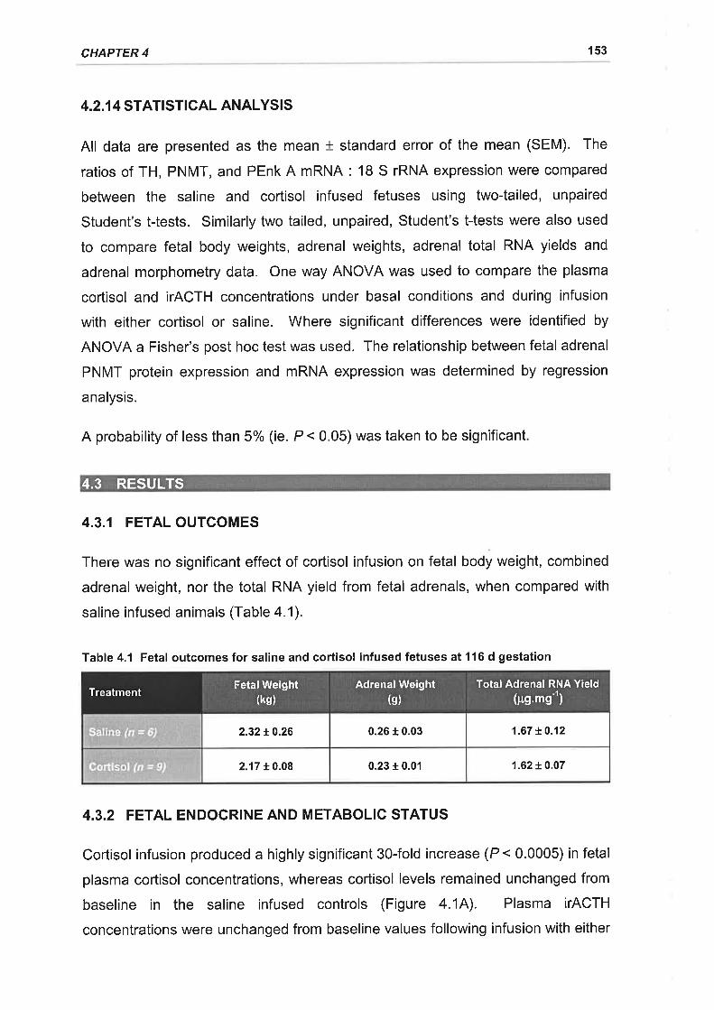
CHAPTER 4 153
4.2.1 4 STATI STICAL ANALYS IS
All data are presented as the mean + standard error of the mean (SEM). The
ratios of TH, PNMT, and PEnk A mRNA : 18 S rRNA expression were compared
between the saline and cortisol infused fetuses using two-tailed, unpaired
Student's t-tests. Similarly two tailed, unpaired, Student's t-tests were also used
to compare fetal body weights, adrenal weights, adrenal total RNA yields and
adrenal morphometry data. One way ANOVA was used to compare the plasma
cortisol and |TACTH concentrations under basal conditions and during infusion
with either cortisol or saline. Where significant differences were identified by
ANOVA a Fisher's post hoc test was used. The relationship between fetal adrenal
PNMT protein expression and mRNA expression was determined by regression
analysis.
A probability of less than 5% (ie. P < 0.05) was taken to be significant
4.3 RESULTS
4.3.1 FETAL OUTCOMES
There was no significant effect of cortisol infusion on fetal body weight, combined
adrenal weight, nor the total RNA yield from fetal adrenals, when compared with
saline infused animals (Table 4.1).
Table 4.1 Fetal outcomes for saline and cortisol infused fetuses at 116 d gestation
4.3.2 FETAL ENDOCRINE AND METABOLIG STATUS
Cortisol infusion produced a highly significant 3O-fold increase (P < 0.0005) in fetal
plasma cortisol concentrations, whereas cortisol levels remained unchanged from
baseline in the saline infused controls (Figure 4.1A). Plasma iTACTH
concentrations were unchanged from baseline values following infusion with either
2.32t0.26 0.26 r 0.03 1.67 t 0.12
2.17 t 0.08 0.23 r 0.01 1.62 r 0.07
FetalWeight(t<s)
AdrenalWeight(s) (pg.'ng-')
Total Adrenal RNA YieldTreatment
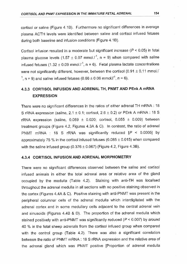
CORTISOL AND PNMT EXPRESSION IN THE IMMATURE FETAL ADRENAL 154
cortisol or saline (Figure 4.18). Furthermore no significant differences in average
plasma ACTH levels were identified between saline and cortisol infused fetuses
during both baseline and infusion conditions (Figure 4.18).
Cortisol infusion resulted in a moderate but significant increase (P < 0.05) in fetal
plasma glucose levels (1.57 t 0.07 mmol.l-1, f = 9) when compared with saline
infused fetuses (1.32+ 0.09 mmol.l-1, n = 6). Fetal plasma lactate concentrations
were not significantly different, however, between the cortisol (0.91 t 0.11 mmol.l-
1, ñ = 9) and saline infused fetuses (0.66 + 0.06 mmol.l-r, n = 6).
4.3.3 CORTISOL INFUSION AND ADRENAL TH, PNMT AND PEnk A mRNA
EXPRESSION
There were no significant differences in the ratios of either adrenal TH mRNA : 18
S rRNA expression (saline, 2.1 + 0.1;cortisol,2.6!0.2) or PEnkA mRNA: 1B S
rRNA expression (saline, 0.089 + 0.020; cortisol, 0.055 + 0.009) between
treatment groups (Figure 4.2, Figures 4.34 & C) ln contrast, the ratio of adrenal
PNMT mRNA : 18 S rRNA was significantly reduced lP <
approximately 75 % in the cortisol infused fetuses (0.085 + 0.015) when compared
with the saline infused group (0.376 t 0.067) (Figure 4.2, Figure 4.38).
4.3.4 CORTISOL INFUSION AND ADRENAL MORPHOMETRY
There were no significant differences observed between the saline and cortisol
infused animals in either the total adrenal area or relative area of the gland
occupied by the medulla (Table 4.2). Staining with anti-TH was localised
throughout the adrenal medulla in all sections with no positive staining observed in
the cortex (Figures 4.4A & C). Positive staining with anti-PNMT was present in the
peripheral columnar cells of the adrenal medulla which interdigitated with the
adrenal cortex and in some medullary cells adjacent to the central adrenal vein
and sinusoids (Figures 4.48 & D). The proportion of the adrenal medulla which
stained positively with anti-PNMT was significantly reduced (P < 0.007) by around
40 % in the fetal sheep adrenals from the cortisol infused group when compared
with the control group (Table 4.2). There was also a significant correlation
between the ratio of PNMT mRNA: 18 S rRNA expression and the relative area of
the adrenal gland which was PNMT positive [Proportion of adrenal medulla
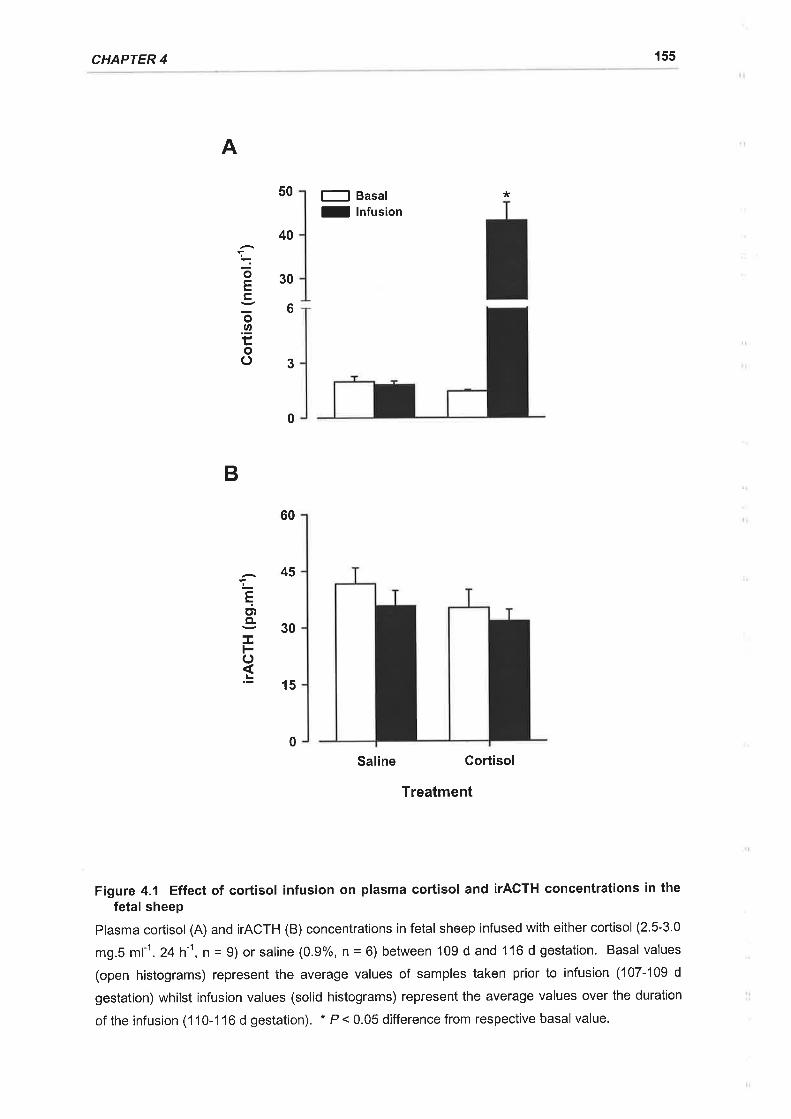
CHAPTER 4 155
A
50
40
30
E Basal
I Infusion
*
oE
õØ
oo
6
3
0
B
EctCL
Fof
60
45
30
15
0
Saline Gortisol
Treatment
Figure 4.1 Effect of coÉisol infusion on plasma cortisol and ITACTH concentrations in thefetal sheep
Plasma cortisol (A) and |TACTH (B) concentrations in fetal sheep infused with either cortisol (2.5-3.0
mg.S ml-1. 24h'1, n = 9) or saline (0.9%, n = 6) between 109 d and 116 d gestation. Basal values
(open histograms) represent the average values of samples taken prior to infusion (107-109 d
gestation) whilst infusion values (solid histograms) represent the average values over the duration
of the infusion (1 10-1 16 d gestation). * P < 0.05 difference from respective basal value.
CORTISOL AND PNñTT EXPRESS'O'I' IN THE ITI'TI'ATURE FETAL ADRENAL 156
Safiftn¡e
Figure 4.2 Representative autoradiographs of Northern blots of total adrenal RNA fromcortisoland saline infused fetal sheep hybridised with TH cDNA PNMT, PEnk A and l8 SrRNA ol igodeoxyn ucleotide probes
Representative autoradiographs of Northern blots after hþridisation of radiolabelled TH cDNA,
PNMT, PEnk A, and 18 S antisense oligonucleotide probes with total RNA (20 ¡rg per lane)
extracted from adrenals collected from fetal sheep at 116 d gestation after infusion of either saline
(open panel, n = 6) or cortisol (2.5-3.0 mg.S ml-1.24 h-r, shaded panel, n = 6)
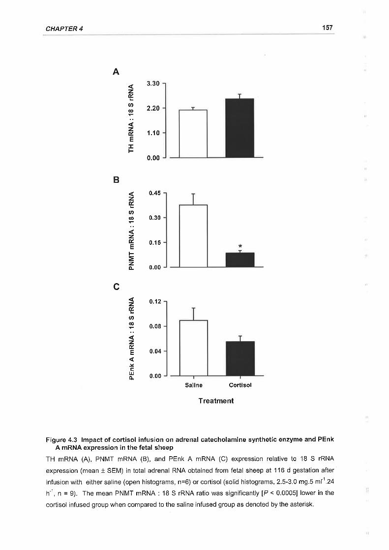
CHAPTER 4 157
A3.30
2.20
1.10
0.00
0.45
0.30
0.15 *
0.00
0.12
0.08
0.04
0.00
Saline Cortisol
Treatment
Figure 4.3 lmpact of cortisol infusion on adrenal catecholamine synthetic enzyme and PEnkA mRNA expression in the fetal sheep
TH mRNA (A), PNMT mRNA (B), and PEnk A mRNA (C) expression relative to 18 S rRNA
expression (mean t SEM) in total adrenal RNA obtained from fetal sheep at 116 d gestation after
infusion with either saline (open histograms, n=6) or cortisol (solid histograms, 2.5-3.0 mg.S m11.24
h-1, ¡ = 9). The mean PNMT mRNA : 18 S rRNA ratio was significantly [P < 0.0005] lower in the
cortisol infused group when compared to the saline infused group as denoted by the asterisk.
B
zÉ.
Ø@
zÉ.EIF
zÉ,
U'oo
zÉ.E
=zo-
zÉ,
U'@
zÉ,E
'ytrlrJo-
c
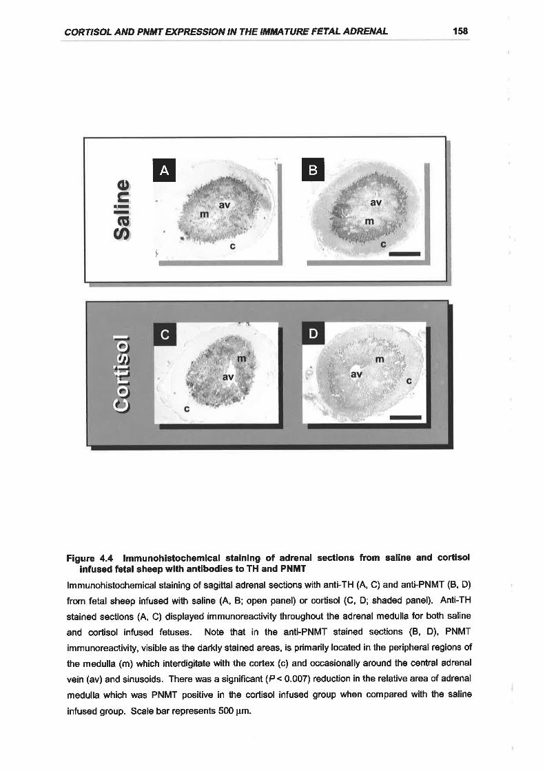
CORTISOL AND PNMT EXPRESS'ON 'N
THE ITI/Iñ'ATURE FETAL ADRENAL 158
tr tr
t
Figure 4.4 lmmunohistochemical staining of adrenal sections from saline and cortisolinfused feta! sheep with antibodies to TH and PNMT
lmmunohistochemical staining of sagittal adrenal sections with anti-TH (A, C) and anti-PNMT (8, D)
from fetal sheep infused with saline (4, B; open panel) or cortisol (C, D; shaded panel). Anti-TH
stained sections (4, C) displayed immunoreactivi$ throughout the adrenal medulla for both saline
and cortisol infused fetuses. Note that in the anti-PNMT stained sections (B, D), PNMT
immunoreactivity, visible as the darkly stained areas, is primarily located in the peripheral regions of
the medulla (m) which interdigitrate with the cortex (c) and occasionally around the central adrenal
vein (av) and sinusoids. There was a significant (P < 0.007) reduction in the relative area of adrenal
medulla which was PNMT positive in the cortisol infused group when compared with the saline
infused group. Scale bar represents 500 pm.
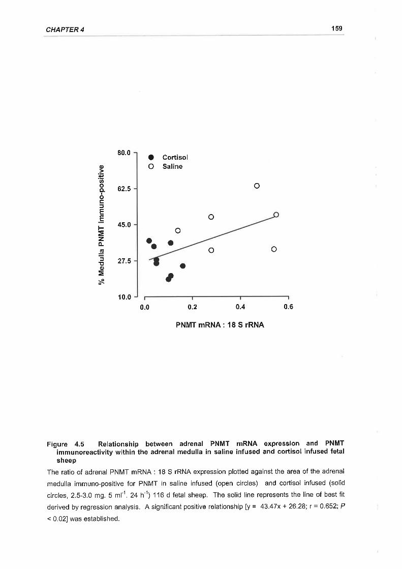
CHAPTER 4 159
80.0 O GortisolO Saline
62.5 o
45.0o
oo
27.5 o,
10.0
0.0 0.2 0.6
PNMT mRNA: 18 S rRNA
Figure 4.5 Relationship between adrenal PNMT mRNA expression and PNMTimmunoreactivity within the adrenal medulla in saline infused and cortisol infused fetalsheep
The ratio of adrenal PNMT mRNA : 1B S rRNA expression plotted against the area of the adrenal
medulla immuno-positive for PNMT in saline infused (open circles) and cortisol infused (solid
circles, 2.5-3.0 mg. 5 ml-1. 24h-1) '116 d fetal sheep. The solid line represents the line of best fit
derived by regression analysis. A significant positive relationship ly = 43.47x + 26.28; r = 0.652; P
< 0.021 was established.
o
ooo
o.lfiØoo.IocE
.Et-=zo-gt,o=s
0.4
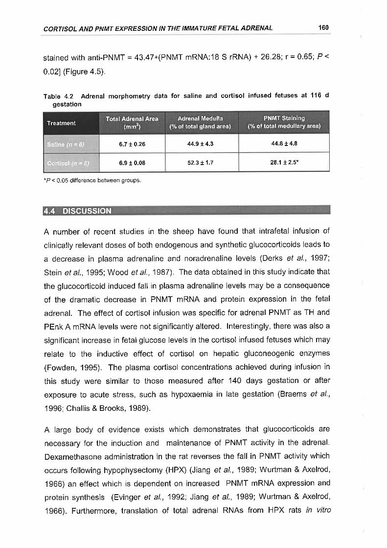
CORTISOL AND PNMT EXPRESSION IN THE IMMATURE FETAL ADRENAL 160
stained with anti-PNMT = 43.47x(PNMT mRNA:18 S rRNA) + 26.28; r = 0.65; P <
0.021 (Figure 4.5).
Table 4.2 Adrenal morphometry data for saline and cortisol infused fetuses at 116 dgestation
*P < 0.05 difference between groups
A number of recent studies in the sheep have found that intrafetal infusion of
clinically relevant doses of both endogenous and synthetic glucocorticoids leads to
a decrease in plasma adrenaline and noradrenaline levels (Derks et al., 1997;
Stein et al., 1995; Wood et al., 1987). The data obtained in this study indicate that
the glucocorticoid induced fall in plasma adrenaline levels may be a consequence
of the dramatic decrease in PNMT mRNA and protein expression in the fetal
adrenal. The effect of cortisol infusion was specific for adrenal PNMT as TH and
PEnk A mRNA levels were not significantly altered. lnterestingly, there was also a
significant increase in fetal glucose levels in the cortisol infused fetuses which may
relate to the inductive effect of cortisol on hepatic gluconeogenic enzymes
(Fowden, 1995). The plasma cortisol concentrations achieved during infusion in
this study were similar to those measured after 140 days gestation or after
exposure to acute stress, such as hypoxaemia in late gestation (Braems et al.,
1996; Challis & Brooks, 19Bg).
A large body of evidence exists which demonstrates that glucocorticoids are
necessary for the induction and maintenance of PNMT activity in the adrenal.
Dexamethasone administration in the rat reverses the fall in PNMT activity which
occurs following hypophysectomy (HPX) (Jiang et al., 1989; Wurtman & Axelrod,
1966) an effect which is dependent on increased PNMT mRNA expression and
protein synthesis (Evinger et al., 1992; Jiang et al., 1989; Wurtman & Axelrod,
1966). Furthermore, translation of total adrenal RNAs from HPX rats in vitro
44.8 +.4.86.710.26 44.9 t4.3
28.1Ì.2.5*6.9 r 0.08 52.3 t.1.7
Total Adrenal Area(..t)
Adrenal Medulla(% of total gland area)
PNMT Staining(% of total medullary area)Treatment
4.4 DTSCUSSTON
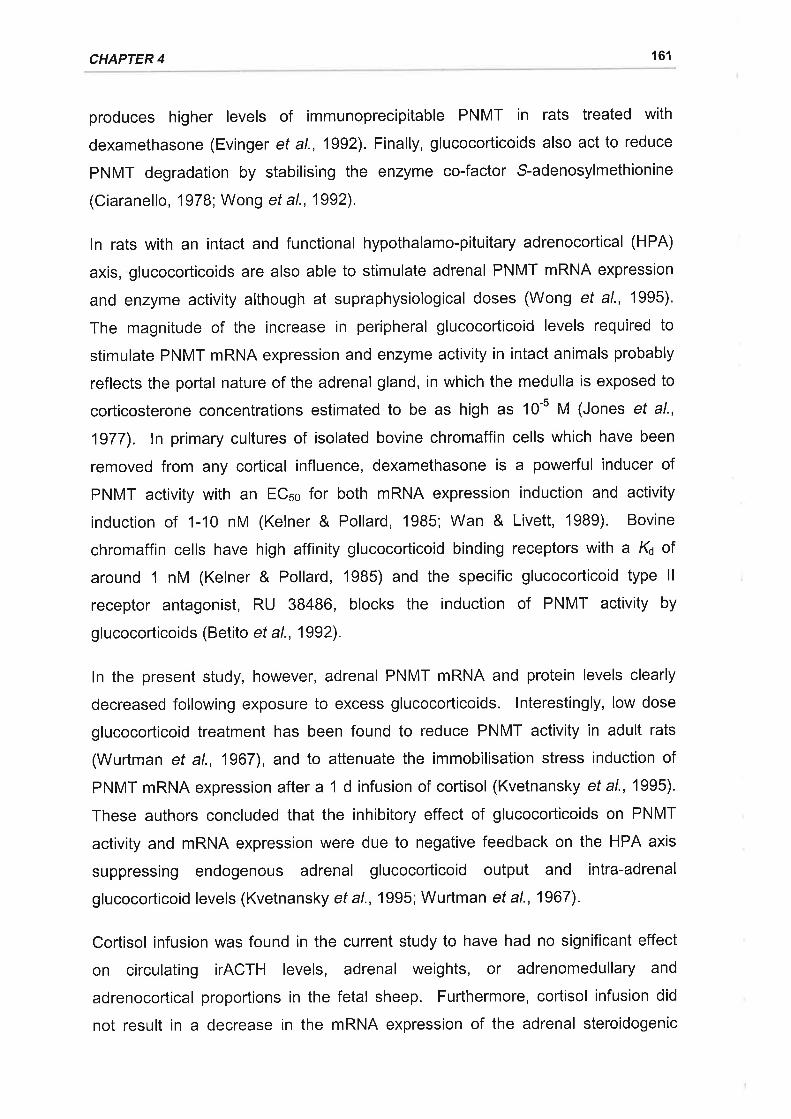
CHAPTER 4 161
produces higher levels of immunoprecipitable PNMT in rats treated with
dexamethasone (Evinger et al., 1992). Finally, glucocorticoids also act to reduce
PNMT degradation by stabilising the enzyme co-factor S-adenosylmethionine
(Ciaranello, 1978; Wong et al., 1992).
ln rats with an intact and functional hypothalamo-pituitary adrenocortical (HPA)
axis, glucocorticoids are also able to stimulate adrenal PNMT mRNA expression
and enzyme activity although at supraphysiological doses (Wong et al., 1995).
The magnitude of the increase in peripheral glucocorticoid levels required to
stimulate PNMT mRNA expression and enzyme activity in intact animals probably
reflects the portal nature of the adrenal gland, in which the medulla is exposed to
corticosterone concentrations estimated to be as high as 10-5 M (Jones et al.,
1977). ln primary cultures of isolated bovine chromaffin cells which have been
removed from any cortical influence, dexamethasone is a powerful inducer of
PNMT activity with an ECso for both mRNA expression induction and activity
induction of 1-10 nM (Kelner & Pollard, 1985; Wan & Livett, 19Bg). Bovine
chromaffin cells have high affinity glucocorticoid binding receptors with a K¿ of
around '1 nM (Kelner & Pollard, 1985) and the specific glucocorticoid type ll
receptor antagonist, RU 38486, blocks the induction of PNMT activity by
glucocorticoids (Betito et al., 1992).
ln the present study, however, adrenal PNMT mRNA and protein levels clearly
decreased following exposure to excess glucocorticoids. lnterestingly, low dose
glucocorticoid treatment has been found to reduce PNMT activity in adult rats
(Wurtman et al., 1967), and to attenuate the immobilisation stress induction of
PNMT mRNA expression after a '1 d infusion of cortisol (Kvetnansky ef a/., 1995).
These authors concluded that the inhibitory effect of glucocorticoids on PNMT
activity and mRNA expression were due to negative feedback on the HPA axis
suppressing endogenous adrenal glucocorticoid output and intra-adrenal
glucocorticoid levels (Kvetnansky ef a\.,1995; Wurtman et a1.,1967).
Cortisol infusion was found in the current study to have had no significant effect
on circulating iTACTH levels, adrenal weights, or adrenomedullary and
adrenocortical proportions in the fetal sheep. Furthermore, cortisol infusion did
not result in a decrease in the mRNA expression of the adrenal steroidogenic
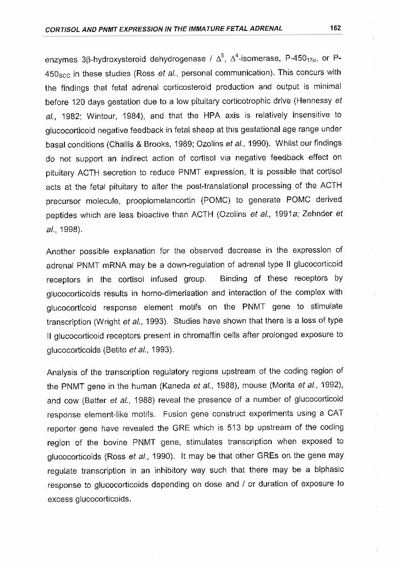
CORTISOL AND PNMT EXPRESSION IN THE IMMATURE FETAL ADRENAL 162
enzymes 3B-hydroxysteroid dehydrogenase / 45, Aa-isomerase, P-450t2o, or P-
450scc in these studies (Ross et al., personal communication). This concurs with
the findings that fetal adrenal corticosteroid production and output is minimal
before 120 days gestation due to a low pituitary corticotrophic drive (Hennessy ef
al., 1982; Wintour, 1984), and that the HPA axis is relatively insensitive to
glucocorticoid negative feedback in fetal sheep at this gestational age range under
basal conditions (Challis & Brooks, 1989; Ozolins et al., 1990). Whilst our findings
do not support an indirect action of cortisol via negative feedback effect on
pituitary ACTH secretion to reduce PNMT expression, it is possible that cortisol
acts at the fetal pituitary to alter the post-translational processing of the ACTH
precursor molecule, proopiomelancortin (POMC) to generate POMC derived
peptides which are less bioactive than ACTH (Ozolins et al., 1991a; Zehnder ef
a/., 1998).
Another possible explanation for the observed decrease in the expression of
adrenal PNMT mRNA may be a down-regulation of adrenal type ll glucocorticoid
receptors in the cortisol infused group. Binding of these receptors by
glucocorticoids results in homo-dimerisation and interaction of the complex with
glucocorticoid response element motifs on the PNMT gene to stimulate
transcription (WrighT et a\.,1993). Studies have shown that there is a loss of type
ll glucocorticoid receptors present in chromaffin cells after prolonged exposure to
glucocorticoids (Betito et al., 1993).
Analysis of the transcription regulatory regions upstream of the coding region of
the PNMT gene in the human (Kaneda et al., 1988), mouse (Morita et al., 1992),
and cow (Batter et al., 1988) reveal the presence of a number of glucocorticoid
response element-like motifs. Fusion gene construct experiments using a CAT
reporter gene have revealed the GRE which is 513 bp upstream of the coding
region of the bovine PNMT gene, stimulates transcription when exposed to
glucocorticoids (Ross et al., 1990). lt may be that other GREs on the gene may
regulate transcription in an inhibitory way such that there may be a biphasic
response to glucocorticoids depending on dose and / or duration of exposure to
excess g lucocorticoids.
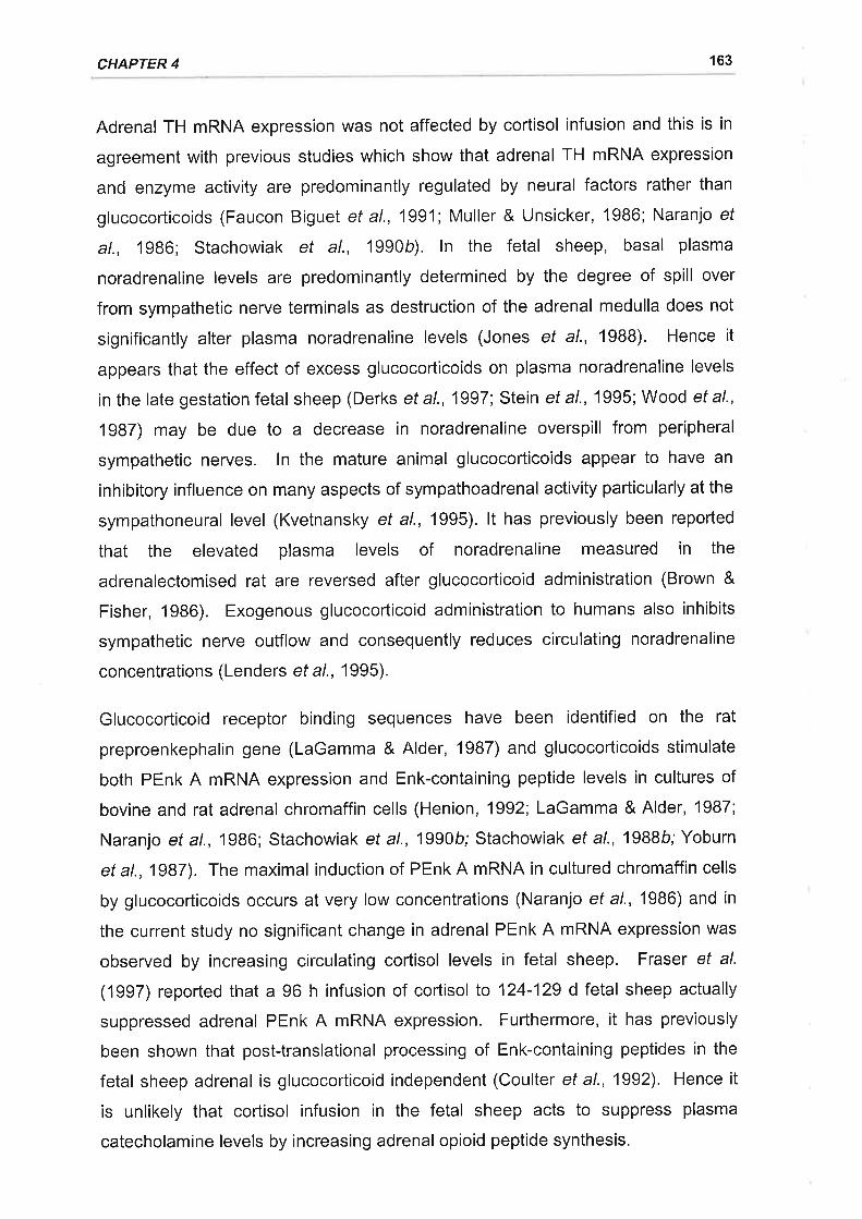
CHAPTER 4 163
Adrenal TH mRNA expression was not affected by cortisol infusion and this is in
agreement with previous studies which show that adrenal TH mRNA expression
and enzyme activity are predominantly regulated by neural factors rather than
glucocorticoids (Faucon Biguet et al., 1991; Muller & Unsicker, 1986; Naranjo ef
al., 1986; Stachowiak et al., 1990b). ln the fetal sheep, basal plasma
noradrenaline levels are predominantly determined by the degree of spill over
from sympathetic nerve terminals as destruction of the adrenal medulla does not
significantly alter plasma noradrenaline levels (Jones et al., 1988). Hence it
appears that the effect of excess glucocorticoids on plasma noradrenaline levels
in the late gestation fetal sheep (Derks et al., 1997 Stein et al.,'1995; Wood et al.,
1987) may be due to a decrease in noradrenaline overspill from peripheral
sympathetic nerves. ln the mature animal glucocorticoids appear to have an
inhibitory influence on many aspects of sympathoadrenal activity particularly at the
sympathoneural level (Kvetnansky et al., 1995). lt has previously been reported
that the elevated plasma levels of noradrenaline measured in the
adrenalectomised rat are reversed after glucocorticoid administration (Brown &
Fisher, 1986). Exogenous glucocorticoid administration to humans also inhibits
sympathetic nerve outflow and consequently reduces circulating noradrenaline
concentrations (Lenders ef a/.,'1995).
Glucocorticoid receptor binding sequences have been identified on the rat
preproenkephalin gene (LaGamma & Alder, 1987) and glucocorticoids stimulate
both PEnk A mRNA expression and Enk-containing peptide levels in cultures of
bovine and rat adrenal chromaffin cells (Henion, 1992; LaGamma & Alder, 1987;
Naranjo et al.,'1986; Stachowiak et al., 1990b; Stachowiak et al., 1988b; Yoburn
et a\.,1987). The maximal induction of PEnk A mRNA in cultured chromaffin cells
by glucocorticoids occurs at very low concentrations (Naranjo et al., 1986) and in
the current study no significant change in adrenal PEnk A mRNA expression was
observed by increasing circulating cortisol levels in fetal sheep. Fraser et al.
(1997) reported that a 96 h infusion of cortisol to 124-129 d fetal sheep actually
suppressed adrenal PEnk A mRNA expression. Furthermore, it has previously
been shown that post-translational processing of Enk-containing peptides in the
fetal sheep adrenal is glucocorticoid independent (Coulter et al., 1992). Hence it
is unlikely that cortisol infusion in the fetal sheep acts to suppress plasma
catecholamine levels by increasing adrenal opioid peptide synthesis.
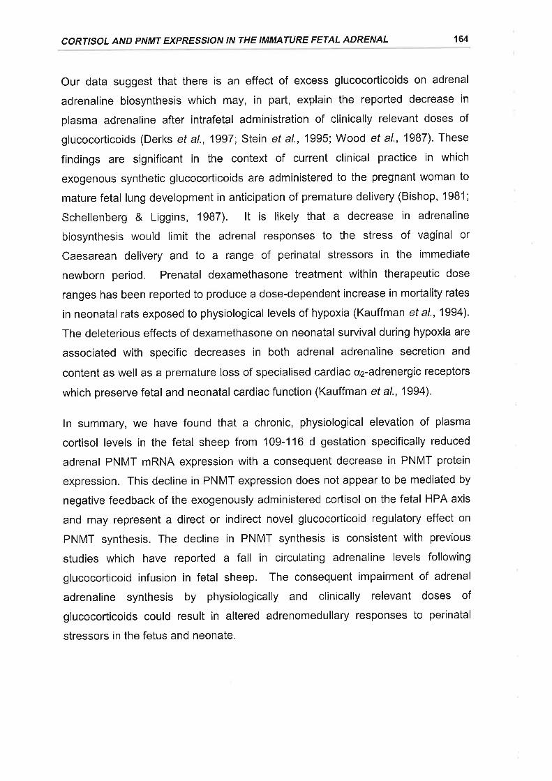
CORTISOL AND PNMT EXPRESSION IN THE IMMATURE FETAL ADRENAL 164
Our data suggest that there is an effect of excess glucocorticoids on adrenal
adrenaline biosynthesis which may, in part, explain the reported decrease in
plasma adrenaline after intrafetal administration of clinically relevant doses of
glucocorticoids (Derks et al., 1997', Stein et al., 1995; Wood et al., 1987). These
findings are significant in the context of current clinical practice in which
exogenous synthetic glucocorticoids are administered to the pregnant woman to
mature fetal lung development in anticipation of premature delivery (Bishop, 1981 ;
Schellenberg & Liggins, 1987). lt is likely that a decrease in adrenaline
biosynthesis would limit the adrenal responses to the stress of vaginal or
Caesarean delivery and to a range of perinatal stressors in the immediate
newborn period. Prenatal dexamethasone treatment within therapeutic dose
ranges has been reported to produce a dose-dependent increase in mortality rates
in neonatal rats exposed to physiological levels of hypoxia (Kauffman et a\.,1994).
The deleterious effects of dexamethasone on neonatal survival during hypoxia are
associated with specific decreases in both adrenal adrenaline secretion and
content as well as a premature loss of specialised cardiac cr2-adrenergic receptors
which preserve fetal and neonatal cardiac function (Kauffman et al., 1994).
ln summary, we have found that a chronic, physiological elevation of plasma
cortisol levels in the fetal sheep from 109-116 d gestation specifically reduced
adrenal PNMT mRNA expression with a consequent decrease in PNMT protein
expression. This decline in PNMT expression does not appear to be mediated by
negative feedback of the exogenously administered cortisol on the fetal HPA axis
and may represent a direct or indirect novel glucocorticoid regulatory effect on
PNMT synthesis. The decline in PNMT synthesis is consistent with previous
studies which have reported a fall in circulating adrenaline levels following
glucocorticoid infusion in fetal sheep. The consequent impairment of adrenal
adrenaline synthesis by physiologically and clinically relevant doses of
glucocorticoids could result in altered adrenomedullary responses to perinatal
stressors in the fetus and neonate.
5. EFFECT OF PLACENTAL RESTRICTION ON TH, PNMT, ANDPEnK A MRNA EXPRESSION IN THE ADRENAL GLAND OF
THE LATE GESTATION SHEEP FETUS
5.1 ¡NTRODUCTTON
The fetus responds to acute episodes of hypoxia with an increase in adrenal
catecholamine ouput (Cheung, 1990; Cohen et al., 1984; Jones et al., 1988) and
in Chapter 3 it was reported that there are also associated changes in adrenal TH
and PNMT gene expression. lt is not yet established if exposure of the fetus to
chronic stress in utero evokes long term changes in catecholamine synthetic
enzyme and proenkephalin A (PEnk A) gene expression which could potentially
impact upon the synthesis and secretion of adrenomedullary catecholamines.
An inability of the placenta to supply the developing fetus with adequate oxygen
and nutrients has been implicated in the pathogenesis of intrauterine growth
restriction (see Robinson ef a/., 1985 and Robinson et al., 1995 for reviews).
Cordocentesis studies in the human demonstrate that small for gestational age
fetuses are frequently hypoxaemic and hypoglycaemic (Economides ef al., 1991).
Catecholamines and their metabolites are also elevated in the amniotic fluid and
umbilical cord blood in growth retarded human fetuses (Divers et al., 1981;
Lagercrantz et al., 1980; Puolakka et al.,'1984; Weiner & Robillard, 1988).
Experimental models of IUGR in which the fetus is chronically hypoxaemic there
are also sustained increases in the fetal plasma concentrations of catecholamines
(Gagnon et al., 1994; Hooper et al., 1990; Simonetta et al., 1997). Despite
increased plasma catecholamines under basal conditions with experimental IUGR,
studies both in the rat (Shaul et al., 1989) and lamb (Jones & Robinson, 1983)
reveal that the catecholamine response, particularly adrenaline, is impaired in
response to an additional acute hypoxic challenge.
lntrauterine growth restriction and chronic fetal hypoxaemia are therefore
associated with long term changes in basal circulating catecholamine levels and
adrenomedullary responsiveness to subsequent acute stressors. Such changes
may reflect alterations in fetal catecholamine secretion, synthesis, and
metabolism. lt is unknown to what degree any of these determinants of circulating
catecholamine levels are altered in the fetal adrenal gland in response to chronic
165
PLACENTAL RESTRICTION AND THE FETAL ADRENAL 166
stress. The aim of this study was therefore to compare the adrenal expression of
TH, PNMT, and PEnk A mRNA in growth restricted and normally grown fetal
sheep in late gestation. For this study a well characterised experimental model of
IUGR was utilised which involved the surgical restriction of placental implantation
in the sheep thus restricting placental growth and function (Alexander, 1964;
Owens et al., 1989).
All procedures were approved by the University of Adelaide Standing Committee
on Ethics in Animal Experimentation.
5.2.1 ANIMALS AND SURGERY
Twenty one Border Leicester x Merino ewes were used in this study. ln ten ewes,
the majority of visible endometrial caruncles, were removed from the uterus prior
to conception as previously described (Robinson et al., 1979a). This procedure
limits the number of placental cotyledons formed following mating and hence
restricts placental and fetal growth. Carunclectomy was performed under
anaesthesia induced by intravenous injection of sodium thiopentone (1.25 g;
Pentothal, Rhone Merieux, Pinkenba, QLD, Australia) and maintained with
halothane (3-4 % in oxygen; Fluothane, lCl, Melbourne, VlC, Australia). Ewes
were administered 2 ml o'f antibiotics (250 mg.ml-1 procaine penicillin ,250 mg.ml-1
dihydrostreptomycin sulphate, 20 mg.ml-1 Procaine HCI; Penstrep llium, Troy
Laboratories, Smithfield, NSW, Australia) intramuscularly prior to surgery. The
uterus was exteriorised through a low abdominal midline incision and
subsequently opened along the antimesometrial border from the level of the cervix
to close to the uterotubule junction in both horns. Most of the visible endometrial
caruncles were then excised and the incisions in the uterus were sutured in a
single layer using 2 I 0 cat gut (Ethicon, Johnson and Johnson, Sydney, NSW,
Australia) through the myometrium and serosa. Once sutured the uterus was
replaced and the abdomen closed in two layers, the peritoneum and rectus sheet,
followed by the skin and sub cutaneous tissue, using 4 I 0 coaled Vicryl (Ethicon).
Ewes were kept under observation for 4-7 d post surgery and following a minimum
of 10 weeks recovery entered a mating program with the date of mounting taken
5.2 METHODS
CHAPTER 5 167
as day 0 of pregnancy. Pregnancies were confirmed by ultrasound at
approximately 60 d gestation.
ln the 10 carunclectomised ewes (Placental Restriction group) and 11 control
ewes a second surgery was carried out between 102 and 112 d gestation under
general anaesthesia as described above. Catheters were inserted into the fetal
and maternal carotid artery and jugular vein as well as the amniotic sac as
described in Chapter 3. Catheters were flushed and filled with heparinised saline
and the fetal catheters were exteriorised through an excision in the ewes flank.
5.2.2 POST OPERATIVE CARE
Following surgery the ewes were housed in individual metabolic crates in animal
holding rooms under a 12 h light-dark cycle and fed lucerne chaff once daily
between 0900 and 1300 h with water ad libitum. Sodium ampicillin (500 mg,
Austrapen, Commonwealth Serum Laboratories, Melbourne, VlC, Australia) was
administered intra-amniotically to all fetal sheep for 4 consecutive days post
operatively.
5.2.3 POST MORTEM AND TISSUE COLLECTION
The ewes were electively killed between 140 and 141 d gestation with an
overdose of sodium pentobarbitone (8.125 g; Lethabarb, Virbac, Peakhust, NSW,
Australia) and both the uterus and fetus were removed by hysterotomy. The
fetuses were weighed and then decapitated and the fetal adrenal glands were
quickly dissected, weighed, and either snap frozen in liquid N2 or placed into ice-
cold paraformaldehyde fixative. Placental cotyledons were individually dissected
from the uteri of carunclectomised (n = 7) and control ewes (n = 3) and weighed.
5.2.4 BLOOD SAMPLING PROTOCOL
Fetal (3.5 ml) and maternal (5 ml) arterial blood samples were collected every
second day between 104 and 140 d gestation. An additional 0.5 ml of whole
arterial blood was also collected in order to determine arterial POz, pH, PCO2, 02
saturation (SOz) and haemoglobin (Hb) content using an ABL 330 acid base
analyser and an OSM2 Hemoximeter (Radiometer, Copenhagen, Denmark).
Blood samples were collected into heparinised tubes on ice and centrifuged at
PLACENTAL RESTRICTION AND THE FETAL ADRENAL 168
4C at 1500 g for 10 min. The plasma was separated and a 1 ml aliquot was
placed into a polypropylene tube (12 x 75 mm) containing antioxidant (50 pl of 2
mM ascorbic acid) for subsequent catecholamine analysis. All plasma aliquots
were subsequently snap frozen in liquid nitrogen and stored at -70'C until
analysed for catecholamines and glucose.
5.2.5 PLASMA GLUGOSE DETERMINATION
Plasma concentrations of glucose were measured across gestation in placentally
restricted (PR, n = 9) and control fetuses (n = 9) using a COBAS MIRA automated
Analysing System (Cobas, Roche Diagnostics, Basle, Switzerland). The system
determined plasma glucose concentrations by enzymatic analysis using
hexokinase and glucose-6-phosphate dehydrogenase and measuring the
formation of NADH spectrophotometrically at 340 nm. The intra-assay coefficient
of variation was 1.4%.
5.2.6 PLASMA CATECHOLAMINE EXTRACTION
Catecholamines were extracted by alumina absorption from gestational plasma
samples obtained from PR (n = 7) and control (n = 6) fetuses and measured in
accordancewiththemethodsof (Schuijers etal.,1986). Thawed'1 ml aliquotsof
plasma were transferred to borosilicate glass culture tubes (12 x 75 mm)
containing 10 mg of acid washed alumina (Hart Analytical, Melbourne, Australia)
and 30 pl of 10 mM sodium metabisulphite. An additional 1 ml of distilled water
was added to the plasma, as well as 100 pl of DHBA (20 pmol / 100 pl 0.01M
sodium metabisulphite) which was used as an internal standard to monitor
catecholamine recovery. Tris-EDTA buffer (400 pl, lMTris base,2 % EDTA, pH
8.6) was added to the tubes which were then vigorously agitated at 4"C for 40 min
on a multi-tube vortexer (Baxter Healthcare Corp., Miami, FL, USA). The samples
were subsequently centrifuged at 1800 g for 10 min at 4'C and the supernatant
was discarded. The alumina was then washed 4 times with distilled deionised
water (0.5 ml) and following the last wash the samples were centrifuged at 1800 g
for'10 min at 4"C, with the supernatant again being discarded. The
catecholamines were desorbed from the alumina by the addition of perchloric acid
(100 pl, 0.1 M in 0.01 M sodium metabisulphite) prior to the samples being
CHAPTER 5 169
vortexed for a further 10 min at 4'C. Following centrifugation at 1800 g for 10 min
at 4"C the supernatant was aspirated and filtered through 0.45 pm syringe filters
(Filter type HV; Nihon Millipore Kogyo K.K., Yonezawa, Japan) to remove any
remaining alumina particles. Samples were stored at -70'C after extraction until
they were assayed using reverse phase HPLC combined with electrochemical
detection. The average recovery of catecholamines with this extraction procedure
was 59.2 + 0.9 %.
5.2.7 CATECHOLAMINE MEASUREMENT
30 pl aliquots of the extracted samples were injected onto a BAS 200A HPLC
system (Bioanalytical Systems lnc., West Lafayette, lN, USA) using a CMA 200
refrigerated microsampler (CMA / Microdialysis AB, Stockholm, Sweden) aI 4"C.
Separation of catecholamines was achieved using a 3 pm ODS, 100 x 3.2 mm,
phase ll cartridge column (Bioanalytical Systems lnc.) and an aqueous mobile
phase which consisted of (mM: sodium acetate, 50; citric acid, 20;
octanesulphonicacid,3.TS; EDTA,2;and methanol 5%v lv,ala pH of 4.3. The
mobile phase flow rate was 0.7 ml.min-1 and the potential of the glassy carbon
detector electrode was set at + 750 mV vs. a Ag / AgCl reference electrode.
Catecholamines were automatically quantified by peak area integration using an
IBM compatible personal computer equipped with Barspec Data System
chromatographic software (Barspec Ltd., Rehovot, lsrael). Peak areas were
compared with to those obtained for catecholamine standards of known
concentration (Norepinephrine Bitartrate, Epinephrine Bitartrate, and DHBA
Hydrobromide; 20 pmol / 100 pl 0.4 M PCA; Bioanalytical Systems lnc.) to
determine the noradrenaline and adrenaline content of the samples. The
sensitivity of the catecholamine assay was 0.07 pmol.ml-1 for both noradrenaline
and adrenaline and the intra- and inter-assay coefficients of variation were less
than 10 %.
5.2.8 RNA ISOLATION AND ANALYSIS
Total adrenal RNA was extracted according to the method of Chirgwin et al. 1979,
as outlined in Chapter 2. Briefly, adrenals from 6 PR fetuses and 6 control fetuses
were homogenised in 4 M guanidine thiocyanate solution and ultracentrifuged at
PLACENTAL RESTRICTION AND THE FETAL ADRENAL 170
130 000 gthrough a5.7 MCsCI cushion. The RNA pelletwas then resuspended
in water and purified by re-precipitation with 0.3 M sodium acetate and absolute
ethanol. The RNA pellet was washed in 70 % ethanol and again resuspended in
water. Maximum absorbance was measured spectrophotometrically at 260 nm
and 280 nm and used to quantify nucleic acid purity, yield, and concentration.
RNA solutions were kept at -70' C until use.
5.2.9 PROBES AND PROBE LABELLING
The probes utilised in this study and the radiolabelling methods for the probes are
described in detail in Chapter 2. To summarise, oligonucleotide antisense probes
complementary to nucleotides 361-389 of the peptide coding region of bovine
PNMT mRNA (29 mer, Wan ef al., 19BB), nucleotides 627-671 of the peptide
coding region of bovine PEnk A mRNA (44 mer, Wan et al', 1988), and
nucleotides 151-180 of the rat 1B S rRNA (30 mer, Chan et al., 1984) sequence
were synthesised (Bresatec, Thebarton, SA, Australia). A 1.7 kb bovine TH cDNA
(D'Mello et al., 1988) was used to detect TH mRNA. The oligonucleotides were
end labelled using T4 polynucleotide kinase (7.9 U.pl-1, Pharmacia, North Ryde,
NSW, Australia) and y-[32P]ATP (4000 Ci.mmol-1 ,Bresatec) in accordance with the
method described for the end labelling of oligonucleotides by Sambrook et al.
(1989). The cDNA probe was labelled by the random priming method with cx-
¡32e1OCte (3000 Ci.mmol-r, Bresatec) and Klenow fragment (6.4 U.pl-t,
Pharmacia) using an oligolabelling kit (Pharmacia) as per manufacturer's
instructions.
5.2.10 NORTHERN BLOT ANALYSIS
Northern blotting was performed as described by Sambrook et al. (1989) and
outlined in detail in Chapter 2. Total RNA samples (20 pg) were denatured in
2.2M formaldehyde and 50 % (v / v) formamide at 55'C for 10 min. The
denatured total RNA was subsequently separated using gel electrophoresis with
1o/o agarose containing 2.2 M formaldehyde and then transferred by capillary
blotting to Zetaprobe nylon membranes (Biorad, Richmond, CA, USA).
Membranes were washed in 10 x SSC for 10 min at room temperature before
being baked for t h at 80"C and stored flat at -20"C until hybridisation.
CHAPTER 5 171
For oligonucleotide probes the membranes were prehybridised at 50"C for 18 h in
a solution consisting of 5 x SSC, 20 mM NaHzPO¿, pH 7.2, 7 % SDS, 5 x
Denhardt's, and 100 pg.ml-r of heat denatured Salmon sperm DNA. Membranes
were hybridised with 1-2 x 10⁶ cpm.ml-1 of labelled oligonucleotide in 30 ml of
fresh hybridisation solution for 20 h at 50'C. The same protocol was used for the
TH cDNA probe, however the hybridisation solution consisted of 50 % (v I v)
deionised formamide, 5 x SSPE, 7 % SDS, pH 7.2, and 100 pg.ml-1 of heat
denatured salmon sperm DNA, with hybridisation being carried out at 42C.
Following hybridisation the membranes were washed in 1 x SSC and 0.1 % SDS
at their hybridisation temperatures (1 x 30 min). The membranes were then
washed at their hybridisation temperature in 0.1 x SSC and 0.1 % SDS (2 x
30 min), briefly air dried, and exposed to a Fuji BAS-Ills phosphorimager plate
(Fuji Photo Co., Tokyo, Japan) for 24-48 h. Autoradiographs were visualised
using a Fuji BAS 1000 phosphorimager (Fuji Photo Co.) and quantitated using Fuji
MacBAS software (Fuji Photo Co.).
5.2.1 1 I M M U NOHISTOCH EM ISTRY
Adrenals from PR (n = 6) and control (n = 6) fetuses were dissected into halves
perpendicular to their poles at postmortem and immersed in ice-cold
paraformaldehyde fixative (4 % in sodium phosphate buffer, 0.1 M, pH 6.4) for
24 h. Following fixation the adrenals were washed in ice cold sodium phosphate
buffer (2 x 2a fi. After washing, adrenals were stored at 4"C in 70 % ethanol
before being embedded in paraffin wax.
Transverse adrenal sections (5 pm) were cut on a microtome and mounted onto
glass slides coated with poly-t--lysine. Semi adjacent sections were either
processed for TH and PNMT immunoreactivity using a Vectastain avidin-biotin
peroxidase complex kit (PK-4001, Vector Laboratories, Burlingame, CA, USA) or
stained with haematoxylin and eosin. The antibodies and immunohistochemical
procedures used in this study have been described in detail in Chapter 4. Briefly,
the sections were deparaffinised by immersion in Histoclear (National Diagnostics,
Somerville, NJ, USA) and then rehydrated by immersion through a graded ethanol
series and rinsed in distilled water. Endogenous peroxidase activity was
quenched by a 30 min incubation with 0.5 % HzOz in methanol. Sections were
PLACENTAL RESTRICTION AND THE FETAL ADRENAL 172
washed (1 x 20 min, 0.01 M PBS, pH 7.0) and then pre-incubated with blocking
solution (3 % NGS in 0.01 M PBS containing 1 % BSA) for 30 min. Following pre-
incubation the sections were covered with primary antibody at the appropriate
dilution (rabbit anti-bovine PNMT, 1:1500; rabbit anti-rat TH, 1:1000) in PBS
containing 1 % BSA. Sections were incubated overnight in an air tight humidified
container at 4"C. Following incubation with the primary antibody sections were
incubated with biotinylated goat anti-rabbit secondary antibody (Vectastain kit PK-
4001)for 60 min at room temperature and then with ABC (Vectastain k¡t PK-4001)
for a further 60 min. The sections were washed with PBS (0.01 M,3 x 5 min)
following each incubation. The sections were subsequently covered with DAB (0.5
mg.ml-1 in PBS buffer and 0,02 % HzOz) at room temperature for 10 min. Sections
were finally washed in PBS (3 x 5 min), dehydrated and coverslipped.
5.2.12ADRE NAL MORPHOM ETRY
The transverse areas of a number of adrenal compartments were determined from
mid glandular sections. lmages of adrenal sections were captured using a CCD
black and white video camera (SSC-M37OCE, Sony Corp., Tokyo, Japan)
mounted on a dissecting microscope via an SZ-CTV photomount tube (Olympus
Optical Co., Tokyo, Japan). The image was digitised as a grey scale image using
a Power Mac 8500 I 120 (Apple, Cupertino, CA, USA) equipped with NIH lmage
V1.61 software. Total adrenal area was obtained by defining and measuring the
boundary around the adrenal section image. Relative vascular area was
calculated by subtracting the area of the section positively stained with
Haematoxylin and Eosin from the total area of the section. The area of the
medulla was defined as the area of the gland which stained positively with anti-
TH. The areas of the sections which were positively stained for PNMT was also
calculated.
5.2.13 DATA ANALYSIS
All data are presented as the mean t standard error of the mean (SEM). A mean
gestational value for each of the blood gas variables and plasma glucose
concentrations were calculated for each animal in the PR and control groups and
the group mean values were compared using one-tailed Student's t-tests. Adrenal
TH, PNMT, and PEnk A mRNA: 18 rRNA ratios in the PR and control groups
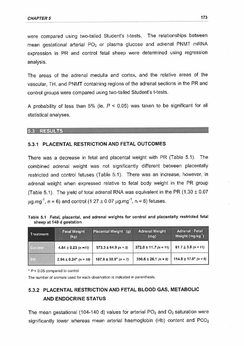
CHAPTER 5 173
were compared using two-tailed Student's t-tests. The relationships between
mean gestational arterial POz or plasma glucose and adrenal PNMT mRNA
expression in PR and control fetal sheep were determined using regression
analysis.
The areas of the adrenal medulla and cortex, and the relative areas of the
vascular, TH, and PNMT containing regions of the adrenal sections in the PR and
control groups were compared using two-tailed Student's t-tests.
A probability of less than 5% (ie. P < 0.05) was taken to be significant for all
statistical analyses.
5.3.I PLACENTAL RESTRICTION AND FETAL OUTCOMES
There was a decrease in fetal and placental weight with PR (Table 5.1). The
combined adrenal weight was not significantly different between placentally
restricted and control fetuses (Table 5.1). There was an increase, however, in
adrenal weight when expressed relative to fetal body weight in the PR group
(Table 5.1). The yield of total adrenal RNA was equivalent in the PR (1.30 t 0.07
pg.mg-1, n = 6) and control (1.27 t 0.07 pg.mg-1, n = 6) fetuses.
Table 5.1 Fetal, placental, and adrenal weights for control and placentally restricted fetalsheep at 140 d gestation
* P < 0.05 compared to control
The number of animals used for each observation is indicated in parenthesis.
5.3.2 PLACENTAL RESTRICTION AND FETAL BLOOD GAS, METABOLIC
AND ENDOCRINE STATUS
The mean gestational (104-140 d) values for arterial POz and Oz saturation were
significantly lower whereas mean arterial haemoglobin (Hb) content and PCOz
5.3 RESULTS
81.7 t 3.8 (n = 11)573.3 + 64.9 (n = 3) 372.0 t 11.7 (n = 11)4.64 t 0.23 (n =11)
114.8 L 17.0* (n = 9)2.94 t 0.24" (n = 10) 187.6 t 30.8* (n = 7) 350.61 26.1 (n = 9)
FetalWeight(ks)
PlacentalWeight (g) AdrenalWeight(ms)
Adrenal : FetalWeight (mg.kg-')Treatment
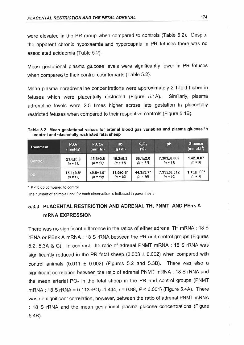
PLACENTAL RESTRICTION AND THE FETAL ADRENAL 174
were elevated in the PR group when compared to controls (Table 5.2). Despite
the apparent chronic hypoxaemia and hypercapnia in PR fetuses there was no
associated acidaemia (Table 5.2).
Mean gestational plasma glucose levels were significantly lower in PR fetuses
when compared to their control counterparts (Table 5.2).
Mean plasma noradrenaline concentrations were approximately 2.l-fold higher in
fetuses which were placentally restricted (Figure 5.14). Similarly, plasma
adrenaline levels were 2.5 times higher across late gestation in placentally
restricted fetuses when compared to their respective controls (Figure 5.18).
Table 5.2 Mean gestational values for arterial blood gas variables and plasma glucose incontrol and placentally restricted fetal sheep
* P < 0.05 compared to control
The number of animals used for each observation is indicated in parenthesis
5.3.3 PLACENTAL RESTRICTION AND ADRENAL TH, PNMT, AND PENK A
mRNA EXPRESSION
There was no significant difference in the ratios of either adrenal TH mRNA : 18 S
rRNA or PEnk A mRNA : 18 S rRNA between the PR and control groups (Figures
5.2, 5.34 & C). ln contrast, the ratio of adrenal PNMT mRNA : 18 S rRNA was
significantly reduced in the PR fetal sheep (0.003 t 0.002) when compared with
control animals (0.011 t 0.002) (Figures 5.2 and 5.38). There was also a
significant correlation between the ratio of adrenal PNMT mRNA : 18 S rRNA and
the mean arterial POz in the fetal sheep in the PR and control groups (PNMT
mRNA: 18 S rRNA =0.113xPO2 -1.444, r = 0.88, P< 0.001) (Figure 5.4A). There
was no significant correlation, however, between the ratio of adrenal PNMT mRNA
: 18 S rRNA and the mean gestational plasma glucose concentrations (Figure
5.48).
7.363r0.009(n = 11)
1.42t0.07(n=9)
45.6+0.8(n = 11)
10.2r0.3(n = 11)
68.r12.0(n = 11)
23.610.9(n = 11)
1.13t0.09*(n=9)
44.3t3.7*(n = 10)
7.355*0.012(n = 10)
15.1+0.8*(n = 10)
49.5t1.0*(n = 10)
11.5+0.6*(n = 10)
P"GOz(mmHg)
Hb(g / dl)
Glucose(mmol.l1)
SuOzPuOz pHTreatment
t%t(mmHg)
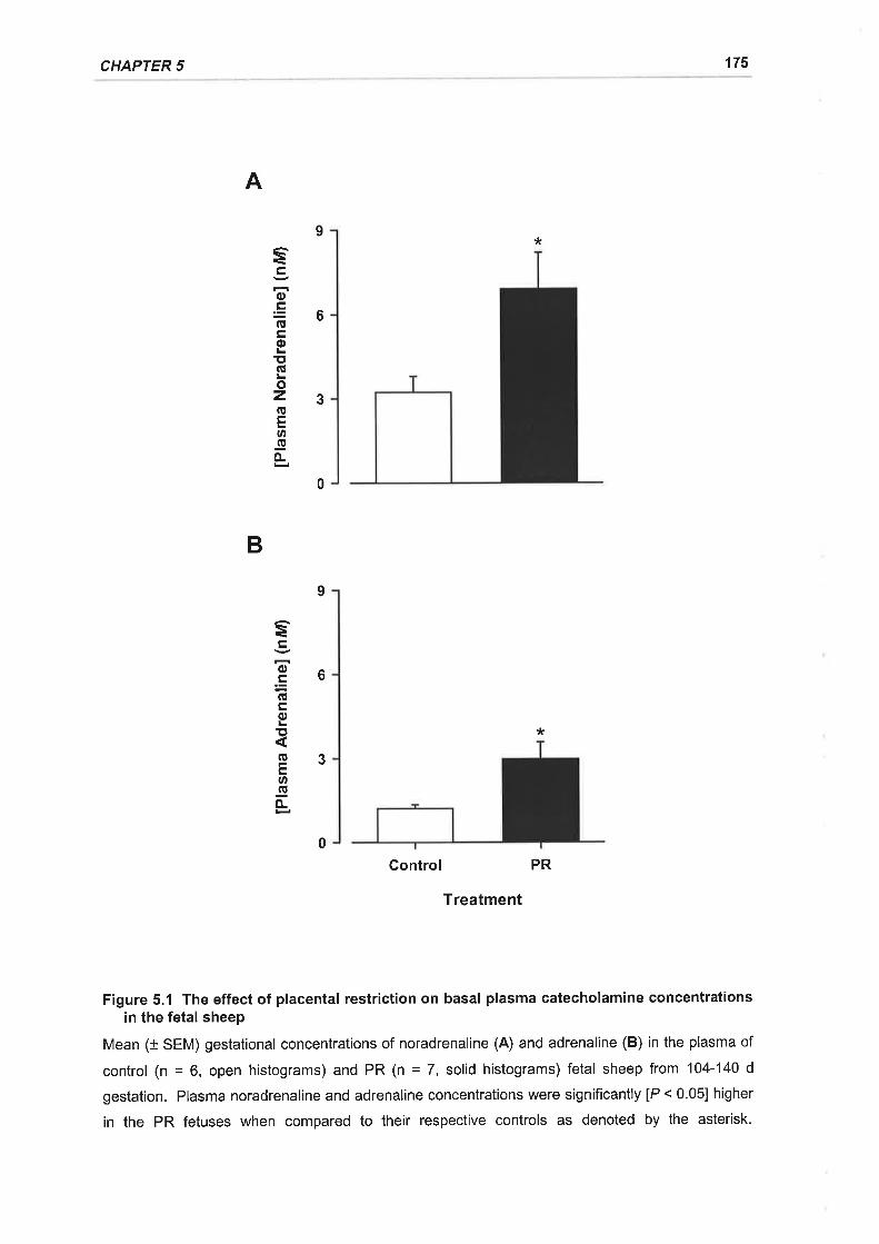
CHAPTER 5 175
A
*
B
*
Control PR
Treatment
Figure 5.1 The effect of placental restriction on basal plasma catecholamine concentrationsin the fetal sheep
Mean (+ SEM) gestational concentrations of noradrenaline (A) and adrenaline (B) in the plasma of
control (n = 6, open histograms) and PR (n = 7, solid histograms) fetal sheep from 104-'140 d
gestation. Plasma noradrenaline and adrenaline concentrations were significantly [P < 0.05] higher
in the PR fetuses when compared to their respective controls as denoted by the asterisk.
9
6
3
soE(!trEE'(ú
ozGEo-go-
6
3
è
oE(ú
oïtßEØ-go-
0
9
0
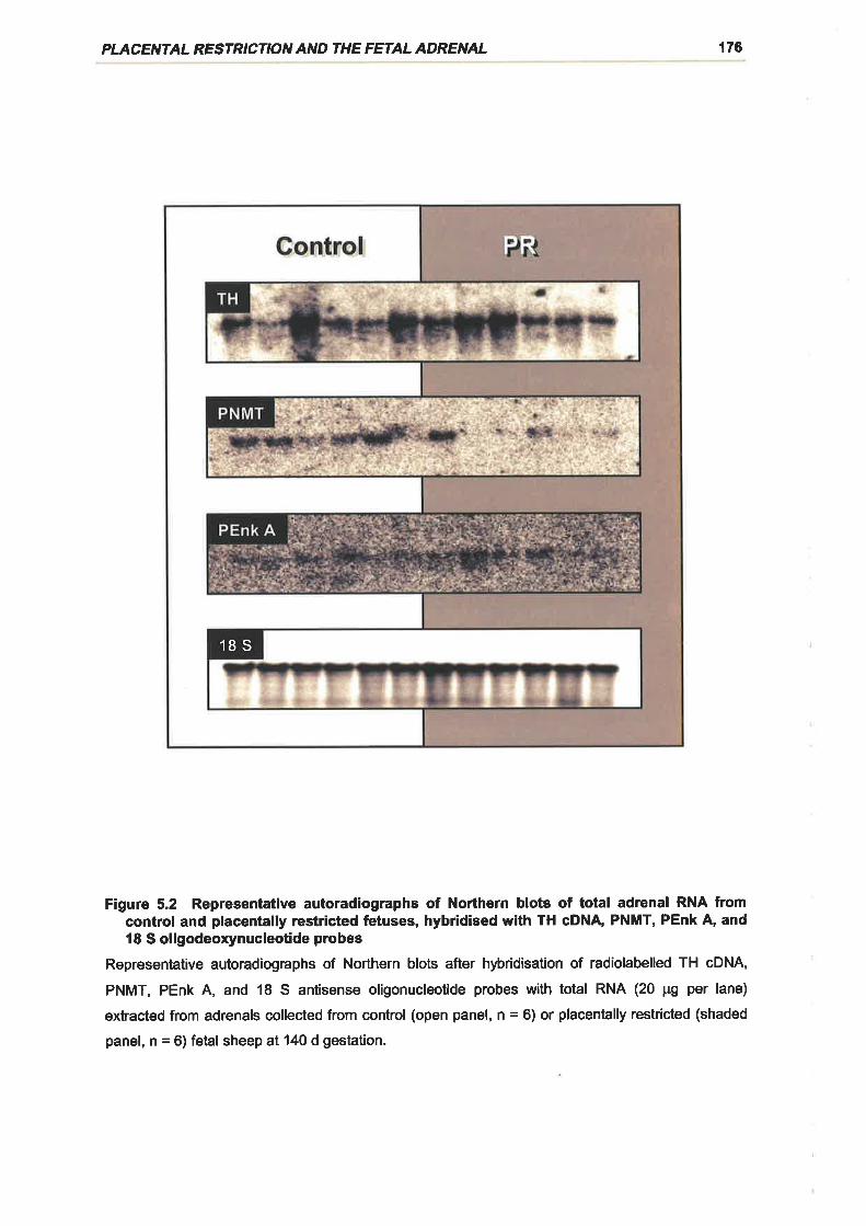
PLACENTA¿ RESTR'CTION AND THE FETAL ADRENAL 176
18S
Figure 5.2 Representative autoradiographs of Northern blots of total adrenal RNA fromcontrol and placentally restricted fetuses, hybridised with TH cDNA PNMT, PEnk A, andl8 S oligodeoxynucleotide probes
Representative autoradiographs of Northern blots after hþridisation of radiolabelled TH cDNA,
PNMT, PEnk A, and 18 S antisense oligonucleotide probes with total RNA (20 pg per lane)
extracted from adrenals collected from control (open panel, n = 6) or placentally restricted (shaded
panel, n = 6) fetal sheep al14O d gestation.
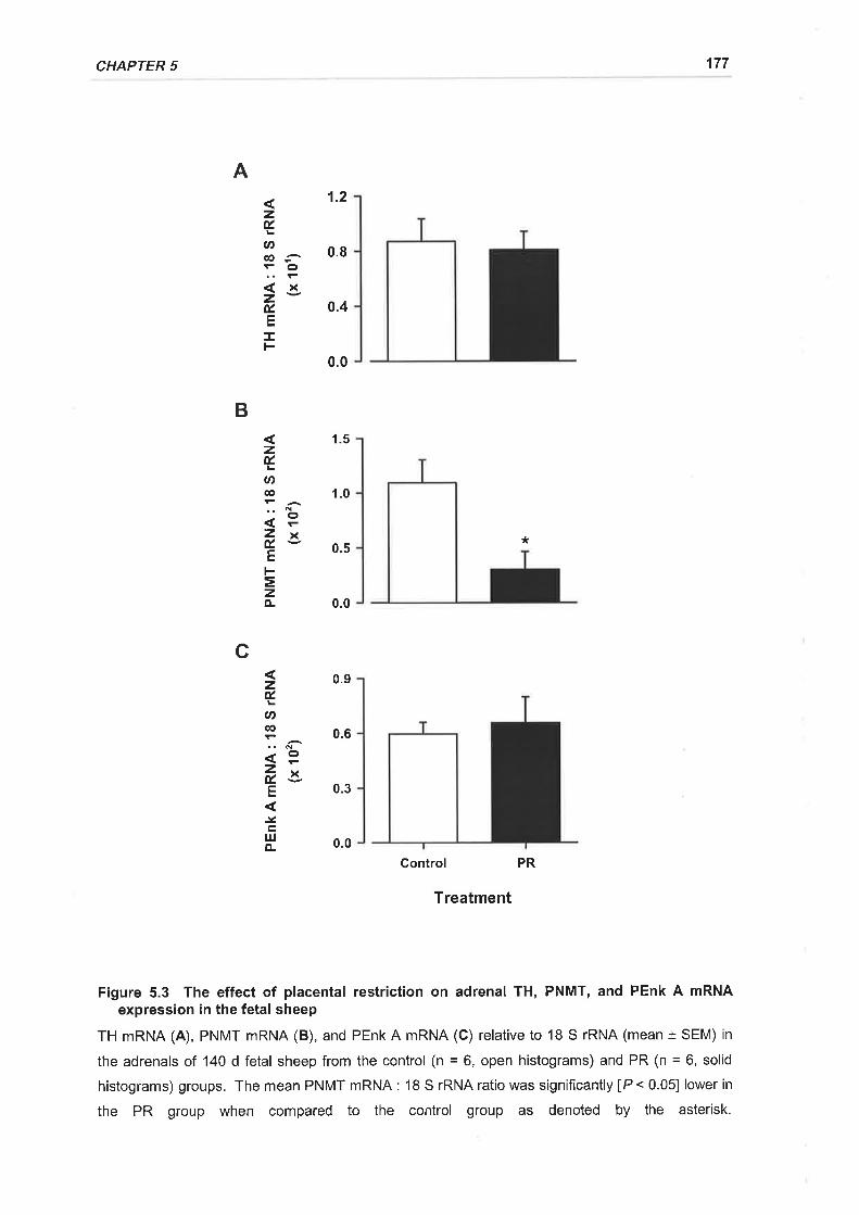
CHAPTER 5 177
1.2
0.8
0.4
1.5
1.0
0.5*
0.0
0.9
0.3
0.0
Control PR
Treatment
Figure 5.3 The effect of placental restriction on adrenal TH, PNMT, and PEnk A mRNAexpression in the fetal sheep
TH mRNA (A), PNMT mRNA (B), and PEnk A mRNA (C) relative to 1B S rRNA (mean t SEM) in
the adrenals of 140 d fetal sheep from the control (n = 6, open histograms) and PR (n = 6, solid
histograms) groups. The mean PNMT mRNA : 18 S rRNA ratio was significantly [P < 0.05] lower in
the PR group when compared to the control group as denoted by the asterisk.
A
zÉ,
U)@- o
2É,E
zÉ.
(t>
@
':ËZxÉ.v
E
=zÀ
0.6
zÉ.
U'ct
zÉ. -aE
.Ycl¡Jo-
0.0
B
c
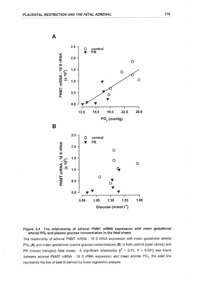
PLACENTAL RESTRICTION AND THE FETAL ADRENAL 178
A
2.5 O controlYPR
2.0o
1.5
1.0 V o
0.5
0.0 Y
12.0 15.5 19.0 22.5 26.0
pO, (mmHg)
2.5 O controlYPR
o
o
Y0.0 Y v Y
0.80 1.05 1.30 1.55 1.80
Glucose (mmol.l-r)
Figure 5.4 The relationship of adrenal PNMT mRNA expression with mean gestationalaÉerial PO2 and plasma glucose concentration in the fetal sheep
The relationship of adrenal PNMT mRNA: 18 S rRNA expression with mean gestational arterial
PO2 (A) and mean gestational plasma glucose concentrations (B) in both control (open circles) and
PR (closed triangles) fetal sheep. A significant relationship tf = 0.70, P < 0.0011 was found
between adrenal PNMT mRNA: 18 S rRNA expression and mean arterial POz, the solid line
represents the line of best fit derived by linear regression analysis.
o
oY
zÉ.
ct,o
N.'o<Fzxú-EFEzo-
oq
o
Vo
4 2'o
ØP ñ^ 1.s"o<FzxÉ- 1.0E
= 0.5
o-
Y
B
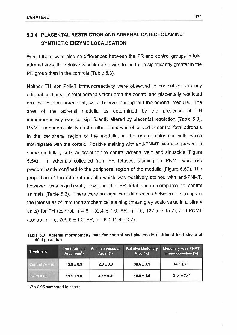
CHAPTER 5 179
5.3.4 PLACENTAL RESTRICTION AND ADRENAL CATECHOLAMINE
SYNTHETIC ENZYME LOCALISATION
Whilst there were also no differences between the PR and control groups in total
adrenal area, the relative vascular area was found to be significantly greater in the
PR group than in the controls (Table 5.3).
Neither TH nor PNMT immunoreactivity were observed in cortical cells in any
adrenal sections. ln fetal adrenals from both the control and placentally restricted
groups TH immunoreactivity was observed throughout the adrenal medulla. The
area of the adrenal medulla as determined by the presence of TH
immunoreactivity was not significantly altered by placental restriction (Table 5.3).
PNMT immunoreactivity on the other hand was observed in control fetal adrenals
in the peripheral region of the medulla, in the rim of columnar cells which
interdigitate with the cortex. Positive staining with anti-PNMT was also present in
some medullary cells adjacent to the central adrenal vein and sinusoids (Figure
5.54). ln adrenals collected from PR fetuses, staining for PNMT was also
predominantly confined to the peripheral region of the medulla (Figure 5.58). The
proportion of the adrenal medulla which was positively stained with anti-PNMT,
however, was significantly lower in the PR fetal sheep compared to control
animals (Table 5.3). There were no significant differences between the groups in
the intensities of immunohistochemical staining (mean grey scale value in arbitrary
units) for TH (control, n = 6, 102.4 + 1.0; PR, n = 6, 122.5 + 15.7), and PNMT
(control, n = 6, 209.5 t 1.0; PR, n = 6,211.8 t 0.7).
Table 5.3 Adrenal morphometry data for control and placentally restricted fetal sheep at140 d gestation
44.6 t 4.012.9 r 0.9 2.8 + 0.8 38.6 + 3.1
21.4 t7.4*l l.9 r 1.0 5.2 r.0.4* 40.8 r 1.6
Total AdrenalArea (mm2)
Relative VascularArea (%)
Relative MedullaryArea (%)
Medullary Area PNMTlmmunopositive (%)Treatment
* P < 0.05 compared to control
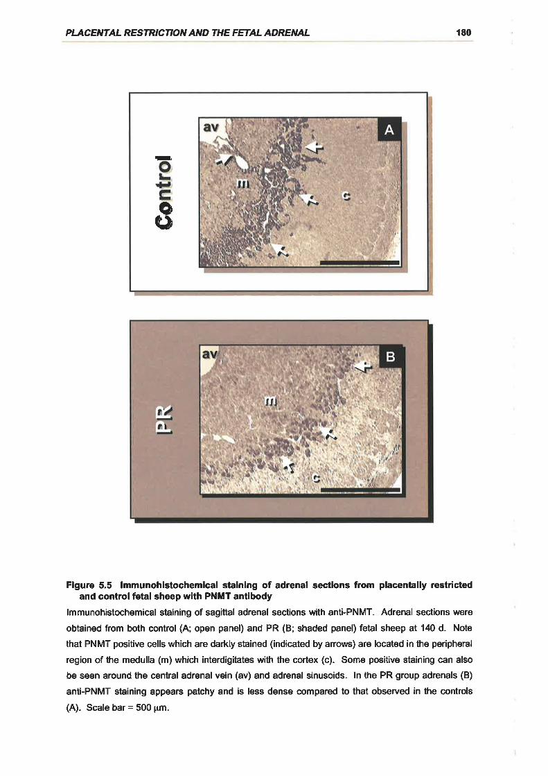
PLACENTA¿ RESTR'CTION AND THE FETAL ADRENAL 180
ae*
Figure 5.5 lmmunohistochemical staining of adrenal sections from placentally restrictedand controlfetal sheep with PNMT antibody
lmmunohistochemical staining of sagittal adrenal sections with anti-PNMT. Adrenal sections were
obtained from both control (A; open panel) and PR (B; shaded panel) fetial sheep at 140 d. Note
that PNMT positive cells which are darkly stained (indicated by anows) are located in the peripheral
region of the medulla (m) which interdigitates with the cortex (c). Some positive staining can also
be seen around the central adrenal vein (av) and adrenal sinusoids. ln the PR group adrenals (B)
anti-PNMT straining appears patchy and is less dense compared to that observed in the controls
(A). Scale bar = 500 pm.
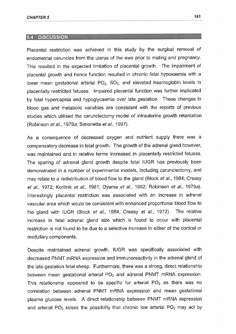
CHAPTER 5 181
s.4 DtscussloN
Placental restriction was achieved in this study by the surgical removal of
endometrial caruncles from the uterus of the ewe prior to mating and pregnancy.
This resulted in the expected limitation of placental growth. The impairment of
placental growth and hence function resulted in chronic fetal hypoxaemia with a
lower mean gestational arterial POz, SOz, and elevated haemoglobin levels in
placentally restricted fetuses. lmpaired placental function was further implicated
by fetal hypercapnia and hypoglycaemia over late gestation. These changes in
blood gas and metabolic variables are consistent with the reports of previous
studies which utilised the carunclectomy model of intrauterine growth retardation
(Robinson et al., 1979a;Simonetta et al., 1997).
As a consequence of decreased oxygen and nutrient supply there was a
compensatory decrease in fetal growth. The growth of the adrenal gland however,
was maintained and in relative terms increased in placentally restricted fetuses.
The sparing of adrenal gland growth despite fetal IUGR has previously been
demonstrated in a number of experimental models, including carunclectomy, and
may relate to a redistribution of blood flow to the gland (Block et al., 1984; Creasy
et al., 1972; Koritnik et al., 1981; Oyama et al., 1992; Robinson et al., 1979a).
lnterestingly placental restriction was associated with an increase in adrenal
vascular area which would be consistent with enhanced proportional blood flow to
the gland with IUGR (Block et al,, 1984; Creasy et al., 1972). The relative
increase in fetal adrenal gland size which is found to occur with placental
restriction is not found to be due to a selective increase in either of the cortical or
medullary components.
Despite maintained adrenal growth, IUGR was specifically associated with
decreased PNMT mRNA expression and immunoreactivity in the adrenal gland of
the late gestation fetal sheep. Furthermore, there was a strong, direct relationship
between mean gestational arterial POz and adrenal PNMT mRNA expression.
This relationship appeared to be specific for arterial POz as there was no
correlation between adrenal PNMT mRNA expression and mean gestational
plasma glucose levels. A direct relationship between PNMT mRNA expression
and arterial POz raises the possibility that chronic low arterial POz may act by
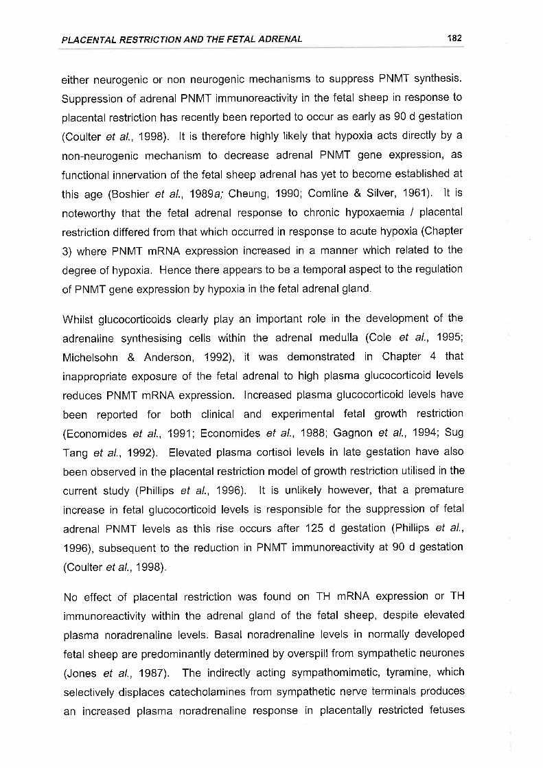
PLACENTAL RESTRICTION AND THE FETAL ADRENAL 182
either neurogenic or non neurogenic mechanisms to suppress PNMT synthesis.
Suppression of adrenal PNMT immunoreactivity in the fetal sheep in response to
placental restriction has recently been reported to occur as early as 90 d gestation
(Coulter et al., 1998). lt is therefore highly likely that hypoxia acts directly by a
non-neurogenic mechanism to decrease adrenal PNMT gene expression, as
functional innervation of the fetal sheep adrenal has yet to become established at
this age (Boshier et al., 1989a; Cheung, 1990; Comline & Silver, 1961). lt is
notewofthy that the fetal adrenal response to chronic hypoxaemia / placental
restriction differed from that which occurred in response to acute hypoxia (Chapter
3) where PNMT mRNA expression increased in a manner which related to the
degree of hypoxia, Hence there appears to be a temporal aspect to the regulation
of PNMT gene expression by hypoxia in the fetal adrenal gland.
Whilst glucocorticoids clearly play an important role in the development of the
adrenaline synthesising cells within the adrenal medulla (Cole et al., 1995;
Michelsohn & Anderson, 1992), it was demonstrated in Chapter 4 that
inappropriate exposure of the fetal adrenal to high plasma glucocorticoid levels
reduces PNMT mRNA expression. lncreased plasma glucocorticoid levels have
been reported for both clinical and experimental fetal growth restriction
(Economides ef al., 1991; Economides et al., 1988; Gagnon et al., 1994; Sug
Tang et al., 1992). Elevated plasma cortisol levels in late gestation have also
been observed in the placental restriction model of growth restriction utilised in the
current study (Phillips et al., 1996). lt is unlikely however, that a premature
increase in fetal glucocorticoid levels is responsible for the suppression of fetal
adrenal PNMT levels as this rise occurs after 125 d gestation (Phillips et al.,
1996), subsequent to the reduction in PNMT immunoreactivity at 90 d gestation
(Coulter et al., 1998).
No effect of placental restriction was found on TH mRNA expression or TH
immunoreactivity within the adrenal gland of the fetal sheep, despite elevated
plasma noradrenaline levels. Basal noradrenaline levels in normally developed
fetal sheep are predominantly determined by overspill from sympathetic neurones
(Jones et al., 1987). The indirectly acting sympathomimetic, tyramine, which
selectively displaces catecholamines from sympathetic nerve terminals produces
an increased plasma noradrenaline response in placentally restricted fetuses
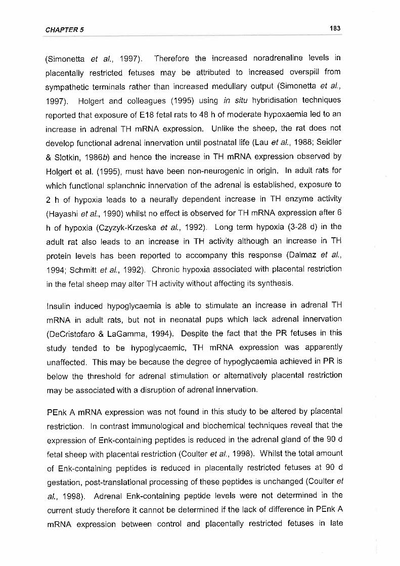
CHAPTER 5 183
(Simonetta et al., 1997). Therefore the increased noradrenaline levels in
placentally restricted fetuses may be attributed to increased overspill from
sympathetic terminals rather than increased medullary output (Simonetta et al.,
1997). Holgert and colleagues (1995) using in situ hybridisation techniques
reported that exposure of E18 fetal rats to 48 h of moderate hypoxaemia led to an
increase in adrenal TH mRNA expression. Unlike the sheep, the rat does not
develop functional adrenal innervation until postnatal life (Lau et a\.,1988; Seidler
& Slotkin, 1986b) and hence the increase in TH mRNA expression observed by
Holgert et al. (1995), must have been non-neurogenic in origin. ln adult rats for
which functional splanchnic innervation of the adrenal is established, exposure to
2 h of hypoxia leads to a neurally dependent increase in TH enzyme activity
(Hayashi et a1.,1990) whilst no effect is observed for TH mRNA expression after 6
h of hypoxia (Czyzyk-Krzeska et al., 1992). Long term hypoxia (3-28 d) in the
adult rat also leads to an increase in TH activity although an increase in TH
protein levels has been reported to accompany this response (Dalmaz et al.,
1994', Schmitt et al., 1992). Chronic hypoxia associated with placental restriction
in the fetal sheep may alter TH activity without affecting its synthesis.
lnsulin induced hypoglycaemia is able to stimulate an increase in adrenal TH
mRNA in adult rats, but not in neonatal pups which lack adrenal innervation
(DeCristofaro & LaGamma, 1994). Despite the fact that the PR fetuses in this
study tended to be hypoglycaemic, TH mRNA expression was apparently
unaffected. This may be because the degree of hypoglycaemia achieved in PR is
below the threshold for adrenal stimulation or alternatively placental restriction
may be associated with a disruption of adrenal innervation.
PEnk A mRNA expression was not found in this study to be altered by placental
restriction. ln contrast immunological and biochemical techniques reveal that the
expression of Enk-containing peptides is reduced in the adrenal gland of the 90 d
fetal sheep with placental restriction (Coulter et a\.,1998). Whilst the total amount
of Enk-containing peptides is reduced in placentally restricted fetuses at 90 d
gestation, post-translational processing of these peptides is unchanged (Coulter ef
al., 1998). Adrenal Enk-containing peptide levels were not determined in the
current study therefore it cannot be determined if the lack of difference in PEnk A
mRNA expression between control and placentally restricted fetuses in late
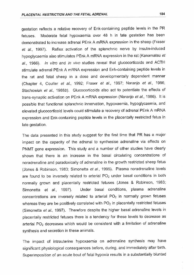
PLACENTAL RESTRICTION AND THE FETAL ADRENAL 184
gestation reflects a relative recovery of Enk-containing peptide levels in the PR
fetuses. Moderate fetal hypoxaemia over 48 h in late gestation has been
demonstrated to increase adrenal PEnk A mRNA expression in the sheep (Fraser
et al., 1997). Reflex activation of the splanchnic nerve by insulin-induced
hypoglycaemia also stimulates PEnk A mRNA expression in the rat (Kanamatsu ef
al., 1986). ln vitro and rn viyo studies reveal that glucocorticoids and ACTH
stimulate adrenal PEnk A mRNA expression and Enk-containing peptide levels in
the rat and fetal sheep in a dose and developmentally dependent manner
(Chapter 4, Coulter et al., 1992; Fraser et al., 1997; Naranjo et al., 1986;
Stachowiak et al., 1988b). Glucocorticoids also act to potentiate the effects of
trans-synaptic activation on PEnk A mRNA expression (Naranjo et a1.,1986). lt is
possible that functional splanchnic innervation, hypoxaemia, hypoglycaemia, and
elevated glucocorticoid levels could stimulate a recovery of adrenal PEnk A mRNA
expression and Enk-containing peptide levels in the placentally restricted fetus in
late gestation.
The data presented in this study suggest for the first time that PR has a major
impact on the capacity of the adrenal to synthesise adrenaline via effects on
PNMT gene expression. This study and a number of other studies have clearly
shown that there is an increase in the basal circulating concentrations of
noradrenaline and paradoxically of adrenaline in the growth restricted sheep fetus
(Jones & Robinson, 1983; Simonetta et al., 1995). Plasma noradrenaline levels
are found to be inversely related to arterial POz under basal conditions in both
normally grown and placentally restricted fetuses (Jones & Robinson, 1983;
Simonetta et al., 1997). Under basal conditions, plasma adrenaline
concentrations are inversely related to arterial POz in normally grown fetuses
whereas they are be positively correlated with POz in placentally restricted fetuses
(Simonetta et al., 1997). Therefore despite the higher basal adrenaline levels in
placentally restricted fetuses there is a tendency for these levels to decrease as
arterial POz decreases which would be consistent with a limitation of adrenaline
synthesis and secretion in these animals.
The impact of intrauterine hypoxaemia on adrenaline synthesis may have
significant physiological consequences before, during, and immediately after birth.
Superimposition of an acute bout of fetal hypoxia results in a substantially blunted
CHAPTER 5 185
adrenaline secretory response in placentally restricted fetuses whilst a large
noradrenaline response occurs which is similar in relative magnitude to that which
is observed in control fetuses (Jones & Robinson, 1983). The mobilisation of
glucose and the suppression of insulin secretion in response to acute hypoxic
challenge are also impaired in placentally restricted fetuses which would be
consistent with a reduced adrenaline secretory response (Jones & Ritchie, 1978b;
Jones & Robinson, 1983; Padbury et al., 1987c; Robinson et al., 1983a). Growth
restriction of fetal sheep by single umbilical artery ligation results in an inability of
the fetus to maintain the adrenaline response to delivery (Oyama et al., 1992). ln
addition to the failure to maintain adrenaline secretion in response to delivery,
there are associated disruptions in blood pressure as well as glucose and free
fatty acid mobilisation in growth retarded fetuses (Oyama et al., 1992). The
effects of intrauterine growth retardation may persist postnatally as 1 d old rat
pups, growth retarded by chronic ligation of the maternal uterine artery, display
impaired adrenaline secretory responses to acute hypoxia (Shaul et al., 1989).
lnterestingly, chronic prenatal hypoxia in the rat is also associated with long term
reductions of PNMT protein levels within respiratory neurones of the medulla
oblongata which may compromise responses to acute hypoxia in early postnatal
life (White & Lawson, 1997).
ln summary, placental restriction and chronic hypoxaemia resulted in a specific
suppression of PNMT mRNA levels and a concomitant decrease in the PNMT
containing region of the fetal adrenal. As placental restriction was not associated
with any changes in adrenal TH and PEnk A mRNA levels, or in the amount and
intensity of immunochemical staining for TH, it appears that adrenaline,
noradrenaline, and Enk-containing peptide synthesis are differentially regulated in
the adrenal of the growth restricted fetus in late gestation. The potential limitation
of adrenaline synthesis and secretion may have serious consequences for the
homeostatic responses of the fetus to subsequent acute physiological challenges
and the transition to postnatal life.
6. SUMMARY AND CONCLUSIONS
The fetal adrenal medulla is a key source of circulating catecholamines during
intrauterine stress, such as the imposition of hypoxaemia (Cohen et al., 1984;
Comline & Silver, 1961; Jones et al., 1988). There have been relatively few
studies, however, which have investigated the impact of physiological stress upon
catecholamine synthesis in the fetal adrenal medulla in a long gestation species
such as the sheep. Given that the catecholamine synthetic capacity of the fetal
adrenal medulla will determine its capacity to meet increased secretory demands
(Bygdeman & von Euler, 1958; Wakade et a\.,1988) this is an important area for
investigation. The studies presented in this thesis have examined the gene
expression of the catecholamine synthetic enzymes, TH and PNMT, in the adrenal
gland of the fetal sheep during development and in response to acute and chronic
stress. Adrenal PEnk A mRNA expression was also examined as intra-adrenal
opioid peptides have been implicated in the regulation of catecholamine secretion
during stress in the fetus (Padbury et al., 1987b). This final chapter will
summarise the main findings and implications of this thesis and discuss directions
for future research.
ln Chapter 2 the expression of adrenal TH, PNMT, and PEnk A mRNA was
determined in the fetal sheep adrenal between 80 and 146 d gestation. This
developmental window encompasses a number of key aspects of adrenal
development in the fetal sheep, including the establishment of functional
splanchnic innervation (Boshier et al., 1989a; Comline & Silver, 1961) and the
maturation and activation of the fetal HPA axis (Challis & Brooks, 1989). Whilst
catecholamines and enkephalin-containing peptides are co-stored within the
adrenal chromaffin cells of the fetal sheep (McMillen et al., 1988; Wan et al.,
1989b), there was a differential regulation of TH, PNMT, and PEnk A mRNA
expression across mid to late gestation. Adrenal TH and PEnk A mRNA
expression peaked al 125 d gestation coincident with the onset of establishment
of functional cholinergic innervation of the adrenal gland by the splanchnic nerve.
TH and PEnk A mRNA expression then decreased after 140 d gestation. PNMT
mRNA expression on the other hand increased in late gestation and peaked at
140-146 d in parallel with the activation of fetal HPA axis and the prepartum
increase in cortisol output from the fetal adrenal cortex. ln Chapter 2 I have
186
CHAPTER 6 187
demonstrated that blockade of nicotinic receptors suppressed adrenal TH mRNA
levels during basal or normoxic conditions. This indicates that adrenal TH mRNA
expression is maintained by splanchnic nerve activation in the late gestation
sheep fetus. lt is case however, that basal adrenal PNMT mRNA expression was
not suppressed by the addition of hexamethonium to the late gestation sheep
fetus. lt appears likely therefore that the expression of these two catecholamine
synthetic enzyme genes are differentially regulated in the adrenal during late
gestation. lt would be of interest to determine the effects of disruption of
splanchnic innervation to the adrenal, by nerve sectioning or fetal athyroidism,
upon the ontogenetic expression of adrenal TH, PNMT, and PEnk A mRNA.
The interaction of the HPA axis in the regulation of catecholamine synthetic
enzymes and enkephalin peptides has been well studied in the adult rat and in cell
culture. Surprisingly, there is relatively little information on the regulation of
catecholamine synthetic enzymes in the fetus by the HPA axis, particularly in a
long gestation species. Hypophysectomy in the fetal sheep results in the failure of
the adrenaline content of the fetal adrenal to increase in late gestation (Comline ef
al., 1970), and also has a number of consequences for the localisation, synthesis,
and processing of the enkephalin containing peptides (Coulter et al., 1989a;
Coulter et al., 1991 ; Coulter et al., 1992). Further understanding of the role of the
HPA axis in the developmental regulation of adrenal TH, PNMT, and PEnk A
mRNA expression could be gained in studies which prevent the prepartum
activation of the HPA axis by either hypothalamic-pituitary disconnection or the
use of 1 1P hydroxylase inhibitors, such as metyrapone which inhibit cortisol
synthesis.
Acute fetal hypoxaemia can potentially arise from a number of situations (Giussani
et al., 1994). Catecholamines secreted from the fetal adrenal gland in response to
hypoxia elicit a number of cardiovascular and metabolic adaptations (Jones et al.,
1988). ln the functionally innervated adrenal gland, hypoxia stimulates
catecholamine secretion by reflex neural stimulation (Biesold et al., 1989; Cheung,
1990; Jensen & Hanson, 1995; Seidler& Slotkin, 1985). Factors associated with
neural stimulation are well established to regulate catecholamine synthetic
enzyme gene expression in the adult adrenal medulla. Prior to adrenal
innervation however, hypoxia stimulates catecholamine secretion from the adrenal

SU MMARY AND CON C LU SIONS 188
gland by a'direct'non-neurogenic mechanism (Cheung, 1990; Comline & Silver,
1966; Comline & Silver, 1961; Seidler & Slotkin, 1985). ln Chapter 3 itwas found
that acute exposure to moderate hypoxaemia reduced TH mRNA and increased
PNMT expression in the fetal adrenal gland in a manner which related to the
degree of change in arterial POz. This effect of hypoxaemia was evident before
the development of functional splanchnic innervation to the adrenal, implicating a
non-neurogenic regulation of TH and PNMT gene expression. After the
development of functional splanchnic innervation however, the regulation of TH
and PNMT mRNA expression in response to acute hypoxia was dependent upon
reflex nicotin ic stimulation.
The non-neurogenic regulation of TH mRNA and PNMT mRNA expression in
adrenomedullary chromaffin cells in responses to changes in oxygen tension has
yet to be examined. Future studies would be required to focus upon the
determination of the second messenger systems and transcription factors
activated at the cellular level in adrenomedullary chromaffin cells in response to
hypoxia. This would then provide further insight into the potential mechanisms
whereby hypoxia could directly influence gene expression independently of neural
stimulation in adrenomedullary chromaffin cells.
Cortisol is also secreted from the fetal adrenal cortex in response to a number of
prenatal stressors (Akagi & Challis, 1990; Ozolins et al., 1991b; Rose et al., 1978).
It has been established that glucocorticoids play an important role in the induction
and maintenance of adrenal PNMT expression and adrenaline synthesis in
chromaffin cells in culture and in the developing and mature rat adrenal in vivo.
There is also indirect evidence to suggest that in the fetal sheep glucocorticoids
are important for the regulation of adrenaline synthesis (Comline et al., 1970).
Paradoxically, however, physiologically and clinically relevant doses of
glucocorticoids act to decrease plasma noradrenaline and adrenaline levels in the
fetal sheep (Derks et al., 1997; Stein et al., 1995; Wood et al., 1987). ln Chapter
4 the effect of a physiological increase in plasma cortisol levels between 109 and
1 16 d gestation was examined and it was found that there was a specific decrease
in adrenal PNMT mRNA and protein expression whilst TH and PEnk A mRNA
expression were not substantially altered. The specific decrease in PNMT mRNA
expression with increased glucocorticoid levels implies that the reduction in

CHAPTER 6 189
adrenaline levels observed in previous studies maybe attributable to a decrease in
adrenal adrenaline synthesis. We have made the novel observation that a
precocious increase in plasma cortisol levels in the fetal sheep actually has a
suppressive effect on adrenal PNMT expression.
The mechanisms by which increased peripheral cortisol levels act to suppress
adrenal PNMT mRNA expression in the fetal sheep clearly needs to be addressed
given the clinical significance of prenatal glucocorticoid treatment (Schellenberg &
Liggins, 1987). lt is unlikely that the exogenous cortisol is acting to feedback
negatively on the fetal HPA axis to reduce intra-adrenal cortisol levels but adrenal
venous effluent could be collected during intrafetal cortisol infusion to ensure that
there is no reduction in adrenal cortisol output. A possible mechanism for the
glucocorticoid induced suppression of PNMT mRNA expression is that cortisol
may act to downregulate of type ll glucocorticoid receptors (GR) on the
adrenomedullary chromaffin cells (Betito ef al., 1993). GR are involved in the
stimulation of PNMT gene expression (Betito et al., 1992; Wan & Livett, 1989) and
a reduction in GR may negatively impact upon PNMT mRNA expression.
Molecular and immunological techniques could be used to study the expression of
GR mRNA and protein in the adrenals of cortisol infused fetal sheep and their
relationship to PNMT mRNA expression. Glucocorticoids have also been
implicated in the long term programming of fetal cardiovascular function (Dodic ef
al., 1999a; Dodic et al., 1999b) and it would be interesting to determine if the
impact of prenatal cortisol infusion on PNMT mRNA expression also continues
into postnatal life. ln the rat, prenatal administration of dexamethasone at 817-
E19 decreases adrenal adrenaline content at postnatal day 1 and led to an
impaired adrenaline secretory response of these neonatal rat pups in response to
hypoxia (Kauffman et a|.,1994).
It can be speculated that the reduction in plasma noradrenaline levels found in this
study after cortisol infusion is not due to a reduction in adrenal TH gene
expression. Glucocorticoids have been reported to reduce sympathetic outflow
(Lenders et al., 1995) hence a reduction in sympathetic outflow in response to
glucocorticoid treatment may be responsible for the reduction in basal plasma
noradrenaline levels observed in the fetal sheep. PEnk A mRNA levels were not
significantly altered in response to glucocorticoid infusion and hence it is unlikely
SU MMARY AN D CO NC LU SIO NS 190
that an increase in enkephalin-containing peptide synthesis at the pre-translational
level is responsible for the inhibitory effects of glucocorticoids upon basal
catecholamine output.
lntrauterine growth retardation is a relatively common pathological condition
frequently associated with chronic fetal hypoxaemia and hypoglycaemia
(Economides ef al., 1991; Robinson et al., 1985). Plasma catecholamine levels
are increased in both clinical IUGR and experimental paradigms (Lagercrantz ef
al., 1980; Simonetta^et al., 1997). ln Chapter 4 adrenal TH, PNMT, and PEnk A
mRNA expression were examined at 140 d following experimental IUGR in fetal
sheep by placental restriction. Despite elevated plasma catecholamine
concentrations in PR fetal sheep, adrenal PNMT mRNA expression and
immunoreactivity were specifically decreased at 140 d gestation. Curiously,
PNMT mRNA expression exhibited a strong positive relationship with mean
gestational levels of arterial POz. This is in contrast to acute hypoxia where
adrenal PNMT mRNA increases with decreasing POz (Chapter 3), therefore there
appears to be a temporal aspect to the regulation of PNMT gene expression in the
fetal sheep by oxygen. There was no significant effect of PR on adrenal TH and
PEnk A mRNA expression at 140 d gestation.
It appears reasonable to conclude that the increased basal output of adrenaline
and noradrenaline with PR are not attributable to increased synthesis within the
fetal adrenal gland, or reduced levels of intra-adrenal opioid peptides. Recent
isotope dilution studies in the fetal sheep indicate that the increased circulating
catecholamine levels in IUGR fetuses are due to enhanced spill-over from the
sympathoadrenal system (Smolich, 1999). lt has previously been demonstrated in
our laboratory that tyramine, which selectively displaces catecholamines from
sympathetic nerve terminals, elicits significantly higher plasma noradrenaline and
adrenaline levels in PR sheep (Simonetta et al., 1997). Therefore the increase in
basal catecholamine levels in PR fetal sheep appears to be due to increased
overspill from developing sympathetic neurones. This possibility could be studied
by measuring plasma catecholamine concentrations in PR fetal sheep following
adrenal demedullation or chemical destruction of the peripheral sympathetic
neryous system with guanethidine or 6-hydroxydopamine.
CHAPTER 6 191
The adrenaline secretory capacity of the fetal sheep adrenal is paradoxically
reduced with PR despite an increase in basal plasma adrenaline levels. This may
therefore affect the reserve of the fetal adrenal gland to respond to further acute
or chronic stress. There is evidence to suggest that this is indeed the case as PR
fetal sheep have been reported to have a reduced adrenaline secretory response
to superimposed acute hypoxia (Jones & Robinson, 1983). A number of
metabolic responses attributable to increased adrenaline levels in response to
hypoxaemia are also blunted in PR fetal sheep (Jones & Robinson, 1983;
Robinson et al., 1983a). The impaired adrenaline secretory response to hypoxia
persists in neonatal rats which have been subjected to the Wigglesworth model of
IUGR (Shaul et al., 19Bg). Based on strong epidemiological evidence, IUGR has
been proposed to be a risk factor in the development of cardiovascular and
metabolic disturbances in adult life (Barker, 1999). lt would be of interest to
determine if the effect of IUGR on the adrenaline synthetic capacity of the adrenal
gland in the sheep persists into post natal life.
ln summary the studies presented in this thesis demonstrate that there is a
differential regulation of adrenal catecholamine synthetic enzyme and PEnk A
gene expression in the fetal sheep with development. There is also differential
regulation of adrenal catecholamine synthetic enzyme gene expression during
acute hypoxaemia in the fetal sheep, before and after functional splanchnic
innervation. Cortisol, which is also released from the adrenal during fetal
hypoxaemia, was demonstrated to have a novel suppressive effect on adrenal
PNMT expression in the fetal sheep. Experimental IUGR with chronic hypoxaemia
was established to result in a specific reduction in adrenal PNMT mRNA
expression and immunoreactivity which was closely related to arterial oxygenation
over gestation in the fetal sheep. Clearly intrauterine stress impacts upon
catecholamine synthetic enzyme gene expression in the fetal sheep. Long term
alterations in adrenal catecholamine synthetic enzyme gene expression will
influence the catecholamine synthetic capacity of the fetal and neonatal gland and
hence may result in altered secretory responses to homeostatic challenges.
Future functional and molecular studies will be required to determine the full array
of non-neurogenic and neural factors which regulate catecholamine synthetic
enzyme gene expression during intrauterine stress.
BIBL¡OGRAPHY
Abate, C., Patel, L., Rauscher, F.J. & Curran, T. (1990). Redox regulation of Fos and Jun
DNA-binding activity in vitro. Science,249, 1157-1161.
Acker, H. (1994). Mechanisms and meaning of cellular oxygen sensing in the organism
Respiration Physiology, 95, 1 -1 0.
Acker, H., Bolling,8., Delpiano, E., Dufau, E., Gorlach, A. & Holtermann, G. (1992). The
meaning of HzOz generation in carotid body cells for POz chemoreceplion. Journal ofthe Autonomic Nervous System, 41, 41-52.
Acker, H. & Xue, D. (1995). Mechanisms of Oz sensing in the carotid body in comparison
with other O2-sensing cells. N/PS, 10, 211-216.
Adams, M.8., Simonetta, G. & McMillen, LC. (1996). The non-neurogenic catecholamine
response of the fetal adrenal to hypoxia is dependent on activation of voltage
sensitive Ca2* chann els. Developmental Brain Research,94, 182-189.
Adnan, N.A.M. & Hawthorne, J.N. (1981)
activation of muscarinic receptors
Neurochemistry, 36, 1 858-1 860.
Phophatidylinositol labelling in response to
in bovine adrenal medulla. Journal of
Afar, R., Silverman, R., Aguanno, A. & Albert, V.R. (1996). Positive and negative
elements contribute to the cell-specific expression of the rat dopamine p-hydroxylase
gene. Molecular Brain Research, 36, 79-92.
Afework, M., Tomlinson, A. & Burnstock, G. (1994). Distribution and co-localization of
nitric oxide synthase and NADPH-diaphorase in the adrenal gland of developing,
adult, and aging Sprague-Dawley rats. Cell lrssue Research,276, 133-141.
Agata, Y., Padbury, J.F., Ludlow, J.K., Polk, D.H. & Humme, J.A. (1986). The effect of
chemical sympathectomy on catecholamine release at birth. Pediatric Research,20,1338-1344.
Aguilera, G., Millan, M.A., Hauger, R.L. & Catt, K.J. (1991). Corticotropin-releasing factor
receptors : distribution and regulation in brain, pituitary, and peripheral tissues.
Annals of the New York Academy of Sciences,512,48-66.
Akagi, K., Berdusco, E.T.M. & Challis, J.R.G. (1990). Cortisol inhibits the ACTH but not
AVP response to hypoxemia in fetal lambs at days 123-128 of gestation. Journal ofDevelopmental Physiology, 1 4, 319-324.
Akagi, K. & Challis, J.R.G. (1990). Threshold of hormonal and biophysical responses to
acute hypoxaemia in fetal sheep at different gestational ages. Canadian Journal ofPhysiology and Pharmacology, 68, 549-555.
192
BIBLIOGRAPHY 193
Akaike,4., Sasa, M., Tamurâ, Y., Ujihara, H. & Takaori, S. (1993). Effects of protein
kinase C on the muscarinic excitation of rat adrenal chromaffin cells. Japanese
Journal of Physiology, 6'1, 1 45-1 48.
Albillos, 4., Carbonê, E., Gandia, L., Garcia, A.G, & Pollo, A. (1996). Opioid inhibition of
Ca2* channel subtypes in bovine chromaffin cells : selectivity of action and voltage-
dependence. European Journal of Neuroscience, 8, 1561-1570.
Alexander, G. (1964). Studies on the placenta of the sheep (Ovis aries): effect of surgical
reduction in the number of caruncles. Journal of Reproduction and Fertility,7,307-321.
Alexander, G. & Bell, A.W. (1982). The role of the adrenal gland in the metabolic
response of young lambs to cold. Journal of Developmental Physiology,4,53-73.
Alexander, G. & Stevens, D. (1980). Sympathetic innervation and the development of
structure and function of brown adipose tissue: studies on lambs chemically
sympathectomized in utero with 6-hydroxydopamine. Journal of Developmental
Physiology, 2, 119-137 .
Allen, J.M., Adrian, T.E., Polak, J.M. & Bloom, S.R. (1983). Neuropeptide Y (NPY) in the
adrenal gland. Journal of the Autonomic Neruous System,9, 559-563.
Aloe, L. & Levi-Montalcini, R. (1979). Nerve growth factor-induced transformation of
immature chromaffin cells in vivo into sympathetic neurons: Effects of antiserum to
nerve growth factor. Proceedings of the National Academy of Sciences USA, 76,
1246-125Q.
Anderson, D.J. (1989). Cellular'neoteny': a possible developmental basis for chromaffin
cell plasticity. Trends in Genetics, 5, 174-178.
Anderson, D.J. (1993). Molecular control of cell fate in the neural crest
sympathoadrenal lineage. Annual Reviews of Neuroscience, 16, 1 29-1 58.
The
Anderson, D.J. & Axel, R.A. (1986). Bipotential neuroendocrine precursor whose choice
of cell fate is determined by NGF and glucocorticoids. Ce\\,47,1079-1090.
Anderson, D.J., Carnahan, J., Michelsohn, A. & Patterson, P.H. (1991). Antibodymarkersidentify a common progenitor to sympathetic neurons and chromaffin cells in vivo,
and reveal the timing of commitment to neuronal differentiation in the
sympathoadrenal lineage. Journal of Neuroscience, 11, 3507-3519.
Anderson, D.J. & Michelson, A. (1989). Role of glucocorticoids in the chromaffin-neuron
developmental decision. lnternational Journal of Developmental Neuroscience, 7,
475-487.
Anderson, K., Robinson, P.J. & Marley, P.D. (1992). Cholinoceptor regulation of cyclic
AMP levels in bovine adrenal medullary cells. Brifish Journal of Pharmacology, 106,
360-366.
BIBLIOGRAPHY 194
Andersson, K.K., Cox, D.D., Que, L., Flatmark, T. & Haavik, J. (1988). Resonance
Raman studies on the blue-green-coloured bovine adrenal tyrosine 3-
monooxygenase (tyrosine hydroxylase). Evidence that the feed back inhibitors
adrenaline and adrenaline are coordinated to iron. Journal of Biological Chemistry,
263, 18621-18626.
Andersson, K.K., Vassort, C., Brennan, 8.A., Que, L., Haavik, J., Flatmark, T., Gros, F. &
Thibault, J. (1992). Purification and characterization of the blue-green rat
phaeochromocytoma (PC12) tyrosine hydroxylase with a dopamine-Fe (lll) complex.
Reversal of endogenous feedback inhibition by phosphorylation of serine-40.
Biochemical Journal, 284, 687-695.
Araujo, D.M. & Collier, B. (1987). Effect of endogenous opioid peptides on acetylcholine
release from the cat superior cervical ganglion : selective effect of a heptapeptide.
Journal of Neuroscience, 7, 1698-1704.
Archer, S.L., Huang, J.M.C., Reeve, H.L., Hampl, V., Tolarova, S., Michelakis, E. & Weir,
E.K. (1996). Differential distribution of electrophysiologically distinct myocytes inconduit and resistance arteries determines their response to nitric oxide and hypoxia.
Circulation Research, 78, 431 -442.
Arimura, A. (1998), Perspectives on pituitary adenylate cyclase activating polypeptide
(PACAP) in the neuroendocrine, endocrine, and nervous system. Japanese Journal
of Physiology, 48, 301 -331.
Arimura, A. & Shioda, S. (1995). Pituitary adenylate cyclase activating polypeptide
(PACAP) and its receptors : Neuroendocrine and endocrine interaclion. Frontiers in
Neuroendocrinology, 1 6, 53-88.
Aunis, D., Guerold, 8., Bader, M.-F, & Ciesielski-Treska, J. (1980). lmmunocytochemical
and biochemical demonstration of contractile proteins in chromaffin cells in culture.
Neuroscience, 5, 2261 -2277 .
Babinski, K., Bodart, V., Roy, M., Delean, A. & Ong, H. (1996). Pituitary adenylate-
cyclase activating polypeptide (PACAP) evokes long-lasting secretion and de novo
biosynthesis of bovine adrenal medullary neuropeptides. Neuro peptides, 30, 572-582.
Bader, M.-F. & Aunis, D. (1983). The 97-kD alpha-actinin-like protein in chromaffin
granule membranes from adrenal medulla : evidence for localization on the
cytoplasmic surface and for binding to actin filaments. Neuroscience, 8, 165-181 .
Baetge, E.E., Suh, Y.H. & Joh, T.H. (1986). Complete nucleotide and deduced amino acid
sequence of bovine phenylethanolamine N-methyltransferase: partial amino acid
homology with rat tyrosine hydroxylase. Proceedings of the National Academy ofScience USA, 83, 5454-5458.
BIBLIOGRAPHY 195
Bailey, S.W., Dillard, S.8., Thomas, K.B. & Ayling, J.E. (1989). Changes in the cofactor
binding of bovine striatal tyrosine hydroxylase at the physiological pH upon cAMP-
dependent phosphorylation mapped with tetrahydropterin analogues. Biochemistry,
28,494-504.
Baizer, L. & Weiner, N. (1985). Nerve growth factor treatment enhances nicotine-
stimulated dopamine release and increases in cyclic 3':5'-monophosphate levels in
PC12 cell cultures. Journal of Neuroscience, 5, 1176-1179.
Baker, P,F. & Knight, D.E. (1978). Calcium-dependent exocytosis in bovine adrenal
medullary cells with leaky plasma membranes. Nafure,276,620-622.
Ball, K.T., Gunn, T.R., Power, G.G., Asakura, H. & Gluckman, P.D. (1995). A potential
role for adenosine in the inhibition of non-shivering thermogenesis in the sheep.
Pediatric Research, 37, 303-309.
Balla, T., Baukal,4.J., Eng, S. & Catt, K.J. (1991). Angiotensin ll receptor subtypes and
biological responses and biological responses in the adrenal cortex and medulla.
Molecular Pharmacology, 40, 401 -406.
Barker, D.J. (1999). Fetal origins of cardiovascular disease. Annals of Medicine, 31, 3-6
Barrett, C.T., Heymann, M.A. & Rudolph, A.M. (1972). Alpha and beta adrenergic
receptor activity in fetal sheep. American Journal of Obstetrics and Gynecology,112,1114-1121.
Basset, J.M. & Symonds, M.E. (1998). The selective þ, agonist ritrodine, unlike
epinephrine or norepinephrine prematurely activates thermogenesis in fetal brown
adipose tissue during prolonged infusion. American Journal of Physiology, 275,
R112-R119.
Basset, J.M., Weeding, C.M. & Hanson, C. (1990). Desensitization of B-receptormediated responses to epinephrine in fetal lambs by prolonged ritrodine
administration. Pediatric Research, 28, 388-393.
Bassett, J.M. & Hanson, C. (1998). Catecholamines inhibit growth in the fetal sheep in the
absence of hypoxemia. American Journal of Physiology,274, R1536-R1545.
Battaglia, F.C. & Meschia, G. (1978). Principal substrates of fetal metabolism
Physiolog ical Reviews, 58, 499-527 .
Batter, D.K., D'Mello, S.R., Turzai, L.M., Hughes, H.8., Gioio,A.E. &Kaplan, B.B. (1988).
The complete nucleotide sequence and structure of the gene encoding bovine
phenylethanolamine N-methyltransferase. Journal of Neuroscience Research, '19,
367-376.
BIBLIOGRAPHY 196
Behrman, R.E., Lees, M.H., Peterson, E.N., De Lannoy, C.W. & Seeds, A.E. (1970).
Distribution of the circulation in the normal and asphyxiated fetal primate. American
Journal of Obstetrics and Gynecology, 108, 956-969.
Beitner-Johnson, D., Shull, G.E., Dedman, J.R, & Millhorn, D.E. (1997). Regulation of
gene expression by hypoxia : a molecular approach. Respiration Physiology,110,87-97.
Bennett, M.K., Calakos, N. & Scheller, R.H. (1992). Syntaxin: a synaptic protein
implicated in docking of synaptic vesicles at presynaptic active zones. Science,257,255-259.
Bergman, B. (1981). Beta-mimetics and the preterm neonatal lung. An experimental and
clinical study. Acta Physiologica Scandanavica, Suppl. 497,1-52.
Berridge, M.J. (1984). lnositol trisphosphate and diacylglycerol as second messengers
B ioch em ica I J o u rnal, 220, 345-360.
Berridge, M.J., Dawson, R.M.C., Downes, C.P., Heslop, J.P. & lrvine, R.F. (1983).
Changes in the levels of inositol phosphates after agonist-dependent hydrolysis of
membrane phosphoinositides. Biochemical Journal, 212, 473-482.
Best, J.4., Chen, Y., Piech, K.M. & Tank, A.W. (1995). The response of the tyrosine
hydroxylase gene to cyclic AMP is mediated by two cyclic AMP-response elements.
Journal of Neurochemistry, 65, 1934-1943.
Betito, K., Diorio, J., Meaney, M.J. & Boksa, P. (1992). Adrenal phenylethanolamine N-
methyltransferase induction in relation to glucocorticoid receptor dynamics: evidence
that acute exposure to high cortisol levels is sufficient to induce the enzyme. Journal
of Neurochemistry, 58, 1 853-1 862.
Betito, K., Diorio, J., Meaney, M.J. & Boksa, P. (1993). Glucocorticoid receptors in bovine
adrenal medullary cells in culture : regulation by cyclic nucleotides. Neuroscience, 54,
263-273.
Biales, 8., Dichter, M. & Tischler, A. (1976). Electrical excitability of cultured adrenal
chromaffin cells. Journ al of Physiology (London), 262, 7 43-7 53.
Biesold, D., Kurosawa, M., Sato, A. & Trzebski, A. (1989). Hypoxia and hypercapnia
increase the sympathoadrenal medullary functions in anesthetized, artificially
ventilated rats. Japanese Journal of Physiology,39, 511-522.
Biguet, N.F., Buda, M., Lamouroux,4., Samolyk, D. & Mallet, J. (1986). Time course of
changes of TH mRNA in rat brain and adrenal medulla after a single injection of
reserpine. The EMBO Journal, 5,287-291.
Biscoe, T.J. & Duchen, M. (1990a). Monitoring POz bythe carotid chemoreceptor. N/PS,
5,229-233.
BIBLIOGRAPHY 197
Biscoe, T.J. & Duchen, M.R. (1990b). Cellular basis of signal transduction in carotid
chemoreceptors. American Journal of Physiology, 258, L27 1-L27 8.
Bishop, E.H. (1981). Acceleration of fetal pulmonary maturity. Obstetrics and Gynecology,
58,485-51S.
Bland, R.D. (1988). Fetal and neonatal lung fluid balance. Seminars in Perinatology, 12,
124-133.
Block, 8.S., Llanos, A.J. & Creasy, R.K. (1984). Responses of the growth retarded fetus
to acute hypoxemia. American Journal of Obstetrics and Gynecology,148, 878-885.
Block,8.S., Schlafer, D.H., Wentworth, R.4., Kreitzer, L.A. & Nathanielsz, P.W. (1990).
Regional blood flow distribution in fetal sheep with intrauterine growth retardationproduced by decreased umbilical placental perfusion. Journal of Developmental
Physiology, 13, 81-85.
Bloom, S.R., Edwards, A.V. & Jones, C.T. (1988). The adrenal contribution to the
neuroendocrine responses to splanchnic nerve stimulation in conscious calves.
Journal of Physiology (London) , 397,513-526.
Bloom, S.R., Edwards, A.V. & Jones, C.T. (1987). Adrenal cortical responses tovasoactive intestinal peptide in conscious hypophysectomized calves. Journal ofPhysiology (London), 391, 441-450.
Blottner, D. & Baumgarten, H.-G. (1992). Nitric oxide synthase (NOS)-containing
sympathoadrenal cholinergic neurons of the rat IMl-cell column : evidence from
histochemistry, immunohistochemistry, and retrograde labelling. Journal ofComparative Neurology, 316, 45-55.
Blottner, D., Weatermann, R., Grothe, C., Bohlen, P. & Unsicker, K. (1989). Basic
fibroblast growth factor in the adrenal gland. Possible trophic role for preganglionic
neurons. European Journal of Neuroscience, 1, 47 1-478.
Boarder, M.R., Marriott, D. & Adams, M. (1987). Stimulus-secretion coupling in cultured
chromaffin cells. Biochemical Pharmacology, 36, 163-167 .
Bobrovskaya, L., Cheah, T.8., Bunn, S.J. & Dunkley, P.R. (1998). Tyrosine hydroxylase in
bovine adrenal chromaffin cells : Angiotensin ll-stimulated activity and
phosphorylation of Ser1e, Ser31, Serao. Journal of Neurochemistry,70,2565 - 2573.
Bocking, 4.D., Gagnon, R,, White, S.E., Homan, J., Milne, K.M. & Richardson, B.S.
(1988). Circulatory responses to prolonged hypoxaemia in the fetal sheep. American
Journal of Obstetrics and Gynecology,159, 1418-1424.
Bocking,4.D., McMillen, 1.C., Harding, R. & Thorburn, G.D. (1986). Effect of reduced
uterine blood flow on fetal and maternal cortisol. Journal of Developmental
Physiology, 8, 237 -245.
BIBLIOGRAPHY 198
Boddy, K., Dawes, G.S., Fisher, T., Pinter, S. & Robinson, J.S. (1974). Foetal respiratory
movements, electrocortical and cardiovascular responses to hypoxemia and acidemia
in sheep. Journal of Physiology (London),243, 599-618.
Bohn, M.C., Goldstein, M. & Black, l.B. (1981). Role of glucocorticoids in expression of
the adrenergic phenotype in rat embryonic adrenal gland. Developmental Biology,82,
1-10.
Boksa, P. (1990). Dopamine release from bovine adrenal medullary cells in culture
Journal of the Autonomic Nervous System,30,63-74.
Boksa, P. & Livett, B.G. (1984). Substance P protects against desensitization of the
nicotinic response in isolated adrenal chromaffin cells. Journal of Neurochemistry,42,
618-627.
Boog, G., Brahim, M,B. & Gandar, R, (1975). Beta-mimetic drugs and possible prevention
of respiratory distress syndrome. British Journal of Obstetrics and Gynaecology, S2,
285-288.
Boshier, D.P., Gavin, C.B. & Holloway, H.(1989a). Morphometric analyses of adrenal
gland growth in fetal and neonatal sheep ll. The adrenal medulla with some
observations on its ultrastructure. Journal of Anatomy,167, 15-30.
Boshier, D.P. & Holloway, H. (1989b). Morphometric analyses of adrenal gland growth in
fetal and neonatal sheep. L The adrenal cortex. Journal of Anatomy,'167,1-14.
Bossu, J.L., De Waard, M. & Feltz, A.(1991). lnactivation characteristics reveal two
calcium currents in adult bovine chromaffin cells. Journal of Physiology (London) ,
437,603-620.
Boyd, P.A. & Scott, A. (1985). Quantitative structural studies on human placentas
associated with pre-eclampsia, essential hypertension and intrauterine growth
retardation . British Journal of Obstetrics and Gynaecology,92,714-721.
Boyle, D.W., Lecklitner, S. & Liechty, E.A. (1996). Effect of prolonged uterine blood flow
reduction on fetal growth in sheep. American Journal of Physiology,270, R246-R253.
Brace, R.A. & Brittingham, D.S. (1986). Fetal vascular pressure and heart rate responses
to non-labour uterine contractions. American Journal of Physiology,251, R409-R416.
Brace, R.A. & Cheung, C.Y. (1987). Role of catecholamines in mediating fetal blood
volume decrease during hypoxia. American Journal of Physiology,243, R520-R525.
Braems, G.A., Matthews, S.G. & Challis, J.R.G. (1996). Differential regulation of pro-
opiomelanocortin messenger RNA in the pars distalis and pars intermedia of the
pituitary gland following prolonged hypoxaemia in fetal sheep. Endocrinology, 137,
2731-2738.
BIBLIOGRAPHY 199
Brandt, 8.L., Hagiwara, S., Kidokoro, Y. & Miyazaki, S. (1976). Action potentials in the rat
chromaffin cell and effects of acetylcholine. Journal of Physiology (London), 263,
417-439.
Brenneman, A.R. & Kaufman, S.(1964). The role of tetrahydropteridine in the enzymatic
conversion of tyrosine to 3,4 dihydroxyphenylalanine. Biochemical and Biophysical
Research Communications, 17, 177 -1 83.
Breslow, M.J. (1992). Regulation of adrenal medullary and cortical blood flow. American
Journal of Physiology, 262, H1317-H 1330.
Breslow, M.J., Jordan, D.A., Thellman, S.T. & Traystman, R.J. (1987). Neural control of
adrenal medullary and cortical blood flow during haemorrhage. American Journal ofP hysi ol ogy, 252, H521-H 528.
Breslow, M.J., Tobin, J.R., Bredt, D.S., Feriis, C.D., Snyder, S.H. & Traystman, R.J.
(1992). Role of nitric oxide in adrenal medullary vasodilation during catecholamine
secretion. European Journal of Pharmacology, 210, 105-1 06.
Briand, R., Yamaguchi, N., Gagne, J., Kimura, T., Farley, L., Foucart, S., Nadeau, R. &
de Champlain, J. (1990). Corelease of neuropeptide Y like immunoreactivity with
catecholamines from the adrenal gland during splanchnic nerve stimulation in
anesthetized dogs. Canadian Journal of Physiology and Pharmacology, 68,363-369.
Brooks, J,C. & Treml, S. (1983). Catecholamine secretion by chemically skinned cultured
chromaffin cells. Journ al of Neurochemistry, 40, 468-473.
Brown, L.A.S. & Longmore, W.J. (1981). Adrenergic and cholinergic regulation of lung
surfactant secretion in the isolated perfused rat lung and in the alveolar type ll cell in
culture. Journal of Biological Chemistry, 256, 66-72.
Brown, M.J., Olver, R.E., Ramsden, C.A., Strang, L.B.&Walters, D.V. (1983). Effects of
adrenaline and of spontaneous labour on the secretion and absorption of lung liquid
in the fetal lamb. Journal of Physiology (London),344, 137-152.
Brown, M.R. & Fisher, L.A. (1986). Glucocorticoid suppression of the sympathetic nervous
system and adrenal medulla. Life Sciences, 39, 1003-1012.
Bruhn, T.O., Engeland, W.C., Anthony, E.L.P., Gann, D.S. & Jackson, l.M.D. (1987).
Corticotropin-releasing factor in the dog adrenal medulla is secreted in response to
hemorrhag e. Endocilnology, 120, 25-33.
Brundin, T. (1966). Studies on pre-aortal paraganglia of newborn rabbits. Acta
Physiologica Scandanavica, 290 (Suppl), 1 -70,
Buckler, K.J. & Vaughan-Jones, R.D. (1994). Effects of hypoxia on membrane potential
and intracellular calcium in rat neonatal carotid body type I cells. Journal ofPhysiology (London), 47 6, 423-428.
BIBLIOGRAPHY 200
Bulenda, D. & Gratzl, M. (1985). Matrix free Ca2* in isolated chromaffin vesicles
B ioche m ica I J o u rnal, 24, 7 7 60-7 7 65.
Bunn, H.F. & Poyton, R.O. (1996). Oxygen sensing and molecular adaption to hypoxia
Physiological Reviews, 76, 839-885.
Bunn, S.J. & Marley, P.D. (1989). Effects of angiotensin ll on cultured bovine
adrenomedullary cells. Neuropeptides, 13, 121-132.
Bunn, S.J., Marley, P.D.& Livett, B.G. (1988). The distribution of opioid binding subtypes
in the bovine adrenal medulla. Neuroscience, 27, 1081-1094.
Bunn, S.J., Marley, P.D.& Livett, B.G. (1990). Receptor stimulated formation of inositol
phosphates in cultures of bovine adrenal medullary cells : The effects of bradykinin,
bombesin and neurotensin. Neuropeptides, 15, 187-194.
Burgoyne, R.D. (1991). Control of exocytosis in adrenal chromaffin cells. Biochimica et
Biophysica Acta, 107 1, 17 4-202.
Burgoyne, R.D. (1984). Mechanisms of secretion from adrenal chromaffin cells.
Biochimica et Biophysica Acta, 779, 201-216.
Burgoyne, R.D. &Williams, G. (1997). NSF and SNAP are presenton adrenal chromaffin
granules. FEBS Letters, 414, 349-352.
Butler, T.G., Simonetta, G., Roberts, M.L. & McMillen, LC. (1995). lmpactof gestational
age on the catecholamine responses of the fetal sheep adrenal to cholinergic
stimulation in vitro. Quañerly Journal of Experimental Physiology,80,767-777 .
Bygdeman, S. & von Euler, U. (1958). Resynthesis of catechol hormones in the cat's
adrenal medulla. Acta Physiologica Scandanavica, 44, 375-383.
Bzoskie, L., Blount, L., Kashiwai, K., Tseng, Y.T., Hay, W.W. & Padbury, J.F. (1995)
Placental norepinephrine clearance: in vivo measurement and physiological role
American Journal of Physiology, 269, 8145-E149.
Cachero, T.G., Rigual, R., Rocher, A. & Gonzalez, C. (1996). Cholera and pertussis
toxins reveal multiple regulation of cAMP levels in the rabbit carotid body. European
J o u rn a I of N e u ro sci e nce, 8, 2320-2327 .
Cahill,4.L., Eertmoed,4.L., Mangoura, D. & Perlman, R.L. (1996). Differential regulation
of phenylethanolamine N-methyltransferase expression in two distinct subpopulations
of chromaffin cells. Journal of Neurochemistry, 67, 1217-1224.
Cambier, J.C., Newell, N.K., Justement, L.8., McGuire, J.C., Leach, K.L. & Chen, Z.Z.
(1987). la binding ligands and cAMP stimulate nuclear translocation of PKC in p
lym phocyte s. N ature, 327, 629-632.
BIBLIOGRAPHY 201
Campbell, D.G., Hardie, D.G. & Vulliet, P.R. (1986). ldentification of the fourphosphorylation site in the N-terminal region of tyrosine hydroxylase. Journal ofBiological Chemistry, 261, 10489-1 0492.
Carlson, S., Skarphedinsson, J.O., Delle, M., Hoffman, P. & Thoren, P. (1992). Reflex
changes in post and preganglionic sympathetic adrenal nerve activity and
postganglionic renal nerve activity upon arterial baroreceptor activation and during
severe haemorrhage in the rat. Acta Physiologica Scandanavica,144,317-323.
Carlson, S., Skarphedinsson, J.O., Jennische, E., Delle, M. & Thoren, P. (1990).
Neurophysiological evidence for and characterization of the post-ganglionic
innervation of the adrenal gland in the ral Acta Physiologica Scandanavica,'140,
491-499.
Carnahan, J.F. & Patterson, P.H. (1991a). Generation of monoclonal antibodies that bind
preferentially to adrenal chromaffin cells and the cells of embryonic sympathetic
ganglia. Journal of Neuroscience, 11, 3493-3506.
Carnahan, J.F. & Patterson, P.H. (1991þ). lsolation of the progenitor cells of the
sympathoadrenal lineage from embryonic sympathetic ganglia with the SA
monoclonal antibodies . Journal of Neuroscience, 11, 3520-3530.
Carter, 4.M., Richardson, 8.S., Homan, J., Towstoless, M. & Challis, J.R.G. (1993).
Regional adrenal blood flow responses to adrenocorticotrophic hormone in fetal
sheep. American Journal of Physiology ,264,8264-8269.
Cassady, G. (1981). The small-for-date infant. ln Neonatology:Pathophysiology andManagement of the Newborn. ed. Avery, G.B. pp. 262-286. Philadelphia: J.B.
Lippincott Co.
Castanas, E., Bourhim, N., Giraud, P., Boudouresque, F., Cantou, P. & Oliver, C.
(1985a). lnteraction of opiates with opioid binding sites in the bovine adrenal medulla
: ll. lnteraction with r sites, Journalof Neurochemistry,45, 688-699.
Castanas, E., Bourhiffi, N., Giraud, P., Bourdouresque, F., Cantou, P. & Oliver, C.
(1985b). lnteraction of opiates with opioid binding sites in the bovine adrenal medulla
: l. lnteraction with ô and p sites. Journal of Neurochemistry,45,677-687.
Casteilla, L., Forest, C., Robelin, J., Ricquier, D., Lomnet, A. & Ailhaud, G. (1987).
Characterization of mitochondrial-uncoupling protein in bovine fetus and newborn
calf . American Journal of Physiology,252, E627-E636.
Ceccatelli, S., Dagerlind, 4., Schalling, M., Wikstrom, A.-C., Okret, S., Gustafsson, J.4.,
Goldstein, M. & Hokfelt, T. (1989). The glucocorticoid receptor in the adrenal gland is
localized to the cytoplasm of adrenaline cells. Acta Physiologica Scandanavica, 137,
559-560.
BIBLIOGRAPHY 202
Celler, B. & Schram, L.P. (1981). Pre and postganglionic sympathetic nerves of the rat
American Journal of Physiology,241, R55-R61.
Cetrulo, C.L. & Freeman, R. (1977). Bioelectric evaluation in intrauterine growth
retardation . Clinical Obstetrics and Gynecology,20, 979-989.
Challis, J.R.G. & Brooks, A.N. (1989). Maturation and activation of hypothalamic-pituitary-
adrenal function in fetal sheep. Endocrine Reviews,10,182-204.
Champigny, O., Ricquier, D., Blondel, O., Mayers, R.M., Briscoe, M.G. & Holloway, B.R.
(1991). Beta3-adrenergic receptor stimulation restores message and expression of
brown-fat mitochondrial uncoupling protein in dogs. Proceedings of the National
Academy of Sciences USA, 88, 10774-10777.
Chan, Y.-L., Gutell, R., Noller, H.F. &Wool, l.G. (1984). The nucleotide sequence of a rat
18 S ribosomal ribonucleic acid gene and a proposal for the secondary structure of
18 S ribosomal ribonucleic acid. Journal of Biological Chemistry,259,224-230.
Cheek, T.R. & Burgoyne, R.D. (1985). Effect of activation of muscarinic receptors on
intracellular free calcium and secretion in bovine adrenal chromaffin cells. Biochimica
et Biophysica Acta, 846, 167-173.
Cheek, T.R., O'sullivan, A.J., Moreton, R.8., Berridge, M.J. & Burgoyne, R.D. (1989).
Spatial localization of stimulus-induced rise in cytosolic Ca2* in bovine adrenal
chromaffin cells. FEBS Letters,247, 429-434.
Chen, C. & Schofield, G.G. (1995). Nitric oxide donors enhanced Ca2* currents and
blocked noradrenaline-induced Ca2* current inhibition in rat sympathetic neurons.
Journal of Physiology (London), 482, 521-531.
Chen, Y., Best, J.4., Nagamoto, K. & Tank, A.W. (1996). Regulation of tyrosine
hydroxylase gene expression by the m1 muscarinic acetylcholine receptor in rat
pheochromocytoma cells. Molecular Brain Research, 40, 42-54.
Chen, Y.M., Dixon, W.R. & Wakade, A.R. (1989). The effect of etorphin on the secretion
of endogenous catecholamines and total tritium evoked by nerve and acetylcholine
stimulation in perfused rat adrenal glands. Life Sciences,44, 167-174.
Cheng, J.8., Goldfien, A. & Ballard, P.L. (1980). Glucocorticoids increase p-adrenergic
receptors in fetal rabbit. Endocrinology, 107, 1646-1648.
Cheng, J.-T., Chen, C.-F., Shum, A.Y.C., Wang, J.-W. & Chen, H.l. (1992). lncrease of
plasma neuropeptide Y-like immunoreactivity following chronic hypoxia in the rat.
N eu rosci ence Letters, 1 40, 21 1 -21 4.
Cheung, C.Y. (1989). Direct adrenal medullary catecholamine response to hypoxia in fetal
sheep. Journal of Neurochemistry ,52,148-153.
BIBLIOGRAPHY 203
Cheung, C.Y. (1990). Fetal adrenal medulla catecholamine response to hypoxia-direct
and neural components. American Journal of Physiology, 258, R1340-R1346.
Cheung, C.Y. & Brace, R.A. (1988). Norepinephrine effects on fetal cardiovascular and
endocrine systems. American Journal of Physiology,254, H734-H741.
Cheung, C.Y. & Holzwarth, M.A. (1986). Fetal adrenal VIP
medullary catecholam ine secretio n. Peptides, 7, 41 3-41 8.
distribution and effect on
Chirgwin, J.M., Przybyla, A.E., MacDonald, R.J. & Rutter, W.J. (1979). lsolation of
biologically active ribonucleic acid from sources enriched in ribonuclease.
B ioch em i stry,'l 8, 5294-5299.
Chiu, R., Boyle, W.J., Meek, J., Smeal, T., Hunter, T. & Karin, M. (1988). The c-Fos
protein interacts with c-Jun I AP-1 to stimulate transcription of AP-1 responsive
genes. Cell, 54, 541 -552.
Chomczynski, P. (1993). A reagent for the single-step simultaneous isolation of RNA,
DNA, and proteins from cell and tissue samples. BiotechniqLtes,15, 532-537.
Chomczynski, P. & Mackey, K. (1994). One-hour downward capillary blotting of RNA at
neutral pH. Analytical Biochemistry, 221, 303-305.
Chomczynski, P. & Sacchi, N. (1987). Single-step method of RNA isolation by acid
guanidinium thiocyanate-phenol chloroform extraction. Analytical Biochemistry, 162,
1 56-1 59.
Chowdhury, P.S., Guo, X., Wakade, T.D., Przywara, D.A. & Wakade, A.R. (1994).
Exocytosis from a single rat chromaffin cell by cholinergic and peptidergic
neurotransmitters. Neuroscience, 59, 1-5.
Christenson, J.C., Dairman, W. & Undenfriend, S. (1970). Preparation and propertiesof a
homogenous aromatic L-amino acid decarboxylase from hog kidney. Archives ofBiochemistry and Biophysics, 141, 356-367.
Chuang, D.-M. & Costa, E. (1974). Biosynthesis of tyrosine hydroxylase in rat adrenal
medulla after exposure to cold. Proceedings of the National Academy of Sciences ofthe U9A,71,4570-4574.
Ciaranello, R.D. (1978). Regulation of phenylethanolamine N-methyltransferase synthesis
and degradation l. regulation by rat adrenal glucocorticoids. Molecular Pharmacology,
14,478-489.
Ciaranello, R.D. & Black, l.B. (1971). Kinetics of the glucocorticoid-mediated induction of
phenylethanolamine N-methyltransferase in the hypophysectomized rat. Biochemical
P h arm acol ogy, 20, 3529-3532.
Ciaranello, R.D., Wooten, G.F. & Axelrod, J. (1975). Regulation of dopamine B-
hydroxylase in rat adrenal glands. Journal of Biological Chemistry ,250,3204-3211.
BIBLIOGRAPHY 204
Ciaranello, R.D., Wooten, G.F. & Axelrod, J. (1976). Regulation of rat adrenal dopamine
B-hydroxylase. ll. Receptor interaction in the regulation of enzyme synthesis and
regulation. Brain Research, 113, 349-362.
Claffey, K.P., Wilkison, W.O. & Spiegelman, B.M. (1992). Vascular endothelial growth
factor: regulation by cell differentiation and activated second messenger pathways.
Journal of Biological Chemistry, 267, 16317 -16322.
Clapp, J.F., Szeto, H.H., Larrow, R., Hewitt, J. & Mann, L.l. (1981). Fetal metabolic
response to experimental placental vascular damage. American Journal ofObstetrics and Gynecology, 140, 446-451.
Clapp, J.F., Szeto, H.H., Larrow, R., Hewitt, J. & Mann, L.l. (1980). Umbilical blood flow
response to embolization of the uterine circulation. American Journal of Obstetrics
and Gynecology, 138, 60-67.
Clary, D.O., Griff, LC. & Rothman, J.E. (1990). SNAPs, a family of NSF attachment
proteins involved in intracellular membrane fusion in animals and yeast. Ce\L61,709-721.
Cochard, P., Goldstein, M. & Black, l.B. (1979). lnitial development of the noradrenergicphenotype in autonomic neuroblasts of the rat embryo rn vivo. Developmental
Biology,T'1,100-114.
Cochard, P., Goldstein, M. & Black, l.B. (1978). Ontogenetic appearances and
disappearance of tyrosine hydroxylase and catecholamines in the rat embryo.
Proceedings of the National Academy of Sciences USA, 75,2986-2990.
Cohen, W.R., Piasecki, G.J., Cohn, H.E., Susa, J.B. & Jackson, B.T. (1991).
Sympathoadrenal responses during hypoglycemia, hyperinsulinemia, and hypoxemia
in the ovine fetus. American Journal of Physiology,261, E95-E102.
Cohen, W.R., Piasecki, G.J., Cohn, H.E., Young, J.B. & Jackson, B.T. (1984). Adrenal
secretion of catecholamines during hypoxemia in fetal lambs. Endocrinology, 114,
383-390.
Cohn, H.E., Sacks, E.J., Heyman, M.A. & Rudolph, A.M. (1974). Cardiovascular
responses to hypoxaemia and acidemia in fetal lambs. American Journal ofObstetrics and Gynecology, 120, 817 -824.
Coker, G.T., Vinnedge, L. & O'Malley, K. (1988). Characterization of rat and human
tyrosine hydroxylase genes : Functional expression of both promoters in neuronal
and non-neuronal cell types. Biochemical and Biophysical Research
Communications, 157, 1341-1347 .
BIBLIOGRAPHY 205
Cole, T.J., Blendy, J.4., Monaghan, 4.P., Krieglstein, K., Schmid, W., Aguzzi, 4.,
Fantuzzi, G., Hummler, E., Unsicker, K. & Schutz, G. (1995). Targeted disruption of
the glucocorticoid receptor gene blocks adrenergic chromaffin cell development and
severely retards lung maturation. Genes and Development,9, 1608-1621 .
Comb, M., Seeburg, P.H., Adelman, J., Eiden, L. & Herbert, E. (1982). Primary structure
of the human Met- and Leu-enkephalin precursor and its mRNA. Nature,295, 663-
666.
Comline, R.S. & Silver, M. (1966). Development of activity in the adrenal medulla of the
foetus and new-born animal. British Medical Bulletin ,22 (1), 16-20.
Comline, R.S. & Silver, M. (1961). The release of adrenaline and noradrenaline from the
adrenal glands of the foetal sheep. Journal of Physiology (London), 156, 424-444.
Comline, R.S., Silver, M. & Silver, l.A. (1970). Effect of foetal hypophysectomy on
catecholamine levels in the lamb adrenal during prolonged gestation. Nature, 225,
739-740.
Conforti, L. & Millhorn, D.E. (1997). Selective inhibition of a slow-inactivating voltage-
dependent K* channel in rat PC12 cells by hypoxia. Journal of Physiology (London),
502,293-305.
Corbet, A.J.S., Flax, P. & Rudolph, A.J. (1977). Role of autonomic nervous system
controlling surface tension in fetal rabbit lungs. Journal of Applied Physiology, 43,
1 039-1 045.
Corcoran, J.J., Wilson, S.P. & Kirshner, N.(1984). Flux of catecholamines through
chromaffin vesicles in cultured bovine adrenomedullary cells. Journal of Biological
C hem i stry, 259, 6208-621 4.
Coulombre,4.J., Johnston, M.C. & Weston, J.A. (1974). Conference on neural crest in
normal and abnormal embryogenesis. Developmental Biology,36, 1-5.
Coulter, C.L., Brown, C.A. & McMillen, l.C. (1990a). The molecular weight profile of
enkephalin-containing peptides in the sheep adrenal gland changes during
development. Endocrinology, 127, 330-336.
Coulter, C.L., Giraud,4.S., Hooper, S.8., Parker, L. & McMillen, l.C. (1990b). Fetal
asphyxia stimulates an increase in fetal plasma catecholamines and [Met]-enkephalin-arg6-phe7 (MERF) in the late gestation sheep fetus. Journal ofDevelopmental Physiology, 1 4, 267 -272.
Coulter, C.L., McMillen, l.C. & Browne, C.A. (1988). The catecholamine content of the
perinatal rat adrenal gland. General Pharmacology, 19, 825-828.
Coulter, C.L., McMillen, 1.C., Robinson, J.S. & Owens, J.A. (1998). Placental restriction
alters adrenal medullary development in the midgestation sheep fetus. Pediatric
Research, 44, 656-662.
BIBLIOGRAPHY 206
Coulter, C,L., Mulvogue, H.M., Young, 1.R., Browne, C.A. & McMillen, l.C, (1989a). Effect
of fetal hypophysectomy on the localization of the catecholamine biosynthetic
enzymes and enkephalins in the adrenal medulla of the fetal sheep. Journal ofE n d ocri nol ogy, 121, 425-430.
Coulter, C.L., Young, 1.R., Browne, C.A. & McMillen, LC. (1991). Different roles for the
pituitary and adrenal cortex in the control of enkephalin peptide localization and
cortico-medullary interaction in the sheep adrenal during development.
N e u roe nd ocri no I ogy, 53, 281 -286.
Coulter, C.L., Young, 1.R., Browne, C.A. & McMillen, l.C. (1992). The effect of fetal
hypophysectomy with or without ACTH replacement on the molecular weight profile
of enkephalin-containing peptides in the adrenal medulla of the fetal sheep. Journal
of Endocrinology, 134, 369-375.
Coulter, C.L., Young, 1.R., Browne, C.A. & McMillen, l.C. (1989b). Effect of
hypophysectomy on the growth and development of the sheep adrenal gland : a
morphometric study. Acta Endocrinologica (Copenhagen), 120, 301-307.
Coupland, R.E. (1980). The development and fate of catecholamine secreting endocrine
cells. ln Biogenic Amines in Development. ed. Parvez, H. & Parvez, S. pp. 3-28.
Amsterdam: Elsevier / North Holland Biomedical Press.
Coupland, R.E. (1965). Ihe Natural History of the Chromaffin Cel/. London: Longmans.
Coupland, R.E. & Holmes, R.L. (1958). The distribution of cholinesterase in the adrenal
glands of the rat, cat, and rabbit. Journal of Physiology (London), '141, 97-106.
Coupland, R.E, & Selby, J.E, (1976). The blood supply of the adrenal medulla
comparative study. Journal of Anatomy,'122, 539-551.
Coupland, R.E. & Tomlinson, A. (1989). The development and maturation of adrenal
medullary chromaffin cells of the rat in vivo : A descriptive and quantitative study.
lnternational Journal of Developmental Neuroscience, 7, 419-438.
Coupland, R.S. & Weakley, B.S. (1968). Developing chromaffin tissue in the rabbit:Anelectron microscopic study. Journal of Anatomy, 102, 425-455.
Court, D.J., Parer, J.T., Block, B.S. & Llanos, A.J. (1984). The effects of B-adrenergicblockade on blood flow distribution during hypoxemia in fetal sheep. Journal ofDevelopmental Physiology, 6, 349-358.
Craviso, G.L., Hemelt, V.B. & Waymire, J.C. (1992). Nicotinic cholinergic regulation of
tyrosine hydroxylase gene expression and catecholamine synthesis in isolated bovine
ad rena I ch romaff i n cells. J ou rn al of N e u roch e m i stry, 59, 2285-2296.
Craviso, G.L., Hemelt, V.B. & Waymire, J.C. (1995). The transient nicotinic stimulation of
tyrosine hydroxylase gene transcription in bovine adrenal chromaffin cells is
independent of c-fos gene activalion. Molecular Brain Research,29,233-244.
a
BIBLIOGRAPHY 207
Creasy, R.K., Barrett, C.T., deSwiet, M., Kahanpaa, K.V. & Rudolph, A.M. (1972).
Experimental intrauterine growth retardation in the sheep. American Journal of
Obstetrics and Gynecology, 112, 566-573.
Critchley, J.4., Ellis, P., Henderson, C.G., Ungar, A. & West, C.P. (1986). Muscarinic and
nicotinic mechanisms in the responses of the adrenal medulla of the dog and cat to
reflex stimuli and to cholinomimetic drugs. British Journal of Pharmacology, S9,831-
835.
Critchley, J.A.J.H., Ellis, P. & Ungar, A. (1980). The reflex release of adrenaline and
noradrenaline from the adrenal glands of cats and dogs. Journal of Physiology
(London), 298,71-78.
Critchley, J.A.J.H., Maclean, M.R. & Ungar, A. (1988). lnhibitory regulation by co-release
peptides of catecholamine secretion by the canine adrenal medulla. British Journal ofPharmacology, 93, 383-386.
Czyzyk-Krzeska, M.F. (1997). Molecular aspects of oxygen sensing in physiological
adaption to hypoxia. Respiration Physiology, 110, 99-111.
Czyzyk-Krzeska, M.F., Bayliss, D.4., Lawson, E.E. & Millhorn, D.E. (1992). Regulation of
tyrosine hydroxylase gene expression in the rat carotid body by hypoxia. Journal ofNeurochemistry, 58, 1 538-1 546.
Czyzyk-Krzeska, M.F. & Beresh, J.E. (1996). Characterization of the hypoxia inducible
protein binding site within the pyrimidine rich tract in the 3' untranslated region of the
tyrosine hydroxylase RNA. Journal of Biological Chemistry, 271, 3293-3299.
Czyzyk-Krzeska, M.F., Dominski, Z.,Kole, R. & Millhorn, D.E. (1994a). Hypoxia stimulates
binding of a cytoplasmic protein to a pyrimidine-rich sequence in the 3'-untranslated
region of rat tyrosine hydroxylase mRNA. Journal of Biological Chemistry,269, 9940-
9945.
Czyzyk-Krzeska, M.F., Furnari, 8.4., Lawson, E.E. & Millhorn, D.E. (1994b). Hypoxia
increases rate of transcription and stability of tyrosine hydroxylase mRNA in
pheochromocytoma (PC12) cells. Journalof Biological Chemistry,269,760-764.
Czyzyk-Krzeska, M.F., Paulding, W.R., Beresh, J.E. & Kroll, S.L. (1997). Post-
transcriptional regulation of tyrosine hydroxylase gene expression by oxygen in PC12
cells. Kidney lnternational, 51, 585-590.
Dagerlind,4., Brimijoin, S., Goldstein, M. & Hokfelt, T. (1993). Effects of antibodies
against acetylcholinesterase on the expression of peptides and catecholamine
synthesizing enzymes in the rat adrenal gland. Neuroscience, 54, 1079-1090.
Dagerlind,4., Goldstein, M. & Hokfelt, T. (1990). Most ganglion cells in the rat adrenal
medulla are noradrenergic. Neuroreport, 1, 137 -140.
BIBLIOGRAPHY 208
Dagerlind,4., Pelto-Huikko, M., Diez, M. & Hokfelt, T. (1995). Adrenal medullaryganglion
neurons project into the splanchnic nerve. Neuroscience Letters,69, 1019-1023.
Dalmaz, Y., Pequignot, J.-M., Cottet-Emard, J.-M. & Peyrin, L. (1994). Adrenal response
to long-term hypoxia is still increased after carotid body denervation in rat. Journal ofApplied Physiology, 76, 1049-1054.
Damase-Michel, C., Giraud, P., Portolafl, G., Montastruc, J.L., Montastruc, P. & Tran,
M.A. (1994). Effects of insulin on the release of neuropeptide Y, [Mets]enkephalin and
catecholamines from dog adrenal medulla. European Journal of Pharmacology,25S,
277-279.
Damase-Michel, C., Tavernier, G., Giraud, P., Montastruc, J.L., Montastruc, P. & Tran,
M.A. (1993). Effects of clonidine, dihydralazine and splanchnic nerve stimulation on
the release of neuropeptide Y, Met-enkephalin and catecholamines from dog adrenal
medulla. N au nyn Schmiedebergs Archives of Pharmacology, 348, 379-384.
Daubner, S.C. & Fitzpatrick, P.F. (1993). Alleviation of catecholamine inhibition of tyrosine
hydroxylase by phosphorylation at serine-40. Advances in Experimental Medicine and
Biology, 338, 87-92.
Daubner, S.C., Lauriano, C., Haycock, J.W. & Fitzpatrick, P.F. (1992). Site directed
mutagenesis of serine 40 of rat tyrosine hydroxylase : effects of dopamine and
cAMP-dependent phosphorylation on enzyme activity. Journal of Biological
Chemistry, 267, 12639-12646.
Dawes, G.S., Fox, H.E., Leduc,8.M., Liggins, G.C. & Richards, R.T. (1972). Respiratory
movements and rapid eye movement sleep in the foetal lamb. Journal of Physiology
(London), 220, 1 1 9-1 43.
Dawes, G.S., Lewis, 8.V., Milligan, J.E., Roach, M.R. & Talner, N.S. (1968). Vasomotor
responses in the hind limbs of foetal and new-born lambs to asphyxia and aortic
chemoreceptor stimulation. Journal of Physiology (London), 195, 55-81 .
Dawson, T.M., Bredt, D.S., Fotuhi, M., Hwang, P.M. & Snyder, S.H. (1991). Nitric oxide
synthase and neuronal NADPH diaphorase are identical in brain and peripheral
tissues. Proceedings of the National Academy of Sciences USA, 88,7797-7801.
Dawson, T.M. & Snyder, S.H. (1994). Gases as biological messengers: nitric oxide and
carbon monoxide in the brain. Journal of Neuroscience,14,5147-5159.
de Onis, M., Blossner, M. & Villar, J. (1998). Levels and patterns of intrauterine growth
retardation in developing countries. European Journal of Clinical Nutrition,52 Suppl.
1, S5-S15.
DeCristofaro, J.D, & LaGamma, E.F. (1994). Neonatal stress:effects of hypoglycemia
and hypoxia on adrenal tyrosine hydroxylase gene expression. Pediatric Research,
36,719-723.
BIBLIOGRAPHY 209
DeGallardo, M.R., Freire, F. & Tramezzani, J.H. (1974). The organ of Zuckerkandl in the
newborn rat and its postnatal involution. Acta Physiologica Latino Americana, 24,
290-304,
Delpiano, M.A. & Acker, H. (1991). Hypoxia increases the cyclic AMP content of the cat
carotid body rn vitro. Journal of Neurochemistry, 57,291-297 .
Delpiano, M.A. & Hescheler, J. (1989). Evidence for a POz sensitive K* channel in the
type-1 cells of the rabbit carotid body. FEBS Leffers, 249,195-198.
Derks, J.8., Guissani, D.4., Jenkins, S.L., Wentworth, R.4., Visser, G.H.A., J.F., P. &Nathanielsz, P.W. (1997). A comparative study of cardiovascular, endocrine and
behavioural effects of betamethasone and dexamethasone administration to fetal
sheep. Journal of Physiology (London), 499,217-226.
Derome, G,, Tseug, R., Mercier, P., Lemaire, l. & Lemaire, S. (1981). Possible muscarinic
regulation of catecholamine secretion mediated by cyclic GMP in isolated bovine
adrenal chromaffin cells. Biochemical Pharmacology, 30, 855-860.
Devaskar, S.U., Ganguli, S., Styer, D., Devaskar, U.P. &Sperling, M.A. (1984). Glucagon
and glucose dynamics in sheep: evidence for glucagon resistance in the fetus.
Am e ri ca n J o u r n a I of P h y s i o I og y, 246, 8256-E265.
Dhillon, D.P., Barer, G.R. & Walsh, M. (1984). The enlarged carotid body of the
chronically hypoxic and chronically hypoxic and hypercapnic rat : A morphometric
analysis. Quarterly Journal of Experimental Physiology, 69, 301 -31 7.
DiCicco-Bloom, E. & Black, l.B. (1989). Depolarization and insulin-like growth factor-1
(lGF-1) differentially regulate the mitotic cycle in cultured rat sympathetic neuroblasts.
Brain Research, 491, 403-496.
Dilberto, E.J. & Allen, P.L. (1981). Mechanism of dopamine B-hydroxylation.Semihydroascorbate as the enzymatic oxidation product of ascorbate. Journal ofBiological Chemistry, 256, 3385-3393.
Divers, W.A., Wilkes, M.M., Babaknia, A., Hill, L.M., Quilligan, E.J. & Yen, S.S.C. (1981)
Amniotic fluid catecholamines and metabolites in intrauterine growth retardation
American Journal of Obstetrics and Gynecology,141, 608-610.
Divon, M.Y., Haglund,8., Nisell, H., Otterblad, P.O. & Westgren, M. (1998). Fetal and
neonatal mortality in the postterm pregnancy: the impact of gestational age and fetal
growth restriction. American Journal of Obstetrics and Gynecology, 178,726-731.
D'Mello, S.R., Turzai, L.M., Gioio, A.E, & Kaplan, B.B. (1989). lsolation and structural
characterization of the bovine tyrosine hydroxylase gene. Journal of Neuroscience
Research, 23, 31-41 .
BIBLIOGRAPHY 210
D'Mello, S.R., Weisberg, E.P., Stachowiak, M.K., Turzai, L.M., Gioio, A.M. & Kaplan, B.B
(1988). lsolation and nucleotide sequence of a cDNA clone encoding bovine adrenal
tyrosine hydroxylase: comparative analysis of tyrosine hydroxylase gene products.
Journal of Neuroscience Research, 19, 440-449.
Dodic, M., Peers, 4., Coghlan, J.P., May, C.N., Lumbers, E.R., Yu, Z. & Wintour, E.M.
(1999a). Altered cardiovascular hemodynamics and baroreceptor-heart rate reflex in
adult sheep after prenatal exposure to dexamethasone. Clinical Science, 97, 103-
1 09.
Dodic, M., Wintour, E.M., Whitworth, J.A. & Coghlan, J.P. (1999b). Effect of steroid
hormones on blood pressure. Clinical and Experimental Pharmacology and
Physiology, 26, 550-552.
Dohi, T., Morita, K. & Tsujimoto, A. (1983). Effect of sodium azide on catecholamine
release from isolated adrenal gland and on guanylate cyclase. European Journal ofP harmacology, 94, 33 1 -335.
Donoghue, S., Felder, R.8., Jordan, D. & Spyer, K.M. (1984). The central projections of
carotid baroreceptors and chemoreceptors in the cat : A neurophysiological study.
Journal of Physiology (London), 347,397-409.
Douglas, W.W. (1975). Secretomotor control of adrenal medullary secretion: synaptic,
membrane, and ionic events in stimulus-secretion coupling. ln Handbook ofPhysiology, Endocrinology, Adrenal Gland, Vol Vl, Section 7. ed. Blaschko, H.,
Sayers, G. & Smith, A.D. pp. 367-388. Washington D.C.: American Physiological
Society.
Douglas, W.W,, Kanno, T. & Samson, S.R. (1967). Effects of acetylcholine and other
medullary secretagogues and antagonists on the membrane potential of adrenal
chromaffin cells: an analysis employing techniques of tissue culture. Journal ofPhysiology (London), 1 88, 1 07-120.
Douglas, W.W. & Poisner, A.M. (1962). On the mode of action of acetylcholine in evoking
adrenal medullary secretion : increased uptake of calcium during the secretory
response. Journal of Physiology (London), 162,385-392.
Douglas, W.W. & Poisner, A.M. (1961). Stimulation of uptake of calcium-45 in the adrenal
gland by acetylcholine. Nafure, 192, 1299.
Douglas, W.W. & Rubin, R.P. (1963). The mechanism of catecholamine release from the
adrenal medulla and the role of calcium in stimulus-secretion coupling. Journal ofPhysiology (London), 167, 288-310.
Douglas, W.W. & Rubin, R.P. (1961). The role of calcium in the secretory response of the
adrenal medulla to acetylcholine. Journal of Physiology (London),159,40-57.
BIBLIOGRAPHY 211
Doupe,4.J., Landis, S.C. & Patterson, P.H. (1985). Environmental influences in the
development of neural crest derivatives : glucocorticoids, growth factors, and
chromaffin cell plasticity. Journal of Neuroscience, 5,2119-2142.
Dry, K.L., Phillips, J.H. & Dart, A.M. (1991). Catecholamine release from bovine adrenal
chromaffin cells during anoxia or metabolic inhibition. Circulation Research. 69, 466-
474.
Dumont, M. & Lemaire, S. (1984). Opioid receptors in bovine adrenal medulla. Canadian
Journal of Physiology and Pharmacology,62, 1284-1291.
Dun, N.J., Dun, S.L., Wu, S.Y. & Forstermann, U. (1993). Nitric oxide synthaseimmunoreactivity in rat superior cervical ganglia and adrenal glands. Neuroscience
Letters, 158, 51-54.
Dun, N.J., Tang, H., Dun, S.L., Huang, R., Dun, E.C. & Wakade, A.R. (1996). Pituitaryadenylate cyclase activating polypeptide-immunoreactive sensory neurons innervate
rat adrenal medulla. Brain Research,716, 11-21.
Dunkley, P.R., Cote,4., Harrison, S.M., Herd, L., Hall, A. & Powis, D.A. (1996). Tyrosinehydroxylase phosphorylation in bovine adrenal chromaffin cells. Clonidine stimulatesbasal but inhibits nicotinic receptor evoked phosphorylation. Biochemical
Pharmacology, 51, 239-245.
Dunlap, C.E., Sundberg, D.K. & Rose, J.G. (1985). Characterization of opioid peptides
from maternal and fetal sheep adrenal glands. Peptides,6,483-489.
Dunn, L.A. & Holz, R.W. (1983). Catecholamine secretion from digitonin-treated adrenalm ed u I lary ch romaff i n cells. J o u rn a I of B i olog ica I C h e m i stry, 258, 4989-4993.
Dzau, V.J., Ellison, K.E., Brody, T., lngelfinger, J. & Pratt, R.E. (1987). A comparativestudy of the distributions of renin and angiotensinogen messenger ribonucleic acidsin rat and mouse tissues. Endocrinology,120,2334-2338.
Eberhard, D.A. & Holz, R.W. (1987). Cholinergic stimulation of inositol phosphate
formation in bovine adrenal chromaffin cells : distinct nicotinic and muscarinic
mechanism s. Journal of Neurochemistry, 49, 1634-1643.
Ebert,8.L., Firth, J.F. & Ratcliffe, P.J. (1995). Hypoxia and mitochondrial inhibitorsregulate expression of glucose transporter-1 via distinct ci+acting sequences.Journal of Biological Chemistry, 270, 29083-29089,
Ebert, S.N., Balt, S.L., Hunter, J.P.B., Gashler, 4., Sukhatme, V. & Wong, D.L. (1994)
Egr-1 activation of rat adrenal phenylethanolamine N-methyltransferase gene
Journal of Biological Chemistry, 269, 20885-20898.
Ebert, S.N. & Wong, D.L. (1995). Differential activation of the rat phenylethanolamine N-
methyltransferase gene by sp1 and egr-1 . Journal of Biological Chemistry, 270,17299-17305.
BIBLIOGRAPHY 212
Economides, D.L., Nicolaides, K.H. & Campbell, S. (1991). Metabolic and endocrine
findings in appropriate and small for gestational age fetuses. Journal of Perinatal
Medicine,19, 97-105.
Economides, D.L., Nicolaides, K.H., Linton, E.A., Perry, L.A. & Chard, T. (1988). Plasma
cortisol and adrenocorticotropin in appropriate and small for gestational age fetuses.
Fetal Therapy, 3, 158-164.
Edwards, A.V. (1982). Adrenal catecholamine output in response to stimulation of the
splanchnic nerve in bursts in the conscious calf . Journal of Physiology (London),327,
409-419.
Edwards, A.V., Furness, P.N. & Helle, K.B. (1980). Adrenal medullary responses to
stimulation of the splanchnic nerve in the conscious caff. Journal of Physiology
(London),308, 15-27.
Edwards, A.V. & Jones, C.T. (1993a). Adrenal cortical and medullary responses to
acetylcholine and vasoactive intestinal peptide in conscious calves. Journal ofPhysiology (London),468, 51 5-527 .
Edwards, A.V. & Jones, C.T. (1994). Adrenal responses to the peptide PACAP in
conscious functionally hypophysectomized calves, American Journal of Physìology,
266, E870-E876.
Edwards, A.V. & Jones, C.T. (1993þ). Autonomic control of adrenal function. Journal ofAnatomy, 183,291-307 .
Edwards, A.V. & Jones, C.T. (1987). The effect of splanchnic nerve section on the
sensitivity of the adrenal cortex to adrenocorticotrophin in the fetal sheep. Journal ofPhysiology (London),390, 23-31 .
Edwards, A.V. & Jones, C.T. (1988). Secretion of corticotrophin releasing factorfrom the
adrenal during splanchnic nerve stimulation in conscious calves. Journal ofPhysiology (London), 400, 89-1 00.
Edwards, A.V., Jones, C,T. & Bloom, S.R. (1986). Reduced adrenal cortical sensitivitytoACTH in lambs with cut splanchnic nerves. Journal of Endocrinology,l l0, 81-85.
Ehrlich, M.E., Evinger, M.J., Joh, T.H. & Teitelman, G. (1989). Do glucocorticoids induce
adrenergic differentiation in adrenal cells of neural crest origin? Developmental Brain
Research, 50, 129-137 .
Eiden, L.E., Eskay, R.L., Scott, J., Pollard, H. & Hotchkiss, A.J. (1983). Primary cultures
of bovine chromaffin cells synthesize and secrete vasoactive intestinal polypeptide
(VlP). Life Sciences, 33, 687-693.
Eiden, L.E., Giraud, P., Dave, J.R., Hotchkiss, A.J. & Affolter, H.-U. (1984). Nicotinic
receptor stimulation activates enkephalin release and biosynthesis in adrenal
chromaffin cells. Nafu re, 312, 661 -663.
BIBLIOGRAPHY 213
Eiden, L.E., Giraud, P., Hotchkiss, A. & Brownstein, M.J. (1982). Enkephalins and VIP in
human pheochromocytomas and bovine adrenal chromaffin cells. Advances in
Biochemical Psychopharmacology, 33, 387-395.
El Maghraby, M.& Lever, J.D. (1980). Typification and differentiation of medullary cells in
the developing rat adrenal - A histochemical and electron microscopic study. Journal
of Anatomy, 131, 103-1 20.
Elfvin, L.G. (1967). The development of the secretory granules in the rat adrenal medulla.
Journal of Ultrastructure Research, 27, 45-62.
Eliot, R.J., Klein, A.H., Glatz, T.H., Nathanielsz, P.W. & Fisher, D.A. (1981). Plasma
norepinephrine, epinephrine, and dopamine concentrations in maternal and fetal
sheep during spontaneous parturition and in premature sheep during cortisol induced
parturition. Endocrinology, 108, 1678-1682.
Eliot, R.J., Lam, R. & Leake, R.D. (1980). Plasma catecholamine concentrations in infants
at birth and during the first 48 hours of life. Journal of Pediatrics, 96, 31 1-315.
Enhorning, G., Chamberlain, D., Contreras, C., Burgoyne, R. & Robertson, B. (1977).
lsoxsuprine-induced release of pulmonary surfactant in the rabbit fetus. American
Journal of Obstetrics and Gynecology,129, 197-202.
Eranko, O., Lempinen, M. & Raisanen, L. (1966). Adrenaline and noradrenaline in the
organ of Zuckerkandl and adrenals of newborn rats treated with hydrocortisone. Acfa
Physiologica Scandanavica, 66, 253-254.
Erath, H.G., Boerth, R.C. & Graham, T.P. (1982). Functional significance of reduced
cardiac sympathetic innervation in the newborn dog. American Journal of Physiology,
243, H20-H26.
Espinoza, M., Riquelmê, R., Germain, 4.M., Tevah, J., Parer, J.T. & Llanos, A.J. (1989)
Role of endogenous opioids in the cardiovascular responses to asphyxia in fetal
sheep. American Journal of Physiology, 256, R1063- R1068.
Evangelista, R., Ray, P. & Lewis, R.V. (1982). A "trypsin-like" enzyme in adrenal
chromaffin granules : a proenkephalin processing enzyme. Biochemical and
Biophysical Research Communications, 106, 895-902.
Evers, J.L., De Haan, J., Jongsma, H.W., Crevels, A.J., Arts, T.H. & Martin, C.B. (1981).
The preejection period of the fetal cardiac cycle. ll. Maternal common internal iliac
artery occlusions. European Journal of Obstetrics and Gynecology and Reproductive
Biology, 11, 419-433.
Evinger, M.J., Ernsberger, P., Regunathan, S., Joh, T.H. & Reis, D.J. (1994). A single
transmitter regulates gene expression through two separate mechanisms: cholinergic
regulation of phenylethanolamine N-methyltransferase mRNA via nicotinic and
muscarinic pathways. Journal of Neuroscience,'14, 2106-2116.
BIBLIOGRAPHY 214
Evinger, M.J., Towle, A.C., Park, D.H., Lee, P. & Joh, T.H. (1992). Glucocorticoids
stimulate transcription of the rat phenylethanolamine N-methyltransferase (PNMT)
gene rn vivo and in vitro. Cellular and Molecular Neurobiology,12, 193-215'
Eyzaguirre, C. & Zapata, P. (1984). Perspectives in carotid body research. Journal of
Applied Physiology, 57, 931-957.
Fandrey, J., Frede, S. & Jelkmann, W. (1994). Role of hydrogen peroxide in hypoxia-
induced erythropoietin production. Biochemical Journal, 303, 507-51 0.
Faucon Biguet, N., Vyas, S. & Mallet, J. (1991). /n vitro and in vivo regulation of the
expression of the tyrosine hydroxylase gene. Journal of Physiology (Paris),85, 105-
1 09.
Faxelius, G., Hagnevik, K., Lagercrantz, H., Lundell, B
Catecholamine surge and lung function after delivery
Childhood, 58, 262-266.
& lrestedt, L. (1983).
Archives of Disease in
Faxelius, G., Lagercrantz, H. & Yao, A. (1984). Sympathoadrenal activity and peripheral
blood flow after birth: comparison in infants delivered vaginally and by cesarean
section. Journal of Pediatrics, 105, 144-148.
Feldberg, W. & Lewis, G.P. (1964). The actions of peptides on the adrenal medulla.
Release of adrenaline by bradykinin and angiotensin. Journal of Physiology (London),
171,239-251.
Feldberg, W., Minz, B. & Tsudzimura, H. (1934). The mechanism of the nervous
discharge of adrenali ne. J ournal of Physiology (London), 81, 286-304.
Felig, P., Sherwin, R.S. & Solomon, V. (1979). Hormonal interactions in the regulation of
blood glucose. Recent Progress in Hormone Research,35,501-529.
Fenwick, E.M., Marty, A, & Neher, E. (1982a). A patch-clamp study of bovine chromaffin
cells and of their sensitivity to acetylcholine. Journal of Physiology (London), 331,
577-597.
Fenwick, E.M., Marty, A.& Neher, E. (1982þ). Sodium and calcium channels in bovine
chromaffin cells. Journal of Physiology (London),331, 599-635.
Fernando, J.C.R., Abdallah, E.A.M., Evinger, M., Forray, C. & El-Fakahany, E.E. (1991).
The presence of an M4 subtype muscarinic receptor in the bovine adrenal medulla
revealed by mRNA and receptor binding analyses. European Journal ofPharmacology, 207, 297 -303.
Fidone, S., Gonzalez, C. & Yoshizaki, K. (1982). Effects of low oxygen on the release of
dopamine from the rabbit carotid body rn situ. Journal of Physiology (London), 333,
93-1 1 0.
BIBLIOGRAPHY 215
Fidone, S.J. & Gonzalez, C. (1986). lnitiation and control of chemoreceptoractivity in the
carotid body. ln Handbook of Physiology, Section 3; The Respiratory System. ed.
Fishman, A.P. pp. 247-312. Maryland: American Physiological Society.
Fidone, S.J., Gonzalez, C., Obeso,4., Gomez-Nino, A. & Dinger, B. (1990). Biogenic
amine and neuropeptide transmitters in carotid body chemotransmission :
experimental findings and perspectives. ln Hypoxia : The Adaptations. ed. Sutton,
J.R., Coates, G. & Remmers, J.E. pp. 116-126. Toronto: Decker.
Finley, J.C.W. & Katz, D.M. (1992). The central organization of carotid body afferent
projections to the brainstem of the rat. Brain Research, 572, 108-1 16.
Finn, D., Lomax, M.A. & Trayhurn, P. (1998). An immunohistochemical and in situ
hybridisation study of postnatal development of uncoupling protein-1 and uncoupling
protein 1 mRNA in lamb perirenal brown adipose tissue. Cell and Tissue Research,
294,461-466.
Fisher, S.K., Holz, R.W. & Agranoff, B.W. (1981). Muscarinic receptors in chromaffin cell
cultures mediate enhanced phospholipid labelling but not catecholamine secretion.
Journal of Neurochemistry, 37, 491-497 .
Fitzpatrick, P.F. (1993). Mechanistic studies of tyrosine hydroxylase. Advances in
Experimental Medicine and Biology, 338, 81-86.
Fleminger, G., Ezva, E., Kilpatrick, D,L. & Udenfriend, S. (1983). Processing of
enkephalin-containing peptides in isolated bovine adrenal chromaffin granules.
Proceedings of the National Academy of Sciences USA, 80, 6418-6421.
Fleminger, G., Lahm, G.-W. & Udenfriend, S. (1984). Changes in rat adrenal
catecholamines and proenkephalin metabolism after denervation. Proceedings of the
National Academy of Sciences USA, 81, 7985-7988.
Fluharty, S.J., Snyder, G.L., Stricker, E.M. & Zigmond, M.J. (1983). Short- and long-term
changes in adrenal tyrosine hydroxylase activity during insulin-induced hypoglycemia
and cold stress. Brain Research,267,384-387.
Fluharty, S.J., Snyder, G.L., Zigmond, M.J. & Stricker, E.M. (1985). Tyrosine hydroxylase
activÌty and catecholamine biosynthesis in the adrenal medulla of rats during stress.
Journal of Pharmacology and Experimental Therapeutics, 233, 32-38.
Forsberg, E.J., Rojas, E. & Pollard, H.B. (1986). Muscarinic receptor enhancement of
nicotine-induced catecholamine secretion may be mediated by phosphoinositide
metabolism in bovine adrenal chromaffin cells. Journal of Biological Chemistry,261,
4915-4920.
Forsythe, J.A., Jiang, 8.H., lyer, N.V., Agani, F., Leung, S.W., Koos, R.D. & Semenza,
G.L. (1996). Activation of vascular endothelial growth factor gene transcription by
hypoxia inducible growth factor. Molecular and Cellular Biology,16,4604-4613.

BIBLIOGRAPHY 216
Fossom, L.H., Carlson, C.D. & Tank, A.W. (1991þ). Stimulation of tyrosine hydroxylase
gene transcription rate by nicotine in rat adrenal medulla. Molecular Pharmacology,
40,193-202.
Fossom, L.H., Sterling, C.& Tank, A.W. (1991a). Activation of tyrosine hydroxylase by
nicotine in rat adrenal gland. Journal of Neurochemistry, 57,2070-2077.
Fowden, A.L. (1995). Endocrine regulation of fetal growth
Developmenf, 7, 351 -363.
Reproduction, Fertility, and
Fowden, A.L., Mao, X.Z. & Comline, R.S. (1986). Effects of pancreatectomy on the
growth and metabolite concentrations of the sheep fetus. Journal of Endocrinology,
110,225-231.
Fowden,4.L., Mijovic, J. & Silver, M. (1993). The effects of cortisol on hepatic and renal
gluconeogenic enzyme activities in the sheep fetus during late gestation. Journal ofEndocri nology, 137, 21 3-222.
Fowler, V.M. & Pollard, H,B. (1982a). Chromaffin granule membrane-F-actin interactions
are calcium sensitive. Nature,295, 336-339,
Fowler, V.M. & Pollard, H.B. (1982b). ln vitro reconstitution of chromaffin granule-
cytoskeleton interactions : ionic factors influencing the association of F-actin with
purified chromaffin granule membranes. Journal of Cellular Biochemistry, 18, 295-
311.
Fraser, M., Matthews, S.G., Braems, G., Jeffray, T. & Challis, J.R.G. (1997).
Developmental regulation of preproenkephalin (PENK) gene expression in the
adrenal gland of the ovine fetus and newborn lamb. Journal of Endocrinology, 155,
143.
Fried, G., Wikstrom, L.-M., Franck, J. & Rokaeus, A. (1991). Galanin and neuropeptideY
in chromaffin granules from the guinea pig. Acta Physiologica Scandanavica, 142,
487-493.
Frischenschlager, 1., Schmidt, W. & Winkler, H. (1983). ls lysolecithin an in vivo
constituent of chromaffin granules? Journal of Neurochemistry,4l, 148Q-1483.
Funakoshi, H., Okuno, S. & Fujisawa, S. (1991). Different effects on activity caused by
phosphorylation of tyrosine hydroxylase at serine-4O by three multifunctional protein
kinases. Journal of Biological Chemistry, 266, 15614-15620.
Gagnon, C., Otten, U. & Thoenen, H. (1976a). lncreased synthesis of dopamine p-
hydroxylase in cultured rat adrenal medullae after in vivo administration of reserpine.
Journal of Neu rochemistry, 27, 259-265.
BIBLIOGRAPHY 217
Gagnon, C., Schotz, R., Otten, U. & Thoenen, H. (1976þ). Synthesis, subcellular
distribution, and turnover of dopamine B-hydroxylase in organ cultures of sympathetic
ganglia and adrenal medullae. Journal of Neurochemistry,2T, 1083-1089.
Gagnon, R., Challis, J., Johnston, L. & Fraher, L. (1994). Fetal endocrine responses to
chronic placental embolization in the late-gestation ovine fetus. American Journal ofObstetrics and Gynecology, 179, 929-938.
Ganfornina, M.D. & Lopez-Barneo, J. (1992). Potassium channel types in arterial
chemoreceptor cells and their selective modulation by oxygen. Journal of General
Physiology, 100, 401 -426.
Ganfornina, M.D. & Lopez-Barneo, J. (1991). Single K* channels in membrane patches of
arterial chemoreceptor cells are modulated by O, tension. Proceedings of the
National Academy of Sciences USA, 88,2927-2930.
Gashler,4.L., Swaminathan, S. & Sukhatme, V.P. (1993). Anovel repression module, an
extensive activation domain, and a bipartite nuclear localization signal defined in the
immediate-early transcription factor Egr-1. Molecular and Cellular Biology,l3, 4556-
4571.
Gaspo, R., Lemarche, L., de Champlain, J. & Yamaguchi, N. (1997). Canine adrenal
catecholamine response to VIP is blocked by PACAP6-27 in vivo. American Journal ofPhysiology, 272, R1606-R1 61 2.
Gaumann, D.M. & Yaksh, T.L. (1988). Adrenal and intestinal secretion of catecholamines
and neuropeptides during splanchnic artery occlusion shock. Circulatory Shock, 26,
391-407.
Geis, W.P., Tatooles, C.J., Prioloa, D.V. & Friedman, W.F. (1975). Factors influencing
neurohumoral control of the heart in the newborn dog. American Journal ofPhysiology, 228, 1685-1 689.
George, R.L., Haycock, J.W., Johnston, J.P., Craviso, G.L. & Waymire, J.C. (1989). ln
vitro phosphorylation of bovine adrenal chromaffin cell tyrosine hydroxylase by
endogenous protein kinases. Journal of Neurochemistry, 52,274-284.
Geraghty, D.P., Livett, 8.G., Rogerson, F.M. & Burcher, E. (1990). A novel substance P
binding site in bovine adrenal medulla. Neuroscience Letters,112,276-281.
Giussani, D.4., Spencer, J.A.D. & Hanson, M.A. (1994). Fetal cardiovascular reflex
responses to hypoxaemia. Fetal and Maternal Medicine Review,6, 17-37.
Giussani, D.4., Spencer, J.A.D., Moore, P.J., Bennet, L. & Hanson, M,A. (1993). Afferent
and efferent components of the cardiovascular reflex responses to acute hypoxia in
term fetal sheep. Journal of Physiology (London),461,431-449.
BIBLIOGRAPHY 218
Gizang-Ginsberg, E. & Ziff, E.B. (1994). Fos family members successively occupy the
tyrosine hydroxylase gene AP-1 site after nerve growth factor or epidermal growth
factor stimulation and can repress transcription. Molecular Endocrinology, 8,249-262.
Gleadle, J.M., Ebert, 8.L., Firth, J.D. & Ratcliffe, P.J. (1995). Oxygen regulated
expression of angiogenic growth factors: effects of hypoxia, transition metals and
chelating agents. American Journal of Physiology,268, C1362-C1368.
Glickman, J.A. & Challis, J,R.G. (1980). The changing response to pattern of sheep fetal
adrenal cells throughout the course of gestation. Endocrinology,l06, 1371-1376.
Goadsby, P.J. (1985). Brainstem activation of the adrenal medulla in the cat. Brain
Research, 327, 241 -248.
Goc, 4., Norman, S.A., Puchacz, E., Stachowiak, E.K., Lukas, R.J. & Stachowiak, M.K.
(1992). A S'-flanking region of the bovine tyrosine hydroxylase gene is involved in cell
specific expression, activation of gene transcription by phorbol ester, and
transactivation by c-fos and c-jun. Molecular and Cellular Neurosciences,3,383-394.
Goc, A. & Stachowiak, M.K. (1994). Bovine tyrosine hydroxylase gene-promoter regions
involved in basal and angiotensin ll-stimulated expression in non-transformed adrenal
medullary cells. Journal of Neurochemistry, 62, 834-843.
Goldberg, M.A., Dunning, S.P. & Bunn, H.F. (1988). Regulation of the erythropoietin
gene: evidence that the oxygen sensor is a heme protein. Science,242, 1412-1415.
Goldberg, M.A. & Gaut, C.C. (1992). Erythropoietin mRNA levels are governed by both
the rate of gene transcription and post-transcriptional events. Blood,77,271-277.
Goldberg, M.A. & Schneider, T.J. (1994). Similarities between the oxygen-sensing
mechanisms regulating the expression of vascular endothelial growth factor and
erythropoietin. Journal of Biological Chemistry, 269, 4355-4359.
Goncalves, C.-A., Hall,4., Sim, A.T.R., Bunn, S.J., Marley, P.D., Cheah, T.B. & Dunkley,
P.R. (1997). Tyrosine hydroxylase phosphorylation in digitonin-permeabilized bovine
adrenal chromaffin cells : the effect of protein kinase and phosphatase inhibitors on
serle and serao phosphorylation. Journal of Neurochemistry, 69, 2387 -2396.
Gonzalez, C., Almarez, L., Obeso, A. & Rigual, R. (1994). Carotid bodychemoreceptors
From natural stimuli to sensory discharges . Physiological Reviews,74, 829-898.
Gootman, P.M., Buckley, N.M. & Gootman, N. (1979). Postnatal maturation of neural
control of the circulation. Reviews of Perinatal Medicine, S, 1-72.
Gosney, J.R. (1985). Adrenal corticomedullary hyperplasia in hypobaric hypoxia. Journal
of Pathology, 1 46, 59-64.
BIBLIOGRAPHY 219
Gozes, 1., O'Connor, D.T. & Bloom, F.E. (1983). A possible high molecular weight
precursor to vasoactive intestinal polypeptide sequestered into pheochromocytoma
chromaffin granules. Regulatory Peptides, 6, 111-1 19.
Greene, L.A. & Tischler, A.S. (1987). PC12 pheochromocytoma cultures inneu robiolog ical resea rch. Adva nce s i n Cel I u I a r N e u rob iology, 3, 37 3-41 4.
Griguere, V., Cote, J. & Labrie, F.(1981). Characteristics of alpha-adrenergic stimulation
of adrenocorticotropin secretion in rat anterior pituitary cells. Endocrinology, 109,
757-762.
Grothe, C., Hofmanî, H., Verhofstadt, A.A.J. & Unsicker, K.(1985). Nerve growth factor
and dexamethasone specify the catecholaminergic phenotype of cultured rat
chromaffin cells : dependence on developmental stage. Developmental Brain
Research,21, 125-132.
Gu, W., Jones, C.T. & Parer, J.T. (1985). Metabolic and cardiovascular effects on fetal
sheep of sustained reduction of uterine blood flow. Journal of Physiology (London),
368,109-129.
Gubler, U., Seeburg, P., Hoffman,8.J., Gage, L.P. & Udenfriend, S. (1982). Molecular
cloning establishes proenkephalin as precursor of enkephalin-containing peptides.
Nature,295, 206-208.
Guidotti, A. & Costa, E. (1973). lnvolvement of adenosine 3':5'-monophosphate in the
activation of tyrosine hydroxylase elicited by drugs. Science,179,902-904.
Gunn, T.R., Ball, K.T. & Gluckman, P.D. (1993). Withdrawal of placental prostaglandins
permits thermogenic response in fetal sheep brown adipose tissue. Journal ofApplied Physiology, 7 4, 998-1004.
Gunn, T.R. & Gluckman, P.D. (1989). The endocrine control of the onset of
thermogenesis at birth. Bailliere's Clinical Endocrinology and Metabolism,3, 869-886.
Guo, X. & Wakade, A.R. (1994). Differential secretion of catecholamines in response to
peptidergic and cholinergic transmitters in rat adrenals. Journal of Physiology
(London), 475, 539-545.
Haidan, A., Bornstein, S.R., Glasow, 4., Uhlmann, K., Lubke, C. & Ehrhart-Bornstein, M.
(1998). Basal steroidogenic activity of adrenocortical cells is increased 10-fold by
coculture with chromaffin cells. Endocrinology, 139, 772-780.
Hamill, O.P., Marty, 4., Neher, E., Sakmann, B. & Sigworth, F.J. (1981). lmproved patch-
clamp techniques for high-resolution current recording from cells and cell-free
membrane patches. Pflugers Archives-European Journal of Physiology, 391, 85-100.
Hammond, W.S. & Yntema, C.L. (1947). Depletion in the thoracolumnar sympathetic
system following removal of the neural crest in the chick. Journal of Comparative
N eurology, 85, 237 -265.
BIBLIOGRAPHY 220
Hanbauer, 1., Karoum, F., Hellstrom, S. & Lahiri, S. (1981). Effects of hypoxia lasting up
to one month on the catecholamine content of the carotid body, Neuroscience,6, 81-
86.
Hanbauer, 1., Lovenberg, W. & Costa, E. (1977).lnduction of tyrosine 3-monooxygenase
in carotid body of rats exposed to hypoxic conditions. Neuropharmacology, 16,277-
282.
Harding, J.E., Jones, C.T. & Robinson, J.S. (1985). Studies on experimental growth
retardation in sheep. The effects a small placenta in restricting transport to and
growth of the fetus. Journal of Developmental Physiology, T, 427-442.
Harish, O.H., Kao, L.S., Raffaniello, R., Wakade, A.R. & Schneider, A,S. (1987)' Calcium
dependence of muscarinic receptor-mediated catecholamine secretion from the
perfused rat adrenal medulla. Journal of Neurochemistry,4S, 1730-1735.
Harrington, C.A., Lewis, E.J., Krezemien, D. & Chikaraishi, D.M. (1987). ldentification and
cell type specificity of the tyrosine hydroxylase gene promoter. Nucleic Acids
Research, 1 5, 2363-2384.
Harrison, F.A. & McDonald, l.R. (1966). The arterial supply to the adrenal gland of the
sheep. Journalof Anatomy, 100, 189-202.
Harrison, R.G. (1951). A comparative study of the vascularisation of the of the adrenal
gland in the rabbit, rat, and cat. Journal of AnatomY,85, 12-23.
Harrison, R.G. & Asling, C.W. (1955). The anatomy and functional significance of the
vascularisation of the adrenal gland in the rhesus monkey (Macaca mulatta). Journal
of Anatomy, 89, 106-1 1 3.
Harrison, R.G. & Hoey, M.J. (1960). Ihe Adrenal Circulation Oxford: Blackwell
Hashimoto, H., lshihata, T., Shigemoto, R., Mori, K. & Nagata, S. (1993). Molecular
cloning and tissue distribution of a receptor for pituitary adenylate cyclase-activating
polypeptide . Neuron, 11, 333-342.
Hashimoto, K., Murakami, K., Hattori, T., Niimi, M., Fujino, K. & Ota, Z. (1984).
Corticotropin-releasing factor (CRF)-like immunoreactivity in the adrenal medulla.
Peptides, 5,707-711 .
Hay, W.W. (1995). Regulation of placental metabolism by glucose supply. Reproduction,
Fertility, and Development, 7, 365-375.
Hayashi, Y., Miwa, S., Lee, K., Koshimura, K., Hamahata, K., Hasegawa, H., Fujiwara, M.
& Watanabe, Y. (1990). Enhancement of in vivo tyrosine hydroxylation in the rat
adrenal gland under hypoxic conditions. Journal of Neurochemistry , 54, 1115-1121.
BIBLIOGRAPHY 221
Haycock, J.W. (1993). Multiple signalling pathways in bovine chromaffin cells regulate
tyrosine hydroxylase phosphorylation at Ser1e, Ser31, Serao. Neurochemical Research,
18,15-26.
Haycock, J.W. (1990). Phosphorylation of tyrosine hydroxylase in situ at serine 8, 19,31,
40. Journal of Biological Chemistry,265, 11682-11691.
Haycock, J.W. (1996). Short and long term regulation of tyrosine hydroxylase in
chromaffin cells by VIP and PACAP. Annals of the New York Academy of Sciences,
805,219-230.
Haycock, J.W., Ahn, N.G., Cobb, M.H. & Krebs, E.G. (1992). ERKI and ERK2, two
microtubule-associated protein 2 kinases, mediate the phosphorylation of tyrosine
hydroxylase at serine 31 in situ. Proceedings of the National Academy of Sciences
usA, 89,2365-2369.
Haycock, J.W., Bennett, W.F., George, R.J. & Waymire, J.C. (1982). Multiple site
phosphorylation of tyrosine hydroxylase. Differential regulation in situ by 8-bromo-
cAMP and acetylcholine. Journal of Biological Chemistry, 257, 13699-13703.
Haycock, J.W. & Wakade, A.R. (1992), Activation and multiple-site phosphorylation of
tyrosine hydroxylase in perfused rat adrenal glands. Journal of Neurochemistry, 58,
57-64.
Heim, T. & Hull, D. (1968). The blood flow and oxygen consumption of brown adipose
tissue in the new-born rabbit. Journal of Physiology (London), 186, 42-55.
Helle, K.8., Reed, R.K., Pihl, K.E. & Serck-Hanssen, G. (1984). Membrane dopamine B-
hydroxylase : a precursor for the soluble enzyme in the bovine adrenal medulla.
lnternational Journal of Biochemistry, 16, 641 -650.
Hemmick, L.M., Ross, M.E. & Evinger, M.J. (1995). Regulation of PNMT gene promoter
constructs transfected into the TE 671 human medullablastoma cell line.
Neuroscience Letters, 201, 77 -80.
Henion, P.D. (1992). Developmental regulation of Ieucine-enkephalin expression in
adrenal chromaffin cells by glucocorticoids and innervation. Journal of Neuroscience,
12,3818-3827.
Henion, P.D. & Landis, S.C. (1990), Asynchronous appearance and topographic
segregation of neuropeptide-containing cells in the developing rat adrenal medulla.
Journal of Neuroscience, 10, 2886-2896.
Hennessy, D.P., Coghlan, J.P., Hardy, K.J. & Wintour, E.M. (1982). Development of the
pituitary-adrenal axis in chronically cannulated ovine fetuses. Journal ofDevelopmental Physiology, 4, 339-352.
BIBLIOGRAPHY 222
Hervonen, A. (1971). Development of catecholamine storing cells in human fetal
paraganglia and adrenal medulla. Acta Physiologica Scandanavica, 368 (Suppl), 1-
94.
Hexum, T.D., Majane, E.A., Russett, L.R. & Yang, H.Y. (1987). Neuropeptide Y release
from the adrenal medulla after cholinergic receptor stimulation. Journal ofP h a rm a co I o gy a n d E xp e ri m e ntal T h e ra p e uti cs, 243, 927 -930 .
Heymans, C. & Neil, ELondon: Churchill.
(1958). Reflexogenic Areas of the Cardiovascular System.
Higgins, L.S. & Berg, D.K. (1988). Cyclic AMP-dependent mechanism regulates
acetylcholine receptor function on bovine adrenal chromaffin cells and discriminates
between old and new receptors. Journal of Cell Biology, 107, 1157-1165,
Higuchi, H., Costa, E. & Yang, H.Y. (1988). Neuropeptide Y inhibits the nicotine-mediated
release of catecholamines from bovine adrenal chromaffin cells. Journal ofPharmacology and Experimental Therapeutics, 244, 468-47 4.
Hinson, J.P., Cameron, L.4., Purbrick, A. & Kapas, S. (1994). The role of neuropeptides
in the regulation of adrenal vascular tone : effects of vasoactive intestinal
polypeptide, substance P, neuropeptide Y, neurotensin, Met-enkephalin, and Leu-
enkephalin on perfusion medium in the intact perfused rat adrenal. Regulatory
Peptides,51, 55 - 61 .
Hiremagalur, 8., Nankova, 8., Nitahara, J., Zeman, R. & Sabban, E. (1993). Nicotine
increases expression of tyrosine hydroxylase gene involvement of a protein kinase A-
mediated pathway. Journal of Biological Chemistry, 268, 23704-237 1 1 .
Hirning, L.D., Fox, 4.P., McCleskey, E.W., Olivera, 8.M., Thayer, S.4., Miller, R.J. &Tsien, R.W. (1988), Dominant role of N-type Ca channels in evoked release of
norepinephrine from sympathetic neurons. Science, 239, 57-61.
Ho, V.T. & Bunn, H.F. (1996). Effects of transition metals on the expression of transition
metals on the expression of the erythropoietin gene: further evidence that the oxygen
sensor is a heme protein. Biochemical and Biophysical Research Communications,
223,175-180.
Hodel, A., Schafer, T., Gerosa, D. & Burger, M.M. (1994). ln chromaffin cells, the
mammalian Seclp homologue is a syntaxin 1A-binding protein associated with
chromaffin granules. Journal of BiologÌcal Chemistry, 269, 8623-8626.
Hodgkin, D.D,, Gilbert, R.D. & Power, G.G. (1988). ln vivo brown fat response tohypothermia and norepinephrine in the ovine fetus. Journal of Developmental
Physiology, 10, 383-391 .
BIBLIOGRAPHY 223
Hoeffler, J.P., Deutsch, P.J., Lin, J. & Habener, J.F. (1989). Distinct adenosine 3',5'-monophosphate and phorbal ester-responsive signal transduction pathways converge
at the level of transcriptional activation by the interactions of DNA binding proteins.
Molecular Endocrinology, 3, 868-880.
Hoeldtke, R., Lloyd, T. & Kaufman, S. (1974). An immunochemical studyof the induction
of tyrosine hydroxylase in rat adrenal glands. Biochemical and Biophysical Research
Communications, 57, 1 045-1 053.
Hoffman, E.C., Reyes, H., Chu, F.-F., Sander, F., Conley, L.H., Brooks, B.A. &
Hankinson, O.(1991). Cloning of a factor required for activity of the Ah (dioxin)
receptor. Science, 252, 954-958.
Hohne-Zell, 8., Ecker, A., Weller, U. & Gratzl, M. (1994). Synaptobrevin cleavage by the
tetanus toxin light chain is linked to the inhibition of exocytosis in chromaffin cells.
FEBS Letters, 355, 1 31-134.
Hokfelt, T., Lundberg, J.M., Lagercrantz, H., Tatemoto, K., Mutt, V., Lindberg, J.,
Terenius, L., Everitt,8.J., Fuxe, K., Agnati, L. & GoldsteÌn, M. (1983). Occurrence of
neuropeptide Y (NPY)-like immunoreactivity in catecholamine neurons in the human
medulla oblongata. Neuroscience Letters, 36, 217 -222.
Holgert, H., Aman, K., Cozzari, C., Hartman, 8.K., Brimijoin, S., Emson, P., Goldstein, M.
& Hokfelt, T. (1995a). The cholinergic innervation of the adrenal gland and its relation
to enkephalin and nitric oxide synthase. Neuroreport,6,2576-2580.
Holgert, H., Dagerlind, A. & Hokfelt, T. (1996a). Phenotype of intra-adrenal ganglion
neurons during postnatal development in rat. Journal of Comparative Neurology,371,
603-620.
Holgert, H., Dagerlind,4., Hokfelt, T. & Lagercrantz, H. (1994). Neuronal markers,peptides, and enzymes in nerves and chromaffin cells in the rat adrenal medulla
d uri n g postnata I developme nt. D evelop mental B rai n Rese arch, 83, 35-52.
Holgert, H., Lagercrantz, H., Dagerlind, 4., Hartman, 8.K., Cozarri, C., Brimijoin, S. &Hokfelt, T. (1996b). Effects of immunological sympathectomy on postnatal peptide
expression in the rat adrenal medulla. Developmental Brain Research,97, 88-95.
Holgert, H., Pequignot, J.-M., Lagercrantz, H. & Hokfelt, T, (1995b). Birth-related up-
regulation of mRNA encoding tyrosine hydroxylase, dopamine B-hydroxylase,neuropeptide tyrosine, and prepro-enkephalin in rat adrenal medulla is dependent on
postnatal oxygenation. Pediatric Research, 37, 701 -706.
Holman, M.E., Coleman, H.A., Tonta, M.A. & Parkington, H.C. (1994). Synaptic
transmission from splanchnic nerves to the adrenal medulla of guinea-pigs. Journal
of Physiology (London), 47 8, 1 1 5-124.
BIBLIOGRAPHY 224
Holz, R.W., Senter, R.A. & Frye, R.A. (1982). Relationship between Ca2* uptake and
catecholamine secretion in primary dissociated cultures of adrenal medulla. Journal
of Neurochemistry, 39, 635-646.
Holzwarth, M.A. (1984). The distribution of vasoactive intestinal polypeptide in the rat
adrenal cortex and medulla. Journal of the Autonomic Nervous System, 11, 269-283.
Hook, V.Y.H. (1984). Carboxypeptidase B-like activity for the processing of enkephalin
precursors in the membrane component of bovine adrenomedullary chromaffin
granules. Neuropeptides, 4, 1 17 -126.
Hooper, S.8., Coulter, C.L., Deayton, J.M., Harding, R, & Thorburn, G.D. (1990). Fetal
endocrine responses to prolonged hypoxemia in sheep. American Journal
Physiology, 259, R703-R707.
Hope, 8.T., Michael, G.J., Knigge, K.M. & Vincent, S.P. (1991). Neuronal NADPH
diaphorase is a nitric oxide synthase. Proceedings of the National Academy of
Sciences USA, 88, 2811-2814.
Houchi, H., Hamano, S., Masuda, Y., lshimurâ, Y., Azuma, M., Ohuchi, T. & Oka, M'
(1994), Stimulatory effect of pituitary adenylate cyclase activating peptide on
catecholamine synthesis in cultured bovine adrenal chromaffin cells : involvements of
tyrosine hydroxylase phosphorylation caused by Ca2* influx and cAMP. Japanese
Journal of Pharmacology, 66, 323-330.
Huang, L.E., Arany,2., Livingston, D.M. & Bunn, H.F. (1996). Activation of hypoxia-
inducible transcription factor depends primarily upon redox-sensitive stabilization of
its cx subunit. Journal of Biological Chemistry,271,32253-32259.
Huizinga, J.D., Tomlinson, J. & Pintin-Quezada, J. (1992). lnvolvement of nitric oxide in
nerve-mediated inhibition and action of vasoactive intestinal peptide in colonic
smooth muscle. Journal of Pharmacology and Experimental Therapeutics, 260, 803-
808.
Hulme, E.C., Birdsall, N.J.M. & Buckley, N.J. (1990). Muscarinic receptor subtypes
Annual Review of Pharmacology and Toxicology, 30, 633-673.
Hwang, O. & Joh, T.H. (1993). Effects of oAMP, glucocorticoids, and calcium on
dopamine p-hydroxylase gene expression in bovine chromaffin cells. Journal of
Molecular Neuroscience, 4, 173-1 83.
Hwang, O., Kim, M.L. & Lee, J.D. (1994). Differential induction of gene expression of
catecholamine biosynthetic enzymes and preferential increase in norepinephrine by
forskolin. Biochemical Pharmacology, 48, 1927 -1934.
Hwang, O. & Lee, J.D. (1995). Reduction of dopamine beta-hydroxylase gene expression
by calcium in bovine chromaffin cells. Biochemical and Biophysical Research
Com m u n i catio n s, 21'1, 684-87 1 .
BIBLIOGRAPHY 225
Hwang, O., Park, S.Y. & Kim, K.S. (1997). Protein kinase A coordinately regulates both
basal expression and cyclic AMP-mediated induction of three catecholamine
synthesizing enzyme genes. J ournal of N eurochemistry, 68, 2241 -2247 .
lcard-Liepkalns, C., Biguet, N.F., Vyas, S., Robert, J.J., Sassone-Corsi, P. & Mallet, J.
(1992). AP-1 complex and c-fos transcription are involved in TPA provoked and
trans-synaptic inductions of the tyrosine hydroxylase gene : insights into long term
reg u latory m echanism s. J ou rn al of N eu rosci en ce Rese arch, 32, 290-298.
lkeda, E., Achen, M.G., Breier, G. & Risau, W.(1995). Hypoxia-induced transcriptional
activation and increased mRNA stability of vascular endothelial growth factor in C6
glioma cells. Journal of Biological Chemistry,270, 19761-19766.
lngebretsen, O.C., Terland, O. & Flatmark, T. (1980). Subcellular distribution of ascorbate
in bovine adrenal medulla : evidence for accumulation in chromaffin granules against
a concentration g radie nt. Biochimica et Biophysica Acta, 628, 1 82-1 89.
lnoue, M., Fujishiro, N. & lmanaga, l. (1998). Hypoxia and cyanide induce depolarization
and catecholamine release in dispersed guinea-pig chromaffin cells. Journal ofPhysiology (London), 507, 807-81 8.
lnoue, M. & Kuriyama, H. (1991). Properties of the nicotinic-receptor activated current in
adrenal chromaffin cells of the guinea-pig. Pflugers Archives-European Journal ofPhysiology, 419, 13-20.
lsaacs, J. & Binkley, F, (1977). Glutathione dependent control of protein disulfide-
sulfhydryl content by subcellular fractions of hepatic tissue. Biochimica et Biophysica
Acta, 497, 192-204.
lsobe, K., Nakai, T. & Takuwa, Y. (1993). Ca2.-dependent stimulatory effect of pituitary
adenylate cyclase - activating polypeptide on catecholamine secretion from cultured
porcine adrenal medullary chromaffin cells. Endo cri nology, 132, 17 57 -17 65.
lsobe, K., Yukimasa, N., Nakai, T. & Tukuwa, Y. (1996). Pituitary adenylate cyclase-
activating polypeptide induces gene expression of the catecholamine synthesizing
enzymes, tyrosine hydroxylase and dopamine B-hydroxylase, through 3',5'-cyclic
adenosine monophosphate- and protein kinase C-dependent mechanisms in cultured
porcine adrenal medullary chromaffin cells. Neurop eptides, 30, 1 67 -17 5.
Itoh, A., Miwa, S., Koshimura, K., Akiyama, Y., Takagi, Y., Yamagata, S., Kikuchi, H. &Masaki, T. (1994). lschemia-induced changes in catecholamine release and their
mechanisms: a study using cultured bovine adrenal chromaffin cells. Brain Research,
643,266-275.
Itskovitz, J., LaGamma, E.F., Bristow, J. & Rudolph, A.M. (1991). Cardiovascular
responses to hypoxaemia in sino-aortic denervated fetal sheep. Pediatric Research,
30,381-385.
BIBLIOGRAPHY 226
lwamoto, H.S,, Kaufman, T., Keil, L.C. & Rudolph, A.M. (1989). Responses to acute
hypoxemia in fetal sheep at 0.6-0.7 gestation. American Journal of Physiology,256,
H613-H620.
Jaanus, S.D. & Rubin, R.P. (1974). Analysis of the role of cyclic adenosine 3':5'-
monophosphate in catecholamine release . Journal of Physiology (London), 237, 465-
476.
Jahng, J.W., Houpt, T.A., Joh, T.H. &Wessel, T.C. (1997). Expression of catecholamine-
synthesizing enzymes, peptidylglycine alpha-amidating monooxygenase, and
neuropeptide Y mRNA in the rat adrenal medulla after acute systemic nicotine.
Journal of Molecular Neuroscience, 8, 45-52.
Jansen, A.H., Belick, J., loffe, S. & Chernick, V. (1989). Control of organ blood flow in
fetal sheep during normoxia and hypoxia. American Journal of Physiology,257,H1132-H1139.
Jelkmann, W. (1992). Erythropoietin: structure, control of production, and function
P hysi ol og ical Rev iew s, 7 2, 449-489.
Jensen, A. & Hanson, M.A. (1995). Circulatory responses to acute asphyxia in intact and
chemodenervated fetal sheep near term. Reproduction , Feftility and Development,T,1 351 -1 359.
Jhanwar-Uniyal, M., Beck,8., Jhanwar, Y.S., Burlet, C. & Leibowitz, S.F. (1993).
Neuropeptide projection from arcuate nucleus to parvocellular division ofparaventricular nucleus : specific relation to ingestion of carbohydrate. Brain
Research, 631, 97-1 06.
Jiang, B.-H., Rue, E., Wang, G.L., Roe, R. & Semenza, G.L. (1996). Dimerization, DNA
binding, and transactivation properties of hypoxia-inducible factor 1. Journal ofBiological Chemistry, 27 1, 1777 1 -1777 L
Jiang, C. & Haddad, G. (1994). Oxygen deprivation inhibits a K channel independently of
cytosolic factors in rat central neurons. Journal of Physiology (London),481,15-26.
Jiang, W., Uht, R. & Bohn, M.C. (1989). Regulation of phenylethanolamine N-
methyltransferase (PNMT) mRNA in the rat adrenal medulla by corticosterone.
lnternational Journal of Developmental Neuroscience, 7, 513-520.
Jingami, H., Nakanishi, S., lmura, H. & Numa, S.(1984). The distribution of messenger
RNAs coding for the opioid peptide and related RNA. European Journal ofBiochemistry, 142, 441-447 .
Jobe, A.H. (1988). The role of surfactant in neonatal adaption. Seminars in Perinatology,
12,113-123.
Joh, T.H. & Hwang, O. (1987). Dopamine p-hydroxylase: biochemistry and molecular
biology. Annals of the New York Academy of Sciences , 493, 342-350.
BIBLIOGRAPHY 227
Joh, T.H., Kopet, R. & Goldstein, M. (1969). A kinetic study of particulate bovine adrenal
tyrosine hydroxylase. Biochimica et Biophysica Acta,171, 378-380.
Johnson, R.G., Beers, M.F. & Scarpa, A. (1982). H*-ATPase of chromaffin granules.
Kinetics, regulation and stoichiometry. Journal of Biological Chemistry, 257, 10701-
10707.
Johnson, R.G. & Scarpa, A. (1976). lnternal pH of isolated chromaffin vesicles. Journal ofBiological Chemistry, 251, 2189-2191 .
Johnson, R.G. & Scarpa, A. (1979). Proton-motive force and catecholamine transport in
isolated chromaffin granules. Journal of Biological Chemistry,254,3750-3760.
Johnson, T.S., Young, J.B. & Landsberg, L. (1983). Sympathoadrenal responses to acute
and chronic hypoxia in the rat. Journal of Clinical lnvestigation, Tl, 1263-1272.
Jones, C.T. (1992). Enkephalin peptides in fetal sheep, changes with gestation, origin and
production by the placenta. Journal of Developmental Physiology, 17, 15-20.
Jones, C.T. & Edwards, A.V. (1990). Adrenal responses to corticotrophin-releasing factor
in conscious hypophysectomized calves. Journal of Physiology (London) , 430,25-36.
Jones, C.T. & Ritchie, J.W.K. (1978a). The cardiovascular effects of circulating
catecholamines in fetal sheep . Journal of Physiology (London), 285,381-393.
Jones, C.T. & Ritchie, J.W.K. (1978b). The metabolic and endocrine effects of circulating
catecholamines in fetal sheep . Journal of Physiology (London),285, 395-408.
Jones, C,T. & Robinson, J.S. (1983). Studies on experimental growth retardation in
sheep. Plasma catecholamines in fetuses with small placenta, Journal ofDevelopmental Physiology, 5, 77 -87 .
Jones, C.T. & Robinson, R.O. (1975). Plasma catecholamines in foetal and adultsheep
Journal of Physiology (London),248, 15-33.
Jones, C.T., Roebuck, M.M., Walker, D.W. & Johnston, B.M. (1988). The role of the
adrenal medulla and peripheral sympathetic nerves in the physiological response to
hypoxia. Journal of Developmental Physiology, 10, 17-36.
Jones, C.T., Roebuck, M.M., Walker, D.W., Lagercrantz, H. & Johnston, B.M. (1987).
Cardiovascular, metabolic, and endocrine effects of chemical sympathectomy and of
adrenal demedullation in fetal sheep. Journal of Developmental Physiology, 9, 347-
367.
Jones, C.T. & Rolph, T.P. (1985). Metabolism during fetal life:afunctional assessmentof
metabolic development. Physiological Reviews, 65, 357-430.
BIBLIOGRAPHY 228
Jones, M.T., Hillhouse, E.W. & Burdon, J.L. (1977). Dynamics and mechanics of
corticosteroid feedback at the hypothalamus and anterior pituitary gland. Journal ofEndocrinology, 7 3, 405-417 .
Joseph, S.K., Thomas, 4.P., Williams, R.J., lrvine, R.F. & Williamson, J.R. (1984). Myo-
inositol 1,4,5-trisphosphate. A second messenger for the hormonal mobilization of
intracellular Ca2* in liver. Journal of Biotogicat Chemistry,259,3077-3081,
Kanamatsu, T., Unsworth, C.D., Diliberto, E.J., Viveros, O.H. & Hong, J.S. (1986). Reflex
splanchnic nerve stimulation increases levels of proenkephalin A-related peptides in
the rat adrenal medulla. Proceedings of the National Academy of Sciences USA, 83,
9245-9249.
Kaneda, N., lchinosê, H., Kobayashi, K., Oka, K., Kishi, F,, Nakazawa, 4., Kurosawa, Y.,
Fujita, K, & Nagatsu, T. (1988). Molecular cloning of cDNA and chromosomal
assignment of the gene for human phenylethanolamine N-methyltransferase, the
enzyme responsible for epinephrine biosynthesis. Journal of Biological Chemistry,
263,7672-7677 .
Kao, L.-S. & Schneider, A.S. (1986). Calcium mobilization and catecholamine secretion in
adrenal chromaffin cells. Journal of Biological Chemistry, 261, 4881-4888.
Kao, L.-S. & Schneider, A.S. (1985). Muscarinic receptors on bovine chromaffin cells
mediate a rise in cytosolic calcium that is independent of extracellular calcium.
J o u r n a I of B i o I og i ca I C h e m i stry, 260, 20 1 I -2022.
Kauffman, K.S., Seidler, F.J. & Slotkin, T.A. (1994). Prenatal dexamethasone exposure
causes loss of neonatal hypoxia tolerance: cellular mechanisms. Pediatric Research,
35, 515-522.
Kayaalp, S.O. & Neff, N.H. (1979a). Cholinergic muscarinic receptors of bovine adrenal
medulla. Neuropharmacology, 18, 909-91 1 .
Kayaalp, S.O. & Neff, N.H. (1979b). Muscarinic receptor binding in rat adrenal medulla.
European Journal of Pharmacology, 57, 255-257 .
Kelner, K.L. & Pollard, H.B. (1985). Glucocorticoid receptors and regulation of
phenylethanolamine N-methyltransferase activity in cultured chromaffin cells. Journal
of Neuroscience, 5, 21 61 -21 68.
Kent, C. & Parker, K.G. (1993). Effects of ACTH and aminoglutethimide on the
catecholamine content and chromaffin cell morphology of the adrenal medulla of the
neonatal rat. Journal of Anatomy, 183, 601-607.
Keogh, R. & Marley, P.D.(1991). Regulation of cyclic AMP levels by calcium in bovine
adrenal medullary cells. Journal of Neurochemistry, 57, 1721-1728.
BIBLIOGRAPHY 229
Kesse, W.K., Parker, T.L. & Coupland, R.E. (1988). The innervation of the adrenal gland
l. The source of pre- and post-ganglionic nerve fibres to the rat adrenal gland.
Journal of Anatomy, 157, 33-41 .
Khalil,2., Livett, B.G. & Marley, P.D. (1986). The role of sensory fibres in the rat
splanchnic nerve in the regulation of adrenomedullary secretion during stress.
Journal of Physiology (London), 370, 201-215.
Khalil, Z.,Marley, P.D. & Livett, B.G. (1988a). Effect of substance P on nicotine-induced
desensitization of bovine adrenal chromaffin cells : possible receptor subtypes. Bratn
Research, 459, 282-288.
Khalil, 2., ltlarley, P.D. & Livett, B.G. (1988þ). Mammalian tachykinins modulate the
nicotinic secretory response of cultured bovine adrenal chromaffin cells. Brain
Research, 459, 289-297 .
Kidokoro, Y. & Ritchie, A.K. (1980). Chromaffin cell action potentials and their possible
role in adrenaline secretion from rat adrenal medulla. Journal of Physiology (London),
307, 199-216.
Kilbourne, E.J., Nankova, 8.8., Lewis, E.J., McMahon, 4., Osaka, H., Sabban, D.B. &Sabban, E.L. (1992). Regulated expression of the tyrosine hydroxylase gene by
membrane depolarization identification of the responsive element and possible
second messengers. Journal of Biological Chemistry, 267,7563-7569.
Kilbourne, E.J. & Sabban, E.L. (1990). Differential effect of membrane depolarization on
levels of tyrosine hydroxylase and dopamine B-hydroxylase mRNAs in PC12
pheochromocytoma cells. Molecular Brain Research, S, 121-127.
Kim, J.S., Nam, J.S., Chae, H.D. & Kim, K.T. (1997). A protein kinase C-activatingphorbol ester enhances transcription of the human DBH gene through a cyclic AMP
response element in SK-N-BE(2)C cells. Molecular Brain Research, 51, 154-160.
Kim, K.S., lshiguro, H., Tinti, C., Wagner, J. & Joh, T.H. (1994), The cAMP-dependentprotein kinase regulates transcription of the dopamine B-hydroxylase gene. Journal ofN eu roscie nce, 1 4, 7 2OO-7 207 .
Kim, K.S., Lee, M.K., Caroll, J. &Joh, T.H. (1993a). Both basal and inducibletranscriptionof the tyrosine hydroxylase gene are dependent upon a cAMP response element.
Journal of Biological Chemistry, 268, 15689-1 5695.
Kim, K.S., Park, D.H., Wessel, T.C., Song, 8., Wagner, J.A. & Joh, T.H. (1993b). Adualrole for the cAMP dependent protein kinase in tyrosine hydroxylase gene expression.
Proceedings of the National Academy of Sciences of the USA, 90, 3471-3475.
King, R.J. & Clements, J.A. (1972). Surface active materials from dog lung. ll.
Composition and physiologic correlations. American Journal of Physiology, 223,715-726.
BIBLIOGRAPHY 230
Kirshner, N. (1975). Biosynthesis of the catecholamines. ln Handbook of Physiology,
Endocrinology, Adrenal Gland, Vol Vl, Section 7. ed. Blaschko, H., Sayers, G. &Smith, A.D. pp. 341-355. Washington, D.C.: American Physiological Society.
Kirshner, N. (1962). Uptake of catecholamines by a particulate fraction of the adrenal
medulla. Journal of Biological Chemistry, 237, 2311-2317 .
Klaus, S., Casteilla, L., Bouillard, F. & Ricquier, D. (1991). The uncoupling protein UCP: a
membranous mitochondrial ion carrier exclusively expressed in brown adipose tissue.
lnternational Journal of Biochemistry, 23,791-801 .
Klaus, S., Choy, L., Champigny, O., Cassard-Doulcier, A.M., Ross, S., Spiegelman, B. &
Ricquier, D. (1994). Characterization of the novel brown adipocyte cell line HIB 18.
Adrenergic pathways involved in the regulation of uncoupling protein respiration.
Journal of Cell Science, 107,313-319.
Klein, 4.H., Reviczky, A., Chou, P., Padbury, J. & Fisher, D.A. (1983). Development of
brown adipose tissue thermogenesis in the ovine fetus and newborn. Endocrinology,
112, 1662-1 666.
Kley, N., Loeffler, J.P., Pittius, C.W. & Hollt, V. (1986). Proenkephalin A gene expression
in bovine adrenal chromaffin cells is regulated by changes in electrical activity. The
EMBO Journal, 5, 967-970.
Kley, N., Loeffler, J.-P., Pittius, C,W. & Hollt, V. (1987). lnvolvement of ion channels in the
induction of proenkephalin A gene expression by nicotine and cAMP in bovine
chromaffin cells. Journal of Biological Chemistry, 262, 4083-4089.
Knight, D.E. & Baker, P.F. (1986). Observations on the muscarinic activation of
catecholamine secretion in the chick adren al. Neuroscience, 19, 357-366.
Knight, D.E. & Baker, P.F. (1983). The phorbal ester TPA increases the affinity of
exocytosis for calcium in "leaky" adrenal medullary cells. FEBS letters,160, 98-100.
Knight, D.E. & Kesteven, N.T. (1983). Evoked transient intracellular free Ca2* changes
and secretion in isolated bovine adrenal medullary cells. Proceedings of the Royal
Society of London (Biology),218, 177-199.
Knutzen, V.K. & Sher, G. (1982). The major determinants of perinatal mortality in a large
metropolitan hospital. Results of a retrospective study. Journal of Reproductive
Medicine, 27,395-400.
Kobayashi, K., Kurosawa, Y., Fujita, K. & Nagatsu, T. (1989). Human dopamine B-
hydroxylase gene : Two mRNA types having different 3' terminal regions are
produced through alternative polyadenylation. Nucleic Acids Research, 17, 1089-
1102.
Koistinaho, J. (1991). Nicotine induced Fos-like immunoreactivity in rat sympatheticganglia and adrenal medulla. Neuroscience Letters, 128, 47-51.
BIBLIOGRAPHY 231
Kojima, K., Kilpatrick, D.L., Stern,4.S., Jones, B.N. & Udenfriend, S. (1982)
Proenkephalin : a general pathway for enkephalin biosynthesis in animal tissues
Archives of Biochemistry and Biophysics, 215, 638-643.
Kollee, L.A.A., Nonnens, L.A,H., Trijbels, J.M.F., Veerkamp, J.H. & Janssen, A.J.M.
(1979). Experimental intrauterine growth retardation in the rat: evaluation of the
Wigglesworth model. Early Human Development, 3, 295-300.
Kondo, H. (1985). lmmunohistochemical analysis of the localization of neuropeptides in
the adrenal gland. Archivum Histologicum Japonicum. Nippon Soshikigaku Kiroku,
48, 453-481.
Kondo, H., Kuramoto, H. & Fujita, T. (1986). An immuno-electron-microscopic study of
the localization of VIP- like immuno reactivity in the adrenal gland of the rat. Cell and
Ilssue Research, 245, 531 -538.
Kondo, H., Wolosewick, J.J. & Pappas, G.D. (1982). The microtrabecular lattice of the
adrenal medulla revealed by polyethylene glycol embedding and stereo electron
microscopy. Journal of Neuroscience, 2, 57-65.
Kondo, H. & Yui, R. (1984). Co-existence of enkephalin and adrenalin in the frog adrenal
gland. Histochemistry, 80, 243-246.
Konishi, S., Tsunoo, A. & Otsuka, M. (1979). Enkephalins presynaptically inhibit
cholinergic transmission in sympathetic ganglia. Nature, 282, 515-516.
Koritnik, D.R., Humphrey, W.D., Kaltenbach, C.C. & Dunn, T.G. (1981). Effects of
maternal undernutrition on the development of the ovine fetus and the associated
changes in growth hormone and prolaclin. Biology of Reproduction,24, 125-137.
Krieger-Bauer, H.l. & Gratzl, M. (1982). Uptake of Ca2* by isolated vesicles from adrenal
medulla. Biochimica et Biophysica Acta,691, 61-70.
Kroll, S.L. &Czyzyk-Krzeska, M.F. (1998). Role of HzOz and heme-containing Oz sêrìsors
in hypoxic regulation of tyrosine hydroxylase gene expression. American Journal ofPhysiology, 27 4, C1 67 -C17 4.
Kruger, C., Haunstetter, 4., Gerber, S., Serf, C., Kaufmann, 4., Kubler, W. & Haass, M.
(1995). Nicotine-induced exocytotic norepinephrine release in guinea-pig heart,
human atrium and bovine adrenal chromaffin cells : modulation by single components
of ischaemia. Journal of Molecular and Cellular Cardiology, 27, 1491-1 506.
Kumakura, K., Karoum, F., Guidotti, A. & Costa, E. (1980). Modulation of nicotinic
receptors by opiate receptor agonists in cultured adrenal chromaffin cells. Nafure,
283,489-492.
Kumer, S.C. & Vrana, K.E. (1996). lntricate regulation of tyrosine hydroxylase activity and
gene expression. Journal of Neurochemistry, 67, 443-462.
BIBLIOGRAPHY 232
Kummer, W. & Acker, H. (1995). lmmunohistochemical demonstration of four subunits of
neutrophil NAD(P)H oxidase in type I cells of the carotid body. Journal of Applied
Physiology, 7 8, 1904-1 909.
Kuramoto, H., Kondo, H. & Fujita, T. (1986). Neuropeptide tyrosine (NPY)-like
immunoreactivity in adrenal chromaffin cells and intra-adrenal nerve fibres of rats.
Anatom ical Record, 21 4, 321 -328.
Kvetnansky, R., Gewirtz, G., P., Weise, V.K. & Kopin, l.J. (1971). Catecholamine
synthesizing enzymes in the rat adrenal gland during exposure to cold. American
Journal of Physiology, 220, 928-931 .
Kvetnansky, R., Pacak, K., Fukuhara, K., Viskupic, E., Hiremagular, 8., Nankovâ, 8.,
Goldstein, D.S., Sabban, E.L. & Kopin, l.J. (1995). Sympathoadrenal system in
stress. lnteraction with the hypothalamic-pituitary-adrenocortical system. Annals ofthe New York Academy of Sciences,771,131-158.
Laakso, M., Edelman, S.V., Brechtel, G. & Baron, A.D. (1992). Effects of epinephrine on
insulin-mediated glucose uptake in whole body and leg muscle in humans: role of
blood flow. American Journal of Physiology, 263, 8199-E204.
Lafeber, H.N., Rolph, T.P. & Jones, C.T. (1985). Studies on the growth of the fetal guinea
pig. The effects of ligation of the uterine artery on organ growth and development.
Journal of Developmental Physiology, 6, 441-459.
LaGamma, E.F. & Alder, J.E. (1987). Glucocorticoids regulate adrenal opiate peptides
Molecular Brain Research, 2, 125-130.
LaGamma, E.F., DeCristofaro, J.D., Agarwal, B.L. & Weisinger, G. (1989). Ontogeny of
the opiate phenotype : an approach to defining transsynaptic mechanisms at the
molecular level in the rat adrenal medulla. lnternational Journal of Developmental
Neuroscience, 7, 499-51 1 .
Lagercrantz, H. & Bistoletti, P. (1973). Catecholamine release in the newborn infant at
birth. Pediatric Research, 11, 889-893.
Lagercrantz, H., Sjoquist, 8., Bremme, K., Lunell, N. & Somell, C. (1980). Catecholamine
metabolites in amniotic fluid as indicators of intrauterine stress. American Journal ofObstetrics and Gynecology, 136, 1 067 -1070.
Lagercrantz, H. & Slotkin, T.A. (1986). The "stress" of being born. Scientific American,
254,92-102.
Lahiri, S. (1994). Chromophores in Oz chemoreception: the carotid body model. N/PS,9,1 61 -1 65.
Laliberte, F., Laliberte, M.-F., Alhenc-Gelas, F. & Chevillard, C. (1987). Cellular and
subcellular immunohistochemical localization of angiotensin-converting enzyme in the
rat adrenal gland. Laboratory lnvestigation, 56, 364-371.
BIBLIOGRAPHY 233
Lamouce, S., Martineau, D. & Yamaguchi, N. (1999). Modulation of adrenal
catecholamine release by PACAP in vivo. American Journal of Physiology, 276,
R162-R170.
Lau, C., Franklin, M., McCarthy, L., Pylypiw, A. & Ross, L.L. (1988). Thyroid hormone
control of preganglionic innervation of the adrenal medulla and chromaffin cell
development in the rat. An ultrastructural, morphometric and biochemical evaluation.
Developmental Brain Research, 44, 109-117 .
Lau, C., Ross, L.L., Whitmore, W.L. & Slotkin, T,A. (1987). Regulation of adrenal
chromaffin cell development by the central monoaminergic system: differential control
of norepinephrine and epinephrine levels and secretory responses. Neuroscience ,
22,1067-1075.
Laudron, P. (1975). Evidence for a localization of dopamine B-hydroxylase within the
chromaffin granules. FEBS Letters, 52, 132-134.
Laudron, P. & Belpaire, F. (1968). Tissue fractionation and catecholamines. ll.
lntracellular distribution patterns of tyrosine hydroxylase, dopa decarboxylase,
dopamine p-hydroxylase, phenylethanolamine N-methyltransferase and monoamine
oxidase in adrenal medulla. Biochemical Pharmacology, 17, 1 127 -1 1 40.
Lawson, E.E., Brown, E.R., Torday, G.S., Madansky, D.L. & Taeusch, H.W. (1978). The
effect of epinephrine on tracheal fluid flow and surfactant efflux in fetal sheep.
American Review of Respiratory Disease,118, 1023-1026.
Leboulenger, F., Leroux, P., Delarue, C., Tonon, M.C., Charney, Y., Dubois, P.-M., Coy,
D.H. & Vaudry, H. (1983). Co-localization of vasoactive intestinal peptide (VlP) and
enkephalins in chromaffin cells of the adrenal gland of amphibia : Stimulation of
corticosteroid production by YlP. Life Sciences,32, 375-383.
Lebowitz, E.4., Novick, J.S. & Rudolph, A.M. (1972). Development of myocardial
sympathetic innervation in the fetal lamb. Pediatric Research,6, 887-893.
LeDouarin, N., Dulac, C., Dupin, E. & Cameron-Curry, P, (1991). Glial cell Iineages in the
neural crest. Glia,4, 175-184.
LeDouarin, N.M. (1980). The ontogeny of the neural crest in avian embryo chimeras
Nature,286, 663-669.
LeDouarin, N.M. & Dupin, E. (1993). Cell lineage analysis in neural crest ontogeny
Journal of Neurobiology, 24, 146-161 .
Lee, K., Miwa, S., Fujiwarâ, M., Magaribuchi, T. & Fujiwara, M. (1987). Differential effects
of hypoxia on the turnover of norepinephrine and epinephrine in the heart, adrenalgland, submaxillary gland and stomach. Journal of Pharmacology and Experimental
T h era p e uti cs, 240, 954-958.
BIBLIOGRAPHY 234
Lenders, J.W., Golczynska, A. & Goldstein, D.S. (1995). Glucocorticoids, sympathetic
activity, and presynaptic alpha2-adrenoreceptor function in humans. Journal ofClinical Endocrinology and Metabolism, 80, 1804-1808.
Levin, E.Y., Levenberg, B. & Kaufman, S. (1960). The enzymatic conversion of 3,4-
dihydroxyphenylethylamine to norepinephrine. Journal of Biological Chemistry ,235 ,
2080-2086.
Levitt, M., Spector, S., Sjoerdsma, A. & Undenfriend, S. (1965). Elucidation of the rate
limiting step in norepinephrine biosynthesis in the perfused guinea-pig heart. Journal
of Pharmacology, 148, 1 -8.
Lewis, A.8., Donovan, M. & Platzker, A.C.G. (1980), Cardiovascular responses to
autonomic blockade in hypoxemic fetal lambs. Biology of the Neonate, 37,233-242.
Lewis, A.B. & Sischo, W. (1985). Cardiovascular and catecholamine responses tohypoxaemia in chemically sympathectomized fetal lambs. Developmental
Pharmacology and Therapeutics, 8, 129-140.
Lewis, R.V., Stern, A.S., Kilpatrick, D.L., Gerber, L.D., Rossier, J., Stein, S. & Udenfriend,
S. (1981). Marked increases in large enkephalin-containing polypeptides in the rat
adrenal gland following denervation . Journal of Neuroscience, 1, 80-82.
Li, C. & McDonald, T.J. (1997). Source of corticotropin-releasing hormone-like innervation
of the fetal sheep adrenal gland. Brain Research,767, 87-91 .
Liang, C.-S., Doherty, J.U., Faillace, R,, Maekawa, K., Arnold, S., Gavras, H. & Hood,
W.B. (1982). lnsulin infusion in conscious dogs. Effects on systemic and coronary
hemodynamics, regional blood flows, and plasma catecholamines. Journal of Clinical
lnvestigation, 69, 1321 -1326.
Liggins, G.C. (1994). The role of cortisol in preparing the fetus for birth. Reproduction,
Fertility, and Development, 6, 1 41-150.
Liggins, G.C., Fairclough, R.J., Grieves, S.4., Kendall, J.Z. & Knox, B.S. (1973). The
mechanism of initiation in the parturition of the ewe. Recent Progress in Hormonal
Research,29, 11 1 -1 59.
Liggins, G.C. & Schellenberg, J.C. (1988). Endocrine control of lung development. ln lheEndocrine Control of the Fetus. ed. Kunzel, W. & Jensen, A. pp. 236-245. Berlin:
Springer Verlag.
Lingg, G., Fischer-Colbrie, R., Schmidt, W. & Winkler, H. (1983). Exposure of an antigen
of chromaffin granules on cell surface during exocytosis. Nature,301, 610-61 1 .
Linnoila, R.1., Diaugustine, R.P., Hervonen, A. & Miller, R.J. (1980). Distribution of (Mets)
and (Leu5)-enkephalin, vasoactive intestinal polypeptide and substance P-like
immunoreactivities in human adrenal glands. Neuroscience, 5, 2247 -2259.
BIBLIOGRAPHY 235
Little, R. & Orberg, B. (1975). Circulatory responses to stimulation of the carotid body
chemoreceptors in cat. Acta Physiologica Scandanavica, 93, 34-51.
Livett, B.G. (1984). Adrenal medullary chromaffin cells in vitro. Physiological Reviews,64,1103-1161.
Livett, B.G. (1993), Chromaffin cells: roles for vesicle proteins and Ca2* in hormone
secretion and exocytosis. flPS, 14,345-348.
Livett, 8.G., Day, R., Elde, R.P. & Howe, P.R.C. (1982). Co-storage of enkephalins and
adrenaline in the bovine adrenal medulla. Neuroscience, T, 1323-1332.
Livett,8.G., Dean, D.M., Whelan, L.G., Udenfriend, S. & Rossier, J. (1981). Co-releaseof
enkephalin and catecholamines from cultured adrenal chromaffin cells. Nafure,289,317-319.
Livett,8.G., Kozousek, V., Mizobe, F. & Dean, D.M. (1979). Substance P inhibits the
nicotinic activation of chromaffin cells. Nature, 278, 256-257.
Livett, B.G. & Marley, P.D. (1993). Non-cholinergic control of adrenal catecholamine
secretion. Journal of Anatomy, 183, 277-289.
Livett, 8.G., Marley, P.D., Wan, D.C.-C. & Zhou, X.-F. (1990). Peptide regulation of
adrenal medullary function. Journal of NeuralTransmission,29 (Suppl), 77-89.
Livett, B.G. & Zhou, X.-F. (1991). Substance P interactions with the nicotinic response
Annals of the New York Academy of Sciences, 632, 249-262.
Lonart, G., Wang, J. & Johnson, K.M. (1992). Nitric oxide induces neurotransmitter
release from hippocampal slices. European Journal of Pharmacology,22O,271-272.
Loncar, D. (1991). Development of Thermogenic Adipose Tissue. lnternational Journal ofDevelopmental Biology, 35, 321 -333.
Lopez, M.G., Moro, M.A., Castillo, C.F., Artalejo, C.R. & Garcia, A.G. (1989). Variable
voltage-dependent blocking effect of nitrendipine, verapamil, diltiazem, cinnarizine
and cadmium on adrenomedullary secretion. British Journal of Pharmacology, 96,
725-731.
Lopez-Barneo, J. (1996). Oxygen-sensing by ion channels and the regulation of cellular
functions. I/NS, 19, 435-440.
Lopez-Barneo, J. (1994), Oxygen-sensitive ion channels: how ubiquitous are they? I/NS,
17,133-135.
Lopez-Barneo, J., Benot, A.R. & Urena, J. (1993). Oxygen sensing and the
electrophysiology of arterial chemoreceptor cells. N/PS, 9, 191-195.
BIBLIOGRAPHY 236
Lopez-Barneo, J., Lopez-Lopez, J.R., Urena, J. & Gonzalez, C. (1988).
Chemotransduction in the carotid body: K* current modulated by POz in type I
chemoreceptor cells. Science, 241, 580-582.
Lopez-Lopez, J.,Gonzalez, C., Urena, J. & Lopez-Barneo, J. (1989). Low pO2 selectively
inhibits K channel activity in chemoreceptor cells of the mammalian carotid body.
Journal of General Physiology,93, 1001-1015.
Lovenberg, W., Weisibach, H. & Undenfriend, S. (1962). Aromatic-L-amino acid
decarboxylase. Journal of Biological Chemistry, 237, 89-93.
Lundberg, J.M., Fried, G., Pernow, J. & Theodorsson-Norheim, E.(1986). Co-release of
neuropeptide Y and catecholamines upon adrenal activation in the cat. Acta
Physiologica Scandanavica, 126, 231 -238.
Lundberg, J.M., Terenius, L., Hokfelt, T. & Goldstein, M. (1983). High levels of
neuropeptide Y (NPY) in peripheral noradrenergic neurons in various mammals
including man. Neuroscience Letters, 42, 167 -172.
Lundberg, J.M., Terenius, L., Hokfelt, T. & Tatemoto, K. (1984). Comparative
immunohistochemical and biochemical analysis of pancreatic polypeptide-like
peptides with special reference to the presence of neuropeptide Y in central and
peripheral neurons. Journal of Neuroscience, 4, 2376-2386.
MacFarland, W.E. & Davenport, H.A. (1941). Adrenal innervation. Journal of Comparative
N eurology, 7 5, 21 9-234.
Magistretti, P.J., Hof, P.R., Martin, J.L., Dietl, M. & Palacios, J.M. (1988). High- and low-
affinity binding sites for vasoactive intestinal peptide (VlP) in the rat kidney revealed
by light microscopic autoradiography. Regulatory Peptides, 23, 1 45-1 52.
Malhotra, R.K. & Wakade, A.R. (1987a). Non-cholinergic component of rat splanchnic
nerves predominates at low neuronal activity and is eliminated by naloxone. Journal
of Physiology (London), 383, 639-652.
Malhotra, R.K. & Wakade, A.R. (1987b). Vasoactive intestinal polypeptide stimulates the
secretion of catecholamines from the rat adrenal gland. Journal of Physiology
( Lo ndo n ), 388, 285-294.
Malhotra, R.K., Wakade, T.D. & Wakade, A.R. (1988a). Comparison of secretion of
catecholamines from the rat adrenal medulla during continuous exposure to nicotine,
muscarine or excess K. Neuroscience, 26, 31 3-320.
Malhotra, R.K., Wakade, T.D. & Wakade, A.R. (1989). Cross-communication between
acetylcholine and VIP in controlling catecholamine secretion by affecting cAMP,
inositol triphosphate, protein kinase C, and calcium in rat adrenal medulla. Journal ofNeuroscience, 9, 4150-4157 .
BIBLIOGRAPHY 237
Malhotra, R.K., Wakade, T.D. & Wakade, A.R. (1988þ). Vasoactive intestinal polypeptide
and muscarine mobilize intracellular Ca2* through breakdown of phosphoinositides to
induce catecholamine secretion. Journal of Biological Chemistry,263,2123-2126.
Malhotra, V., Orci, L., Glick, 8.S., Block, M.R. & Rothman, J.E. (1988c). Role of N-
ethylmaleimide-sensitive transport component in promoting fusion of transport
vesicles with cisternae of the Golgi stack. Cell, 54,221-227.
Malmejac, J. (1964). Activity of the adrenal medulla and its regulation. Physiological
Reviews, 44, 186-218.
Maniatis, T., Goodbourn, S. & Fisher, J.A. (1987). Regulation of inducible and tissue-
specific gene expression. Science, 236, 1237-1245.
Margolis, F.L., Roffi, J. & Jost, A. (1966). Norepinephrine methylation in fetal rat adrenals
Science, 154, 275-276.
Marletta, M.A. (1993). Nitric oxide synthase structure and mechanism. Journal ofB io log i cal C h e m i stry, 268, 1 2231 -1 2234.
Marley, E. & Prout, G.l. (1965). Physiology and pharmacology of the splanchnic adrenal
medullary junction. Journal of Physiology (London), 180, 483-513.
Marley, P.D., Bunn, S.J., Wan, D.C.-C., Allen, A.M. & Mendelsohn, F.A.O. (1989).
Localization of angiotensÌn ll binding sites in the bovine adrenal medulla using alabelled specific antagonist. Neuroscience, 28, 777-787 .
Marley, P.D., Mcleod, J., Anderson, C. & Thomson, K.A. (1995a). Nerves containing
nitric oxide synthase and their possible function in the control of catecholamine
secretion in the bovine adrenal medulla. Journal of the Autonomic Nervous System,
54,184-194.
Marley, P.D., Mitchelhill, K.l. & Livett, B.G. (1986). Metorphamide, a novel endogenous
adrenal opioid peptide, inhibits nicotine-induced secretion from bovine adrenal
chromaffin cells. Brain Research,363, 10-17.
Marley, P.D., Thomson, K.A. & Bralow, R.A, (1995þ). Protein kinase A and nicotinic
activation of bovine adrenal tyrosine hydroxylase. British Journal of Pharmacology,
114,1687-1693.
Marley, P.D., Thomson, K.A. & Smardencas, A. (1993). Non-cholinergic nervous control
of catecholamine secretion from perfused bovine adrenal glands. Journal ofPhysiology (London), 465, 489-500.
Marron, R., Stern, Y., Kanner, B, & Schuldiner, S. (1983). Functional asymmetry of the
amine transporter from chromaffin granules. Journal of Biological Chemistry, 258,11476-11481.
BIBLIOGRAPHY 238
Martin-Body, R.L., Robson, G.J. & Sinclair, J.D. (1986), Restoration of hypoxic respiratory
responses in the awake rat after carotid body denervation by sinus nerve section.
Journal of Physiology (London), 380, 61-73.
Martinez, 4., Padbury, J., Shames, L. & Evans, C. (1988)
epinephrine release during hypoxia in fetal sheep :
cardiovascular effects . Pediatric Research, 23, 343-347.
McNicol, A.M.
pathology
Press.
Naloxone potentiates
Dose response and
Martinez,4.M., Padbury, J.F., Burnell, E.E,, Thio, S.L. & Humme, J. (1990). The effects
of hypoxia on (methionine) enkephalin peptide and catecholamine release in fetal
sheep. Pediatric Research, 27, 52-55.
Masquilier, D. & Sassone-Corsi, P. (1992). Transcriptional cross-talk: Nuclear factors
CREM and CREB bind to AP-1 sites and inhibit activation by Jun. Journal ofBiol og i ca I Ch e m i stry, 267, 22460-22466.
McMahon, A. & Sabban, E.L. (1992). Regulation of expression of dopamine P-
hydroxylase in PC12 cells by glucocorticoids and cyclic AMP analogues. Journal ofNeurochemistry, 59, 2040-2047 .
McMillen, 1.C., Mulvogue, H.M., Coulter, C.L., Browne, C.A. & Howe, P.R.C. (1988).
Ontogeny of catecholamine-synthesizing enzymes and enkephalins in the sheep
adrenal medulla : an immunocytochemical study. Journal of Endocrinology,118,221-
226.
McMillen, LC., Simonetta, G., Roberts, M.L. & Adams, M.B. (1997). Catecholamines,
enkephalins and the response of the fetal adrenal medulla to hypoxaemia. Equine
Veterinary Journal, Suppl. 24, 68-73.
McMillian, M.K., Tuominen, R.K,, Hudson, P.M,, Suh, H.H. & Hong, J,S. (1992).
Angiotensin ll receptors are coupled to crconotoxin-sensitive Ca2* influx in bovine
adrenal medullary chromaffin cells. Journal of Neurochemistry, 58, 1285-1291.
(1992). The human adrenal gland. Aspects of structure, function, and
ln The Adrenal Gland. ed. James, V.H.T. pp. 1-42. New York: Raven
Merklin, R.J. (1962). The arterial supply of the suprarenal gland. Anatomical Record,144,
359-372.
Mesiano, S., Young, 1.R., Baxter, R.C., Hintz, R.L., Browne, C.A. & Thorburn, G.D.
(1987). Effect of hypophysectomy with or without thyroxine replacement on growth
and circulating concentrations of insulin-like growth factors land ll in the fetal lamb.
Endocrinology, 120, 1 821 -1830.
Michelsohn, A.M. & Anderson, D.J, (1992). Changes in competence determine the timing
of two sequential glucocorticoid effects on sympathoadrenal progenitors. Neuron, 8,
589-604.
BIBLIOGRAPHY 239
Migally, N. (1979). lnnervation of the mouse adrenal cortex. Anatomical Record,194, 105-
112.
Mikhail, Y. & Mahran,Z. (1965). lnnervation of the cortical and medullary portions of the
adrenal gland of the rat during postnatal life. Anatomical Record,152, 431-438.
Milley, J.R., Papacostas, J.S. & Tabata, B. (1986). Effect of insulin on uptake of metabolic
substrates by the sheep fetus. American Journal of Physiology,251, E349-E356.
Mills, L. & Nurse, C. (1993). Chronic hypoxia in vitro increases volume of dissociated
carotid body chemoreceptors . Neuroreport, 4, 619-622.
Minchenko, A., Bauer, T., Salceda, S. & Caro, J. (1994). Hypoxic stimulation of vascular
endothelial growth factor expression in vitro and in vivo. Laboratory lnvestigations,
71,374-379.
Minior, V.K. & Divon, M.Y, (1998). Fetal growth restriction at term: myth or reality?
Obstetrics and Gynecology, 92, 57 -60.
Misbahuddin, M. & Oka, M. (1988). Muscarinic stimulation of guinea pig adrenal
chromaffin cells stimulates catecholamine secretion without significant increase in
Ca2* uptak e. Neuroscience Letters, 87 , 266-270.
Miyake, Y., Murata, Y., Hesser, J. & Tyner, J. (1991). Cardiovascular responses to
norepinephrine and arginine vasopressin infusion in chronically catheterized fetal
lambs. Journal of Reproductive Medicine, 36, 735-7 40.
Mochizuki-Oda, N., Takeuchi, Y., Matsumura, K., Oosawa, Y. & Watanabe, Y. (1997).
Hypoxia induced catecholamine release and intracellula t Caz* increase via
suppression of K* channels in cultured rat adrenal chromaffin cells. Journal ofNeurochemistry, 69, 377-387 .
Mohamed, A.4., Parker, T.L. & Coupland, R.E. (1988). The innervation of the adrenal
gland. ll. The source of spinal afferent nerve fibres to the guinea pig adrenal gland.
Journal of Anatomy, 60, 5'1-58.
Molina, C.4., Foulkes, N.S., Lalli, E. & Sassone-Corsi, P. (1993). lnducibility and negative
autoregulation of CREM: an alternative promoter directs the expression of ICER, an
early response repressor. Cell,75, 875-886.
Molinoff, P.8., Brimijoin, S., Weinshilboum, R. & Axelrod, J. (1970). Neurally mediated
increase in dopamine-B-hydroxylase activity. Proceedings of the National Academy ofScÌences, 66, 453-458.
Moller, K. & Sundler, F. (1996). Expression of pituitary adenylate cyclase activating
peptide (PACAP) and PACAP type I receptors . Regulatory Peptides,63, 129-139.
Moncada, S., Palmer, R.M.J. & Higgs, E.A. (1991). Nitric oxide:physiology,pathophysiology and pha rmacology. P harm acolog i ca I Revi ew s, 43, 1 09-1 42.
BIBLIOGRAPHY 240
Montague, P.R., Gancayco, C.D., Winn, M.J., Marchase, R.B. & Freidlander, M.J. (1994).
Role of NO production in NMDA receptor-mediated neurotransmitter release in
cerebral cortex. Science, 263, 973-977.
Morgan, D.A., Balon, T.W., Ginsberg, B.H. & Mark, A.L. (1993). Non-uniform regional
sympathetic nerve responses to hyperinsulinemia in rats. American Journal ofP hy s i ol o g y, 264, R423-R427 .
Morita, K., Bell, R.4., Siddall, B.J. & Wong, D.L. (1996). Neural stimulation of Egr-1
messenger RNA expression in the rat adrenal gland : possible relation tophenylethanolamine N-methyltransferase gene regulation. Journal ofPharmacological and Experimental Therapeutics, 279, 379-385.
Morita, K., Dohi, T., Kitayamâ, S., Koyama, Y. & Tsujimoto, A. (1987). Enhancement of
stimulation-evoked catecholamine release from cultured bovine adrenal chromaffin
cells by forskolin. Journal of Neurochemistry, 48,243-247.
Morita, K., Dohi, T., Kitayama, S. & Tsujimoto, A. (1985). Enhancement of acetylcholine-
evoked catecholamine release from perfused dog adrenals by elevating cAMP levels.
Archives I nternationales de P harmacodynamie et de Therapie, 27 5, 208-21 6.
Morita, K., Ebert, S.N. &Wong, D.L. (1995a). Role of transcription factor Egr-1 in phorbol
ester-induced phenylethanolamine N-methyltransferase gene expression. Journal ofBiological Chemistry, 19, 11161-11167 .
Morita, K., Minami, N., Suemitsu, T., Miyasako, T. & Dohi, T. (1995b). Cyclic AMP
enhances acetylcholine (ACh) induced ion fluxes and catecholamine release by
inhibiting Na*, K*-ATPase and participates in the responses to ACh in cultured bovine
adrenal medullary chromaffin cells. Journal of NeuralTransmission,l0O,lT-26.
Morita, S., Kobayashi, K., Hidaka, H. & Negatsu, T. (1992). Organization and complete
nucleotide sequence of the gene encoding mouse phenylethanolamine N-
methyltransferase. Molecular Brain Research, 13, 31 3-31 9.
Moro, M.4., Michelena, P., Sanchez-Garcia, P., Palmer, R., Moncada, S. & Garcia, A.G.
(1993). Activation of adrenal medullary L-arginine: nitric oxide pathway by stimuli
which induce the release of catecholamines. European Journal of Pharmacology-
Molecular Pharmacology Section, 246, 213-218.
Muller, T.H. & Unsicker, K. (1986). Nerve growth factor and dexamethasone modulate
synthesis and storage of catecholamines in cultured rat adrenomedullary chromaffin
cells: Dependence on postnatal age. Journal of Neurochemistry,46,516-524.
Mulligan, E., Lahiri, S. & Storey, B.T.(1981). Carotid body Oz chemoreception and
mitochondrial oxidative phosphorylation. Journal of Applied Physiology,51,438-446.
BIBLIOGRAPHY 241
Murotsuki, J., Challis, J.R.G., Han, V.K.M., Fraher, J. & Gagnon, R. (1997). Chronicfetalplacental embolization and hypoxaemia cause hypertension and myocardial
hypertrophy in fetal sheep. American Journal of Physiology, 272, R201-R207.
Musacchio, J.M. (1969). Beef adrenal medulla dihydropteridine reductase. Biochimica et
Biophysica Acta, 191, 485-487 .
Nagatsu, T., Levitt, M. & Udenfriend, S. (1964). Tyrosine hydroxylase-the initial step in
norepi n eph ri ne biosynthesis. J ou rn al of B iochem i stry, 239, 291 0-2917 .
Nakai, T. & Yamada, R. (1978). The secretion of catecholamines in newborn babies with
special reference to fetal distress. Journal of Perinatal Medicine,6, 39-45.
Naranjo, J.R., Mocchetti, 1., Schwartz, J.P. & Costa, E. (1986). Permissive effect of
dexamethasone on the increase of proenkephalin mRNA induced by depolarization of
chromaffin cells. Proceedings of the National Academy of Science USA, 83, 1513-
1517.
Narindrasorasak, S., Brichenden,4., Ball, E. & Sanwal, B.D. (1987). Regulation of protein
kinase C by cAMP and a tumor promoter in skeletal myoblasts. Journal of Biological
Chem i stry, 262, 1 0497 -10503,
Naujoks, K.W., Korsching, S., Rohrer, H. & Thoenen, H. (1982). Nerve growth factor-
mediated induction of tyrosine hydroxylase and of neurite outgrowth in cultures of
bovine adrenal chromaffin cells : dependence on developmental stage.
Developmental Biology, 92, 365-379.
Newton, E.R., Kennedy, J.L., Louis, F., Cetrulo, C.L., Sbarra, A. & Feingold, M. (1987).
Obstetric diagnosis and perinatal mortality. American Journal of Perinatology,4,300-304.
Nicholls, D.G. & Locke, R.M. (1984). Thermogenic mechanisms in brown fat
Physiological Reviews, 64, 1 -64.
Niggli, V., Knight, D.E., Baker, P.F., Vigny, A. & Henry, J.P. (1984). Tyrosine hydroxylase
in "leaky" adrenal medullary cells evidence for in situ phosphorylation by separate
Ca2* and cyclic AMP-dependent systems, Journal of Neurochemistry,43, 646-658.
Niijima, A. & Winter, A.L. (1968). The effect of catecholamines on unit activity in afferent
nerves from the adrenal glands. Journal of Physiology (London), 195, 647-656.
Nilsson, S.F. (1991). Neuropeptide Y (NPY) : a vasoconstrictor in the eye, brain and other
tissues in the rabbit. Acta Physiologica Scandanavica, 141, 455-467 .
Nishijima, M.K., Breslow, M.J., Raff, H, &Traystman, R.J. (1989). Regional adrenal blood
flow during hypoxia in anesthetized, ventilated dogs. American Journal of Physiology,
256, H94-H100.
BIBLIOGRAPHY 242
Nishizuka, Y. (1984). Turnover of inositol phospholipids and signal transduction. Science,
225,1365-1370.
Njus, D., Knoth, J., Cook, C. & Kelley, P.M. (1983). Electron transfer across the
chromaffin granule membrane . Journal of Biological Chemistry, 258, 27 -30.
Njus, D., Knoth, J. & Zallakian, M. (1981). Proton linked transport in chromaffin granules
Current Topics in Bioenergetics,ll, 107-147.
Njus, D., Sehr, P.4,, Radda, G.K., Ritchie, G.A. & Sealey, P.J. (1978). Phosphorus-31
nuclear magnetic resonance studies of active proton translocation in chromaffin
granules. Biochemistry, 17, 4337 -4343.
Noda, M., Furutani, Y., Takahashi, H., Toyosato, M., Hirose, T., lnayama, S., Nakanishi,
S. & Numa, S. (1982). Cloning and sequence analysis of cDNA for bovine
preproenkephalin. N ature, 295, 202-206.
Norenberg, W., Bek, M., Limberger, N., Takeda, K. & llles, P. (1995). lnhibition of
nicotinic acetylcholine receptor channels in bovine adrenal chromaffin cells by Y¡{ypeneuropeptide receptors via the adenylate cyclase / protein kinase A system. Naunyn
Schmiedebergs Archives of Pharmacology, 351, 337 -347.
Norris, M.L. & Millhorn, D.E. (1995). Hypoxia-induced protein binding to O2-responsive
sequences on the tyrosine hydroxylase gene. Journal of Biological Chemistry,270,23774-23779.
Nougues, J., Reyne, Y., Champigny, O., Holloway,8., Casteilla, L. & Ricquier, D. (1993).
The beta 3-adrenoreceptor agonist lCl-D7114 is not as efficient on reinduction of
uncoupling protein mRNA in sheep as it is in dogs and smaller species. Journal ofAnim al Science, 7 1, 2388-2394.
Nylund, F., Lagercrantz, H. & Funnell, N.O. (1979). Catecholamines in fetal blood during
birth in man. Journal of Developmental Physiology,1,427-430.
O'Farrell, M. & Marley, P.D.(1997). Multiple calcium channels are required for pituitary
adenylate cyclase-activating polypeptide-induced catecholamine secretion from
bovine cultured adrenal chromaffin cells. Naunyn Schmiedebergs Archives ofPharmacology, 356, 536-542.
Okuno, S. & Fujisawa, H. (1991). Conversion of tyrosine hydroxylase to stable and
inactive form by the end products. Journal of Neurochemistry,5T, 53-60.
Okuno, S. & Fujisawa, H. (1985). A new mechanism for regulation of tyrosine 3-
monooxygenase by end product and cyclic AMP-dependent protein kinase. Journal ofBiochem istry, 260, 2633-2635.
Olasmaa, M., Guidotti, A. & Costa, E. (1992). Vasoactive intestinal polypeptide facilitates
tyrosine hydroxylase induction by cholinergic agonists in bovine adrenal chromaffin
cells. Molecular Pharmacology, 41, 456-464.
BIBLIOGRAPHY 243
Oomori, Y., Okuno, S., Fujisawâ, H., luchi, H., lshikawa, K., Satoh, Y. & Ono, K. (1994).
Ganglion cells immunoreactive for catecholamine synthesizing enzymes,
neuropeptide Y, and vasoactive intestinal polypeptide in the rat adrenal gland. Cel/
and Tissue Research, 27 5, 201 -213.
O'sullivan, A.J. & Burgoyne, R.D. (1989). A comparison of bradykinin, angiotensin ll and
muscarinic stimulation of cultured bovine adrenal chromaffin cells. Bioscience
Reports,9,243-252.
O'Sullivan, A.J. & Burgoyne, R.D. (1990). Cyclic GMP regulates nicotine-induced
secretion from cultured bovine adrenal chromaffin cells : effects of 8-bromo-cyclic
GMP, atrial natriuretic peptide, and nitroprusside (nitric oxide). Journal ofNeurochemistry, 54, 1 805-1 808.
O'Sullivan, 4.J., Cheek, T.R., Moreton, R.8., Berridge, M.J. & Burgoyne, R.D. (1989).
Localization and heterogeneity of agonist-induced changes in cytosolic calcium
concentration in single bovine adrenal chromaffin cells from video imaging of fura-2.
The EMBO Journal ,8, 401-411.
Owen, P.J., Marriott, D.B. & Boarder, M.R. (1989). Evidence for dihydropyridine sensitive
and conotoxin-insensitive release of noradrenaline and uptake of calcium in adrenal
chromaffin cells. Brifish Journal of Pharmacology, 97, 1 33-1 38.
Owens, J.4., Owens, P.C. & Robinson, J.S. (1989). Experimental growth retardation:
metabolic and endocrine aspects. ln Advances in Fetal Physiology: Reviews inHonour of G.C. Liggins. ed. Gluckman, P.D., Johnston, B.M. & Nathanielsz, P.W. pp.
263-286. New York: Perinatology Press.
Oyama, K., Padbury, J., Chappell, 8., Martinez, A., Stein, H. & Humme, J. (1992). Single
umbilical artery ligation-induced fetal growth retardation : effect on postnatal
adaption. American Journal of Physiology, 263, E57 5-E583.
Ozolins, 1.2., Antolovich, G.C., Browne, C.4., Perry, R.4., Robinson, P.M., Silver, M. &McMillen, l.C. (1991a). Effect of adrenalectomy or long term cortisol or
adrenocorticotrophin (ACTH)-releasing factor infusion on the concentration and
molecular weight distribution of ACTH in fetal sheep. Endocrinology,l29, 1942-1950.
Ozolins, 1.2., Young, l.R. & McMillen, LC. (1990). Effect of cortisol infusion on basal and
corticotrophin releasing factor (CRF)-stimulated plasma ACTH concentrations in the
sheep fetus after surgical isolation of the pituitary. Endocrinology, 127, 1833-1840.
Ozolins, 1.2., Young, l.R. & McMillen, LC. (1991þ). Surgical disconnection of the fetal
hypothalamus and pituitary abolishes the ACTH response to hypoxaemia and
hypog lycaem ia d u ri n g late gestation. E n docri n ology, 1 30, 2438-2445.
Padbury, J., Agata, Y., Ludlow, J., lkegami, M., Baylen, B. & Humme, J. (1987a). Effectoffetal adrenalectomy on catecholamine release and physiologic adaption at birth in
sheep. Journal of Clinical lnvestigation, 80, 1096-1 103.
BIBLIOGRAPHY 244
Padbury, J.F. (1989). Functional maturation of the adrenal medulla and peripheral
nervous system. Bailliere's Clinical Endocrinology and Metabolism, 3, 689-705.
Padbury, J.F., Agata, Y., Polk, D.H., Wang, D.L. & Callegari, C.C. (1987þ). Neonatal
adaption: naloxone increases the catecholamine surge at birth. Pediatric Research,
21,590-593.
Padbury, J.F., Jacobs, H.C., Lam, R.W., Conaway, D., Jobe, A.H. & Fisher, D.A. (1984).
Adrenal epinephrine and the regulation of pulmonary surfactant release in neonatal
rabbits. Experimental Lung Research, 7, 177-186.
Padbury, J.F., Ludlow, J.K., Ervin, M.G., Jacobs, H.C. & Humme, J.A. (1987c)
Thresholds for physiological effects of plasma catecholamines in fetal sheep
American Journal of Physiology, 252, 8530-8537 .
Padbury, J.F. & Martinez, A.M. (1988). Sympathoadrenal system activity at birth
integration of postnatal adaption. Seminars in Perinatology, 12, 163-172.
Padbury, J.F., Martinez, A.M., Thio, S.L. & Burnell, E. (1989). lntegrated neuroendocrine
stress responses in fetal sheep. ln Molecular Biology of Sfress. pp. 143-151:Alan R.
Liss.
Padbury, J.F., Polk, D.H., Newnham, J.P. & Lam, R.W. (1985). Neonatal adaptiongreater sympathoadrenal response in pre-term than full-term fetal sheep at birth
Am e ri ca n J o u rn a I of P h y s i o I og y, 248, E443-8449.
Padbury, J.F., Roberman,8., Oddie, T.H., Hobel, C.J. & Fisher, D.A. (1982).
catecholamine release in response to labor and delivery. Obstetrics
Gynaecology, 60, 607-61 1 .
Fetal
and
Pappano, A.J. (1977). Ontogenetic development of autonomic neuroeffector transmission
and transmitter reactivity in embryonic and fetal hearts. Pharmacological Reviews,
29, 3-33.
Papparella,4., Berard, D. & Stonestreet, B.S. (1994). Circulatory and metabolic effects of
anemia in hyperinsulinemic ovine fetuses. American Journal of Physiology, 266,
H250-H257,
Parer, J.T., Krueger, T.R. & Harris, J.L. (1980). Fetal oxygen consumption and
mechanisms of heart rate response during artificially produced late decelerations of
fetal heart rate in sheep. American Journal of Obstetrics and Gynecology,'136, 478-
482.
Park, S.Y., Choi, H.J. & Hwang, O. (1999). Regulation of basal expression of
catecholamine-synthesizing enzyme genes by PACAP. Molecules and Cells, 9, 146-
151.
Parker, T.L., Kesse, W.K., Mohamed, A.A. & Afework, M. (1993). The innervation of the
mammalian adrenal gland. Journal of Anatomy, 183, 265-276.
BIBLIOGRAPHY 245
Patvez, S. & Parvez, H. (1974). Placental transfer of [3H]-epinephrine and its metabolites
to the fetal heart during variable hormone treatments. Hormone Research, 5, 321-
330.
Patey, G., Liston, D. & Rossier, J. (1985). Characterization of a new 23 kDalton
enkephalin-containing protein in the bovine adrenal medulla, Neuropeptldes, 5, 513-
51 6.
Patey, G., Liston, D. & Rossier, J. (1984). Characterization of new enkephalin-containing
peptides in the adrenal medulla by immunoblotting. FEBS Letters, 172, 303-308.
Patrick, R.L. & Kirshner, N. (1971a). Acetylcholine-induced stimulation of catecholamine
recovery in denervated rat adrenals after reserpine-induced depletion. MolecularPharmacology, 7, 389-396.
Patrick, R.L. & Kirshner, N. (1971ö). Effect of stimulation on the levels of tyrosine
hydroxylase, dopamine B-hydroxylase, and catecholamines in intact and denervated
rat adrenal glands. Molecular Pharmacology, T, 87-96.
Patterson, P.H. (1990). Control of cell fate in a vertebrate neurogenic lineage. Cell,62,1 035-1 038.
Paulick, R.P., Meyers, R.L., Rudolph, C.D. & Rudolph, A,M. (1991). Hemodynamic
responses to cr-adrenergic blockade during hypoxemia in the fetal lamb. Journal ofDevelopmental Physiology, 16, 63-69,
Peach, M.J. (1971). Adrenal medullary stimulation induced by angiotensin l, angiotensin lland analogues. Circulation Research, 28, Suppl ll, 107-117.
Peeters, L.L.H., Sheldon, R.E., Jones, M.D., Makowski, E.L. & Meschia, G. (1979). Blood
flow to fetal organs as a function of arterial oxygen content. American Journal ofObstetrics and Gynecology, 135, 637-646.
Pelto-Huikko, M. (1989). lmmunocytochemical localization of neuropeptides in the rat
adrenal medulla. Journal of Electron Microscopic Techniques, 12, 364-37 9.
Pelto-Huikko, M., Dagerlind, A., Kononen, J., Lundberg, J.M., Villar, M., Koistinaho, J.,
Bravo, R. & Hokfelt, T. (1995). Neuronal regulation of c-fos, c-jun, and junB
immediate-early genes in rat adrenal medulla. Journal of Neuroscience, 15, 1854-
1 868.
Pelto-Huikko, M., Salminen, T. & Hervonen, A. (1985). Localisation of enkephalins in
adrenaline cells and nerves innervating adrenaline cells in the rat adrenal medulla.
H i stoch em i stry, 82, 37 7 -383.
Pequignot, J.M., Cottet-Emard, J.M., Dalmaz, Y. & Peyrin, L. (1987). Dopamine and
norepinephrine dynamics in rat carotid body during long-term hypoxia. Journal of the
Autonomic Neruous System, 2'1, 9-1 4.
BIBLIOGRAPHY 246
Pequignot, J.M., Hellstrom, S., Forsgren, S., Cottet-Emard, J.M. & Peyrin, L. (1991).
Transection of carotid sinus nerve inhibits the turnover of dopamine and
norepinephrine in long-term hypoxic carotid bodies: A biochemical and morphometric
study. Journal of the Autonomic Nervous System,32, 165-176.
Pequignot, J.M., Hellstrom, S. & Johansson, C. (1984). lntact and sympathectomized
carotid bodies of long-term hypoxic rats : A morphometric ultrastructural study.
Journal of Neurocytology, 13, 481 -493.
Perrin, D., Germeshausen, A., Soling, H.D., Wuttke, W. & Jarry, H. (1995). Enhanced
cAMP production mediates the stimulatory action of pituitary adenylate cyclase
activating polypeptide (PACAP) on in vitro catecholamine secretion from bovine
adrenal chromaffin cells. Experimental and Clinical Endocrinology and Diabetes,103,
81-87.
Peters, T.J., Golding, J., Butler, N.R., Fryer, J.G., Lawrence, C.J. & Chamberlain, G.V.P.
(1983). Plus ca change: predictors of birthweight in two national studies. British
Journal of Obstetrics and Gynaecology,90, 1040-1045.
Phillipe, M, (1983). Fetal catecholamines. American Journal of Obstetrics and
Gynecology, 1 46, 840-855.
Phillips, 1.D., Simonetta, G., Owens, J.A., Robinson, J.S., Clarke, LJ. & McMillen, l.C.
(1996). Placental restriction alters the functional development of the pituitary-adrenal
axis in the sheep fetus during late gestation. Pediatric Research,40,861-866.
Phillips, J.H. (1982). Dynamic aspects of chromaffin granule structure. Neuroscience, T,
1 595-1 609.
Phillips, J.H. (1981). Transport of Ca2* and Na* across the chromaffin-granule membrane
Biochemical Journal, 200, 99-1 07.
Phillips, J.H., Allison, Y.P. & Morris, S.J. (1977). The distribution of calcium, magnesium
and copper and iron in the bovine adrenal medulla. Neuroscience,2, 147-152.
Phillips, J.H. & Apps, D.K. (1980). Stoichiometry of catecholamine / proton exchange
across the chromaffin cell membrane . Biochemical Journal,200, 99-107.
Phillips, J.H., Burridge, K., Wilson, S.P. & Kirshner, N. (1983). Visualization of the
exocytosis / endocytosis secretory cycle in cultured adrenal chromaffin cells. Journal
of Cell Biology,97, 1906-1917.
Phillips, M.1., Speakman, E.A. & Kimura, B. (1993). Levels of angiotensin and molecular
biology of the tissue renin angiotensin systems. Regulatory Peptides,43,1-20.
Piech-Dumas, K.M. & Tank, A.W. (1999). CREB mediates the cAMP-responsiveness of
the tyrosine hydroxylase gene: use of an antisense RNA strategy to produce CREB-
deficient PC12 cell lines. Molecular Brain Research,7O,219-230.
BIBLIOGRAPHY 247
Plotsky, P.M., Otto, S., Toyama, T. & Sutton, S. (1990). Lack of correlation between
immunoreactive corticotropin-releasing factor concentration profiles in hypophysial-
portal and peripheral plasma. Journal of Neuroendocrinology, 2, 65-69.
Pocotte, S.L., Frye, R.4., Senter, R.A., TerBrush, D.R. & Lee, S.A. (1985). Effects of
phorbal ester on catecholamine secretion and protein phosphorylation in adrenal
medulla cell cultures. Proceedings of the National Academy of Sciences USA., 82,
930-934.
Pocotte, S.L., Holz, R.W. & Ueda, T. (1986). Cholinergic receptor-mediated
phosphorylation and activation of tyrosine hydroxylase in cultured bovine adrenal
chromaffin cells. Journal of Neurochemistry,46, 61 0-622.
Pohorecky, L. & Rust, J.H. (1968). Studies on the cortical control of the adrenal medulla
in the rat. Journal of Pharmacology and Experimental Therapeutics , 162,227-238.
Pohorecky, L.A. & Wurtman, R.J. (1968). lnduction of epinephrine-forming enzyme by
glucocorticoids : Steroid hydroxylation and induction effects. Nature,219,392-394.
Pomerantz, J.E., L¡, C., Nathanielsz, P.W. & McDonald, T.J. (1996). Corticotropin-
releasing hormone-like axons in the adrenal glands of fetal and postnatal sheep.
Journal of the Autonomic Nervous System,59, 87-90.
Post, J.M., Hume, J.R., Archer, S.L. & Weir, E.K. (1992). Direct role for potassium
channel inhibition in hypoxic pulmonary vasoconstriction. American Journal ofP hysi ology, 262, C882-C890.
Powis, D.A. & O'Brien, K.J. (1991). Angiotensin ll increases catecholamine release from
bovine adrenal medulla but does not enhance that evoked by K. depolarization or
carbachol. Journal of Neurochemistry, 57, 1461-1469.
Powis, D.A., O'Brien, K.J., Harrison, S.M., Jarvie, P.E. & Dunkley, P.R. (1996). Mn2. can
substitute 'Íor Caz* in causing catecholamine secretion but not for increasing tyrosine
hydroxylase phosphorylation in bovine adrenal chromaffin cells. Cel/ Calcium, 19,
419-429.
Prentki, M., Wollheim, C.B. & Lew, P.D. (1984). Ca2* homeostasis in permeabilized
human neutrophils. Characterization of Ca2*-sequestering pools and the action of
inositol 1,4,5-trisphosphate. Journal of Biological Chemistry,259, 13777-13782.
Puolakka, J., Kauppila, A. & Vuori, J. (1984). Amniotic fluid norepinephrine concentration
as an indicator of fetal sympathetic nervous activity. Gynecologic and Obstetric
I nv e sti g ati on, 17, 265-268.
Pyner, S. & Coote, J.H. (1994). A comparison between the adult rat and neonate rat of
the architecture of sympathetic preganglionic neurones projecting to the superior
cervical ganglion, stellate ganglion and adrenal medulla. Journal of the Autonomic
Nervous System, 48, 1 53-1 66.
BIBLIOGRAPHY 248
Racz, K., Pinet, F., Gasc, J.M., Guyene, T.T. & Corvol, P. (1992). Coexpression of renin,
angiotensinogen, and their messenger ribonucleic acids in adrenal tissues. Journal ofClinical Endocrinology and Metabolism, 75, 730-737,
Ramu,4., Levine, M. & Pollard, H. (1983). Chemical evidence that catecholamines are
transported across the chromaffin granule membrane as noncationic species,
Proceedings of the National Academy of Sciences USA, 80,2107-2111.
Ransone, L.J. & Verma, l. (1990). Nuclear proto-oncogenes Fos and Jun. Annual Review
of Cell Biology,6, 539-557.
Raymond, R. & Millhorn, D. (1997). Regulation of tyrosine hydroxylase gene expression
during hypoxia: The role of Ca2* and PKC. Kidney lnternational,51,536-541.
Reiffen, F.U. & Gratzl, M. (1986). Chromogranins, widespread in endocrine and nervous
tissue bind Ca2*. FEBS Letters,195, 327-330.
Reuss, M.L., Parer, J.T., Harris, J.L. & Kreuger, T.R. (1982). Hemodynamic effects of
alpha-adrenergic blockade during hypoxia in fetal sheep. American Journal ofObstetrics and Gynecology, 142, 410-415.
Rigual, R., Gonzalez,8., Gonzalez, C. & Fidone, S. (1986). Synthesis and release of
catecholamines by the cat carotid body rn vitro. Brain Research, 374, 101-109.
Riley, S.C., Boshier, D.P., Luu-The, V,, Labrie, F. & Challis, J.R.G. (1992).
lmmunohistochemical localization of 3p-hydroxysteroid / ^5-44-isomerase,
tyrosine
hydroxylase and phenylethanolamine N-methyl transferase in adrenal glands of
sheep fetuses throughout gestation and in neonates. Journal of Reproduction and
Fertility, 96, 127 -134.
Rius, R.4., Guidotti, A. & Costa, E. (1994). Pituitary adenylate cyclase activating
polypeptide (PACAP) potently enhances tyrosine hydroxylase (TH) expression in
adrenal chromaffin cells. life Sciences, 54, 1735-1743.
Robinson, LM,, Yamada, M., Carrion-Vazquez, M., Lennon, V.A. & Fernandez, J.M.
(1996). Specialized release zones in chromaffin cells examines with pulsed-laser
imaging. Cell Calcium, 20, 181-201.
Robinson, J., Chidzanja, S., Kind, K., Lok, F., Owens, P. & Owens, J. (1995). Placental
control of fetal growth. Reproduction, Fertility, and Development,7,333-344.
Robinson, J.S. (1979). Growth of the fetus. British Medical Bulletin, 35, 137-144
Robinson, J.S., Falconer, J. & Owens, J.A. (1985). lntrauterine growth retardation
Clinical and experimental. Acta Paediatrica Scandanavica, Suppl. 319, 135-142.
Robinson, J.S., Jones, C.T. & Kingston, E.J. (1983a). Studies on experimental growth
retardation in sheep. The effects of maternal hypoxaemia. Journal of Developmental
Physiology, S,89-100.
BIBLIOGRAPHY 249
Robinson, J.S., Jones, C.T. & Thorburn, G.D. (1977a). The effects of hypoxaemia in fetal
sheep. Journalof Clinical Pathology,30, Suppl. (Roy.Coll. Path.l1'1,127-133.
Robinson, J.S., Kingston, E.J., Jones, C.T. & Thorburn, G.D. (1979a). Studies on
experimental growth retardation in sheep. The effect of removal of endometrial
caruncles on fetal size and metabolism. Journal of Developmental Physiology, 1,
379-398.
Robinson, P.M., Perry, R.4., Hardy, K.J., Coghlan, J.P. & Scoggins, B.A. (1977b). The
innervation of the adrenal cortex in the sheep, Ovis ovis. Journal of Anatomy, 124,
117-129.
Robinson, P.M., Rowe, E.J. & Wintour, E.M. (1979b). The histogenesis of the adrenal
cortex in the fetal sheep . Acta Endocrinologica (Copenhagen), 91, 134-149.
Robinson, R.L., Culberson, J.L. & Carmichael, S.W. (1983b). lnfluence of hypothalamic
stimulation on the secretion of adrenomedullary catecholamines. Journal of the
Autonomic Nervous System, 8, 89-96.
Rose, J.C., MacDonald,4.4., Heymann, M.A. & Rudolph, A.M. (1978). Developmental
aspects of the pituitary-adrenal axis response to haemorrhagic stress in lamb fetuses
in utero. Journal of Clinical lnvestigation,61,424-432.
Roskoski, R., Vulliet, P.R. & Glass, D.B. (1987). Phosphorylation of tyrosine hydroxylase
by cyclic GMP-dependent protein kinase. Journalof Neurochemistry,43, 840-845.
Ross, M.E., Evinger, M.J., Hyman, M.J., Carroll, J.M., Mucke, L., Comb, M., Reis, D,J.,
Joh, T.H. & Goodman, H.M. (1990). ldentification of a functional glucocorticoid
response element in the phenylethanolamine N-methyltransferase promoter using
fusion genes introduced into chromaffin cells in primary culture. Journal ofN eu roscience, I 0, 520-530.
Roth, D, & Burgoyne, R.D. (1994). SNAP-2S is present in a SNARE complex in adrenal
chromaffin cells. FEBS Letters, 351, 207 -210.
Roth, D. & Burgoyne, R.D. (1995). Stimulation of catecholamine secretion from adrenal
chromaffin cells by 14-3-3 proteins is due to reorganisation of the cortical actin
network. FEBS Letters, 374,77-81.
Rowe, J.W., Young, J.8., Minaker, K.L., Stevens, 4.L., Pallotta, J. & Landsberg, L.
(1981). Effect of insulin and glucose infusions on sympathetic nervous system activity
in normal men. Diabefes, 30, 219-225.
Rubin, R.P. & Miele, E. (1968). A study of the differential secretion of epinephrine and
norepinephrine from the perfused cat adrenal gland. Journal of Pharmacology and
Experimental Therapeutics, 164, 1 15-121 .
Rudolph, A.M. (1985). Distribution and regulation of blood flow in the fetal and neonatal
lamb. Circulation Research, 57, 811-821.
BIBLIOGRAPHY 250
Rudolph,4.M., lwamoto, H.S. & Teitel, D.F. (1988). Circulatory Changes at Birth. Journal
of Perinatal Medicine,l6 Suppl. 1,9-21.
Rundle, S.E., Canny, 8.J., Robinson, P.M. & Funder, J.W. (1988). lnnervation of the
sheep adrenal cortex : an immunohistochemical study with rat corticotrophin-
releasing factor antiserum. Neuroendocrinology, 48, 8-1 5.
Ruppersberg, J.P., Stocker, M., Pongs, O., Heinemann, S.H., Frank, R. & Koenen, M,
(1991). Regulation of fast inactivation of cloned mammalian l6(A) channels by
cysteine oxidation. Nature, 352, 7 11-7 14.
Rychkov, G.Y., Adams, M.8., McMillen, LC. & Roberts, M.L, (1998). Oxygen-sensing
mechanisms are present in the chromaffin cells of the sheep adrenal medulla before
birth. Journal of Physiology (London),509, 887-893.
Sabban, E.L. & Goldstein, M. (1984). Subcellular site of biosynthesis of the catecholamine
synthetic enzymes in bovine adrenal medulla. Journal of Neurochemistry, 43, 1663-
1 668.
Sadler, T.W. (1985). Central nervous system. ln Langman's Medical Embryology. ed
Tracy, T.M. pp. 363-369. Baltimore: Williams & Wilkins .
Saiani, L. & Guidotti, A. (1982). Opiate receptor-mediated inhibition of catecholamine
release in primary cultures of bovine adrenal chromaffin cells. Journal ofNeurochemistry, 39, 1 669-1 676.
Said, S.l. (1976). Evidence for secretion of vasoactive intestinal peptide by tumours of
pancreas, adrenal medulla, thyroid and lung : support for the unifying APUD concept.
Clinical Endocrinology, 5, 201-204.
Sambrook, J., Fritsch, E.F. & Maniatis, T. (1989). Molecular Cloning. A Laboratory
Manual. Cold Spring Harbor: Cold Spring Harbor Laboratory Press.
Sasaoka, T., Kaneda, N., Kurosawa, Y., Fujita, K. & Nagatsu, T. (1989). Structure of
human phenylethanolamine N-methyltransferase gene : existence of two types of
mRNA with different transcription initiation sites. Neurochemistry lnternational, 15,
555-565.
Sassone-Corsi, P. (1995). Transcriptive factors responsive to oAMP. Annual Reviews ofCell and Developmental Biology, 11, 355-377.
Schalling, M., Dagerlind, A., Brene, S., Hallmafl, H., Djurfeldt, M., Perssofl, H., Terenius,
L., Goldstein, M., Schlesinger, D. & Hokfelt, T. (1988). Coexistence and gene
expression of phenylethanolamine N-methyltransferase, tyrosine hydroxylase, and
neuropeptide tyrosine in the rat and bovine adrenal gland. Proceedings of the
National Academy of Sciences of the USA, 85, 8306-8310.
Schellenberg, J.C. & Liggins, G.C. (1987). New approaches to hormonal acceleration of
fetal lung maturation. Journal of Perinatal Medicine,15,447-452.
BIBLIOGRAPHY 251
Schiff, D., Stern, L. & Leduc, J. (1966). Chemical thermogenesis in newborn infants:
catecholamine secretion and the plasma non-esterified fatty acid response to cold
exposure. Pediatrics, 37, 577 -582.
Schmitt, P., Garcia, C., Soulier, V., Pujol, J.F. & Pequignot, J.M. (1992). lnfluence of long
term hypoxia on tyrosine hydroxylase in the rat carotid body and adrenal gland.
Journal of the Autonomic Nervous System, 40, 13-20.
Schroder, H.J., Huneke, 8., Klug, A., Stegner, H., Carstensen, M. & Leichtweiss, H.P.
(1987). Fetal sheep temperatures in utero during cooling and application of
triiodothyronine, norepinephrine, propranolol, and suxamethonium. Pflugers Archives-
European Journal of Physiology, 410,376-384.
Schuijers, J.A,, Walker, D.W., Browne, C.A. & Thorburn, G.D. (1986). Effect of
hypoxaemia on plasma catecholamines in intact and immunosympathectomized fetal
lambs. American Journal of Physiology,251, R893-R900.
Schultzberg, M., Hokfelt, T., Lundberg, J.M., Terenius, L., Elfvin, L.-G. & Elde, R. (1978a).
Enkephalin-like immunoreactivity in nerve terminals in sympathetic ganglia and
adrenal medulla and in adrenal medullary gland cells. Acta Physiologica
Scandanavica, 103, 475-477 .
Schultzberg, M., Lundberg, J.M., Hokfelt, T., Terenius, L., Brandt, J., Elde, R.P. &
Goldstein, M. (1978b). Enkephalin like immunoreactivity in gland cells and nerve
terminals of the adrenal medulla. Neuroscience, 3, 1 169-1 186.
Schuster, S.J., Badiavas, E.V., Costa-Giomi, P., Weinman, R., Erslev, A.J. & Caro, J.
(1989). Stimulation of erythropoietin gene transcription during hypoxia and cobalt
exposure. Blood, 73, 13-16.
Seidl, K. & Unsicker, K. (1989). The determination of the adrenal medullary cell fate
during embryogenesis. Developmental Biology, 136, 481 -490.
Seidler, F.J., Brown, K.K., Smith, P.G. & Slotkin, T.A. (1987). Toxic effects of hypoxia on
neonatal cardiac function in the rat: alpha-adrenergic mechanisms. Toxicology
Letters, 37,79-84.
Seidler, F.J. & Slotkin, T.A. (1985). Adrenomedullary function in the neonatal rat:
responses to acute hypoxia. Journal of Physiology (London), 358, 1-16.
Seidler, F.J. & Slotkin, T.A. (1986a). Non-neurogenic adrenal catecholamine release in
the neonatal rat: exocytosis or diffusion? Developmental Brain Research, 28, 274-
277.
Seidler, F.J. & Slotkin, T.A. (1986b). Ontogeny of adrenomedullary responses to hypoxia
and hypoglycaemia: role of splanchnic innervation. Brain Research Bulletin, 16, 11-
14.
BIBLIOGRAPHY 252
Semenza, G.L., Roth, P.H., Fang, H.-M. & Wang, G.L. (1994). Transcriptional regulation
of genes encoding glycolytic enzymes by hypoxia inducible factor-1 . Journal ofB iol og ica I C h em i stry, 269, 237 57 -237 63.
Semenza, G.L. & Wang, G.L. (1992). A nuclear factor induced by hypoxia via de novo
protein synthesis binds to human erythropoietin gene enhancer at site required for
transcriptional activati on. Molecular and Cellular Biology, 12, 5447 -5454.
Seo, H., Yang, C., Kim, H.S. & Kim, K.S. (1996). Multiple protein factors interact with the
cis-regulatory elements of the proximal promoter in a cell-specific manner and
regulate transcription of the dopamine beta-hydroxylase gene. Journal ofNeuroscience, 1 6, 41 02-41 1 2.
Serck-Hanssen, G. & Christiansen, E.N. (1973). Uptake of calcium in chromaffin granules
of bovine adrenal medulla stimulated in vitro. Biochimica et Biophysica Acta,307,404-414.
Shaskus, J., Greco, D., Asnan, L.P. & Lewis, E.J. (1992). A bifunctional genetic
regulatory element of the rat dopamine p-hydroxylase gene influences cell type
specificity and second messenger mediated transcription. Journal of Biological
Chemistry, 267, 1 8821 -18830.
Shaul, P.W., Cha, C.-J.M. & Oh, W. (1989). Neonatal sympathoadrenal response to acute
hypoxia: impairment after experimental growth retardation. Pediatric Research, 25,
466-472.
Shiman, R., Akino, M. & Kaufman, S. (1970). Solubilization and partial purification of
tyrosine hydroxylase from bovine adrenal medulla. Journal of Biological Chemistry,
246, 1330-1 340.
Shimoda, K., Shen, G.H., Pfeiffer, R.F., McComb, R.D. & Yang, H.Y. (1993). Antiserum
against neuropeptide Y enhances the nicotine-mediated release of catecholamines
from cultured rat adrenal chromaffin cells. Neurochemistry lnternational, 23,71-77 .
Shivers,8.D., Gorcs, T.J., Gottschall, P.E. &Arimura, A. (1991). Two high affinitybindingsites for pituitary adenylate cyclase-activating polypeptide have different tissue
distributions. Endocrinology, 128, 3055-3065.
Shukla, R. & Wakade, A.R. (1991). Functional aspects of calcium channels of splanchnic
neurons and chromaffin cells of the rat adrenal medulla. Journal of Neurochemistry ,
56, 753-758.
Shweiki, D., ltin,4., Soffer, D. & Keshet, E. (1992). Vascular endothelial growth factor
induced by hypoxia-initiated angiogenesis. Nafure, 359, 843-845.
Siegel, R.E., Eiden, L.E. & Pruss, R.M. (1985). Multiple populations of neuropeptide-
containing cells in cultures of the bovine adrenal medulla. Developmental Brain
Research, 17, 267-270.
BIBLIOGRAPHY 253
Siegel, S.R. & Fischer, D.A. (1980). Ontogeny of the renin-angiotensin-aldosterone
system in the fetal and newborn lamb. Pediatric Research,14,99-102.
Sies, H., Gerstenecker, C., Menzel, H. & Flohe, L.(1972). Oxidation in the NADP system
and release of GSSG from hemoglobin free perfused rat liver during peroxidatic
oxidation of glutathion by hydroperoxides. FEBS Letters, 27, 17 1-175.
Silva, J.E. (1988). Full expression of uncoupling protein gene requires the concurrence of
norepineph ri ne a nd tri iodothyro nine. M ol ecu I ar En d ocri n ology, 2, 7 06-7 13.
Simonetta, G., Rourke, A.K., Owens, J.A., Robinson, J.S. & McMillen, l.C. (1995). Growth
restriction in the fetal sheep: a model to investigate the fetal origins of adult disease.
Proceedings of the Australian Physiological and Pharmacological Society, 26, 119P.
Simonetta, G., Rourke, A.K., Owens, J.A., Robinson, J.S. & McMillen, l.C. (1997). lmpact
of placental restriction on the development of the sympathoadrenal system. Pediatric
Research, 42, 805-811 .
Simonetta, G., Young, 1.R., Coulter, C.L., Hey, N.J. & McMillen, l.C. (1993). Fetal
adrenalectomy does not affect circulating enkephalins in the sheep fetus during late
gestation. Neuroendocrinology, 57, 408-415.
Simonetta, G., Young, l.R. & McMillen, l.C. (1996a). Plasma catecholamine and met-
enkephalin-Argo-Phe7 responses to hypoxaemia after adrenalectomy in the fetal
sheep. Journal of the Autonomic Nervous System, 60, 108-1 14.
Simonetta, G., Young, l.R. & McMillen, l.C. (1996b). The role of the pituitary-thyroid axis
in the developmental regulation of enkephalin containing peptides in the superior
cervical ganglia and adrenal of the fetal sheep. Journal of the Autonomic Nervous
System,59, 34-40.
Simonetta, G., Young, l.R. & McMillen, l.C. (1996c). Thyroxine replacement after
hypophysectomy alters the pattern of enkephalin localisation in the adrenal medulla
of the fetal sheep . Journal of the Autonomic Nervous System,59, 60-65.
Sinclair, J.D. (1987). Respiratory drive in hypoxia: carotid body and other mechanisms
compared. N/PS, 2, 57 -60.
Sinclair, P., Gibbs, A.H., Sinclair, J,F. & de Matteis, F. (1979). Formation of cobaltprotoporphyrin in the liver of rats. A mechanism for the inhibition of liver haem
biosynthesis by inorganic cobalt. Biochemical Journal, 178, 529-538.
Sinclair, P.R., Sinclair, J,F., Bonkowsky, H.L., Gibbs, A.H. & de Matteis, F. (1982).
Formation of cobalt protoporphyrin by chicken hepatocytes in culture. Relationship to
decrease of S-aminolaevulinate synthase caused by cobalt. Biochemical
Pharmacology, 31, 993-999.
BIBLIOGRAPHY 254
Slotkin, T.A., Kavlock, R.J., Cowdery, T., Orband, L., Bartolome, M., Whitmore, W.L. &
Bartolome, J. (1986). Effects of neonatal methylmercury exposure on adrenergic
receptor binding sites in peripheral tissues of the developing raL. Toxicology, 41,95-1 06.
Slotkin, T.4., Orband-Miller, L. & Queen, K.L. (1987). Do catecholamines contribute to the
effects of neonatal hypoxia on development of brain and heart? lnfluence of
concurrent alpha-adrenergic blockade on ornithine decarboxylase activity.
lnternational Journal of Developmental Neuroscience, 5, 135-143.
Slotkin, T.A. & Seidler, F.J. (1988). Adrenomedullary catecholamine release in the fetus
and newborn: secretory mechanisms and their role in stress and survival. Journal ofDevelopmental Physiology, 10, 1-16.
Smith, B.T. (1977). Cell line A 549: A model system for the study of alveolar type ll cell
function. American Review of Respiratory Disease, 115,285.
Smith, P.G., Slotkin, T.A. & Mills, E, (1982). Development of sympathetic ganglionic
transmission in the neonatal rat: Pre- and post-ganglionic nerve response to asphyxia
and 2-deoxyglucose. Neuroscience, 7, 501-507.
Smith, V., Smith, D.S,, Winkler, H. & Ryan, J.W, (1973). Exocytosis in the adrenal
medulla demonstrated by freeze-etching. Science, 179, 79-82.
Smolich, J.J. (1999). Catecholamine kinetics in the fetus and newborn. Proceedings ofthe lnternational Fetal Physiology Symposium.
Snijders, R.J., Abbas,4., Melby, O., lreland, R.M. & Nicolaides, K.H. (1993). Fetal plasma
erythropoietin concentration in severe growth retardation. American Journal ofObstetrics and Gynecology, 168, 615-61 9.
Soffer, L.J., Dorfman, R,l. & Gabrilove, J.L. (1961), The anatomy, morphologic structure
and embryology of the adrenals, ln The Human Adrenal Gland. pp. 11-30.
Philadelphia: Lea and Febiger.
Sollner, T., Whiteheart, S.W., Brunner, M., Erdjument-Bromagê, H., Geromanos, S.,
Tempst, P. & Rothman, J.E. (1993). SNAP receptors implicated in vesicle targeting
and fusion. Nature, 362, 318-324.
Sommers-Smith, S.K. & Giannopoulos, G. (1983). ldentification of B-adrenergic receptors
in pulmonary alveolar type ll cells. Life Sciences,33,2071-2078.
Soothill, P.W., Nicolaides, K.H. & Campbell, S. (1987). Prenatal asphyxia,
hyperlacticaemia, hypoglycaemia, and erythroblastosis in growth retarded fetuses.
British Medical Journal (Clinical Research Edition), 294, 1051-1053.
BIBLIOGRAPHY 255
Soulier, V., Gestreau, C., Borghini, N., Dalmaz, Y., Cottet-Emard, J.M. & Pequignot, J.M.
(1997). Peripheral chemosensitivity and central integration: neuroplasticity of
catecholaminergic cells under hypoxia. Comparative Biochemistry and Physiology,
1184,1-7.
Sourkes, T. (1966). Dopa decarboxylase; substrates, coenzymes, inhibitors.
Pharmacological Reviews, 1 8, 53-60.
Sperling, M.A., Ganguli, S., Leslie, N. & Landt, K.(1984). Fetal-perinatal catecholamine
secretion: role in perinatal glucose homeostasis. American Journal of Physiology,
247, E69-E74.
Srivastava, M., Duong, L.T. & Fleming, P.J. (1984). Cytochrome b-561 catalyzes
transmembrane electron transfer. Journal of Biological Chemistry,259, 8072-8075.
Stachowiak, M., Sebbane, R., Stricker, E.M., Zigmond, M.J. & Kaplan, B.B. (1985). Effect
of chronic cold exposure on tyrosine hydroxylase mRNA in rat adrenal gland. Brain
Research, 359, 356-359.
Stachowiak, M., Stricker, E.M., Zigmond, M.J. & Kaplan, B.B. (1988a). A cholinergic
antagonist blocks cold stress-induced alterations in rat adrenal tyrosine hydroxylase
mRNA. Brain Research, 427, 193-195.
Stachowiak, M.K., Fluharty, S.J., Stricker, E.M., Zigmond, M.J. & Kaplan, B.B. (1986).
Molecular adaptions in catecholamine biosynthesis induced by cold stress and
sympathectomy. Journal of Neuroscience Research, 16, 13-24.
Stachowiak, M.K., Goc, A., Hong, J.S., Kaplan, B.B. & Stachowiak, E.K. (1990a). Neural
and hormonal regulation of the tyrosine hydroxylase gene in adrenal medullary cells :
Participation of c-fos and AP-1 factors. Molecular and Cellular Neurosciences, 1,
202-213.
Stachowiak, M.K., Goc, A., Hong, J.-S., Poisner, A., Jiang, H.-K. & Stachowiak, E.K.
(1994). Regulation of tyrosine hydroxylase gene expression in depolarized non-
transformed bovine adrenal medullary cells: second messenger systems and
promoter mechanisms. Molecular Brain Research, 22, 309-319.
Stachowiak, M.K., Hong, J.S. & Viveros, O.H. (1990þ). Coordinate and differential
regulation of phenylethanolamine N-methyltransferase, tyrosine hydroxylase, and
proenkephalin mRNAs by neural and hormonal mechanisms in cultured medullary
cells. Brain Research, 510, 277 -288.
Stachowiak, M.K., Jiang, H.K., Poisner, A.M., Tuominen, R.K. & Hong, J.S. (1990c). Short
and long term regulation of catecholamine biosynthetic enzymes by angiotensin in
cultured adrenal medullary cells. Journal of Biological Chemistry,265, 4694-4702.
BIBLIOGRAPHY 256
Stachowiak, M.K., Lee, P.H.K., Rigual, R.J., Viveros, O.H. & Hong, J.S. (1988þ). Roles of
the pituitary-adrenocortical axis in control of the native and cryptic enkephalin levels
and proenkephalin mRNA in the sympathoadrenal system of the rat. Molecular Brain
Research, 3,263-274.
Stachowiak, M.K,, Sar, M., Tuominen, R.K., Jiang, H.-K., An, S., ladarola, M.J., Poisner,
A.M. & Hong, J.S. (1990d). Stimulation of adrenal medullary cells rn vivo and in vitro
induces expression of c-fos proto-oncogene. Oncogene, S, 69-73.
Stahlman, M.T. & Gray, M.E. (1978). Anatomical development and maturation of the lung
Clinics in Perinatology, 5, 181-196.
Starlinger, H., Acker, H. & Heinrich, R. (1983). Dopamine beta-hydroxylase activityof the
cat carotid body under different arterial Oz and COz conditions. Journal ofNeurochemistry, 41, 1 533-1 537.
Stauderman, K.A. & Pruss, R.M. (1990). Different patterns of agonist-stimulated
increases of 3H-inositol phosphate isomers and cytosolic Ca** in bovine adrenal
chromaffin cells : comparison of the effects of histamine and angiotensin ll. Journal ofN eu rochem i stry, 54, 946-953.
Stauderman, K.A. & Pruss, R.M. (1989). Dissociation of Ca** entry and Ca** mobilisation
responses to angiotensin ll in bovine adrenal chromaffin cells. Journal of Biological
Chemistry, 264, 18349-1 8355.
Stein, H.M,, Martinez,4., Oyama, K., Blount, L. & Padbury, J.F. (1995). Effect of
corticosteroids on free and sulfoconjugated catecholamines at birth in premature
newborn sheep. American Journal of Physiology,268, E28-832.
Stein, P.E., White, S.E., Homan, J., Fraher, L., McGarrigle, H.H.G., Hanson, M.A. &Bocking, A.D. (1998). Fetal endocrine responses to prolonged reduced uterine blood
flow are altered following bilateral sectioning of the carotid sinus and vagus nerves.
Journal of Endocrinology, 157, 1 49-1 55.
Steinsland, O,S., Passo, S.S. & Nahas, G.G. (1970). Biphasic effect of hypoxia on
adrenal catecholamine content . American Journal of Physiology, 218,995-998.
Stemple, D.L., Mahanthappa, N.K. &Anderson, D.J. (1988). Basic FGF induces neuronal
differentiation, cell division, and NGF dependence in chromaffin cells : a sequence of
events in sympathetic development, Neuron, 1, 517-525.
Stern, C.D., Artinger, K.B. & Bronner-Fraser, M.(1991). Tissue interactions affecting
migration and differentiation of neural crest cells in the chick embryo. Development,
113,207-216.
Stern, L., Lees, M.H. & Leduc, J. (1965). Environmental temperature, oxygen
consumption, and catecholamine secretion in newborn infants. Pediatrics,36, 367-
373.

BIBLIOGRAPHY 257
Stoehr, S.J., Smolen, J.E., Holz, R.W. & Agranoff, B.W. (1986). lnositol trisphosphate
mobilizes intracellular calcium in permeabilised adrenal chromaffin cells. Journal ofNeurochemistry, 46, 637-640.
Strack,4.M., Sawyer, W,8., Platt, K.B. & Loewy, A.D. (1989). CNS cell groups regulating
the sympathetic outflow to adrenal gland as revealed by transneuronal cell body
labelling with pseudorabies virus. Brain Research, 491,274-296.
Streb, H., lrvine, R.F., Berridge, M.J. & Schulz, l. (1983). Release of Ca2* from anonmitochondrial intracellular store in pancreatic acinar cells by inositol-1,4,5-
trisphosphate. Nature, 306, 67-69.
Strittmatter, S.M., DeSouza, E.8., Lynch, D.R. & Snyder, S.H. (1986). Angiotensin -
converting enzyme localized in the rat pituitary and adrenal glands by 3H-captopril
autoradiography. Endocrinology, 118, 1 690-1 699,
Stuehr, D.J., Kwon, N.S., Nathan, C.F. & Griffith, O.W. (1991). Nol-hydroxy-L-arginine is
an intermediate in the biosynthesis of nitric oxide from L-arginine. Journal ofB io I og ical C h e m i stry, 266, 6259-6263.
Sug Tang, A., Bocking,4.D., Brooks,4.N., Hooper, S., White, S.E., Jacobs, R.A., Fraher,
L.J. & Challis, J.R.G, (1992). Effects of restricting uteroplacental blood flow on
concentrations of corticotrophin releasing hormone, adrenocorticotrophin, cortisol and
prostaglandin E2 in the sheep fetus during pregnancy. Canadian Journal ofPhysiology and Pharmacology, 70, 1396-1402.
Sun, M.-K. & Reis, D.J. (1994). Central neural mechanisms mediating excitation of
sympathetic neurons by hypoxia. Progress in Neurobiology,44, 197-219.
Sutherland, C., Alterio, J., Campbell, D.G., LeBourdelles, 8., Mallet, J., Haavik, J. &
Cohen, P. (1993). Phosphorylation and activation of human tyrosine hydroxylase in
vitro by mitogen-activated protein (MAP) kinase and MAP-kinase-activated kinases 1
and 2. European Journal of Biochemistry, 217, 715-722.
Swilem, A.-M.F., Hawthorn, J.N. & Azilla, N. (1983). Catecholamine secretion by perfused
bovine adrenal medulla in response to nicotinic activation is inhibited by muscarinic
receptors. Biochemical Pharmacology, 32, 3873-387 4.
Tabarin,4., Chen, D., Hakanson, R. & Sundler, F. (1994). Pituitary adenylate cyclase-
activating peptide in the adrenal gland of mammals: distribution, characterisation and
responses to drugs. Neuroendocrinology, 59, '1 13-1 19.
Tagaya, M., Wilson, D.W., Brunner, M., Arango, N. & Rothman, J.E. (1993). Domain
structure of an N-ethylmaleimide-sensitive fusion protein involved in vesicular
transport. Journal of Biological Chemistry, 268, 2662-2666.
BIBLIOGRAPHY 258
Tanaka, K., Shibuya, 1., Uezona, Y., Ueta, Y., Toyohira, Y., Yanagihara, N., lzumi, F.,
Kanno, T. & Yamashita, H. (1998). Pituitary adenylate cyclase-activating polypeptide
causes Ca2* release from ryanodine / caffeine stores through a novel pathway
independent of both inositol trisphosphates and cyclic AMP in bovine adrenal
medullary cells. Journal of Neurochemistry, 70, 1652-1661 .
Tangalakis, K., Coghlan, J.P., Connell, J., Crawford, R., Darling, P., Hammond, V.E.,
Haralambidis, J,, Penschow, J. & Wintour, E.M. (1989). Tissue distribution and levels
of gene expression of three steroid hydroxylases in ovine fetal adrenal glands. Acfa
En d ocri n ol og ica (Cope n h ag e n), 120, 225-232.
Terbush, D.R., Bittner, M.A. & Holz, R.W. (1988). Ca2* influx causes rapid translocation ofprotein kinase C to membranes. Studies of the effects of secretagogues in adrenal
chromaffin cells. Journal of Biological Chemistry, 263, 18873-18879.
Terbush, D.R. & Holz, R.W. (1986). Effects of phorbal esters, diacylglycerol and
cholinergic agonists on the subcellular distribution of protein kinase C in intact or
digitonin -permeabilized adrenal chromaffin cells. Journal of Biological Chemistry,
261, 17099-1 71 06.
Thoenen, H., Mueller, R.A. & Axelrod, J. (1970). Neuronally dependent induction of
adrenal phenylethanolamine N-methyltransferase by 6-hydroxydopamine.
Biochemical Pharmacology, 19, 669-673.
Thoenen, H., Mueller, R.A. & Axelrod, J. (1969). Trans-synaptic induction of adrenal
tyrosine hydroxylase. Journal of Pharmacology and Experimental Therapeutics, 169,
249-254.
Thomas, R.M., Canning, C.E. & Coates, P.M. (1983). Erythropoietin and cord
haemoglobin in the regulation of fetal erythropoiesis. Brifish Journal of Obstetrics and
Gynaecology, 90, 795-800.
Thomas, S.A. & Palmiter, R.D. (1997). Thermoregulatory and metabolic phenotypes of
mice lacking noradrenaline and adrenaline. Nature, 387, 94-97.
Thompson, R.J., Jackson, A. & Nurse, C.A. (1997). Developmental loss of hypoxic
chemosensitivity in rat adrenomedullary chromaffin cells. Journal of Physiology
(London), 498, 503-51 0.
Tillet, Y., Krieger, M. &Thibault, J. (1997). lsolation and characterization of ovine tyrosine
hydroxylase mRNA. Journal of Neurochemistry, 68, 2161-2169.
Tinti, C., Conti,8., Cubells, J.F., Kim, K.-S., Baker, H. &Joh, T.H. (1996). lnduciblecAMPearly repressor can modulate tyrosine hydroxylase gene expression after stimulation
of cAMP synthesis. J ou rnal of Biological Chemistry, 27 1, 2537 5-25381 .
BIBLIOGRAPHY 259
Tischler,4.S., Lee, Y.C., Perlman, R.L., Costopoulos, D. & Bloom, S.R. (1985).
Production of "ectopic" vasoactive intestinal peptide-like immunoreactivity in normal
human chromaffin cell cultures. Life Sciences, 37, 1881-1 886.
Tischler, A.S., Lee, Y.C., Perlman, R.L., Costopoulos, D., Slayton, V.W. & Bloom, S.R.
(1984). Production of "ectopic" vasoactive intestinal peptide-like and neurotensin-like
immunoreactivity in human pheochromocytoma cell cultures. Journal ofN e u rosci e n ce, 4, 1 398-1 404.
Toledano, M.B. & Leonard, W.J. (1991). Modulation of transcription factor NF-kB binding
activity by oxidation-reduction in vitro. Proceedings of the National Academy ofSciences USA, 88, 4328-4332.
Tonshoff, C., Hemmick, L. & Evinger, M.J. (1997). Pituitary adenylate cyclase activatingpolypeptide (PACAP) regulates expression of catecholamine biosynthetic enzyme
genes in bovine adrenal chromaffin cells. Journal of Molecular Neuroscience,9, 127-
140.
Townsend, S.F., Rudolph, C.D. & Rudolph, A.M. (1991). Cortisol induced perinatal
hepatic gluconeogenesis in the lamb. Journal of Developmental Physiology, 16,71-79.
Trayhurn, P., Thomas, M.E. & Keith, J.S. (1993). Postnatal development of uncouplingprotein, uncoupling protein mRNA, and GLUT4 in adipose tissues of goats. American
Journal of Physiology, 265, R676-R682.
Trifaro, J.M. (1969). Phospholipid metabolism and adrenal medullary activity. l. Effect of
acetylcholine on tissue uptake and incorporation of orthophosphate-32P into
nucleotides and phospholipids of bovine adrenal medullae. Molecular Pharmacology,
5,382-393.
Tschernitz, C., Laslop,4., Eiter, C., Kroesen, S. & Winkler, H. (1995). Biosynthesis of
large dense-core vesicles in PC12 cells: effects of depolarization and second
messengers on the mRNA levels of their constituents. Molecular Brain Research,31,131-140.
Tsubaki, M., Nakayama, M., Okuyama, E. & lchikawa, Y. (1997). Existence of two heme
B centers in cytochrome b561 from bovine adrenal chromaffin vesicles as revealed
by a new purification procedure and EPR spectroscopy. Journal of Biochemistry,272,23206-23210.
Tsujimoto,4., Morita, K., Nishikawa, T. & Yamada, S.(1980). Cyclic nucleotide elevationpreceding catecholamine release in isolated dog adrenals. Archives lnternationales
de Pharmacodynamie et de Therapie, 245, 262-270.
Tuominen, R.K., Hudson, P.M., McMillian, M.K., Ye, H., Stachowiak, M.K. & Hong, J.S.
(1991). Long -term activation of protein kinase C byangiotensin ll in cultured bovine
adrenal medullary cells. Journal of Neurochemistry, 56, 1292-1298.
BIBLIOGRAPHY 260
Turkel, S.B. & ltabashi, H.H. (1974). The natural history of neuroblastic cells in the fetal
adrenal gland. American Journal of Pathology, 76, 225-244.
Twitchell, W.A. & Rane, S.G. (1994). Nucleotide-independent modulation of Ca2*-
dependent K* channel current by a p{ype opioid receptor. Molecular Pharmacology,
46,793-798.
Uchiyama, Y., Morita, K., Kitayama, S., Suemitsu, T., Minami, N., Miyasako, T. & Dohi, T.
(1994). Possible involvement of nitric oxide in acetylcholine-induced increase of
intracellular Ca2* concentration and catecholamine release in bovine adrenal
chromaffin cells. Japanese Journal of Pharmacology, 65,73-77 .
Udenfriend, S., Zaltzman-Nirenberg, P.& Nagatsu, T.(1965). lnhibitors of purified beef
adrenal tyrosine hydroxylas e. Biochemical Pharmacology, 1 4, 835-845.
Ungar, A. & Phillips, J.H. (1983). Regulation of the adrenal medulla. Physiological
Reviews, 63,787-843.
Unsicker, K. (1985). Embryonic development of rat adrenal medulla in transplants to the
anterior chamber of the eye. Developmental Biology,108,259-268.
Unsicker, K. (1971). On the innervation of the rat and pig adrenal cortex. Zeitschrift furZellforschung, 116, 1 51 -1 56.
Unsicker, K., Drisch, 8., Otten, J. & Thoenen, H. (1978a). Nerve growth factor-inducedfiber outgrowth from isolated rat adrenal chromaffin cells: impairment by
glucocorticoids. Proceedings of the National Academy of Sciences USA, 75, 3498-
3502.
Unsicker, K., Habura-Fluh, O. & Zwarg, U. (1978ö). Different types of small granule
containing cells and neurons in the guinea pig adrenal medulla. Cell and lr'ssue
Research, 1 89, 1 09-1 30.
Unterman, T.G., Simmons, R.4., Glick, R.P. & Ogata, E.S. (1993). Circulating levels of
insulin, insulin-like growth factor-l (lGF-l), IGF-ll, and IGF-binding proteins in the
small for gestational age fetal rat. Endocrinology,132,327-336.
Varndell, 1.M., Polak, J.M., Allen, J.M., Terenghi, G. & Bloom, S.R. (1984). Neuropeptide
tyrosine (NPY) immunoreactivity in norepinephrine-containing cells and nerves of the
mammalian adrenal gland. Endocrinology, 114, 1460-1462.
Varndell, 1.M., Tapia, F.J., DeMey, J., Rush, R.4., Bloom, S.R. & Polak, J.M. (1982).
Electron immunohistochemical localisation of enkephalin like material in
catecholamine containing cells of the carotid body, the adrenal medulla, and in
pheochromocytomas of man and other mammals. Journal of Histochemistry andCytochem i stry, 30, 682-690.
BIBLIOGRAPHY 261
Vaupel, R., Jarry, H., Schlomer, H.-T. & Wuttke, W. (1988). Differential response of
substance P-containing subtypes of adrenomedullary cells to different stressors.
En d ocri no logy, 1 23, 21 40-21 45.
Verhofstad, A.A.J., Coupland, R.E., Parker, T.R. & Goldstein, M. (1985).
lmmunohistochemical and biochemical study on the development of the
noradrenaline and adrenaline storing cells of the adrenal medulla of the rat. Cell and
Iissue Research, 242, 233-243.
Verhofstad, A.A.J., Hokfelt, T., Goldstein, M., Steinbusch, H.W.M. & Joosten, H.W.J.
(1979). Appearance of tyrosine hydroxylase, aromatic amino-acid decarboxylase,
dopamine p-hydroxylase and phenylethanolamine N-methyltransferase during
ontogenesis of the adrenal medulla. Cell and lissue Research,200, 1-13.
Villar, J. & Belizan, J.M. (1982). The relative contribution of prematurity and fetal growth
retardation to low birthweight in developing and developed societies. American
Journal of Obstetrics and Gynaecology, 143,793-798.
Vinson, G.P. & Hinson, J.P. (1992). Blood flow and hormone secretion in the in the
adrenal gland. ln The Adrenal Gland. ed. James, V.H.T. pp.71-87. New York: Raven
Press.
Vinson, G.P., Pudney, J.A. & Whitehouse, B.J. (1985). The mammalian adrenal
circulation and the relationship between blood flow and steroidogenesis. Journal ofE ndocri nol ogy, 1 05, 285-294.
Vogel, K.S. & Weston, J.A. (1990). The sympathoadrenal lineage in avian embryos. l.
Adrenal chromaffin cells lose neuronal traits during embryogenesis. Developmental
Biology, 139, 1-12.
Vyas, S., Faucon-Biguet, N. & Mallet, J. (1990). Transcriptional and post-transcriptional
regulation of tyrosine hydroxylase gene by protein kinase C. The EMBO Journal,9,3707-3712.
Wada,4., Arita, M., Takara, H., Sumikawa, K., Uezono, Y. & lzumi, F.(1989a). lnhibition
of vecuronium of carbachol-induced influx of "N"*, 45Ca2+ and secretion of
catecholamines in cultured bovine adrenal medullary cells. Naunyn-Schmiedebergs
Archives of Pharmacology, 340, 605-609.
Wada, A., Takara, H., lzumi, F., Kobayashi, H. & Yanagihara, N. (1985). lnflux of 22Na
through acetylcholine receptor-associated Na channels : Relationship between "Nainflux, ouCa influx and secretion of catecholamines in cultured bovine adrenal medulla
cells. Neuro science, 15, 283-292.
Wada, 4., Uezono, Y., Arita, M., Tsuji, K., Yanagihara, N., Kobayashi, H. & lzumi, F.
(1989b). High-affinity and selectivity of neosurugatoxin for the inhibition of '2Na influx
via nicotinic receptor ion channel in cultured bovine adrenal medullary cells :
Comparative study with histrionicotoxin. Neuroscience, 33, 333-339.
BIBLIOGRAPHY 262
Wada, A., Yashima, N., lzumi, F., Kobayashi, H. & Yanagihara, N. (1984). lnvolvement of
Na influx in acetylcholine receptor mediated secretion of catecholamines from
cu ltured bovine adrenal medulla cells. Neuro science Letters, 47, 7 5-80.
Wahlestedt, C., Regunathan, S. & Reis, D.J. (1992). ldentification of cultured cells
selectively expressing Y r, Yz-, or Yg- type receptors for neuropeptide Y / peptide YY.
Life Sciences, 50, 7 -12.
Wakade, A.R. (1988). Noncholinergic transmitter(s) maintains secretion of
catecholamines from rat adrenal medulla for several hours of continuous stimulation
of splanchnic neurons. Journal of Neurochemistry,50, 1302-1308.
Wakade, A.R. (1981). Studies on secretion of catecholamines evoked by acetylcholine or
transmural stimulation of the rat adrenal gland. Journal of Physiology (London),313,463-480.
Wakade,4.R., Guo, X., Strong, R., Arimura, A. & Haycock, J. (1992). Pituitaryadenylatecyclase-activating polypeptide (PACAP) as a neurotransmitter in rat adrenal medulla.
Regulatory Peptides, 37, 331.
Wakade, 4.R,, Malhotra, R.K. & Wakade, T.D. (1986). Phorbal ester facilitates asCa
accumulation and catecholamine secretion by nicotine and excess K* but not by
muscarine in rat adrenal medulla. Nature,321, 698-700.
Wakade, A.R. & Wakade, T.D. (1983). Contribution of nicotinic and muscarinic receptors
in the secretion of catecholamines evoked by endogenous and exogenous
acetylcholin e. Neuroscience, 10, 973-97 L
Wakade, A.R., Wakade, T.D. & Malhotra, R.K. (1988). Restoration of catecholamine
content of previously depleted adrenal medulla in vitro : lmportance of synthesis in
maintaining the catecholamine stores. Journal of Neurochemistry, 51,820-829.
Wakade, T.D., Blank, M.A., Malhotra, R.K., Pourcho, R. & Wakade, A.R. (1991). Thepeptide VIP is a neurotransmitter in rat adrenal medulla : Physiological role in
controlling catecholamine secretio n. J ournal of Physiology (London), 444, 349-362.
Walker, D.W. & Schuijers, J.A. (1989). Effect of thyroidectomy on cardiovascular
responses to hypoxia and tyramine infusion in fetal sheep. Journal of Developmental
Physiology, 12, 337 -345.
Wallace, E.F., Evans, C.J., Jurik, S.M., Mefford, LN. & Barchas, J.D. (1982).
Carboxypeptidase B activity from adrenal medulla-is it involved in the processing of
proenkeph alin? Life Sciences, 31, 1793-1796.
Walter, D.V. & Olver, R.E. (1978). The role of catecholamines in lung liquid absorption at
btrth. Pediatric Research, 12, 239-242.
BIBLIOGRAPHY 263
Wan, D.C.-C., Choi, C.-L. & Livett, B.G. (1989a). Developmental expression of
proenkephalin A mRNA and phenylethanolamine N-methyltransferase mRNA in
foetal sheep adrenal medulla. lnternational Journal of Developmental Neuroscience,
7,489-498.
Wan, D.C.-C. & Livett, B.G. (1989). lnduction of phenylethanolamine N-methyltransferase
expression by glucocorticoids in cultured bovine adrenal chromaffin cells. European
Journal of Pharmacology, 172, 107 -1 1 5.
Wan, D.C.-C., Marley, P.D. & Livett, B.G. (1991a). Coordinate and differential regulation
of proenkephalin A and PMNT mRNA expression in cultured bovine adrenal
chromaffin cells: responses to oAMP elevation and phorbal esters. Molecular Brain
Research,9, 135-142.
Wan, D.C.-C., Marley, P.D. & Livett, B.G. (1991b). Coordinate and differential regulation
of proenkephalin A and PNMT mRNA expression in cultured bovine adrenal
chromaffin cells : responses to secretory stimuli. Molecular Brain Research, 9, 103-
111.
Wan, D.C.-C., Scanlon, D., Choi, C.-L., Bunn, S.J., Howe, P.R.C. & Livett, B.G. (1989þ).
Co-localization of RNAs coding for phenylethanolamine N-methyltransferase and
proenkephalin A in bovine and ovine adrenals. Journal of the Autonomic Nervous
System,26,231-240.
Wan, D.C.-C., Scanlon, D., Choi, C.-L., Hudson, P. & Livett, B.G. (1987). Synthesis of aspecific oligodeoxyribonucleotide probe and its use for studying proenkephalin Agene expression. Neuroscience Letters, 76, 74-80.
Wang, G.L., Jiang, B.-H., Rue, E.A. &Semenza, G.(1995a). Hypoxia-induciblefactor 1 is
a basic-helix-loop-helix PAS heterodimer regulated by cellular Oz tension.
Proceedings of the National Academy of Sciences USA, 92, 5510-5514.
Wang, G.L., Jiang, B.-H. & Semenza, G.L. (1995b). Effect of altered redox states on
expression and DNA-binding activity of hypoxia-inducible factor 1. Biochemical andBiophysical Research Communications, 212, 550-556.
Wang, G.L., Jiang, B.-H. & Semenza, G.L. (1995c). Effect of protein kinase and
phosphatase inhibitors on expression of hypoxia-inducible factor 1. Biochemical and
Biophysical Research Communications, 216, 669-675.
Wang, G.L. & Semenza, G.L. (1993a). Characterization of hypoxia-inducible factor 1 and
regulation of DNA binding activity by hypoxia. Journal of Biological Chemistry, 268,
21513-21518.
Wang, G.L. & Semenza, G.L. (1993b). Desferrioxamine induces erythropoietin gene
expression and hypoxia-inducible factor 1 DNA-binding activity: implications for
models of hypoxia signal transduction. Blood, 82, 3610-3615.
BIBLIOGRAPHY 264
Wang, G.L. & Semenza, G.L. (1993c). General involvement of hypoxia-inducible factor 1
in transcriptional response to hypoxia. Proceedings of the National Academy ofScrences USA, 90, 4304-4308.
Wang, G.L. & Semenza, G.L. (1995). Purification and characterization of hypoxia
inducible factor 1. Journal of Biochemistry,270, 1230-1237.
Wang, 2.2., Stensaas, L.J., DeVente, J., Dinger, B. & Fidone, S. (1991).
lmmunocytochemical localization of oAMP and cGMP in cells of the rat carotid body
following natural and pharmacological stimulation. Histochemistry,96,523-530.
Warnes, K.E., Morris, M.J., Symonds, M.E., Phillips, 1.D., Clarke, 1.J., Owens, J.A. &McMillen, l.C. (1998). Effects of increasing gestation, cortisol, and maternal
undernutrition on hypothalamic neuropeptide Y expression in the sheep fetus. Journal
of Neuroendocrinology, 10, 51-57 .
Watanabe, T., Hiramatsu, K., Ohmori, Y. & Paik, Y.-K. (1990). Histo- and cytochemical
studies on the distribution of acetylcholinesterase-positive fibres in the goat adrenalgland. Anatomia, Histologia, Embryologia, 19, 245-254.
Watanabe, T., Masuo, Y., Matsumoto, H., Suzuki, N., Ohtaki, T., Masuda, Y., Kitada, C.,
Tsuda, M. & Fujino, M.(1992). Pituitary adenylate cyclase activating polypeptide
provokes cultured rat adrenal chromaffin cells to secrete adrenaline. Biochemical andBiophysical Research Communications, 182, 403-411.
Watanabe, T., Shimamoto, N., Takahashi, A. & Fujino, M. (1995). PACAP stimulates
catecholamine secretion from adrenal medulla : a novel noncholinergic secretagogue.
American Journal of Physiology, 269, E903-E909.
Waymire, J.C., Craviso, G.L., Lickteig, K., Johnston, J.P., Baldwin, C. & Zigmond, R.E.
(1991). Vasoactive intestinal peptide stimulates catecholamine biosynthesis in
isolated adrenal chromaffin cells : evidence for a cyclic AMP-dependentphosphorylation and activation of tyrosine hydroxylase. Journal of Neurochemistry,
57,1313-1324.
Waymire, J.C., Johnston, J.P., Hummer - Lickteig, K., Lloyd, 4., Vigny, A. & Craviso, G.L.
(1988). Phosphorylation of bovine adrenal chromaffin cell tyrosine hydroxylase.
Temporal correlation of acteylcholine's effect on site phosphorylation, enzyme
activation, and catecholamine synthesis. Journal of Biological Chemistry,263, 12439-
12447.
Weakley, B.S. & Coupland, R.E. (1965). An electron microscopic study on the
development of chromaffin cells in the rabbit. Journal of Anatomy,99, 937.
Webb, P.D. (1980). Development of the adrenal cortex in fetal sheep-an ultrastructural
study. Journal of Developmental Physiology,2, 161-181.
BIBLIOGRAPHY 265
Webster, K.A. (1987). Regulation of glycolytic enzyme RNA transcriptional rates by
oxygen availability in skeletal muscle cells. Molecular and Cellular Biochemistry,77,19-28.
Weiner, C.P. & Robillard, J.E. (1988). Atrial natriuretic factor, digoxin-like immunoreactive
substance, norepinephrine, epinephrine, and plasma renin activity in human fetuses
and their alteration by fetal disease. American Journal of Obstetrics and Gynecology,
159,1353-1360.
Weiner, N. (1975). Control of the biosynthesis of adrenal catecholamines by the adrenal
medulla. ln Handbook of Physiology, Endocrinology, Adrenal Gland, Vol Vl, Section7. ed. Blaschko, H., Sayers, G. & Smith, A.D. pp. 357-366. Washington D.C.:
American Physiological Society.
Weisberg, E.P., Baruchin, 4., Stachowiak, M.K., Stricker, E.M., Zigmond, M.J. & Kaplan,
B.B. (1989). lsolation of a rat adrenal cDNA clone encoding phenylethanolamine N-
methyltransferase and cold induced alterations in adrenal PNMT mRNA and protein.
Molecular Brain Research, 6, 159-166.
Wessel, T.C. & Joh, T.H. (1992). Parallel upregulation of catecholamine synthesizingenzymes in rat brain and adrenal gland : effects of reserpine and correlation with
immediate early gene expression . Molecular Brain Research, 15,349-360.
Wesselingh, S., Li, Y. & Blessing, W.W. (1989). PNMT-containing neurones in the rostral
medulla C1, C3 groups are transneuronally labelled after injection of herpes simplexvirus type 1 into the adrenal gland. Neuroscience Letters, 106, 99-104.
Wharton, J., Gordon, L., Byrne, J., Herzog, H., Selbie, L.4., Moorê, K., Sullivan, M.H.,
Elder, M.G., Moscoso, G., Taylor, K.M. & et al. (1993). Expression of the human
neuropeptide tyrosine Y1 receptor. Proceedings of the National Academy of SciencesusA, 90, 687-691.
White, L.A. & Marletta, M.A. (1992), Nitric oxide synthase is a cytochrome P¿so-like
hemoprotein. Biochemistry, 31, 6627-6631 .
White, L.D. & Lawson, E.E. (1997). Effects of chronic prenatal hypoxia on tyrosinehydroxylase and phenylethanolamine N-methyltransferase messenger RNA and
protein levels in medulla oblongata of postnatal rat. Pediatric Research,42,455-462.
Widmark, C., Hokegard, K.-H., Lagercrantz, H., Lilja, H. & Rosen, K.G. (1989).
Electrocardiographic waveform changes and catecholamine responses during acute
hypoxia in the immature and mature fetal lamb. American Journal of Obstetrics andGynecology, 1 60, 1 245-1250.
Wilburn, L.4., Goldsmith, P.C., Chang, K.J. & Jaffe, R.B. (1986). Ontogeny of enkephalinand catecholamine-synthesizing enzymes in the primate fetal adrenal medulla.
Journal of Clinical Endocrinology and Metabolism, 63, 974-980.
BIBLIOGRAPHY 266
Wilson, D.F., Mokashi,4., Chugh, D., Vinogradov, S., Osanai, S. & Lahiri, S. (199a). The
primary oxygen sensor of the cat carotid body is cytochrome a¡ of the mitochondrial
respiratory chain. FEBS Letters, 351, 370-374.
Wilson, D.W., Whiteheart, S.W., Wiedmann, M., Brunner, M. & Rothman, J.E. (1992). A
multisubunit particle implicated in membrane fusion. Journal of Biological Chemistry,
117,531-538.
Wilson, S.P. (1988). Vasoactive intestinal polypeptide elevates cyclic AMP levels and
potentiates secretion in bovine adrenal chromaffin cells. Neuropeptides, 11, 17-21.
Wilson, S.P., Chang, K.-J,& Viveros, O.H. (1982). Proportional secretion of opioid
peptides and catecholamines from adrenal chromaffin cells in culture. Journal ofNeuroscience, 2, 1150-1 156.
Wilson, S.P. & Kirshner, N. (1983). Calcium-evoked secretion from digitonin
permeabilized adrenal medullary chromaffin cells. Journal of Biological Chemistry,
258,4994-5000,
Winkler, H. (1977). The biogenesis of adrenal chromaffin granules, Neuroscience,2,657-683.
Winkler, H. (1987). Composition and transport function of membranes of chromaffin
granules. Annals of the New York Academy of Sciences, 493, 252-258.
Winkler, H. (1976). The composition of adrenal chromaffin granules
controversial results. Neuroscience, 1, 65-70.
an assessment of
Winkler, H., Apps, D.K. & Fischer-Colbrie, R. (1986). The molecular function of adrenal
chromaffin granules: established facts and unresolved topics. Neuroscience,'18,261-290.
Winkler, H., Seitzen, M. & Schober, M. (1987). The life cycle of catecholamine-storing
vesicles. Annals of the New York Academy of Sciences, 493, 3-19.
Winkler, H. & Westhead, E. (1980). The molecular organization of adrenal chromaffin
granules. Neuroscience, 5, 1 803-1 823.
Wintour, E.M. (1984). Developmental aspects of the hypothalamic-pituitary-adrenal axis
Journal of Developmental Physiology, 6, 291 -299.
Wintour, E.M., Barnes, A., Cahill, F., Hardy, K.J., Horacek, l. & Scoggins, B.A. (1979).
Potassium : aldosterone relationships in pregnant ewes and chronically cannulated
ovine fetuses. Pediatric Research, 13, 265-267.
Wintour, E.M., Brown, E,H., Denton, D.A., Hardy, K.J., McDougall, J.G., Oddie, C.J. &Whipp, G.T. (1975). The ontogeny and regulation of the secretion of the ovine foetal
adrenal-in vitro and in vivo studies. Endocrinology, 79, 301-316.
BIBLIOGRAPHY 267
Wolf, H., Oosting, H. & Treffers, P.E. (1989). Second-trimester placental volume
measurement by ultrasound: prediction of fetal outcome. American Journal ofObstetrics and Gynecology, 160, 121-126.
Wong, D.L., Bildstein, C.L., Siddall, 8., Lesage, A. & Yoo, Y.S. (1993). Neural regulation
of phenylethanolamine N-methyltransferase in vivo : transcriptional and translational
changes. Molecular Brain Research, 18, 107-114.
Wong, D.L., Hayashi, R.J. & Ciaranello, R.D. (1985). Regulation of biogenic amine
methyltransferases by glucocorticoids via S-adenosylmethionine and its metabolizing
enzymes, methionine adenosyltransferase and S-adenosylhomocysteine hydrolase.
Brain Research, 330, 209-216.
Wong, D.L., Lesage,4., Siddall, B. & Funder, J.W. (1992). Glucocorticoid regulation of
phenylethanolamine N- methyltransferase in vivo. FASEB Journal,6, 3310-3315.
Wong, D.L., Siddall, B. & Wang, W. (1995). Hormonal control of rat adrenal
phenylethanolamine N-methyltransferase enzyme activity, the final critical pathway.
N eu ro psycho ph a rm acology, 1 3, 223-234.
Wong, D.L. & Wang, W. (1994). Neural control of dopamine beta-hydroxylase in vivo:
acute and chronic effects. Molecular Brain Research,25,57-66.
Wong, P.C., Hart, S.D., Zaspel, 4.M., Chiu, A.T., Ardecky, J., Smith, R.D. & et al. (1990).
Functional studies on non-peptide angiotensin ll subtype-specific ligands. DuP 753
(All-1) and PD 123177 (All-2). Journal of Pharmacology and Experimental
Therapeutics, 255, 584-592.
Wood, C.E., Cheung, C.Y. & Brace, R.A. (1987). Fetal heart rate, arterial pressure, and
blood volume responses to cortisol infusion. American Journal of Physiology, 253,
R904-R909.
Wood, S.M., Gleadle, J.M., Pugh, C.W., Hankinson, O. & Ratcliffe, P.J. (1996). The role
of the aryl hydrocarbon receptor nuclear translocator (ARNT) in the hypoxic induction
of gene expression . Journal of Biological Chemistry, 27'1, 151 17 -1 5123.
Woodhams, P.L., Allen, Y.S., McGovern, J., Allen, J.M., Bloom, S.R., Balazs, R. & Polak,
J.M. (1985). lmmunohistochemical analysis of the early ontogeny of the neuropeptide
Y system in rat brain. Neuroscience,15,173-202.
Wright, A.P.H., Zilliacus, J., McEwan, 1.J., Dahlman-Wright, K., Almlof, T., Carlstedt-
Duke, J. & Gustafsson, J.-4. (1993). Structure and function of the glucocorticoid
receptor. Journal of Steroid Biochemistry and Molecular Biology,47,11-19.
Wurtman, R. & Axelrod, J. (1966). Control of enzymatic synthesis of adrenaline in the
adrenal medulla by adrenal cortical steroids. Journal of Biological Chemistry, 241,
2301-2305.
BIBLIOGRAPHY 268
Wurtman, R.J. & Axelrod, J. (1965). Adrenaline synthesis : control by the pituitary gland
and adrenal glucocorticoids. Science, 150, 1464-1465.
Wurtman, R.J., Noble, E.P. & Axelrod, J. (1967). lnhibition of enzymatic synthesis of
epinephrine by low doses of glucocorticoids. Endocrinology, 80, 825-828.
Wurzburger, R.J. & Musacchio, J.M. (1971). Subcellular distribution and aggregation of
bovine adrenal tyrosine hydroxylase. Journal of Pharmacology and Experimental
Therapeutics, 177, I 55-1 68.
Yaffe, H., Parer, J.T., Block, B.S. & Llanos, A.J. (1987). Cardiorespiratory response tograded reductions of uterine blood flow in the sheep fetus. Journal of Developmental
Physiology, 9, 325-336.
Yanagihara, N., lsosaki, M., Ohuch¡, T. & Oka, M. (1979). Muscarinic receptor-mediated
increase in cyclic GMP level in isolated bovine adrenal medullary cells. FEBS Letters,
105,296-298.
Yeo, C.J., Jaffe, B.M, & Zinner, M.J. (1984). The effects of intravenous substance P on
hemodynamics and regional blood flow in conscious dogs. Surgery,95, 175-182.
Yoburn,8.C., Franklin, S.O., Calvano, S.E. & lnturrisi, C.E. (1987). Regulation of rat
adrenomedullary enkephalin by glucocorticoids. Life Sciences, 40, 2495-2503.
Yoon, P.S. & Sharp, R.R. (1985). Ca2* and proton transport in chromaffin granule
membranes : A proton NMR study. Biochemistry, 24, 7269-7273.
Yoshikawa, M., Saito, H., Sano, T., Ohuchi, T., lshimura,Y., Morita, K., Saito, S. & Oka,
M. (1990). Localization and release of immunoreactive vasoactive intestinal
polypeptide in bovine adrenal medulla. Neuroscience Letters, 111, 7 5-79.
Yoshimasa, T,, Sibley, D.R., Bouvier, M., Lefkowitz, R.J. & Caron, M.G, (1987). Cross-
talk between cellular signalling pathways suggested by phorbal-ester-induced
adenylate cyclase phosphorylation. Nature, 327, 67-70.
Young, J.8., Rosa, R.M. & Landsberg, L.(1984). Dissociation of sympathetic nervous
system, and adrenomedullary responses. American Journal of Physiology,247,835-E40.
Youngson, C., Nurse, C.4., Yeger, H. & Cutz, E. (1993). Oxygen sensing in airway
chemoreceptors. Nature, 365, 1 53-1 55.
Zehnder, T.J., Valego, N.K., Schwartz, J., Green, J. & Rose, J.C. (1998). Cortisol infusion
depresses the ratio of bioactive to immunoreactive ACTH in adrenalectomized sheep
fetuses. American Journal of Physiology,274, E391-E396.
Zhou, X.-F. & Livett, B.G. (1990). Substance P increases catecholamine secretion from
perfused rat adrenal glands evoked by prolonged field stimulation. Journal ofP hy s iology (Lo nd o n ), 425, 321 -334.
BIBLIOGRAPHY 269
Zhou, X.-F., Marley, P.D. & Livett, B.G. (1990a). Role of capsaicin-sensitive neurons in
catecholamine secretion from rat adrenal glands. European Journal ofPharmacology, 186, 247 -255.
Zhou, X.-F., Marley, P.D. & Livett, B.G. (1990þ). Substance P has biphasic effects on
catecholamine secretion evoked by electrical stimulation of perfused rat adrenal
glands in vitro. Journal of the Autonomic Nervous System, 31, 31-40.
Zhou, X.-F., Marley, P.D. & Livett, B.G. (1991a), Substance P modulates the time course
of nicotinic but not muscarinic catecholamine secretion from perfused adrenal glands
of rat. British Journal of Pharmacology, 104, 159-165.
Zhou, X.-F., Oldfield, B.J. & Livett, B.G. (1991b). Substance P-containing sensory
neurons in the rat dorsal root ganglia innervate the adrenal medulla. Journal of the
Autonom ic Nervous System, 33, 247 -254.
Zhu, W.H., Conforti, L., Czyzyk-Krzeska, M.F. & Millhorn, D.E. (1996). Membrane
sensitive depolarization in PC-12 cells during hypoxia is regulated by an Oz-sensitive
K* current. American Journal of Physiotogy,271, C658-C665.
Zigmond, R.E., Schwarzschild, M.A. & Rittenhouse, A.R. (1989). Acute regulation oftyrosine hydroxylase by nerve activity and by neurotransmitters via phosphorylation.
Annual Reviews of Neuroscience, 12, 415-461.


