Embed Size (px)
Citation preview
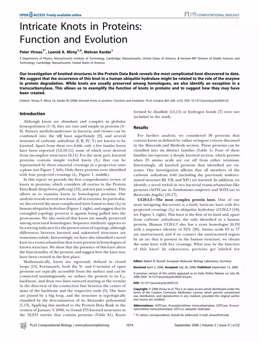
Intricate Knots in Proteins:Function and EvolutionPeter Virnau
1*, Leonid A. Mirny
1,2, Mehran Kardar
1
1 Department of Physics, Massachusetts Institute of Technology, Cambridge, Massachusetts, United States of America, 2 Harvard–MIT Division of Health Sciences and
Technology, Cambridge, Massachusetts, United States of America
Our investigation of knotted structures in the Protein Data Bank reveals the most complicated knot discovered to date.We suggest that the occurrence of this knot in a human ubiquitin hydrolase might be related to the role of the enzymein protein degradation. While knots are usually preserved among homologues, we also identify an exception in atranscarbamylase. This allows us to exemplify the function of knots in proteins and to suggest how they may havebeen created.
Citation: Virnau P, Mirny LA, Kardar M (2006) Intricate knots in proteins: Function and evolution. PLoS Comput Biol 2(9): e122. DOI: 10.1371/journal.pcbi.0020122
Introduction
Although knots are abundant and complex in globularhomopolymers [1–3], they are rare and simple in proteins [4–8]. Sixteen methyltransferases in bacteria and viruses can becombined into the a/b knot superfamily [9], and severalisozymes of carbonic anhydrase (I, II, IV, V) are known to beknotted. Apart from these two folds, only a few insular knotshave been reported [5,6,10,11], some of which were derivedfrom incomplete structures [6,11]. For the most part, knottedproteins contain simple trefoil knots (31) that can berepresented by three essential crossings in a projection ontoa plane (see Figure 1, left). Only three proteins were identifiedwith four projected crossings (41, Figure 1, middle).
In this report we provide the first comprehensive review ofknots in proteins, which considers all entries in the ProteinData Bank (http://www.pdb.org) [12], and not just a subset. Thisallows us to examine knots in homologous proteins. Ouranalysis reveals several new knots, all in enzymes. In particular,we discovered the most complicated knot found to date (52) inhumanubiquitin hydrolase (Figure 1, right), and suggest that itsentangled topology protects it against being pulled into theproteasome. We also noticed that knots are usually preservedamong structural homologues. Sequence similarity appears tobe a strong indicator for thepreservationof topology, althoughdifferences between knotted and unknotted structures aresometimes subtle. Interestingly, we have also identified a novelknot in a transcarbamylase that is not present in homologues ofknown structure. We show that the presence of this knot altersthe functionality of the protein, and suggest how the knot mayhave been created in the first place.
Mathematically, knots are rigorously defined in closedloops [13]. Fortunately, both the N- and C-termini of openproteins are typically accessible from the surface and can beconnected unambiguously: we reduce the protein to its Ca-backbone, and draw two lines outward starting at the terminiin the direction of the connection line between the center ofmass of the backbone and the respective ends [5]. The linesare joined by a big loop, and the structure is topologicallyclassified by the determination of its Alexander polynomial[1,13]. Applying this method to the Protein Data Bank in theversion of January 3, 2006, we found 273 knotted structures inthe 32,853 entries that contain proteins (Table S1). Knots
formed by disulfide [14,15] or hydrogen bonds [7] were notincluded in the study.
Results
For further analysis, we considered 36 proteins thatcontain knots as defined by rather stringent criteria discussedin the Materials and Methods section. These proteins can beclassified into six distinct families (Table 1). Four of thesefamilies incorporate a deeply knotted section, which persistswhen 25 amino acids are cut off from either terminus.Interestingly, all knotted proteins thus identified are en-zymes. Our investigation affirms that all members of thecarbonic anhydrase fold (including the previously undeter-mined isozymes III, VII, and XIV) are knotted. In addition, weidentify a novel trefoil in two bacterial transcarbamylase-likeproteins (AOTCase in Xanthomonas campestris and SOTCase inBacteroides fragilis) [16,17].UCH-L3—The most complex protein knot. One of our
most intriguing discoveries is a fairly intricate knot with fiveprojected crossings (52) in ubiquitin hydrolase (UCH-L3 [18];see Figure 1, right). This knot is the first of its kind and, apartfrom carbonic anhydrases, the only identified in a humanprotein. Human UCH-L3 also has a yeast homologue [6,19]with a sequence identity of 32% [20]. Amino acids 63 to 77are unstructured, and if we connect the unstructured regionby an arc that is present in the human structure, we obtainthe same knot with five crossings. What may be the functionof this knot? In eukaryotes, proteins get labeled for
Editor: Robert B. Russell, European Molecular Biology Laboratory, Germany
Received April 3, 2006; Accepted July 28, 2006; Published September 15, 2006
A previous version of this article appeared as an Early Online Release on July 28,2006 (DOI: 10.1371/journal.pcbi.0020122.eor).
DOI: 10.1371/journal.pcbi.0020122
Copyright: � 2006 Virnau et al. This is an open-access article distributed under theterms of the Creative Commons Attribution License, which permits unrestricteduse, distribution, and reproduction in any medium, provided the original authorand source are credited.
Abbreviations: AOTCase, N-acetylornithine transcarbamylase; SOTCase, N-succi-nylornithine transcarbamylase; UCH-L3, ubiquitin hydrolase
* To whom correspondence should be addressed. E-mail: [email protected]
PLoS Computational Biology | www.ploscompbiol.org September 2006 | Volume 2 | Issue 9 | e1221074

degradation by ubiquitin conjugation. UCH-L3 performsdeconjugation of ubiquitin, thus rescuing proteins fromdegradation. The close association of the enzyme withubiquitin should make it a prime target for degradation atthe proteasome. We suggest that the knotted structure ofUCH-L3 makes it resistant to degradation. In fact, the first
step of protein degradation was shown to be ATP-dependentprotein unfolding by threading through a narrow pore (;13A in diameter) of a proteasome [21,22]. Such threading intothe degradation chamber depends on how easily a proteinunfolds, with more stable proteins being released back intosolution [23] and unstable ones being degraded. If ATP-dependent unfolding proceeds by pulling the C-terminus intoa narrow pore [21], then a knot can sterically preclude suchtranslocation, hence preventing protein unfolding anddegradation. While arceabacterial proteasome PAN wasshown to process proteins from its C- to N-terminus [21], itcannot be ruled out that some eukaryotic proteasomesprocess proteins in the N- to C-direction, thus requiringprotection of both termini. Unfolding of a knotted protein bypulling may require a long time for global unfolding anduntangling of the knot. Unknotted proteins, in contrast, havebeen shown to become unstable if a few residues are removedfrom their termini [24], suggesting that threading a few (5–10)residues into a proteasomal pore would be sufficient tounravel an unknotted structure. At both termini, UCH-L3contains loops entangled into the knot protecting both endsagainst unfolding if pulled. It should also be noted that bothN- and C-termini are stabilized by a number of hydrophobicinteractions with the rest of the protein. The C-terminus is
Figure 1. Examples of the Three Different Types of Knots Found in Proteins
Colors change continuously from red (first residue) to blue (last residue). A reduced representation of the structure, based on the algorithm described in[1,6,36], is shown in the lower row.(Left) The trefoil knot (31) in the YBEA methyltransferase from E. coli (pdb code 1ns5; unpublished data) reveals three essential crossings in a projectiononto a plane.(Middle) The figure-eight knot (41) in the Class II ketol-acid reductoisomerase from Spinacia oleracea (pdb code 1yve [26]) features four crossings. (Onlythe knotted section of the protein is shown.)(Right) The knot 52 in ubiquitin hydrolase UCH-L3 (pdb code 1xd3 [18]) reveals five crossings. Pictures were generated with Visual Molecular Dynamics(http://www.ks.uiuc.edu/Research/vmd) [43].DOI: 10.1371/journal.pcbi.0020122.g001
PLoS Computational Biology | www.ploscompbiol.org September 2006 | Volume 2 | Issue 9 | e1221075
Synopsis
Several protein structures incorporate a rather unusual structuralfeature: a knot in the polypeptide backbone. These knots areextremely rare, but their occurrence is likely connected to proteinfunction in as yet unexplored fashion. The authors’ analysis of thecomplete Protein Data Bank reveals several new knots that, alongwith previously discovered ones, may shed light on such con-nections. In particular, they identify the most complex knotdiscovered to date in a human protein, and suggest that itsentangled topology protects it against unfolding and degradation.Knots in proteins are typically preserved across species andsometimes even across kingdoms. However, there is also oneexample of a knot in a protein that is not present in a closely relatedstructure. The emergence of this particular knot is accompanied by ashift in the enzymatic function of the protein. It is suggested thatthe simple insertion of a short DNA fragment into the gene maysuffice to cause this alteration of structure and function.
Intricate Knots in Proteins

particularly stable—residues 223 to 229 are hydrophobic andform numerous contacts at 5 A with the rest of the structure.
We would like to stress that this hypothesis needs to betested by experiments. Different proteins may also providedifferent levels of protection against degradation, dependingon structural details, the depth of the knot, and its complex-ity. Recently, a knot in the red/far-red light photoreceptorphytochrome A in Deinococcus radiodurans was identified [11](see Materials and Methods). Although sequence similaritysuggests that the knot may also be present in planthomologues, we cannot be certain. In plants, the red-absorbing form is rather stable (half-life of 1 wk), but thefar-red–absorbing form is degraded upon photoconversion
by the proteasome with a half-life of 1–2 h in seedlings (andsomewhat longer in adult plants) [25].Evolutionary aspects. As expected, homologous structures
tend to retain topological features. The trefoil knot incarbonic anhydrase can be found in isozymes ranging frombacteria and algae to humans (Table 1). Class II ketol-acidreductoisomerase comprises a figure-eight knot present inEscherichia coli [10] and spinach [26] (see Figure 1, middle), andS-adenosylmethione synthetase contains a deep trefoil knot inE. coli [5,27] and rat [28]. It appears that particular knots haveindeed been preserved throughout evolution, which suggests acrucial role for knots in protein enzymatic activity andbinding.
Table 1. List of Knotted PDB Entries (January 2006)
Protein Knot Family Protein Species PDB Code Length Knot Knotted Core
a/b knot YbeA-like E. coli 1ns5 153 31 69–121 (32)
T. maritime 1o6d 147 31 68–117 (30)
S. aureus 1vh0 157 31 73–126 (31)
B. subtilis 1to0 148 31 64–116 (32)a
tRNA(m1G37)-methyltransferase TrmD H. influenza 1uaj 241 31 93–138 (92)a
E. coli 1p9p 235 31 90–130 (89)a
SpoU-like RNA 29-O ribose mtf. T. thermophilus 1v2x 191 31 96–140 (51)
H. influenza 1j85 156 31 77–114 (42)
T. thermophilus 1ipa 258 31 185–229 (29)a
E. coli 1gz0 242 31 172–214 (28)
A. aeolicus 1zjr 197 31 95–139 (58)
S. viridochromog. 1x7p 265 31 192–234 (31)
YggJ C-terminal domain-like H. influenza 1nxz 246 31 165–216 (30)
B. subtilis 1vhk 235 31 158–208 (27)b
T. thermophilus 1v6z 227 31 103–202 (25)c
Hypothetical protein MTH1 (MT0001) A. M. Thermoautotr. 1k3r 262 31 48–234 (28)
Carbonic anhydrases Carbonic anhydrase N. gonorrhoeae 1kop 223 31 36–223 (0)
Carbonic anhydrase I H. sapiens 1hcb 258 31 29–256 (2)
Carbonic anhydrase II H. sapiens 1lug 259 31 30–256 (3)
Bos Taurus 1v9e 259 31 32–256 (3)
Dunaliella salina 1y7w 274 31 37–270 (4)
Carbonic anhydrase III Rattus norv. 1flj 259 31 30–256 (3)
H. sapiens 1z93 263 31 28–254 (9)
Carbonic anhydrase IV H.sapiens 1znc 262 31 32–261 (1)
Mus musculus 2znc 249 31 32–246 (3)b
Carbonic anhydrase V Mus musculus 1keq 238 31 7–234 (4)
Carbonic anhydrase VII H. sapiens 1jd0 260 31 28–257 (3)
Carbonic anhydrase XIV Mus Musculus 1rj6 259 31 29–257 (2)
Miscellaneous Ubiquitin hydrolase UCH-L3 H. sapiens 1xd3A 229 52 12–172 (11)d,e
S. cerevisiae (synth.) 1cmxA 214 31 9–208 (6)b,d
S-adenosylmethionine synthetase E. coli 1fug 383 31 33–260 (32)
Rattus norv. 1qm4 368 31 30–253 (29)b
Class II ketol-acid reductoisomerase Spinacia oleracea 1yve 513 41 239–451 (62)
E. coli 1yrl 487 41 220–435 (52)
Transcarbamylase-like B. fragilis 1js1 324 31 169–267 (57)
X. campestris 1yh1 334 31 171–272 (62)
‘‘Protein’’ describes the name or the family of the knotted structure. ‘‘Species’’ refers to the scientific name of the organism from which the protein was taken for structure determination.‘‘PDB code’’ gives one example Protein Data Bank entry for each knotted protein: additional structures of the same protein can be found using the SCOP classification tool [9]. ‘‘Length’’describes the number of Ca-backbone atoms in the structure. ‘‘Knot’’ refers to the knot type which was discovered in the protein: 31, trefoil; 41, figure-eight knot; 52, 2nd knot with fivecrossings according to standard knot tables [13]. The core of a knot is the minimum configuration which stays knotted after a series of deletions from each terminus; in brackets weindicate how many amino acids can be removed from either side before the structure becomes unknotted (see Materials and Methods).aStructure is fragmented and becomes knotted when missing sections are joined by straight lines. The size of the knotted core refers to the thus-connected structure. The knot is alsopresent in at least one fragment.bStructure is fragmented and only knotted when missing sections are joined by straight lines. Fragments are unknotted.c1v6z is currently not classified according to SCOP (version 1.69). Sequence similarity suggests that it is part of the a/b knot fold. 1v6zB contains a shallow composite knot (31#41), whichturns into a regular trefoil when two amino acids are cut from the N-terminus. (The random closure [see Materials and Methods] determines a trefoil right away.)d1uch contains the same structure as 1xd3. If the missing section in the center of this structure is joined by a straight line, it becomes knotted (52). In the yeast homologue (1cmx), aminoacids 63 to 77 are unstructured, and if we replace the missing parts by a straight line, we obtain a trefoil knot that has been identified before [6]. If we connect the unstructured region byan arc present in the human structure, we obtain the same knot with five crossings.eVisual inspection reveals that the calculated size of the knotted core is too small.DOI: 10.1371/journal.pcbi.0020122.t001
PLoS Computational Biology | www.ploscompbiol.org September 2006 | Volume 2 | Issue 9 | e1221076
Intricate Knots in Proteins

UCH-L3 in human and yeast share only 33% [29] of theirsequences, but contain the same 5-fold knot as far as we cantell from the incomplete structure in yeast. It is not only likelythat all species in between have the same knot—the linkbetween sequence and structure may also be used to predictcandidates for knots among isozymes or related proteins forwhich the structure is unknown. For example, UCH-L4 inmouse has 96% sequence identity with human UCH-L3. Thesimilarity with UCH-L6 in chicken is 86%, and with UCH-L1about 55%. Indeed, a reexamination of the most recentProtein Data Bank entries revealed that UCH-L1 contains thesame 52 knot as UCH-L3. (See the Update section—thestructure was not yet part of the January Protein Data Bankrelease on which this paper is based.) Unfortunately, themethod is not foolproof because differences between knottedand unknotted structures are sometime subtle. As we willdemonstrate in the next paragraph, a more reliable estimatehas to consider the conservation of major elements of theknot, like loops and threads.
AOTCase—How a protein knot can alter enzymatic activity.Somewhat surprisingly, we also identified a pair of homo-logues for which topology is not preserved. N-acetylornithinetranscarbamylase (AOTCase [17]) is essential for the argininebiosynthesis in several major pathogens. In other bacteria,animals, and humans, a homologous enzyme (OTCase)processes L-ornithine instead [30]. Both proteins have twoactive sites. The first one binds carbamyl phosphate to theenzyme. The second site binds acetylornithine in AOTCasesand L-ornithine in OTCases, enabling a reaction withcarbamyl phosphate to form acetylcitrulline or citrulline,respectively [17, 31].
AOTCase in X. campestris has 41% sequence identity withOTCase from Pyrococcus furiosus [32] and 29% with humanOTCase [31]. As demonstrated in Figure 2, AOTCase containsa deep trefoil knot which is not present in OTCase (Figure 2,
right) and which modifies the second active site. The knotconsists of a rigid proline-rich loop (residues 178–185),through which residues 252 to 256 are threaded and affixed.As elaborated in [17], the reaction product N-acetylcitrullinestrongly interacts with the loop and with Lys252. Access tosubsequent residues is, however, restricted by the knot. L-norvaline in Figure 2 (right) is very similar to L-ornithine butlacks the N-e atom of the latter to prevent a reaction withcarbamyl phosphate. As the knot is not present in OTCase,the ligand has complete access to the dangling residues 263–268 and strongly interacts with them [31]. This leads to arotation of the carboxyl-group by roughly 1108 around theCa–Cb bond [17].This example demonstrates how the presence of a knot can
modify active sites and alter the enzymatic activity of aprotein—in this case, from processing L-ornithine to N-acetyl-L-ornithine. It is also easy to imagine how thisalteration happened: a short insertion extends the loop andmodifies the folding pathway of the protein.
Discussion
Nature appears to disfavour entanglements, and evolutionhas developed mechanisms to avoid knots. Human DNAwraps around histone proteins, and the rigidity of DNAallows it to form a spool when it is fed into a viral capsid. Oneend also stays in the loading channel and prevents subsequentequilibration [33]. Knotted proteins are rare, although thereason is far less well understood. Can the absence ofentanglement be explained in terms of particular statisticalensembles, or is there an evolutionary bias? And how do thesestructures actually fold?Knots are ubiquitous in globular homopolymers [1–3,8], but
rare in coil-like phases [1,34–36]. It is likely that even a flexiblepolymer will at least initially remain unknotted after a
Figure 2. Structures of Transcarbamylase from X. campestris with a Trefoil Knot and from Human without a Knot
(Left) Knotted section (residues 171–278) of N-acetylornithine transcarbamylase from X. campestris with reaction product N-acetylcitrulline (pdb code1yh1 [17]) and interacting side chains.(Right) Corresponding (unknotted) section (residues 189–286) in human ornithine transcarbamylase (pdb code 1c9y [31]) with inhibitor L-norvaline andcarbamyl phosphate. Colors change continuously from red (first residue in the section) to blue (last residue in the section). The two proteins have anoverall sequence identity of 29% [41]. Pictures were generated with VMD [43].DOI: 10.1371/journal.pcbi.0020122.g002
PLoS Computational Biology | www.ploscompbiol.org September 2006 | Volume 2 | Issue 9 | e1221077
Intricate Knots in Proteins

collapse from a swollen state. In proteins, the free energylandscape is considerably more complex, which may allowmost proteins to stay unknotted. The secondary structure andthe stiffness of the protein backbone may shift the length scaleat which knots typically appear, too [8]. If knotted proteins arein fact more difficult to degrade, it might also be disadvanta-geous for most proteins to be knotted in the first place.
Unfortunately, few experimental papers address foldingand biophysical aspects of knots in proteins. In recent work[37], Jackson and Mallam reversibly unfolded and folded aknotted methyltransferase in vitro, indicating that chaper-ones are not a necessary prerequisite. In a subsequent study[38], the authors provide an extensive kinetic analysis of thefolding pathway. In conclusion, we would like to express ourhope that this report will inspire more experiments in thissmall but nevertheless fascinating field.
Materials and Methods
To determine whether a structure is knotted, we reduce theprotein to its backbone, and draw two lines outward starting at thetermini in the direction of the connection line between the center ofmass of the backbone and the respective ends. These two lines arejoined by a big loop, and the structure is classified by thedetermination of its Alexander polynomial [1,13]. To determine thesize of the knotted core, we delete successively amino acids from theN-terminus until the protein becomes unknotted [1,6]. The proce-dure is repeated at the C-terminus starting with the last structure thatcontained the original knot. For each deletion, the outward pointingline through the new termini is parallel to the respective linescomputed for the full structure. The thus determined size should,however, only be regarded as a guideline. A better estimate can beachieved by looking at the structure.
In Table 1 we include knotted structures with no missing aminoacids in the center of the protein. (A list of potentially knottedstructures with missing amino acids can be found in Table S3.)Technically, the numbering of the residues in the mmcif file has to besubsequent, and no two amino acids are allowed to be more than 6 Aapart. In addition, the knot has to persist when two amino acids arecut from either terminus. We have further excluded structures forwhich unknotted counterexamples exist (e.g., only one nuclearmagnetic resonance structure among many is knotted or anotherstructure of the same protein is unknotted). If a structure isfragmented, the knot has to appear in one fragment and in theresulting structure obtained from connecting missing sections bystraight lines. Other knotted structures are only considered when atleast one additional member of the same structural family [9]contains a knot according to the criteria above.
The enforcement of these rules leads to the exclusion of thebluetongue virus core protein [6] (41) and photoreceptor phyto-chrome A in D. radiodurans [11] (31), which have been previouslyidentified as being knotted. Both structures are fragmented andbecome knotted only when a few missing fragments are connected bystraight lines. In the viral core protein, the dangling C-terminusthreads through a loose loop and becomes knotted in one out of twocases. On the other hand, the photoreceptor phytochrome A appearsto contain a true knot. Notably, our analysis suggests that the thusconnected structure of phytochrome A contains a figure-eight knotinstead of a trefoil as reported in [11]. Moreover, we excluded astructure of the Autographa California nuclear polyhedrosis virus, whichcontains a knot according to our criteria. However, the N-terminus isburied inside the protein and the knot only exists because of ourspecific connection to the outside.
To further validate our criteria, we implemented an alternativemethod [4,8,39] that relies on the statistical analysis of multiplerandom closures. We arbitrarily chose two points on a sphere (which
has to be larger than the protein) and connected each with oneterminus. The two points can be joined unambiguously, and theresulting loop was analyzed by calculating the Alexander polynomial.We repeated the procedure 1,000 times, and defined the knot as themajority type.
Applying this analysis, we discovered 241 knotted structures in theProtein Data Bank. All 241 structures are also present in the 273structures (Table S1) that were identified by our method, and the knottype is the same. Themissing32 structures (Table S2) aremostly shallowknots and were already rejected according to our extended criteria.The random closure also correctly discards rare structures with buriedtermini. In conclusion, the method used in this paper is considerablyfaster but requires a slightly increased inspection effort. Ourobservations agree with [8], which provides an extensive comparisonof closures applied to proteins. A complete listing of knotted ProteinData Bank structures is given in the Supporting Information.
Update. Recently, the structure of human UCH-L1 was solved andreleased [40]. The protein shares 55% sequence identity with UCH-L3[41], and it contains the same 5-fold knot. UCH-L1 is highly abundantin the brain, comprising up to 2% of the total brain protein [42]. Thestructure of UCH-L1 was not yet part of the January Protein DataBank edition on which the rest of this study is based. We also noticedseveral new structures of knotted transcarbamylase-like proteins.
Supporting Information
Table S1. List of Knotted Protein Data Bank Entries
Found at DOI: 10.1371/journal.pcbi.0020122.st001 (79 KB DOC).
Table S2. List of Knotted Entries from Table S1 That BecomeUnknottedWhenEnds AreConnected by theRandomClosureMethod
Found at DOI: 10.1371/journal.pcbi.0020122.st002 (28 KB DOC).
Table S3. List of Structures That Become Knotted When MissingSections Are Joined by Straight Lines
Found at DOI: 10.1371/journal.pcbi.0020122.st003 (35 KB DOC).
Accession Numbers
The Protein Data Bank (http://www.pdb.org) accession numbers forthe structures discussed in this paper are human UCH-L3 (1xd3),UCH-L3 yeast homologue (1cmx), human UCH-L1 (2etl), photo-receptor phytochrome A in D. radiodurans (1ztu), class II ketol-acidreductoisomerase in E. coli (1yrl), class II ketol-acid reductoisomerasein spinach (1yve), S-adenosylmethione synthetase in E. coli (1fug), S-adenosylmethione synthetase in rat (1qm4), AOTCase from X.campestris (1yh1), SOTCase from B. fragilis (1js1), OTCase from P.furiosus (1a1s), OTCase from human (1c9y), bluetongue virus coreprotein (2btv), and baculovirus P35 protein in Autographa Californianuclear polyhedrosis virus (1p35).
Acknowledgments
Upon completion of this work we became aware of a related study [8],which independently identified the knots in UCH-L3 and SOTCase in are-examination of protein knots. PV would like to acknowledgediscussions with Francois Nedelec and with Olav Zimmermann, inwhich they proposed the potential link between protein knots anddegradation. LM and PV would also like to thank Rachel Gaudet for adiscussion about the function of ubiquitin hydrolase.
Author contributions. MK conceived the study. PV designed andwrote the analysis code. PV and LM analyzed the data. PV, LM, andMK wrote the paper.
Funding. This work was supported by National Science Foundationgrant DMR-04–26677 and by Deutsche Forschungsgemeinschaft grantVI 237/1. LM is an Alfred P. Sloan Research Fellow.
Competing interests. The authors have declared that no competinginterests exist.
References
1. Virnau P, Kantor Y, Kardar M (2005) Knots in globule and coil phases of amodel polyethylene. J Am Chem Soc 127: 15102–15106.
2. Mansfield ML (1994) Knots in Hamilton cycles. Macromolecules 27: 5924–5926.
3. Lua RC, Borovinskiy AL, Grosberg AY (2004) Fractal and statistical
properties of large compact polymers: A computational study, Polymer45: 717–731.
4. Mansfield ML (1994) Are there knots in proteins? Nat Struct Mol Bio 1:213–214.
5. Mansfield ML (1997) Fit to be tied. Nat Struct Mol Bio 4: 166–167.6. Taylor WR (2000) A deeply knotted protein structure and how it might
fold. Nature 406: 916–919.
PLoS Computational Biology | www.ploscompbiol.org September 2006 | Volume 2 | Issue 9 | e1221078
Intricate Knots in Proteins

7. Taylor WR, Lin K (2003) Protein knots—A tangled problem. Nature 421: 25.8. Lua RC, Grosberg AY (2006) Statistics of knots, geometry of conformations,
and evolution of proteins. PLOS Comp Biol 2: e45.9. Murzin AG, Brenner SE, Hubbard T, Chothia C (1995) SCOP: A structural
classification of proteins database for the investigation of sequences andstructures. J Mol Biol 247: 536–540 http://scop.mrc-lmb.cam.ac.uk/scop.
10. Tyagi R, Duquerroy S, Navaza J, Guddat LW, Duggleby RG (2005) Thecrystal structure of a bacterial Class II ketol-acid reductolsomerase:Domain conservation and evolution. Protein Sci 14: 3089–3100.
11. Wagner JR, Brunzelle JS, Forest KT, Vierstra RD (2005) A light-sensing knotrevealed by the structure of the chromophore-binding domain ofphytochrome. Nature 438: 325–331.
12. Berman HM, Westbrook J, Feng Z, Gilliland G, Bhat TN, et al. (2000) TheProtein Data Bank. Nucleic Acids Res 28: 235–242. (The Protein Data Bankis athttp://www.pdb.org. Accessed 22 August 2006.)
13. Adams CC (1994) The knot book: An elementary introduction to themathematical theory of knots. New York: W. H. Freeman. 306 p.
14. Liang C, Mislow K (1995) Topological features of protein structures: Knotsand links. J Am Chem Soc 177: 4201–4213.
15. Takusagawa F, Kamitori S (1996) A real knot in protein. J Am Chem Soc118: 8945–8946.
16. Shi D, Gallegos R, DePonte J III, Morizono H, Yu X, et al. (2002) Crystalstructure of a transcarbamylase-like protein from the anaerobic bacteriumBacteroides fragilis at 2.0 A resolution. J Mol Biol 320: 899–908.
17. Shi D, Morizono H, Yu X, Roth L, Caldovic L, et al. (2005) Crystal structureof N-acetylornithine transcarbamylase from Xanthomonas campestris: A novelenzyme in a new arginine biosynthetic pathway found in several eubacteria.J Biol Chem 280: 14366–14369.
18. Misaghi S, Galardy PJ, Meester WJN, Ovaa H, Ploegh HL et al. (2005)Structure of the ubiquitin hydrolase Uch-L3 complexed with a suicidesubstrate. J Biol Chem 280: 1512–1520.
19. Johnston SC, Riddle SM, Cohen RE, Hill CP (1999) Structural basis for thespecificity of ubiquitin C-terminal hydrolases. EMBO J 18: 3877–3887.
20. Holm L, Sander C (1996) Mapping the protein universe. Science 273: 595–602. (The Dali Database is located at http://ekhidna.biocenter.helsinki.fi/dali/start. Accessed 22 August 2006.)
21. Navon A, Goldberg AL (2001) Proteins are unfolded on the surface of theATPase ring before transport into the proteasome. Mol Cell 8: 1339–1349.
22. Pickart CM, VanDemark AP (2000) Opening doors into the proteasome.Nat Struct Mol Bio 7: 999–1001.
23. Kenniston JA, Baker TA, Sauer RT (2005) Partitioning between unfoldingand release of native domains during ClpXP degradation determinessubstrate selectivity and partial processing. Proc Natl Acad Sci U S A 102:1390–1395.
24. Neira JL, Fersht AR (1999) Exploring the folding funnel of a polypeptidechain by biophysical studies on protein fragments. JMol Biol 285: 1309–1333.
25. Clough RC, Vierstra RD (1997) Phytochrome degradation. Plant CellEnviron 20: 713–721.
26. Biou V, Dumas R, Cohen-Addad C, Douce R, Job D, et al. (1997) The crystal
structure of plant acetohydroxy acid isomeroreductase complexed withNADPH, two magnesium ions and a herbicidal transition state analogdetermined at 1.65 A resolution. EMBO J 16: 3405–3415.
27. Fu Z, Hu Y, Markham GD, Takusagawa F (1996) Flexible loop in thestructure of S-adenosylmethionine synthetase crystallized in the tetragonalmodification. J Biomol Struct Dyn 13: 727–739.
28. Gonzalez B, Pajares MA, Hermoso JA, Alvarez L, Garrido F, et al. (2000) Thecrystal structure of tetrameric methionine adenosyltransferase from ratliver reveals the methionine-binding site. J Mol Biol 300: 363–375.
29. Sander C, Schneider R (1991) Database of homology-derived proteinstructures. Proteins: Struct Funct Genet 9: 56–68.
30. Morizono H, Cabrera-Luque J, Shi D, Gallegos R, Yamaguchi S, et al. (2006)Acetylornithine transcarbamylase: A novel enzyme in arginine biosynthesis.J Bacteriol 188: 2974–2982.
31. Shi D, Morizono H, Aoyagi M, Tuchman M, Allewell NM (2000) Crystalstructure of human ornithine transcarbamylase complexed with carbamylphosphate and L-Norvaline at 1.9 A resolution. Proteins: Struct FunctGenet 39: 271–277.
32. Villeret V, Clantin B, Tricot C, Legrain C, Roovers M, et al. (1998) Thecrystal structure of Pyrococcus furiosus ornithine carbamoyltransferasereveals a key role for oligomerization in enzyme stability at extremelyhigh temperatures. Proc Natl Acad Sci U S A 95: 2801–2806.
33. Arsuaga J, Vasquez M, Trigueros S, Sumners DW, Roca J (2002) Knottingprobability of DNA molecules confined in restricted volumes: DNAknotting in phage capsids. Proc Natl Acad Sci U S A 99: 5373–5377.
34. Janse van Rensburg EJ, Sumners DW, Wassermann E, Whittington SG(1992) Math Gen 25: 6557–6566.
35. Deguchi T, Tsurusaki K (1997) Universality in random knotting. Phys Rev E55: 6245–6248.
36. Koniaris K, Muthukumar M (1991) Self-entanglement in ring polymers. JChem Phys 95: 2873–2881.
37. Jackson SE, Mallam AL (2005) Folding studies on a knotted protein. J MolBiol 346: 1409–1421.
38. Mallam AL, Jackson SE (2006) Probing nature’s knots: The folding pathwayof a knotted homodimeric protein. J Mol Biol 359: 1420–1436.
39. Millett K, Dobay A, Stasiak A (2005) Linear random knots and their scalingbehavior. Macromolecules 38: 601–606.
40. Das C, Hoang QQ, Kreinbring CA, Luchansky SJ, Meray RK, et al. (2006)Structural basis for conformational plasticity of the Parkinson’s disease-associated ubiquitin hydrolase UCH-L1. Proc Natl Acad Sci U S A 103:4675–4680.
41. Krissinel E, Henrick K (2004) Secondary-structure matching (SSM), a newtool for fast protein structure alignment in three dimensions. Acta CrystD60: 2256–2268.
42. Wilkinson KD, Lee KM, Deshpande S, Duerksen-Hughes P, Boss JM, et al.(1989) The neuron-specific protein PGP 9.5 is a ubiquitin carboxyl-terminal hydrolase. Science 246: 670–673.
43. Humphrey W, Dalke A, Schulten K (1996) VMD—Visual moleculardynamics. J Molec Graphics 14: 33–38.
PLoS Computational Biology | www.ploscompbiol.org September 2006 | Volume 2 | Issue 9 | e1221079
Intricate Knots in Proteins


