Embed Size (px)
Citation preview

THE JOURNAL OF BIOLOGICAL CHEMISTRY 0 1993 by The American Society for Biochemistry and Molecular Biology, Inc
Vol. 268, No. 1, Issue of January 5, pp. 527-533,1993 Printed in U.S.A.
Isolation and Characterization of a Novel Serine Proteinase Complexed with az-Macroglobulin from Porcine Gastric Mucosa*
(Received for publication, June 19, 1992)
Takashi UchinoS, Yasuko SakuraiS, Masaaki Nishigail, Takayuki Takahashi$, Hideo Arakawag, Atsushi Ikais, and Kenji TakahashiSll From the $Department of Biophysics and Biochemistry, Faculty of Science, The University of Tokyo, Bunkyo-ku, Tokyo 113 and the $Laboratory of Biodynamics, Tokyo Institute of Technology, Nagatsuta, Midori-ku, Yokohama 227, Japan
Porcine stomach mucosa was found to contain a 740- kDa protein having endopeptidase activity toward peptide 4-methylcoumaryl-7-amide substrates and low molecular mass peptides. This protein was purified to an apparent homogeneity by a series of chromato- graphic steps on DEAE-cellulose, Sepharose CL-4B, hydroxylapatite, and fast protein liquid chromatogra- phy Mono Q columns. The protein was shown to be a complex of the plasma proteinase inhibitor a2-macro- globulin and a 25-kDa endopeptidase. The enzyme ac- tivity was completely inhibited by diisopropyl fluoro- phosphate, p-amidinophenylmethanesulfonyl fluoride, leupeptin, antipain, bovine pancreatic trypsin inhibi- tor, soybean trypsin inhibitor, and ovomucoid, indicat- ing that the entrapped enzyme is a serine proteinase. The proteinase could be released from az-macroglobu- lin by mild acid treatment and the released enzyme showed activity toward protein substrates.
Substrate specificity studies using synthetic and pep- tide substrates indicated that the enzyme preferen- tially hydrolyzes Arg-X bonds and, to a much lesser extent, Lys-X bonds, and is apparently distinct from thrombin, kallikrein, plasmin, and other trypsin-like proteinases so far reported including tryptase. Thus, the present enzyme is thought to be a novel type of serine proteinase. The proteinase associated with aZ- macroglobulin was also found in porcine intestinal mu- cosa, but not in plasma.
The stomach is a dilated segment of the digestive tract whose main function is to digest ingested food. The best studied stomach enzyme involved in the process of digestion in this organ is the aspartic proteinase pepsin (1-5). In contrast to this extracellular proteinase, very little is known of intracellular proteinases, although recently cathepsin E has been isolated and characterized (2, 6-9) and its structure analyzed (10-12). Mucosal extracts indeed contain a variety of peptidase activities and most of these peptidases are thought to be involved in intracellular protein turnover (13- 15). In addition, there must be some peptidases that function as proteolytic processing enzymes for certain biologically im- portant proteins and peptides and their precursors since bioactive peptides such as gastrin, enkephalin, somatostatin, glucagon, and other peptides are known to be present in the
* The costs of publication of this article were defrayed in part by the payment of page charges. This article must therefore be hereby marked “aduertisernent” in accordance with 18 U.S.C. Section 1734 solely to indicate this fact.
7 To whom correspondence should be addressed Dept. of Biophys- ics and Biochemistry, Faculty of Science, The University of Tokyo, Bunkyo-ku, Tokyo 113, Japan. Tel.: 3-5689-5607; Fax: 3-5684-2394.
stomach (16). However, none of the peptidases involved in the proteolytic processing of such peptides have yet been identified.
In an attempt to elucidate the functional role of proteolytic enzymes in the mammalian stomach, a high molecular weight protein that preferably hydrolyzes peptide 4-methylcoumaryl- 7-amide (MCA)’ substrates at the Arg-MCA bond was found in the homogenate. In the present study, we purified the protein and characterized it in detail. The results reveal that the enzyme activity is due to an a2-macroglobulin-associated serine proteinase with a molecular weight of about 25,000. Further investigation of the enzymatic properties including substrate specificity and effects of various inhibitors indicated that this proteinase is distinct from any other known serine endepeptidases thus far described.
EXPERIMENTAL PROCEDURES AND RESULTS~
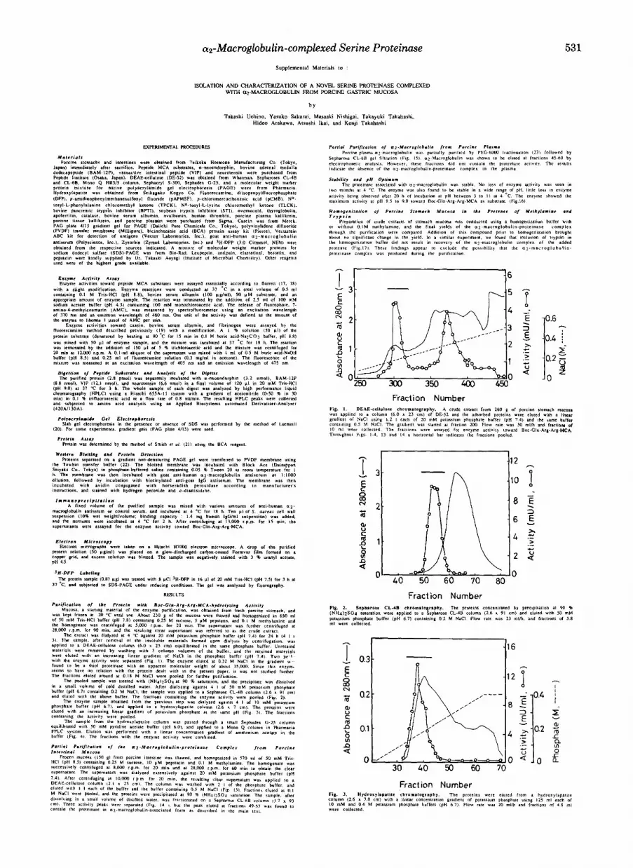
Purification of a Protein with Boc-Gln-Arg-Arg-MCA Hy- drolyzing Actiuity from Porcine Stomach Mucosa-Table I summarizes the purification of a protein hydrolyzing Boc- Gln-Arg-Arg-MCA from porcine stomach mucosa. The de- tailed procedure is described in the Miniprint section. Anion- exchange column chromatography (DE-52 and Mono &) al- ways gave rise to low recovery of activity, but inclusion of these columns was eventually found to yield the protein in satisfactory purity as described in the following section. From 230 g of the mucosa, 1.4 mg of the protein was obtained with an overall yield of 2.2%.
Purity and Molecular Weight-Native PAGE using a gra- dient gel gave a single protein band associated with the activity (Fig. 5). Its apparent molecular mass was estimated to be approximately 730,000 by PAGE and 750,000 by gel filtration on Sepharose CL-GB (Fig. 6). Fig. 7 shows the results of SDS-PAGE of the sample in the presence and absence of 2-mercaptoethanol. A major protein band corresponding to M, = 360,000 and a very faint band corresponding to M, = 100,000 were observed under nonreducing conditions, while a major protein band ( M , = 95,000) and other minor bands (M, = 80,000, 56,000, and 54,000) were separated under reducing
The abbreviations used are: MCA, 4-methylcoumaryl-7-amide; SDS, sodium dodecyl sulfate; PAGE, polyacrylamide gel electropho- resis; DFP, diisopropyl fluorophosphate; TPCK, Nu-tosyl-L-phenyl- alanine chloromethyl ketone; TLCK, N”-tosyl-L-lysine chloromethyl ketone; BOC, t-butyloxycarbonyl; Z, benzyloxycarbonyl; Bz, benzoyl; SUC, succinyl.
Portions of this paper (including “Experimental Procedures,” part of “Results,” Table I, and Figs. 1-4,6,8-11, and 13-17) are presented in miniprint at the end of this paper. Miniprint is easily read with the aid of a standard magnifying glass. Full size photocopies are included in the microfilm edition of the Journal that is available from Waverly Press.
~~
527

528 az-Macroglobulin-complexed Serine Proteinase
Relative Activity (%I FIG. 5. PAGE analysis of the purified protein. The sample
was separately applied to two well positions of a gradient PAGE gel (4-15%) without SDS. After electrophoresis at 4 “C, one lane was stained with Coomassie Brilliant Blue R-250 (lane 2), while the other was sliced into pieces of 3-mm width for overnight extraction in 0.5 ml of the routine assay buffer. Aliquots of the extracts were assayed for enzyme activity toward Boc-Gln-Arg-Arg-MCA and the relative activities are shown. Lane 1 shows the separation of molecular weight marker proteins (Pharmacia LKB Biotechnology Inc.): thyroglobulin (670 kDa), apoferritin (440 kDa), lactate dehydrogenase (140 kDa), and albumin (66 kDa).
A B
r, -
22- L
FIG. 7. SDS-PAGE analysis of the purified protein. A, the protein (2 pg) was electrophoresed in 7.5% PAGE gel in SDS under nonreducing (lane 1 ) and reducing (lane 2 ) conditions. The gels were stained with Coomassie Brilliant Blue. B, the protein (0.87 pg) was incubated with 8 pCi of [3H]DFP in 20 mM Tris-HC1 (pH 7.5) and subjected to SDS-PAGE using 7.5% gel under reducing conditions, followed by fluorography. Molecular weight marker proteins used are: dimer of human a*-macroglobulin subunit (360 kDa), myosin (200 kDa), &galactosidase (116 kDa), phosphorylase b (97 kDa), bovine serum albumin (66 kDa), ovalbumin (43 kDa), carbonic anhydrase (31 kDa), and soybean trypsin inhibitor (22 kDa).
conditions. However, the sample treated with the serine pro- teinase inhibitor DFP (3H-labeled) produced only a single band with a molecular mass of approximately 28,000 on SDS- PAGE with autoradiography (Fig. 7 B ) .
Association of a Serine Proteinase with a2-Macroglobulin- The results described above suggested that the purified pro- tein is a complex of the plasma protein inhibitor a2-macro- globulin with a serine proteinase. To substantiate this point, several experiments were performed. The purified protein cross-reacted with goat anti-human a2-macroglobulin anti- body (Fig. 8). Furthermore, direct electron microscopic obser- vation of the protein revealed two similar molecular shapes (Fig. 9). One of them looks like the letter H, while the other is a rounded structure characterized by two thick units located roughly in parallel with small material between them. They are the characteristic shapes of a2-macroglobulin that under- went a drastic conformational change after reacting with proteinase (24).
Release of a Serine Proteinase from the a2-Macroglobulin- proteinase Complex-The results shown in Fig. 7 suggest that the proteinase is dissociable from the a2-macroglobulin-pro- teinase complex. It was found that exposure of the purified protein to pH 3 for 12 h did not cause significant change in the activity toward Boc-Gln-Arg-Arg-MCA substrate, but re-
sulted in a remarkable increase in the activity toward protein substrates (Fig. 10). In addition, goat anti-human a2-macro- globulin antibody no longer precipitated the proteinase activ- ity when the purified protein was previously treated at pH 3 (data not shown). These results clearly indicate that the proteinase is noncovalently associated with a2-macroglobulin, and is freed from steric restraint upon the acid treatment.
In order to obtain the pure enzyme from the acid-treated sample, several column chromatographic steps were tried under various conditions. The enzyme activity was completely lost on ion-exchange columns (Mono Q, Mono S, and DE- 52). Poor recovery (about 20%) of the activity was also ob- served on the gel filtration over a Sephacryl S-300 column. Thus, none of these columns was found to be suitable for the purification. It should be noted that the gel filtration gave a single activity peak eluting at a position corresponding to MI = 25,000 (data not shown), a value close to that obtained in the [3H]DFP labeling experiment.
Action on Synthetic Substrates-No significant difference was observed when the activities toward a few MCA substrates were compared before and after the acid treatment of the protein. This was reasonable since the substrates are small enough to freely reach the active site irrespective of whether the proteinase is associated with the inhibitor. Thus, the activities on synthetic substrates were tested using the enzyme associated with a2-macroglobulin and the results are shown in Table 11. The specificities of human thrombin (MI = 34,000), porcine tissue kallikrein (M1=30,000), and porcine plasmin (MI = 80,000) are also included for comparison.
The enzyme was most active toward Boc-Gln-Gly-Arg- MCA. Boc-Gln-Arg-Arg-MCA, Boc-Gln-Ala-Arg-MCA, Boc- Phe-Ser-Arg-MCA, and Boc-Leu-Gly-Arg-MCA were hydro- lyzed fairly well by the enzyme and to similar extents. A common feature of the substrates hydrolyzable by the enzyme was the presence of an Arg residue at the PI position. Sub- strates containing Lys-MCA bonds were much less susceptible to the enzyme, and substrates for chymotrypsin were not cleaved at all. Little or no cleavage occurred when the MCA- derivatives of an amino acid or a dipeptide were tested. Thrombin very rapidly hydrolyzed the substrates Boc-Val- Pro-Arg-MCA and Boc-Gln-Ala-Arg-MCA, while the tissue kallikrein most rapidly cleaved Boc-Gln-Ala-Arg-MCA, Boc-
TABLE I1 Enzyme activities toward various MCA substrates
Enzyme activities were determined at pH 9.0 with 50 pM substrates as described under “Experimental Procedures” and are expressed as percent of activity toward Boc-Gln-Arg-Arg-MCA.
Substrate Present Thrombin Kallikrein Plasmin enzyme (human) (porcine) (porcine)
Boc-Gln-Arg-Arg-MCA 100 100 100 100 Boc-Gln-Gly-Arg-MCA 201 354 63 10 Boc-Gln-Ala-Arg-MCA 121 3300 305 55 Boc-Phe-Ser-Arg-MCA 105 142 97 11 Boc-Leu-Gly-Arg-MCA 90 304 37 5 Boc-Leu-Thr-Arg-MCA 48 - - - Boc-Gly-Arg-Arg-MCA 42 11 13 12 Boc-Val-Pro-Arg-MCA 41 4160 217 24 Boc-Leu-Lys-Arg-MCA 40 42 - 30 Boc-Val-Leu-Lys-MCA 13 - 285 57
Z-Phe-Arg-MCA 16 - - Z-Arg-Arg-MCA 3 Bz-Arg-MCA 0.4 - 71 Suc-Ala-Ala-Pro-Phe-MCA 0 Suc-Leu-Leu-Val-Tyr-MCA 0 Arg-MCA 0 0 5 0 Leu-MCA 0 - -
Boc-Glu-Lys-Lys-MCA 11 - - - -
- - - -
- - - - - -
- ’ Not tested.

a2-Macroglobulin-complexed Serine Proteinase 529
Val-Leu-Lys-MCA, and Boc-Val-Pro-Arg-MCA. Plasmin cleaved Boc-Gln-Arg-Arg-MCA efficiently, but hydrolyzed Boc-Gln-Gly-Arg-MCA, a good substrate for the present en- zyme, rather slowly. These results clearly show that the specificity of the current proteinase is different from those of thrombin, tissue kallikrein, and plasmin.
Action on Peptide Substrates-Four peptide substrates were tested for hydrolysis by the proteinase, and the results of high performance liquid chromatography analysis of the digests are shown in Fig. 11. As summarized in Fig. 12, cleavages occurred only on the COOH-terminal side of Arg or Lys residues. The specificity is thus consistent with that found with MCA substrates.
Effects of Inhibitors-The effects of various inhibitors on the activity were examined, and the results are shown in Table 111. DFP, p-amidinophenylmethanesulfonyl fluoride, antipain, leupeptin, and bovine pancreatic trypsin inhibitor inhibited strongly the activity of the az-macroglobulin-asso- ciated proteinase. Strong inhibition by soybean trypsin inhib- itor and ovomucoid was observed only after the purified protein was treated at pH 3, indicating again the release of the proteinase from az-macroglobulin by this treatment. These results clearly indicate that the enzyme is a trypsin- like serine proteinase.
Presence of the az-Macroglobulin-Proteinase Complex in Porcine Intestinal Mucosa-In order to examine whether or
83 3 1-
a-Neoendorphin YGSLFdW%
5211
BAM-1PP Tv
YGGFMRRVGFIPE
7 9 11 v r I VIP HSDAVFTDNMRLRKQMAVKK~SI~-NHz
36
Neurotensin pELYUJKPRRPYIL T
FIG. 12. Sites and extents of cleavage by the proteinase in peptide substrates. Arrowheads show the sites of cleavage, and the number above each arrowhead indicates the estimated extent of cleavage in percent of the peptide bond. Large and small arrowheads show major and minor cleavage sites, respectively. The amino acid sequences of peptides are shown in one-letter codes. pE, pyroglutamic acid residue, -NH2, amide.
TABLE I11 Effects of inhibitors on the proteinase activity
Inhibitor Concentration Inhibition
DFP 2 98 p-Amidinophenylmethanesulfonyl fluoride 0.5 97 Iodoacetic acid 1 9 p-Chloromercuribenzoic acid EDTA
0.4 4 1 23
o-Phenanthroline TLCK
1 3
TPCK 0.2 34 0.25 4
Benzamidine 1 69 Bestatin 0.065 9 Amastatin 0.042 0 Antipain 0.1 98 Leupeptin Elastatinal
0.1 98 0.2
Pepstatin 11
0.01 a Bovine pancreatic trypsin inhibitor 0.1 mg/ml 98 (95)” Soybean trypsin inhibitor 0.1 mg/ml 44 (100) Ovomucoid 0.1 mg/ml 0 (98)
treated at pH 3.
mM %
a Values in parentheses are the results obtained with the sample
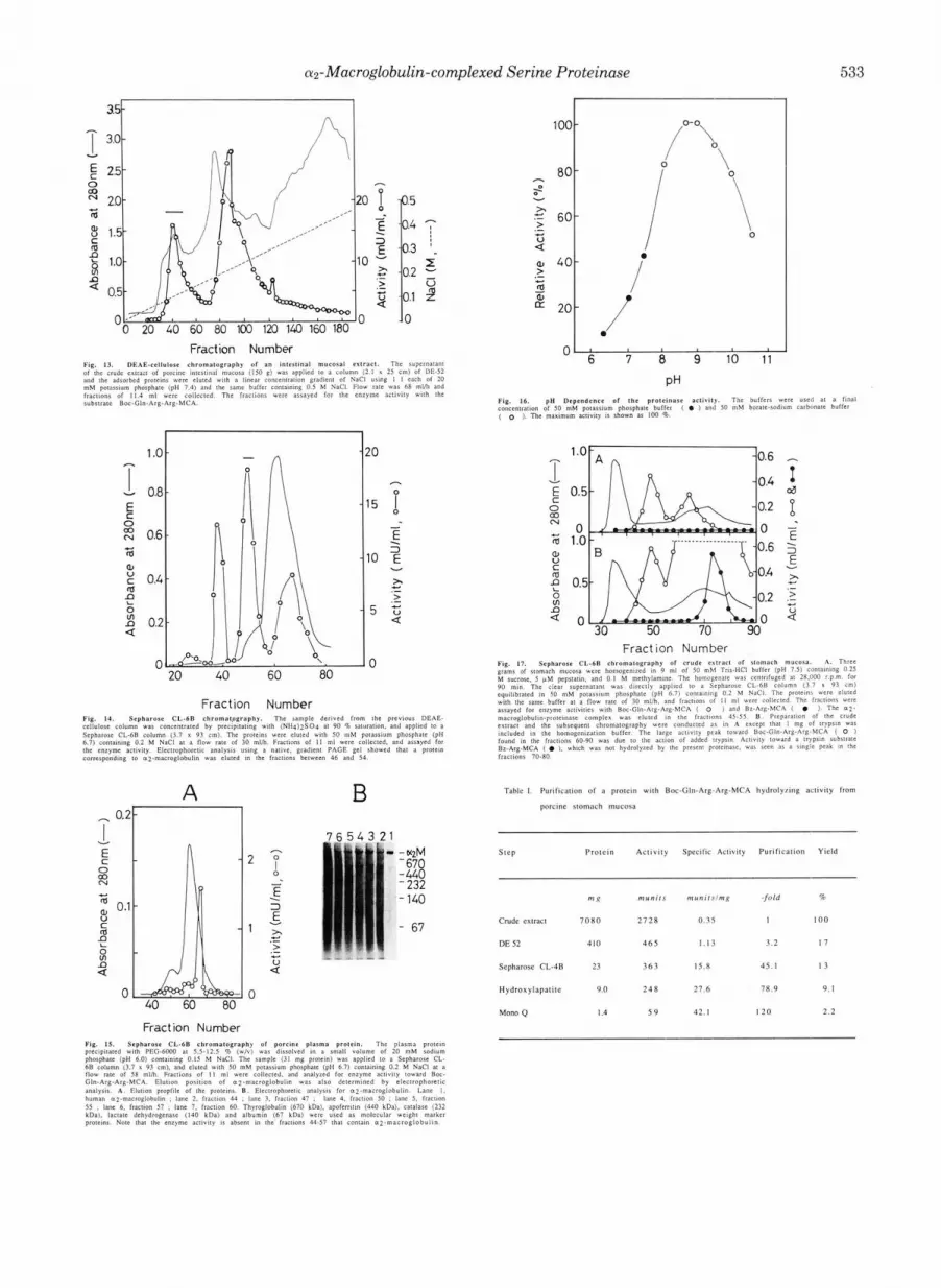
not the same a2-macroglobulin-proteinase complex is present in the mucosa of the porcine intestine, a partial purification experiment was conducted as described in the Miniprint section. Electrophoretic analysis of the fractions obtained by chromatography on Sepharose CL-GB revealed that wmac- roglobulin was eluted in fractions 45-50, which corresponded to one of the peaks with proteinase activity (Fig. 14). Associ- ation of the proteinase with cuz-macroglobulin was confirmed by assaying the activity in the gel slice extracts as described under “Experimental Procedures.” The activities toward var- ious MCA substrates were assayed for the proteinase in this peak. The relative activities were essentially the same as those of the purified stomach enzyme, suggesting that the same serine proteinase is present in the intestinal mucosa in the az-macroglobulin-associated form. The specific enzyme activ- ity toward Boc-Gln-Arg-Arg-MCA was estimated to be 23.9 milliunits/mg a2-macroglobulin (the amount of the inhibitor was densitometrically determined in PAGE of the above Sepharose CL-GB fraction). The value is about half the spe- cific activity (42.1 milliunits/mg protein) of the az-macroglob- ulin-protein complex purified from stomach.
As shown in Fig.15, porcine plasma did not contain a significant amount of the inhibitor-proteinase complex.
DISCUSSION
In the present study we have isolated from porcine stomach mucosa a high molecular weight protein (approximately M, = 740,000) with Boc-Gln-Arg-Arg-MCA hydrolyzing activity. Detailed characterization of the protein revealed that it is a complex of a major plasma protein, a2-macroglobulin, and an enzyme. This enzyme, which is responsible for the activity, was demonstrated to be a serine peptidase with a molecular mass of approximately 25,000 Da.
Cleavage specificity studies using synthetic and peptide substrates indicated that the enzyme preferentially hydrolyzes Arg-X bonds and, to a much lesser extent, Lys-X bonds. In addition, the enzyme appears to contain multiple amino acid side chain binding sites in the active site (25,26). As suggested by the finding that tripeptide MCA substrates such as Boc- Gln-Arg-Arg-MCA and Boc-Gln-Gly-Arg-MCA, but not di- peptide MCA substrates such as 2-Arg-Arg-MCA and Z-Phe- Arg-MCA, are good substrates, filling at least four binding sites (SS, Sz, S1, and Sl’) seems to be a prerequisite for hydrolysis. Although further systematic kinetic studies are required to clarify the specificity of each binding site, the SI subsite evidently favors basic (Arg, Lys) side chains. The enzyme has only endopeptidase activity, and its association with the plasma proteinase inhibitor a*-macroglobulin is con- sistent with the well-known fact that the inhibitor interacts only with enzymes having endopeptidase activity (27).
The mechanism of inhibition of proteinase activity by az- macroglobulin is thought to be physical trapping of protein- ases by the inhibitor (27, 28). We suspected that the enzyme in the present study might be one of the well-characterized proteinases. Several authors have reported that isolated az- macroglobulin fractions have low kallikrein-like (29, 30) and trypsin-like (31) activities. More recently, thrombin associ- ated with a2-macroglobulin was isolated from mammalian cells cultured in the presence of the small proteinase inhibitor leupeptin (32,331. In this context, it is particularly interesting to compare the current proteinase with kallikrein (34) and thrombin (35) since these enzymes appear to have overlapping enzymatic properties such as molecular size (Mr = 25,000- 35,000) and endopeptidase nature with trypsin-like cleavage specificity. However, the results shown in Table I1 indicate that the at-macroglobulin-associated enzyme has a substrate

530 az-Macroglobulin-complexed Serine Proteinase
specificity clearly distinct from those of tissue kallikrein and thrombin.
It should be noted that the substrate specificity of the enzyme is somewhat similar to that of rat lung tryptase; the relative activities of the tryptase toward Boc-Phe-Ser-Arg- MCA, Boc-Val-Pro-Arg-MCA, and Boc-Val-Leu-Lys-MCA are 100, 50.6, and 6.1 (36), respectively, while those of the present enzyme are 100, 39, and 12.9, respectively (Table 11). In contrast, the behavior of the two enzymes toward polypep- tide proteinase inhibitors such as bovine pancreatic trypsin inhibitor and soybean trypsin inhibitor is different. The pro- teinase dissociated from az-macroglobulin has been com- pletely inhibited by the above inhibitors, but the tryptase was reported to be inhibited to a limited extent even at a higher inhibitor concentration (36). Furthermore, native tryptase is known to be a tetramer (Mr = 144,000) consisting of two species of subunits (M, = 30,900 and 31,600) (37, 38). Smith et al. (37) reported the failure of pure a2-macroglobulin to inhibit human lung tryptase, suggesting that this enzyme may be too large to be trapped by a2-macroglobulin (28). Additional differences between the two enzymes are also observed in pH profiles for stability and activity. From these considerations, the enzyme found in the current study is thought to be different from tryptase. Thus, we tentatively suggest that the proteinase is a new protein which has not been described before.
We have considered the possibility that the a2-macroglob- ulin-proteinase complex is artificially produced during puri- fication. Since methylamine treatment is known to rapidly inactivate a2-macroglobulin (39), addition of the reagent could prevent the proteinase trapping by inhibitor if the enzyme is originally present in a free form in the tissue homogenate. However, purification experiments with or without methyla- mine produced no significant difference in the yield of the a2- macroglobulin-proteinase complex. Furthermore, we could not find the corresponding free proteinase in the homogenate of the stomach mucosa and the yield of the a2-macroglobulin- proteinase complex did not change significantly whether it was purified from fresh mucosa immediately after killing or after storage of the mucosa at -20 "C for months. Moreover, inclusion of trypsin in the homogenization buffer neither resulted in recovery of the a2-macroglobulin-trypsin complex nor affected the activity in fractions containing the az-mac- roglobulin complex of the present enzyme. These results strongly suggest that the proteinase-az-macroglobulin com- plex does not form during storage or after homogenization and that the enzyme is only present in a form associated with a2-macroglobulin in uiuo. The same complex is also present in the intestinal mucosa, but not in the plasma.
At present, the physiological significance of the complex in the digestive organs is not clear. The enzyme is very stable when complexed with az-macroglobulin, but appears to be- come rather unstable after release from a2-macroglobulin. Therefore, a2-macroglobulin may be important in stabilizing the enzyme and/or restricting the enzyme action only to low molecular weight peptides. To clarify its physiological roles, further studies, including detailed characterization of the trapped enzyme, identification of cells producing the enzyme,
and localization of the a2-macroglobulin-proteinase complex in the tissues, are necessary. However, the present finding that the proteinase associated with a2-macroglobulin hydro- lyzes peptide substrates at the Arg-X and Lys-X bonds tempts us to speculate its possible involvement in the proteolytic processing or degradation of some bioactive peptides known to be present in the gastrointestinal tract. Such a possibility is now under examination.
Acknowledgments-We thank Dr. Takaaki Aoyage (Institute of Microbial Chemistry, Tokyo, Japan) for the generous gift of leupep- tin, antipain, elastatinal, bestatin, and pepstatin. We also thank Dr. Senarath B. P. Athauda for his valuable suggestion.
REFERENCES 1. Fruton, J. S. (1971) The Enzymes 3 , 119-164 2. Samloff, I. M. (1969) Gastroenterology 67,659-669
4. Sogawa, K., FuJn-Kuriyama, Y., Mizukami, Y., Ichihara, Y., and Takahashi, 3. Kageyama, T., and Takahashi, K. (1983) J. Biochem. (Tokyo) 9 3 , 743-754
5. Hayano, T., Sogawa, K., Ichihara, Y., Fujii-Kuriyama, Y., and Takahashi, K. (1983) J. Biol. Chem. 258,5306-5311
K 11988) J RIA C h m . 283. 1382-1385 6. Kageyama, T., and Takahashi, K. (1980) J. Biochem. (Tokyo) 87,725-735
8. Samloff, I. M., Taggart, R. T., Shiraishi, T., Branch, T., Reid, W. A., Heath, 7. Matsuzaki, O., and Takahashi, K. (1988) Biomed. Res. 9,515-523
9. Athauda, S. B. P., Takahashi, T., Inoue, H., Ichinose, M., and Takahashi, R. W., Valler, M. J., and Kay, J. (1987) Gostroenteroloty 9 3 , 77-84
K. (1991) FEBS Lett. 292,53-56 10. Azuma, T., Pals, G., Mohandas, T. K., Couvreur, J. M., and Taggart, R. T.
(1989) J. Biol. Chem. 2 6 4 , 16748-16753 11. Athauda, S. B: P., Matsuzaki, O., Kageyama, T., and Takahashi, K. (1990)
Biochem. Bw hys Res Commun. 166,878-885 12. Athauda, S. B. 6., Takahashi, T., Kageyama, T., and Takahashi, K. (1991)
Biochem. BW hys Res Commun. 176,152-158 13. Woolley, D. E.,&ucker, J: S., Green, G., and Evanson, J. M. (1976) Biochem.
J. 163, 119-126 14. de Bruin, P. A. F., Griffioen, G., and Verspaget, H. W. (1987) Cancer Res.
47,4654-4657 15. Barrett, A. J. (1977) in Proteinases in Mammalian Cells and Tissues
(Barrett, A. J., ed) pp. 209-248, North-Holland Publishmg Co., Amster-
>"", - . - . . . . . . . - - - , . - - - . .
16.
17. 18. 19. 20. 21.
22.
Fujita, T., Kanno, T., and Kohayashi, S. (1988) in The Paraneuron, pp.
Barrett, A. J., and Kirschke H. (1981) Methods Enzymol. 80, 535-561 Barrett, A. J. (1980) Biochem. J. 187,909-912
Sogawa, K., and Takahashi,'K. (1978) J. Biochem. (Tokyo) 84,763-770 Laemmli, U. K. (1970) Nature 227,680-685 Smith, P. K., Krohn, R. I., Hermanson, G . T., Mallia, A. K., Gartner, F.
H., Provenzano, M. D., FuJlmoto, E. K., Goeke, N. M., Olson, B. J., and Klenk, D. C. (1985) Anal. Biochem. 160,76-85
Towbin. H.. Staehelin. T.. and Gordon. J . (1979) Proc. Natl. Acad. Sci.
dam
165-184, Springer-Verlag, Tokyo
~~
U. S. A. 76,4350-4354 '
23. Osada, T., Sasaki, T., and Ikai, A. (1988) J. Biochem. (Tokyo) 103 , 212-
24. Nishigai, M., Osada, T., and Ikai, A. (1985) Biochim. Biophys. Acta 8 3 1 , 217
25. Schechter, I., and Berger, A. (1967) Biochem. Biophys. Res. Commun. 2 7 , 236-241
26. Schechter, I., and Berger, A. (1968) Biochem. Biophys. Res. Commun. 32 , 157-162
27. Starkey, P. M., and Barrett, A. J. (1977) in Proteinases in Mammalion Cells 898-902
and Tissues (Barrett, A. J., ed) pp. 663-696, North-Holland Publishing Co., Amsterdam
. .
28. Barrett, A. J., and Starkey, P. M. (1973) Biochem. J. 133, 709-724 29. Vogt. W., and Dugal, B. (1976) Naungu-Schmiederbmg's Arch. Pharmacol.
294,75-84 - 30. McConnell, D. J. (1972) J. Clin. Invest. 61,1611-1623 31. Laurell, C.-B., and Jeppsson, J.-0. (1975) in The P h m a Proteins (Putnum,
33. Tsuii. A.. Arai. T.. Furcinitti, P. S., Langmore, J. P., and Kurachi, K. (1991) 32. Tsuji, A., ant% urachi, K. (1989) J . Biol. Chem. 264,16093-16099
F. W., ed) 229-264, Academic Press, New York
34. Fiedler. F.. Fin[. E.: Tschesche. H.. and Fritz. H. (1981) Methods Enzymol. B"i&him. B b hys Acta 1078,85-93-
80,493L532 ' '
46,156-176
239,436-443
, .
35. Lundblad, R. L., Kingdon, H. S., and Mann, G . M. (1976) Methods Enzymol.
36. Kido, H., Fukusen, N., and Katsunuma, N. (1985) Arch. Biochem. Biophys.
37. Smith. T. J.. Houdand. M. W.. and Johnson, D. A. (1984) J. Biol. Chem. 269 , 11046-11651 '
38. Cromlish, J. A., Seidah, N. G., Marcinkiewicz, M., Hamelin, J., Johnson,
39. Travis, J., and Salvesen, G . S. (1983) Annu. Rev. Biochem. 62,655-709 D A,, and Chretien, M. (1987) J. Biol. Chem. 262,1363-1373
Continued on next page.
cr2-Macroglobulin-complexed Serine Proteinase Supplemental Materials IO ;
531
ISOLATION AND CHARACTERIZATION OF A NOVEL SERINE PROTEINASE CXX4F"F'D WITH arMACROGLOBULlN FROM PORCINE GASTRIC MUCOSA
b y
Takashi Uchino. Yasuko Sakurai. Masaaki Nishigai. Takayuki Takahashi. Hidco Arskawa. Atsushi Ikai. and K e j i Takahashi

532 a2-Macroglobulin-complexed Serine Proteinase
A B
B
4 M - - 14nm 19nm
17nm
700 -
600 -
500 ~
400L ",. 100
0 (
J
a2-Macroglobulin-complexed Serine Proteinase 533
v
: 0 00 N
m U 0
C m
e
e n 51 U
100 -
80 -
60 -
40 -
20 -
A - I 0 . 2 7 1 B
Fraction Number
T
..- m
1.0 0.6
- E E . 3
v
Tahlc I, Purdwa~ion of a prolcm with Roe-Gln.Arg-Arg-MCA hydrolytin? activily from
porcine stomach mucasa
SlCP Prolc in Act iv i ty Spfeafic Aclivily Purificalion Yield
DE 52 4 1 0 4 6 5 1.13 3.2 17
Sepharose CL.4B 23 3 6 3 IS.8 45 .1 1 3
Hydraxylapalile 9.0 2 4 8 27 .6 7R.9 9 . I
Mono Q I .4 5 9 42. I I 2 0 2 . 2


