Embed Size (px)
Citation preview

Clin. exp. Immunol. (1991) 82, 17-24
Isotype distribution of mucosal IgG-producing cells in patientswith various IgG subclass deficiencies
D. E. NILSSEN, R. SODERSTROM*, P. BRANDTZAEG, K. KETT, L. HELGELAND, G. KARLSSONt,T. SODERSTROM: & L. A. HANSON$ Laboratory for Immunohistochemistry and Immunopathology (LIIPA T), Institute
of Pathology and Department of Medicine A, University of Oslo, The National Hospital, Rikshospitalet, Oslo, Norway,*Department of Medicine, tDepartment of Otorhinolaryngology, and +Department of Clinical Immunology, Sahlgren's Hospital,
University of Goteborg, Goteborg, Sweden
(Acceptedfor publication 11 July 1990)
SUMMARY
The subclass distribution of IgG-producing immunocytes was examined by immunohistochemistryin nasal and rectal mucosa of infection-prone patients with untreated IgG subclass deficiencies.Biopsy specimens from the two sites were obtained in 18 clinically and serologically well-characterized adult subjects; only a nasal or rectal sample was available from nine similar patients.Chronic lung disease was common in the patient groups with selective serum IgG1 deficiency andcombined IgGI and IgG3 deficiency, whereas the other categories of patients had mainly upperairway and other mild infections. Serum IgG2 or IgG3 deficiency was usually expressed also at thecellular level in rectal mucosa, and the proportion of rectal IgG1 cells was significantly correlatedwith the IgGI level (r= 0 90, P < 0-001). Likewise, there tended to be a decreased expression of theactual subclass at the cellular level in nasal mucosa of patients with serum IgG or IgG2 deficiency.Conversely, the median nasal proportion of IgG3 cells was remarkably unaffected by a deficiency ofthis subclass in serum and rectal mucosa. Interestingly, these patients rather tended to have raisedIgG3 and reduced IgG2 cell proportions in their nasal mucosa, although this apparent local IgG3compensation was nevertheless strongly correlated with the serum IgG3 level (r= 0-87, P < 0002).These disparities may reflect different antigenic and mitogenic exposure of the two tissue sites; forexample, a persistent protein bombardment of the nasal mucosa that could conceivably overridelocally a B cell maturation defect. The possible clinical consequences of such variable mucosalexpression of IgG subclass deficiencies remain to be studied.
Keywords IgG subclass deficiencies immunoglobulin-producing cells nasal and rectal mucosa
mucosal immunity immunodeficiency
INTRODUCTION
Most human secretory tissues normally contain a substantialalthough varying number of IgM-, IgG-, and IgD-producingcells in addition to the predominating IgA immunocytes(Brandtzaeg et al., 1979). The proportion of IgG cells is small(3-5%) in normal intestinal mucosa (Crabbe, Carbonara &Heremans, 1965; Brandtzaeg et al., 1985) but considerablygreater in respiratory mucosa. Thus, clinically normal nasalmucosa shows an IgG cell distribution of about 11% inglandular areas and 49% in the stroma beneath the surfaceepithelium; a moderate degree ofchronic rhinitis will on averageraise these proportions to 24% and 68%, respectively (Brandt-zaeg, 1985).
Correspondence: D. E. Nilssen, LIIPAT, Riskhospitalet, N-0027Oslo 1, Norway.
17
Information has recently become available concerning syn-thesis of the four IgG subclasses in the human gut (Scott et al.,1986) and the normal distribution of mucosal immunocytesproducing them (Bjerke & Brandtzaeg, 1990; Helgeland et al.,1991). Other studies have considered the influence on thisdistribution of certain gut diseases such as ulcerative andCrohn's colitis (Kett, Rognum & Brandtzaeg, 1987), chronicgastritis (Valnes & Brandtzaeg, 1989) and coeliac disease(Rognum et al., 1989). IgGl is the major mucosal IgG subclassin health and disease. A notable difference between the intestinaland nasal mucosa is that IgG2 dominates over IgG3 immuno-cytes in the former, whereas the reverse is often true in the latter(Brandtzaeg et al., 1986).
The primary aim of this investigation was to examine byimmunohistochemistry the IgG immunocyte subclass distribu-tion in nasal and rectal mucosa of serologically IgG subclass-deficient patients. The availability of a serologically and clini-
ADONIS 0009910491000046

D. E. Nilssen et al.
Table 1. Infections and associated diseases in the patients in relation to their serological IgG subclass deficiencies
Clinical features Distribution of clinical features according to immunodeficiency
InfectionsRecurrent rhinopharyngitisRecurrent rhinosinusitisChronic bronchitisConjunctivitisCystitisOsteomyelitisUnclassified
Associated diseasesChronic lung diseaseDiabetes mellitusProtein-loosing gastropathyHypothyreoid disease
Serological IgGsubclass deficiency*
+ ++++++++++
++ +
+
+ +++++
+ ++
+++
+
+
++ ++ + + ++++
++
IgG l (2) IgG2 (4) IgG3 (10) IgGl+2(l) IgGI+3(6) IgG2+3(2) IgGI+2+3(2)
* Number of patients in parentheses.
cally well-characterized patient material at the University ofGoteborg (Soderstrom et al., 1986; Soderstrom, Soderstrom &Hanson, 1987) encouraged us to study relations between theexpression of IgG subclass deficiency in serum and mucosa, andthe associated clinical manifestations. A preliminary study hadsuggested that the nasal expression ofIgG subclass deficiency atthe cellular level does not necessarily conform with the isotypedefect seen in serum (Brandtzaeg et al., 1986).
MATERIALS AND METHODS
PatientsTwenty-seven serologically and clinically well-characterizedIgG subclass-deficient patients (15 women and 12 men; medianage 37 years, range 19-73) were included in the study. They werereferred to Sahlgren's Hospital, the University of Goteborg,mainly because of recurrent infections ofthe respiratory tract; inaddition, one-third of the patients had chronic lung disease(mainly bronchial asthma) and a few had associated diseaseslike insulin-dependent diabetes mellitus, protein-loosing gastro-pathy, and hypothyreoid disease (Table 1). All patients had totalserum IgG, IgA and IgM levels within the normal range. Theywere subjected to thorough clinical investigation, and the IgGsubclass deficiency was confirmed in at least two serum samples,obtained 3-6 months apart (not during or immediately after anacute infection). Sixteen of the patients were included in acontrolled study of the effect of immunoglobulin prophylaxis(Soderstrom et al., manuscript in preparation). The meannumber of days with infection per year in these patients was91 2, as determined during a prospective 12-month period withsaline injections. None of the patients was given immunoglobu-lin during a period of at least 6 months before the biopsy wasperformed. Informed consent was obtained from all patients,and the study was approved by the Ethic's Committee of theUniversity of Goteborg.
Serum immunoglobulin quantificationsAll immunoglobulin determinations were performed by singleradial immunodiffusion according to Mancini, Carbonara &
Fig. 1 Two-colour immunofluorescence staining for IgG (a b rhoda-mine) and IgG3 (c,d, fluorescein) in same field. (a,c) Section of nasalmucosa with superficial moderate inflammation; (b,d) section ofnormalrectal mucosa. Both specimens were obtained from the same patientwith serological IgG3 deficiency. (c) In nasal mucosa there was a raisedproportion of IgG3-producing cells (21%), whereas (d) rectal mucosalIgG3 cells were reduced in harmony with the serological IgG3 subclassdefect. Examples of identical cells in the two panels are arrowed. Bar 40Pm.
18

IgG subclass deficiencies
Table 2. Biopsy material categorized according to histological findings and serological IgG subclass deficiencies of thepatients
Biopsy site andhistology* Distribution of histological findings according to immunodeficiency
Nasal mucosaNormal + + + + +Chronic inflammation
slight ++ +++++ + ++ +moderate + + +severe + +
Rectal mucosaNormal ++ ++ +++++++++ + ++++++ + ++Chronic inflammation
Serological IgGsubclass deficiency IgG I IgG2 IgG3 IgG I + 2 IgGI + 3 IgG2+ 3 IgG I + 2+ 3
* Based on tissue sections stained with haematoxylin and eosin.
Heremans (1965) using heavy chain-specific rabbit anti-IgG,anti-IgA, and anti-IgM (Dako immunoglobulins, Dakopatts,Glostrup, Denmark) and the Behring standard reference serum(Behringwerke, Marburg-Lahn, FRG). IgG subclass deficien-cies were evaluated by quantification of serum IgG subclasseswith monoclonal antibodies (HP6007, clone JL512; HP6008,clone GOM 1; HP601 0, clone Z64; and HP601 1, clone RJ4;Unipath, Bedford, UK) in single radial immunodiffusionperformed in gel containing 3% polyethylene glycol. A serologi-cal subclass deficiency was diagnosed as a concentration belowthe normal adult population range as determined by Oxelius(1979).
Biopsy specimensNasal mucosal specimens were excised from the inferior turbi-nate bone at least 4 cm from the nasal tip. Rectal mucosalspecimens were obtained from the distal third of the rectum,mainly 5-10 cm up on the posterior wall. In 18 patients mucosalspecimens were obtained from both sites; nine patients con-sented only to a nasal or rectal biopsy.
Control specimens of nasal mucosa from six previouslyreported patients with IgG subclass deficiencies (Brandtzaeg etal., 1986) were evaluated for inter-observer reproducibility.
ImmunohistochemistryThe tissue specimens were prior to ethanol fixation and paraffinembedding extracted in cold phosphate-buffered (pH 7 5)isotonic saline for 48 h to avoid interstitial background staining(Brandtzaeg, 1974). Several serial sections from each tissueblock were cut at 6 tm, de-waxed and subjected to immuno-fluorescence staining. The two-colour method used has beendescribed previously, including characteristics of the fluoro-chrome conjugates (Brandtzaeg et al., 1986), the source of IgGsubclass-specific monoclonal antibodies (Jefferis et al., 1985),and other details of the staining procedure (Kett et al., 1987).Briefly, each section was subjected to paired staining for one ofthe four IgG subclasses and for total IgG. These sections werefirst incubated with murine monoclonal antibody (ascites1:800) to IgGI (HP 6070, clone 2C7), IgG2 (HP 6009, cloneGOM2), IgG3 (HP 6047, clone CB1-AH7), or IgG4 (HP 6011,
clone RJ4) and subsequently with a mixture of FITC-labelledrabbit anti-mouse IgG and rhodamine B sulphonyl chloride-labelled anti-human IgG. An additional section from each tissuesite was subjected to conventional histological evaluation.
Microscopy and cell countingAll sections were evaluated without knowledge of the immuno-logical or clinical status of the actual patient. Fluorescent cellswere observed in a Leitz Orthoplan microscope equipped withx 25 and x 40 immersion objectives, an x 10 ocular, and aPloem-type vertical illuminator with interference filters forselective observation of green or red emission. For each tissuespecimen an average of 778 cells (range 200-2517) showing red(class-specific) cytoplasmic staining were examined for con-comitant green (subclass-specific) fluorescence. The proportionof cells containing one of the four subclasses was then calculatedin relation to the total number of IgG-producing immunocytesdetected in the evaluated area of the same section (Fig. 1).Several sections often had to be counted for each subclass inorder to obtain a sufficiently high number of IgG cells, especiallyin the gut mucosa. On average, the proportions ofgreen cells (allfour subclasses) added up to slightly below 100% (range 90-103%) for each specimen.
Microscopic evaluation based on staining with haematoxy-lin and eosin showed normal histology of rectal mucosa in allpatients, whereas slight-to-moderate chronic inflammation wasoften seen in nasal mucosa (Table 2).
IgG immunocyte reference distributionThe normal subclass distribution of IgG-producing cells wasdetermined by paired staining as described above in 10 jejunalcontrol specimens with no histological or immunological abnor-malities. Similar data recorded previously in subjects withoutknown IgG subclass deficiency were obtained for nasal mucosa(Brandtzaeg et al., 1986), normal ileal mucosa (Bjerke &Brandtzaeg, 1990) and normal colonic mucosa (Helgeland et al.,1991).
Statistical analysisCorrelations between the proportions of the four IgG subclassesobserved at the two mucosal sites and the IgG-subclass levels in
19

D. E. Nilssen et al.
200P
CX
60 r
C g~Ig2 Ig _
100 - (b)
0i
20 F |11 Il | |1
60
40~~~~~~~~~~~~~~~~~~~~~
20
IgG1 IgG2 IgG3 IgG4 IgGI |1G2 IgG3 IgG4 IgG1 IgG2 IgG3 IgG4 IgGI IgGZ | IgG3 1G4J
IgG1, 2 | IgG1, 3' IgG2, 3 IgG1- 3
Fig. 2. Median percentage distribution of IgG subclass-producing cells in intestinal (0) and nasal (0) mucosa. (a) Control subjects andpatients with single serological IgG subclass deficiencies; (b) patients with combined serological IgG-subclass deficiencies. A, jejunum;0, ileum; U, colon; 0, rectum.
20
IgG subclass deficiencies
80
30 _
Fig. 3. Riand seru
single or
loon (a)
070 _-
0
0 60
0)
=C- 50(9
a)
0)0
80
60
40
*
.0
0
is30 40 50 60 70 80 90 100 c
Serum IgG1 fraction (%)0)
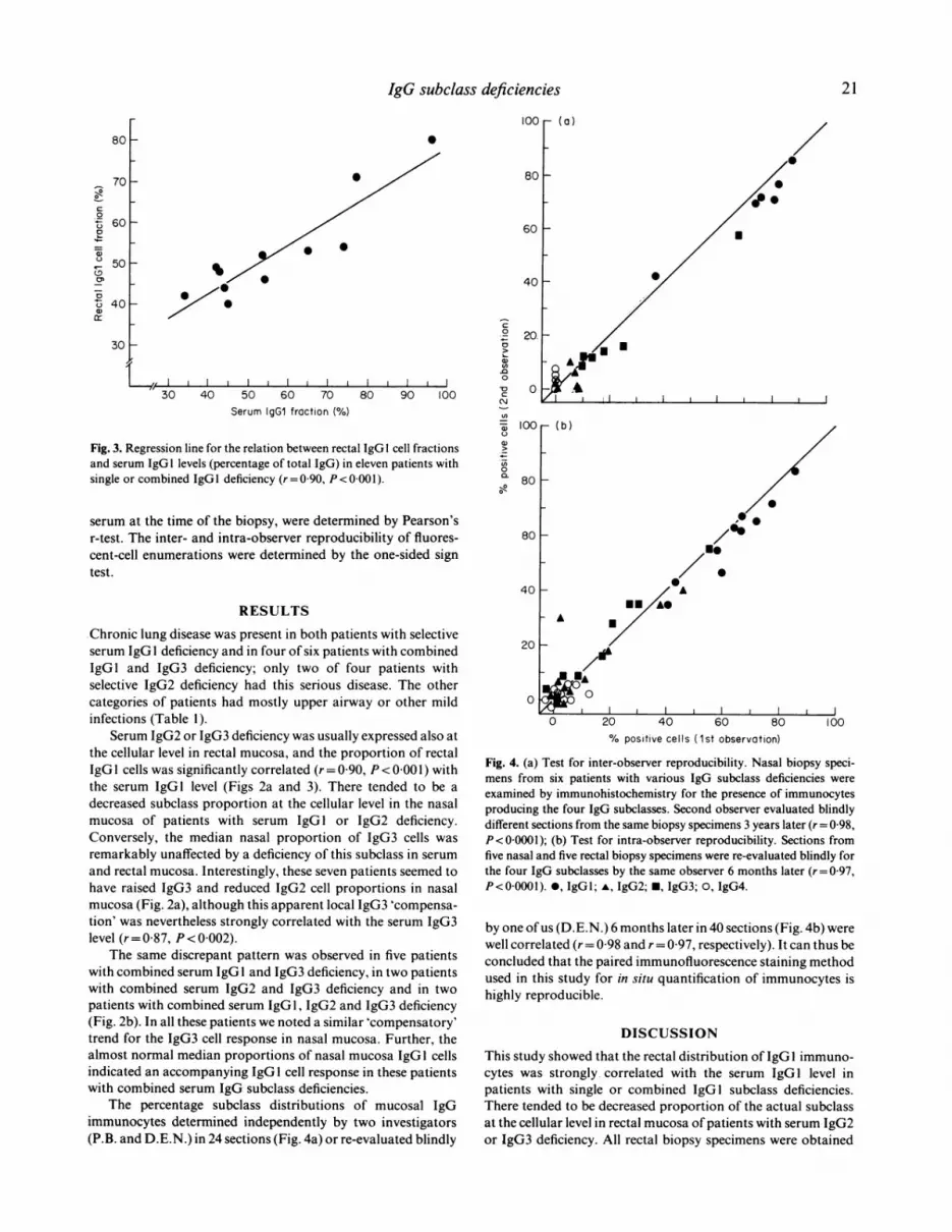
egression line for the relation between rectal IgG 1 cell fractionsm IgG I levels (percentage of total IgG) in eleven patients withcombined IgGI deficiency (r=0 90, P<0 001).
serum at the time of the biopsy, were determined by Pearson'sr-test. The inter- and intra-observer reproducibility of fluores-cent-cell enumerations were determined by the one-sided signtest.
20.
0
.
100r (b)
80
0
0
80
40 [RESULTS
Chronic lung disease was present in both patients with selectiveserum IgGI deficiency and in four of six patients with combinedIgGi and IgG3 deficiency; only two of four patients withselective IgG2 deficiency had this serious disease. The othercategories of patients had mostly upper airway or other mildinfections (Table 1).
Serum IgG2 or IgG3 deficiency was usually expressed also atthe cellular level in rectal mucosa, and the proportion of rectalIgGI cells was significantly correlated (r= 0 90, P < 0-001 ) withthe serum IgGl level (Figs 2a and 3). There tended to be a
decreased subclass proportion at the cellular level in the nasalmucosa of patients with serum IgG1 or IgG2 deficiency.Conversely, the median nasal proportion of IgG3 cells was
remarkably unaffected by a deficiency of this subclass in serumand rectal mucosa. Interestingly, these seven patients seemed tohave raised IgG3 and reduced IgG2 cell proportions in nasalmucosa (Fig. 2a), although this apparent local IgG3 'compensa-tion' was nevertheless strongly correlated with the serum IgG3level (r= 0-87, P < 0002).
The same discrepant pattern was observed in five patientswith combined serum IgG 1 and IgG3 deficiency, in two patientswith combined serum IgG2 and IgG3 deficiency and in twopatients with combined serum IgG 1, IgG2 and IgG3 deficiency(Fig. 2b). In all these patients we noted a similar 'compensatory'trend for the IgG3 cell response in nasal mucosa. Further, thealmost normal median proportions of nasal mucosa IgGI cellsindicated an accompanying IgG I cell response in these patientswith combined serum IgG subclass deficiencies.
The percentage subclass distributions of mucosal IgGimmunocytes determined independently by two investigators(P.B. and D.E.N.) in 24 sections (Fig. 4a) or re-evaluated blindly
20 F
0o
-A/ A
A /
/0
i ,-0 20 40 60 80
% positive cells (1st observation)100
Fig. 4. (a) Test for inter-observer reproducibility. Nasal biopsy speci-mens from six patients with various IgG subclass deficiencies were
examined by immunohistochemistry for the presence of immunocytesproducing the four IgG subclasses. Second observer evaluated blindlydifferent sections from the same biopsy specimens 3 years later (r = 0 98,P< 00001); (b) Test for intra-observer reproducibility. Sections fromfive nasal and five rectal biopsy specimens were re-evaluated blindly forthe four IgG subclasses by the same observer 6 months later (r= 0 97,P<000001). 0, IgGl; A, IgG2; *, IgG3; 0, IgG4.
by one of us (D.E.N.) 6 months later in 40 sections (Fig. 4b) werewell correlated (r= 0-98 and r= 0 97, respectively). It can thus beconcluded that the paired immunofluorescence staining methodused in this study for in situ quantification of immunocytes ishighly reproducible.
DISCUSSION
This study showed that the rectal distribution of IgGI immuno-cytes was strongly correlated with the serum IgGI level inpatients with single or combined IgGl subclass deficiencies.There tended to be decreased proportion of the actual subclassat the cellular level in rectal mucosa of patients with serum IgG2or IgG3 deficiency. All rectal biopsy specimens were obtained
21
E~~~~~~~
D. E. Nilssen et al.
from patients with a histologically normal mucosa. Also, theclinical features of these patients indicated preservation ofintestinal immune defence.
Conversely, a remarkable lack of relation was noted betweenthe serum IgG subclass levels and the median proportions ofnasal mucosal IgG3 (and sometimes also IgGl) subclass-producing immunocytes. This finding was in agreement with apreliminary study (Brandtzaeg et al., 1986) of a few of theimmunodeficient patients included in the present investigation.Thus, a reduced serum IgG3 level was not reflected by adecreased nasal IgG3 cell response, although the mucosal IgG3'compensation' in the upper respiratory tract was neverthelessstrongly correlated with the serum IgG3 level. A similar'compensatory' trend was noted for both IgG3 and IgGI innasal mucosa of patients with a combined serum IgG I and IgG3deficiency, and particularly for IgG3 in those with combinedIgG2 and IgG3 or IgGl, IgG2 and IgG3 deficiency. It wasnoteworthy that IgG3-deficient patients, despite a normalserum IgG2-subclass level often showed a strikingly reducedproportion of IgG2 immunocytes in their nasal mucosa.However, this might well be an arithmetic reflection of theirincreased IgG3 cell proportion.
In contrast to rectal mucosa, histological evaluation of nasalmucosa often showed slight to moderate chronic inflammationin agreement with the predominant clinical finding of rhino-pharyngitis. We did not attempt to enumerate the nasal IgG-producing cells in terms ofimmunocyte density, because of theirheterogeneous distribution (Brandtzaeg, 1984). The determina-tion of subclass proportions was performed in areas withabundant glandular elements and in the superficial stroma; thelatter compartment often contained numerous lymphoid cells asa reflection of chronic inflammation. It seemed justified tocombine our counts obtained from both locations in themucosa, however, because previously we have been unable toshow any difference in the subclass distribution between surface-and gland-associated IgG-producing nasal immunocytes(Brandtzaeg et al., 1987).
The small amounts ofIgG that normally reach the gut lumenby passive diffusion are rapidly degraded (Haneberg & Endre-sen, 1976). IgG is therefore probably of little importance forintestinal surface defence; this may partly explain the preserva-tion of a healthy gut mucosa in our IgG subclass-deficientpatients. This conclusion is in agreement with previous reportson patients with more generalized B cell deficiency (hypo-gammaglobulinaemia), who showed only minor clinical, func-tional or structural abnormalities in the gut (Eidelman, 1976;Nilssen et al., 1989).
IgG probably plays a more important role at mucosalsurfaces in the upper respiratory tract. At least 90% of IgG innasal fluid has been shown to be of serum origin (Mygind,Weeke & Ullman, 1975). In animal experiments, serum andexocrine IgG molecules have shown identical complement-dependent bactericidal and opsonizing activities (Eddie, Schul-kind & Robbins, 1971), and IgG was found to afford immunolo-gic exclusion of soluble antigens in the upper respiratory tract(Stokes, Soothill & Turner, 1975): Mucosal 'leakage' of intersti-tial IgG is enhanced by inflammatory processes (Brandtzaeg,Fjellanger & Gjeruldsen, 1970; Rossen, Kasel & Couch, 1971),and the classical experiments by Fazekas de St. Groth, Donnel-ley & Graham (1951) on 'pathotopic potentiation' of localimmunity suggested that serum-derived IgG may have an
important protective function in the respiratory tract. Indeed,IgG seems to be the major antibody class operating in the lowerrespiratory tract (Newhouse, Sanchis & Bienenstock, 1976). It isnot surprising, therefore, that lack of IgG or one or more of itssubclasses has clinical consequences, mainly in the respiratorytract.
The respiratory mucosa is constantly exposed to a heavybombardment of antigens and mitogens. The nature of theantigen plays an important part in the IgG subclass expression;thus IgGl and IgG3 antibodies are elicited in T cell-dependentresponses by protein antigens like viruses or tetanus toxoid(Papadea & Check, 1989). In contrast, IgG2 antibodies areproduced in response to many bacterial antigens includingcarbohydrates (Shakib & Stanworth, 1980). The IgG subclassesshow considerable biological differences with IgG 1 and IgG3being better complement-activating antibodies and more opso-nic than IgG2 and IgG4 (Unkeless, Fleit & Mellman, 1981). Thediscrepancies between the expression of IgG3 subclass defi-ciency at the two mucosal sites observed in our study may reflectdifferent antigenic and mitogenic loads, for example persistentprotein exposure (virus?) of the nasal mucosa which couldlocally override a B cell maturation defect. Such stimulatorydifferences would probably also be operating normally andexplain the fact that there often is a preference of IgG3 overIgG2 cells in nasal mucosa of subjects with an intact immunesystem (Brandtzaeg et al., 1986). It remains to be examinedwhether the apparent 'compensatory' production of IgG3 in theupper respiratory tract of patients with serum deficiency of thissubclass might be of any protective significance along with IgG Iand secretory IgA antibodies.
More severe chronic bronchitis, and even chronic lungdisease with bronchial asthma or bronchiectasis, occurred in thegroup with selective IgGI deficiency, and especially when it wascombined with IgG3 deficiency, as described earlier (Oxelius etal., 1986; Bj0rkander et al., 1986). The absence of IgGI is oftenassociated with the same clinical symptoms as in generalizedhypogammaglobulinaemia as IgGl makes up the largest pro-portion (60-70%) of total IgG (Papadea & Check, 1989). Thisfact probably explains why these patients often have a history oflung disease and increased susceptibility to pyogenic infections(Schur et al., 1970).
The genetic regulation of IgG subclass expression includesnumerous rearrangements and recombinatorial events generat-ing antibody diversity; this complexity allows for error (Papa-dea & Check, 1989). IgG subclass deficiencies may occur as aresult of defects in the constant heavy chain (CH) genes or in theregulation of their switching on chromosome 14. The gene orderon the second (CH,3, CH.;I, CH,,I) and third (C,.2, C;4, C.2) segment(Flanagan & Rabbits, 1982; Conley, Brown & Bartelt, 1987)apparently influences the expression of IgG subclass deficien-cies. This may explain the tendency for combined deficiencies toinclude mainly IgGl and IgG3 or IgG2 and IgG4 (Hammar-str6m et al., 1984a). B cells switching downstream for one CHgene to another usually deletes the intervening upstream DNAsequences, thus preventing backward switches (Hammarstr6met al., 1984b). Predominating vectorial switching on the first twogene segments in the sequence CHP*CH6-+CHY3--CHyI-+CH21during local immune responses in the upper respiratory tract hasbeen suggested previously by our laboratory; such a mechanismwould be in agreement with the normal predominance of IgA 1,IgGl and IgG3 immunocytes in nasal mucosa, in that order
22

IgG subclass deficiencies 23
(Brandtzaeg et al., 1986), and a local expansion of IgD alongwith IgG immunocytes at this site in many patients with IgAdeficiency (Brandtzaeg et al., 1979; 1987).
Our main conclusion is that IgG subclass deficiencies, asrevealed in serum, were usually expressed also at the cellularlevel in rectal mucosa. It was remarkable that in patients withlow levels of serum IgG3, the proportion of this subclass locallyproduced in nasal mucosa seemed to be little affected. Thisapparent local IgG3 compensation was nevertheless positivelycorrelated with the serum IgG3 subclass level. IgG3 and IgGlantibodies may be ofprotective significance on the surface of therespiratory mucosa. However, because of their phlogisticproperties these subclasses may also be involved in mucosalimmunopathology. These immunological aspects are obviouslyonly part of a complex interplay normally taking place in therespiratory tract. Little is known of beneficial or detrimentaleffects exerted by T cells, macrophages, mast cells, goblet cellsand other more poorly defined effector cells present in themucosa; further studies of patients with various types ofimmunodeficiency may contribute to a better understanding ofthese effects.
ACKNOWLEDGMENTSThis study was supported by grants from the Norwegian ResearchCouncil for Science and the Humanities, the Norwegian Cancer Society,Dr A. Malthes Legacy for Internal Medicine, Sverre S. S0rensensFoundation for Rheumatological Research, Oslo, Norway, the SwedishMedical Research Council (no. 215), The Hesselman Foundation,Bohuslandstinget and Kabi Vitrum, Stockholm, Sweden. We thank thetechnical staff at LIIPAT, Mr E. Jenssen and Dr G. Hansson forvaluable assistance.
REFERENCES
BJERKE, K. & BRANDTZAEG, P. (1990) Terminally differentiated humanintestinal B cells. IgA and IgG subclass-producing immunocytes indistal ileum, including Peyer's patches, compared with lymph nodesand palatine tonsils. Scand. J. Immunol. 32, 61.
BJORKANDER, J., BENGTSSON, U., OXELIUS, V.-A. & HANSON, L.A. (1986)Symptoms in patients with lowered levels of IgG subclasses, with or
without IgA deficiency, and effects of immunoglobulin prophylaxis.Mongr. Allergy, 20, 157.
BRANDTZAEG, P. (1974) Mucosal and glandular distribution ofimmuno-globulin components. Immunohistochemistry with a cold ethanol-fixation technique. Immunology, 26, 1 101.
BRANDTZAEG, P. (1984) Immune function of human nasal mucosa andtonsils in health and disease. In Immunology of the Lung and UpperRespiratory Tract (ed. by J. Bienenstock) p. 28, McGraw-Hill, NewYork.
BRANDTZAEG, P. (1985) Cells producing immunoglobulins and otherimmune factors in the human nasal mucosa. Protides biol. Fluids, 32,363.
BRANDTZAEG, P., FJELLANGER, I. & GJERULDSEN, S.T. (1970) Humansecretory immunoglobulins. I Salivary secretions from individualswith normal or low levels of serum immunoglobulins. Scand JHaematol, Suppl. 12, 1.
BRANDTZAEG, P., GJERULDSEN, S.T., KORSRUD, F., BAKLIEN, K.,BERDAL, P. & EK, J. (1979) The human secretory immune systemshows striking heterogeneity with regard to involvement of J-chainpositive IgD immunocytes. J. Immun. 122, 503.
BRANDTZAEG, P., KARLSSON, G., HANSSON, G., PETRUSON, B., BJOR-KANDER, J. & HANSON, L.A. (1987) The clinical condition of IgA-deficient patients is related to the proportion of IgD- and IgM-producing cells in their nasal mucosa. Clin. exp. Immunol. 67, 626.
BRANDTZAEG, P., KETT, K., ROGNUM, T.O., SODERSTROM, R., BJOR-KANDER, J., S6DERSTROM, T., PETRUSSON, B. & HANSON, L.A. (1986)Distribution of mucosal IgA and IgG subclass-producing immuno-cytes and alterations in various disorders. Mongr. Allergy, 20, 179.
BRANDTZAEG, P., VALNES, K., SCOTT, H., ROGNUM, T.O., BJERKE, K. &BAKLIEN, K. (1985) The human gastrointestinal secretory immunesystem in health and disease. Scand. J. Gastroenterol. 20 (Suppl. 1 14),17.
CONLEY, M.E., BROWN, P. & BARTELT, M.S. (1987) IgG subclasspotential of surface IgM-negative and surface IgM-positive humanperipheral blood B cells. Clin. Immunol. Immunopathol. 43, 21 1.
CRABBt, P.A., CARBONARA, A.G. & HEREMANS, J.F. (1965) The normalhuman intestinal mucosa as a major source ofplasma cells containingyA-immunoglobulin. Lab. Invest. 20, 235.
EDDIE, D.S., SCHULKIND, M.L. & ROBBINS, J.B. (1971) The isolation andbiologic activities of purified secretory IgA and IgG anti-Salmonellatyphimurium "O'' antibodies from rabbit intestinal fluid and colos-trum. J. Immunol. 106, 181.
EIDELMAN, S. (1976) Intestinal lesions in immune deficiency. Hum.Pathol. 7, 427.
FAZEKAS DE ST. GROTH, S., DONNELLEY, M. & GRAHAM, D.M. (1951)Studies in experimental immunology of influenza. VIII. Pathotopicadjuvants. Aust. J. exp. Biol. med. Sci. 29, 323.
FLANAGAN, J.G. & RABBITS, T.H. (1982) Arrangement of humanimmunoglobulin heavy chain constant region genes implies evolu-tionary duplication of a segment containing y, £ and a genes. Nature,300, 709.
HAMMARSTROM, L., GRANSTROM, M., OXELIUS, V.-A., PERSSON, A.A. &SMITH, C.I.E. (1984a) IgG subclass distribution of antibodies againstS. aureus teichoic acid and a-toxin in normal and immunodeficientdonors. Clin. exp. Immunol. 55, 593.
HAMMARSTROM, L., MELLSTEDT, H., PERSSON, M.A.A., SMITH, C.I.E. &AHRE, A. (1984b) IgA subclass distribution in paraproteinemias:suggestion of an IgG-IgA subclass switch pattern. Acta pathol.microbial. immunol. Scand. [Sect. C], 92, 207.
HANEBERG, B. & ENDRESEN, C. (1976) Fragments ofimmunoglobulins inhuman faeces. Acta pathol. microbial. Scand. 84, 31.
HELGELAND, L., TYSK, C., KETT, K., ANDERSEN, S.N. & BRANDTZAEG, P.(1991) Evaluation of genetic impact on the mucosal IgG-subclassresponse in inflammatory bowel disease. In Proceedings of the 6thInternational Congress of Mucosal Immunology (In press).
JEFFERIS, R., REIMER, C.B., SKVARIL, F., DE LANGE, G., LING, N.R.,LOWE, J., WALKER, M.R., PHILLIPS, D.J., ALOISIO, C.H., WELLS,T.W., VAERMAN, J.P., MAGNUSON, C.G., KUBAGAWA, H., COOPER,M., VARTDAL, F., VANDVIK, B., HAAIJMAN, J.J., MAKELA, O.,SARNESTO, A., LANDO, Z., GERGELY, J., RAJNAVOLGYI, E., LASZLO, G.,RADL, J. & MOLINARO, G.A. (1985) Evaluation of monoclonalantibodies having specificity for human IgG subclasses: Results of anIUIS/WHO collaborative study. Immunol. Lett. 10, 223.
KETT, K., ROGNUM, T.O. & BRANDTZAEG, P. (1987) Mucosal subclassdistribution of immunoglobulin G-producing cells is different inulcerative colitis and Crohn's disease of the colon. Gastroenterology,93, 919.
MANCINI, G., CARBONARA, A.0. & HEREMANS, J.F. (1965) Immunoche-mical quantitation of antigens by single radial immunodiffusion.Immunohistochemistrv, 2, 235.
MYGIND, N., WEEKE, B. & ULLMAN, S. (1975) Quantitative determina-tion of immunoglobulin in nasal secretions. Int. Arch. Allergy appl.Immunol. 49, 99.
NEWHOUSE, M., SANCHIS, J. & BIENENSTOCK, J. (1976) Lung defensemechanism. N. Engl. J. Med. 295, 1045.
NILSSEN, D.E., H6VERSTAD, T., FRdLAND, S.S. & MIDTVEDT, T. (1989)Short-chain fatty acids and other intestinal microflora-associatedcharacteristics in feces of patients with severe B-cell immunodefi-ciency. Scand. J. Gastroenterol. 24, 21.
OXELIUS, V.-A. (1979) IgG subclass levels in infancy and childhood.Acta paediatr. Scand. 68, 23.

24 D. E. Nilssen et al.
OXELIUS, V.-A., HANSON, L.A., BJ0RKANDER, J., HAMMARSTROM, L. &SJOHOLM, k (1986) IgG3 deficiency: common in obstructive lungdisease. Monogr. Allergy, 20, 106.
PAPADEA, C. & CHECK, I.J., (1989) Human immunoglobulin G andimmunoglobulin G subclasses; Biochemical, genetic, and clinicalaspects. Crit. Rev. c/in. Lab: Sci. 27, 27.
ROGNUM, T.O., KETT, K., FAUSA, O., BENGTSSON, U., KILANDER, A.F.,SCOTT, H., GAARDER, P.I. & BRANDTZAEG, P. (1989) Raised numberof jejunal IgG2-producing cells in untreated adult coeliac diseasecompared with food allergy. Gut, 30, 1574.
ROSSEN, R.D., KASEL, J.A. & COUCH, R.B. (1971) The secretory immunesystem: its relation to respiratory viral infection. Prog. Virol. 13, 194.
SCHUR, P.H., BOREL, H., GELFAND, E.W., ALPER, C.A. & ROSEN, F.S.(1970) Selective gamma-G globulin deficiencies in patients withrecurrent pyogenic infections. N. Engl. J. Med. 283, 631.
SCOTT, M.G., NAHM, M.H., MACKE, K., NASH, G.S., BERTOVICH, M.J.& MACDERMOTT, R.P. (1986) Spontaneous secretion of IgG sub-classes by intestinal mononuclear cells: differences between ulcerativecolitis, Crohn's disease, and controls. Clin. exp. Immunol. 66, 209.
SHAKIB, F. & STANWORTH, D.R. (1980) Human IgG subclasses in healthand disease. Ricerca clin. Lab. 10, 463.
SODERSTROM, T., SODERSTR6M, R. & HANSON, L.A. (1987) Immuno-globulin G subclasses in immunodeficiency. Ann. clin. Res. 19, 280.
SODERSTROM, T., S6DERSTROM, R., BENGTSSON, U., BJORKANDER, J.,HELLSTRAND, K., HOLM, J. & HANSON, L.A. (1986) Clinical andimmunological evaluation of patients low in single or multiple IgGsubclasses, in immunoglobulin subclass deficiencies. Monogr. Allergy,20, 135.
STOKES, C.R., SOOTHILL, J.F. & TURNER, M.W. (1975) Immuneexclusion is a function of IgA. Nature, 255, 745.
UNKELESS, J.C., FLEIT, H. & MELLMAN, J.S. (1981) Structural aspectsand heterogeneity of immunoglobulin Fc-receptors. Adt'. Immunol.31, 247.
VALNES, K. & BRANDTZAEG, P. (1989) Subclass distribution of mucosalIgG-producing cells in gastritis. Gut, 30, 322.


![ISOTYPE und Weltsprachen / ISOTYPE and the Universal Languages [Deutsch]](https://img.pdfslide.net/doc/110x75/635a57159d85dc43cb06ede5/isotype-und-weltsprachen-isotype-and-the-universal-languages-deutsch.jpg)


