Embed Size (px)
Citation preview
JIVS The Chamber of Veterinary e-ISSN: 2602-3490
Surgeons, Istanbul Abbr. Title: J Ist Vet Sci
Journal of Istanbul Veterinary Sciences
(JIVS)
Journal home page: www.jivs.net
http://dergipark.gov.tr/http-www-jivs-net
Since 2017
Editor-in-Chief Prof. Dr. Erdal MATUR
Istanbul University, Faculty of Veterinary Medicine, Department of Physiology, Turkey
Editor Prof. Dr. Mukaddes ÖZCAN
Istanbul University, Faculty of Veterinary Medicine, Department of Physiology, Turkey
Editor Prof. Dr. Alper YILMAZ
Istanbul University, Faculty of Veterinary Medicine, Department of Animal Breeding & Husbandry, Turkey
Statistical Editor Assoc. Prof. Dr. Bilge ACAR BOLAT
Istanbul University, School of Business Administration,
Department of Quantitative Methods. Turkey
Language Editor Pelin Burcu Daştan
Journal of Istanbul Veterınary Scıences
Journal homepage: www.jivs.net http://dergipark.gov.tr/http-www-jivs-net
This work is licensed under the Creative Commons Attribution 4.0 International License.
Volume 4; Issue 3, April 2020 E-ISSN: 2602-3490
Editorial Board
Ahmet Gülçubuk Istanbul University, Turkey
Ali Belge Adnan Menderes University, Turkey
Ayşen Gargılı Marmara University, Turkey
Brain Nielsen Michigan State University, USA
Ebru Yalçın Uludağ University, Turkey
Kemal Özdem Öztabak Istanbul University, Turkey
Kutlay Gürbulak Erciyes University, Turkey
Laman Mamedova Kansas State University, USA
Maria Tsantarliotou Aristotle University, Greece
Mehmet Ali Bal Kahramanmaraş Sütçü İmam University, Turkey
Murat Yıldırım Istanbul University, Turkey
Oytun Okan Şenel Ankara University, Turkey
Ömer Akıneden Justus-Liebig Univ. Gießen, Germany
Rıfat Mutuş Gelişim University, Turkey
Serhat Pabuçcuoğlu Istanbul University, Turkey
Şahin Arslan Kafkas University, Turkey
Urs Giger Pennsylvania University, USA
Ümit Cirit Dicle University, Turkey
Yücel Meral Ondokuz Mayıs University, Turkey
Zdenek Knotek University of Veterinary and Pharmaceutical Sciences Brno, Czech Republic
Indexes and Platforms
Turkey citation index, Google scholar,
ResearchBib, Bielefeld Academic Search
Engine, Cosmos IF, İnternational Institute
of organized research(I2OR), Scientific
İndexing service, CrossRef, Eurasian
Scientific Journal Index, Open AIRE,
General Impact factor, Directory of
Research Journals Indexing (DRJI), ASOS
index.
Contents
Journal of Istanbul Veterınary Scıences
Journal homepage: www.jivs.net http://dergipark.gov.tr/http-www-jivs-net
This work is licensed under the Creative Commons Attribution 4.0 International License.
Pages
Deniz KARAKCI, Nilay SEYİDOĞLU, Oğuz MERHAN, Kadir BOZUKLUHAN The preventive role of different doses Spirulina platensis on lipid peroxidation and antioxidant status in healthy rats
90-95
Aikerim KUMONDOROVA, Kemal METİNER Protective immune studies against fungi 96-101
İl̇han İRİM, Aykut Asım AKBAŞ, Mustafa SAATCI Determination of fertility traits of sheep and growth characteristics of Chios crossbred male lambs reared under local breeder conditions
102-108
Hasan ATALAY, Fatih KAHRIMAN Estimation of relative feed value, relative forage quality and net energy lactation values of some roughage samples by using near infrared reflectance spectroscopy
209-118
Mohammad ESLAMPANAH, Mohammad ABDİGOUDARZİ, Mohammad Hasan HABLOLVARİD
Identification of acinar cells of salivary gland in blood fed female ticks (Hyalomma anatolicusm anatolicum) by light microscopy
119-124
Hasan ATALAY, Fatih KAHRIMAN, Firat ALATÜRK
Estimation of dry matter, crude protein and starch values in mixed feeds by near-infrared reflectance (NIR) spectroscopy
125-130
Denisa PÉREZ GAUDİO, Flavio PÉREZ, Gustavo BRETSCHNEİDER
First report of a perforated abomasal ulcer in a beef heifer calf in Argentina 131-135
Olushola ADELEYE, Emmanuel OKOH, Adenike ADELEYE, Fakilahyel Musa MSHELBWALA, Abiodun ADETOMİWA, James APANTAKU, Ngozichukwu ABOAJAH, Ladoke DUROTOYE, Johnny Olufemi OLUKUNLE
Ameliorative effects of Allium cepa Linn. scaly leaves extract on reproductive dysfunctions in streptozotocin-induced diabetic Wistar rats
136-144
Mahmut KESKİN, Kenan SEZER
Investigation of the prevalence of Babesia canis in dogs in the center of Antalya province
145-149
90
Research Article
Volume: 4, Issue: 3 December 2020 Pages: 90-95
The preventive role of different doses Spirulina platensis on lipid peroxidation and antioxidant status in healthy rats
Journal of Istanbul Veterınary Scıences
Deniz Karakci1, *Nilay Seyidoglu2, Oguz Merhan3, Kadir Bozukluhan4
1.Department of Biochemistry, Faculty of Veterinary Medicine, Tekirdag Namik Kemal University, Tekirdag, Turkey. 2.Department of Physiology, Faculty of Veterinary Medicine, Tekirdag Namik Kemal University, Tekirdag, Turkey. 3.Department of Biochemistry, Faculty of Veterinary Medicine, Kafkas University, Kars, Turkey. 4.Kars School of Higher Vocational Education, Kafkas University, Kars, Turkey. Karakci, D. ORCID: 0000-0002-1884-1874, Seyidoğlu N. ORCID: 0000-0002-2817-5131, Merhan O. ORCID: 0000-0002-3399-0667, Bozukluhan K. ORCID: 0000-0003-4929-5156
ABSTRACT
There are several antioxidant supplements using for reproductivity and life quality, especially herbal ones. Nowadays, herbal antioxidants especially Spirulina platensis has been still interested due to protective role on oxidant antioxidant balance and health. The present study, we aimed to evaluate the effects of different doses of S. platensis on important oxidant molecule MDA (TBA, oxidant malondialdehyde), and individual antioxidants as GPx (glutathione peroxidase), CAT (catalase) and SOD (superoxide dismutase) in healthy rats. For this purpose, we used thirty Wistar Albino male rats in three groups: Control, Low Dose Spirulina (500 mg/kg) and High Dose Spirulina (1000 mg/kg). S. platensis additives were given by oral gavage daily under a long forty five day of trial. At the end of the study, interestingly, all the antioxidants GPx, CAT, SOD and the oxidant MDA lipid peroxidation values were decreased in group high dose Spirulina compared to Control (p < 0.05). In spite of these decreases, testis weights and indexes were increased in group high dose Spirulina compared to Control significantly. The testis weights and indexes were evaluated for normal health of animals. It can be considered that due to the excessive protein and antioxidants features of S. platensis, oxidant and antioxidant mechanisms may be changed. However it can be said that Spirulina can compensate the homeostasis and health of animals. It is also suggested that the applications and different doses of S. platensis are needed to be assayed for further studies .
Keywords: antioxidant Spirulina platensis, testis index, rat.
DOI: https://doi.org/10.30704/http-www-jivs-net.793250
To cite this article: Karakci, D., Seyidoglu, N., Merhan, O., Bozukluhan, K. (2020). The preventive role of different doses Spirulina platensis on lipid peroxidation and antioxidant status in healthy rats. Journal of Istanbul Veterinary Sciences, 4(3), 90-95. Abbreviated Title: J Ist Vet Sci
Free radicals are important reactive molecules which designate for oxidative stress imbalance between oxidative and antioxidative status. Although the oxidant molecules have a role on cellular damage with radical oxygen species, the antioxidant molecules suppress and scavenge free radicals. The most important oxidative molecule known as
malondialdehyde (MDA) damages cells or tissues in stressful situations such as diseases, over nutrition or high protein, and thereby oxidative-antioxidative balance reduces. Also, all biological molecules in cells such as proteins, lipids, DNA or RNA can be damage during oxidative status. Especially, if protein gets oxidation, several functional changes can be existed in
*Corresponding Author: Nilay Seyidoğlu E-mail: [email protected]
Journal home page: www.jivs.net http://dergipark.gov.tr/http-www-jivs-net
Article History
Received: 10.09.2020 Accepted: 09.10.2020 Available online: 12.10.2020
This work is licensed under the Creative Commons Attribution 4.0 International License.
Introduction
91
organism for example inactivation of DNA, decompose the protein peroxides and damage molecules (Halliwell and Whiteman, 2004). Nevertheless, antioxidants have a protective role on cellular mechanism against to oxidative damage (Misra and Niyogi, 2009; Firat et al., 2011). Glutathione (GSH), glutathione peroxidase (GPx), catalase (CAT) and superoxide dismutase (SOD) are the best-known antioxidant markers called as enzymatic antioxidants. These enzymatic molecules are capable of either removing or scavenging free radicals and their actions. Spirulina platensis is the most popular antioxidant herbal due to its rich features and protection efficiency against to diseases. This natural antioxidant includes 60-70 % proteins, 4-7 % essential fatty acids (a-linoleic acid), 20% carbohydrates, 6-7% minerals, pigments (phycocyanin, b-carotene) and some special vitamins (Goksan and Kilic, 2009; Yang and Zhang, 2009; Yusuf et al., 2016). The antioxidant, anti-inflammatory, antimicrobial activities of S. platensis were reported by several researchers (Kitada et al., 2009; Mahdi et al., 2019; Seyidoglu et al., 2019). S. platensis contains special antioxidant molecules such as carotenoids, phycocyanin, xanthophylls, and phycobilins which have a key role on cardiotoxicity, hepatotoxicity, carcioneogenesis, tumor destruction and cancer (Mohan et al., 2006; Karkos et al., 2011; Ibañez et al., 2012; Abdel-Daim et al., 2013; Wu et al., 2016). All these researchers reported that especially phycocyanin, an extract of Spirulina, could protect the body against to oxidative stress. Martin et al. (2007) indicated that when the reactive oxygen species is occurred because of lipid peroxidation, antioxidant substances of Spirulina can prevent peroxidation and oxidative stress. Also, Mansour et al. (2006) explained that Spirulina may serve as an antioxidant by its oxygen quenching properties for free radicals due to its carotenoid content. Physiological changes in oxidative status have been correlated with differences of organ weights and functions due to production of higher reactive oxygen species, such as testis (Vernet et al., 2004; Aitken and Roman, 2008; Bashandy et al., 2016). It was reported that food utilization and protein catabolism increase in oxidative reaction, and thereby organ weights decrease (Aitken and Roman, 2008). Sarkar et al. (2003) observed the reduction in the testicular weight due to germ cell loss in rats. Testicular weight loss seems to be a feature of infertility belongs to oxidant-antioxidant imbalance. Also, it was reviewed that in spite of other organ weights, testis weights may provide a sensitive alert for studies. Especially in immature animals, testicular weights measurement is accepted to interpretation the closely linking with
body weight, testicular size and testis index (Greaves, 2007). Also, increase of the testis weights and testicular index are accepted as the normal growth of the animals. As the use of natural antioxidants increases, more studies are necessary to evaluate the protective effects for health. In this study, it’s aimed to investigate the oxidant and antioxidant efficiency of S. platensis on health and to identify the effects on testicular weights in healthy rats which fed by low and high doses of S. platensis under a long period of forty five day trial.
Materials and Methods Animal housing and diets: The experimental protocols were in accordance with the National Institute of Health Guide for the Care and Use of Laboratory Animals and approved by the Animal Care and Use Committee of University. The study was carried out with the permission of University Animal Experimentation Local Ethics Committee (Approval no: 2017/04-4). Thirty, adult Wistar albino, male rats aged 7-8 weeks old and average weight 180-200 g were used in this study. Rats were housed under standard laboratory conditions (Lights: 12-hour light/dark/day, Humidity: 55% and Temperature: 24 ± 25 °C). The animals were housed in stainless steel cages and divided into three groups forty five day of trial. The groups were as follows: 1.Control (with basal diet); 2. Low dose Spirulina-LSp (with 500 mg/kg S. platensis); 3.High dose Spirulina-HSp (with 1000 mg/kg S.platensis). Rats were given ad libitum access to commercial rodent diet (Table1). S. platensis (Egert, Izmir-Turkey) was applied to rat by oral gavage daily was provided and modified according to the literatures (Nagaoka et al., 2005; Moreira et al., 2011). Measurements: Blood samples were collected by puncture of heart under short (2-3 minutes) isoflurane anesthesia at the end of the study. Laparotomy was done for conceiving the reproductive system. Both testes (without epididymis) and gonadal fats were
* The basal diet was formulated and projected to take on maintenance requirements according to the NRC, 1995.
Karakci et al., 2020/ Journal of Istanbul Veterinary Sciences. Volume 4, Issue 3, pp: 90-95
Table 1. Basal diet formulation*
Contents
Metabolize energy (ME-kj) Crude protein (%) Crude fat (%) Crude fiber (%) Crude ash (%)
2000-2500 23 3 7 8
92
removed and weighted with precision weighing. (Sartorius, BL210S) immediately. Blood specimens were centrifuged in the same days within 30 minutes at 2000 × g for 10 min at 4 °C and the plasma was stored at -80 °C until analyses day. Antioxidant enzymes in plasma SOD, GPx and CAT enzyme activities were determined with commercial kits by microplate reader. MDA levels were measured according to the colorimetric method by spectrophotometer (Epoch, Biotek, Vermont, USA) that reported by Yoshoiko et al. (1979). Antioxidant Parameters Kit Methods for SOD, GPx, CAT: Antioxidant enzymes activities SOD (Cat. No:706002), GPx (Cat. No:703102), and CAT (Cat. No:707002) in plasma were determined with commercial kits (Cayman Chemical Company, Michigan, USA) by microplate spectrophotometer (Epoch, Biotek, Vermont, USA). SOD Assay Kit uses tetrazolium salt for detection of superoxide radicals produced by xanthine oxidase and hypoxanthine. GPx Assay Kit measures GPx activity indirectly by two reactions with glutathione reductase (GR). Oxidized glutathione (GSSG), produced on reduction of hydroperoxide by GPx and is turned to reduced state by GR and NADPH. Catalase (CAT) assay kit utilizes the peroxidation function of CAT for determination enzyme activity. The method is based on the reaction of enzyme with methanol in the presence of H2O2 optimal concentration. Lipid Peroxidation Manual Method for MDA: MDA levels were measured for lipid peroxidation according to the colorimetric method that reported by Yoshoiko et al. (1979) with microplate spectrophometer. Thiobarbituric acid (TBA) method were evaluated using the spectrophotometer. The reaction of thiobarbituric acid (TBA) with MDA, one of the aldehyde products of lipid peroxidation (Hodges et al., 1999). 0.5 plasma was mixed with 2.5 ml of 20% trichloroacetic acid (TCA) in a 14 ml centrifuge tube. 1ml of 0.6 % TBA was added to the mixture and warmed for 30 min in a boiling water bath then done cooling procedure. Then it was mixed into a 4 ml of nbutyl-alcohol layer in a separation tube and MDA content in the plasma was determined from the absorbance at 532 nm by spectrophotometer. Thiobarbituric reactive substances (TBARS) in the plasma was determined from the absorbance at 532 nm by spectrophotometer. Statistical Assessment: Statistical analyses were performed with SPSS (Version 20.0). Data were tested for normality distribution and variance homogeneity assumptions. All the values were grouped, and the
means and standard errors were calculated. One-way ANOVA was applied to all parameters to examine the differences between groups. Differences were considered significant at p < 0.05. If the differences between groups was provided to be significant (p < 0.05), differences evaluated by Tukey’s test. On the other hand, in non-homogenous groups, differences between means were analyzed by Kruskal Wallis and following by Mann Whitney U test between groups one by one.
Results The MDA, SOD, CAT and GPx values of all groups were provided in Table 2. Although there was no statistical difference between groups LSp and Control, there was a significant decrease in MDA in group HSp compared to Control (p: 0.001; 8.05 ± 0.06 and 4.39 ± 0.04 μmol L-1 group Control and HSp, respectively). Besides that, the antioxidants parameters (SOD, CAT and GPx) were tended to increase in group LSp than Control (p > 0.05, Table 2). However, interestingly the statistical decreases were determined in group HSp compared to Control (p: 0.001; p: 0.001 and p: 0.001 SOD, CAT and GPx, respectively) as shown in Table 2.
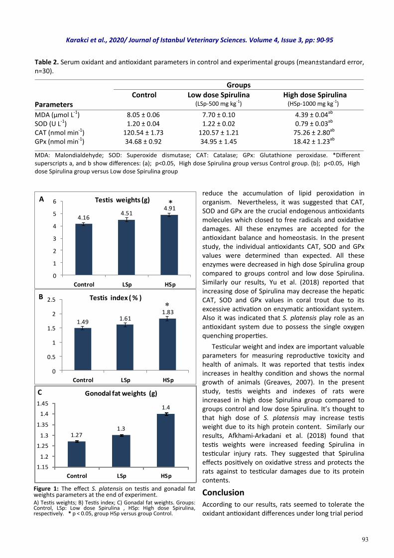
The testis and gonadal fats weights and testis indexes were shown in Figure 1. There were significant changes in testis weight in group HSp compared to Control (p: 0.017; 4.16 ± 0.16 and 4.91 ± 0.19 g, Control and HSp respectively) as figured in Figure 1a. Also, there was significant increase in testis index in group HSp than Control (Figure 1b; p: 0.008; 1.49 % and 1.83 % , Control and HSp respectively). However, no differences were observed about gonadal fat in all groups (Figure 1c; 1.27±0.08 , 1.30±0.10 and 1.40±0.13 Control, LSp and HSp respectively).
Discussion This study, we assessed the oxidant and antioxidant efficiency of S. platensis in healthy rats. We also provide evidence that forty five day of trial feeding with low and high doses of Spirulina observed a protective effect on testis weight and gonadal fat. Lipid peroxidation changes are linked to MDA and oxidative damage in cell, tissue and organs. Insight of the literatures, MDA levels increase due to free radicals activation resulting from fatty acids in tissue damage. Thereby, the antioxidant defense system cells are activated and the oxidant activities are reduced (Urso and Clarkson, 2003). In the present study, the MDA value in high dose Spirulina group was found lower compared to both control and low dose Spirulina groups. It’s thought that S. platensis may
Karakci et al., 2020/ Journal of Istanbul Veterinary Sciences. Volume 4, Issue 3, pp: 90-95
93
Figure 1: The effect S. platensis on testis and gonadal fat weights parameters at the end of experiment. A) Testis weights; B) Testis index; C) Gonadal fat weights. Groups: Control, LSp: Low dose Spirulina , HSp: High dose Spirulina, respectively. * p < 0.05, group HSp versus group Control.
reduce the accumulation of lipid peroxidation in organism. Nevertheless, it was suggested that CAT, SOD and GPx are the crucial endogenous antioxidants molecules which closed to free radicals and oxidative damages. All these enzymes are accepted for the antioxidant balance and homeostasis. In the present study, the individual antioxidants CAT, SOD and GPx values were determined than expected. All these enzymes were decreased in high dose Spirulina group compared to groups control and low dose Spirulina. Similarly our results, Yu et al. (2018) reported that increasing dose of Spirulina may decrease the hepatic CAT, SOD and GPx values in coral trout due to its excessive activation on enzymatic antioxidant system. Also it was indicated that S. platensis play role as an antioxidant system due to possess the single oxygen quenching properties.
Testicular weight and index are important valuable parameters for measuring reproductive toxicity and health of animals. It was reported that testis index increases in healthy condition and shows the normal growth of animals (Greaves, 2007). In the present study, testis weights and indexes of rats were increased in high dose Spirulina group compared to groups control and low dose Spirulina. It’s thought to that high dose of S. platensis may increase testis weight due to its high protein content. Similarly our results, Afkhami-Arkadani et al. (2018) found that testis weights were increased feeding Spirulina in testicular injury rats. They suggested that Spirulina effects positively on oxidative stress and protects the rats against to testicular damages due to its protein contents.
Conclusion According to our results, rats seemed to tolerate the oxidant antioxidant differences under long trial period
Table 2. Serum oxidant and antioxidant parameters in control and experimental groups (mean±standard error, n=30).
Groups Parameters
Control
Low dose Spirulina (LSp-500 mg kg-1)
High dose Spirulina (HSp-1000 mg kg-1)
MDA (μmol L-1) SOD (U L-1) CAT (nmol min-1) GPx (nmol min-1)
8.05 ± 0.06 1.20 ± 0.04
120.54 ± 1.73 34.68 ± 0.92
7.70 ± 0.10 1.22 ± 0.02
120.57 ± 1.21 34.95 ± 1.45
4.39 ± 0.04ab 0.79 ± 0.03ab
75.26 ± 2.80ab 18.42 ± 1.23ab
MDA: Malondialdehyde; SOD: Superoxide dismutase; CAT: Catalase; GPx: Glutathione peroxidase. *Different superscripts a, and b show differences: (a); p<0.05, High dose Spirulina group versus Control group. (b); p<0.05, High dose Spirulina group versus Low dose Spirulina group
4.164.51
4.91
0
1
2
3
4
5
6
Control LSp HSp
Testis weights (g) *A
1.491.61
1.83
0
0.5
1
1.5
2
2.5
Control LSp HSp
Testis index ( % )B *
1.271.3
1.4
1.15
1.2
1.25
1.3
1.35
1.4
1.45
Control LSp HSp
Gonodal fat weights (g)C
Karakci et al., 2020/ Journal of Istanbul Veterinary Sciences. Volume 4, Issue 3, pp: 90-95
94
and normal rearing conditions. This could be the ability of rats to compensate the S. platensis and endogenous antioxidants together on health and organ metabolism. In conclusion, insight of the literatures, within our results, total antioxidant status should be determined to evaluate the individual antioxidant better. However, it’s thought that S. platensis may be one of the most important herbal food and represents an antioxidant supplement for homeostasis. It’s also thought that the doses must be determined due to antioxidant properties of Spirulina. However, more studies are necessary to clarify the efficiency of testicular health and antioxidant correlation, and for future works.
References Abdel-Daim, M. M., Abuzead, S. M. & Halawa S. M.
(2013). Protective role of Spirulina platensis against acute deltamethrin-induced toxicity in rats. Plos One, 8(9), e72991.
Afkhami-Arkadani, M., Hasanzadeh, S., Shahrooz, R., Delirezh, N. & Malekinejad, H. (2018). Antioxidant effects of Spirulina platensis (Arthrospira platensis) on cyclophosphamide-induced testicular injury in rats. Veterinary Research Forum, 9, 35-41.
Aitken, R. J. & Roman, S. D. (2008). Antioxidant systems and oxidative stress in the testes. Oxidative Medicine and Cellular Longevity, 1, 15-24.
Bashandy, S. A. E., El Awdan, S. A., Ebaid, H. & Alhazza, I. M. (2016). Antioxidant potential of Spirulina platensis mitigates oxidative stress and reprotoxicity induced by sodium arsenite in male rats. Oxidative Medicine and Cellular Longevity, 27174351.
Devasagayam, T., Tilak, J., Boloor, K., Sane, K. S., Ghaskadbi, S. S. & Lele, R. (2004). Free radicals and antioxidants in human health: current status and future prospects. Journal of the Association of Physicians of India, 52, 4-10.
Firat, O., Cogun, H., Yuzereroglu, T., Gok, G., Kargin, F. & Kotemen, Y. (2011). A comparative study on the effects of a pesticide (cypermethrin) and two metals (copper, lead) to serum biochemistry of Nile tilapia, Oreochromis niloticus. Fish Physiology and Biochemistry, 37, 657-666.
Goksan, T. & Kilic, C. (2009). Growth and biochemical composition of Spirulina platensis Geitler in summer period under the conditions of Çanakkale Turkey. Asian Journal of Chemistry, 21, 4947-4950.
Greaves, P. (2007). Male genital tract. In: histopathology of preclinical toxicity studies. 3rd ed. (661-716). US: Elsevier Science Press.
Halliwell, B. & Whiteman, M. (2004). Measuring reactive species and oxidative damage in vivo and in cell culture: how should you do it and what do the results mean? British Journal of Pharmacology, 142(2), 231–255.
Hodges, D. M., DeLong, J. M., Forney, C. F. & Prange, R. K. (1999). Improving the thiobarbituric acid-reactive-substances assay for estimating lipid peroxidation in plant tissues containing anthocyanin and other interfering compounds. Planta, 207, 604-611.
Ibañez, E., Herrero, M., Mendiola, J. A. & Castro-Puyana, M. (2012). Extraction and characterization of bioactive compounds with health benefits from marine resources: macro and micro algae, cyanobacteria, and invertebrates. In Marine Bioactive Compounds. (pp. 55-98). US: Springer.
Kalafati, M., Jamurtas, A. Z., Nikolaidis, M. G., Paschalis, V., Theodorou, A. A., Sakellariou, G. K., Koutedakis, Y. & Kouretas, D. (2010). Ergogenic and antioxidant effects of spirulina supplementation in humans. Medicine & Sciences in Sports & Exercise, 42, 142-151.
Karkos, P. D., Leong, S. C., Karkos, C. D., Sivaji, N. & Assimakopoulos, D. A. (2011). Spirulina in clinical practice: evidence-based human applications. Evidence-Based Complementary and Alternative Medicine, 2011, 1-4.
Kitada, K., Macmudah, S., Sasaki, M., Goto, M., Nakashima, Y., Kumamoto, S. & Hasegawa, T. (2009). Antioxidant and antibacterial activity of nutraceutical compounds from Chlorella vulgaris extracted in hydrothermal condition. Separation Science and Technology, 44, 1228-1239.
Mahdi, T., Akineh, Y., Ghodrat, R. M., Mojtaba, N. & Soleiman, M. (2019). The effect of Spirulina platensis meal on antioxidant gene expression, total antioxidant capacity, and lipid peroxidation of rainbow trout (Oncorhynchus mykiss). Fish Physiology and Biochemistry, 45, 977-986.
Mansour, N., Mcniven, M. & Richardson, G. (2006). The effect of dietary supplementation with blueberry, a-tocopherol or astaxanthin on oxidative stability of Arctic char (Salvelinus alpinus) semen. Theriogenology, 66, 373-382.
Martin, D. A., Afonso, L. O., Hosoya, S., Lewis-Mccrea, L. M., Valente, L. M. & Lall, S. P. (2007). Effects of moderately oxidized dietary lipid and the role of vitamin E on the stress response in Atlantic halibut (Hippoglossus hippoglossus L.). Aquaculture, 272, 573-580.
Karakci et al., 2020/ Journal of Istanbul Veterinary Sciences. Volume 4, Issue 3, pp: 90-95
95
Misra, S. & Niyogi, S. (2009). Selenite causes cytotoxicity in rainbow trout (Oncorhynchusmykiss) hepatocytes by inducing oxidative stress. Toxicology In Vitro, 23, 1249-1258.
Mohan, I. K., Khan, M., Shobha, J. C., Naidu, M. U., Prayag, A., Kuppusamy, P. & Kutula, V. K. (2006). Protection against cisplatin-induced nephrotoxicity by Spirulina in rats. Cancer Chemotherapy Pharmacology, 58, 802-808.
Moreira, L. M., Rocha, A. S. R., Ribeiro, C. L. G., Rodrigues, R. S. & Soares, L. S. (2011). Nutritional evaluation of single-cell protein produced by Spirulina platensis. African Journal of Food Science, 5, 799-805.
Nagaoka, S., Shimizu, K., Kaneko, H., Shibayama, F., Morikaw, K., Kanamaru, Y., Otsuka, A., Hirahashi, T. & Toshimitsu, K. (2005). A novel protein C-phycocyanin plays a crucial role in the hypocholesterolemic action of Spirulina platensis concentrate in rats. Journal of Nutrition, 135, 2425-2430.
Sarkar, M., Chaudhuri, G. R., Chattopadhyay, A. & Biswas, N. M. (2003). Effect of sodium arsenite on spermatogenesis, plasma gonadotrophins and testosterone in rats. Asian Journal of Andrology, 5, 27-31.
Seyidoglu, N., Gurbanli, R., Koseli, E., Cengiz, F. & Aydin, C. (2019). The effects of Spirulina (Arthrospira) platensis on morphological and hematological parameters evoked by social stress in male rats. Journal of Istanbul Veterinary Sciences, 3, 21-27.
Urso, M. L. & Clarkson, P. M. (2003). Oxidative stress, exercise and antioxidant supplementation. Toxicology, 189, 41-54.
Vernet, P., Aitken, R. J. & Drevet, J. R. (2004). Antioxidant strtategies in the epidydimis. Molecular Cellular Endocrinology, 216, 31-39.
Yang, L. & Zhang L. M. (2009). Chemical structural and chain conformational characterization of some bioactive polysaccharides isolated from natural sources. Carbohydrate polymers, 76, 349-361.
Yoshoiko, T., Kawada, K. & Shimada, T. (1979). Lipid peroxidation in maternal and cord blood and protective mechanism against active-oxygen toxicity in the blood. American Journal of Obstetrics & Gynecology, 135, 372-376.
Yu, W., Wen, G., Lin, H., Yang, Y., Huang, X., Zhou, C., Zhang, Z., Duan, Y., Huang, Z. & Li, T. (2018). Effects of dietary Spirulina platensis on growth performance, hematological and serum biochemical parameters, hepatic antioxidant status, immune responses and disease resistance of Coral trout Plectropomus leopardus (Lacepede, 1802). Fish and Shellfish Immunology, 74, 649-655.
Yusuf, M. S., Hassan, M. A., Abdel-Daim, M. M., El Nabtiti, A. S., Ahmed, A. M., Moawed, S. A., El-Sayed, A. K. & Cui, H. (2016). Value added by Spirulina platensis in two different diets on growth performance, gut microbiota, and meat quality of Japanese quails. Veterinary World, 9,1287-1293.
Wu, Q., Liu, L., Miron, A., Klimova, B., Wan, D. & Kuca, K. (2016). The antioxidant, immuno-modulatory, and anti-inflammatory activities of Spirulina: An overview. Archive Toxicology, 90, 1-24.
Karakci et al., 2020/ Journal of Istanbul Veterinary Sciences. Volume 4, Issue 3, pp: 90-95
96
Review Article
Volume: 4, Issue: 3 December 2020 Pages: 96-101
Protective immune studies against fungi
Journal of Istanbul Veterınary Scıences
Aikerim Kumondorova1, Kemal Metiner2
1. Istanbul University Cerrahpasa, Institute of Graduate Studies, 34320, Avcılar/ Istanbul, Turkey. 2. Istanbul University-Cerrahpasa, Faculty of Veterinary Medicine, Department of Microbiology, Buyukcekmece Campus 34500, Istanbul, Turkey. Kumondorova, A. ORCID: 0000-0001-7341-1597, Metiner, K. ORCID: 0000-0003-4105-5852.
ABSTRACT
The immune system is the host's defence against different agents and infections. Understanding the complex and highly dynamic interactions between fungi and host cells in a tissue-specific manner is crucial to facilitate the development of new therapeutic approaches to infections. Generally fungal pathogens rarely cause diseases in immunologically competent individuals. However, commensal and non-pathogenic environmental fungi can cause life-threatening infections in individuals with immune deficiency. Understanding the molecular and cellular bases of immunity to fungi has progressed significantly over the past few years. Despite close interactions with fungi today, how the immune system protects humans and animals from fungal pathogens has not been fully elucidated compared to the immune response to bacteria or viruses. The immune system is the host's defence against various foreign proteins and infections. Understanding the complex and highly dynamic interactions between fungi and host cells is crucial for the development of new therapeutic approaches to infections. Researchers from 15 countries in Europe, Asia, Australia, North and South America have provided the last five years review and original research articles that consist of a wide range of fungal pathogens, disease, effector, regulatory cells and molecular pathways of host immune responses to fungal exposure. In this review, we summarize an outline of the recent findings, perspectives, and reviews about the complex and highly dynamic interactions between fungi and host cells and a contemporary understanding of protective immunity against fungi. This review will allow an overview of the most exciting recent advances in antifungal immunity, discoveries that will help pave the way for the development of new strategies that are seriously needed to combat these devastating diseases.
Keywords: adaptive immunity, innate immunity, fungal infections, host immune system
DOI: https://doi.org/10.30704/http-www-jivs-net.778761
To cite this article: Kumondorova, A., Metiner, K. (2020). Protective immune studies against fungi. Journal of Istanbul Veterinary Sciences, 4(3), 96-101. Abbreviated Title: J Ist Vet Sci
Fungi are eukaryotic heterotrophs with potentially more than 5 million species that can be found in any environment. Although there are a vast number of fungi in the world, only a small number (about 300 species) can cause disease in humans and animals. The most successful pathogens among these share the ability to grow at the physiologic temperature of
endothermic vertebrates and consequently colonize or infect only susceptible hosts. Most of the disease-causing fungi are opportunistic pathogens. They only cause disease under certain conditions - such as when the immune system becomes weakened (Templeton et al. 2018). Immune-competent humans and animals are mostly resistant to fungal infections that were
*Corresponding Author: Aikerim Kumondorova E-mail: [email protected]
Journal home page: www.jivs.net http://dergipark.gov.tr/http-www-jivs-net
Article History
Received: 10.08.2020 Accepted: 17.10.2020 Available online: 22.10.2020
This work is licensed under the Creative Commons Attribution 4.0 International License.
Introduction
97
investigated rare and remained imperfectly understood throughout much of life history. In any case, since 1980, the prevalence of opportunistic fungal diseases has steadily increased in parallel with increases in individuals with acquired immune deficiencies or those receiving immune suppressive or myeloablative therapies. For example, chemotherapy, immunosuppressive drugs, and HIV infection cause disruption of the immune system, which is stated by researchers, which means that fungi can infect these vulnerable patients more easily (Fisher et al., 2018). Fungi can cause many different types of infections. These can result in widespread skin and mucosal infections, serious life-threatening systemic infections, sepsis and organ failure. In both cases, there is a limited number of treatments available and they do not currently have vaccines to prevent these infections. Therefore, there is a growing interest in studies on fungal biology and host-fungal interactions that can identify the immediate need for new treatments, new antifungal goals, or alternative. Continuous exposure of humans and animals to both commensal and environmental fungi require a strong immune system for tolerance and protection while limiting collateral damage caused by excessive or harmful inflammations has been reported by researchers (Bongomin et al., 2017).
Fungal Infections and Immunity Fungi are common inhabitants of host barrier surfaces such as the oral cavity, skin, vagina, gut, lungs. And the immune system has co-evolved and adapted to their presence over millions of years. Changed immune status, usually due to treatment with immunosuppressive drugs or sometimes caused by inherited weaknesses in host defense, leads to increased susceptibility to different fungal infections. Fungi are associated with a wide variety of diseases in mammals, starting from cutaneous lesions and acute self-limiting pulmonary manifestations in immune-competent individuals to inflammatory diseases and severe life-threatening infections in immune-compromised patients. Mucosal infections are more prevalent than invasive infections and are a major cause of morbidity. In contrast to bacterial and viral infections, an effective vaccine against fungal infections has not been developed yet. Currently, available antifungal drugs are only partly successful in treating invasive fungal infections (Campos et al., 2015). Fungi infections with high contagiousness are caused by some fungal species in three opportunistic fungi. These three genera are Candida spp., Aspergillus spp., and Cryptococcus spp. These species
can exist in two morphological forms: yeasts with different cell wall compositions (simultaneous reproduction with conidia formation asexual cell forms) and hyphae branching multicellular structures, tubular filaments). While hypha morphology is generally associated with tissue infestation, it is stated that the conidial form is associated with colonization, which suggests different host recognition mechanisms and explains the contrast in virulence (Kumar et al., 2018). Researchers have found that Candida species remain the fourth most crucial factor in hospital-acquired bloodstream infections. However, invasive aspergillosis and other mold infections, mostly caused by Aspergillus fumigatus and A. terreus are the leading causes of infection-related deaths in patients with hematopoietic stem cell transplantation (Khanna et al., 2016). In the past two decades, the immunopathogenesis of fungal infections has been described primarily in terms of Th1 / Th2 balance. Although the Th1 mediated defense guided by IL-12 / IFNg is at the center of immunity against fungi, other cytokines and T cell-dependent protection routes are now considered obsolete concepts. In the defense against fungi, due to new research, the immune response through Th17 can play a role in the formation of inflammation attributed to their uncontrolled responses, and chronic inflammatory events can be associated with recurrent fungal infections (Romani, 2008). A recent study shows that host-specific receptors recognize fungal-specific ligands and activate signal cascades that initiate the phagocytosis of fungi, pro-inflammatory mediators, the formation of reactive oxygen reactions, the accumulation of inflammatory cells into the sites of infection and the activation of acquired immunity. A better understanding of the molecular mechanisms that form the basis of defense against fungi provides important infrastructure for the development of protective vaccines, therapeutic drugs, and research of basic strategies (Hohl et al., 2006). Leib und Gut-Landmann et al. (2012), in their published article, focused on the recognition of fungi by the host immune system, concepts emerging in effector mechanisms, creating protective T cell responses and developing vaccine-based treatments for vulnerable patient groups, and analyzed the natural and adapted immune system against fungal pathogens. In their study, they also emphasized that IL-1β murine production is critical for host defense in mouse models exposed to candidiasis. Production and release of IL-1β require two independent signals: one
Kumondorova and Metiner, 2020/ Journal of Istanbul Veterinary Sciences. Volume 4, Issue 3, pp: 96-101
98
regulates the transcription and translation of pro-IL-1β, and the other causes its proteolytic cleavage to transform into active IL-1β. Fungi trigger both stages of IL-1β synthesis by CLR-dependent caspase activation by combining inflammasomes with different subunit composition. The NOD-like receptor NLRP3 (Nucleotide oligomerization domain-like receptor family, pyrin domain containing 3) and the adaptor protein ASC (Apoptosis-associated speck-like protein containing a CARD) form the scaffold of the NLRP3 inflammasome for caspase-1-activation. Mice with NLRP3 and ASC deficiency have no caspase-1 activation and IL-1β secretion. In addition, it was noticed that increased mortality in response to Candida albicans infection as well. Moreover, in humans, allelic variations in NLRP3 have been associated with recurrent vulvovaginal candidiasis cases. Khanna et al. (2016) have investigated how immunology and host genetics could help during fungal infections; especially they focused on Candida spp. and Aspergillus spp. invasions. They realized that genetic and immunological defects in innate and adaptive immune systems led to an increased risk of invasive fungal infections among those who received chemotherapy or transplant. These differences have been argued, in part, from the individual genetic makeup that will increase or decrease the susceptibility to infections. Based on this hypothesis, many researchers analyzed whether single nucleotide polymorphisms in genes involved in immune responses against fungal pathogens influenced susceptibility to infections. In summary, this study showed how genetic polymorphisms could predispose to the development of invasive fungal infections, especially in the signal pathways of innate immune cells. Another study by Jiang (2016) published a review on immunology and immune genetics. The review focused on two main host immune responses (natural and adaptive) against fungi and large immune cells such as macrophages, neutrophils, dendritic cells, T and B cells. He tried to clarify the mechanical aspects of the antifungal effect of each cell type and how these cells and their mechanisms can support future vaccine strategies. He also stated that the natural and adaptive immune response against fungi is important, but remains poorly understood for today. Furthermore, Uehling et al. (2017) published an article, which was named as “Do fungi have an innate immune response?’’ In this review, they tried to find answers about the biological interactions of fungi, especially between plants and animals. Scientists
focused on fungi NLR proteins and how they may use similar mechanisms to recognize and respond to heterospecific species. The NOD-like receptors (NLRs) contribute to the recognition and discrimination process in plants and animals as well. As a result, they outlined of fungus similarities and differences with their plant and animal counterparts, and proposed future directions elucidating aspects of fungal immune systems. They noticed the evolutionary success of the kingdom Fungi and the diversity of fungal biotic interactions with plants and animals, how fungi had developed the ability to identify and respond to interacting organisms also. However, mechanisms for such monitoring and response are just beginning to be understood. Understanding NLR-mediated fungal immunity in pathogenic fungi reveals specific targets for drug development to activate fungal cell death. Lionakis et al., (2017) published a study focusing on the contemporary understanding of protective immunity against fungi. This review is based on information from animal models and patients with primary immunodeficiency disorders (PIDD). In particular, these models were patients who did not have an allergic or toxin-mediated fungal disease. In addition, this study attempted to actively explain the fungal recognition mechanism and immune activation, together with an in-depth study of the fungal cell wall structure. The cell wall of the fungi contains polysaccharide and lipid layers that activate the immune system. The cell wall is located outside the plasma membrane, and this wall consists of several layers: the innermost layer consists of chitin, a N-acetylglucosamine polymer. The external layer is formed by immune reactive β-(1,3) and β-(1,6) glucans, which are hidden by many fungi (Erwig & Gow., 2016). Also, these scientists have studied different relationships between various cell receptors such as C-type lectin receptors, toll-like receptors, NOD-like receptors and other CD14 receptors. Together, they also studied various mechanisms in detail between the fungus and the host. They showed how the fungi enter the host cell and what processes will follow step by step. As a result, these scientists have demonstrated recent advances that the antifungal binding of the dectin1 / CARD9 (Caspase recruitment domain-containing protein 9) and IL-17 pathways in antifungal immunity. Another conclusion was that the discovery of β-glucan–induced trained immunity and conserved sterilizing immunity-mediating epitopes lays the foundation for clinical trials to test vaccine protection against multiple fungal genera and species (Garfoot et al., 2016).
Kumondorova and Metiner, 2020/ Journal of Istanbul Veterinary Sciences. Volume 4, Issue 3, pp: 96-101
99
Recent studies have also shown in the article of Kumar et al., (2018). It stated that immunological and genetic studies had shown the crucial role of human immune disorders in fungal infections. In contrast to viral and antibacterial infections, experimental vaccine against fungal infections has not been developed yet, and available antifungal drugs are only partly successful in treating patients and animals with fungal infections. The research team also noticed the most invasive fungal infections such as Candida spp., Aspergillus spp., and Cryptococcus spp from three genera and analyzed in detail about their features. For instance, how they are evading host-induced programmed cell death. Especially, how the A. fumigatus expresses the gene AfBir1 during this kind of process, this gene is homologous to the human Survivin gene, which contains a BIR domain that is involved in the suppression of apoptosis by caspase inhibition and it was reported that these findings highlight the potential for identifying drug targets in the pathogen genome. It was also mentioned about the human gut, which is present CLRs dectin-1 and dectin-3 are PRRs that are important in mediating anti-fungal responses to intestinal fungi. Upon colonization of mouse intestine with C. albicans, some fungal PRRs such as dectin-1, dectin2, and Mincle were highly expressed in gut-resident CX3CR1+ mononuclear phagocytes (MNPs) than in dendritic cells. Specific diminution of CX3CR1+ MNPs in mice resulted in a decrease in anti-fungal Th17 cells and IgG antibody responses against intestinal C. albicans, not against systemic infection. These findings highlight the importance of tissue-specific cellular functions and tissue-specific cellular functions in fungal infections. It was also investigated the effect of genetic variations in the human CX3CR1 gene on immunity to fungal infections in patients with inflammatory bowel disease. The article mentioned about another interferon pathway, type III IFNs (IFN-λs), as a crucial regulator of antifungal neutrophil responses against A. fumigatus (Leonardi et al., 2018). Along with another study which named as “Immunity to Human Fungal Pathogens: Mechanisms of Host Recognition, Protection, Pathology, and Fungal Interference” by Templeton et al. (2018) was given a short brief about the mechanisms of immunity to human fungal pathogens. Additionally, they stated that early fungal recognition and inflammation provide critical signals that drive adaptive antifungal immunity. However, important variations encountered by the host innate immune system, and evolutionary adaptation of pathogenic fungi, drive diverse disease outcomes ranging from tolerance,
clearance and resolution to dissemination and immense inflammation. They also report that the exposure of hyphae of the dematiaceous mold Curvularia lunata to human THP1 monocytes resulted in increased inflammatory IL-8 and regulatory IL-10, offering a possible mechanism for the proficiency of this species to cause chronic infections in immune-competent individuals. Another article also says that hypha of the yeast C. albicans induced low levels of cytokine secretion from human monocytes, with the highest levels from yeast and intermediate levels from pseudohypha, and cell wall mannan depletion partially reversed these responses (Mukaremera et al., 2017). It is important to mention a recent study by Freitas et al., (2019) about fungal extracellular vesicles as potential targets for immune interventions. The article reports how important the release of extracellular vesicles by fungi and about its basic cellular process. The scientists from this study stated that vesicles may might play a pivotal role in the establishment of fungal infections, as they can interact with the host immune system to bring out multiple outcomes. They observed that, depending on the fungal pathogen, extracellular vesicles could exacerbate or attenuate fungal infections. The research shows the interaction between fungal extracellular vesicles and the host immune system and how an understanding of the mechanisms that regulate those interactions might be useful for the development of new adjuvants as well as the improvement of protective immune responses against infectious or noninfectious diseases.
Vaccination
Despite today's medical need, there is no vaccine commercially used for opportunistic mycoses. Mutants, whose virulence is attenuated, represent one of the best ways to stimulate immunity and provide maximum protection, as evidenced by experimental models of blastomycosis, histoplasmosis and coccidioidomycosis (Wüthrich et al., 2011). Besides, vaccination with attenuated mutants induces antifungal memory CD8 + T cells, maintained for at least six months without numerical or functional loss (Nanjappa et al., 2012). While these vaccine strains are unlikely to be safe in people with weakened immunity, the information from experimental vaccine models is crucial for the rational design of human vaccine candidates in the future. The mechanisms of protecting experimental vaccines prepared against candidiasis, aspergillosis and endemic mycoses owe Th17 and Th1 cells, especially for human vaccines (Wüthrich et al., 2011). In summary, fungal vaccines protect by T cell activation and antibodies. T cell-
Kumondorova and Metiner, 2020/ Journal of Istanbul Veterinary Sciences. Volume 4, Issue 3, pp: 96-101
100
based vaccines mediate immunity acquired by the production of inflammatory cytokines (IFN-y, TNF, IL-17 and IL-22), which are killed by phagocytes and regulate the synthesis of epithelial cell-secreted antimicrobial proteins such as cathelicidines and histatins (Romani, 2011). Antibodies neutralize virulence factors (e.g., adhesins), inhibit the growth of fungi and contribute to protection by stimulating direct killing, opsonophagocytosis and complement activation. Thus, strategies using both ways of adaptive immune response have been reported to be the most rational and successful vaccine candidates (Huang et al., 2010). Study by Ural and Ulutas focused on the Trichophyton verrucosum vaccine. To explain in detail, the immune response of animals was observed by naturally infecting the T. verrucosum agent to racehorses. The aim of this study was to investigate how effective the T. verrucosum vaccine against the horses infected by T. equinum. Cross immune effects in these animals have been observed. A total of 25 racehorses between the ages of 2 and 14 received random intramuscular lyophilized T. verrucosum vaccine. Clinical evaluations were recorded at the beginning, middle and end of the treatment. At the end of the study, the clinical picture was significantly reduced in vaccinated horses (P <.001). The symptoms of trichophytosis in all horses vaccinated started to decrease gradually within 7 to 12 days after vaccination. Complete clinical remission was detected within 28 to 42 days, and all horses treated became culture negative within 25 days after starting treatment. No clinical improvement was observed in nine racehorses used for control purposes, which were never vaccinated throughout the study. It was found that any infection was not noticed in all horses that received the vaccine within ten months of vaccination. As a result, in this study, they stated that the inactive T. verrucosum vaccine creates a safe and effective immune response for racehorses infected by T. equinum (Ural and Ulutas., 2008). In another study in Europe, the effectiveness of an inactive vaccine for the treatment of dermatophytosis in felines was investigated in detail via a controlled-double-blind multicenter GCP study. Fifty-five cats with dermatophytosis confirmed by fungal culture, caused by Trichophyton mentagrophytes or Microsporum canis, were vaccinated intramuscularly. The vaccine was administered intramuscularly every two weeks. Clinical symptoms were evaluated on days 14, 28 and 42. Clinical symptoms were recorded and considered according to their severity. Whether the
applied vaccine is working was analysed through the number of lesions decreased and the severity of the lesion decreased. The primary assessment point was made for cats under one-year-old and cats with the first infection. At this point, the effect of the vaccine applied to young cats was significantly more successful than placebo cats used for control purposes (total of lesions: p = 0.0446; scored score x number: p = 0.0405). In cats with more severe lesions, the difference in vaccine administration for the second time was more pronounced. The affected exotic cats also improved using these parameters. Based on this study, the inactivated vaccine investigated stated that the clinical signs of dermatophytosis can be used as a treatment protocol to accelerate healing in younger severely affected cats and cats with initial infection (Westhoff et al., 2010).
Conclusion
Despite significant advances in our understanding of host immunity to fungal exposure and infection, the treatment of fungal diseases has not progressed beyond the use of a limited repertoire of antifungal drugs that are rendered increasingly ineffective by emerging fungal resistance. In conclusion, the recent studies described in original research and review articles in this survey provide a positive direction for the future of antifungal immune therapy. Present achievements by fungal immunologists have significantly increased our awareness of the basic mechanisms of innate and adaptive immunity, inflammation, regulation of antifungal immune responses at molecular, cell, tissue, and organismal levels. We hope these articles will stimulate further research in terms of novel antifungal therapies, with more investment in this research area now needed to stimulate interest in solving current and future challenges posed by a fungal disease.
References Bongomin, F., Gago, S., Oladele, R. O., & Denning, D.
W. (2017). Global and multinational prevalence of fungal diseases-estimate precision. Journal of Fungi (Basel), 3:E57.
Campos, J. H., Soares, R. P., Ribeiro, K., Andrade, A.C., Batista, W. L., & Torrecilhas, A. C. (2015). Extracellular vesicles: role in inflammatory responses and potential uses in vaccination in cancer and infectious diseases. Journal of Immunology Research, 2015:832057.
Freitas, M. S., Bonato, V. L. D., Pessoni, A. M., Rodrigues, M.L., Casadevall, A., & Almeifa, F. (2019).
Kumondorova and Metiner, 2020/ Journal of Istanbul Veterinary Sciences. Volume 4, Issue 3, pp: 96-101
101
Fungal extracellular vesicles as potential targets for immune interventions. Journal of Clinical Mycrobiology (M Sphere), 4:e00747-19.
Fisher, M. C., Hawkins, N. J., Sanglard, D., & Gurr, S. J. (2018). Worldwide emergence of resistance to antifungal drugs challenges human health and food security. Science, 360, 739–42.
Erwig, L. P., & Gow, N. A. (2016). Interactions of fungal pathogens with phagocytes. Nature Reviews Microbiology,14(3),163–176.
Garfoot, A. L., Shen, Q., WXthrich, M., Klein, B. S., & Rappleye, C. A. (2016). The Eng1 β-glucanase enhances histoplasma virulence by reducing β-glucan exposure. American Society for Microbiology, MBio; 7(2):e01388–e01315.
Huang, H., Ostroff, G. R., Lee, C. K., Specht, C. A., & Levitz, S. M. (2010). Robust stimulation of humoral and cellular immune responses following vaccination with antigen-loaded beta-glucan particles. MBio, 1(3):e00164.
Khanna, N., Stuehler, C., Lünemann, A., Agnieszka, W., Pierre-Yves B., & LeibundGut-Landmann, S. (2016). Host response to fungal infections-how immunology and host genetics could help to identify and treat patients at risk. Swiss Medical Weekly, 21, 146:w14350.
Kumar, V., van de Veerdonk, F.L., & Netea, M. G. (2018). Antifungal immune responses: emerging host–pathogen interactions and translational implications. Genome Medicine, 10:39.
LeibundGut-Landmann, S., Wüthrich, M., & Hohl, T. M. (2012). Immunity to Fungi. Current opinion in immunology, 24(4), 449-458.
Leonardi, I., Li, X., Semon, A., Li, D., Doron, I., Putzel, G., Bar, A., Rescigno, M., McGovern, D. P. B., Pla, J., & Iliev, D. I. (2018). CX3CR1+ mononuclear phagocytes control immunity to intestinal fungi. Science. 359, 232–236.
Lionakis, M. S., Iliev, I. D., & Hohl, T. M. (2017). Immunity against fungi. Journal of clinical investigation insight, 2(11):e93156.
Mukaremera, L., Lee, K. K., Mora-Montes, H. M., & Gow, N. A. R. (2017). Candida albicans yeast, pseudohyphal, and hyphal morphogenesis differentially affects Immune recognition. Front Immunol, 7, 8:629.
Nanjappa, S. G., Heninger, E., Wüthrich, M., Sullivan, T., & Klein, B. (2012). Protective antifungal memory CD8+ T cells are maintained in the absence of CD4+ T cell help and cognate antigen in mice. Journal of Clinical Investigation, 122, 987-999.
Romani, L. (2008). Cell mediated immunity to fungi: a
reassessment. Medical Mycology, 46(6), 515-529.
Romani, L. (2011). Immunity to fungal infections. Nature Reviews Immunology, 11, 275-288.
Jiang, S. (2016). Immunity against fungal infections. Immunology and immunogenetics insights, 8, 3-6.
Hohl, T. M., Rivera, A., & Pamer, E. G. (2006). Immunity to fungi. Current Opinion in Immunology, 18(4), 465-472.
Templeton, S. P., Rivera, A., Hube, B., & Jacobsen, I. D. (2018). Immunity to Human Fungal Pathogens: Mechanisms of host recognition, protection, pathology, and fungal interference. Frontiers in Immunology. 9:2337.
Uehling, J., Deveau, A., & Paoletti, M. (2017). Do fungi have an innate immune response? An NLR-based comparison to plant and animal immune systems. PLoS Pathogens, 13(10): e1006578.
Ural, K., & Ulutas. B. (2008). Immunization with Trichophyton verrucosum vaccine in hunter/Jumper and dressage horses with naturally occurring Trichophyton equinum infection: A Prospective, randomized, double-blinded, placebo-controlled clinical trial. Journal of Equine Veterinary Science, 28(10),590-593.
Westhoff, D. K., Kloes, M. C., Orveillon, F. X., Farnow, D., Elbers, K., & Mueller, R. C. (2010). Treatment of feline dermatophytosis with an inactivated fungal vaccine. The Open Mycology Journal, 4, 10-17.
Wüthrich, M., Hung, C. Y., Gern, B. H., Pick-Jacobs, J. C., Galles, K. J., Filutowicz, H. I., Cole, G. T., & Klein, B. S. (2011). A TCR transgenic mouse reactive with multiple systemic dimorphic fungi. Journal of Immunology, 187, 1421-1431.
Wüthrich, M., Gern, B., Hung, C. Y., Ersland, K., Rocco, N., Pick-Jacobs, J., Galles, K., Filutowicz, H., Warner, T., Evans M., Cole, G., & Klein, B. (2011). Vaccine-induced protection against 3 systemic mycoses endemic to North America requires Th17 cells in mice. Journal of Clinical Investigation, 121, 554-568.
Kumondorova and Metiner, 2020/ Journal of Istanbul Veterinary Sciences. Volume 4, Issue 3, pp: 96-101
102
Research Article
Volume: 4, Issue: 3 December 2020 Pages: 102-108
Determination of fertility traits of sheep and growth characteristics of Chios crossbred male lambs reared under local breeder conditions
Journal of Istanbul Veterınary Scıences
İlhan İRİM1, Aykut Asım AKBAŞ2* , Mustafa SAATCI3
1. Degree of Veterinary Medicine, Döşemealtı District Directorate of Agriculture and Forestry, Antalya, Turkey. 2. Department of Animal Science, Faculty of Veterinary Medicine, Burdur Mehmet Akif Ersoy University, Burdur, Turkey. 3. Department of Animal Science, Fethiye Faculty of Agriculture, Muğla Sıtkı Koçman University, Muğla, Turkey. I. Irim ORCID: 0000-0003-3422-195. A.A. Akbaş ORCID: 0000-0003-2235-9439. M. Saatcı ORCID: 0000-0003-3697-8804.
ABSTRACT
This study was conducted to determine the fertility of Chios x Kıvırcık and Chios x Cine Caparı crossbreed sheep (94 ewes) and the growth characteristics and liveability values of crossbred lambs (62 male kids) under local breeder conditions. The birth and lamb rate and litter size values of Chios x Kıvırcık ewes and Chios x Cine Caparı ewes were detected as 93%, 1.21 and 1.29; 93%, 1.12 and 1.20. respectively. The liveability traits of crossbred lamb for each genotype on the 120th day of age were 81.06% and 84.00%, respectively. The average live weights on birth and 120th day of age were detected as 3.97 kg and 26.89 kg; 3.86 kg and 25.86 kg, respectively. For the same periods, the average of body measurements such as height at withers, rump height, body length and chest girth were detected as 38.08 cm, 38.27 cm, 35.79 cm and 37.76 cm; 59.67 cm, 59.61 cm, 57.18 cm and 74.71 cm respectively for Chios x Kıvırcık lambs and also determined as 37.18 cm, 37.49 cm, 35.36 cm and 36.59 cm; 59.44 cm, 59.30 cm, 56.68 cm and 74.43 cm, respectively for Chios x Cine Caparı lambs. While flocks had an statistically significant effect in generally on all growth periods, except birth; differences between genotypes were statistically significant for the last two measurement periods. It was thought that the animals having higher production levels and also adaptability could be reared in the region with suitable management, breeding and crossbreding systems.
Keywords: Chios, crossbreeding, fertility, growth
DOI: https://doi.org/10.30704/http-www-jivs-net.803490
To cite this article: Irim, I., Akbas, A. A., Saatci, M. (2020). Determination of fertility traits of sheep and growth characteristics of Chios crossbred male lambs reared under local breeder conditions. Journal of Istanbul Veterinary Sciences, 4(3), 102-108. Abbreviated Title: J Ist Vet Sci
Livestock activity includes positive contributions in terms of utilizing labor force idle and animal feed, allowing regular cash flow and decreasing risk in the enterprise and migration from the rural are (Öztürk and Karkacıer, 2008). Sheep breeding, which is one of the mentioned livestock activities, has been performed
in different locations from time immemorial. The fact that there is no need for expensive animal shelter and equipment as in the other livestock activities in sheep breeding, most of the ration need is met by roughage, and this roughage is mostly met from pasture makes sheep breeding a livestock branch with low input
*Corresponding Author: Aykut Asım Akbaş E-mail: [email protected]
Journal home page: www.jivs.net http://dergipark.gov.tr/http-www-jivs-net
Article History
Received: 01.10.2020 Accepted: 17.10.2020 Available online: 28.10.2020
This work is licensed under the Creative Commons Attribution 4.0 International License.
Introduction
* This study was prepared from first author’s master thesis.
103
(Ergün et al., 2006). Through sheep breeding, which has an important place among animal production activities in the world, the pastures and grazing lands, which are not used for other purposes in various countries, are used as far as possible (Günaydın, 2009). The geographical location, pasture structure, and climate conditions of Turkey provide the appropriate conditions for sheep breeding. When considering this situation, the importance of sheep breeding increases further; the adaptation skill of sheep in the areas where agricultural production is unproductive shows that the developing countries such as Turkey may turn this disadvantage into an opportunity (Görür et al., 2012). In Anatolian culture, sheep has a material and moral value and sheep breeding in Turkey is performed in agricultural enterprise or in the form of village herds, plateau or migratory herds (Yılmaz et al., 2014) The current population of sheep which exceeds 37 million (Turkish Statistical Institute, 2020) is mostly composed of the populations including native breeds with low yield and feeding is mainly performed based on grazing (Ertuğrul et al., 2010). When considering that majority of the sheep breeds in Turkey are composed of the breeds with low yield, many improvement studies are performed in terms of increasing the mentioned yield. Chios breed, which has a thin, fat-free and long tail and is included in prolific breeds (Hatziminaoglou et al., 1996), is one of the important breeds used for this purpose. It is remarkable that the studies on native sheep breeds in Turkey are generally performed in public enterprises. However, the studies on determining the morphological and physiological characteristics and the yield of sheep in breeder conditions have importance in terms of providing the performance of more effective livestock development policies (Karaca et al., 1996). In this respect, it is understood that the yield levels of animals should be revealed through the studies to be conducted under local breeding conditions. The aim of the current study was to investigate the fertility of Chios x Kıvırcık (CK) and Chios x Cine Caparı (CC) crossbreed sheep and the growth characteristics and survival rates of crossbred lambs.
Materials and methods
Animals and data collection: The present study was carried out in Chios x Kıvırcık (F1) and Chios x Cine Caparı (F1) crossbreed flocks reared under local breeder condition in Bekilli district of Denizli province. The five flock which consisted for aim of a project conducted with a local foundation under breeder conditions were detected. 2 years old dams were
provide the all farmers fitting the purpose of local foundation’s project. While some fertility traits such as conception rate, birth rate, lamb rate and litter size were performed described by Akcapınar (2000) of 94 ewes, the birth weights, live weights and some morphological body measurements such as height at withers, rump height, body length and chest girth of 62 single male lambs described by Elmaz et al. (2011) indicative for growth characteristics until 120th day of age were defined. In general, the animals in all the flocks followed were kept at pasture between 06:00 and 18:00 at the times when pasture was suitable. They were additionally fed by barley/wheat feed grinder when they returned back from pasture. After the lambs lived with their mothers for about one month after delivery, they were taken to a place without their mothers and they were allowed to suck milk from their mothers twice a day as one in the morning and one in the evening. The lambs continued to suck milk until they were at the age of 3 months and after this period, they started to be taken to pasture together with the sheep. In addition, after the lambs were 1 month old, they were fed with lamb grower feed. As the measurements of the lambs used in the study were performed in the flocks, no additional care and feeding condition was provided for the animals. Also, no hybridization system was applied in the flocks followed similarly and the hybrid herds distributed to the breeders within the scope of the project mentioned above and, therefore, the flocks at F1 level raised locally under the current conditions were explored on site and the sizes of these crossbred lambs were measured. Statistical analyses: All statistical analyses were carried out using Minitab 16.1 statistical package (2010). A descriptive statistical analysis was applied on the data related to reproduction characters. Chi-Square test was used for statistical evaluation of the data in order to compare survival rates of lambs for different examination periods. A statistical model with the fixed effects (genotype and flocks) was used for determining the least square (LS) means of the weight and body measurement traits. The effects of the factors with their interactions on growth performance were analyzed by using generalized linear model (GLM) procedure with birth weight as a linear covariate. When the dual interactions between the groups were examined, the interaction analyses were not performed since no statistical significance was found. Additionally, Tukey’s analysis was employed in controlling significance of differences between sub-groups (P<0.05).
Irim et al., 2020/ Journal of Istanbul Veterinary Sciences. Volume 4, Issue 3, pp: 102-108
104
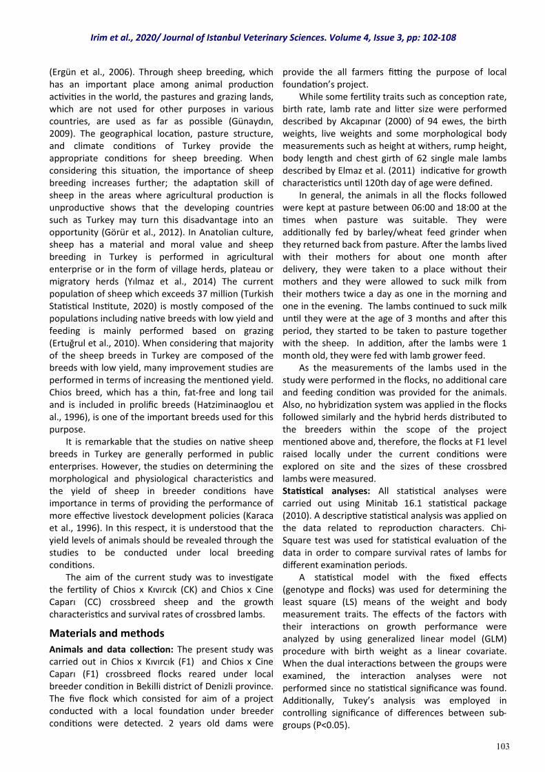
Some fertility characteristics of five different Chios x Kıvırcık (CK) and Chios x Cine Caparı (CC) flocks were presented in Table 1. While the conception rate, birth rate, lamb rate, litter size, single birth rate and twinning rates in Chios x Kıvırcık crossbred sheep were found to be 98%, 93%, 1.21, 1.29, 71% and 29% respectively, the same values were detected as 97%, 93%, 1.12, 1.20, 80% and 20% for Chios x Cine Caparı crossbred sheep respectively.
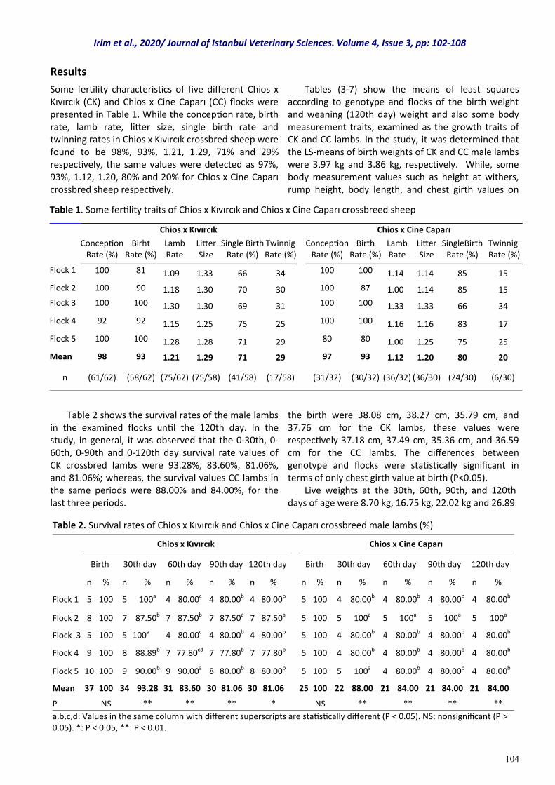
Table 2 shows the survival rates of the male lambs in the examined flocks until the 120th day. In the study, in general, it was observed that the 0-30th, 0-60th, 0-90th and 0-120th day survival rate values of CK crossbred lambs were 93.28%, 83.60%, 81.06%, and 81.06%; whereas, the survival values CC lambs in the same periods were 88.00% and 84.00%, for the last three periods.
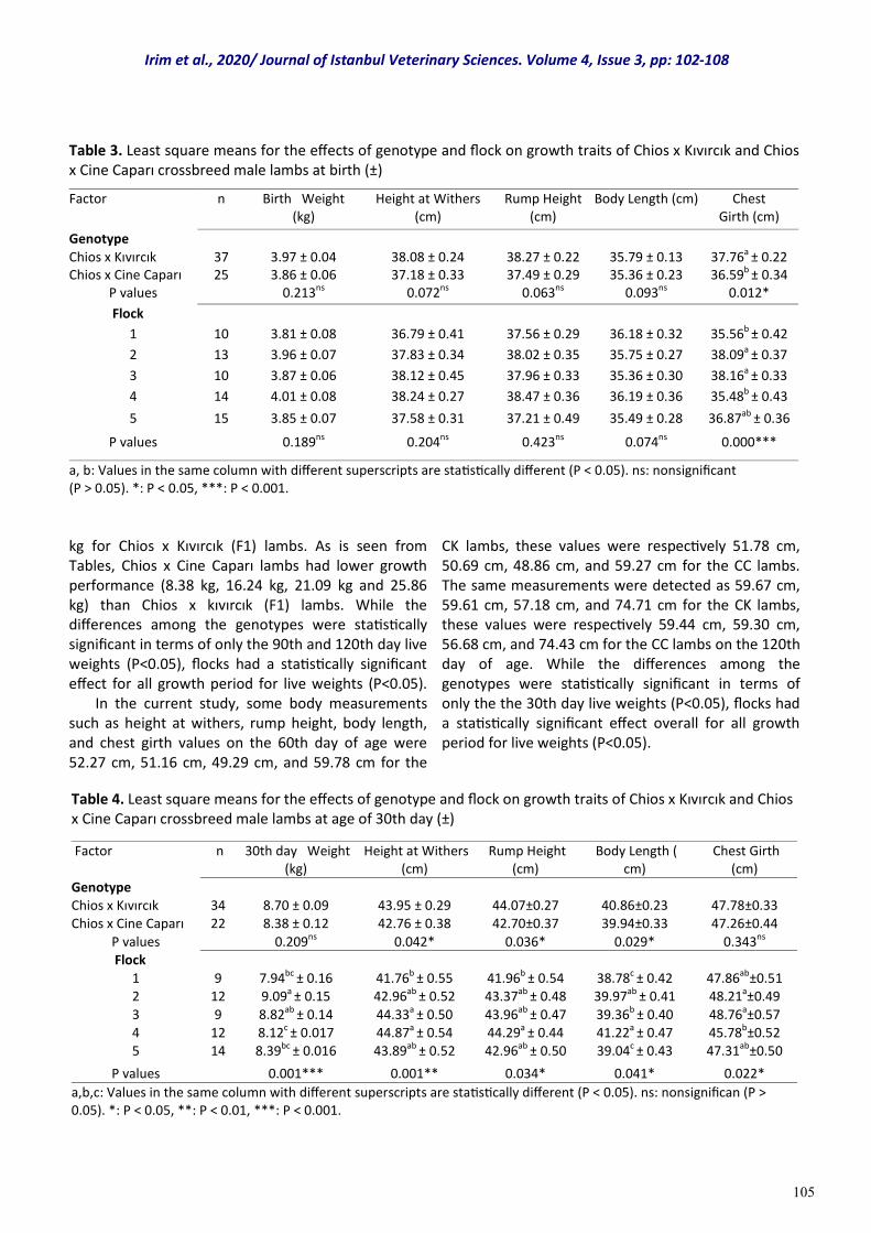
Tables (3-7) show the means of least squares according to genotype and flocks of the birth weight and weaning (120th day) weight and also some body measurement traits, examined as the growth traits of CK and CC lambs. In the study, it was determined that the LS-means of birth weights of CK and CC male lambs were 3.97 kg and 3.86 kg, respectively. While, some body measurement values such as height at withers, rump height, body length, and chest girth values on
the birth were 38.08 cm, 38.27 cm, 35.79 cm, and 37.76 cm for the CK lambs, these values were respectively 37.18 cm, 37.49 cm, 35.36 cm, and 36.59 cm for the CC lambs. The differences between genotype and flocks were statistically significant in terms of only chest girth value at birth (P<0.05). Live weights at the 30th, 60th, 90th, and 120th days of age were 8.70 kg, 16.75 kg, 22.02 kg and 26.89
Results
Table 1. Some fertility traits of Chios x Kıvırcık and Chios x Cine Caparı crossbreed sheep
Chios x Kıvırcık Chios x Cine Caparı
Conception Rate (%)
Birht Rate (%)
Lamb Rate
Litter Size
Single Birth Rate (%)
Twinnig Rate (%)
Conception Rate (%)
Birth Rate (%)
Lamb Rate
Litter Size
SingleBirth Rate (%)
Twinnig Rate (%)
Flock 1 100 81 1.09 1.33 66 34 100 100 1.14 1.14 85 15
Flock 2 100 90 1.18 1.30 70 30 100 87 1.00 1.14 85 15
Flock 3 100 100 1.30 1.30 69 31 100 100 1.33 1.33 66 34
Flock 4 92 92 1.15 1.25 75 25 100 100 1.16 1.16 83 17
Flock 5 100 100 1.28 1.28 71 29 80 80 1.00 1.25 75 25
Mean 98 93 1.21 1.29 71 29 97 93 1.12 1.20 80 20
n (61/62) (58/62) (75/62) (75/58) (41/58) (17/58) (31/32) (30/32) (36/32) (36/30) (24/30) (6/30)
Table 2. Survival rates of Chios x Kıvırcık and Chios x Cine Caparı crossbreed male lambs (%)
Chios x Kıvırcık Chios x Cine Caparı
Birth 30th day 60th day 90th day 120th day Birth 30th day 60th day 90th day 120th day
n % n % n % n % n % n % n % n % n % n %
Flock 1 5 100 5 100a 4 80.00c 4 80.00b 4 80.00b 5 100 4 80.00b 4 80.00b 4 80.00b 4 80.00b
Flock 2 8 100 7 87.50b 7 87.50b 7 87.50a 7 87.50a 5 100 5 100a 5 100a 5 100a 5 100a
Flock 3 5 100 5 100a 4 80.00c 4 80.00b 4 80.00b 5 100 4 80.00b 4 80.00b 4 80.00b 4 80.00b
Flock 4 9 100 8 88.89b 7 77.80cd 7 77.80b 7 77.80b 5 100 4 80.00b 4 80.00b 4 80.00b 4 80.00b
Flock 5 10 100 9 90.00b 9 90.00a 8 80.00b 8 80.00b 5 100 5 100a 4 80.00b 4 80.00b 4 80.00b
Mean 37 100 34 93.28 31 83.60 30 81.06 30 81.06 25 100 22 88.00 21 84.00 21 84.00 21 84.00
P NS ** ** ** * NS ** ** ** **
a,b,c,d: Values in the same column with different superscripts are statistically different (P < 0.05). NS: nonsignificant (P > 0.05). *: P < 0.05, **: P < 0.01.
Irim et al., 2020/ Journal of Istanbul Veterinary Sciences. Volume 4, Issue 3, pp: 102-108
105
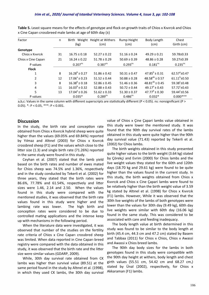
kg for Chios x Kıvırcık (F1) lambs. As is seen from Tables, Chios x Cine Caparı lambs had lower growth performance (8.38 kg, 16.24 kg, 21.09 kg and 25.86 kg) than Chios x kıvırcık (F1) lambs. While the differences among the genotypes were statistically significant in terms of only the 90th and 120th day live weights (P<0.05), flocks had a statistically significant effect for all growth period for live weights (P<0.05). In the current study, some body measurements such as height at withers, rump height, body length, and chest girth values on the 60th day of age were 52.27 cm, 51.16 cm, 49.29 cm, and 59.78 cm for the
CK lambs, these values were respectively 51.78 cm, 50.69 cm, 48.86 cm, and 59.27 cm for the CC lambs. The same measurements were detected as 59.67 cm, 59.61 cm, 57.18 cm, and 74.71 cm for the CK lambs, these values were respectively 59.44 cm, 59.30 cm, 56.68 cm, and 74.43 cm for the CC lambs on the 120th day of age. While the differences among the genotypes were statistically significant in terms of only the the 30th day live weights (P<0.05), flocks had a statistically significant effect overall for all growth period for live weights (P<0.05).
Table 3. Least square means for the effects of genotype and flock on growth traits of Chios x Kıvırcık and Chios x Cine Caparı crossbreed male lambs at birth (±)
Factor n Birth Weight (kg)
Height at Withers (cm)
Rump Height (cm)
Body Length (cm) Chest Girth (cm)
Genotype Chios x Kıvırcık 37 3.97 ± 0.04 38.08 ± 0.24 38.27 ± 0.22 35.79 ± 0.13 37.76a ± 0.22 Chios x Cine Caparı 25 3.86 ± 0.06 37.18 ± 0.33 37.49 ± 0.29 35.36 ± 0.23 36.59b ± 0.34
P values 0.213ns 0.072ns 0.063ns 0.093ns 0.012*
Flock
1 10 3.81 ± 0.08 36.79 ± 0.41 37.56 ± 0.29 36.18 ± 0.32 35.56b ± 0.42
2 13 3.96 ± 0.07 37.83 ± 0.34 38.02 ± 0.35 35.75 ± 0.27 38.09a ± 0.37
3 10 3.87 ± 0.06 38.12 ± 0.45 37.96 ± 0.33 35.36 ± 0.30 38.16a ± 0.33
4 14 4.01 ± 0.08 38.24 ± 0.27 38.47 ± 0.36 36.19 ± 0.36 35.48b ± 0.43
5 15 3.85 ± 0.07 37.58 ± 0.31 37.21 ± 0.49 35.49 ± 0.28 36.87ab ± 0.36
P values 0.189ns 0.204ns 0.423ns 0.074ns 0.000***
a, b: Values in the same column with different superscripts are statistically different (P < 0.05). ns: nonsignificant (P > 0.05). *: P < 0.05, ***: P < 0.001.
Table 4. Least square means for the effects of genotype and flock on growth traits of Chios x Kıvırcık and Chios x Cine Caparı crossbreed male lambs at age of 30th day (±)
Factor n 30th day Weight (kg)
Height at Withers (cm)
Rump Height (cm)
Body Length ( cm)
Chest Girth (cm)
Genotype Chios x Kıvırcık 34 8.70 ± 0.09 43.95 ± 0.29 44.07±0.27 40.86±0.23 47.78±0.33 Chios x Cine Caparı 22 8.38 ± 0.12 42.76 ± 0.38 42.70±0.37 39.94±0.33 47.26±0.44
P values 0.209ns 0.042* 0.036* 0.029* 0.343ns Flock
1 9 7.94bc ± 0.16 41.76b ± 0.55 41.96b ± 0.54 38.78c ± 0.42 47.86ab±0.51 2 12 9.09a ± 0.15 42.96ab ± 0.52 43.37ab ± 0.48 39.97ab ± 0.41 48.21a±0.49
3 9 8.82ab ± 0.14 44.33a ± 0.50 43.96ab ± 0.47 39.36b ± 0.40 48.76a±0.57 4 12 8.12c ± 0.017 44.87a ± 0.54 44.29a ± 0.44 41.22a ± 0.47 45.78b±0.52 5 14 8.39bc ± 0.016 43.89ab ± 0.52 42.96ab ± 0.50 39.04c ± 0.43 47.31ab±0.50
P values 0.001*** 0.001** 0.034* 0.041* 0.022*
a,b,c: Values in the same column with different superscripts are statistically different (P < 0.05). ns: nonsignifican (P > 0.05). *: P < 0.05, **: P < 0.01, ***: P < 0.001.
Irim et al., 2020/ Journal of Istanbul Veterinary Sciences. Volume 4, Issue 3, pp: 102-108
106
Discussion In the study, the birth rate and conception rate obtained from Chios x Kıvırcık hybrid sheep were quite higher than the values (69.05% and 69.84%) reported by Yılmaz and Altınel (2003) for Chios x Kıvırcık crossbred sheep (F1) and the values which close to the litter size (1.3) and single birth rate (71.26%) reported in the same study were found in this study. Ceyhan et al. (2007) stated that the lamb yield based on the birth rates and number of ewes mated for Chios sheep was 74.5% and 1.36%, respectively and in the study conducted by Tekerli et al. (2002) for three years, they stated that the birth rates were 86.6%, 77.78% and 61.54%, respectively and litter sizes were 1.46, 2.14 and 2.50. When the values found in this study were compared with the mentioned studies, it was observed that the birth rate values found in this study were higher and the lambing rate was lower. The high birth and conception rates were considered to be due to controlled mating applications and the intense keep up with mechanisms in the following period. When the literature data were investigated, it was observed that number of the studies on the fertility rate criteria of Chios x Cine Caparı crossbred sheep was limited. When data reported in Cine Caparı breed registry were compared with the data obtained in this study, it was observed that the birth rate and the litter size were similar values (GDARP, 2009). While, 30th day survival rate obtained from CK lambs was higher than survival value (89.51) at the same period found in the study by Altınel et al. (1998), in which they used CK lambs, the 30th day survival
value of Chios x Çine Çapari lambs value obtained in this study were lower the mentioned study. It was found that the 90th day survival rates of the lambs obtained in this study were quite higher than the 90th day survival value (71.43) reported by Tekerli et al. (2002) for Chios lambs. The birth weights obtained in this study presented quite higher values to the birth weight (3.64 kg) stated by Çörekçi and Evrim (2000) for Chios lambs and the live weight values they stated for the 60th and 120th days (18.70 kg and 29.61 kg) were found to be quite higher than the values found in the current study. In this study, the birth weights obtained from Chios x Kıvırcık and Chios x Cine Çapari lambs were found to be relatively higher than the birth weight value of 3.59 kg stated by Altınel et al. (1998) for Chios x Kıvırcık (F1) lambs. However, While it was observed that the 30th live weights of the lambs of both genotypes were lower than the values for 30th day (9.49 kg), 60th day live weights were similar with 60th day (16.06 kg) found in the same study. This was considered to be associated with care and feeding inadequacy. The body length value at birth determined in this study was found to be similar to the body length at birth (45.4 cm, 44.3 cm and 47.2 cm) stated by Basem and Tabbaa (2011) for Chios x Chios, Chios x Awassi and Awassi x Chios breed lambs. The 90th day body sizes for the lambs in both genotypes found in this study were compatible with the 90th day height at withers, body length and chest girth values (55.51 cm, 54.42 cm and 68.27 cm,) stated by Ünal (2002), respectively, for Chios x Akkaraman (F1) lambs.
Table 5. Least square means for the effects of genotype and flock on growth traits of Chios x Kıvırcık and Chios x Cine Caparı crossbreed male lambs at age of 60th day (±)
Factor n Birth Weight (kg)
Height at Withers (cm)
Rump Height (cm)
Body Length (cm)
Chest Girth (cm)
Genotype Chios x Kıvırcık 31 16.75 ± 0.18 52.27 ± 0.22 51.16 ± 0.24 49.29 ± 0.21 59.78±0.33
Chios x Cine Caparı 21 16.24 ± 0.22 51.78 ± 0.29 50.69 ± 0.39 48.86 ± 0.28 59.27±0.39
P values 0.207ns 0.387ns 0.293ns 0.181ns 0.235ns
Flock 1 8 16.28b ± 0.27 51.86 ± 0.42 50.31 ± 0.47 47.85b ± 0.31 62.57a±0.47
2 12 17.06a ± 0.23 51.52 ± 0.44 50.88 ± 0.28 48.58ab ± 0.57 61.11b±0.50
3 8 16.38b ± 0.18 52.86 ± 0.45 51.46 ± 0.36 48.81ab ± 0.45 59.38c±0.48
4 11 16.03b ± 0.32 52.88 ± 0.43 50.72 ± 0.44 49.17a ± 0.43 57.72c±0.43
5 13 17.04a ± 0.26 51.62 ± 0.33 51.30 ± 0.37 47.77b ± 0.30 59.44c±0.56
P values 0.035* 0.289ns 0.486ns 0.032* 0.000***
a,b,c: Values in the same column with different superscripts are statistically different (P < 0.05). ns: nonsignificant (P > 0.05). *: P < 0.05, ***: P < 0.001.
Irim et al., 2020/ Journal of Istanbul Veterinary Sciences. Volume 4, Issue 3, pp: 102-108
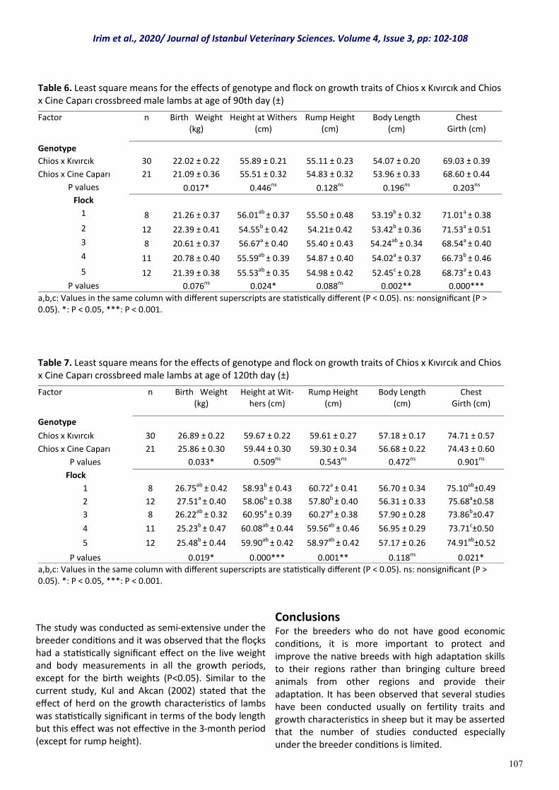
107
The study was conducted as semi-extensive under the breeder conditions and it was observed that the floçks had a statistically significant effect on the live weight and body measurements in all the growth periods, except for the birth weights (P<0.05). Similar to the current study, Kul and Akcan (2002) stated that the effect of herd on the growth characteristics of lambs was statistically significant in terms of the body length but this effect was not effective in the 3-month period (except for rump height).
Conclusions For the breeders who do not have good economic conditions, it is more important to protect and improve the native breeds with high adaptation skills to their regions rather than bringing culture breed animals from other regions and provide their adaptation. It has been observed that several studies have been conducted usually on fertility traits and growth characteristics in sheep but it may be asserted that the number of studies conducted especially under the breeder conditions is limited.
Table 6. Least square means for the effects of genotype and flock on growth traits of Chios x Kıvırcık and Chios x Cine Caparı crossbreed male lambs at age of 90th day (±)
Factor n Birth Weight (kg)
Height at Withers (cm)
Rump Height (cm)
Body Length (cm)
Chest Girth (cm)
Genotype
Chios x Kıvırcık 30 22.02 ± 0.22 55.89 ± 0.21 55.11 ± 0.23 54.07 ± 0.20 69.03 ± 0.39
Chios x Cine Caparı 21 21.09 ± 0.36 55.51 ± 0.32 54.83 ± 0.32 53.96 ± 0.33 68.60 ± 0.44
P values 0.017* 0.446ns 0.128ns 0.196ns 0.203ns
Flock
1 8 21.26 ± 0.37 56.01ab ± 0.37 55.50 ± 0.48 53.19b ± 0.32 71.01a ± 0.38
2 12 22.39 ± 0.41 54.55b ± 0.42 54.21± 0.42 53.42b ± 0.36 71.53a ± 0.51
3 8 20.61 ± 0.37 56.67a ± 0.40 55.40 ± 0.43 54.24ab ± 0.34 68.54a ± 0.40
4 11 20.78 ± 0.40 55.59ab ± 0.39 54.87 ± 0.40 54.02a ± 0.37 66.73b ± 0.46
5 12 21.39 ± 0.38 55.53ab ± 0.35 54.98 ± 0.42 52.45c ± 0.28 68.73a ± 0.43
P values 0.076ns 0.024* 0.088ns 0.002** 0.000***
a,b,c: Values in the same column with different superscripts are statistically different (P < 0.05). ns: nonsignificant (P > 0.05). *: P < 0.05, ***: P < 0.001.
Table 7. Least square means for the effects of genotype and flock on growth traits of Chios x Kıvırcık and Chios x Cine Caparı crossbreed male lambs at age of 120th day (±)
Factor n Birth Weight (kg)
Height at Wit-hers (cm)
Rump Height (cm)
Body Length (cm)
Chest Girth (cm)
Genotype
Chios x Kıvırcık 30 26.89 ± 0.22 59.67 ± 0.22 59.61 ± 0.27 57.18 ± 0.17 74.71 ± 0.57
Chios x Cine Caparı 21 25.86 ± 0.30 59.44 ± 0.30 59.30 ± 0.34 56.68 ± 0.22 74.43 ± 0.60
P values 0.033* 0.509ns 0.543ns 0.472ns 0.901ns
Flock
1 8 26.75ab ± 0.42 58.93b ± 0.43 60.72a ± 0.41 56.70 ± 0.34 75.10ab±0.49
2 12 27.51a ± 0.40 58.06b ± 0.38 57.80b ± 0.40 56.31 ± 0.33 75.68a±0.58
3 8 26.22ab ± 0.32 60.95a ± 0.39 60.27a ± 0.38 57.90 ± 0.28 73.86b±0.47
4 11 25.23b ± 0.47 60.08ab ± 0.44 59.56ab ± 0.46 56.95 ± 0.29 73.71c±0.50
5 12 25.48b ± 0.44 59.90ab ± 0.42 58.97ab ± 0.42 57.17 ± 0.26 74.91ab±0.52
P values 0.019* 0.000*** 0.001** 0.118ns 0.021*
a,b,c: Values in the same column with different superscripts are statistically different (P < 0.05). ns: nonsignificant (P > 0.05). *: P < 0.05, ***: P < 0.001.
Irim et al., 2020/ Journal of Istanbul Veterinary Sciences. Volume 4, Issue 3, pp: 102-108
108
Many of the fertility trait findings obtained from Chios x Kıvırcık and Chios x Cine Capari sheep that are raised locally have similarities among the genotypes but especially the superiority in favor of Chios x Kıvırcık crossbred in lamb yield, litter size and twinning rate is remarkable. In addition, it was determined that the findings obtained from both genotypes were quite low compared to Chios breed in terms of the lamb yield and they were similar with the lamb yield values of Kıvırcık and Cine Caparı breeds. In the light of these findings, it is considered that the income of local people who live on lambs may be increased by implementing planned reverse Crossbreeding applications and the regular recording studies.
References Akçapınar, H. (2000). Koyun yetiştiriciliği. 2nd ed.
Ankara, Turkey: İsmat Publishing.
Altınel, A., Evrim, M., Özcan, M., Başpınar, H., & Deligözoğlu, F. (1998). Studies on possibility of producing high quality lambs by crossbreeding among Sakız, Kıvırcık and German Black Headed mutton sheep. Turkish Journal of Veterinary and Animal Sciences, 22, 257-265
Basem, M. A., & Tabbaa, M. (2011). Comparison of body weight and dimensions at birth and weaning among Awassi and Chios breeds and their crosses. Jordan Journal of Agricultural Sciences, 7(4), 656-666.
Ceyhan, A., Erdoğan, İ., & Sezenler, T (2007). Some production characteristics of Kıvırcık, Gokceada and Sakız breeds of sheep conserved as gene resources. Journal of Tekirdag Agricultural Faculty, 4(2), 211-217.
Çörekçi, Ş. G., & Evrim, M. (2000). Comparative studies on the production performances of Chios and İmroz sheep kept under semi-intensive condition III. The effect of some environmental factors on production characteristics. Turkish Journal of Veterinary and Animal Sciences, 24, 553-561 (in Turkish with an abstract in English).
Elmaz, Ö., Saatcı, M., Dağ, B., Aktaş, H. A., Ata, A., Gülay, Ş.M., & Mamak, N. (2011). Honamlı keçi ırkının çeşitli özelliklerinin tespiti. Project of TUBITAK, No: 109R020.
Ergün, A., Çolpan, İ., Yıldız, G., Küçükersan, S., Tuncer, Ş. D., Yalçın, S., Küçükersan, M. K., & Şehu, A. (2006). Animal nutrition and nutritional diseases. 3rd ed. Ankara, Turkey: Pozitif Publishing.
Ertuğrul, M., Savaş, T., Dellal, G., Taşkın, T., Koyuncu, M., Cengiz, F., Dağ, B., Koncagül, S., & Pehlivan E. (2010, January 11-15). Türkiye küçükbaş hayvancılığının iyileştirilmesi. Paper presented at
Türkiye Ziraat Mühendisliği VII. Teknik Kongresi, Ankara, Turkey.
General Directorate of Agricultural Research and Policies (GDARP). (2009). Domestic animal genetic resources in Turkey. Ankara, Turkey.
Görür, O. T., Taşkın, Y., Çıplak, E., Göktaş, G., Polat, N., Akın, K., Özcan, T., Yüksel, S., Artık, M., & Sarıca, Y. (2012, June 8-9). Küçükbaş hayvancılığı genel bakış. Küçükbaş Hayvancılık Çalıştay Raporu, Hakkari, Turkey.
Günaydın, G. (2009). Economic and political analysis of Turkish sheep breeding sector. Uludağ Üniversitesi Ziraat Fakültesi Dergisi, 23(2), 15-32.
Hatziminaoglou, I., Georgoudis, A., Zervas, N., & Boyazoglu. J. (1996). Prolific breeds of Greece. In M. H. Fahmy (Ed). Prolific sheep pp. 73-92. UK: CAB International Press.
Karaca, O., Altın, T., & Okut, H. (1996). Some parameters estimation on the live weights changes of Karakaş ewes in the rural farms. Yüzüncü Yıl Üniversitesi Ziraat Fakültesi Dergisi, 6(3), 59-72.
Kul, S., & Akcan, A. (2002). Survival ability, growth and some body measurements in Awassi and East Friesian x Awassi crossbred (F1) lambs. Uludağ Üniversitesi Veteriner Fakültesi Dergisi, 21, 109-114.
Minitab (2010). Minitab 16.1.1 for Windows. State College, PA, USA: Minitab Inc.
Öztürk, D., & Karkacıer, O. (2008). Süt sığırcılığı yapan işletmelerin ekonomik analizi: Tokat ili Yeşilyurt ilçesi örneği. Gaziosmanpaşa Üniversitesi Ziraat Fakültesi Dergisi, 25(1), 15-22.
Tekerli, M., Gündoğan, M., Akıncı, Z., & Akcan, A. (2002). Determination of production characteristics of Akkaraman, Daglic, Chios and Awassi sheep in the conditions of Afyon Province. I - Fertility and survival rate. Lalahan Hayvancılık Araştırma Enstitüsü Dergisi, 42(2), 29-36
Turkish Statistical Institute (2020). Hayvansal Üretim İstatistikleri. http://www.tuik.gov.tr/PreTablo.do?alt_id=1002/ *Accessed 19 July 2020+.
Ünal, N. (2002). Akkaraman ve Sakız x Akkaraman F1 kuzularda yaşama gücü, büyüme ve bazı vücut ölçüleri. Turkish Journal of Veterinary and Animal Sciences, 26, 109-116.
Yılmaz, A., Altınel, A. (2003). The fertility and milk production traits of chios X kıvırcık (F1) ewes mated with german black-headed mutton rams to produce crossbred slaughter lambs, in comparison with kıvırcık and turkish merino ewes. İstanbul Üniversitesi Veteriner Fakültesi Dergisi, 29(2), 221-227.
Irim et al., 2020/ Journal of Istanbul Veterinary Sciences. Volume 4, Issue 3, pp: 102-108
109
Research Article
Volume: 4, Issue: 3 December 2020 Pages: 109-118
Estimation of relative feed value, relative forage quality and net energy lactation values of some roughage samples by using near infrared reflectance spectroscopy
Journal of Istanbul Veterınary Scıences
Hasan ATALAY1*, Fatih KAHRIMAN2 1. Department of Animal Nutrition and Nutritional Diseases, Faculty of Veterinary Medicine, Balikesir University, 10145, Balıkesir, Turkey, ORCİD ID. 0000-0002-5744-7538. 2. Department of Field Crops, Faculty of Agriculture, Çanakkale Onsekiz Mart University, 17100, Çanakkale, Turkey, ORCID ID: 0000-0001-6944-0512
ABSTRACT
The aim of this study is to determine the relative feed value (RFV), relative forage quality (RFQ) and net energy lactation (NEL) values of some roughage samples, based on the measurements taken from two different near infrared reflectance (NIR) devices. Corn silage (n = 18), alfalfa (n = 9), oat grass hay (n = 15), wheat straw (n = 10) were used as roughage samples and NIR measurements were taken from these samples in two ways. In the study, two different NIR (NIR1, NIR2) devices were used and nutrient values of roughage samples which were used were determined. The reference chemical analyzes of the roughage samples used in the study were made in the laboratory as dry matter. After the determination of the nutritional values, by adding these nutritional values (Dry matter, ether extract, crude protein, ash, neutral detergent fiber, acid detergent fiber) to the RFV, RFQ, NEL equations, RFV, RFQ and NEL values of each roughage sample were calculated. Meanwhile, the roughage samples used in the study were also analyzed in NIR devices and after the nutritional values were determined, these nutritional values were written in the RFV, RFQ, NEL equations thus RFV, RFQ, NEL values of each roughage samples were calculated separately for each device. Relationships between predictions obtained from NIR1 and NIR2 devices and reference chemical analysis values were determined by statistical tests. It was determined that the type of device and sample used had an important effect on the relationships between the results obtained from the reference analyzes and the predictions based on NIR measurements. Regression coefficients between RVF, RFQ and NEL values obtained from NIR1 and NIR2 devices and RVF, RFQ and NEL values obtained from reference (REF) analysis were determined to be 0.37 and 0.50. Among the roughage types, the highest similarity between NIR results and reference analysis results was found in alfalfa samples. The study results showed that indirect estimation by NIR relating to RVF, RFQ and NEL parameters is possible depending on the sample type and that there is a need for calibration improvement studies to determine these parameters directly with NIR.
Keywords: roughages, relative feed value, relative forage quality, net energy lactation, NIR spectroscopy
DOI: https://doi.org/10.30704/http-www-jivs-net.791669
To cite this article: Atalay, H., Kahriman, F. (2020). Estimation of relative feed value, relative forage quality and net energy lactation values of some roughage samples by using near infrared reflectance spectroscopy. Journal of Istanbul Veterinary Sciences, 4(3), 109-118. Abbreviated Title: J Ist Vet Sci
Roughages are the most abundant and inexpensive source of feed that are extensively used in ruminant feeding. The quality of the roughages is primarily indicated by the amount and composition of their structural carbohydrate contents. Specifically, the NDF and ADF fractions that form the cell wall of plants are
important parameters determining their intake and digestibility. Various indexes are used to express the quality of roughages depending on their NDF and ADF content. These indexes are relative feed value (RFV) and relative forage quality (RFQ). Relative feed value (RFV) is an estimated index used in the quality
*Corresponding Author: Hasan Atalay E-mail: [email protected]
Journal home page: www.jivs.net http://dergipark.gov.tr/http-www-jivs-net
Article History
Received: 07.09.2020 Accepted:27.10.2020 Available online: 28.10.2020
This work is licensed under the Creative Commons Attribution 4.0 International License.
Introduction
110
assessment of alfalfa grass and other roughages. Relative forage quality (RFQ) is another feed evaluation system that considers NDF digestibility. The RFQ is a more developed index than the RFV index, as it better reflects the expected performance of cattle consuming roughage. The digestibility of NDF determines rumen fullness and digesta flow rate ultimately affecting the dry matter consumption. Total digestible nutrients (TDN) are used to determine the RFQ (Hayırlı 2016; Sheaffer et al., 1995; Ball et al., 2007). Various analyzes such as weende analysis, Van soest analysis, in vitro ruminal and enzymatic techniques are used to determine the nutrient matter content of roughages. However, since chemicals are used in all these techniques, time and cost calculations are required to be made. Studies continue to explore more economically efficient and less time consuming alternative methods in analyzing the chemical composition of the feeds and forages. Near infrared reflectance spectroscopy (NIRS) is one of the important alternatives studied on this subject. The NIRS method is an analysis method that calculates the nutrient content of feeds based on mathematical modeling without using chemicals (Goldman et al., 1987; Parrini et al,. 2019; Ünal, 2005; Kellems and Church, 2010). Near Infrared Analysis is a method developed and used by the feed industry to evaluate the nutrient content of feeds and determine their quality levels (Marten et al., 1989). Yang et al. reported that NIR spectroscopy can be used successfully to determine the content of crude protein (CP), ADF, NDF and water soluble carbohydrate (WSC) in Italian ryegrass (Lolium multiflorum) (Yang et al., 2017). In another study (Rushing et al., 2016) on the use of NIR in feed quality measurements in wild rye (Elymus glabriflorus), calibration models were created for different feed quality evaluations such as relative feed value as well as general quality evaluations such as ADF and NDF. Research findings showed that feed quality evaluations for the feed type used can be successfully performed with NIR (Rushing et al., 2016). Most of the studies conducted have focused on general quality evaluations such as ADF, NDF and CP content. to best of our knowledge, there is no study on the calculation of the indexes used in the measurement of feed quality such as RFV and RFQ with the values determined by NIR and their association with the indexes calculated over the values obtained with reference analysis. No study has addressed the effect of the measured feed sample type *as fed (in natural condition) or as dry basis (dry matter)+ on NIR estimation results. It is thought that studies on this
subject will provide important findings on how feed quality evaluations can be made with NIR in a more practical way. Therefore, the purpose of this study is to investigate whether it is possible to determine the Relative Feed Value (RFV), RFQ and lactation energy (NEL) of some roughages by using the NIR analysis results and to determine the effect of the sample form (as fed basis or dry matter basis) used in the analyzes and the effect of feed group from which the sample was taken.
Materials and Methods
The feed samples (corn silage, alfalfa, oat grass hay, wheat straw) used in the research were produced in Balıkesir. Roughage samples were taken fresh and weighed 0.5 kg from farms and brought to the laboratory in airtight bags and kept at -20°C until analysis. Wet feed samples (silage) with known weights were dried in the oven for 48 hours at 60°C until their weight did not change and the dry matter level was determined. Air-dried and weight known feed samples were dried in the oven at 105 ° C for 4 hours and the dry matter level was determined (AOAC, 1997). Dried feed samples were ground in the mill (Retsch ZM 200 ultra centrifugal mill, 1 mm sieve) for analysis. Reference analysis (Chemical analysis): Reference (chemical) analysis of roughage samples used in the study were made in Balıkesir University Veterinary Faculty Animal Nutrition and Nutrition Diseases Laboratory, according to specified method for dry matter (DM, method 934.01), ash (method 942.05), crude protein (CP, method 990.03), ether extract (EE, method 920.39) (AOAC, 1997) and reference analysis values (nutrient values) were determined. (NDF) and (ADF) analysis of feed samples were made by using Gerhard FT12 Fiber Analyzer (Gerhardt, 2010) automatic device according to the methods reported by Van Soest et al. (1991). By using the nutrient values (NDF, ADF, DM, CP, EE, ash) obtained as a result of the analysis of feed samples, RFV, RFQ, and NEL levels were calculated according to the equations reported in NRC (2001) (Samiei et al., 2015). NIR spectroscopy measurements: NIR measurements were performed on two different NIR devices. One of these devices is a desktop NIR device (Spectrastar 2400D, Unity Scientific, USA) and the other is a portable NIR device (Dinamica Generale Agri NIR Analyzer, Italy). To make spectral measurements from roughage samples and to determine the feed quality characteristics in the desktop NIR device, spectral data of the feed samples (corn silage, alfalfa, oat grass hay,
Atalay, H., & Kahriman, F. 2020/ Journal of Istanbul Veterinary Sciences. Volume 4, Issue 3, pp: 109-118
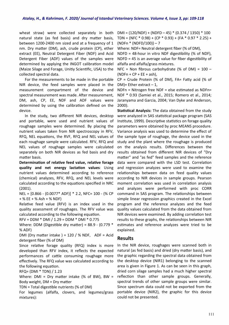
111
wheat straw) were collected separately in both natural state (as fed basis) and dry matter basis, between 1200-2400 nm sized and at a frequency of 1 nm. Dry matter (DM), ash, crude protein (CP), ether extract (EE), Neutral Detergent Fiber (NDF) and Acid Detergent Fiber (ADF) values of the samples were determined by applying the INGOT calibration model (Maize Silage and Forage, Unitiy Scientific, USA) to the collected spectral data. For the measurements to be made in the portable NIR device, the feed samples were placed in the measurement compartment of the device and spectral measurement was made. After measurement, DM, ash, CP, EE, NDF and ADF values were determined by using the calibration defined on the device. In the study, two different NIR devices, desktop and portable, were used and nutrient values of roughage samples were determined. By placing the nutrient values taken from NIR spectroscopy in RFV, RFQ, NEL equations, the RVF, RFQ and NEL values of each roughage sample were calculated. RFV, RFQ and NEL values of roughage samples were calculated separately on both NIR devices as fed basis and dry matter basis. Determination of relative feed value, relative forage quality and net energy lactation values: Using nutrient values determined according to reference (chemical) analyzes, RFV, RFQ, and NEL levels were calculated according to the equations specified in NRC (2001). NEL = *0.866 - (0.0077* ADF)+ * 2.2, NFC= 100 - (% CP + % EE + % Ash + % NDF) Relative feed value (RFV) is an index used in the quality assessment of roughages. The RFV value was calculated according to the following equation. RFV = DDM * DMI / 1.29 = DDM * DMI * 0.775 Where: DDM (Digestible dry matter) = 88.9 - (0.779 * % ADF) DMI (Dry matter intake ) = 120 / % NDF, ADF = Acid detergent fiber (% of DM) Since relative forage quality (RFQ) index is more developed than RFV index, it reflects the expected performances of cattle consuming roughage more effectively. The RFQ value was calculated according to the following equation. RFQ= (DMI * TDN) / 1.23 Where: DMI = Dry matter intake (% of BW), BW = Body weight, DM = Dry matter TDN = Total digestible nutrients (% of DM) For legumes (alfalfa, clovers, and legumes/grass mixtures):
DMI = (120/NDF) + (NDFD – 45) * (0.374 / 1350) * 100 TDN = (NFC * 0.98) + (CP * 0.93) + (FA * 0.97 * 2.25) + (NDFn * (NDFD/100)) – 7 Where: NDF= Neutral detergent fiber (% of DM), NDFD = 48-hour in vitro NDF digestibility (% of NDF), NDFD = 45 is an average value for fiber digestibility of alfalfa and alfalfa/grass mixtures. NFC = Non fibrous carbohydrate (% of DM) = 100 – (NDFn + CP + EE + ash), CP = Crude Protein (% of DM), FA= Fatty acid (% of DM)= Ether extract – 1, NDFn = Nitrogen free NDF = else estimated as NDFn= NDF * 0.93 (Samiei et al., 2015; Romero et al., 2014; Jaranyama and Garcia, 2004; Van Dyke and Anderson, 2000). Statistical Analysis: The data obtained from the study were analyzed in SAS statistical package program (SAS Institute, 1999). Descriptive statistics on forage quality parameters were obtained by proc MEANS procedure. Variance analysis was used to determine the effect of the sample type of roughage, the device used in the study and the plant where the roughage is produced on the analysis results. Differences between the results obtained from different NIR devices of "Dry matter" and "as fed" feed samples and the reference data were compared with the LSD test. Correlation and regression analyzes were used to examine the relationships between data on feed quality values according to NIR devices in sample groups. Pearson moment correlation was used in correlation analysis and analyzes were performed with proc CORR command in SAS program. The relationships between simple linear regression graphics created in the Excel program and the reference analyzes and the feed quality values calculated from the data obtained from NIR devices were examined. By adding correlation test results to these graphs, the relationships between NIR estimates and reference analyzes were tried to be explained.
Results
In the NIR device, roughages were scanned both in natural (as fed basis) and dried (dry matter basis), and the graphic regarding the spectral data obtained from the desktop device (NIR1) belonging to the scanned area is given in Figure 1. As can be seen in this graph, dried corn silage samples had a much higher spectral reflection than other sample groups. Generally, spectral trends of other sample groups were similar. Since spectrum data could not be exported from the portable device (NIR2), the graphic for this device could not be presented.
Atalay, H., & Kahriman, F. 2020/ Journal of Istanbul Veterinary Sciences. Volume 4, Issue 3, pp: 109-118
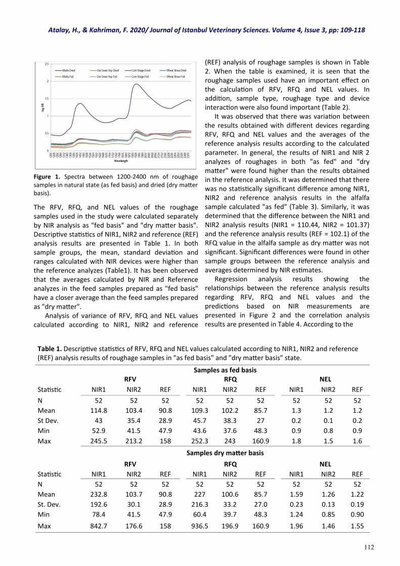
112
Figure 1. Spectra between 1200-2400 nm of roughage samples in natural state (as fed basis) and dried (dry matter basis).
The RFV, RFQ, and NEL values of the roughage samples used in the study were calculated separately by NIR analysis as "fed basis" and "dry matter basis". Descriptive statistics of NIR1, NIR2 and reference (REF) analysis results are presented in Table 1. In both sample groups, the mean, standard deviation and ranges calculated with NIR devices were higher than the reference analyzes (Table1). It has been observed that the averages calculated by NIR and Reference analyzes in the feed samples prepared as "fed basis" have a closer average than the feed samples prepared as "dry matter". Analysis of variance of RFV, RFQ and NEL values calculated according to NIR1, NIR2 and reference
(REF) analysis of roughage samples is shown in Table 2. When the table is examined, it is seen that the roughage samples used have an important effect on the calculation of RFV, RFQ and NEL values. In addition, sample type, roughage type and device interaction were also found important (Table 2). It was observed that there was variation between the results obtained with different devices regarding RFV, RFQ and NEL values and the averages of the reference analysis results according to the calculated parameter. In general, the results of NIR1 and NIR 2 analyzes of roughages in both "as fed" and "dry matter" were found higher than the results obtained in the reference analysis. It was determined that there was no statistically significant difference among NIR1, NIR2 and reference analysis results in the alfalfa sample calculated "as fed" (Table 3). Similarly, it was determined that the difference between the NIR1 and NIR2 analysis results (NIR1 = 110.44, NIR2 = 101.37) and the reference analysis results (REF = 102.1) of the RFQ value in the alfalfa sample as dry matter was not significant. Significant differences were found in other sample groups between the reference analysis and averages determined by NIR estimates. Regression analysis results showing the relationships between the reference analysis results regarding RFV, RFQ and NEL values and the predictions based on NIR measurements are presented in Figure 2 and the correlation analysis results are presented in Table 4. According to the
Table 1. Descriptive statistics of RFV, RFQ and NEL values calculated according to NIR1, NIR2 and reference (REF) analysis results of roughage samples in "as fed basis" and "dry matter basis" state.
Samples as fed basis RFV RFQ NEL
Statistic NIR1 NIR2 REF NIR1 NIR2 REF NIR1 NIR2 REF
N 52 52 52 52 52 52 52 52 52
Mean 114.8 103.4 90.8 109.3 102.2 85.7 1.3 1.2 1.2
St Dev. 43 35.4 28.9 45.7 38.3 27 0.2 0.1 0.2
Min 52.9 41.5 47.9 43.6 37.6 48.3 0.9 0.8 0.9
Max 245.5 213.2 158 252.3 243 160.9 1.8 1.5 1.6
Samples dry matter basis
RFV RFQ NEL
Statistic NIR1 NIR2 REF NIR1 NIR2 REF NIR1 NIR2 REF
N 52 52 52 52 52 52 52 52 52
Mean 232.8 103.7 90.8 227 100.6 85.7 1.59 1.26 1.22
St. Dev. 192.6 30.1 28.9 216.3 33.2 27.0 0.23 0.13 0.19
Min 78.4 41.5 47.9 60.4 39.7 48.3 1.24 0.85 0.90
Max 842.7 176.6 158 936.5 196.9 160.9 1.96 1.46 1.55
Atalay, H., & Kahriman, F. 2020/ Journal of Istanbul Veterinary Sciences. Volume 4, Issue 3, pp: 109-118
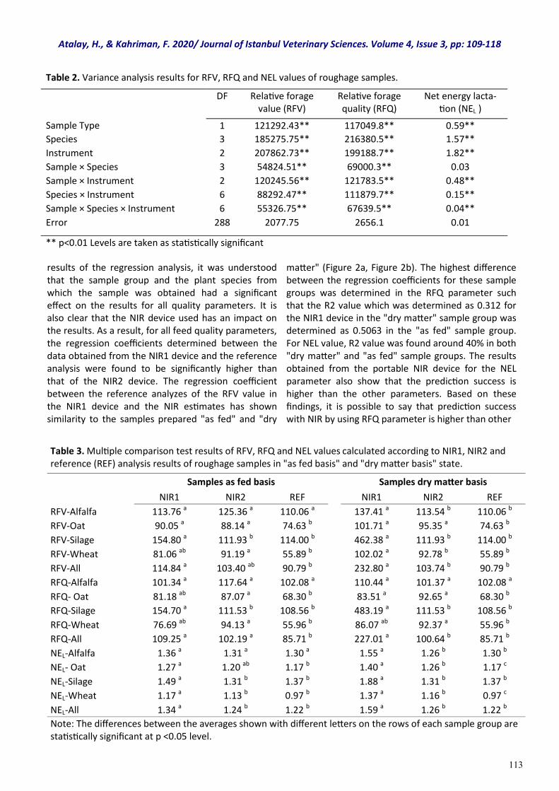
113
results of the regression analysis, it was understood that the sample group and the plant species from which the sample was obtained had a significant effect on the results for all quality parameters. It is also clear that the NIR device used has an impact on the results. As a result, for all feed quality parameters, the regression coefficients determined between the data obtained from the NIR1 device and the reference analysis were found to be significantly higher than that of the NIR2 device. The regression coefficient between the reference analyzes of the RFV value in the NIR1 device and the NIR estimates has shown similarity to the samples prepared "as fed" and "dry
matter" (Figure 2a, Figure 2b). The highest difference between the regression coefficients for these sample groups was determined in the RFQ parameter such that the R2 value which was determined as 0.312 for the NIR1 device in the "dry matter" sample group was determined as 0.5063 in the "as fed" sample group. For NEL value, R2 value was found around 40% in both "dry matter" and "as fed" sample groups. The results obtained from the portable NIR device for the NEL parameter also show that the prediction success is higher than the other parameters. Based on these findings, it is possible to say that prediction success with NIR by using RFQ parameter is higher than other
Table 2. Variance analysis results for RFV, RFQ and NEL values of roughage samples.
DF Relative forage value (RFV)
Relative forage quality (RFQ)
Net energy lacta-tion (NEL )
Sample Type 1 121292.43** 117049.8** 0.59**
Species 3 185275.75** 216380.5** 1.57**
Instrument 2 207862.73** 199188.7** 1.82**
Sample × Species 3 54824.51** 69000.3** 0.03
Sample × Instrument 2 120245.56** 121783.5** 0.48**
Species × Instrument 6 88292.47** 111879.7** 0.15**
Sample × Species × Instrument 6 55326.75** 67639.5** 0.04**
Error 288 2077.75 2656.1 0.01
** p<0.01 Levels are taken as statistically significant
Table 3. Multiple comparison test results of RFV, RFQ and NEL values calculated according to NIR1, NIR2 and reference (REF) analysis results of roughage samples in "as fed basis" and "dry matter basis" state.
Samples as fed basis Samples dry matter basis
NIR1 NIR2 REF NIR1 NIR2 REF
RFV-Alfalfa 113.76 a 125.36 a 110.06 a 137.41 a 113.54 b 110.06 b
RFV-Oat 90.05 a 88.14 a 74.63 b 101.71 a 95.35 a 74.63 b
RFV-Silage 154.80 a 111.93 b 114.00 b 462.38 a 111.93 b 114.00 b
RFV-Wheat 81.06 ab 91.19 a 55.89 b 102.02 a 92.78 b 55.89 b
RFV-All 114.84 a 103.40 ab 90.79 b 232.80 a 103.74 b 90.79 b
RFQ-Alfalfa 101.34 a 117.64 a 102.08 a 110.44 a 101.37 a 102.08 a
RFQ- Oat 81.18 ab 87.07 a 68.30 b 83.51 a 92.65 a 68.30 b
RFQ-Silage 154.70 a 111.53 b 108.56 b 483.19 a 111.53 b 108.56 b
RFQ-Wheat 76.69 ab 94.13 a 55.96 b 86.07 ab 92.37 a 55.96 b
RFQ-All 109.25 a 102.19 a 85.71 b 227.01 a 100.64 b 85.71 b
NEL-Alfalfa 1.36 a 1.31 a 1.30 a 1.55 a 1.26 b 1.30 b
NEL- Oat 1.27 a 1.20 ab 1.17 b 1.40 a 1.26 b 1.17 c
NEL-Silage 1.49 a 1.31 b 1.37 b 1.88 a 1.31 b 1.37 b
NEL-Wheat 1.17 a 1.13 b 0.97 b 1.37 a 1.16 b 0.97 c
NEL-All 1.34 a 1.24 b 1.22 b 1.59 a 1.26 b 1.22 b
Note: The differences between the averages shown with different letters on the rows of each sample group are statistically significant at p <0.05 level.
Atalay, H., & Kahriman, F. 2020/ Journal of Istanbul Veterinary Sciences. Volume 4, Issue 3, pp: 109-118
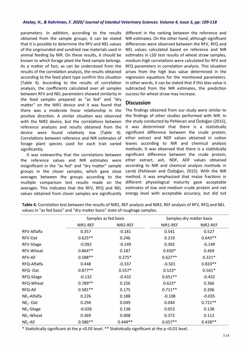
114
parameters. In addition, according to the results obtained from the sample groups, it can be stated that it is possible to determine the RFV and NEL values of the ungrounded and sundried raw materials used in animal feeding by NIR. On these results, it should be known to which forage plant the feed sample belongs. As a matter of fact, as can be understood from the results of the correlation analysis, the results obtained according to the feed plant type confirm this situation (Table 4). According to the results of correlation analysis, the coefficients calculated over all samples between RFV and NEL parameters showed similarity in the feed samples prepared as "as fed" and "dry matter" on the NIR1 device and it was found that there was a moderate linear relationship in the positive direction. A similar situation was observed with the NIR2 device, but the correlations between reference analyzes and results obtained from the device were found relatively low (Table 4). Correlations between reference and NIR estimates of forage plant species used for each trait varied significantly. It was noteworthy that the correlations between the reference values and NIR estimates were insignificant in the "as fed" and "dry matter" sample groups in the clover samples, which gave close averages between the groups according to the multiple comparison test results made on the averages. This indicates that the RFV, RFQ and NEL values obtained from clover samples are significantly
different in the ranking between the reference and NIR estimates. On the other hand, although significant differences were observed between the RFV, RFQ and NEL values calculated based on reference and NIR estimates in LSD test results of wheat straw samples, medium-high correlations were calculated for RFV and RFQ parameters in correlation analysis. This situation arises from the high bias value determined in the regression equations for the mentioned parameters. In other words, it can be stated that if this bias value is subtracted from the NIR estimates, the prediction success for wheat straw may increase.
Discussion The findings obtained from our study were similar to the findings of other studies performed with NIR. In the study conducted by Pehlevan and Özdoğan (2015), it was determined that there is a statistically significant difference between the crude protein, ether extract and NDF values obtained in cotton leaves according to NIR and chemical analysis methods. It was observed that there is a statistically significant difference between the crude protein, ether extract, ash, NDF, ADF values obtained according to NIR and chemical analysis methods in carob (Pehlevan and Özdoğan, 2015). With the NIR method, it was emphasized that maize fractions in different physiological maturity gave acceptable estimates of low and medium crude protein and net energy level with acceptable accuracy, but did not
Table 4. Correlation test between the results of NIR1, REF analysis and NIR2, REF analysis of RFV, RFQ and NEL values in "as fed basis" and "dry matter basis" state of roughage samples.
Samples as fed basis Samples dry matter basis
NIR1-REF NIR2-REF NIR1-REF NIR2-REF
RFV-Alfalfa 0.357 -0.181 0.541 0.527
RFV-Oat 0.625** 0.246 0.219 0.643**
RFV-Silage -0.092 -0.149 0.392 -0.149
RFV-Wheat 0.864** 0.187 0.650* 0.469
RFV-All 0.588** 0.275* 0.627** 0.321*
RFQ-Alfalfa 0.448 -0.337 -0.501 0.833**
RFQ- Oat 0.877** 0.557* 0.523* 0.561*
RFQ-Silage -0.132 -0.432 0.651** -0.432
RFQ-Wheat 0.789** 0.256 0.623* 0.366
RFQ-All 0.581** 0.175 0.711** 0.206
NEL-Alfalfa 0.226 0.188 -0.108 -0.035
NEL- Oat 0.294 0.049 0.044 0.721**
NEL-Silage -0.028 0.138 0.053 0.138
NEL-Wheat 0.369 0.008 0.372 0.112
NEL-All 0.586** 0.449** 0.657** 0.428**
* Statistically significant at the p <0.05 level. ** Statistically significant at the p <0.01 level.
Atalay, H., & Kahriman, F. 2020/ Journal of Istanbul Veterinary Sciences. Volume 4, Issue 3, pp: 109-118
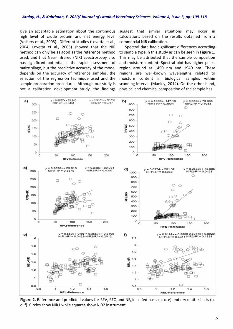
115
give an acceptable estimation about the continuous high level of crude protein and net energy level (Volkers et al., 2003). Different studies (Lovetta et al., 2004; Lovetta et al., 2005) showed that the NIR method can only be as good as the reference method used, and that Near-infrared (NIR) spectroscopy also has significant potential in the rapid assessment of maize silage, but the predictive accuracy of the model depends on the accuracy of reference samples, the selection of the regression technique used and the sample preparation procedures. Although our study is not a calibration development study, the findings
suggest that similar situations may occur in calculations based on the results obtained from a commercial NIR calibration. Spectral data had significant differences according to sample type in this study as can be seen in Figure 1. This may be attributed that the sample composition and moisture content. Spectral plot has higher peaks region around at 1450 nm and 1940 nm. These regions are well-known wavelengths related to moisture content in biological samples within scanning interval (Manley, 2014). On the other hand, physical and chemical composition of the sample has
Figure 2. Reference and predicted values for RFV, RFQ and NEL in as fed basis (a, c, e) and dry matter basis (b, d, f). Circles show NIR1 while squares show NIR2 instrument.
Atalay, H., & Kahriman, F. 2020/ Journal of Istanbul Veterinary Sciences. Volume 4, Issue 3, pp: 109-118
116
important effect on the spectral data/plot obtained from the NIR measurement (Pasquini 2003). The same samples can give different spectroscopic information due to differences in their particle size, moisture content as well as other biochemical compounds such as protein, ether extract and starch content. We used roughage samples in the forms of "as fed" and "dry basis" and their biochemical composition show high variability. We must notice that this case must have strong effect on the obtained results from NIR measurements and of course prediction results. The results of this study have revealed that NIRS method can be used in the quantitative determination of RFV, RFQ and NEL values of roughage. It was determined that used sample type had a significant effect on estimation results, and forage type of roughage was also found to have an effect on the results. The most successful results were obtained from alfalfa samples in the calculation of RFV, RFQ and NEL values from NIR analysis results. In addition to these results, it was observed that the determinations of RFV, RFQ and NEL values may vary according to the used instrument. Even though portability is an important function, it was found out that NIR2 instrument resulted in more deviations compared to NIR1, excluding silage samples. In desktop NIR instrument, the difference between results of silage samples and reference analysis results was determined to be higher than portable NIR instrument. There are several studies for desktop and portable NIR instruments to analyze biological samples. Most of these studies focused on calibration development for both types of instrument. Henn et al. (2016) compared portable versus desktop NIR instrument for analyzing sugar content of different syrup samples. Yu et al. (2020) compare the desktop and portable NIR instrument for discrimination of peanut samples based on fatty acid content. They found comparable results from portable devices with desktop instrument. Our study is not a calibration development research and our findings showed that portable instruments are not sufficient for indirect estimation of RFV, RFQ and NEL values in different roughage samples. Similar results were reported a previous study (Vander Schaaf, 2013) focused on the comparing the prediction performances of desktop and portable (the same instrument in here) NIR instruments for analyzing the components of corn silage and alfalfa hay. This study showed that two instruments gave similar results for dry matter (r=0.86) but no strong correlation for other components such as protein, ash, and fat content. Our results in agreement in this finding and we could
associate this result with the measurement capacity of the devices and the type of sample used. The portable device used in this study has a short scanning interval compared to desktop NIR instrument and it has also low scanning intensity. Therefore desktop instrument may give similar results to ref analyses.
Conclusions The results indicated that the NIRS method can be used to quantitatively determine the RFV, RFQ and NEL values of roughages. It was determined that the used sample type had a significant effect on the estimation results, and also the type of forage plant from which the roughage was prepared was also effective on the results. The most successful results in the calculation of RFV, RFQ and NEL values based on NIR analysis results were obtained from clover samples. In addition to these findings, it was observed that the determinations of RFV, RFQ and NEL values may change depending on the device used. Although portability is an important function, it has been found that the NIR2 showed more deviation in the results than the NIR1 device, with the exception of silage samples. As to the desktop NIR device, the difference between the results obtained from the silage samples and the reference analysis results was higher than those of the portable NIR device. In the light of the findings of this research, it has been understood that there is a need for calibration development studies to determine RFV, RFQ and NEL values directly with NIR and also special calibrations are needed for roughage sample groups.
Acknowledgments This project was funded by Balıkesir University, Scientific Research Projects Commission (BAP: 15-02/90, 15-02/091, 15-02/092). The authors would like to thank Serkan ATEŞ (Department of Animal and Rangeland Sciences, Oregon State University, Corvallis, OR 97333) for his contribution.
Conflict of interest Authors have no conflict of interest to declare.
Atalay, H., & Kahriman, F. 2020/ Journal of Istanbul Veterinary Sciences. Volume 4, Issue 3, pp: 109-118
117
AOAC (1997). Association of Official Analytical Chemists, 16th ed., Washington, D.C. USA.
Ball, D. M., Hoveland, C. S., & Lacefield, G. D. (2007). Adapted from Southern Forages, 4 th Ed. International Plant Nutrition Institute. U.S.A.
Gerhardt (2010). Analytical systems documents. Gerhardt GmbH & Co.KG Cäsariusstraße 97, D-53639, Königswinter.
Goldman, A., Genizi, A., Yulzari, A., & Seligman, N. G. (1987). Improving the reliability of the two stage in vitro assay for ruminant feed digestibility by calibrasyon against intwo data from a wide range of sources. Animal Feed Science and Technology, 18, 233-245.
Hayırlı, A. (2016). Evaluation of roughages. Atatürk Üniversitesi, Rasyonel Hayvancılık ARGE Ata Teknokent, Erzurum.
Henn, R., Schwab, A., & Huck, C. W. (2016). Evaluation of bench-top versus portable near- infrared spectroscopic method combined with multivariate approaches for the fast and simultaneous quantitative analysis of main sugars in syrup formulations. Food Control, 68, 97-104.
Jaranyama, P., & Garcia, A. D. (2004). Understanding relative feed value (RFV) and relative forage quality (RFQ). College of Agric and Biological Sci, South Dakota State University, USDA.
Lovetta, D. K., Deavilleb, E. R., Moulda, R., Givensb, D. I., & Owena, E. (2004). Using near infrared reflectance spectroscopy (NIRS) to predict the biological parameters of maize silage. Animal Feed Science and Technology, 115(1-2), 179-187.
Lovetta, D. K., Deavilleb, E. R., Givensb, D. I., Finlayc, M., & Owena, E. (2005). Near infrared reflectance spectroscopy (NIRS) to predict biological parameters of maize silage: effects of particle commination, oven drying temperature and the presence of residual moisture. Animal Feed Science and Technology, 120(3-4), 323-332.
Marten, G. C., Shenk, J. S., & Barton, F. E. (1989). Near infrared reflectance spectroscopy (NIRS): Analysis of forage quality. U.S. Department of agriculture, agriculture handbook No. 643 (revised with supplements), 110 p.
Manley, M. (2014). Near-infrared spectroscopy and hyperspectral imaging: non-destructive analysis of biological materials. Chemical Society Reviews, (43), 8200-8214.
NRC (2001). Nutrient requirements of dairy cattle. 7th rev. ed. , Washington DC, USA: National Academy Press.
Parrini, S., Acciaioli, A., Franci, O., Pugliese, C., & Bozzi, R. (2019). Near infrared spectroscopy technology for prediction of chemical composition of natural fresh pastures, Journal of Applied Animal Research, 47(1), 514-520.
Pasquini, C. (2003). Near infrared spectroscopy: fundamentals, practical aspects and analytical applications. Journal of the Brazilian Chemical Society, 14(2), 198-219.
Pehlevan, F., & Özdoğan, M. (2015). Comparison between chemical and near Infrared reflectance spectroscopy
methods for determining of nutrient content of some alternative feeds. Journal of Tekirdag Agricultural Faculty, 12(2), 1-10.
Kellems, R. O., & Church, D. C. (2010). Livestock feeds and feeding (Alp, M., Kocabağlı, N. Ed.) Istanbul, Turkey, Nobel.
Romero, J. J., Castillo, M. S., Burns, J. C., Moriel, P., & Davidson, S. (2014). Forage quality: concepts and practices, NC State University College of Agriculture and Life Sciences, Published by North Carolina Cooperative Extension.
Rushing, J. B., Saha, U. K., Lemus, R., Sonon, L., & Baldwin, B. S. (2016). Analysis of some important forage quality attributes of southeastern wildrye (Elymus glabriflorus) using near infrared reflectance spectroscopy. American Journal of Analytical Chemistry (7), 642-662.
Samiei, A., Liang, J. B., Ghorbani, G. R., Hirooka, H., Mahyari, S. A., Sadri, H., & Tufarelli, V. (2015). Relationship between dietary energy level, silage butyric acid and body condition score with subclinical ketosis incidence in dairy cows. Advances in Animal and Veterinary Sciences, 3(6), 354-361.
SAS (1999). Institute SAS V8 user manual, SAS Cary, NC, USA.
Sheaffer, C. C., Peterson, M. A., McCaslin, M., Volenec, J. J., Cherney, J. H., Johnson, K. D., Woodward, W. T., & Viands, D. R. (1995). Acid detergent fiber, neutral detergent fiber concentration and relative feed value. North American Alfalfa Improvement Conference, Minneapolis.
Ünal Y. (2005). Near intrared reflektans spektroskopinin hayvan besleme bilim alanında kullanım imkânları. Lalahan Hayvancılık Araştırma Enstitüsü Dergisi, 45(1) 33-39.
Van Soest, P. J., Robertson, J. B., & Lewis, B. A. (1991). Methods for dietary fiber, neutral detergent fiber and non starch polysaccharides in relation to animal nutrition. Journal Dairy Science, (74), 3583-3597.
Van Dyke, N. J., & Anderson, P. M. (2000). Interpreting a forage analysis. Alabama Cooperative Extension, Circular ANR-890.
Vander Schaaf, D. (2013). Near infrared reflectance spectroscopy comparison of dairy one and agrinir forage analyzer, California Polytechnic State University, San Luis Obispo.
Volkers, K. C., Wachendorf, M., Loges, R., Jovanovic, N., & Taube, F. (2003). Prediction of the quality of forage maize by near-infrared reflectance spectroscopy. Animal Feed Science and Technology, 109(3), 183-194.
Yang, Z, Nie, G., Pan, L., Zhang, Y., Huang, L., Ma, X., Zhang, X. (2017). Development and validation of near infrared spectroscopy for the prediction of for age quality parametersin Lolium multiflorum. Peer Journal 2017;5 :e3867.
Yu, H., Liu, H., Wang, Q., & Van, Ruth S. (2020). Evaluation of portable and benchtop NIR for classification of high oleic acid peanuts and fatty acid quantitation. LWT, 128, *109398+.
References
Atalay, H., & Kahriman, F. 2020/ Journal of Istanbul Veterinary Sciences. Volume 4, Issue 3, pp: 109-118
119
Research Article
Volume: 4, Issue: 3 December 2020 Pages: 119-124
Identification of acinar cells of salivary gland in blood fed female ticks (Hyalomma anatolicusm anatolicum) by light microscopy
Journal of Istanbul Veterınary Scıences
Eslampanah, M. 1*, Abdigoudarzi, M.2, Hablolvarid M.H. 1
1.Depatment of Pathology, Razi Vaccine and Serum Research Institute, Agricultural Research, Education and Extension Organization, Karaj, 3197619751, Iran. 2. Department of Parasitology, Razi Vaccine and Serum Research Institute, Agricultural Research, Education and Extension Organization, Karaj 3197619751, Iran. M. Eslampanah ORCID: 0000-0002-2585-9001. M. Abdigoudarzi ORCID: 0000-0002-9239-5972. M. H. Hablolvarid ORCID: 0000-0002-6408-4341.
ABSTRACT
Ticks play an important role in human and veterinary medicine particularly due to their ability to transmit protozoan pathogens. This study was undertaken on salivary gland of tick using histological methods to decrease cost and budget to determine the presence of tick-borne pathogens of medical and veterinary importance. Ticks have been proved as carrier or vector of pathogenic protozoa by separating salivary gland and using histological methods. This study provides the morphological and histological properties of the salivary glands of semi-engorged Hyalomma anatolicum anatolicum females. Unfed ticks solely were placed on cattle’s ear for feeding and females were collected, and placed in glass vials containing 70% ethanol. Collected ticks were studied and identified morphologically. Dorsal exoskeleton removed with a scalpel and salivary glands were separated by suitable forceps. Then Salivary glands were fixed in 10% formalin for further studies by light microscopy. Samples were stained with hematoxylin-eosin (H&E) for investigation under light microscope. The histological results show that the glandular tissue in females is combined with a system of ducts and the salivary glands of H. a. anatolicum consisted of three types of acinus (acinus I, II and III). The type I acinus was agranular and showed slight morphological changes during feeding. There were five granular cell types in the type II acinus, and three granular cell types in type III acinus. Data achieved here will help in understanding of the cellular morphology and general histology of these organs in this specie, preparing important information for the creation of scientific bases which will contribute to the development of more specific and efficient methods of control.
Keywords: Hyalomma anatolicum anatolicum, salivary gland, tick
DOI: https://doi.org/10.30704/http-www-jivs-net.796509
To cite this article: Eslampanah, M., Abdigoudarzi, M., Hablolvarid, M.H. (2020). Identification of acinar cells of salivary gland in blood fed female ticks (Hyalomma anatolicusm anatolicum) by light microscopy. Journal of Istanbul Veterinary Sciences, 4(3), 119-124. Abbreviated Title: J Ist Vet Sci
There are three types of salivary gland acini in ixodidae female ticks (Nunes et al, 2006; Hall-Mendelin et al, 2011; Šimo et al, 2017; Goddard & Varela-Stokes., 2009; Bior et al, 2002; El-Kady et al, 2001). Type I acinus are directly attached to the anterior region of the main salivary duct. The type I acinar cells appear as the cells with large nuclei. The numbers of cell, the
shapes of the cells as well as granularity does not change during the period of feeding (Zhou et al, 2013; Bowman & Sauer, 2005). Type II acinus connects to lobular ducts. This type consists of three cell types; a, b and c cell (Šimo et al, 2017; Bowman & Sauer, 2005). Type III acinus is occupied posterior part of the lobulated mass of the gland. They are connected to
*Corresponding Author: Mohammad Eslampanah E-mail: [email protected]
Journal home page: www.jivs.net http://dergipark.gov.tr/http-www-jivs-net
Article History
Received: 17.09.2020 Accepted: 12.11.2020 Available online: 16.11.2020
This work is licensed under the Creative Commons Attribution 4.0 International License.
Introduction
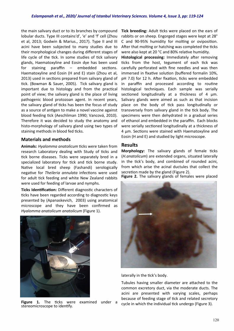
120
the main salivary duct or to its branches by compound lobular ducts. Type III contains‘d’, ‘e’ and ‘f’ cell (Zhou et al, 2013; Godwin & Marius., 2017). Type II and III acini have been subjected to many studies due to their morphological changes during different stages of life cycle of the tick. In some studies of tick salivary glands, Haematoxyline and Eosin dye has been used for staining paraffin – embedded sections. Haematoxyline and Eosin (H and E) stain (Zhou et al, 2013) used in sections prepared from salivary gland of tick. (Bowman & Sauer, 2005). Tick salivary gland is important due to histology and from the practical point of view; the salivary gland is the place of living pathogenic blood protozoan agent. In recent years, the salivary gland of ticks has been the focus of study as a source of antigen to make a novel vaccine against blood feeding tick (Aeschliman 1990; Vancová, 2010). Therefore it was decided to study the anatomy and histo-morphology of salivary gland using two types of staining methods in blood fed ticks.
Materials and methods
Animals: Hyalomma anatolicum ticks were taken from research Laboratory dealing with Study of ticks and tick borne diseases. Ticks were separately bred in a specialized laboratory for tick and tick borne study. Native local bred sheep (Fashandi) serologically negative for Theileria annulata infections were used for adult tick feeding and white New Zealand rabbits were used for feeding of larvae and nymphs.
Ticks Identification: Different diagnostic characters of ticks have been regarded according to diagnostic keys presented by (Apanaskevich, 2003) using anatomical microscope and they have been confirmed as Hyalomma anatolicum anatolicum (Figure 1).
Figure 1. The ticks were examined under a stereomicroscope to identify.
Tick breeding: Adult ticks were placed on the ears of rabbits or on sheep. Engorged stages were kept at 28°C and 90-95% humidity for molting or oviposition. After that molting or hatching was completed the ticks were also kept at 20 °C and 80% relative humidity. Histological processing: Immediately after removing ticks from the host, tegument of each tick was carefully perforated with fine needles and was then immersed in fixative solution (buffered formalin 10%, pH 7.0) for 12 h. After fixation, ticks were embedded in paraffin and processed according to routine histological techniques. Each sample was serially sectioned longitudinally at a thickness of 4 µm. Salivary glands were aimed as such as that incision place on the body of tick pass longitudinally or transversely from salivary gland in the tick body. The specimens were then dehydrated in a gradual series of ethanol and embedded in the paraffin. Each blocks were serially sectioned longitudinally at a thickness of 4 µm. Sections were stained with Haematoxyline and Eosin (H and E) and studied by light microscope.
Results Morphology: The salivary glands of female ticks (H.anatolicum) are extended organs, situated laterally in the tick’s body, and combined of rounded acini, from which arise the acinal ductules that collect the secretion made by the gland (Figure 2). Figure 2. The salivary glands of females were placed
laterally in the tick’s body.
Tubules having smaller diameter are attached to the common excretory duct, via the moderate ducts. The acini are presented with varying scales, perhaps because of feeding stage of tick and related secretory cycle in which the individual tick undergo (Figure 3).
Eslampanah et al., 2020/ Journal of Istanbul Veterinary Sciences. Volume 4, Issue 3, pp: 119-124
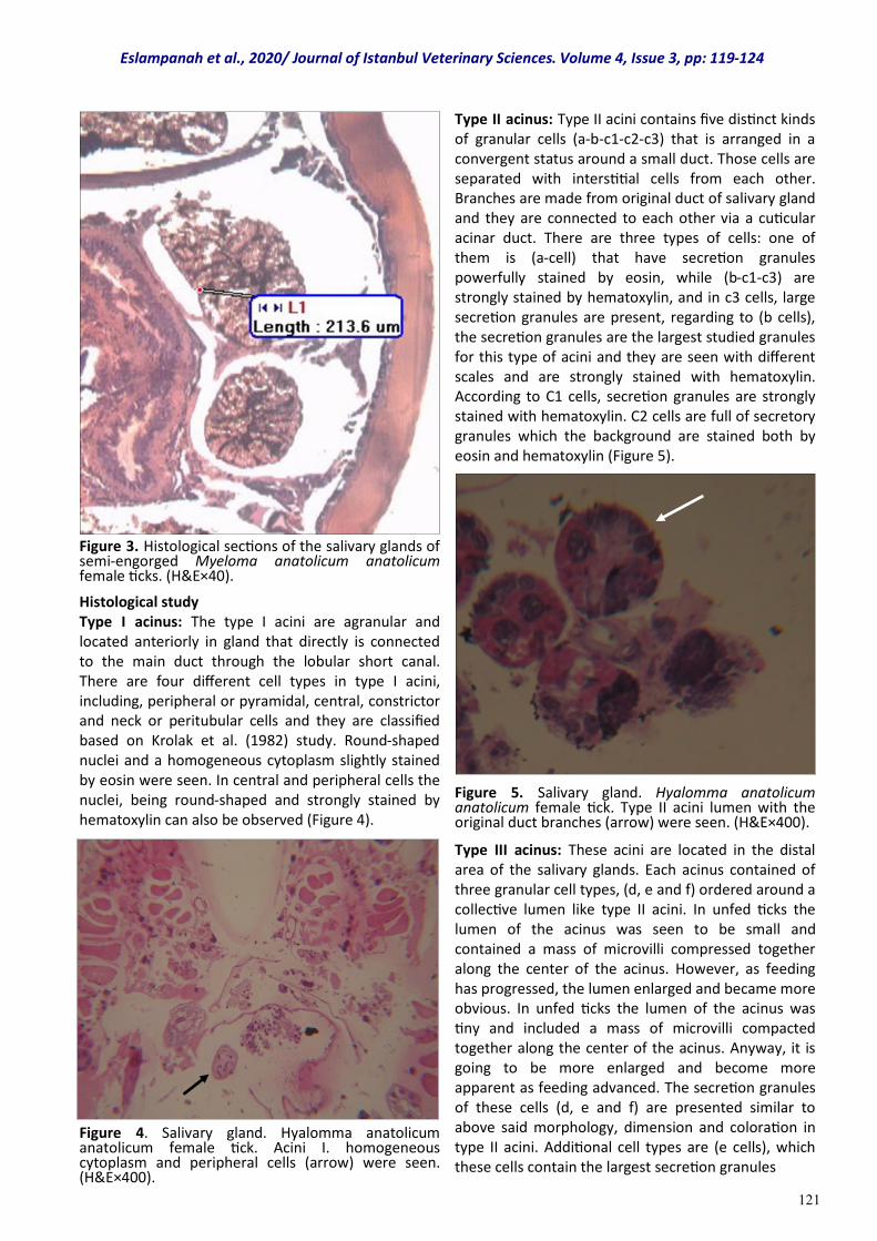
121
Figure 3. Histological sections of the salivary glands of semi-engorged Myeloma anatolicum anatolicum female ticks. (H&E×40).
Histological study Type I acinus: The type I acini are agranular and located anteriorly in gland that directly is connected to the main duct through the lobular short canal. There are four different cell types in type I acini, including, peripheral or pyramidal, central, constrictor and neck or peritubular cells and they are classified based on Krolak et al. (1982) study. Round-shaped nuclei and a homogeneous cytoplasm slightly stained by eosin were seen. In central and peripheral cells the nuclei, being round-shaped and strongly stained by hematoxylin can also be observed (Figure 4).
Figure 4. Salivary gland. Hyalomma anatolicum anatolicum female tick. Acini I. homogeneous cytoplasm and peripheral cells (arrow) were seen. (H&E×400).
Type II acinus: Type II acini contains five distinct kinds of granular cells (a-b-c1-c2-c3) that is arranged in a convergent status around a small duct. Those cells are separated with interstitial cells from each other. Branches are made from original duct of salivary gland and they are connected to each other via a cuticular acinar duct. There are three types of cells: one of them is (a-cell) that have secretion granules powerfully stained by eosin, while (b-c1-c3) are strongly stained by hematoxylin, and in c3 cells, large secretion granules are present, regarding to (b cells), the secretion granules are the largest studied granules for this type of acini and they are seen with different scales and are strongly stained with hematoxylin. According to C1 cells, secretion granules are strongly stained with hematoxylin. C2 cells are full of secretory granules which the background are stained both by eosin and hematoxylin (Figure 5).
Figure 5. Salivary gland. Hyalomma anatolicum anatolicum female tick. Type II acini lumen with the original duct branches (arrow) were seen. (H&E×400).
Type III acinus: These acini are located in the distal area of the salivary glands. Each acinus contained of three granular cell types, (d, e and f) ordered around a collective lumen like type II acini. In unfed ticks the lumen of the acinus was seen to be small and contained a mass of microvilli compressed together along the center of the acinus. However, as feeding has progressed, the lumen enlarged and became more obvious. In unfed ticks the lumen of the acinus was tiny and included a mass of microvilli compacted together along the center of the acinus. Anyway, it is going to be more enlarged and become more apparent as feeding advanced. The secretion granules of these cells (d, e and f) are presented similar to above said morphology, dimension and coloration in type II acini. Additional cell types are (e cells), which these cells contain the largest secretion granules
Eslampanah et al., 2020/ Journal of Istanbul Veterinary Sciences. Volume 4, Issue 3, pp: 119-124
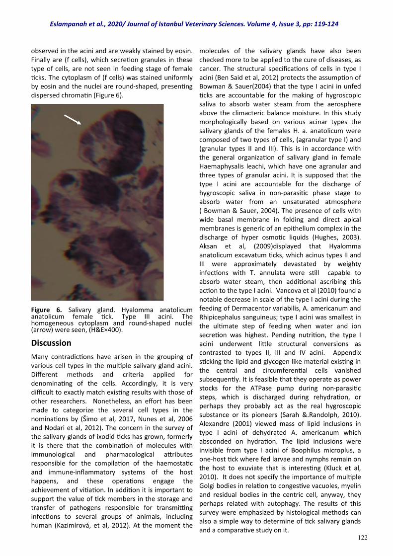
122
observed in the acini and are weakly stained by eosin. Finally are (f cells), which secretion granules in these type of cells, are not seen in feeding stage of female ticks. The cytoplasm of (f cells) was stained uniformly by eosin and the nuclei are round-shaped, presenting dispersed chromatin (Figure 6).
Figure 6. Salivary gland. Hyalomma anatolicum anatolicum female tick. Type III acini. The homogeneous cytoplasm and round-shaped nuclei (arrow) were seen, (H&E×400).
Discussion
Many contradictions have arisen in the grouping of various cell types in the multiple salivary gland acini. Different methods and criteria applied for denominating of the cells. Accordingly, it is very difficult to exactly match existing results with those of other researchers. Nonetheless, an effort has been made to categorize the several cell types in the nominations by (Šimo et al, 2017, Nunes et al, 2006 and Nodari et al, 2012). The concern in the survey of the salivary glands of ixodid ticks has grown, formerly it is there that the combination of molecules with immunological and pharmacological attributes responsible for the compilation of the haemostatic and immune-inflammatory systems of the host happens, and these operations engage the achievement of vitiation. In addition it is important to support the value of tick members in the storage and transfer of pathogens responsible for transmitting infections to several groups of animals, including human (Kazimírová, et al, 2012). At the moment the
molecules of the salivary glands have also been checked more to be applied to the cure of diseases, as cancer. The structural specifications of cells in type I acini (Ben Said et al, 2012) protects the assumption of Bowman & Sauer(2004) that the type I acini in unfed ticks are accountable for the making of hygroscopic saliva to absorb water steam from the aerosphere above the climacteric balance moisture. In this study morphologically based on various acinar types the salivary glands of the females H. a. anatolicum were composed of two types of cells, (agranular type I) and (granular types II and III). This is in accordance with the general organization of salivary gland in female Haemaphysalis leachi, which have one agranular and three types of granular acini. It is supposed that the type I acini are accountable for the discharge of hygroscopic saliva in non-parasitic phase stage to absorb water from an unsaturated atmosphere ( Bowman & Sauer, 2004). The presence of cells with wide basal membrane in folding and direct apical membranes is generic of an epithelium complex in the discharge of hyper osmotic liquids (Hughes, 2003). Aksan et al, (2009)displayed that Hyalomma anatolicum excavatum ticks, which acinus types II and III were approximately devastated by weighty infections with T. annulata were still capable to absorb water steam, then additional ascribing this action to the type I acini. Vancova et al (2010) found a notable decrease in scale of the type I acini during the feeding of Dermacentor variabilis, A. americanum and Rhipicephalus sanguineus; type I acini was smallest in the ultimate step of feeding when water and ion secretion was highest. Pending nutrition, the type I acini underwent little structural conversions as contrasted to types II, III and IV acini. Appendix sticking the lipid and glycogen-like material existing in the central and circumferential cells vanished subsequently. It is feasible that they operate as power stocks for the ATPase pump during non-parasitic steps, which is discharged during rehydration, or perhaps they probably act as the real hygroscopic substance or its pioneers (Sarah &.Randolph, 2010). Alexandre (2001) viewed mass of lipid inclusions in type I acini of dehydrated A. americanum which absconded on hydration. The lipid inclusions were invisible from type I acini of Boophilus microplus, a one-host tick where fed larvae and nymphs remain on the host to exuviate that is interesting (Kluck et al, 2010). It does not specify the importance of multiple Golgi bodies in relation to congestive vacuoles, myelin and residual bodies in the centric cell, anyway, they perhaps related with autophagy. The results of this survey were emphasized by histological methods can also a simple way to determine of tick salivary glands and a comparative study on it.
Eslampanah et al., 2020/ Journal of Istanbul Veterinary Sciences. Volume 4, Issue 3, pp: 119-124
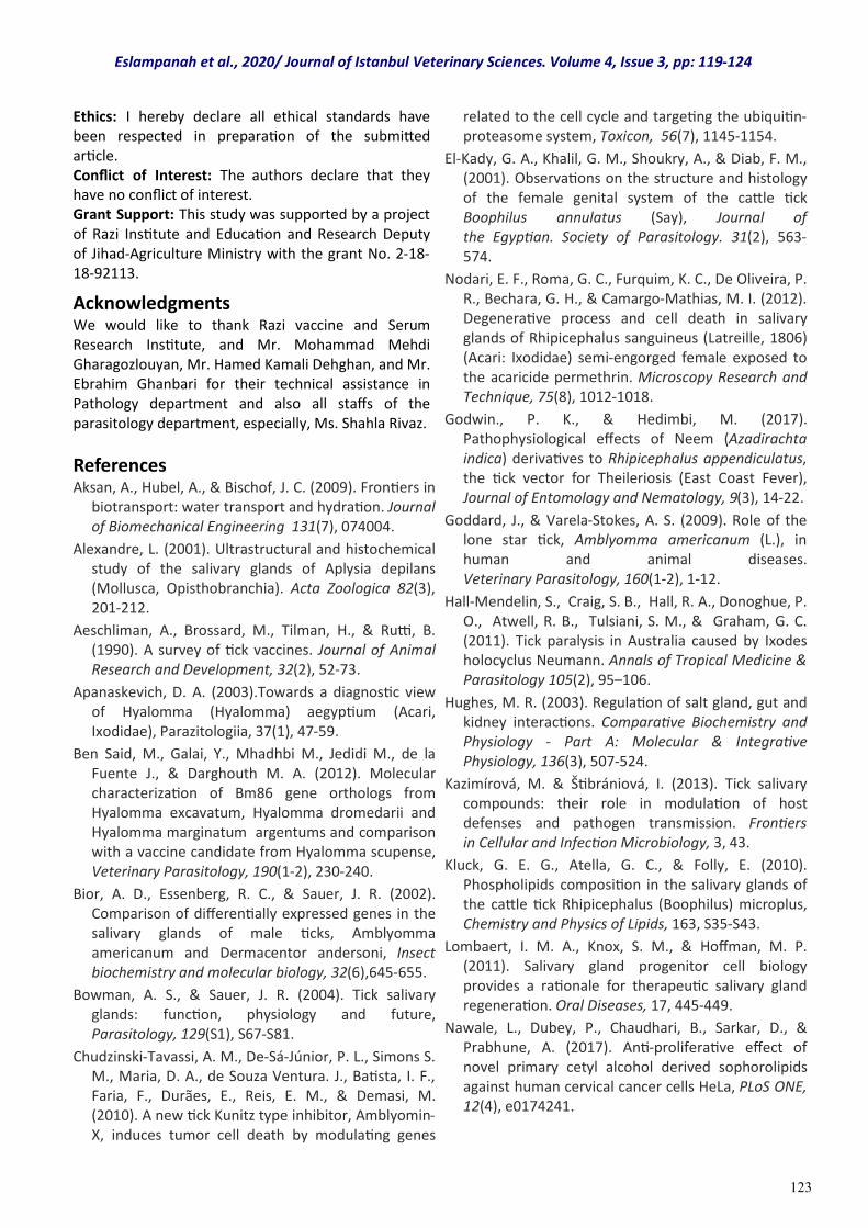
123
Ethics: I hereby declare all ethical standards have been respected in preparation of the submitted article. Conflict of Interest: The authors declare that they have no conflict of interest. Grant Support: This study was supported by a project of Razi Institute and Education and Research Deputy of Jihad-Agriculture Ministry with the grant No. 2-18-18-92113.
Acknowledgments We would like to thank Razi vaccine and Serum Research Institute, and Mr. Mohammad Mehdi Gharagozlouyan, Mr. Hamed Kamali Dehghan, and Mr. Ebrahim Ghanbari for their technical assistance in Pathology department and also all staffs of the parasitology department, especially, Ms. Shahla Rivaz.
References Aksan, A., Hubel, A., & Bischof, J. C. (2009). Frontiers in
biotransport: water transport and hydration. Journal of Biomechanical Engineering 131(7), 074004.
Alexandre, L. (2001). Ultrastructural and histochemical study of the salivary glands of Aplysia depilans (Mollusca, Opisthobranchia). Acta Zoologica 82(3), 201-212.
Aeschliman, A., Brossard, M., Tilman, H., & Rutti, B. (1990). A survey of tick vaccines. Journal of Animal Research and Development, 32(2), 52-73.
Apanaskevich, D. A. (2003).Towards a diagnostic view of Hyalomma (Hyalomma) aegyptium (Acari, Ixodidae), Parazitologiia, 37(1), 47-59.
Ben Said, M., Galai, Y., Mhadhbi M., Jedidi M., de la Fuente J., & Darghouth M. A. (2012). Molecular characterization of Bm86 gene orthologs from Hyalomma excavatum, Hyalomma dromedarii and Hyalomma marginatum argentums and comparison with a vaccine candidate from Hyalomma scupense, Veterinary Parasitology, 190(1-2), 230-240.
Bior, A. D., Essenberg, R. C., & Sauer, J. R. (2002). Comparison of differentially expressed genes in the salivary glands of male ticks, Amblyomma americanum and Dermacentor andersoni, Insect biochemistry and molecular biology, 32(6),645-655.
Bowman, A. S., & Sauer, J. R. (2004). Tick salivary glands: function, physiology and future, Parasitology, 129(S1), S67-S81.
Chudzinski-Tavassi, A. M., De-Sá-Júnior, P. L., Simons S. M., Maria, D. A., de Souza Ventura. J., Batista, I. F., Faria, F., Durães, E., Reis, E. M., & Demasi, M. (2010). A new tick Kunitz type inhibitor, Amblyomin-X, induces tumor cell death by modulating genes
related to the cell cycle and targeting the ubiquitin-proteasome system, Toxicon, 56(7), 1145-1154.
El-Kady, G. A., Khalil, G. M., Shoukry, A., & Diab, F. M., (2001). Observations on the structure and histology of the female genital system of the cattle tick Boophilus annulatus (Say), Journal of the Egyptian. Society of Parasitology. 31(2), 563-574.
Nodari, E. F., Roma, G. C., Furquim, K. C., De Oliveira, P. R., Bechara, G. H., & Camargo-Mathias, M. I. (2012). Degenerative process and cell death in salivary glands of Rhipicephalus sanguineus (Latreille, 1806) (Acari: Ixodidae) semi-engorged female exposed to the acaricide permethrin. Microscopy Research and Technique, 75(8), 1012-1018.
Godwin., P. K., & Hedimbi, M. (2017). Pathophysiological effects of Neem (Azadirachta indica) derivatives to Rhipicephalus appendiculatus, the tick vector for Theileriosis (East Coast Fever), Journal of Entomology and Nematology, 9(3), 14-22.
Goddard, J., & Varela-Stokes, A. S. (2009). Role of the lone star tick, Amblyomma americanum (L.), in human and animal diseases. Veterinary Parasitology, 160(1-2), 1-12.
Hall-Mendelin, S., Craig, S. B., Hall, R. A., Donoghue, P. O., Atwell, R. B., Tulsiani, S. M., & Graham, G. C. (2011). Tick paralysis in Australia caused by Ixodes holocyclus Neumann. Annals of Tropical Medicine & Parasitology 105(2), 95–106.
Hughes, M. R. (2003). Regulation of salt gland, gut and kidney interactions. Comparative Biochemistry and Physiology - Part A: Molecular & Integrative Physiology, 136(3), 507-524.
Kazimírová, M. & Štibrániová, I. (2013). Tick salivary compounds: their role in modulation of host defenses and pathogen transmission. Frontiers in Cellular and Infection Microbiology, 3, 43.
Kluck, G. E. G., Atella, G. C., & Folly, E. (2010). Phospholipids composition in the salivary glands of the cattle tick Rhipicephalus (Boophilus) microplus, Chemistry and Physics of Lipids, 163, S35-S43.
Lombaert, I. M. A., Knox, S. M., & Hoffman, M. P. (2011). Salivary gland progenitor cell biology provides a rationale for therapeutic salivary gland regeneration. Oral Diseases, 17, 445-449.
Nawale, L., Dubey, P., Chaudhari, B., Sarkar, D., & Prabhune, A. (2017). Anti-proliferative effect of novel primary cetyl alcohol derived sophorolipids against human cervical cancer cells HeLa, PLoS ONE, 12(4), e0174241.
Eslampanah et al., 2020/ Journal of Istanbul Veterinary Sciences. Volume 4, Issue 3, pp: 119-124
124
Nunes, E. T., Mathias, M. I., & Bechara, G. H. (2006). Structural and cytochemical changes in the salivary glands of the Rhipicephalus (Boophilus) microplus (CANESTRINI, 1887) (Acari: Ixodidae) tick female during feeding. Veterinary Parasitology, 140(1-2), 114-123.
Randolph, S. E. (2011). Transmission of tick-borne pathogens between co-feeding ticks: Milan Labuda's enduring paradigm, Ticks Tick Borne Dis, 2(4),179-82.
Šimo, L., Kazimirova, M., Richardson, J., & Bonnet, S. I. (2017). The essential role of tick salivary glands and saliva in tick feeding and pathogen transmission. Frontiers in Cellular and Infection Microbiology, 7, 281.
Simons, S. M., Sá Júnior, P. L., Faria, F., Batista, I. F. C., Barros-Batesti, D. M., Labruna, M. B., & Chudzinski-Tavassi A. M. (2011). The action of Amblyomma
cajennense tick salivain compounds of hemostatic system and cytotoxicity in tumor celllines. Biomedicine & Pharmacotherapy, 65(6),443-450.
Vancová, I., Hajnická, V., Slovák, M., Kocáková, P., Paesen, G. C., & Nuttall, P. A. (2010). Evasin-3-like anti-chemokine activity in salivary gland extracts of ixodid ticks during blood-feeding: a new target for tick control. Parasite Immunology, 32(6),460-463.
Zhou, J., Zhou, Y., Cao, J., Zhang, H., & Yu, Y. (2013). Distinctive microRNA profiles in the salivary glands of Haemaphysalis longicornis related to tick blood-feeding. Experimental and Applied Acarology, 59(3),339-349.
Eslampanah et al., 2020/ Journal of Istanbul Veterinary Sciences. Volume 4, Issue 3, pp: 119-124
125
Research Article
Volume: 4, Issue: 3 December 2020 Pages: 125-130
Estimation of dry matter, crude protein and starch values in mixed feeds by near-infrared reflectance (NIR) spectroscopy
Journal of Istanbul Veterınary Scıences
Hasan ATALAY1*, Fatih KAHRIMAN2 , Fırat ALATÜRK2 1. Department of Animal Nutrition and Nutritional Diseases, Faculty of Veterinary Medicine, Balikesir University, 10145, Balıkesir, Turkey. 2. Department of Field Crops, Faculty of Agriculture, Çanakkale Onsekiz Mart University, 17100, Çanakkale, Turkey. H. Atalay ORCID: 0000-0002-5744-7538. F. Kahriman ORCID: 0000-0001-6944-0512, F. Alatürk ORCID: 0000-0003-3394-5855.
ABSTRACT
This study aims to determine the levels of dry matter, crude protein, and starch widely used in ruminant feeding, in dairy and beef cattle mixed feeds based on the measurements taken from two different Near-Infrared Reflectance (NIR) devices. The chemical analyses of the mixed feed samples used in the study were performed and the reference values (REF) were determined. In the research, two different NIR devices, a Benchtop-NIR and a Portable-NIR, were used and the nutrient values of mixed feeds were analyzed with these devices. It was determined that there were statistically significant differences between the reference method and NIR results for protein and starch content. Although the differences between the mean values were significant, the reference analyses results and NIR measurements were similar in some ways. According to the correlation coefficients calculated between the reference analyses with Benchtop-NIR and Portable-NIR devices, there was a weak correlation in the dry matter content, while a strong positive correlation existed in the protein (r = 0.72 for Portable, r = 0.93 for Benchtop NIR) and starch content. In the study, 30 different mixed feed (dairy cattle feed, beef cattle feed) values that are commonly used in ruminant feeding were measured.
Keywords: mixed feed, starch, near-infrared reflectance (NIR)
DOI: https://doi.org/10.30704/http-www-jivs-net.786427
To cite this article: Atalay, H., Kahriman, F., Alatürk, F. (2020). Estimation of dry matter, crude protein and starch values in mixed feeds by near-infrared reflectance (NIR) spectroscopy. Journal of Istanbul Veterinary Sciences, 4(3), 125-130 Abbreviated Title: J Ist Vet Sci
Animal products are aimed to be obtained by using vegetables in ruminant feeding. Consuming animal products is critically important for a healthy society. However, the nutritional content of mixed feeds varies according to the feed crude materials used. Minimizing the cost of feed, the most important cost of the livestock sector, is vital for livestock enterprises. The feed should not only be economical but also have quality standards. Mixed feeds, prepared for livestock, are widely used in cattle and sheep feeding. As there is a deficit in roughage in our country, the nutritional needs of cattle and sheep are provided with mixed feeds. Factories produce concentrated feed and carry out quality analyses to supply the nutrients required for animals. The nutritional content of the mixed feeds changes according to the nutritional content of the
crude material. Starches, in the class of Non Structural Carbohydrate (NSC), are the main energy sources for lactating dairy cows and it is recommended to be 23-30% in the ration. The amount of starch in the ration is important to avoid nutritional diseases such as acidosis (Usta and Saçaklı, 2013). Considering the living and productivity shares of animals, various nutrients are needed. They need to consume carbohydrates and fats to obtain energy, protein for amino acids, vitamins for enzyme activities, and minerals for nervous and muscular systems (Freeman, 2003). Carbohydrates are the most important energy source of animals. 70-80% of dry matter in plants consists of carbohydrates (Butler et al., 2003).
*Corresponding Author: Hasan Atalay E-mail: [email protected]
Journal home page: www.jivs.net http://dergipark.gov.tr/http-www-jivs-net
Article History
Received: 27.8.2020 Accepted:22.11.2020 Available online: 23.11.2020
This work is licensed under the Creative Commons Attribution 4.0 International License.
Introduction
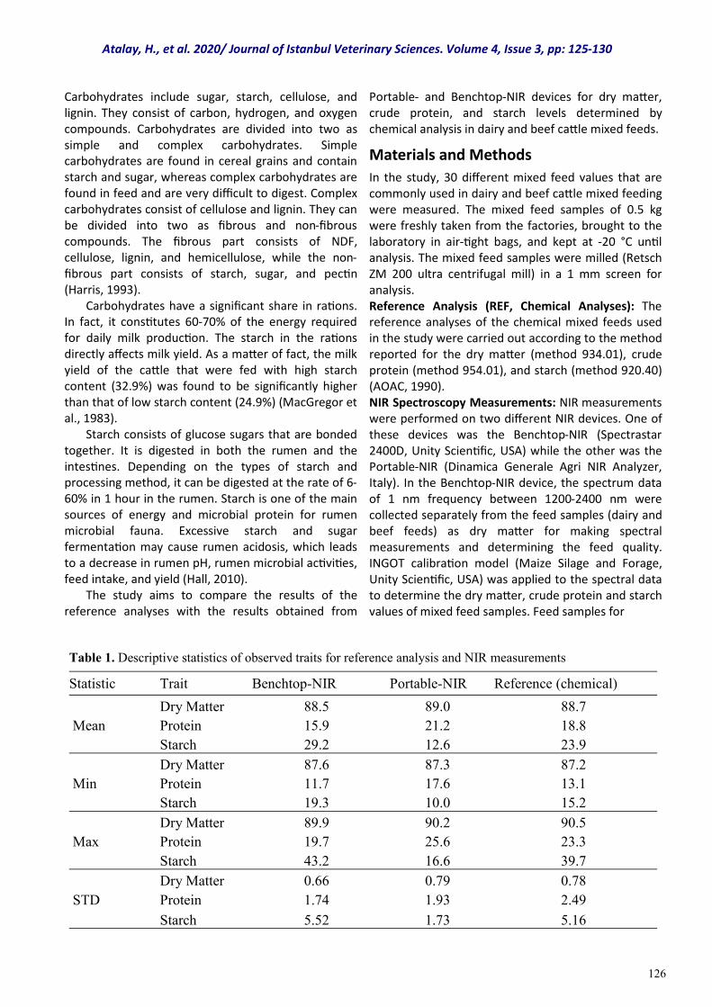
126
Carbohydrates include sugar, starch, cellulose, and lignin. They consist of carbon, hydrogen, and oxygen compounds. Carbohydrates are divided into two as simple and complex carbohydrates. Simple carbohydrates are found in cereal grains and contain starch and sugar, whereas complex carbohydrates are found in feed and are very difficult to digest. Complex carbohydrates consist of cellulose and lignin. They can be divided into two as fibrous and non-fibrous compounds. The fibrous part consists of NDF, cellulose, lignin, and hemicellulose, while the non-fibrous part consists of starch, sugar, and pectin (Harris, 1993). Carbohydrates have a significant share in rations. In fact, it constitutes 60-70% of the energy required for daily milk production. The starch in the rations directly affects milk yield. As a matter of fact, the milk yield of the cattle that were fed with high starch content (32.9%) was found to be significantly higher than that of low starch content (24.9%) (MacGregor et al., 1983). Starch consists of glucose sugars that are bonded together. It is digested in both the rumen and the intestines. Depending on the types of starch and processing method, it can be digested at the rate of 6-60% in 1 hour in the rumen. Starch is one of the main sources of energy and microbial protein for rumen microbial fauna. Excessive starch and sugar fermentation may cause rumen acidosis, which leads to a decrease in rumen pH, rumen microbial activities, feed intake, and yield (Hall, 2010). The study aims to compare the results of the reference analyses with the results obtained from
Portable- and Benchtop-NIR devices for dry matter, crude protein, and starch levels determined by chemical analysis in dairy and beef cattle mixed feeds.
Materials and Methods
In the study, 30 different mixed feed values that are commonly used in dairy and beef cattle mixed feeding were measured. The mixed feed samples of 0.5 kg were freshly taken from the factories, brought to the laboratory in air-tight bags, and kept at -20 °C until analysis. The mixed feed samples were milled (Retsch ZM 200 ultra centrifugal mill) in a 1 mm screen for analysis. Reference Analysis (REF, Chemical Analyses): The reference analyses of the chemical mixed feeds used in the study were carried out according to the method reported for the dry matter (method 934.01), crude protein (method 954.01), and starch (method 920.40) (AOAC, 1990). NIR Spectroscopy Measurements: NIR measurements were performed on two different NIR devices. One of these devices was the Benchtop-NIR (Spectrastar 2400D, Unity Scientific, USA) while the other was the Portable-NIR (Dinamica Generale Agri NIR Analyzer, Italy). In the Benchtop-NIR device, the spectrum data of 1 nm frequency between 1200-2400 nm were collected separately from the feed samples (dairy and beef feeds) as dry matter for making spectral measurements and determining the feed quality. INGOT calibration model (Maize Silage and Forage, Unity Scientific, USA) was applied to the spectral data to determine the dry matter, crude protein and starch values of mixed feed samples. Feed samples for
Atalay, H., et al. 2020/ Journal of Istanbul Veterinary Sciences. Volume 4, Issue 3, pp: 125-130
Table 1. Descriptive statistics of observed traits for reference analysis and NIR measurements
Statistic Trait Benchtop-NIR Portable-NIR Reference (chemical)
Dry Matter 88.5 89.0 88.7
Mean Protein 15.9 21.2 18.8
Starch 29.2 12.6 23.9
Dry Matter 87.6 87.3 87.2
Min Protein 11.7 17.6 13.1
Starch 19.3 10.0 15.2
Dry Matter 89.9 90.2 90.5
Max Protein 19.7 25.6 23.3
Starch 43.2 16.6 39.7
Dry Matter 0.66 0.79 0.78
STD Protein 1.74 1.93 2.49
Starch 5.52 1.73 5.16
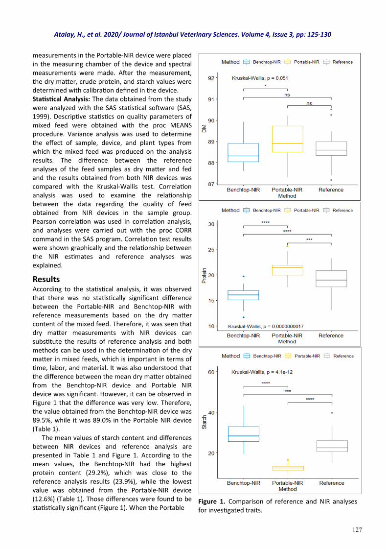
127
measurements in the Portable-NIR device were placed in the measuring chamber of the device and spectral measurements were made. After the measurement, the dry matter, crude protein, and starch values were determined with calibration defined in the device. Statistical Analysis: The data obtained from the study were analyzed with the SAS statistical software (SAS, 1999). Descriptive statistics on quality parameters of mixed feed were obtained with the proc MEANS procedure. Variance analysis was used to determine the effect of sample, device, and plant types from which the mixed feed was produced on the analysis results. The difference between the reference analyses of the feed samples as dry matter and fed and the results obtained from both NIR devices was compared with the Kruskal-Wallis test. Correlation analysis was used to examine the relationship between the data regarding the quality of feed obtained from NIR devices in the sample group. Pearson correlation was used in correlation analysis, and analyses were carried out with the proc CORR command in the SAS program. Correlation test results were shown graphically and the relationship between the NIR estimates and reference analyses was explained.
Results According to the statistical analysis, it was observed that there was no statistically significant difference between the Portable-NIR and Benchtop-NIR with reference measurements based on the dry matter content of the mixed feed. Therefore, it was seen that dry matter measurements with NIR devices can substitute the results of reference analysis and both methods can be used in the determination of the dry matter in mixed feeds, which is important in terms of time, labor, and material. It was also understood that the difference between the mean dry matter obtained from the Benchtop-NIR device and Portable NIR device was significant. However, it can be observed in Figure 1 that the difference was very low. Therefore, the value obtained from the Benchtop-NIR device was 89.5%, while it was 89.0% in the Portable NIR device (Table 1). The mean values of starch content and differences between NIR devices and reference analysis are presented in Table 1 and Figure 1. According to the mean values, the Benchtop-NIR had the highest protein content (29.2%), which was close to the reference analysis results (23.9%), while the lowest value was obtained from the Portable-NIR device (12.6%) (Table 1). Those differences were found to be statistically significant (Figure 1). When the Portable
Figure 1. Comparison of reference and NIR analyses for investigated traits.
Atalay, H., et al. 2020/ Journal of Istanbul Veterinary Sciences. Volume 4, Issue 3, pp: 125-130
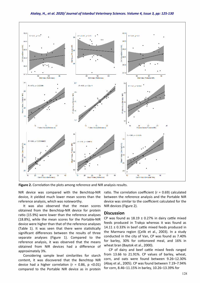
128
NIR device was compared with the Benchtop-NIR device, it yielded much lower mean scores than the reference analysis, which was noteworthy. It was also observed that the mean scores obtained from the Benchtop-NIR device for protein ratio (15.9%) were lower than the reference analyses (18.8%), while the mean scores for the Portable-NIR device were higher than that of the reference analyses (Table 1). It was seen that there were statistically significant differences between the results of three separate analyses (Figure 1). Compared to the reference analysis, it was observed that the means obtained from NIR devices had a difference of approximately 3%. Considering sample level similarities for starch content, it was discovered that the Benchtop NIR device had a higher correlation (r = 0.86, p <0.01) compared to the Portable NIR device as in protein
ratio. The correlation coefficient (r = 0.69) calculated between the reference analysis and the Portable NIR device was similar to the coefficient calculated for the NIR devices (Figure 2).
Discussion CP was found as 18.19 ± 0.27% in dairy cattle mixed feeds produced in Trakya whereas it was found as 14.11 ± 0.33% in beef cattle mixed feeds produced in the Marmara region (Çelik et al., 2003). In a study conducted in the city of Van, CP was found as 7.40% for barley, 30% for cottonseed meal, and 16% in wheat bran (Baytok et al., 2000). CP of dairy and beef cattle mixed feeds ranged from 13.66 to 21.91%. CP values of barley, wheat, corn, and oats were found between 9.26–12.30% (Abaş et al., 2005). CP was found between 7.19–7.94% for corn, 8.46–11.15% in barley, 10.26–13.39% for
Figure 2. Correlation the plots among reference and NIR analysis results.
Atalay, H., et al. 2020/ Journal of Istanbul Veterinary Sciences. Volume 4, Issue 3, pp: 125-130
129
wheat, 13.52–13.83% for wheat bran (thick), and 14.20-15.23% in wheat bran (Göngör et al., 2008). It was also found that that CP was 10.56% for barley, 10.92% in wheat, 8.38% in corn, 12.79% for wheat bran, 29.24% for sunflower seed meal, 32.02% for cotton seed meal, and 44.98% for soybean meal (Alp et al., 1996). In barley varieties, the starch concentration determined by the calculation method (not chemical analysis) was found between 51-64% (Maruz and Çelik, 2020). In addition, the starch content of triticale and wheat varieties measured with NIT SystemInfratec 1241 grain analyzer was found between 62.46-64.65% (Kızılgeçi and Yıldırım, 2017). In a study in which the chemical compositions of poultry feeds used in poultry nutrition were investigated using the chemical and NIRS method, a weak relationship was found in terms of starch content (R2= 0.269 ) while there was a significant relationship between dry matter and crude protein content (R2= 0.7239; 0.9549) (Karaman and Erdemir, 2018). Crude protein and starch content in the total mixed ration (TMR) were estimated by NIRS with good accuracy (R2 ˃ 0.85) in proportion to low standard estimation errors (Mentink et al., 2006). In our study, the results of the correlation analysis performed to examine the differences between the mean values at the sample level are presented in Figure 2. Although there was no statistically significant difference between the methods in terms of dry matter content, the similarity between the different NIR devices and sample analysis results of the reference analyses was quite low according to the correlation analysis. Therefore, it was seen that it would not be correct to use these methods as substitutes at the sample level. According to the results obtained for the protein ratio, it was observed that there was a positive and significant relationship between the results obtained from both NIR devices and the reference analysis. In addition, the results obtained from the Benchtop and Portable-NIR devices showed high similarity. At the sample level, the Benchtop-NIR device had the highest similarity (r = 0.93, p <0.01) whereas a lower similarity (r = 0.72, p <0.01) was determined between Portable-NIR and reference analysis results. Protein is one of the most successfully detected contents in different samples of vegetables (Baye et al., 2006). Therefore, in this study, the results obtained at the sample level were higher for the protein ratio, which was expected. However, it was observed that the success of the Benchtop- and Portable-NIR devices in determining the protein ratio of the fattening feed was different.
The differences between the results obtained from different spectroscopy devices in this study are related to the measurement methods and the calibration models used. The scanning range and frequency of the Portable-NIR device are lower than the Benchtop NIR. However, there may be differences in the calibration and sample compatibility of these estimation models although global calibration models are used in both devices. As a matter of fact, in a study conducted by Tran et al., (2010), the prediction success of local and global models in different feed sources was compared and local models were found to yield more successful results compared to global models.
Conclusion
In this study, the relationship between the reference analysis and the results obtained from the NIR devices having two different properties was investigated by determining the crude protein, starch, and dry matter content of mixed feeds. The results showed that although there were significant differences between the protein and starch content with the reference analyses, there was a remarkable similarity at the sample level according to the correlation analysis. Comparing the sample analyses, it was observed that NIR devices can be used to determine the protein and carbohydrate content of mixed feeds provided that some conversion coefficients need to be developed for the detection. It is useful to carry out different studies for the development of those conversion coefficients by using more samples in future research.
Conflict of interest Authors have no conflict of interest to declare.
References Abaş, İ., Özpınar, H., Kutay, H. C., Kahraman, R., &
Eseceli, H. (2005). Determination of the metabolizable energy (ME) and net energy lactation (NEL) contents of some feeds in the Marmara region by in vitro gas technique. Turkish Journal of Veterinary and Animal Sciences, 29(3), 751-757.
Alp, M., Koçabağlı, N., Kahraman, R., Yetim, M., & Şenel, H. S. (1996). Kanatlı beslenmesinde bullanılan yem maddelerinin ve karma yemlerin besin maddeleri ve enerji kapsamları yönünden değerlendirilmesi. İstanbul Üniversitesi Veteriner Fakültesi Dergisi, 22(1), 9-22.
AOAC (1990). Association of Official Analytical Chemists, 16th ed. Washington, D.C., USA.
Baye, T. M., Pearson, T. C., & Settles, A. M. M. (2006). Development of a calibration to predict maize seed composition using single kernel near infrared spectroscopy. Journal of Ceracel Science, 43, 236-
243.
Atalay, H., et al. 2020/ Journal of Istanbul Veterinary Sciences. Volume 4, Issue 3, pp: 125-130
130
Baytok, E., Muruz, H., Bolat, D., & Aksu, T. (2000). Mera᾽da otlayan süt ineklerine farklı düzeylerde protein ihtiva eden karma yem verilmesinin süt verimi ve kompozisyonuna etkisi. Y.Y.Ü. Veteriner Fakültesi Dergisi, 11(1), 117-120.
Butler, L. D, Cropper, J. B., Johnson, R. H., Norman, A. J., Peacock, G. L., Shaver, P. L., & Spaeth, K.E. (2003). Livestock nutrition, husbandry, and behavior. National range and pasture handbook, 1st ed. United States Department of Agriculture Natural Resources Conservation Service.
Çelik, K., Ertürk, M. M., & Ersoy, İ. E. (2003). Quantitatively Investigating some quality characteristics of compound feeds and feedstuffs sampled out of different feed mills, Akdeniz Üniversitesi Ziraat Fakültesi Dergisi, 16(2), 161-168.
Freeman, S. (2003). Biological Science. Prentice Hall.
Göngör, T., Başalan, M., & Aydoğan, İ. (2008). Kırıkkale yöresinde üretilen bazı kaba yemlerin besin madde miktarları ve metabolize olabilir enerji düzeylerinin belirlenmesi. Ankara Üniversitesi Veteriner Fakültesi Dergisi, 55, 111-115.
Hall, M. B. (2010). Non-fiber carbohydrates. Technical Services Nutritionist F.A.R.M.E. Institute, Inc., Homer, NY.
Harris, B. (1993). Florida Cooperative Extension Service. Nonstructural and structural carbohydrates in dairy cattle rations. Gainesville: Florida Cooperative Extension Service, Institute of Food and Agricultural Sciences, University of Florida.
Karaman, M., & Erdemir, S. (2018). Kanatlı hayvanların beslenmesinde kullanılan bazi karma yemlerin kimyasal kompozisyonunun near infrared reflektans spektroskopi (NIRS) ile belirlenmesi. Black Sea Journal of Agriculture, 1(2), 24-28.
Kızılgeçi, F., & Yıldırım, M. (2017). Bazı tritikale (X
Triticosecale Wittmack) genotiplerinin verim ve kalite özelliklerinin belirlenmesi. Türkiye Tarımsal Araştırmalar Dergisi, 4(1), 43-49.
MacGregor, C. A., Stokes, M. R., Hoover, W. H., Leonard, H. A., Junkins, L. L., Sniffen, Jr. C. J., & Mailman, R. W. (1983). Effect of dietary concentration of total nonstructural carbohydrates on energy and nitrogen metabolism and milk production of dairy cows. Journal of Dairy Science. 66(1), 39-50.
Mentink, R. L., Hoffman, P. C., & Bauman, L. M. (2006). Utility of near-infrared reflectance spectroscopy to predict nutrient composition and in vitro digestibility of total mixed rations. Journal of Dairy Science, 89(6), 2320-2326.
Muruz, H., & Çelik, C. (2020). Bazı arpa (Hordeum vulgare L.) çeşitlerinin in vitro gaz üretimi, organik madde sindirilebilirliği, besin maddeleri içerikleri ve enerji değerlerinin karşılaştırılması. Anadolu Tarım Bilim. Dergisi, 35, 133-139.
SAS Institute 1999, SAS V8 User Manual, SAS Cary, NC.
Tran, H., Salgado, P., Tillard, E., Dardenne, P., Nguyen, X. T., & Lecomte, P. (2010). Global and local predictions of dairy diet nutritional quality using near infrared reflectance spectroscopy, Journal of Dairy Science, 93(10), 4961-4975.
Usta, M., & Saçaklı, P. (2013). Süt ineklerinin beslenmesinde nişastanın önemi ve düşük nişastalı rasyonlarla besleme stratejileri. Erciyes Üniversitesi Veteriner Fakültesi Dergisi, 10(2), 125-133.
Atalay, H., et al. 2020/ Journal of Istanbul Veterinary Sciences. Volume 4, Issue 3, pp: 125-130
131
Case Report
Volume: 4, Issue: 3 December 2020 Pages: 131-135
First report of a perforated abomasal ulcer in a beef heifer calf in Argentina
Journal of Istanbul Veterınary Scıences
Denisa Pérez Gaudio1, Flavio Pérez2, Gustavo Bretschneider3 *
1. Área de Patología, Depto. de Fisiopatología, Centro de Investigación Veterinaria de Tandil (CIVETAN), Universidad Nacional del Centro de la Provincia de Buenos Aires, Tandil, Buenos Aires, Argentina. 2. Eville & Jones. Meat Hygiene Inspection, Norwich, Norfolk, United Kingdom. 3. Instituto Nacional de Tecnología Agropecuaria (INTA) - AER Necochea, Necochea, Buenos Aires, Argentina. Pérez Gaudio, D. ORCID: 0000-0003-2179-7760; Pérez, F. ORCID: 0000-0001-5325-1987; Bretschneider, G. ORCID: 0000-0001-6433-4286
ABSTRACT
Abomasal ulcers are local processes of mucosal autodigestion caused by the disturbance of the balance between protective and aggressive mechanisms in the abomasal mucosa. In order to clarify the etiology, several causes have been discussed and one with a multifactorial origin has been proposed. Signs are mostly non-specific and vary according to different ulcer types. This report describes for the first time in Argentina the clinical case of a perforated abomasal ulcer that induced sudden death in a heifer calf at foot dairy. Necropsy revealed, digestive content in the abdominal cavity and two ulcers in the abomasal mucosa. One ulcer had caused a well-defined 3 cm diameter perforation, which it leading cause has not been determined. Apart from other well known precautions, to minimize the risk of induce perforated abomasal ulcers, stressful management practices should be avoided.
Keywords: calf, abomasal ulcer, perforation
DOI: https://doi.org/10.30704/http-www-jivs-net.794447
To cite this article: Pérez Gaudio, D., Pérez, F., Bretschneider, G. (2020). First report of a perforated abomasal ulcer in a beef heifer calf in Argentina. Journal of Istanbul Veterinary Sciences, 4(3), 131-135. Abbreviated Title: J Ist Vet Sci
Abomasal ulcers, local processes of autodigestion of the mucosa (Kureljušid et al., 2013), represent a significant economic problem and an animal welfare concern. They are an important cause of indigestion in dairy cows, feed-lot beef cattle and calves (Braun et al., 1991; Marshall, 2009). Nevertheless, they seem to be more prevalent in grazing dairy cows in the spring and early summer, and in those cows that have concomitant illnesses (Ceelen, 2010). Abomasal ulcers are the result of pathophysiological conditions, where the balance between the protective and destructive processes is disturbed (Kureljušid et al., 2013). In consequence, the resistance of abomasal mucosa is reduced, due to an increase in the secretion of corticosteroids, gastric acid and pepsin in combination with a decreased synthesis of prostaglandins (Braun et al., 1991).
When it comes to clinical findings, both erosions and ulcers may be found in the abomasum (Smith et al., 1983). Abomasal ulcers can be round, oval or polymorph in shape and clearly demarked from the surrounding mucosa. However, they are often burdened with necrotic debris and blood clots. They usually develop as multiple lesions and solitary ulcers are rarely found. Their radius ranges from a few millimeters up to five centimeters (Marshall, 2009). Unlike erosions, which heal by epithelial regeneration without scar formation, ulcers penetrate the entire thickness of the mucosa and may extend through the submucosa, muscularis externa, and serosa. The central crater of the ulcer has a fibrinonecrotic covering and is surrounded by raised, rounded edges. Healing is by wound contraction and granulation tissue formation, thus resulting in a permanent scar. The
*Corresponding Author: Gustavo Bretschneider E-mail: [email protected]
Journal home page: www.jivs.net http://dergipark.gov.tr/http-www-jivs-net
Article History
Received: 13.09.2020 Accepted: 7.12.2020 Available online: 13.12.2020
This work is licensed under the Creative Commons Attribution 4.0 International License.
Introduction
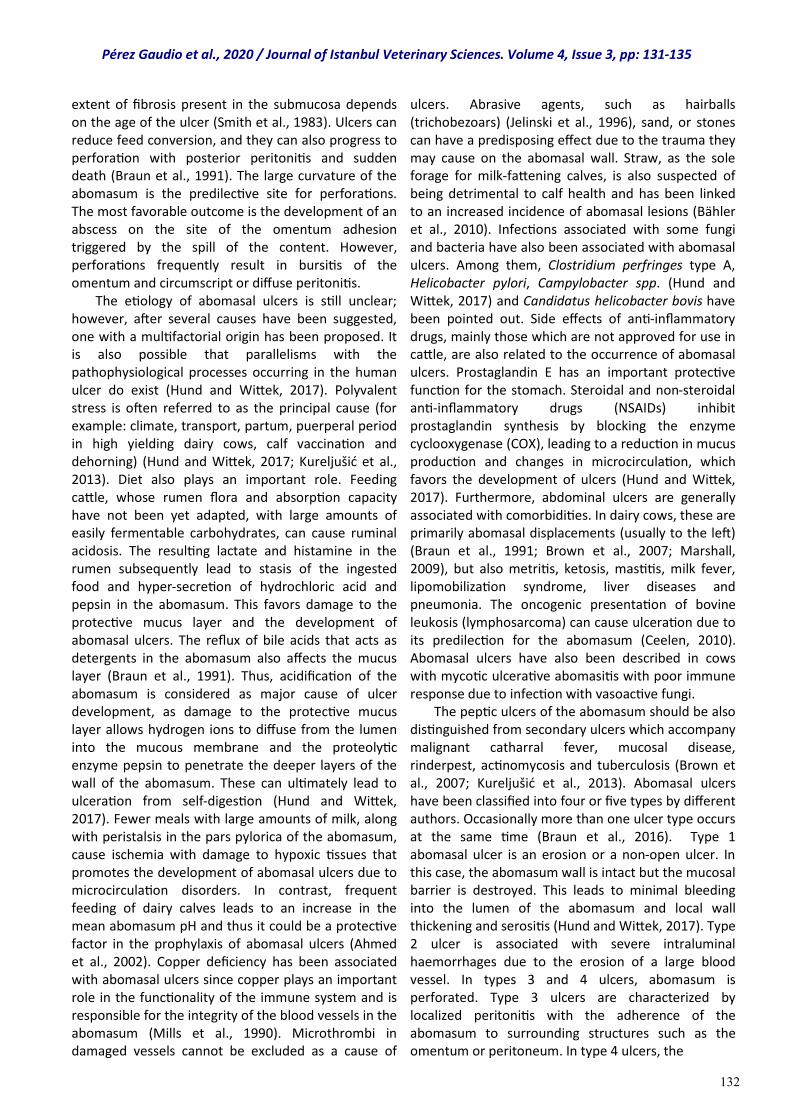
132
extent of fibrosis present in the submucosa depends on the age of the ulcer (Smith et al., 1983). Ulcers can reduce feed conversion, and they can also progress to perforation with posterior peritonitis and sudden death (Braun et al., 1991). The large curvature of the abomasum is the predilective site for perforations. The most favorable outcome is the development of an abscess on the site of the omentum adhesion triggered by the spill of the content. However, perforations frequently result in bursitis of the omentum and circumscript or diffuse peritonitis. The etiology of abomasal ulcers is still unclear; however, after several causes have been suggested, one with a multifactorial origin has been proposed. It is also possible that parallelisms with the pathophysiological processes occurring in the human ulcer do exist (Hund and Wittek, 2017). Polyvalent stress is often referred to as the principal cause (for example: climate, transport, partum, puerperal period in high yielding dairy cows, calf vaccination and dehorning) (Hund and Wittek, 2017; Kureljušid et al., 2013). Diet also plays an important role. Feeding cattle, whose rumen flora and absorption capacity have not been yet adapted, with large amounts of easily fermentable carbohydrates, can cause ruminal acidosis. The resulting lactate and histamine in the rumen subsequently lead to stasis of the ingested food and hyper-secretion of hydrochloric acid and pepsin in the abomasum. This favors damage to the protective mucus layer and the development of abomasal ulcers. The reflux of bile acids that acts as detergents in the abomasum also affects the mucus layer (Braun et al., 1991). Thus, acidification of the abomasum is considered as major cause of ulcer development, as damage to the protective mucus layer allows hydrogen ions to diffuse from the lumen into the mucous membrane and the proteolytic enzyme pepsin to penetrate the deeper layers of the wall of the abomasum. These can ultimately lead to ulceration from self-digestion (Hund and Wittek, 2017). Fewer meals with large amounts of milk, along with peristalsis in the pars pylorica of the abomasum, cause ischemia with damage to hypoxic tissues that promotes the development of abomasal ulcers due to microcirculation disorders. In contrast, frequent feeding of dairy calves leads to an increase in the mean abomasum pH and thus it could be a protective factor in the prophylaxis of abomasal ulcers (Ahmed et al., 2002). Copper deficiency has been associated with abomasal ulcers since copper plays an important role in the functionality of the immune system and is responsible for the integrity of the blood vessels in the abomasum (Mills et al., 1990). Microthrombi in damaged vessels cannot be excluded as a cause of
ulcers. Abrasive agents, such as hairballs (trichobezoars) (Jelinski et al., 1996), sand, or stones can have a predisposing effect due to the trauma they may cause on the abomasal wall. Straw, as the sole forage for milk-fattening calves, is also suspected of being detrimental to calf health and has been linked to an increased incidence of abomasal lesions (Bähler et al., 2010). Infections associated with some fungi and bacteria have also been associated with abomasal ulcers. Among them, Clostridium perfringes type A, Helicobacter pylori, Campylobacter spp. (Hund and Wittek, 2017) and Candidatus helicobacter bovis have been pointed out. Side effects of anti-inflammatory drugs, mainly those which are not approved for use in cattle, are also related to the occurrence of abomasal ulcers. Prostaglandin E has an important protective function for the stomach. Steroidal and non-steroidal anti-inflammatory drugs (NSAIDs) inhibit prostaglandin synthesis by blocking the enzyme cyclooxygenase (COX), leading to a reduction in mucus production and changes in microcirculation, which favors the development of ulcers (Hund and Wittek, 2017). Furthermore, abdominal ulcers are generally associated with comorbidities. In dairy cows, these are primarily abomasal displacements (usually to the left) (Braun et al., 1991; Brown et al., 2007; Marshall, 2009), but also metritis, ketosis, mastitis, milk fever, lipomobilization syndrome, liver diseases and pneumonia. The oncogenic presentation of bovine leukosis (lymphosarcoma) can cause ulceration due to its predilection for the abomasum (Ceelen, 2010). Abomasal ulcers have also been described in cows with mycotic ulcerative abomasitis with poor immune response due to infection with vasoactive fungi. The peptic ulcers of the abomasum should be also distinguished from secondary ulcers which accompany malignant catharral fever, mucosal disease, rinderpest, actinomycosis and tuberculosis (Brown et al., 2007; Kureljušid et al., 2013). Abomasal ulcers have been classified into four or five types by different authors. Occasionally more than one ulcer type occurs at the same time (Braun et al., 2016). Type 1 abomasal ulcer is an erosion or a non-open ulcer. In this case, the abomasum wall is intact but the mucosal barrier is destroyed. This leads to minimal bleeding into the lumen of the abomasum and local wall thickening and serositis (Hund and Wittek, 2017). Type 2 ulcer is associated with severe intraluminal haemorrhages due to the erosion of a large blood vessel. In types 3 and 4 ulcers, abomasum is perforated. Type 3 ulcers are characterized by localized peritonitis with the adherence of the abomasum to surrounding structures such as the omentum or peritoneum. In type 4 ulcers, the
Pérez Gaudio et al., 2020 / Journal of Istanbul Veterinary Sciences. Volume 4, Issue 3, pp: 131-135
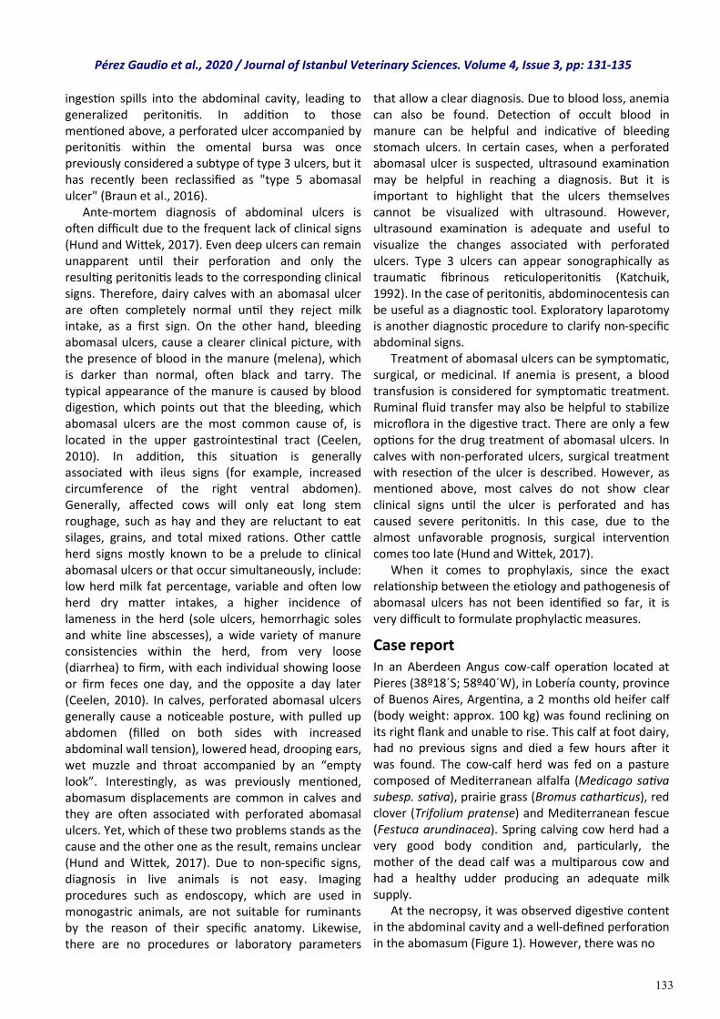
133
ingestion spills into the abdominal cavity, leading to generalized peritonitis. In addition to those mentioned above, a perforated ulcer accompanied by peritonitis within the omental bursa was once previously considered a subtype of type 3 ulcers, but it has recently been reclassified as "type 5 abomasal ulcer" (Braun et al., 2016). Ante-mortem diagnosis of abdominal ulcers is often difficult due to the frequent lack of clinical signs (Hund and Wittek, 2017). Even deep ulcers can remain unapparent until their perforation and only the resulting peritonitis leads to the corresponding clinical signs. Therefore, dairy calves with an abomasal ulcer are often completely normal until they reject milk intake, as a first sign. On the other hand, bleeding abomasal ulcers, cause a clearer clinical picture, with the presence of blood in the manure (melena), which is darker than normal, often black and tarry. The typical appearance of the manure is caused by blood digestion, which points out that the bleeding, which abomasal ulcers are the most common cause of, is located in the upper gastrointestinal tract (Ceelen, 2010). In addition, this situation is generally associated with ileus signs (for example, increased circumference of the right ventral abdomen). Generally, affected cows will only eat long stem roughage, such as hay and they are reluctant to eat silages, grains, and total mixed rations. Other cattle herd signs mostly known to be a prelude to clinical abomasal ulcers or that occur simultaneously, include: low herd milk fat percentage, variable and often low herd dry matter intakes, a higher incidence of lameness in the herd (sole ulcers, hemorrhagic soles and white line abscesses), a wide variety of manure consistencies within the herd, from very loose (diarrhea) to firm, with each individual showing loose or firm feces one day, and the opposite a day later (Ceelen, 2010). In calves, perforated abomasal ulcers generally cause a noticeable posture, with pulled up abdomen (filled on both sides with increased abdominal wall tension), lowered head, drooping ears, wet muzzle and throat accompanied by an “empty look”. Interestingly, as was previously mentioned, abomasum displacements are common in calves and they are often associated with perforated abomasal ulcers. Yet, which of these two problems stands as the cause and the other one as the result, remains unclear (Hund and Wittek, 2017). Due to non-specific signs, diagnosis in live animals is not easy. Imaging procedures such as endoscopy, which are used in monogastric animals, are not suitable for ruminants by the reason of their specific anatomy. Likewise, there are no procedures or laboratory parameters
that allow a clear diagnosis. Due to blood loss, anemia can also be found. Detection of occult blood in manure can be helpful and indicative of bleeding stomach ulcers. In certain cases, when a perforated abomasal ulcer is suspected, ultrasound examination may be helpful in reaching a diagnosis. But it is important to highlight that the ulcers themselves cannot be visualized with ultrasound. However, ultrasound examination is adequate and useful to visualize the changes associated with perforated ulcers. Type 3 ulcers can appear sonographically as traumatic fibrinous reticuloperitonitis (Katchuik, 1992). In the case of peritonitis, abdominocentesis can be useful as a diagnostic tool. Exploratory laparotomy is another diagnostic procedure to clarify non-specific abdominal signs. Treatment of abomasal ulcers can be symptomatic, surgical, or medicinal. If anemia is present, a blood transfusion is considered for symptomatic treatment. Ruminal fluid transfer may also be helpful to stabilize microflora in the digestive tract. There are only a few options for the drug treatment of abomasal ulcers. In calves with non-perforated ulcers, surgical treatment with resection of the ulcer is described. However, as mentioned above, most calves do not show clear clinical signs until the ulcer is perforated and has caused severe peritonitis. In this case, due to the almost unfavorable prognosis, surgical intervention comes too late (Hund and Wittek, 2017). When it comes to prophylaxis, since the exact relationship between the etiology and pathogenesis of abomasal ulcers has not been identified so far, it is very difficult to formulate prophylactic measures.
Case report In an Aberdeen Angus cow-calf operation located at Pieres (38º18´S; 58º40´W), in Lobería county, province of Buenos Aires, Argentina, a 2 months old heifer calf (body weight: approx. 100 kg) was found reclining on its right flank and unable to rise. This calf at foot dairy, had no previous signs and died a few hours after it was found. The cow-calf herd was fed on a pasture composed of Mediterranean alfalfa (Medicago sativa subesp. sativa), prairie grass (Bromus catharticus), red clover (Trifolium pratense) and Mediterranean fescue (Festuca arundinacea). Spring calving cow herd had a very good body condition and, particularly, the mother of the dead calf was a multiparous cow and had a healthy udder producing an adequate milk supply. At the necropsy, it was observed digestive content in the abdominal cavity and a well-defined perforation in the abomasum (Figure 1). However, there was no
Pérez Gaudio et al., 2020 / Journal of Istanbul Veterinary Sciences. Volume 4, Issue 3, pp: 131-135
134
Figure 1. Digestive content in the abdominal cavity and the well-defined perforation found in the abomasum. Arrows show the perforation site.
evidence of peritonitis or adherences. At the opening, the mucosa of the abomasum presented two ulcers, one of 3 cm in diameter with an irregular perforation in the center (Figure 2) and the other, smaller (2 cm diameter) without perforation. Abrasive objects were not found into the digestive tract. Moreover, the rest of the organs did not exhibited evident alterations.
Figure 2. Perforated abomasal ulcer in the calf presented in this study. Arrows show the perforation site.
Discussion and conclusion Considering that most of abomasal ulcers may be subclinical, in absence of dark, tarry manure, it is difficult to conclusively arrive at abomasal ulcers diagnosis. In our case, perforated abomasal ulcer was diagnosed by necropsy examination. The radius of the perforation may play a decisive role for the developing ulcer type, thus, radius of 1 to 3 mm on the perforation site results in type 3 ulcers while perforations from 1.25 to 3 cm result in type 4 ulcerations (Kureljušid et al., 2013). The perforated ulcer presented in this case was of 3 cm diameter (i.e; 1.5 cm radius), which could be classified as type 4 ulcer. In Canadian cow-calf operations, over a decade, 209 calf deaths were reported to be caused by
abomasal ulcers. Of the total, 93.3% of the lesions corresponded to perforating ulcers while 6.7% were in concordance with hemorrhagic ones. The episodes, mostly subclinical, were mainly recorded in calves with ≤ 2 months of age (86% of cases). There was no predisposition associated with sex or breed. It is important to note that none of the previously listed ulceration causes have been scientifically associated as a causative agent of abomasum ulcers of these cows (Jelinski et al., 1996). On the other hand, in particular, hairballs were found in 35 of 46 cases of abomasal ulcers (Wittek et al., 2016). However, the role they played in the development of ulcers has been rejected, considering that the friction exerted by a hairball on the abomasal mucosa would be unable to erode it. In the clinical case reported here, no hairballs were found in the abomasum. In dairy calves in Switzerland, abomasal ulcers are known to be responsible for 25% of all deaths, with a prevalence of 0.2-5.7% (Hund and Wittek, 2017). The fact that the highest number of cases was reported in calves up to 2 months of age, reveals an association between the formation of ulcers and the development of pre-stomachs. The transition process between pre-ruminant to ruminant occurs between the third and eighth week of life. In other words, both the pre-ruminant period (<3 weeks old) and the period of transition to ruminant (3 to 8 weeks old) represent the moments of lifetime in which calves are most susceptible to death from an abomasal perforated ulcer. However, the cause associated with the physiological transition to ruminant which triggers the development of abomasal ulcers, is still unknown (Jelinski et al., 1996). Taking this into consideration the death of the calf reported in this study seems to be supported by data and within risk range of age. Clostridium perfringens and Campylobacter jejuni have been described in relation to abomasal ulcers in calves. Nevertheless, a recent study in dairy calves, found no differences between healthy calves and ulcerated calves with regard to the presence or the absence of Clostridium perfringens. Candidatus helicobacter bovis has been proposed as a new potentially ulcerogenic agent, although its involvement in gastric disease in cattle is presently unknown. Other bacteria, such as Helicobacter spp., which is clearly associated with gastric ulcers in humans, has not been detected in dairy calves. Therefore, the role of these bacteria in the pathogenesis of abomasal ulcers can be classified as low to non-existent (Hund and Wittek, 2017). Differential diagnoses for abomasal ulcers differ between ulcer types and according to the animal´s
Pérez Gaudio et al., 2020 / Journal of Istanbul Veterinary Sciences. Volume 4, Issue 3, pp: 131-135
135
age. Overall, for perforated abomasal ulcers, the differential diagnosis includes, omphalitis, uterine rupture, traumatic reticuloperitonitis rumen bloat, endoparasites, and peritonitis, among others (Hund and Wittek, 2017). For prophylactic purposes, in general, cattle husbandry practices that induce calf stress should be minimized, since particularly, well-developed calves often die by perforated abomasal ulcers within days of these practices (Braun et al., 1991). It is also important to point out that the death, reported herein, corresponds to medical foresights when discussing about a calf born and raised in an extensive pasture-based cow-calf operation system that cares cattle well-being. Despite the relationships in the development of abomasal ulcers remain without being fully understood, their multifactorial genesis is unquestionably. This fact has made it impossible to reach the leading cause of the perforated abomasal ulcer reported herein. Nevertheless, as it was previously described, the most plausible cause may be associated to the pre-ruminant to ruminant transition.
Conflict of interest The authors have no conflicts of interest to declare.
References Ahmed, A. F., Constable, P. D. & Misk, N. A. (2002).
Effect of feeding frequency and route of administration on abomasal luminal pH in dairy calves fed milk replacer. Journal of Dairy Science, 85(6), 1502-1508.
Bähler, C., Regula, G., Stoffel, M. H., Steiner, A. & von Rotz, A. (2010). Effects of the two production programs ‘Naturafarm’ and ‘conventional’ on the prevalence of non-perforating abomasal lesions in Swiss veal calves at slaughter. Research in Veterinary Science, 88(2), 352-360.
Braun, U. (2016). Ascites in cattle: Ultrasonographic findings and diagnosis. Veterinary Clinics: Food Animal Practice, 32(1), 55-83.
Braun, U., Bretscher, R. & Gerber, D. (1991). Bleeding abomasal ulcers in dairy cows. The Veterinary Record, 129(13), 279-284.
Brown, C. C., Baker, D. C., Barker, I. K. (2007). The alimentary system. In Jubb K. F., Kennedy P. C., & Palmer N. (Ed). Pathology of Domestic Animals. 5th ed. (pp 1-297). San Diego, US: Academic Press.
Ceelen, H. (2010, March 1). Prevent abomasal ulcers. Vet’s Corner. Ontario Dairy Farmer, 74-75. Retrieved from http://oabp.ca/wp-content/uploads/2014/02/
Abomasal-Ulcers.pdf.
Hund, A. & Wittek, T. (2017). Labmagengeschwüre beim Rind. Tierärztliche Praxis Ausgabe G: Großtiere/Nutztiere, 45(02), 121-128.
Jelinski, M. D., Ribble, C. S., Campbell, J. R. & Janzen, E.D. (1996). Descriptive epidemiology of fatal abomasal ulcers in Canadian beef calves. Preventive Veterinary Medicine, 26(1), 9-15.
Katchuik, R. (1992). Abomasal disease in young beef calves: surgical findings and management factors. The Canadian Veterinary Journal, 33(7), 459.
Kureljušid, B., Ivetid, V., Savid, B., Jezdimirovid, N., Cvetojevid, Đ., Kureljušid, J. et al. (2013). Pathomorphological characteristics of abomasal ulcers in high-yielding dairy cows. Acta veterinaria, 63(2-3), 237-246.
Marshall, T. S. (2009). Abomasal ulceration and tympany of calves. Veterinary Clinics of North America: Food Animal Practice, 25(1), 209-220.
Mills, K. W., Johnson, J. L., Jensen, R. L., Woodard, L. F. & Doster, A. R. (1990). Laboratory findings associated with abomasal ulcers/tympany in range calves. Journal of Veterinary Diagnostic Investigation, 2(3), 208-212.
Smith, D. F., Munson, L. & Erb, H. N. (1983). Abomasal ulcer disease in adult dairy cattle. The Cornell Veterinarian, 73(3), 213-224.
Wittek, T., Ernstberger, M., Muckenhuber, M. & Flöck, M. (2016). Effects of wheat protein in milk replacers on abomasal emptying rate in calves. Journal of Animal Physiology and Animal Nutrition, 100(2), 264-270.
Pérez Gaudio et al., 2020 / Journal of Istanbul Veterinary Sciences. Volume 4, Issue 3, pp: 131-135
136
Research Article
Volume: 4, Issue: 3 December 2020 Pages: 136-144
Ameliorative effects of Allium cepa Linn. scaly leaves extract on reproductive dysfunctions in streptozotocin-induced diabetic Wistar rats
Journal of Istanbul Veterınary Scıences
Olushola Emmanuel Adeleye,1*, Emmanuel Oluwatosin Okoh1, Adenike Iyabo Adeleye2, Fakilahyel Musa Mshelbwala3, Abiodun Seun Adetomiwa4, James Taiwo Apantaku3, Ngozichukwu Afoma Aboajah1, Ladoke A. Durotoye1, Johnny Olufemi Olukunle1
1. Department of Veterinary Physiology and Pharmacology, College of Veterinary Medicine, Federal University of Agriculture, Abeokuta, Nigeria. 2.Veterinary Teaching Hospital, College of Veterinary Medicine, Federal University of Agriculture, Abeokuta, Nigeria. 3.Department of Veterinary Pathology, College of Veterinary Medicine, Federal University of Agriculture, Abeokuta, Nigeria. 4. Department of Veterinary Surgery and Theriogenology, College of Veterinary Medicine, Federal University of Agriculture, Abeokuta, Nigeria.
ABSTRACT
Diabetes mellitus, an endocrine and metabolic disorder characterized by hyperglycemia and low blood insulin or target organs insensitivity to insulin affects life quality due to its complications. Infertility is a complication in diabetes. Various agents have been used for research on diabetes-induced infertility globally, but there are little documented treatments for diabetes associated infertility. Allium cepa scaly leaves extract (ACSLE) possess anti-oxidant and anti-diabetic activities. This study investigated the effects of ACSLE on reproductive dysfunctions in male diabetic rats. Twenty eight male rats were assigned to 4 groups (n=7): CT (control); DNT (streptozotocin, 60 mg/kg once, intraperitoneal), DT1 and DT2 (streptozotocin, 60 mg/kg once, intraperitoneal, ACSLE 125 and 250 mg/kg rat/day respectively). Organ samples were obtained after 2 weeks and testicular weights recorded. Fasting blood glucose was determined using a digital glucometer. Sperm count, motility, viability and morphology were assessed microscopically. Testes were histologically evaluated. Glucose levels were reduced in DT1 and DT2 compared with DNT. Testes weights increased in DT1and DT2 compared with DNT. Sperm concentration increased in DT1 and DT2 compared with DNT motility increased in DT1 and DT2 compared with DNT; viability increased significantly in DT1 and DT2 compared with DNT. Abnormal sperm morphology decreased in DT1 and DT2 compared with DNT. Testes showed degenerated cells in DNT and no lesions in DT1 and DT2. Reduced blood glucose, improved testicular functions and morphology showed that ACSLE ameliorated reproductive dysfunctions associated with streptozotocin-induced diabetes in male Wistar rats.
Keywords: Allium cepa; diabetes; streptozotocin; reproductive dysfunctions
DOI: https://doi.org/10.30704/http-www-jivs-net.811491
To cite this article: Adeleye, O, E., Okoh, E. O., Adeleye, A. I., Mshelbwala, F. M., Adetomiwa, A. S., Apantaku, J. T., Aboajah, N. A., Durotoye, L. A., Olukunle, J. O. (2020). Ameliorative effects of Allium cepa Linn. scaly leaves extract on reproductive dysfunctions in streptozotocin-induced diabetic Wistar rats. Journal of Istanbul Veterinary Sciences, 4(3), 136-144 Abbreviated Title: J Ist Vet Sci
Diabetes mellitus (DM), a disorder in which the body is unable to properly metabolize carbohydrates is increasing globally and now affects 9.3 % of the world’s adult population (Saeedi et al., 2019). It is estimated that 463 million people had diabetes in 2019 (Saeedi et al., 2019) and without urgent and sufficient actions, it is predicted that 578 million
people will have diabetes in 2030 and the number will increase by 51% (700 million) in 2045 (Saeedi et al., 2019). The disease is characterized by excessive amounts of sugar in the blood and urine; inadequate production and / or utilization of insulin; thirst, hunger and loss of weight (Maiti et al., 2004). DM is a complex metabolic
*Corresponding Author: Olushola Emmanuel Adeleye E-mail: [email protected]
Journal home page: www.jivs.net http://dergipark.gov.tr/http-www-jivs-net
Article History
Received: 16.10.2020 Accepted: 14.12.2020 Available online: 19.12.2020
This work is licensed under the Creative Commons Attribution 4.0 International License.
Introduction
137
disorder that results from defects in insulin secretion, action, or a combination of both (Akkati et al., 2011). DM is grouped into two main types, type I diabetes mellitus (T1DM) and type II diabetes mellitus (T2DM) (American Diabetes Association, 2019). T1DM results from defective beta cells in the islets of Langerhans which could be caused by destruction by various infections, diseases and exposure to various toxic chemicals (Cooke and Plotnick, 2008; Eizirik et al., 2009; Akkati et al., 2011). Major symptoms are polyphagia, polydipsia, polyuria and weight loss (Cooke and Plotnick, 2008; Akkati et al., 2011; Ramachandran, 2014). T2DM is characterized by persistently high blood glucose caused by insulin resistance and usually with relative insulin deficiency (Butler I., 2003; Akkati I., 2011). Risk factors for T2DM are genetic predisposition, hyperlipidemia, lifestyle factors (including obesity, lack of physical activity, poor diet, stress, urbanization) and history of gestational diabetes (Kahn and Hull, 2006; Riserus et al., 2009; Akkati et al., 2011). Glucose metabolism is an important event in spermatogenesis in DM and deleterious effects of DM leads to male infertility (Amidu et al., 2013; Koroglu et al., 2015) via actions at multiple levels including altered spermatogenesis, degenerative and apoptotic changes in testes, altered glucose metabolism in blood-testes barrier, reduced testosterone, luteinizing hormone (LH), follicle stimulating hormone (FSH) synthesis and secretion (Tsounapi et al., 2016); decreased spermatozoa motility (Saumya et al., 2016) and semen volume (Adedara et al., 2015); abnormal spermatozoa morphology (Afifi et al., 2015) and the disruption of seminiferous tubule morphology (Rashid et al., 2015); ejaculatory dysfunction and reduced libido (Sexton and Jarow, 1997; Baccetti et al., 2002; Cavallini, 2006; Scarano et al., 2006; Agbaje et al., 2007; Kilarkaje et al., 2014; Ghanbari et al., 2015). Diabetes is linked with increased oxidative stress (Freitas et al., 1997; Abou-Seif and Youssef, 2004) which may play an important role in the cause of diabetic complications. Physiologically, the balance between reactive oxygen species (ROS) production and antioxidant activity is maintained in the body, but when ROS accumulates in excessive amounts either due to increased generation or impaired clearance, they can cause deleterious effects on sperm cells which can lead to sperm dysfunction (Marti et al., 2007; Agarwal et al., 2008; Tremellen, 2008). The management of DM without negative effects is still challenging to the medical system, which has increased demands for natural and other products
with anti-diabetic activity and little or no side effects (Kameswara et al., 1999; Philippe and Raccah, 2009; Akkati et al., 2011). Plants, plants extract, and the active compound from plants usage to manage diseases are crucial in new drugs discovery (Rupeshkumar et al., 2014). Allium cepa (onion) is a plant that belongs to the Alliaceae family (Shaath and Flores, 1998). It is the most widely cultivated species of the genus Allium (Bindu and Podikunju, 2015) and contains pharmacologically active constituents, including flavonoids (quercetin), organosulfur compounds (propyl thiosulfinate), and phenol components, fructooligosaccharides (FOS) compounds (Slimestad et al., 2007). Allium cepa possess anti-allergic (Roldan et al., 2008; Marefati et al., 2018), anti-inflammatory, and antioxidant activities (Griffiths et al., 2002; Lee and Jung, 2016; Marefati et al., 2018). Peak concentrations of quercetin is attained 7.0 ± 2.9 hours after ingestion, and its elimination half-life is about 11 hours. Also, the plant matrix influences both the rate and extent of absorption (Marefati et al., 2018). Flavonoids are the major phenolics in onions, which can be classified to different subclasses (flavones, flavanones, flavonols, isoflavones, flavanonols, flavanols, chalcones, and anthocyanins) on the basis of the degree of unsaturation and the degree of oxidation of the central ring (P´erez-Gregorio et al., 2010). Flavonoids subclasses can be further differentiated on the basis of the number and nature of substituent groups attached to the rings (P´erez-Gregorio et al., 2010). Flavonols are the most abundant in onions, present as their glycosides, that is, quercetin and kaempferol (Prakash et al., 2007; Santas et al., 2010; Nile et al., 2020) in higher concentration (280-400 mg/kg) than other vegetables (i.e., 100 mg/kg in broccoli, 50 mg/kg in apple) (de Ancos et al., 2015). Anthocyanins, belonging to anthocyanidins, are mainly present in red onions (250 mg/kg), besides having a composition rich in flavonols as yellow onions (de Ancos et al., 2015). Quercetin which is one of the most common flavonoids in A. cepa has been reported to improve diabetic status by decreasing oxidative stress (Mahesh and Menon, 2004; Coskun et al., 2005; Dias et al., 2005) and by reducing the disturbance of hepatic gene expressions (Kobori et al., 2009; Jiet al., 2011).
The objectives of this study were to study the anti-diabetic, antioxidant activity and free radical scavenging effects of Allium cepa scaly leaf extract on high blood glucose level and impaired testicular functions and morphology associated with streptozotocin-induced diabetic male Wistar rats.
Adeleye et al., 2020/ Journal of Istanbul Veterinary Sciences. Volume 4, Issue 3, pp: 136-144
138
Materials and methods
Ethical statement: This experiment was conducted in the experimental animal house at the College of Veterinary Medicine, Federal University of Agriculture, Abeokuta, Nigeria according to the guidance and approval of the committee on animal care ethics and use, Federal University of Agriculture, Abeokuta (CACEU/2019/3224).
Animals: Twenty eight male Wistar rats (150-200g) were used for this study. The rats were housed in the experimental animal house of the college. They were kept in well ventilated standard rat cages at ambient temperature and 12 hour light/darkness period was maintained. The rats were fed standard pelleted rat chow and clean water was given ad libitum. They were kept for 10 days for adaptation prior to start of the experiment.
Plant material: Allium cepa L. (onion) were obtained from a market in Abeokuta, Ogun State. Identification and authentication was done at Department of Pure and Applied Botany, College of Biosciences, Federal University of Agriculture, Abeokuta (FUNAABH0029).
Plant extraction: Allium cepa L. scaly leaves were removed from the onion and reduced into coarse particles. The coarse particles were soaked in 97% ethanol for 72 hours at room temperature. The mixture was filtered using Whatman’s filter paper and the filtrate was evaporated at 600C using a vacuum rotary evaporator. The wet brown residue i.eAllium cepa scaly leaves extract (ACSLE) was allowed to evaporate in vacuo and stored in the refrigerator (40C) until ready to use. Thereafter, 1 g of the residue was dissolved in 20 mL of distilled water to give a concentration of 50 mg/mL.
Plant phytochemical analysis: Standard screening tests for the extract was performed for various plant constituents. Presence or absence of secondary metabolites such as alkaloids, steroidal compounds, phenolic compounds, flavonoids, saponins, tannins, and anthraquinones were screened for using standard procedures(Edeogaet al., 2005).
Diabetes induction: Streptozotocin (STZ) (Sigma Chemical Co., St. Louis, MO, USA) was used to induce diabetes in rats. It was injected intra-peritoneally at a single dose of 60 mg/kg of body weight after being
freshly prepared in ice-cold citrate buffer of pH 4.5 (Akbarzadeh et. al., 2007). Animals with glucose levels above 300 mg/dL were considered diabetic and were included in the study.
Experimental Procedure: The rats were randomly distributed into 4 groups (n=7) after diabetes induction and treated as listed in Table 1. Blood glucose measurement Fasting blood glucose levels were estimated 3 times within a week during the treatment period of two weeks using blood sample obtained from the tail vein of the rats and determined in mg/dL by a digital glucometer (Accu-chek Advantage, Roche Diagnotics, Germany). The animals were fasted for a period of 16 hours before their blood glucose levels were measured. At the end of the 14 days treatment, the rats were euthanized with thiopental injection (50 mg/kg) and a ventral midline abdominal incision was made to expose the reproductive organs. The testes were identified, carefully removed and processed further for sperm evaluation; sperm motility, concentration, viability and morphology as described by Jequier, (2010) and testicular histology.
Statistical Analysis: The differences between the means were analyzed statistically with one-way analysis of variance, followed by Tukey’s comparison test (ANOVA; 95% confidence interval). Values of P<0.05 were taken to imply statistical significance. Results were expressed as mean and standard error of mean and were presented in bar charts. Statistical Package for the Social Science (SPSS) software (version 16.0; SPSS Inc., USA) was used in all data analysis.
Results Phytochemical studies: Tannins, saponins, anthraquinones, flavonoids, sterols, alkaloids and terpenoids were present in the extract (Table 2). Hypoglycemic effects of ACSLE on blood glucose: The mean values of blood glucose levels revealed a reduction in the DT1 (175.3 ± 2.3 mg/dL) and DT2 (165.0 ± 1.8 mg/dL) than that in the DNT (440.0 ± 13.0 mg/dL) after 14 days (Figure 1). Effects of ACSLE on sperm variables: On administration of ACSLE for 14 days, the mean values of sperm motility (77.7 ± 1.5, 80.0 ± 0.0 %), viability (70.0 ± 0.0, 80.0 ± 0.00 %), concentration (130.0 ± 5.9,
Table 1. Animal groupings and daily protocols
Group Protocols
CT Control group received distilled water of 0.5 ml daily.
DNT Diabetic group received 60 mg/kg STZ once
DT1 Received 60 mg/kg STZ once and treated with 125 mg/kg ACSLE (O.E.C.D., 2008), 2 weeks
DT2 Received 60 mg/kg STZ once and treated with 250 mg/kg ACSLE (O.E.C.D., 2008), 2 weeks
Adeleye et al., 2020/ Journal of Istanbul Veterinary Sciences. Volume 4, Issue 3, pp: 136-144
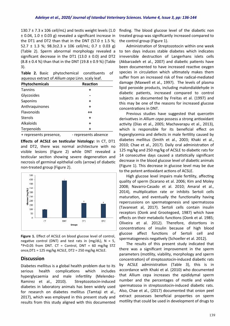
139
130.7 ± 7.3 x 10⁶ cell/mL) and testis weight levels (1.0 ± 0.04, 1.0 ± 0.03 g) revealed a significant increase in the DT1 and DT2 than that in the DNT (57.0 ± 1.5 %; 52.7 ± 1.3 %; 98.3±2.3 x 10⁶ cell/mL; 0.7 ± 0.03 g) (Table 2). Sperm abnormal morphology revealed a significant decrease in the DT1 (13.0 ± 0.0) and DT2 (8.8 ± 0.4 %) than that in the DNT (19.8 ± 0.9 %) (Table 3).
Effects of ACSLE on testicular histology: In CT, DT1 and DT2, there was normal architecture with no visible lesions (Figure 2) while DNT revealed a testicular section showing severe degeneration and necrosis of germinal epithelial cells (arrow) of diabetic non-treated group (Figure 2).
Figure 1. Effect of ACSLE on blood glucose level of control, negative control (DNT) and test rats in (mg/dL), N = 5, *P<0.05 from DNT. CT = Control, DNT = 60 mg/kg STZ once,DT1 = 125 mg/kg ACSLE, DT2 = 250 mg/kg ACSLE.
Discussion
Diabetes mellitus is a global health problem due to its serious health complications which includes hyperglycaemia and male infertility (Melendez-Ramirez et al., 2010). Streptozotocin-induced diabetes in laboratory animals has been widely used for research on diabetes mellitus (Tamtaji et al., 2017), which was employed in this present study and results from this study aligned with this documented
finding. The blood glucose level of the diabetic non treated group was significantly increased compared to the control group (Figure 1). Administration of Streptozotocin within one week to ten days induces stable diabetes which indicates irreversible destruction of Langerhans islets cells (Akbarzadeh et al., 2007) and diabetic patients have been documented to have increased reactive oxygen species in circulation which ultimately makes them suffer from an increased risk of free radical-mediated damage (Maxwell et al., 1997). The levels of plasma lipid peroxide products, including malondialdehyde in diabetic patients, increased compared to control subjects as documented by Freitas et al. (1997) and this may be one of the reasons for increased glucose concentrations in DNT. Previous studies have suggested that quercetin derivatives in Allium cepa possess a strong antioxidant activity (Dias et al., 2005; Machavarapu et al., 2013), which is responsible for its beneficial effect on hyperglycemia and defects in male fertility caused by diabetes mellitus (Smith et al., 2003; Khaki et al., 2010; Chae et al., 2017). Daily oral administration of 125 mg/kg and 250 mg/kg of ACSLE to diabetic rats for 14 consecutive days caused a statistically significant decrease in the blood glucose level of diabetic animals (Figure 1). This decrease in glucose level may be due to the potent antioxidant actions of ACSLE. High glucose level impairs male fertility, affecting quality of sperm (Scarano et al. 2006; Kim and Moley 2008; Navarro-Casado et al. 2010; Amaral et al., 2014), multiplication rate or inhibits Sertoli cells maturation, and eventually the functionality having repercussions on spermatogenesis and spermatozoa (Tavareset al, 2017). Sertoli cells contain insulin receptors (Oonk and Grootegoed, 1987) which have effects on their metabolic functions (Oonk et al. 1985; Oliveira et al. 2012). Therefore, distortions in concentrations of insulin because of high blood glucose affect functions of Sertoli cell and spermatogenesis negatively (Schoeller et al. 2012). The results of this present study indicated that there was a significant improvement in the sperm parameters (motility, viability, morphology and sperm concentration) of streptozotocin-induced diabetic rats by ACSLE administration (Table 3), this is in accordance with Khaki et al. (2010) who documented that Allium cepa increases the epididymal sperm number and the percentages of motile and viable spermatozoa in streptozotocin-induced diabetic rats. Also, Chae et al., (2017) documented that onion peel extract possesses beneficial properties on sperm motility that could be used in development of drugs to
Table 2. Basic phytochemical constituents of aqueous extract of Allium cepa Linn. scaly leaf.
Phytochemicals Reaction
Tannins +
Glycosides -
Saponins +
Anthraquinones +
Flavonoids ++
Sterols +
Alkaloids +
Terpenoids +
+ represents presence, - represents absence
Adeleye et al., 2020/ Journal of Istanbul Veterinary Sciences. Volume 4, Issue 3, pp: 136-144
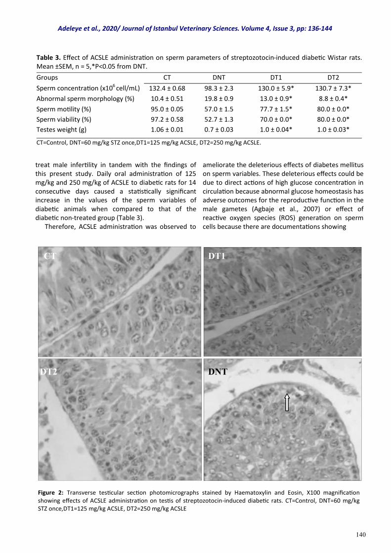
140
treat male infertility in tandem with the findings of this present study. Daily oral administration of 125 mg/kg and 250 mg/kg of ACSLE to diabetic rats for 14 consecutive days caused a statistically significant increase in the values of the sperm variables of diabetic animals when compared to that of the diabetic non-treated group (Table 3). Therefore, ACSLE administration was observed to
ameliorate the deleterious effects of diabetes mellitus on sperm variables. These deleterious effects could be due to direct actions of high glucose concentration in circulation because abnormal glucose homeostasis has adverse outcomes for the reproductive function in the male gametes (Agbaje et al., 2007) or effect of reactive oxygen species (ROS) generation on sperm cells because there are documentations showing
Table 3. Effect of ACSLE administration on sperm parameters of streptozotocin-induced diabetic Wistar rats. Mean ±SEM, n = 5,*P<0.05 from DNT.
Groups CT DNT DT1 DT2
Sperm concentration (x106 cell/mL) 132.4 ± 0.68 98.3 ± 2.3 130.0 ± 5.9* 130.7 ± 7.3*
Abnormal sperm morphology (%) 10.4 ± 0.51 19.8 ± 0.9 13.0 ± 0.9* 8.8 ± 0.4*
Sperm motility (%) 95.0 ± 0.05 57.0 ± 1.5 77.7 ± 1.5* 80.0 ± 0.0*
Sperm viability (%) 97.2 ± 0.58 52.7 ± 1.3 70.0 ± 0.0* 80.0 ± 0.0*
Testes weight (g) 1.06 ± 0.01 0.7 ± 0.03 1.0 ± 0.04* 1.0 ± 0.03*
CT=Control, DNT=60 mg/kg STZ once,DT1=125 mg/kg ACSLE, DT2=250 mg/kg ACSLE.
CT
DT2
DT1
DNT
Figure 2: Transverse testicular section photomicrographs stained by Haematoxylin and Eosin, X100 magnification showing effects of ACSLE administration on testis of streptozotocin-induced diabetic rats. CT=Control, DNT=60 mg/kg STZ once,DT1=125 mg/kg ACSLE, DT2=250 mg/kg ACSLE
Adeleye et al., 2020/ Journal of Istanbul Veterinary Sciences. Volume 4, Issue 3, pp: 136-144
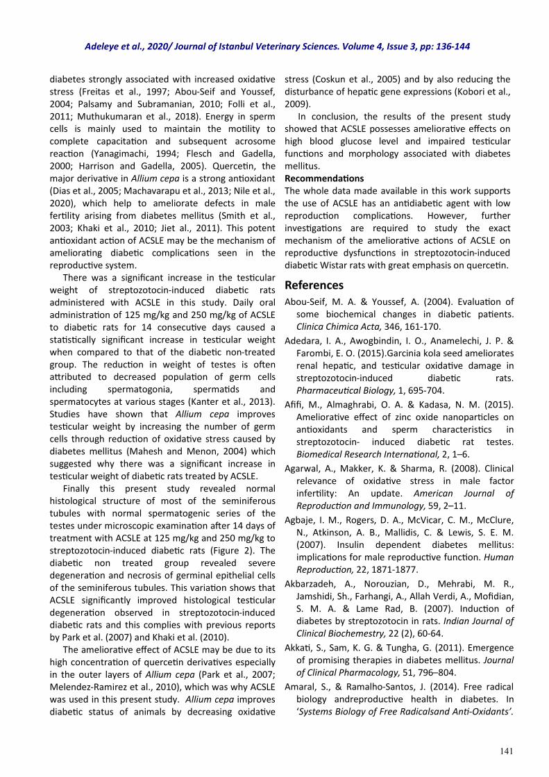
141
diabetes strongly associated with increased oxidative stress (Freitas et al., 1997; Abou-Seif and Youssef, 2004; Palsamy and Subramanian, 2010; Folli et al., 2011; Muthukumaran et al., 2018). Energy in sperm cells is mainly used to maintain the motility to complete capacitation and subsequent acrosome reaction (Yanagimachi, 1994; Flesch and Gadella, 2000; Harrison and Gadella, 2005). Quercetin, the major derivative in Allium cepa is a strong antioxidant (Dias et al., 2005; Machavarapu et al., 2013; Nile et al., 2020), which help to ameliorate defects in male fertility arising from diabetes mellitus (Smith et al., 2003; Khaki et al., 2010; Jiet al., 2011). This potent antioxidant action of ACSLE may be the mechanism of ameliorating diabetic complications seen in the reproductive system. There was a significant increase in the testicular weight of streptozotocin-induced diabetic rats administered with ACSLE in this study. Daily oral administration of 125 mg/kg and 250 mg/kg of ACSLE to diabetic rats for 14 consecutive days caused a statistically significant increase in testicular weight when compared to that of the diabetic non-treated group. The reduction in weight of testes is often attributed to decreased population of germ cells including spermatogonia, spermatids and spermatocytes at various stages (Kanter et al., 2013). Studies have shown that Allium cepa improves testicular weight by increasing the number of germ cells through reduction of oxidative stress caused by diabetes mellitus (Mahesh and Menon, 2004) which suggested why there was a significant increase in testicular weight of diabetic rats treated by ACSLE. Finally this present study revealed normal histological structure of most of the seminiferous tubules with normal spermatogenic series of the testes under microscopic examination after 14 days of treatment with ACSLE at 125 mg/kg and 250 mg/kg to streptozotocin-induced diabetic rats (Figure 2). The diabetic non treated group revealed severe degeneration and necrosis of germinal epithelial cells of the seminiferous tubules. This variation shows that ACSLE significantly improved histological testicular degeneration observed in streptozotocin-induced diabetic rats and this complies with previous reports by Park et al. (2007) and Khaki et al. (2010). The ameliorative effect of ACSLE may be due to its high concentration of quercetin derivatives especially in the outer layers of Allium cepa (Park et al., 2007; Melendez-Ramirez et al., 2010), which was why ACSLE was used in this present study. Allium cepa improves diabetic status of animals by decreasing oxidative
stress (Coskun et al., 2005) and by also reducing the disturbance of hepatic gene expressions (Kobori et al., 2009). In conclusion, the results of the present study showed that ACSLE possesses ameliorative effects on high blood glucose level and impaired testicular functions and morphology associated with diabetes mellitus. Recommendations The whole data made available in this work supports the use of ACSLE has an antidiabetic agent with low reproduction complications. However, further investigations are required to study the exact mechanism of the ameliorative actions of ACSLE on reproductive dysfunctions in streptozotocin-induced diabetic Wistar rats with great emphasis on quercetin.
References Abou-Seif, M. A. & Youssef, A. (2004). Evaluation of
some biochemical changes in diabetic patients. Clinica Chimica Acta, 346, 161-170.
Adedara, I. A., Awogbindin, I. O., Anamelechi, J. P. & Farombi, E. O. (2015).Garcinia kola seed ameliorates renal hepatic, and testicular oxidative damage in streptozotocin-induced diabetic rats. Pharmaceutical Biology, 1, 695-704.
Afifi, M., Almaghrabi, O. A. & Kadasa, N. M. (2015). Ameliorative effect of zinc oxide nanoparticles on antioxidants and sperm characteristics in streptozotocin- induced diabetic rat testes. Biomedical Research International, 2, 1–6.
Agarwal, A., Makker, K. & Sharma, R. (2008). Clinical relevance of oxidative stress in male factor infertility: An update. American Journal of Reproduction and Immunology, 59, 2–11.
Agbaje, I. M., Rogers, D. A., McVicar, C. M., McClure, N., Atkinson, A. B., Mallidis, C. & Lewis, S. E. M. (2007). Insulin dependent diabetes mellitus: implications for male reproductive function. Human Reproduction, 22, 1871-1877.
Akbarzadeh, A., Norouzian, D., Mehrabi, M. R., Jamshidi, Sh., Farhangi, A., Allah Verdi, A., Mofidian, S. M. A. & Lame Rad, B. (2007). Induction of diabetes by streptozotocin in rats. Indian Journal of Clinical Biochemestry, 22 (2), 60-64.
Akkati, S., Sam, K. G. & Tungha, G. (2011). Emergence of promising therapies in diabetes mellitus. Journal of Clinical Pharmacology, 51, 796–804.
Amaral, S., & Ramalho-Santos, J. (2014). Free radical biology andreproductive health in diabetes. In ‘Systems Biology of Free Radicalsand Anti-Oxidants’.
Adeleye et al., 2020/ Journal of Istanbul Veterinary Sciences. Volume 4, Issue 3, pp: 136-144
142
and diagnosis of diabetes: Standards of Medical Care in Diabetes. (2019). Diabetes Care, 42(Suppl. 1): S13–S28.
Amidu, N., Owiredu, W. K., Alidu, H., Sarpong, C., Gyasisarpong, C. K., & Quaye, L. (2013). Association between metabolic syndrome and sexual dysfunction among men with clinically diagnosed diabetes. Diabetology and Metabolic Syndrome, 5, 242-250.
Baccetti, B., Marca, L. A., & Piomboni, P. (2002). Insulin-dependent diabetes in men is associated with hypothalamo-pituitary derangement and with impairment in semen quality. Human Reproduction, 17, 2673-2677.
Bindu, B., & Podikunju, B. (2015). Performance evaluation of onion (Allium Cepa L. Var. Cepa) varieties for their suitability in kollam district. International Journal of Research in Agricultural Science, l(1), 18-20.
Butler, A. E., Janson, J., Bonner Weir, S., Ritzel, R., Rizza, R. A., & Butler, P. C. (2003). Beta‑cell deficit and increased beta‑cell apoptosis in humans with type 2 diabetes. Diabetes,52, 102-110.
Cavallini, G. (2006). Male idiopathic oligo asthenoteratozoospermia. Asian Journal of Andrology. 8, 143–157.
Chae, M. R., Kang, S. J., Lee, K. P., Choi, B. R., Kim, H. K., Park, J. K., Kim, C. Y., & Lee, S. W. (2017). Onion (Allium cepa L.) peel extract (OPE) regulates human sperm motility via protein kinase C-mediated activation of the human voltage-gated proton channel. Andrology, 5(5), 979–989.
Cooke, D. W. & Plotnick,L. (2008). Type 1 diabetes mellitus in pediatrics. Pediatric Review, 29, 374–384.
Coskun, O., Kanter, M., Korkmaz, A., & Oter, S. (2005). Quercetin; a flavonoid antioxidant, prevents and protects streptozotocin-induced oxidative stress and beta-cell damage in rat pancreas. Pharmacology Research, 51, 117-123.
deAncos, B., Colina-Coca, C., Gonzalex-Pena, D. & Sanchez-Moreno, C. (2015). Bioactive compounds from vegetable and fruit by-products. In Biotechnology of Bioactive Compounds. Sources and Applications. Section I, V. K. Gupta and M. G. Tuohy, Eds., pp. 3–36,Wiley Blackwell, 2015.
Dias, A. S., Porawski, M., Alonso, M., Marroni, N., Collado, P. S. & Gonzalez-Gallego, J. (2005). Quercetin decreases oxidative stress, NF-kappaB activation, and iNOS overexpression in liver of streptozotocin-induced diabetic rats. Journal of
Nutrition, 135, 2299-2304.
Edeoga, H. O., Okwu, D. E. & Mbaebie, B. O. (2005).Phytochemical constituents of some Nigerian medicinal plants. African Journal of Biotechnology, 4 (7), 685-688.
Eizirik, D. L., Colli, M. L., & Ortis, F. (2009).The role of inflammation in insulitis and beta‑cell loss in type 1 diabetes. Nature Reviews Endocrinology, 5, 219-26.
Flesch, F. M. & Gadella, B. M. (2000). Dynamics of the mammalian sperm plasma membrane in the process of fertilization. Biochimica et Biophysica Acta, 1469, 197-235.
Folli, F., Corradi, D., Fanti, P., Davalli, A., Paez, A. . Giaccari, A Perego C., & Muscogiuri, G. (2011). The Role of Oxidative Stress in the Pathogenesis of Type 2 Diabetes Mellitus Micro- and Macrovascular Complications: Avenues for a Mechanistic-Based Therapeutic Approach. Current Diabetes Review, 7 (5), 313-324.
Freitas, J. P., Filipe, P. M., & Rodrigo, F. G. (1997). Lipid peroxidation intype 2 normolipidemic diabetic patients. Diabetes Research and Clinical Practice, 36, 71-75.
Ghanbari, E., Nejati, V., Najafi, G., Khazaei, M., & Babaei, M. (2015). Study on the effect of royal jelly on reproductive parameters in streptozotocin-induced diabetic rats. Internal Journal of Fertility and Sterility, 1, 113-120.
Griffiths, G., Trueman, L., Crowther, T., Thomas, B., & Smith, B. (2002). Onions-a global benefit to health. Phytotherapy Research, 16(7), 603-615.
Harrison, R. A., & Gadella, B. M. (2005). Bicarbonate ‑induced membrane processing in sperm capacitation. Theriogenology, 63, 342-51.
Jequier, A. M. (2010). Semen analysis: a new manual and its application to the understanding of semen and its pathology. Asian Journal of Andrology, 12(1), 11–13.
Ji, Y. J., Yeni, L., Min, S. M., Ji, Y. K., & Oran, K. (2011). Effect of quercetin derivatives on diabetic animals. Nutrition & Metabolism, 8, 18-25.
Kahn, S. E., Hull, R. L., & Utzschneider, K. M. (2006). Mechanisms linking obesity to insulin resistance and type 2 diabetes. Nature. 444, 840–846.
Kameswara, R. B., Kesavulu, M. M., Guiri, R. & Apparao, C. H. (1999). Hepatic key enzyme in experimental diabetes. Journal of Ethnopharmacology, 91(1), 109-113.
Kanter, M., Aktas, C. & Erboga, M. (2013). Curcumin attenuates testicular damage, apoptotic germ cell
Adeleye et al., 2020/ Journal of Istanbul Veterinary Sciences. Volume 4, Issue 3, pp: 136-144
143
death, and oxidative stress in streptozotocininduced diabetic rats. Molecular Nutrition and Food Research, 57, 1578-1585.
Khaki, A., Fathiazad, F., Nouri, M., Maleki, N.A., Khamnei, H. J., & Ahmadi, P. (2010). Beneficial effects of quercetin on sperm parameters in streptozotocininduced diabetic male rats Phytotherapy Research, 24, 1285-1291.
Kilarkaje, N., Al-Hussaini, H., & Al-Bader, M. M. (2014). Diabetes‑induced DNA damage and apoptosis are associated with poly (ADP ribose) polymerase 1 inhibition in the rat testis. European Journal of Pharmacology, 737, 29–40.
Kim, S. T., & Moley, K. H. (2008). Paternal effect on embryo quality in diabetic mice is related to poor sperm quality and associated with decreased glucose transporter expression. Reproduction. 136, 313-322.
Kobori, M., Masumoto, S., Akimoto, Y. & Takahashi, Y.(2009). Dietary quercetin alleviates diabetic symptoms and reduces streptozotocin-induced disturbance of hepatic gene expression in mice. Molecular Nutrition and Food Research, 53, 859-868.
Koroglu, P., Senturk, G. E., Yucel, D., Ozakpinar, O. B., Uras, F., & Arbak, S. (2015). The effect of exogenous oxytocin on streptozotocin (STZ) induced diabetic adult rat testes. Fertility and Sterility, 2, 47–54.
Lee, B.K. & Jung,Y.S.(2016).Allium cepaextract and quercetin protect neuronal cells from oxidative stress via PKC-ε inactivation/ERK1/2 activation. Oxidative Medicine and Cellular Longevity, 2016, 2495624.
Machavarapu, M., Sindiri, M. K., & Vangalapati, M.(2017). Optimization of Physico-chemical Parameters for the Extraction of Flavonoids and Phenolic Components from the Skin of Allium cepa. Optimization. Journal of Physiology, 2(7), 110-121.
Mahesh, T., & Menon, V. P. (2004). Quercetinallievates oxidative stress in streptozotocin-induced diabetic rats. Phytotherapy Research, 18, 123-127.
Maiti, R., Jana, D., Das, U. K., & Ghosh, D. (2004). Antidiabetic effect of aqueous extract of seed of tamarindusindica in streptozotocin induced diabetic rats. Journal of Ethnopharmacology, 92, 85-91.
Marefati, N., Eftekhar, N., Kaveh, M., Boskabadi, J., Beheshti, F., Boskabady, M. H. (2018).The Effect of Allium cepa extract on lung oxidant, boskabadi, antioxidant, and immunological biomarkers in ovalbumin-sensitized rats. Medical Principles and Practice, 27,122–128.
Marti, E., Mara, L., Marti, J. I., Muiño-Blanco, T., &
Cebrián-Pérez, J. A. (2007). Seasonal variations in antioxidant enzyme activity in ram seminal plasma. Theriogenology. 67, 1446–1454.
Maxwell, S. R. J., & Lip, G. Y. H. (1997). Free radicals and antioxidants in cardiovascular disease. British Journal of Clinical Pharmacology, 44,307-317.
Melendez-Ramirez, L. Y., Richards, R. J. & Cefalu, W. T. (2010). Complications of type 1 diabetes. Journal of Endocrinology. 39, 351-400.
Muthukumaran, J., Ramachandran, V., Ranga, R. A., Baojun, X., Stephen, S. M. C. (2018). Guava leaf extract diminishes hyperglycemia and oxidative stress, prevents -cell death, inhibits inflammation, and regulates NF-kB signaling pathway in STZ Induced diabetic rats. Biomedical Research International, 5, 1-14.
Navarro-Casado, L., Juncos-Tobarra, M. A., Cha´fer-Rudilla, M., de Onzono, L. I., Bla´zquez-Cabrera, J. A., & Miralles-Garcı´a, J. M. (2010). Effect of experimental diabetes and STZ on male fertility capacity study in rats. Andrology, 31, 584–592.
Nile, A., Gansukh, E., Park, G. S., Kim, D. H., & Hariram Nile, S. (2020). Novel insights on the multi-functional properties of flavonolglucosides from red onion (Allium cepaL) solid waste-In vitro and in silicoapproach. Food Chemistry. 127650.
O.E.C.D. (Organization for Economic Co-operation and Development). (2008): OECD guidelines 407 for the testing of chemicals: Repeated Dose 28- day oral toxicity studies in rodents. https://ntp.niehs.nih.gov/iccvam/suppdocs/feddocs/oecd/oecdtg407-2008.pdf
Oliveira, P. F., Alves, M. G., Rato, L.,Laurentino, S., Silva, J., Sa´,R., Barros, A., Sousa, M., Carvalho, R. A., Cavaco, J. E., & Socorro, S. (2012). Effect of insulin deprivation on metabolism and metabolism associated gene transcript levels of in vitro cultured human Sertoli cells. Biochimica et Biophysica Acta, 1820, 84-89.
Oonk, R. B., Grootegoed, J. A., & van der Molen, H. J.(1985). Comparison of theeffects of insulin and follitropin on glucose metabolism by Sertoli cells fromimmature rats. Molecular and Cellular Endocrinology, 42, 39-48.
Oonk, R. B., & Grootegoed, J. A. (1987). Identification of insulin receptorson rat Sertoli cells. Molecular and Cellular Endocrinology, 49, 51-62.
Palsamy, P., Sivakumar, S., & Subramanian, S. (2010). Resveratrol attenuates hyperglycemia-mediated oxidative stress, proinflammatory cytokines and protects hepatocytes ultrastructure in streptozotocin-nicotinamide-induced experimental
Adeleye et al., 2020/ Journal of Istanbul Veterinary Sciences. Volume 4, Issue 3, pp: 136-144
144
protects hepatocytes ultrastructure in streptozotocin-nicotinamide-induced experimental diabetic rats. Chemico-Biologic Interaction, 186 (2), 200-210.
Park, J., Kim J., & Kim, M. K. (2007). Onion flesh and onion peel enhance antioxidant status in aged rats. Tokyo Journal of Nutrition Science and Vitaminology, 53, 21-29.
P´erez-Gregorio, R. M., Garc´ıa-Falc´on, M. S., Simal-G´andara, J., Rodrigues, A. S., & Almeida, D. P. F. (2010). Identification and quantification of flavonoids in traditional cultivars of red and white onions at harvest. Journal of Food Composition and Analysis, 23(6), 592–598.
Philippe, J., Raccah, D. (2009). Treating type 2 diabetes: how safe are current therapeutic agents? International Journal of Clinical Practice, 63, 321–332.
Prakash, D., Singh, B. N. & Upadhyay, G.(2007). Antioxidant and free radical scavenging activities of phenols fromonion (Allium cepa). Food Chemistry, 102(4), 1389–1393.
Ramachandran, A. (2014). Know the signs and symptoms of diabetes. Indian Journal of Medical Research, 140(5), 579-5816.
Rashid, K. & SilP. C. (2015). Curcumin ameliorates testicular damage in diabetic rats by suppressing cellular stress-mediated mitochondria and endoplasmic reticulum-dependent apoptotic death. Biochimica Biophysica Acta, 3, 70–82.
Riserus, U., Willett, W. C. & Hu, F. B. (2009). Dietary fats and prevention of type 2 diabetes. Progress in Lipid Research, 48, 44-51.
Roldan, E., Sanchez-Moreno, C.,de Anco,B., & Cano, M. P. (2008). Characterisation of onion (AlliumcepaL.) by-products as food ingredients with antioxidant and antibrowning properties. Food Chemistry, 108(3), 907-916.
Rupeshkumar, M., Kavitha, K., & Haldar, P. K. (2014). The role of herbal plants in the diabetes mellitus therapy: an overview. International Journal of Applied Pharmacology, 6, 1-3.
Saeedi, P., Petersohn, I., Malanda, B., Karuranga, S., Unwin, N., Colagiuri, S., Guariguata, L., Motala, A. A., Ogurtsova, K., ShawJ. E., Bright, D., & Williams, R.(2019). Global and regional diabetes prevalence estimates for 2019 and projections for 2030 and 2045: Results from the International Diabetes Federation Diabetes Atlas, 9th edition. Diabetes Research and Clinical Practice, 157 . 107843.
Santas, J., Almajano, M. P., & Carb´o, R. (2010).
Antimicrobial and antioxidant activity of crude onion (Allium cepa, L.) extracts. International Journal of Food Science and Technology, 45(2), 403–409.
Saumya, S. & Basha, P. M. (2016). Fluoride exposure aggravates the testicular damage and sperm quality in diabetic mice: protective role of Ginseng and Banaba. Biological Trace Element Research, 2, 1–14.
Scarano, W. R., Messias, A. G., Oliva, S. U., Klinefelter, G. R., & Kempinas, W. G. (2006). Sexual behaviour, sperm quantity and quality after short-term streptozotocin-induced hyperglycaemia in rats. International Journal of Andrology, 29, 482-488.
Schoeller, E. L., Albanna, G., Frolova, A. I., &Moley, K. H. (2012). Insulin rescues impaired spermatogenesis via the hypothalamicpituitary-gonadal axis in Akita diabetic mice and restores malefertility. Diabetes, 61, 1869-1878.
Sexton, W. J., & JarowJ. P. (1997). Effect of diabetes mellitus upon male reproductive function. Urology Research, 49, 508-513.
Shaath, N. A., & Flores, F. B. (1998). Egyptian onion oil, developments in Food Science. Journal of Nutrition, 40, 443-453.
Slimestad, R., Fossen,T., & Vagen, I. M. (2007). Reviews. Onions: a source of unique dietary flavonoids. Journal of Agriculture and Food Chemistry, 55 (25), 10067–10080.
Smith, C., Lombard, K. A. Peffley, E. B., & Liu. W. (2003). Genetic analysis of quercetin in onion (Allium cepa L.) 'Lady Raider'. Texas Journal of Agriculture and Natural Resources, 16, 24-28.
Tamtaji, O. R., Hossan, H., Sagyed, A. T., Mohamed, B., Seyed, M. T., Moshen, T. & Reza, A. (2017). Protective effects of red onion ethanolic extract on learning and memory impairments in animal of diabetes. Journal of Medicine, 6(3), 249-257.
Tavares, R. S., Portela Joana, M. D., Sousa, M. I., Mota Paula, C., Ramalho-Santos, J., & Amaral, S.(2017). High glucose levels affect spermatogenesis: an in vitro approach. Reproduction Fertility and Development, 29(7), 1369-1378.
Tremellen, K. (2008). Oxidative stress and male infertility a clinical perspective. Human Reproduction Update, 14(3),243-258.
Tsounapi, P., Honda, M., Dimitriadis, F., Kawamoto, B., Hikita, K., Muraoka, K., Saito, M., Sofikitis, N., & Takenaka, A. (2016). Impact of antioxidants on seminal vesicles function and fertilizing potential in diabetic rats. Asian Journal of Andrology, 2, 1-10.
Yanagimachi, R. (1994). Mammalian fertilization. Physiology of Reproduction, 1, 189-317.
Adeleye et al., 2020/ Journal of Istanbul Veterinary Sciences. Volume 4, Issue 3, pp: 136-144
145
Research Article
Volume: 4, Issue: 3 December 2020 Pages: 145-149
Investigation of the prevalence of Babesia canis in dogs in the center of Antalya province
Journal of Istanbul Veterınary Scıences
Mahmut KESKİN1, Kenan SEZER2*, , 1. Altınkum Animal Hospital, 07100, Altınkum, Antalya, Turkey 2. Department of Internal Medicine, Faculty of Veterinary Medicine, Burdur Mehmet Akif Ersoy University, 15030, Burdur, Turkey.
Keskin M. ORCID: 0000-0003-4343-5840. Sezer K. ORCID: 0000-0002-7413-5966.
ABSTRACT
This study was conducted to investigate the prevalence of Babesia canis in Antalya province For this purpose, the blood samples were taken from 200 dogs in the Antalya city centre, the examples were studied by the immune fluorescence antibody test (IFAT). In the study fluo Babesia canis (Biopronix/Italy) test kit was used, 1/32 and higher titters were considered positive. The 37 out of 200 serum samples (18.5%) were found positive for Babesia canis as results of IFA test. As a result; Babesia canis positivity by serological method in dogs in Antalya, which gives an idea about the prevalence of the disease in this province, it would be useful to continue similar studies, using the specific identification method in the wider population.
Keywords: Babesia canis, IFAT, dog
DOI: https://doi.org/10.30704/http-www-jivs-net.802265
To cite this article: Keskin, M., & Sezer, K. (2020). Investigation of the prevalence of Babesia canis in dogs in the center of Antalya province. Journal of Istanbul Veterinary Sciences, 4(3), 145-149. Abbreviated Title: J Ist Vet Sci
Babesiosis is a disease of tropical and subtropical mild climate regions and it appears on wild carnivores as well as dogs (Uilenberg, 2006; Inci et al., 2010; Irwin, 2010) The parasites transmitted from one animal to another by İxodidae tick type, related to Babesia species, take place near the top of the blood protozoon of the dogs (Inci et al., 2010; Irwin, 2010; Kahn, 2005). B. canis canis is transmitted by Dermacentor reticularis in Europe, B. canis vogeli is transmitted by Rhipicephalus sanguineus in tropical and subtropical countries, B. canis rossi is transmitted by Haemaphysalis leachi in South Africa (Boozer and Macintire, 2003; Kahn, 2005). Also in Turkey, Rhipicephalus sanguineus transferred B. canis vogeli shows a wide spread and this suggests that babesiosis on dogs is very common (Mimioglu, 1954; Güler, 1982; Inci et al., 2010). The types of pathogens in dogs are B. canis (2.5X5.0 µm) and B. gibsoni (0.5X3.5 µm) (Boozer and Macintire, 2003; Bilal, 2013). Babesia canis has been
subdivided into three subspecies which are B. canis canis, B. canis vogeli and B. canis rossi (Boozer and Macintire, 2003; Inci et al., 2010). The occurrence of the disease and incubation time change between 10 and 21 days and that changes according to pathogen types and strains (Uilenberg, 2006; Inci et al., 2010). Although babesiosis appears in all dogs, it’s suggested that the susceptibility to infection is higher in younger dogs. Rottweiler, greyhounds, pit bull terrier and American staffordshire terrier race dogs (Birkenheuer et al., 2004; Mathe et el., 2006). It has been reported because of pre-immunization, asymptomatic carrier dogs are more resistance than the dogs completely free from interference about getting infected again (Brandao et al., 2003). Although it’s thought many infected dogs are subclinical carrier, course of the babesiosis disease is per acute, acute and chronic (Freeman et al., 1994). Babesisosis is a disease that is characterized by lethargy, high fever (40-41.3 0C) hemolytic anemia
*Corresponding Author: Kenan Sezer E-mail: [email protected]
Journal home page: www.jivs.net http://dergipark.gov.tr/http-www-jivs-net
Article History
Received: 30.9.2020 Accepted:16.12.2020 Available online: 18.12.2020
This work is licensed under the Creative Commons Attribution 4.0 International License.
Introduction
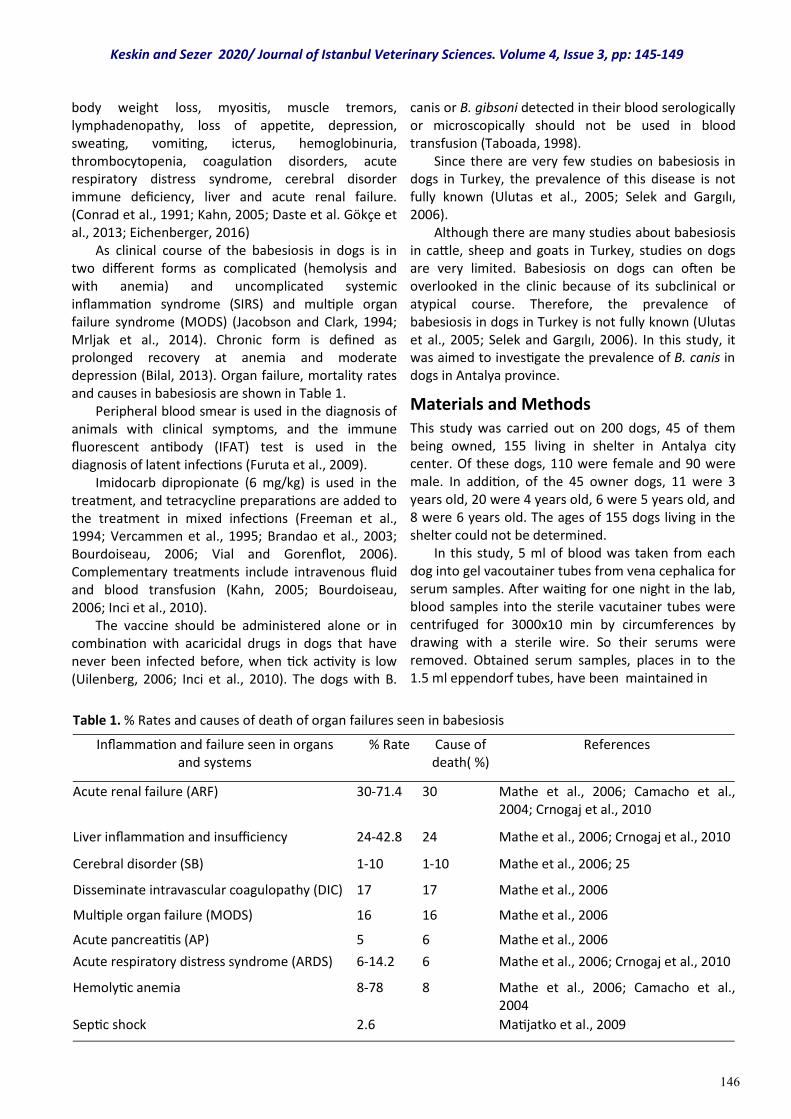
146
body weight loss, myositis, muscle tremors, lymphadenopathy, loss of appetite, depression, sweating, vomiting, icterus, hemoglobinuria, thrombocytopenia, coagulation disorders, acute respiratory distress syndrome, cerebral disorder immune deficiency, liver and acute renal failure. (Conrad et al., 1991; Kahn, 2005; Daste et al. Gökçe et al., 2013; Eichenberger, 2016) As clinical course of the babesiosis in dogs is in two different forms as complicated (hemolysis and with anemia) and uncomplicated systemic inflammation syndrome (SIRS) and multiple organ failure syndrome (MODS) (Jacobson and Clark, 1994; Mrljak et al., 2014). Chronic form is defined as prolonged recovery at anemia and moderate depression (Bilal, 2013). Organ failure, mortality rates and causes in babesiosis are shown in Table 1. Peripheral blood smear is used in the diagnosis of animals with clinical symptoms, and the immune fluorescent antibody (IFAT) test is used in the diagnosis of latent infections (Furuta et al., 2009). Imidocarb dipropionate (6 mg/kg) is used in the treatment, and tetracycline preparations are added to the treatment in mixed infections (Freeman et al., 1994; Vercammen et al., 1995; Brandao et al., 2003; Bourdoiseau, 2006; Vial and Gorenflot, 2006). Complementary treatments include intravenous fluid and blood transfusion (Kahn, 2005; Bourdoiseau, 2006; Inci et al., 2010). The vaccine should be administered alone or in combination with acaricidal drugs in dogs that have never been infected before, when tick activity is low (Uilenberg, 2006; Inci et al., 2010). The dogs with B.
canis or B. gibsoni detected in their blood serologically or microscopically should not be used in blood transfusion (Taboada, 1998). Since there are very few studies on babesiosis in dogs in Turkey, the prevalence of this disease is not fully known (Ulutas et al., 2005; Selek and Gargılı, 2006). Although there are many studies about babesiosis in cattle, sheep and goats in Turkey, studies on dogs are very limited. Babesiosis on dogs can often be overlooked in the clinic because of its subclinical or atypical course. Therefore, the prevalence of babesiosis in dogs in Turkey is not fully known (Ulutas et al., 2005; Selek and Gargılı, 2006). In this study, it was aimed to investigate the prevalence of B. canis in dogs in Antalya province.
Materials and Methods This study was carried out on 200 dogs, 45 of them being owned, 155 living in shelter in Antalya city center. Of these dogs, 110 were female and 90 were male. In addition, of the 45 owner dogs, 11 were 3 years old, 20 were 4 years old, 6 were 5 years old, and 8 were 6 years old. The ages of 155 dogs living in the shelter could not be determined. In this study, 5 ml of blood was taken from each dog into gel vacoutainer tubes from vena cephalica for serum samples. After waiting for one night in the lab, blood samples into the sterile vacutainer tubes were centrifuged for 3000x10 min by circumferences by drawing with a sterile wire. So their serums were removed. Obtained serum samples, places in to the 1.5 ml eppendorf tubes, have been maintained in
Keskin and Sezer 2020/ Journal of Istanbul Veterinary Sciences. Volume 4, Issue 3, pp: 145-149
Table 1. % Rates and causes of death of organ failures seen in babesiosis
Inflammation and failure seen in organs and systems
% Rate Cause of death( %)
References
Acute renal failure (ARF) 30-71.4 30 Mathe et al., 2006; Camacho et al., 2004; Crnogaj et al., 2010
Liver inflammation and insufficiency 24-42.8 24 Mathe et al., 2006; Crnogaj et al., 2010
Cerebral disorder (SB) 1-10 1-10 Mathe et al., 2006; 25
Disseminate intravascular coagulopathy (DIC) 17 17 Mathe et al., 2006
Multiple organ failure (MODS) 16 16 Mathe et al., 2006
Acute pancreatitis (AP) 5 6 Mathe et al., 2006
Acute respiratory distress syndrome (ARDS) 6-14.2 6 Mathe et al., 2006; Crnogaj et al., 2010
Hemolytic anemia 8-78 8 Mathe et al., 2006; Camacho et al., 2004
Septic shock 2.6 Matijatko et al., 2009
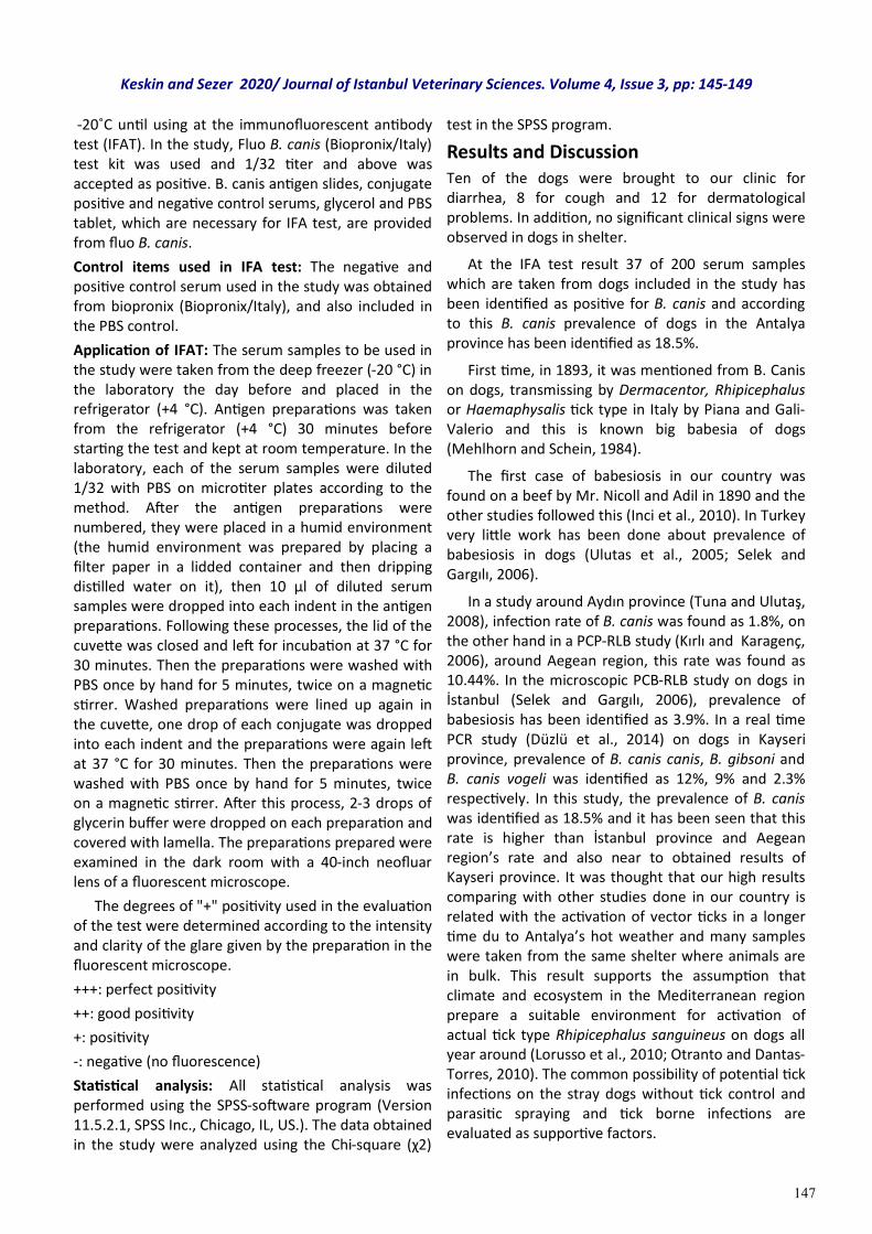
147
-20˚C until using at the immunofluorescent antibody test (IFAT). In the study, Fluo B. canis (Biopronix/Italy) test kit was used and 1/32 titer and above was accepted as positive. B. canis antigen slides, conjugate positive and negative control serums, glycerol and PBS tablet, which are necessary for IFA test, are provided from fluo B. canis.
Control items used in IFA test: The negative and positive control serum used in the study was obtained from biopronix (Biopronix/Italy), and also included in the PBS control.
Application of IFAT: The serum samples to be used in the study were taken from the deep freezer (-20 °C) in the laboratory the day before and placed in the refrigerator (+4 °C). Antigen preparations was taken from the refrigerator (+4 °C) 30 minutes before starting the test and kept at room temperature. In the laboratory, each of the serum samples were diluted 1/32 with PBS on microtiter plates according to the method. After the antigen preparations were numbered, they were placed in a humid environment (the humid environment was prepared by placing a filter paper in a lidded container and then dripping distilled water on it), then 10 µl of diluted serum samples were dropped into each indent in the antigen preparations. Following these processes, the lid of the cuvette was closed and left for incubation at 37 °C for 30 minutes. Then the preparations were washed with PBS once by hand for 5 minutes, twice on a magnetic stirrer. Washed preparations were lined up again in the cuvette, one drop of each conjugate was dropped into each indent and the preparations were again left at 37 °C for 30 minutes. Then the preparations were washed with PBS once by hand for 5 minutes, twice on a magnetic stirrer. After this process, 2-3 drops of glycerin buffer were dropped on each preparation and covered with lamella. The preparations prepared were examined in the dark room with a 40-inch neofluar lens of a fluorescent microscope.
The degrees of "+" positivity used in the evaluation of the test were determined according to the intensity and clarity of the glare given by the preparation in the fluorescent microscope.
+++: perfect positivity
++: good positivity
+: positivity
-: negative (no fluorescence)
Statistical analysis: All statistical analysis was performed using the SPSS-software program (Version 11.5.2.1, SPSS Inc., Chicago, IL, US.). The data obtained in the study were analyzed using the Chi-square (χ2)
test in the SPSS program.
Results and Discussion Ten of the dogs were brought to our clinic for diarrhea, 8 for cough and 12 for dermatological problems. In addition, no significant clinical signs were observed in dogs in shelter.
At the IFA test result 37 of 200 serum samples which are taken from dogs included in the study has been identified as positive for B. canis and according to this B. canis prevalence of dogs in the Antalya province has been identified as 18.5%.
First time, in 1893, it was mentioned from B. Canis on dogs, transmissing by Dermacentor, Rhipicephalus or Haemaphysalis tick type in Italy by Piana and Gali-Valerio and this is known big babesia of dogs (Mehlhorn and Schein, 1984).
The first case of babesiosis in our country was found on a beef by Mr. Nicoll and Adil in 1890 and the other studies followed this (Inci et al., 2010). In Turkey very little work has been done about prevalence of babesiosis in dogs (Ulutas et al., 2005; Selek and Gargılı, 2006).
In a study around Aydın province (Tuna and Ulutaş, 2008), infection rate of B. canis was found as 1.8%, on the other hand in a PCP-RLB study (Kırlı and Karagenç, 2006), around Aegean region, this rate was found as 10.44%. In the microscopic PCB-RLB study on dogs in İstanbul (Selek and Gargılı, 2006), prevalence of babesiosis has been identified as 3.9%. In a real time PCR study (Düzlü et al., 2014) on dogs in Kayseri province, prevalence of B. canis canis, B. gibsoni and B. canis vogeli was identified as 12%, 9% and 2.3% respectively. In this study, the prevalence of B. canis was identified as 18.5% and it has been seen that this rate is higher than İstanbul province and Aegean region’s rate and also near to obtained results of Kayseri province. It was thought that our high results comparing with other studies done in our country is related with the activation of vector ticks in a longer time du to Antalya’s hot weather and many samples were taken from the same shelter where animals are in bulk. This result supports the assumption that climate and ecosystem in the Mediterranean region prepare a suitable environment for activation of actual tick type Rhipicephalus sanguineus on dogs all year around (Lorusso et al., 2010; Otranto and Dantas-Torres, 2010). The common possibility of potential tick infections on the stray dogs without tick control and parasitic spraying and tick borne infections are evaluated as supportive factors.
Keskin and Sezer 2020/ Journal of Istanbul Veterinary Sciences. Volume 4, Issue 3, pp: 145-149
148
Conclusion As a result of the study, it was concluded that Babesia canis positivity detected in dogs in Antalya by serological method, gives an idea about the prevalence of the disease in this province, and It would be useful to continue similar studies in larger populations by specific diagnosis methods towards the agent.
Acknowledgments We thank Burdur Mehmet Akif Ersoy University, Coordinatorship of Scientific Research Projects for all their support to this research.
References Bilal T. (2013). Kedi köpek iç hastalıkları. Istanbul,
Turkey, Nobel Tıp Kitabevi.
Biopronix, Fluo Babesia canis. Agrolabo diagnostic products. Catalogue for Cats, Dogs, and Horses. Code: 27263601 Scarmagno (TO) Italy
Birkenheuer, A. J., Neel, J., Ruslander, D., Levy, M.G., & Breitschwerdt, E. B. (2004). Detection and molecular characterization of a novel large Babesia species in a dog. Veterinary Parasitology, 5,151-160.
Boozer, A. L., & Macintire, D. K. (2003). Canine babesiosis. Veterinary Clinics of North America: Small Animal Practice, 33, 885-904.
Bourdoiseau, G. (2006). Canine babesiosis in France. Veterinary Parasitology, 138, 118-125.
Brandao, L. P., Hagiwara, M. K., & Myiashiro, S.I. (2003). Humoral immunity and reinfection resistance in dogs experimentally inoculated with Babesia canis and either treated or untreated with imidocarb dipropionate. Veterinary Parasitology, 114, 253-265.
Camacho, A. T., Guitian, E. J., Pallas, E., Gestal, J. J., Olmeda, A. S., Goethert, H. K., Telford, S. R., & Spielman, A. (2004). Azotemia and mortality among Babesia microti-like infected dogs. Journal of Veterinary Internal Medicine, 18(2), 141-146.
Conrad, P., Thomford, J., Yamane, I., Whiting, J., & Bosma, L. (1991). Hemolytic anemia caused by Babesia gibsoni infection in dogs. Journal of the American Veterinary Medical Association, 199, 601-605.
Crnogaj, M, Petlevski, R. Mrljak, V, Torti, M, Kucer, N, Matijatko, V, Sacer, I., & Stokovic, I. (2010). Malondialdehyde levels in serum of dogs infected with Babesia canis. Journal Veterinarni Medicina, 55(4),163-171.
Daste, T., Lucas, M. N., & Aumann, M. J. (2013).
Cerebral babesiosis and acute respiratory distress syndrome in a dog. Journal of Veterinary Emergency & Critical Care) 23, 615-623.
Düzlü, Ö, Inci, A., Yıldırım, A., Onder, Z., & Çiloğlu, A. (2014). Köpeklerde kene kaynaklı bazı protozoon ve rickettsial enfeksiyonların Real Time PCR ile araştırılması ve saptanan izolatların moleküler karakterizasyonları. Ankara Üniversitesi Veteriner Fakültesi Dergisi, 61, 275-282.
Eichenberger, R. M., Riond, B., & Willi, B, Hofmann-Lehmann, R., Deplazes, P. (2016). Prognostic markers in acute Babesia canis infections. Journal of Veterinary Internal Medicin, 30, 174-182.
Freeman, M. J., Kirby, B. M., Panciera, D. L., Henik, R. A., Rosin, E., & Sullivan, L. J. (1994). Hypotensive shock syndrome associated with acute Babesia canis infection in a dog. Journal of the American Veterinary Medical Association, 204, 94-96.
Furuta, P. I., Oliveira, T. M. F. S., Teixeira, M. C. A,. Rocha, A. G., Machado, R. Z., & Tinucci-Costa, M. (2009). Comparison between a soluble antigen-based ELISA and IFAT in detecting antibodies against Babesia canis in dogs. Brazilian Journal of Veterinary Parasitology, 18, 41-45.
Gökçe, E., Kırmızıgül, A. H., Taşçı, G. T., Uzlu, E., Gündüz, N., & Vatansever, Z. (2013). Türkiye’de köpeklerde Babesia canis canis’in klinik ve parazitolojik olarak ilk tespiti. Kafkas Üniversitesi Veteriner Fakültesi Dergisi, 19, 717-720.
Güler, S. (1982). Ankara ve civarındaki koyun ve keçilerde kış Ixodide’leri üzerine araştırmalar. Uludağ Üniversitesi Veteriner Fakültesi Dergisi, 1, 45-54.
Inci, A., Düzlü, Ö., & Iça, A. (2010). Babesiidae. In Dumanli N, Karaer Z. (Ed). Veteriner protozooloji, 1. baskı, Ankara. Turkey Medisan Yayınevi.
Irwin, P. J. (2010). Canine babesiosis. Veterinary Clinics of North America: Small Animal Practice, 40, 1141-1156.
Jacobson, L. S., & Clark, I. (1994). The pathophysiology of canine babesiosis: new approaches to an old puzzle. Journal of the South African Veterinary Association, 65, 134-145.
Kahn CM. (2005). The Merck Veterinary Manual, 9th ed. Merck & Co. Inc. New Jersey.
Kırlı, G., & Karagenç, T. (2006). Ege bölgesinde köpek babesiosisinin yaygınlığı. Yüksek Lisans Tezi, Aydın: Adnan Menderes Üniversitesi, Sağlık Bilimleri Enstitüsü. Türkiye
Keskin and Sezer 2020/ Journal of Istanbul Veterinary Sciences. Volume 4, Issue 3, pp: 145-149
149
Lorusso, V, Dantas-Torres, F, Lia, R. P., Tarallo, V. D, Mencke, N., Capelli, G., & Otranto, D. (2010). Seasonal dynamics of the brown dog tick. Rhipicephalus sanguineus, on a confined dog
Mathe, A., Vörös, K., Papp, L., & Reiczigel, J. (2006). Clinical manifestations of canine babesiosis in Hungary (63 cases). Acta Veterinaria Hungarica, 54, 367-385.
Matijatko, V., Kis, I., Torti, M., Brkljacid, M., Kucer, N., Rafaj, R. B., Grden, D., Zivicnjak, T., & Mrljak, V. (2009). Septic shock in canine babesiosis. Veterinary Parasitol, 10(162), 263-270.
Mehlhorn H., & Schein E. (1984). The piroplasm: Life cycle and sexual stages. Advances in Parasitology, 23, 37-103.
Mimioğlu, M. (1954). Die Schildzecken (Ixodidade) der haustiere in der Türkei, Ankara Üniversitesi Veteriner Fakültesi Dergisi, 1, 20-35.
Mrljak, V., Kučer, N., Kuleš, J., Tvarijonaviciute, A., Brkljačid, M., Crnogaj, M., Zivičnjak, T., Smit, I., Ceron, J. J., & Rafaj, R. B. (2014). Serum concentrations of eicosanoids and lipids in dogs naturally infected with Babesia canis. Veterinary Parasitology, 201(1-2), 24-30.
Otranto, D., & Dantas-Torres, F. (2010). Canine and feline vector-borne diseases in Italy: current situation and perspectives. Parasitology, 3, 1-12.
Selek, N. A., Gargılı, A. (2006). İstanbul ili köpeklerinde bulunan Babesia türlerinin teşhisinde mikroskobik ve PCR-RLB bulgularının karşılaştırılması. Doktora Tezi, İstanbul: İstanbul Üniversitesi Sağlık Bilimleri Enstitüsü,
Taboada J. 1995. Canine babesiosis, (pp 315-319). In Bonagura JD (ed): Kirk’s current veterinary therapy: XII. Small animal practice. Philadelphia. US: WB Saunders.
Tuna, G. E., & Ulutaş, B. (2008). Trombositopenili köpeklerde Ehrlichia canis ve Babesia canis enfeksiyonlarının prevalansı. Yüksek Lisans Tezi, Aydın Adnan Menderes Üniversitesi, Aydın, Turkey.
Uilenberg G. (2006). Babesia-a historical overview. Veterinary Parasitology, 138, 3-10.
Ulutaş, B, Bayramlı, G., Ulutaş, P. A., & Karagenç, T. (2005). Serum concentration of some acute phase proteins in naturally occurring canine babesiosis: a preliminary study. Vet Clin Pathol 34, 144-147.
Vercammen, F, De Deken, R., & Maes, L. (1995). Clinical and serological observations on experimental infections with Babesia canis and its diagnosis using the IFAT. Parasitology, 2, 407-410.
Vial, H. J., & Gorenflot, A. (2006). Chemotherapy against babesiosis. Veterinary Parasitology, 138, 147-160.
Keskin and Sezer 2020/ Journal of Istanbul Veterinary Sciences. Volume 4, Issue 3, pp: 145-149


