Embed Size (px)
Citation preview
cancers
Review
Keratinocyte Carcinoma and Photoprevention: The ProtectiveActions of Repurposed Pharmaceuticals, Phytochemicalsand Vitamins
Celina Pihl 1,2,* , Katrine Togsverd-Bo 1,3, Flemming Andersen 4,5, Merete Haedersdal 1,3 , Peter Bjerring 4
and Catharina Margrethe Lerche 1,2
�����������������
Citation: Pihl, C.; Togsverd-Bo, K.;
Andersen, F.; Haedersdal, M.;
Bjerring, P.; Lerche, C.M. Keratinocyte
Carcinoma and Photoprevention: The
Protective Actions of Repurposed
Pharmaceuticals, Phytochemicals and
Vitamins. Cancers 2021, 13, 3684.
https://doi.org/10.3390/
cancers13153684
Academic Editors: Salvador
González, Melissa Gill and
Ángeles Juarranz
Received: 29 June 2021
Accepted: 18 July 2021
Published: 22 July 2021
Publisher’s Note: MDPI stays neutral
with regard to jurisdictional claims in
published maps and institutional affil-
iations.
Copyright: © 2021 by the authors.
Licensee MDPI, Basel, Switzerland.
This article is an open access article
distributed under the terms and
conditions of the Creative Commons
Attribution (CC BY) license (https://
creativecommons.org/licenses/by/
4.0/).
1 Department of Dermatology, Copenhagen University Hospital—Bispebjerg and Frederiksberg,2400 Copenhagen, Denmark; [email protected] (K.T.-B.); [email protected] (M.H.);[email protected] (C.M.L.)
2 Department of Pharmacy, University of Copenhagen, 2100 Copenhagen, Denmark3 Department of Clinical Medicine, University of Copenhagen, 2100 Copenhagen, Denmark4 Department of Dermatology, Aalborg University Hospital, 9100 Aalborg, Denmark; [email protected] (F.A.);
[email protected] (P.B.)5 Private Hospital Molholm, 7100 Vejle, Denmark* Correspondence: [email protected]
Simple Summary: Keratinocyte carcinoma is the most common type of cancer. Sun exposure andultraviolet radiation are significant contributors to the development of carcinogenesis, mediated byDNA damage, increased oxidative stress, inflammation, immunosuppression and dysregulated signaltransduction. Photoprevention involves using different compounds to delay or prevent ultravioletradiation-induced skin cancer. In this review, we look at new avenues for systemic photopreventionthat are based on pharmaceuticals, plant-derived phytochemicals and vitamins. We also investigatethe mechanisms underlying these strategies for preventing the onset of carcinogenesis.
Abstract: Ultraviolet radiation (UVR) arising from sun exposure represents a major risk factor inthe development of keratinocyte carcinomas (KCs). UVR exposure induces dysregulated signaltransduction, oxidative stress, inflammation, immunosuppression and DNA damage, all of whichpromote the induction and development of photocarcinogenesis. Because the incidence of KCs isincreasing, better prevention strategies are necessary. In the concept of photoprevention, protectivecompounds are administered either topically or systemically to prevent the effects of UVR and thedevelopment of skin cancer. In this review, we provide descriptions of the pathways underlyingphotocarcinogenesis and an overview of selected photoprotective compounds, such as repurposedpharmaceuticals, plant-derived phytochemicals and vitamins. We discuss the protective potential ofthese compounds and their effects in pre-clinical and human trials, summarising the mechanisms ofaction involved in preventing photocarcinogenesis.
Keywords: cancer; cancer prevention; keratinocyte carcinoma; mechanism of action; photocarcino-genesis; phytochemicals; skin; skin cancer; ultraviolet radiation
1. Introduction to Photocarcinogenesis
Keratinocyte carcinoma (KC)—consisting of basal cell carcinomas (BCCs) and squa-mous cell carcinomas (SCCs)—is the most common cancer worldwide [1]. Carcinogenesisis strongly impacted by sun exposure, demonstrated by 90% of KC cases being associatedwith ultraviolet (UV) radiation (UVR) exposure [2]. UVR consists of UVA (320–400 nm),UVB (280–320 nm) and UVC (200–280 nm), with only the former two reaching the earth [3].UVR exposure primarily affects the skin. Here, UVR is almost entirely absorbed by theepidermal cells, inducing adverse effects that contribute to UV-induced carcinogenesis, orphotocarcinogenesis.
Cancers 2021, 13, 3684. https://doi.org/10.3390/cancers13153684 https://www.mdpi.com/journal/cancers
Cancers 2021, 13, 3684 2 of 29
Chemical prevention of photocarcinogenesis—or chemical photoprevention—involvesthe administration of compounds that counteract the effects of UVR. This can be achievedby direct absorption or reflection of UV rays—often by topical application—or by targetingthe biological effects of UVR systemically.
In this review, we consider five events induced by UVR that contribute to photocar-cinogenesis: DNA damage, oxidative stress, inflammation, immunosuppression and signaltransduction (Figure 1A–C).
Cancers 2021, 13, x FOR PEER REVIEW 2 of 31
the epidermal cells, inducing adverse effects that contribute to UV-induced carcinogene-sis, or photocarcinogenesis.
Chemical prevention of photocarcinogenesis—or chemical photoprevention—in-volves the administration of compounds that counteract the effects of UVR. This can be achieved by direct absorption or reflection of UV rays—often by topical application—or by targeting the biological effects of UVR systemically.
In this review, we consider five events induced by UVR that contribute to photocar-cinogenesis: DNA damage, oxidative stress, inflammation, immunosuppression and sig-nal transduction (Figure 1A–C).
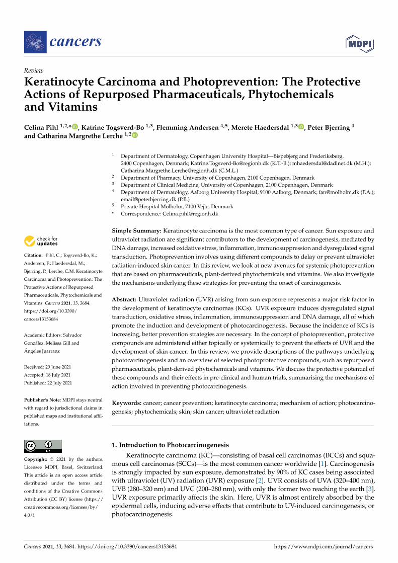
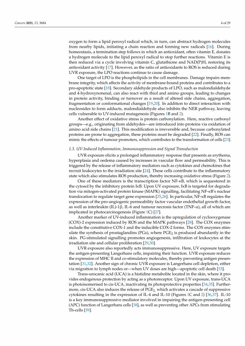
Figure 1. Simplified schematic of (A): UV-induced DNA damage caused by direct UV absorption by the DNA molecules resulting in DNA lesions such as 6–4 photoproducts (6–4PPs) or cyclobutane pyrimidine dimers (CPDs) which are repaired by XPC and the nuclear excision repair pathway; (B): UV-induced oxidative stress caused by increased reactive oxygen species (ROS) production result-ing in damaged DNA (8-oxo-hydroxyguanine lesions), lipid (lipid peroxidation; LPO) and protein (carbonylation and other irreversible changes) molecules; (C): UV-induced inflammation, immuno-suppression and signal transduction caused by dysregulated mitogen-activated protein kinase (MAPK) activity resulting in activation and transcription of inflammatory mediators. Furthermore, UV radiation induces immunosuppressive mediators via the photoisomersiation of trans-urocanic acid (UCA) to cis-UCA. Abbreviations: AMPK: AMP-activated protein kinase, AP-1: activator protein 1, COX-2: cyclooxygenase 2, IL: interleukin, PGE2: prostaglandin E2, TLR4: Toll-like receptor 4.
1.1. UV-Induced DNA Damage A direct target of UVR exposure is DNA. UVR exposure can result in DNA strand
breaks and the formation of 6–4 photoproducts or cyclobutane pyrimidine dimers (CPDs) [4,5]. These lesions are formed in locations with two adjacent pyrimidines and are re-moved by nucleotide excision repair (NER). In brief, the repair mechanism consists of recognition, incision and replication steps that remove and replace the damaged DNA [6], carried out by the xeroderma pigmentosum family of proteins (XPA–G) (Figure 1A). In particular, XPC reportedly plays a key role in recognising UV-induced DNA damage, as confirmed by mutations and deletions often found in SCC patients [7].
If the NER pathway does not function correctly and the dimers are not repaired, →CC TT tandem mutations—also called thymine dimers—may be introduced. These are
often found in the p53 gene of SCC patients and are recognised as an indicator of UV exposure [8]. Mutations in p53 cause genomic instability and generate a microenviron-
Figure 1. Simplified schematic of (A): UV-induced DNA damage caused by direct UV absorption bythe DNA molecules resulting in DNA lesions such as 6–4 photoproducts (6–4PPs) or cyclobutanepyrimidine dimers (CPDs) which are repaired by XPC and the nuclear excision repair pathway;(B): UV-induced oxidative stress caused by increased reactive oxygen species (ROS) productionresulting in damaged DNA (8-oxo-hydroxyguanine lesions), lipid (lipid peroxidation; LPO) andprotein (carbonylation and other irreversible changes) molecules; (C): UV-induced inflammation, im-munosuppression and signal transduction caused by dysregulated mitogen-activated protein kinase(MAPK) activity resulting in activation and transcription of inflammatory mediators. Furthermore,UV radiation induces immunosuppressive mediators via the photoisomersiation of trans-urocanicacid (UCA) to cis-UCA. Abbreviations: AMPK: AMP-activated protein kinase, AP-1: activator protein1, COX-2: cyclooxygenase 2, IL: interleukin, PGE2: prostaglandin E2, TLR4: Toll-like receptor 4.
1.1. UV-Induced DNA Damage
A direct target of UVR exposure is DNA. UVR exposure can result in DNA strandbreaks and the formation of 6–4 photoproducts or cyclobutane pyrimidine dimers(CPDs) [4,5]. These lesions are formed in locations with two adjacent pyrimidines and areremoved by nucleotide excision repair (NER). In brief, the repair mechanism consists ofrecognition, incision and replication steps that remove and replace the damaged DNA [6],carried out by the xeroderma pigmentosum family of proteins (XPA–G) (Figure 1A). Inparticular, XPC reportedly plays a key role in recognising UV-induced DNA damage, asconfirmed by mutations and deletions often found in SCC patients [7].
If the NER pathway does not function correctly and the dimers are not repaired,CC→TT tandem mutations—also called thymine dimers—may be introduced. These areoften found in the p53 gene of SCC patients and are recognised as an indicator of UV
Cancers 2021, 13, 3684 3 of 29
exposure [8]. Mutations in p53 cause genomic instability and generate a microenvironmentthat is conducive to tumour development and progression [9]. Together with mutations inother key proteins, this causes an imbalance in tumour suppressor genes and oncogenes,facilitating photocarcinogenesis (Figure 2).
Cancers 2021, 13, x FOR PEER REVIEW 3 of 31
ment that is conducive to tumour development and progression [9]. Together with muta-tions in other key proteins, this causes an imbalance in tumour suppressor genes and on-cogenes, facilitating photocarcinogenesis (Figure 2).
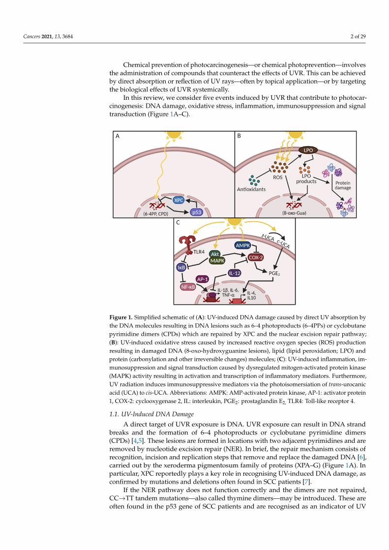
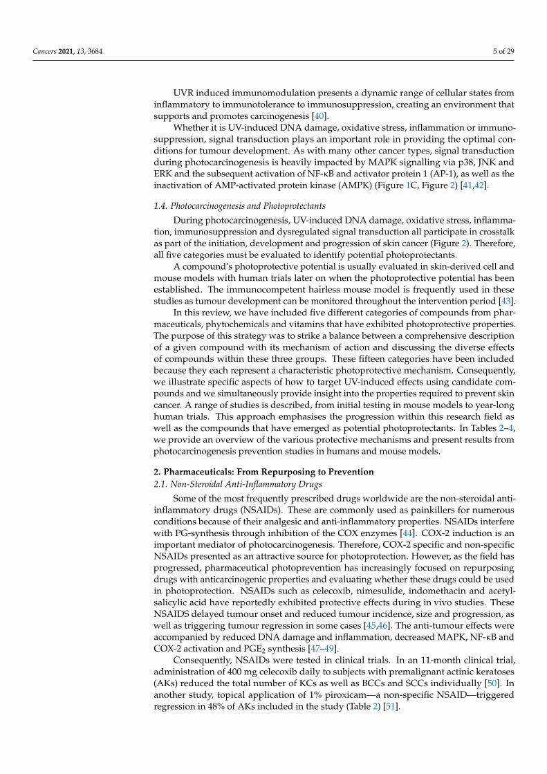
Figure 2. Crosstalk linking UV-induced events: DNA damage, oxidative stress, inflammation, im-munosuppression and dysregulated signal transduction as presented in Figure 1. All five events are involved in crosstalk, creating an environment that promotes photocarcinogenesis. Abbreviations: AMPK: AMP-activated protein kinase, AP-1: activator protein 1, COX-2: cyclooxygenase 2, IL: interleukin, MAPK: mitogen-activated protein kinase, NER: nuclear excision repair, PGE2: prostaglandin E2, ROS: reactive oxygen species, TNF: tumour necrosis factor, t(trans)/c(cis)-UCA: urocanic acid.
1.2. UV-Induced Oxidative Stress and Protein Damage Exposure to UVR increases the production and release of reactive nitrogen and oxy-
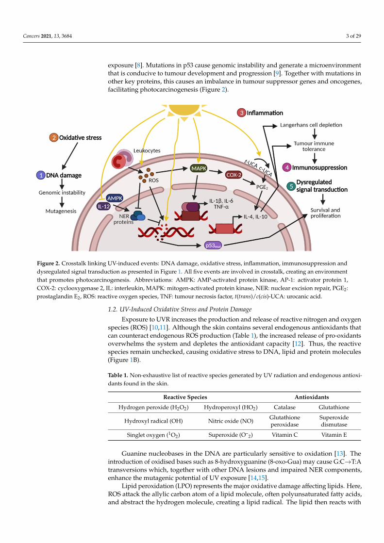
gen species (ROS) [10,11]. Although the skin contains several endogenous antioxidants that can counteract endogenous ROS production (Table 1), the increased release of pro-oxidants overwhelms the system and depletes the antioxidant capacity [12]. Thus, the re-active species remain unchecked, causing oxidative stress to DNA, lipid and protein mol-ecules (Figure 1B).
Table 1. Non-exhaustive list of reactive species generated by UV radiation and endogenous antiox-idants found in the skin.
Reactive Species Antioxidants Hydrogen peroxide
(H2O2) Hydroperoxyl (HO2) Catalase Glutathione
Hydroxyl radical (OH) Nitric oxide (NO) Glutathione peroxidase
Superoxide dismutase
Singlet oxygen (1O2) Superoxide (O–2) Vitamin C Vitamin E
Guanine nucleobases in the DNA are particularly sensitive to oxidation [13]. The in-troduction of oxidised bases such as 8-hydroxyguanine (8-oxo- →Gua) may cause G:C T:A transversions which, together with other DNA lesions and impaired NER components, enhance the mutagenic potential of UV exposure [14,15].
Lipid peroxidation (LPO) represents the major oxidative damage affecting lipids. Here, ROS attack the allylic carbon atom of a lipid molecule, often polyunsaturated fatty acids, and abstract the hydrogen molecule, creating a lipid radical. The lipid then reacts with oxygen to form a lipid peroxyl radical which, in turn, can abstract hydrogen mole-cules from nearby lipids, initiating a chain reaction and forming new radicals [16]. During homeostasis, a termination step follows in which an antioxidant, often vitamin E, donates a hydrogen molecule to the lipid peroxyl radical to stop further reactions. Vitamin E is
Figure 2. Crosstalk linking UV-induced events: DNA damage, oxidative stress, inflammation, immunosuppression anddysregulated signal transduction as presented in Figure 1. All five events are involved in crosstalk, creating an environmentthat promotes photocarcinogenesis. Abbreviations: AMPK: AMP-activated protein kinase, AP-1: activator protein 1,COX-2: cyclooxygenase 2, IL: interleukin, MAPK: mitogen-activated protein kinase, NER: nuclear excision repair, PGE2:prostaglandin E2, ROS: reactive oxygen species, TNF: tumour necrosis factor, t(trans)/c(cis)-UCA: urocanic acid.
1.2. UV-Induced Oxidative Stress and Protein Damage
Exposure to UVR increases the production and release of reactive nitrogen and oxygenspecies (ROS) [10,11]. Although the skin contains several endogenous antioxidants thatcan counteract endogenous ROS production (Table 1), the increased release of pro-oxidantsoverwhelms the system and depletes the antioxidant capacity [12]. Thus, the reactivespecies remain unchecked, causing oxidative stress to DNA, lipid and protein molecules(Figure 1B).
Table 1. Non-exhaustive list of reactive species generated by UV radiation and endogenous antioxi-dants found in the skin.
Reactive Species Antioxidants
Hydrogen peroxide (H2O2) Hydroperoxyl (HO2) Catalase Glutathione
Hydroxyl radical (OH) Nitric oxide (NO) Glutathioneperoxidase
Superoxidedismutase
Singlet oxygen (1O2) Superoxide (O–2) Vitamin C Vitamin E
Guanine nucleobases in the DNA are particularly sensitive to oxidation [13]. Theintroduction of oxidised bases such as 8-hydroxyguanine (8-oxo-Gua) may cause G:C→T:Atransversions which, together with other DNA lesions and impaired NER components,enhance the mutagenic potential of UV exposure [14,15].
Lipid peroxidation (LPO) represents the major oxidative damage affecting lipids. Here,ROS attack the allylic carbon atom of a lipid molecule, often polyunsaturated fatty acids,and abstract the hydrogen molecule, creating a lipid radical. The lipid then reacts with
Cancers 2021, 13, 3684 4 of 29
oxygen to form a lipid peroxyl radical which, in turn, can abstract hydrogen moleculesfrom nearby lipids, initiating a chain reaction and forming new radicals [16]. Duringhomeostasis, a termination step follows in which an antioxidant, often vitamin E, donatesa hydrogen molecule to the lipid peroxyl radical to stop further reactions. Vitamin E isthen reduced via a cycle involving vitamin C, glutathione and NAD(P)H, restoring itsantioxidant activity [17]. However, as the ratio of antioxidants to ROS is reduced duringUVR exposure, the LPO reactions continue to cause damage.
One target of LPO is the phospholipids in the cell membranes. Damage impairs mem-brane integrity, which affects the activity of membrane-bound proteins and contributes to apro-apoptotic state [18]. Secondary aldehyde products of LPO, such as malondialdehydeand 4-hydroxynonenal, can also react with thiol and amino groups, leading to changesin protein activity, binding or turnover as a result of altered side chains, aggregation,fragmentation or conformational changes [19,20]. In addition to direct interaction withnucleosides to form adducts, malondialdehyde also inhibits the NER pathway, leavingcells vulnerable to UV-induced mutagenesis (Figures 1B and 2).
Another effect of oxidative stress is protein carbonylation. Here, reactive carbonylgroups—e.g., originating from aldehydes—are introduced into proteins via oxidation ofamino acid side chains [21]. This modification is irreversible and, because carbonylatedproteins are prone to aggregation, these proteins must be degraded [22]. Finally, ROS canmimic the effects of tumour promoters, which contributes to the transformation of cells [23].
1.3. UV-Induced Inflammation, Immunosuppression and Signal Transduction
UVR exposure elicits a prolonged inflammatory response that presents as erythema,hyperplasia and oedema caused by increases in vascular flow and permeability. This istriggered by the release of inflammatory mediators such as cytokines and chemokines thatrecruit leukocytes to the irradiation site [24]. These cells contribute to the inflammatorystate which also stimulates ROS production, thereby increasing oxidative stress (Figure 2).
One of these mediators is the transcription factor NF-κB, which is sequestered inthe cytosol by the inhibitory protein IκB. Upon UV exposure, IκB is targeted for degrada-tion via mitogen-activated protein kinase (MAPK) signalling, facilitating NF-κB’s nucleartranslocation to regulate target gene expression [25,26]. In particular, NF-κB regulates theexpression of the pro-angiogenic permeability factor vascular endothelial growth factor,as well as interleukin (IL)-1β, IL-6 and tumour necrosis factor (TNF-α), all of which areimplicated in photocarcinogenesis (Figure 1C) [27].
Another marker of UV-induced inflammation is the upregulation of cyclooxygenase(COX)-2 expression induced by ROS and the MAPK pathways [28]. The COX enzymesinclude the constitutive COX-1 and the inducible COX-2 forms. The COX enzymes stim-ulate the synthesis of prostaglandins (PGs), where PGE2 is produced abundantly in theskin. PG-stimulated signalling promotes angiogenesis, infiltration of leukocytes at theirradiation site and cellular proliferation [29,30].
UVR exposure also reportedly acts immunosuppressive. Here, UV exposure targetsthe antigen-presenting Langerhans cells, impairing their function. UVR exposure reducesthe expression of MHC II and co-stimulatory molecules, thereby preventing antigen presen-tation [31,32]. Another sign of chronic UVR exposure is Langerhans cell depletion, eithervia migration to lymph nodes or—when UV doses are high—apoptotic cell death [33].
Trans-urocanic acid (UCA) is a histidine metabolite located in the skin, where it pro-vides endogenous protection by acting as a photoreceptor. Upon UV exposure, trans-UCAis photoisomerised to cis-UCA, inactivating its photoprotective properties [34,35]. Further-more, cis-UCA also induces the release of PGE2, which activates a cascade of suppressivecytokines resulting in the expression of IL-4 and IL-10 (Figures 1C and 2) [36,37]. IL-10is a key immunosuppressive mediator involved in impairing the antigen-presenting cell(APC) function of Langerhans cells [38], as well as preventing other APCs from stimulatingTh-cells [39].
Cancers 2021, 13, 3684 5 of 29
UVR induced immunomodulation presents a dynamic range of cellular states frominflammatory to immunotolerance to immunosuppression, creating an environment thatsupports and promotes carcinogenesis [40].
Whether it is UV-induced DNA damage, oxidative stress, inflammation or immuno-suppression, signal transduction plays an important role in providing the optimal con-ditions for tumour development. As with many other cancer types, signal transductionduring photocarcinogenesis is heavily impacted by MAPK signalling via p38, JNK andERK and the subsequent activation of NF-κB and activator protein 1 (AP-1), as well as theinactivation of AMP-activated protein kinase (AMPK) (Figure 1C, Figure 2) [41,42].
1.4. Photocarcinogenesis and Photoprotectants
During photocarcinogenesis, UV-induced DNA damage, oxidative stress, inflamma-tion, immunosuppression and dysregulated signal transduction all participate in crosstalkas part of the initiation, development and progression of skin cancer (Figure 2). Therefore,all five categories must be evaluated to identify potential photoprotectants.
A compound’s photoprotective potential is usually evaluated in skin-derived cell andmouse models with human trials later on when the photoprotective potential has beenestablished. The immunocompetent hairless mouse model is frequently used in thesestudies as tumour development can be monitored throughout the intervention period [43].
In this review, we have included five different categories of compounds from phar-maceuticals, phytochemicals and vitamins that have exhibited photoprotective properties.The purpose of this strategy was to strike a balance between a comprehensive descriptionof a given compound with its mechanism of action and discussing the diverse effectsof compounds within these three groups. These fifteen categories have been includedbecause they each represent a characteristic photoprotective mechanism. Consequently,we illustrate specific aspects of how to target UV-induced effects using candidate com-pounds and we simultaneously provide insight into the properties required to prevent skincancer. A range of studies is described, from initial testing in mouse models to year-longhuman trials. This approach emphasises the progression within this research field aswell as the compounds that have emerged as potential photoprotectants. In Tables 2–4,we provide an overview of the various protective mechanisms and present results fromphotocarcinogenesis prevention studies in humans and mouse models.
2. Pharmaceuticals: From Repurposing to Prevention2.1. Non-Steroidal Anti-Inflammatory Drugs
Some of the most frequently prescribed drugs worldwide are the non-steroidal anti-inflammatory drugs (NSAIDs). These are commonly used as painkillers for numerousconditions because of their analgesic and anti-inflammatory properties. NSAIDs interferewith PG-synthesis through inhibition of the COX enzymes [44]. COX-2 induction is animportant mediator of photocarcinogenesis. Therefore, COX-2 specific and non-specificNSAIDs presented as an attractive source for photoprotection. However, as the field hasprogressed, pharmaceutical photoprevention has increasingly focused on repurposingdrugs with anticarcinogenic properties and evaluating whether these drugs could be usedin photoprotection. NSAIDs such as celecoxib, nimesulide, indomethacin and acetyl-salicylic acid have reportedly exhibited protective effects during in vivo studies. TheseNSAIDS delayed tumour onset and reduced tumour incidence, size and progression, aswell as triggering tumour regression in some cases [45,46]. The anti-tumour effects wereaccompanied by reduced DNA damage and inflammation, decreased MAPK, NF-κB andCOX-2 activation and PGE2 synthesis [47–49].
Consequently, NSAIDs were tested in clinical trials. In an 11-month clinical trial,administration of 400 mg celecoxib daily to subjects with premalignant actinic keratoses(AKs) reduced the total number of KCs as well as BCCs and SCCs individually [50]. Inanother study, topical application of 1% piroxicam—a non-specific NSAID—triggeredregression in 48% of AKs included in the study (Table 2) [51].
Cancers 2021, 13, 3684 6 of 29
However, although NSAIDs have demonstrated photoprotective potential in bothmouse and human studies, other studies have described the potentially adverse sideeffects of prolonged NSAID use. These include increased risk of cardiovascular events [52],gastrointestinal bleeding [53] and renal failure [54].
2.2. AMPK Activators: Metformin
Increasing evidence has implicated AMPK as a potential target for cancer thera-pies [55]. AMPK as an energy and nutrient sensor can interact with p53 via metaboliccheckpoints to induce cell cycle arrest [56,57]. Additionally, tumours from UV-irradiatedhuman and murine skin display decreased AMPK activation [41], suggesting a role forAMPK activation in photoprotection. Metformin is commonly used to treat diabetes byinhibiting protein kinase A and activating AMPK, leading to decreased gluconeogenesis inthe liver and increased insulin sensitivity in target tissues [58]. Interestingly, metformintreatment is reportedly associated with a 31% decrease in overall cancer risk, compared toother antidiabetic treatments [59].
In keratinocytes, incubation with metformin protected against UV-induced inflamma-tion, as demonstrated by impaired NF-κB activity and reductions in IL-1β, IL-6 and TNF-αexpression [60]. Similarly, a study on nude mice with SCC A431 tumour-cell xenograftsreported impaired NF-κB activity as well as reduced COX-2 expression following injectionwith metformin [61].
Metformin reduced ROS formation and expression of matrix metalloproteinase (MMP)1 and 3 in vitro [62]. During homeostasis, MMPs partake in remodelling and degradation ofthe extracellular matrix [63]. However, MMPs can also stimulate tumour development andangiogenesis. Furthermore, topical application of metformin in hairless mice increased CPDrepair six hours after UVR exposure [41]. In xenografted mice, injection with metforminalso protected against UV-induced proliferation, inducing apoptosis in the tumours alongwith reductions in protein kinase B (Akt), MAPK and NF-κB signalling [61]. Moreover,metformin may specifically target the skin’s cancer stem cell diaspora [64,65], furtherillustrating its photoprotective potential.
In hairless mice, topical and oral administration of metformin delayed tumour onset,decreased tumour multiplicity and volume and stimulated DNA repair (Table 2) [41].In vitro studies using AMPK-knockout cells reported that metformin-induced DNA repairwas dependent on AMPK activation [41].
There have been no clinical studies on the effect of metformin on KCs. However, apopulation-wide study in Taiwanese diabetic patients reported a significantly lower riskof skin cancer incidence in metformin-treated subjects compared to those who had neverreceived metformin [66]. Another recent study across the Icelandic population found asignificantly lower risk of BCCs, but not SCCs, following metformin use [67].
Metformin, therefore, represents a promising candidate for photoprotection, but morestudies are needed, namely clinical trials, to investigate its effects. Furthermore, AMPKactivators such as 5-aminoimidazole-4-carboxamide ribonucleotide (AICAR) [41] andphenformin [68,69] should also be investigated as potential photoprotectants.
2.3. Toll-Like Receptor 4 Antagonism: Resatorvid
Because inflammation is an important event in the development of photocarcino-genesis, some recent research has focused on targeting immunomodulators such as theToll-like receptors (TLRs). In particular, TLR4 has been thought to be a driver in cutaneousinflammation activating MAPK, AP-1 and NF-κB, as well as the expression of IL-1β, IL-6and TNF-α [70] (Figure 1C).
In the skin, TLR4 expression is upregulated in keratinocytes following UVR expo-sure [71]. A study that compared normal skin, sun-damaged skin and AKs from thesame individuals reported that TLR4 expression was confined to the basal layer of theepidermis in normal skin, whereas in response to sun damage, TLR4 was strongly ex-pressed across several epidermal layers, with thicker and more pronounced detection
Cancers 2021, 13, 3684 7 of 29
in the AK samples [72]. Therefore, TLR4 antagonism is an emerging target for limitingUV-induced inflammation.
Resatorvid, or TAK-242, is a small-molecule inhibitor that selectively binds to TLR4and inhibits cellular activity by preventing TLR4 from interacting with adaptor mole-cules [73]. Resatorvid has been reported to have neuroprotective effects following braininjury [74], and is currently under investigation for its anticarcinogenic properties demon-strated in breast and ovarian cancer cell lines in which TLR4 antagonism reduced epithelial–mesenchymal transition and invasion [75].
In a study on irradiated keratinocytes, incubation with resatorvid prevented activationof p38, JNK and Akt. This was accompanied by reductions in AP-1 and NF-κB activity andIL-6 and IL-8 expression [72]. Notably, the study reported that incubation with resatorvidpre- or post-UVR had similar effects, suggesting that these are not caused by UV absorptionbut by resatorvid’s antagonism of TLR4.
Irradiated hairless mice treated with a topical formulation of resatorvid also demon-strated reduced activity of the MAPKs, AP-1 and NF-κB with decreased expression of IL-6,IL-8 and IL-10 [72,76]. When photocarcinogenesis is stimulated experimentally, topicalapplication of resatorvid delayed tumour onset and reduced tumour multiplicity andincidence in hairless mice [76]. Interestingly, this study reported that only when resatorvidwas used as a prevention measure—i.e., administered together with UV—and not an inter-vention treatment—i.e., resatorvid given after UV was terminated—were there reductionsin tumourigenesis (Table 2).
Resatorvid has been tested in a clinical trial as a treatment for septic shock. Althoughit failed to suppress patients’ cytokine levels, resatorvid was generally well tolerated [77].Therefore, resatorvid is a promising candidate for photoprotection, but more studies areneeded to elucidate its mechanism of action, determine whether the protective effects canbe translated to humans and establish a suitable therapeutic window.
2.4. Oestrogen Receptor Signalling: Erb041, 17β-Oestradiol and Phytoestrogens
Another potential pharmaceutical target for skin cancer prevention is the modulationof the oestrogen receptors (ERs)—ERα and ERβ. Both receptors are activated by oestrogen(17β-oestradiol) and related oestrogenic compounds, resulting in target gene transcription.Despite sharing ligands, the two receptors often act antagonistically, with ERβ reportedlyfunctioning as a tumour suppressor to prevent tumour metastasis and proliferation inducedby ERα [78,79]. ERβ specific agonists have been tested in clinical trials to promote theanticarcinogenic effects of ERβwithout stimulating ERα signalling.
Erb-041, or Prinaberel, is an oestrogenic ligand with a similar binding affinity tooestrogen but optimised to selectively bind to ERβ [80]. A clinical trial monitored Erb-041intake for its effects on rheumatoid arthritis over a 12-week period, and although treatmentfailed to show efficacy in patients, the study did report that Erb-041 was well-toleratedand exhibited a good safety profile [81]. In the skin, ERβ expression is normally confinedto the basal layer of the epidermis. This expression is reduced in tumour adjacent skinand lost entirely in skin tissue derived from SCCs. Moreover, ERβ expression is lost inUV-exposed murine skin [82], further suggesting that ERβ signalling is a potential targetfor skin cancer prevention.
One study assessed the effect of Erb-041 in preventing photocarcinogenesis. Treatmentwith Erb-041 restored ERβ expression in both irradiated murine skin and cell cultures, andtopical application of Erb-041 to hairless mice led to a delay in tumour onset. The micein this study also exhibited reductions in tumour incidence, multiplicity and volume aswell as in carcinoma progression. This was accompanied by a decreased inflammatoryresponse manifesting as reduced leukocyte infiltration, hyperplasia and inflammatorycytokines. Furthermore, Erb-041 application resulted in reduced activation of ERK, p38,NF-κB and Akt signalling and decreased expression of proliferation, angiogenesis andepithelial-mesenchymal transition markers [82]. These observations suggest that Erb-041can prevent UV-induced events (Table 2).
Cancers 2021, 13, 3684 8 of 29
Other non-specific oestrogenic compounds have also been tested as photoprotec-tants. For example, 17β-oestradiol injections reduced UV-induced immunosuppressionand associated IL-10 production in male mice [83]. Some phytochemicals also have oe-strogenic properties, as described below (Section 3). These natural phytoestrogens sharestructural similarities with oestrogen and are capable of modulating ER signalling, albeitwith lower potency [84]. Phytoestrogens such as genistein, epicatechin and resveratrolexhibit numerous protective effects [85]; the latter two are discussed in Sections 3.1 and 3.2.
Although activation of ERβ signalling is a potential target for preventing UVR-inducedeffects, more studies are needed to understand how ERβ signalling operates photoprotec-tion. As administration of oestrogenic compounds may also result in some side effects, it ispossible that the less potent phytoestrogens may be suitable alternatives.
2.5. Recent Discoveries in Pre-Clinical Studies: Carvedilol and Bucillamine
Finally, carvedilol and bucillamine have been studied over the past five years aspotential photoprotectants.
Carvedilol is a β-adrenergic receptor (β-AR) antagonist that prevents the bindingof catecholamines such as nor- and epinephrine to the β-AR, licenced for treatment ofhypertension and heart disease. However, catecholamines also reportedly impact carcino-genesis by affecting DNA repair and APCs [86,87]. As keratinocytes express β-AR-2 [88],carvedilol was tested in epidermal cells and hairless mice for its potential in photocarcino-genesis prevention.
In vitro studies reported that following carvedilol treatment, irradiated cells exhibitedreductions in NF-κB and AP-1 activity, PGE2 release and colony formation [89,90], whereastopical application in reconstituted human skin reduced inflammation markers such asCOX-2 and TNF-α expression, as well as hyperplasia and apoptosis [91]. Furthermore,carvedilol treatment decreased inflammatory cytokines and CPDs in hairless mice whenphotocarcinogenesis is stimulated experimentally. These mice also exhibited delays intumour onset and reduced tumour multiplicity and incidence [90].
Bucillamine is a cysteine-derived compound that contains two thiol groups, whichconfer significant antioxidant activity. Bucillamine has been used for more than 30 years inJapan and South Korea as a treatment for rheumatoid arthritis and is well tolerated [92],but it is not licenced for use in the European Union.
Bucillamine shares structural similarities with N-acetylcysteine, a proven photopro-tectant in hairless mice [93], but it is reportedly even more potent as an antioxidant [94].Therefore, the photoprotective potential of bucillamine was evaluated. In irradiated ker-atinocytes and hairless mice, treatment with bucillamine reduced the activation of JNK andcaspase 3 [95]. Furthermore, the mice exhibited reductions in dermal oedema, leukocyteinfiltration, proliferation and p53 expression [95,96]. These results indicate that bucillaminetreatment can prevent UV-induced skin damage in hairless mice. Therefore, future studiesshould investigate its effects on photocarcinogenesis, focusing on whether bucillamine’sinherent antioxidant activity can limit the impact of oxidative stress.
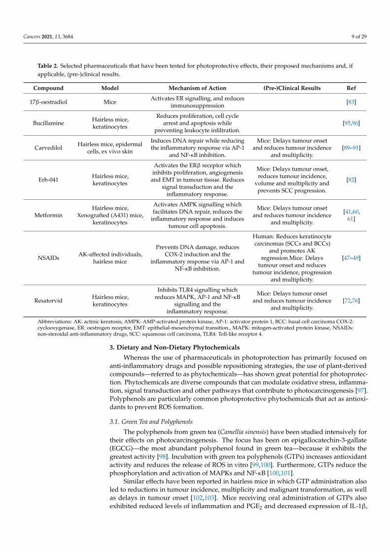
To sum up, the use of pharmaceuticals in photoprotection is an evolving field. Whereaspreviously, much research focused on preventing the induction of COX-2 by implementingspecific inhibitors (NSAIDs), recent studies have involved drug repurposing to avoid theexpensive and time-consuming clinical trials necessary to evaluate novel pharmaceuticals.In this section, we have discussed a selection of compounds that may not be ready touse as treatments but do highlight events and mechanisms that may be targeted in futurestudies. In Table 2, we have provided an overview of the seven compounds discussed inthis section, their proposed mechanisms and potential (pre-)clinical results in human andmouse studies.
Cancers 2021, 13, 3684 9 of 29
Table 2. Selected pharmaceuticals that have been tested for photoprotective effects, their proposed mechanisms and, ifapplicable, (pre-)clinical results.
Compound Model Mechanism of Action (Pre-)Clinical Results Ref
17β-oestradiol Mice Activates ER signalling, and reducesimmunosuppression [83]
Bucillamine Hairless mice,keratinocytes
Reduces proliferation, cell cyclearrest and apoptosis while
preventing leukocyte infiltration.[95,96]
Carvedilol Hairless mice, epidermalcells, ex vivo skin
Induces DNA repair while reducingthe inflammatory response via AP-1
and NF-κB inhibition.
Mice: Delays tumour onsetand reduces tumour incidence
and multiplicity.[89–91]
Erb-041 Hairless mice,keratinocytes
Activates the ERβ receptor whichinhibits proliferation, angiogenesis
and EMT in tumour tissue. Reducessignal transduction and the
inflammatory response.
Mice: Delays tumour onset,reduces tumour incidence,
volume and multiplicity andprevents SCC progression.
[82]
MetforminHairless mice,
Xenografted (A431) mice,keratinocytes
Activates AMPK signalling whichfacilitates DNA repair, reduces the
inflammatory response and inducestumour cell apoptosis.
Mice: Delays tumour onsetand reduces tumour incidence
and multiplicity.
[41,60,61]
NSAIDs AK-affected individuals,hairless mice
Prevents DNA damage, reducesCOX-2 induction and the
inflammatory response via AP-1 andNF-κB inhibition.
Human: Reduces keratinocytecarcinomas (SCCs and BCCs)
and promotes AKregression.Mice: Delays
tumour onset and reducestumour incidence, progression
and multiplicity.
[47–49]
Resatorvid Hairless mice,keratinocytes
Inhibits TLR4 signalling whichreduces MAPK, AP-1 and NF-κB
signalling and theinflammatory response.
Mice: Delays tumour onsetand reduces tumour incidence
and multiplicity.[72,76]
Abbreviations: AK: actinic keratosis, AMPK: AMP-activated protein kinase, AP-1: activator protein 1, BCC: basal cell carcinoma COX-2:cyclooxygenase, ER: oestrogen receptor, EMT: epithelial-mesenchymal transition., MAPK: mitogen-activated protein kinase, NSAIDs:non-steroidal anti-inflammatory drugs, SCC: squamous cell carcinoma, TLR4: Toll-like receptor 4.
3. Dietary and Non-Dietary Phytochemicals
Whereas the use of pharmaceuticals in photoprotection has primarily focused onanti-inflammatory drugs and possible repositioning strategies, the use of plant-derivedcompounds—referred to as phytochemicals—has shown great potential for photoprotec-tion. Phytochemicals are diverse compounds that can modulate oxidative stress, inflamma-tion, signal transduction and other pathways that contribute to photocarcinogenesis [97].Polyphenols are particularly common photoprotective phytochemicals that act as antioxi-dants to prevent ROS formation.
3.1. Green Tea and Polyphenols
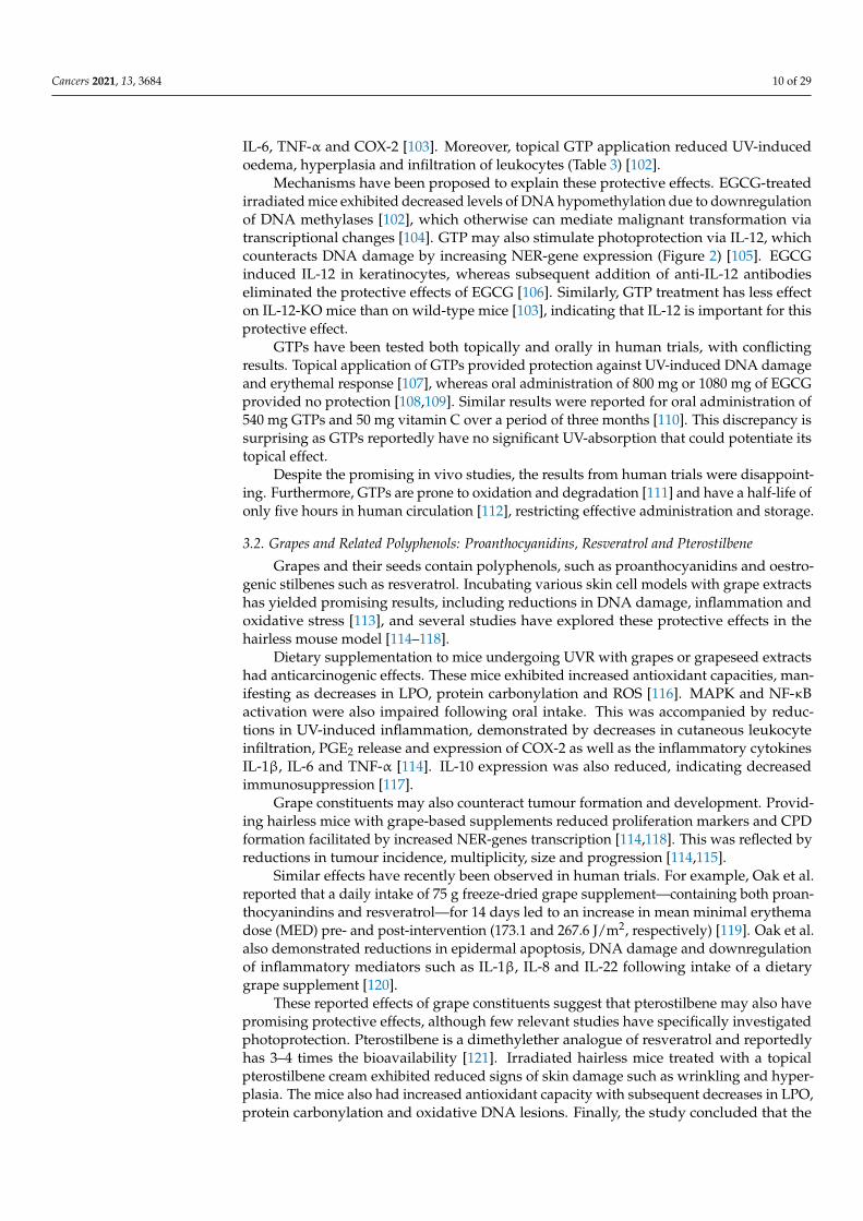
The polyphenols from green tea (Camellia sinensis) have been studied intensively fortheir effects on photocarcinogenesis. The focus has been on epigallocatechin-3-gallate(EGCG)—the most abundant polyphenol found in green tea—because it exhibits thegreatest activity [98]. Incubation with green tea polyphenols (GTPs) increases antioxidantactivity and reduces the release of ROS in vitro [99,100]. Furthermore, GTPs reduce thephosphorylation and activation of MAPKs and NF-κB [100,101].
Similar effects have been reported in hairless mice in which GTP administration alsoled to reductions in tumour incidence, multiplicity and malignant transformation, as wellas delays in tumour onset [102,103]. Mice receiving oral administration of GTPs alsoexhibited reduced levels of inflammation and PGE2 and decreased expression of IL-1β,
Cancers 2021, 13, 3684 10 of 29
IL-6, TNF-α and COX-2 [103]. Moreover, topical GTP application reduced UV-inducedoedema, hyperplasia and infiltration of leukocytes (Table 3) [102].
Mechanisms have been proposed to explain these protective effects. EGCG-treatedirradiated mice exhibited decreased levels of DNA hypomethylation due to downregulationof DNA methylases [102], which otherwise can mediate malignant transformation viatranscriptional changes [104]. GTP may also stimulate photoprotection via IL-12, whichcounteracts DNA damage by increasing NER-gene expression (Figure 2) [105]. EGCGinduced IL-12 in keratinocytes, whereas subsequent addition of anti-IL-12 antibodieseliminated the protective effects of EGCG [106]. Similarly, GTP treatment has less effecton IL-12-KO mice than on wild-type mice [103], indicating that IL-12 is important for thisprotective effect.
GTPs have been tested both topically and orally in human trials, with conflictingresults. Topical application of GTPs provided protection against UV-induced DNA damageand erythemal response [107], whereas oral administration of 800 mg or 1080 mg of EGCGprovided no protection [108,109]. Similar results were reported for oral administration of540 mg GTPs and 50 mg vitamin C over a period of three months [110]. This discrepancy issurprising as GTPs reportedly have no significant UV-absorption that could potentiate itstopical effect.
Despite the promising in vivo studies, the results from human trials were disappoint-ing. Furthermore, GTPs are prone to oxidation and degradation [111] and have a half-life ofonly five hours in human circulation [112], restricting effective administration and storage.
3.2. Grapes and Related Polyphenols: Proanthocyanidins, Resveratrol and Pterostilbene
Grapes and their seeds contain polyphenols, such as proanthocyanidins and oestro-genic stilbenes such as resveratrol. Incubating various skin cell models with grape extractshas yielded promising results, including reductions in DNA damage, inflammation andoxidative stress [113], and several studies have explored these protective effects in thehairless mouse model [114–118].
Dietary supplementation to mice undergoing UVR with grapes or grapeseed extractshad anticarcinogenic effects. These mice exhibited increased antioxidant capacities, man-ifesting as decreases in LPO, protein carbonylation and ROS [116]. MAPK and NF-κBactivation were also impaired following oral intake. This was accompanied by reduc-tions in UV-induced inflammation, demonstrated by decreases in cutaneous leukocyteinfiltration, PGE2 release and expression of COX-2 as well as the inflammatory cytokinesIL-1β, IL-6 and TNF-α [114]. IL-10 expression was also reduced, indicating decreasedimmunosuppression [117].
Grape constituents may also counteract tumour formation and development. Provid-ing hairless mice with grape-based supplements reduced proliferation markers and CPDformation facilitated by increased NER-genes transcription [114,118]. This was reflected byreductions in tumour incidence, multiplicity, size and progression [114,115].
Similar effects have recently been observed in human trials. For example, Oak et al.reported that a daily intake of 75 g freeze-dried grape supplement—containing both proan-thocyanindins and resveratrol—for 14 days led to an increase in mean minimal erythemadose (MED) pre- and post-intervention (173.1 and 267.6 J/m2, respectively) [119]. Oak et al.also demonstrated reductions in epidermal apoptosis, DNA damage and downregulationof inflammatory mediators such as IL-1β, IL-8 and IL-22 following intake of a dietarygrape supplement [120].
These reported effects of grape constituents suggest that pterostilbene may also havepromising protective effects, although few relevant studies have specifically investigatedphotoprotection. Pterostilbene is a dimethylether analogue of resveratrol and reportedlyhas 3–4 times the bioavailability [121]. Irradiated hairless mice treated with a topicalpterostilbene cream exhibited reduced signs of skin damage such as wrinkling and hyper-plasia. The mice also had increased antioxidant capacity with subsequent decreases in LPO,protein carbonylation and oxidative DNA lesions. Finally, the study concluded that the
Cancers 2021, 13, 3684 11 of 29
application of pterostilbene but not resveratrol reduced tumour incidence in these mice(Table 3) [122].
Based on these results, more research should focus on elucidating the effect of grapesin human photoprotection. Furthermore, studies should explore whether the increasedbioavailability of pterostilbene and the improved in vivo results can be translated intoclinical results to provide an additional avenue for protection.
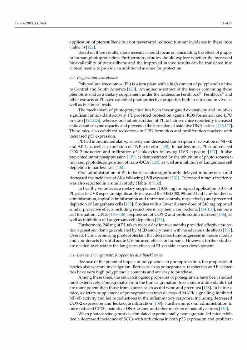
3.3. Polypodium Leucotomos
Polypodium leucotomos (PL) is a fern plant with a high content of polyphenols nativeto Central and South America [123]. An aqueous extract of the leaves containing thesephenols is sold as a dietary supplement under the tradename Fernblock®. Fernblock® andother extracts of PL have exhibited photoprotective properties both in vitro and in vivo, aswell as in clinical trials.
The mechanism of photoprotection has been investigated extensively and involvessignificant antioxidant activity. PL provided protection against ROS formation and LPOin vitro [124,125], whereas oral administration of PL to hairless mice reportedly increasedantioxidant enzyme capacity and prevented the formation of oxidative DNA lesions [126,127].These mice also exhibited reductions in CPD formation and proliferation markers withincreased p53 expression.
PL had immunomodulatory activity and decreased transcriptional activation of NF-κBand AP-1, as well as expression of TNF-α in vitro [128]. In hairless mice, PL counteractedCOX-2 induction and infiltration of leukocytes following UVR exposure [127]. It alsoprevented immunosuppression [129], as demonstrated by the inhibition of photoisomerisa-tion and photodecomposition of trans-UCA [124], as well as inhibition of Langerhans celldepletion in hairless rats [130].
Oral administration of PL to hairless mice significantly delayed tumour onset anddecreased the incidence of AKs following UVR exposure [131]. Decreased tumour incidencewas also reported in a similar study (Table 3) [132].
In healthy volunteers, a dietary supplement (1080 mg) or topical application (10%) ofPL prior to UVR exposure significantly increased the MED (80, 98 and 34 mJ/cm2 for dietaryadministration, topical administration and untreated controls, respectively) and preventeddepletion of Langerhans cells [133]. Studies with a lower dietary dose of 240 mg reportedsimilar protective effects including reductions in erythema and oedema [134,135], sunburncell formation, CPDs [134–136], expression of COX-2 and proliferation markers [136], aswell as inhibition of Langerhans cell depletion [134].
Furthermore, 240 mg of PL taken twice a day for two months provided effective protec-tion against sun damage evaluated by MED and erythema with no adverse side effects [137].Overall, PL is a promising photoprotectant that decreases tumourigenesis in mouse modelsand counteracts harmful acute UV-induced effects in humans. However, further studiesare needed to elucidate the long-term effects of PL on skin cancer development.
3.4. Berries: Pomegranate, Raspberries and Blackberries
Because of the potential impact of polyphenols in photoprotection, the properties ofberries also warrant investigation. Berries such as pomegranate, raspberries and blackber-ries have very high polyphenolic contents and are easy to purchase.
Among these three, the anticarcinogenic properties of pomegranate have been studiedmost extensively. Pomegranates from the Punica granatum tree contain antioxidants thatare more potent than those from sources such as red wine and green tea [138]. In hairlessmice, a dietary supplement of pomegranate extract decreased MAPK signalling, inhibitedNF-κB activity and led to reductions in the inflammatory response, including decreasedCOX-2 expression and leukocyte infiltration [139]. Furthermore, oral administration tomice reduced CPDs, oxidative DNA lesions and other markers of oxidative stress [140].
When photocarcinogenesis is stimulated experimentally, pomegranate-fed mice exhib-ited a decreased incidence of SCCs with reductions in both p53 expression and prolifera-
Cancers 2021, 13, 3684 12 of 29
tion [141]. In human studies, intake of pomegranate over 12 weeks provided protectionagainst UVR, as shown by increases in MED from baseline to post-treatment [142].
Although there have been no clinical trials, raspberries also have great potential as aphotoprotectant. Hairless mice treated topically with a black raspberry extract followingUVR exposure exhibited decreases in tumour multiplicity and size. This was accompaniedby reductions in the number of 8-oxo-Gua lesions and in p53 expression, as well as areduced inflammatory response, which was indicated by decreased neutrophil activationand oedema [143]. Recent studies have also shown that red raspberries exhibit promisingtopical photoprotectant activity. Albeit no data on tumour formation, irradiated micetreated with a red raspberry extract demonstrated reductions in erythemal response, p38,AP-1 and NF-κB activity, together with decreased COX-2 expression. The antioxidantcapacity was also increased in these mice, possibly via nuclear factor erythroid 2-relatedfactor (Nrf-2) activation, which induces transcription of antioxidant-response genes anddecreases 8-oxo-Gua lesions and protein carbonylation [144], mimicking results reportedin vitro [145].
Finally, blackberries and one of their polyphenols, cyanidin-3-glucoside, have dis-played promising photoprotective characteristics in vivo. Topical application of blackberryextract or the polyphenol to hairless mice undergoing UVR showed reductions in boththe inflammatory and oxidative stress responses. These mice exhibited reductions in LPOand oxidative DNA lesions, as well as decreased oedema, hyperplasia and leukocyteinfiltration. Reductions in MAPK signalling and NF-κB activity were also reported, to-gether with subsequent decreases in PGE2 release and expression of iNOS, IL-6 and TNF-α(Table 3) [146,147].
Although some of these studies focused on topical application, the widespread avail-ability of these berries is ideal for oral photoprotection. However, further studies areneeded to confirm the protective potential of berries: extensive mouse studies shouldbe used to elucidate protective mechanisms and clinical trials are needed to investigatewhether berries can prevent KCs in humans.
3.5. Cocoa Flavanols
Polyphenolic compounds can also be found in cocoa as so-called cocoa flavanols.Although chocolate and other cocoa-containing products may have a high cocoa content,the flavanols are often destroyed in processing steps before the final products are generated.Nevertheless, the potential of cocoa extracts in epidermal health has been suggested bypointing to effects such as ROS scavenging, prevention of MAPK and NF-κB activation andinhibition of COX-2 induction [148,149].
Because similar pathways contribute to KC development, the effect of cocoa extractwas tested in hairless mice when photocarcinogenesis was stimulated experimentally.Following oral intake of cocoa extract, these mice had fewer wrinkles and MMP-1 expres-sion was downregulated [150]. Irradiated mice given cocoa extract also exhibited a lowerincidence of invasive SCCs, as well as decreases in mutated p53 expression and PGE2 re-lease [141], indicating increased protection against UV-induced events and carcinogenesis.
Cocoa extract has also proved beneficial in humans. In 2006, a clinical trial spanningover 12 weeks reported that a daily intake of 329 mg cocoa flavanols reduced UV-inducederythema by 25%, whereas a lower intake (27 mg) did not [151]. Other studies exploringshorter and longer intervention periods (1 and 24 weeks, respectively) had similar out-comes, resulting in increases in MED [150,152]. However, two studies investigated theeffects of a higher dose (600 mg) over 12 weeks, and only one of these studies found a protec-tive effect [153,154], suggesting that more research is needed to clarify the photoprotectiveeffects of cocoa flavanols.
As described in this section, dietary and non-dietary phytochemicals have enor-mous potential for photoprotection. Pre-clinical and human trials have shown that thesecompounds have antioxidant, anti-inflammatory and anti-carcinogenic effects in subjectsexposed to UVR (Table 3).
Cancers 2021, 13, 3684 13 of 29
In contrast to pharmaceuticals, which are expensive and may have adverse side effects,phytochemicals are widely available and may be readily included in the daily diet. Further-more, while phytochemical intake at higher concentrations may have some side effects, theyare not as severe as what is demonstrated with certain pharmaceuticals. However, becausesome polyphenolic compounds absorb UVR, observed effects may be due to a sunscreeneffect leaving oral administration inefficient [155]. Therefore, more research is needed toevaluate the use of phytochemicals in long-term systemic photoprevention. These studiesshould focus on dosing, delivery and effectiveness to optimise the protective potential.
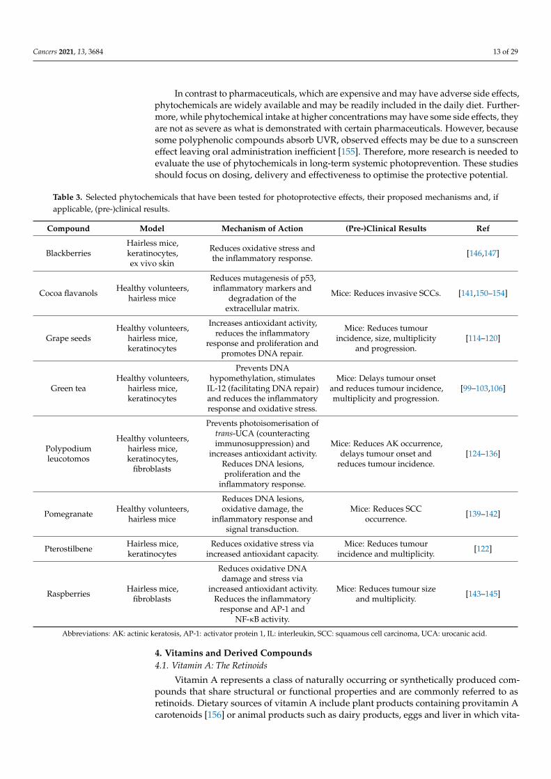
Table 3. Selected phytochemicals that have been tested for photoprotective effects, their proposed mechanisms and, ifapplicable, (pre-)clinical results.
Compound Model Mechanism of Action (Pre-)Clinical Results Ref
BlackberriesHairless mice,keratinocytes,ex vivo skin
Reduces oxidative stress andthe inflammatory response. [146,147]
Cocoa flavanols Healthy volunteers,hairless mice
Reduces mutagenesis of p53,inflammatory markers and
degradation of theextracellular matrix.
Mice: Reduces invasive SCCs. [141,150–154]
Grape seedsHealthy volunteers,
hairless mice,keratinocytes
Increases antioxidant activity,reduces the inflammatory
response and proliferation andpromotes DNA repair.
Mice: Reduces tumourincidence, size, multiplicity
and progression.[114–120]
Green teaHealthy volunteers,
hairless mice,keratinocytes
Prevents DNAhypomethylation, stimulates
IL-12 (facilitating DNA repair)and reduces the inflammatoryresponse and oxidative stress.
Mice: Delays tumour onsetand reduces tumour incidence,multiplicity and progression.
[99–103,106]
Polypodiumleucotomos
Healthy volunteers,hairless mice,keratinocytes,
fibroblasts
Prevents photoisomerisation oftrans-UCA (counteractingimmunosuppression) and
increases antioxidant activity.Reduces DNA lesions,proliferation and the
inflammatory response.
Mice: Reduces AK occurrence,delays tumour onset and
reduces tumour incidence.[124–136]
Pomegranate Healthy volunteers,hairless mice
Reduces DNA lesions,oxidative damage, the
inflammatory response andsignal transduction.
Mice: Reduces SCCoccurrence. [139–142]
Pterostilbene Hairless mice,keratinocytes
Reduces oxidative stress viaincreased antioxidant capacity.
Mice: Reduces tumourincidence and multiplicity. [122]
Raspberries Hairless mice,fibroblasts
Reduces oxidative DNAdamage and stress via
increased antioxidant activity.Reduces the inflammatory
response and AP-1 andNF-κB activity.
Mice: Reduces tumour sizeand multiplicity. [143–145]
Abbreviations: AK: actinic keratosis, AP-1: activator protein 1, IL: interleukin, SCC: squamous cell carcinoma, UCA: urocanic acid.
4. Vitamins and Derived Compounds4.1. Vitamin A: The Retinoids
Vitamin A represents a class of naturally occurring or synthetically produced com-pounds that share structural or functional properties and are commonly referred to asretinoids. Dietary sources of vitamin A include plant products containing provitamin Acarotenoids [156] or animal products such as dairy products, eggs and liver in which vita-
Cancers 2021, 13, 3684 14 of 29
min A is present as retinol, retinal, retinyl esters and retinoic acid (RA) [157,158]. Becausethe photoprotective potential of carotenoids has been explored in other reviews [159], thefollowing description will focus on the latter group.
In vitro studies have shown that retinoids have antioxidant activity that preventsROS formation, in part due to restoration of Nrf2 [160]. Furthermore, retinoids ex-hibit anti-inflammatory properties by reducing AP-1 activity [161,162] and TNF-α expres-sion [163,164]. However, because topical retinoid treatment can cause skin irritation [165,166],the elicited inflammatory responses must be considered in their specific contexts.
Whereas topical application of retinoids protects against DNA damage and apopto-sis in hairless mice [167], the effects on photocarcinogenesis reportedly vary and remaincontroversial. Two separate experiments from the same group led by Epstein et al. show-cased these opposing effects of RA, reporting stimulation of photocarcinogenesis in onestudy [168] but inhibition in another [169]. A similar paradox has been reported by othergroups, with stimulatory [166,170,171], inhibitory [172], and no effect on photocarcinogen-esis in hairless mice treated with RA all being described [173]. No mechanism has beensuggested that can resolve these conflicting results.
Consequently, few human studies have been performed. One study reported that theapplication of a 2% retinyl ester cream protected against thymine dimers and erythemain healthy volunteers 24 hours after UVR treatment [174]. However, topical applicationof a 0.05% all-trans RA cream for eight days had no effect on MED [175]. Although thisdiscrepancy may be caused by differences in the retinoic compounds or their concentrations,it may also reflect the pattern observed in the mouse studies.
Despite this, a recent cohort study reported that increased dietary intake of retinol ledto a decreased risk of SCC [176]. Similarly, in psoriasis patients exposed to psoralen-UVA,systemic retinoid use was associated with a significant reduction in SCC risk [177]. In atrial focusing on subjects with a history of AKs, a dietary supplement of 25,000 IU retinolover five years protected against SCC but not BCC incidence (Table 4) [178]. However,the same group reported a lack of effect in high-risk subjects with a history of KCs [179].Taken together, these results indicate that we still do not know enough about how retinoidsfunction and whether they can consistently provide photoprotection.
4.2. Vitamin B3: Nicotinamide
Vitamin B3 represents a family of water-soluble compounds with similar structuresthat include nicotinamide, nicotinic acid and nicotinamide riboside. These are found infish, meat and wheat, with smaller quantities present in vegetables. Vitamin B3 compoundsact as precursors for the cofactor nicotinamide adenine dinucleotide (NAD+) which isinvolved in ATP metabolism [180–182]. NAD+ deficiency increases the skin’s sensitivity toUVR exposure by reducing genomic stability and preventing DNA repair [183]. Therefore,replenishing NAD+ precursors is a potential strategy in cancer prevention.
NAD+ acts as a substrate for poly ADP-ribose polymerase-1 (PARP-1) and the sirtuinproteins that regulate DNA repair and genomic stability [184,185]. Incubation of irradiatedkeratinocytes with nicotinamide enhanced DNA repair and reduced photolesions [186].Furthermore, pre-treatment also prevented UV-induced inflammation in keratinocytes byreducing the expression of IL-6, IL-10, TNF-α and COX-2 [187,188].
Gensler et al. have described the effect of nicotinamide in mouse models and foundthat topical and systemic administration resulted in decreased tumour incidence in ir-radiated BALB/c mice [189,190]. Moreover, these mice exhibited reductions in tumourdevelopment, tumour multiplicity and UV-induced immunosuppression.
Topical application of a 5% nicotinamide cream also reduced immunosuppression inhealthy human volunteers [191,192]. As an oral delivery study reported similar protectiveresults [193], with no effect on MED following topical application [191], the effects ofnicotinamide are likely caused by protection against UV-induced events rather than sun-screening (Table 4).
Cancers 2021, 13, 3684 15 of 29
The effects of treating sun-damaged individuals with nicotinamide were investigatedin a phase II double-blinded randomised controlled trial. In volunteers with ≥4 palpableAKs, 500 mg dietary supplement reduced the mean AK count over a period of four months(21.6 vs. 34.8 in the control group) [194]. Similar effects were reported over one year in aphase III trial involving high-risk subjects, with dietary supplements reducing the rate ofnew KCs (1.8 per person in nicotinamide-treated subjects vs. 2.4 in control subjects) andSCCs (0.5 vs. 0.7 per person, respectively) [195]. This trial reported few adverse effectsand no additional effect at higher doses, suggesting that 500 mg is a safe and effectivedose. However, no beneficial effects were observed on recurrent carcinomas or followingtreatment discontinuation [195].
The phase III trial was notable for an increase in infections among the treated groupand significant differences in the numbers of skin and mucocutaneous infections [196].Although the authors accepted that all adverse effects should be considered, they also notedthat nicotinamide reportedly increases the clearance of skin infections [197,198]. Nevertheless,Yélamos et al. noted that while an overall reduction in KC rates was observed followingnicotinamide intake, the more aggressive types of SCCs and BCCs apparently increased [199].
Therefore, although nicotinamide has protective effects in human studies, additionaltrials with a focus on patient follow-up and the differences among and subtypes will benecessary to address these concerns.
4.3. Vitamin C
Vitamin C is a key antioxidant found in the skin where it plays a role in keratinocyteviability and maintenance of the epidermal barrier [200]. Vitamin C can be synthesisedfrom D-glucose in plants and almost all non-primates [201], whereas humans can no longerproduce the active enzyme required for this process. Therefore, humans obtain vitamin Cfrom their diet.
The antioxidant properties of vitamin C are well established. In keratinocytes, treat-ment with vitamin C reduced ROS formation [202], oxidative DNA damage [203] andLPO, simultaneously preventing glutathione depletion [204]. Treatment with vitamin Calso affects inflammatory responses, leading to decreased expression and release of IL-1α,IL-6, IL-8 and TNF-α [202,204,205], while also preventing apoptosis and MAPK activation(Table 4) [202,206].
Despite these effects and its role as an antioxidant, the photoprotective potential ofvitamin C is less clear in in vivo studies. In a study performed in 1982, dietary supplemen-tation of vitamin C given to hairless mice led to a delay in UV-induced tumour onset anddecreased tumour incidence [207]. A second study, performed nine years later, confirmedthese observations [208]. However, in 2005, a study involving a similar set-up observed anincrease in UV-induced tumour multiplicity [93], indicating some uncertainty regardingthe effect of vitamin C when more complex models are used.
Furthermore, few clinical trials have investigated the photoprotective effects of vi-tamin C. In a study performed in 2002, 500 mg oral supplement of vitamin C over aneight-week period had no effect on the UV-induced erythemal response measured [209].Studies exploring higher doses of 2 g and 3 g—albeit over shorter periods (7 and 50 days,respectively)—also reported no significant effects [210,211]. Topical application of a 5%cream also offered no protection in healthy volunteers [212,213], and cohort studies lastingmore than ten years have found no evidence that vitamin C intake decreases the incidenceof BCCs and SCCs [214,215].
Because vitamin C alone does not appear to provide photoprotection in vivo, the focusof research has shifted to investigating vitamin C combined with other compounds. Com-bining vitamins C and E has produced promising clinical results and is further describedin Section 4.5 [211,216,217]. Similarly, a topical formulation of vitamin C, ferulic acidand the phenolic compound phloretin conferred protection against UV-induced erythema,apoptotic sunburn-cell production and thymine dimer formation in healthy volunteers [218],indicating that vitamin C in synergy with other compounds may provide sufficient protection.
Cancers 2021, 13, 3684 16 of 29
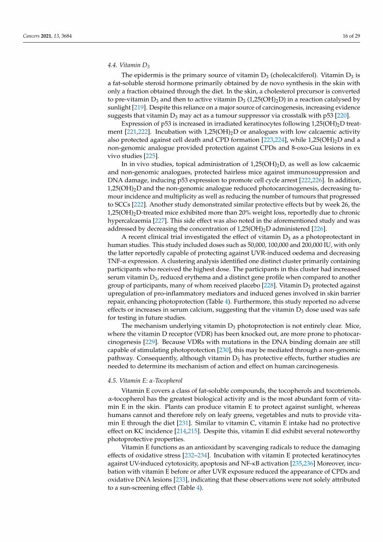
4.4. Vitamin D3
The epidermis is the primary source of vitamin D3 (cholecalciferol). Vitamin D3 isa fat-soluble steroid hormone primarily obtained by de novo synthesis in the skin withonly a fraction obtained through the diet. In the skin, a cholesterol precursor is convertedto pre-vitamin D3 and then to active vitamin D3 (1,25(OH)2D) in a reaction catalysed bysunlight [219]. Despite this reliance on a major source of carcinogenesis, increasing evidencesuggests that vitamin D3 may act as a tumour suppressor via crosstalk with p53 [220].
Expression of p53 is increased in irradiated keratinocytes following 1,25(OH)2D treat-ment [221,222]. Incubation with 1,25(OH)2D or analogues with low calcaemic activityalso protected against cell death and CPD formation [223,224], while 1,25(OH)2D and anon-genomic analogue provided protection against CPDs and 8-oxo-Gua lesions in exvivo studies [225].
In in vivo studies, topical administration of 1,25(OH)2D, as well as low calcaemicand non-genomic analogues, protected hairless mice against immunosuppression andDNA damage, inducing p53 expression to promote cell cycle arrest [222,226]. In addition,1,25(OH)2D and the non-genomic analogue reduced photocarcinogenesis, decreasing tu-mour incidence and multiplicity as well as reducing the number of tumours that progressedto SCCs [222]. Another study demonstrated similar protective effects but by week 26, the1,25(OH)2D-treated mice exhibited more than 20% weight loss, reportedly due to chronichypercalcaemia [227]. This side effect was also noted in the aforementioned study and wasaddressed by decreasing the concentration of 1,25(OH)2D administered [226].
A recent clinical trial investigated the effect of vitamin D3 as a photoprotectant inhuman studies. This study included doses such as 50,000, 100,000 and 200,000 IU, with onlythe latter reportedly capable of protecting against UVR-induced oedema and decreasingTNF-α expression. A clustering analysis identified one distinct cluster primarily containingparticipants who received the highest dose. The participants in this cluster had increasedserum vitamin D3, reduced erythema and a distinct gene profile when compared to anothergroup of participants, many of whom received placebo [228]. Vitamin D3 protected againstupregulation of pro-inflammatory mediators and induced genes involved in skin barrierrepair, enhancing photoprotection (Table 4). Furthermore, this study reported no adverseeffects or increases in serum calcium, suggesting that the vitamin D3 dose used was safefor testing in future studies.
The mechanism underlying vitamin D3 photoprotection is not entirely clear. Mice,where the vitamin D receptor (VDR) has been knocked out, are more prone to photocar-cinogenesis [229]. Because VDRs with mutations in the DNA binding domain are stillcapable of stimulating photoprotection [230], this may be mediated through a non-genomicpathway. Consequently, although vitamin D3 has protective effects, further studies areneeded to determine its mechanism of action and effect on human carcinogenesis.
4.5. Vitamin E: α-Tocopherol
Vitamin E covers a class of fat-soluble compounds, the tocopherols and tocotrienols.α-tocopherol has the greatest biological activity and is the most abundant form of vita-min E in the skin. Plants can produce vitamin E to protect against sunlight, whereashumans cannot and therefore rely on leafy greens, vegetables and nuts to provide vita-min E through the diet [231]. Similar to vitamin C, vitamin E intake had no protectiveeffect on KC incidence [214,215]. Despite this, vitamin E did exhibit several noteworthyphotoprotective properties.
Vitamin E functions as an antioxidant by scavenging radicals to reduce the damagingeffects of oxidative stress [232–234]. Incubation with vitamin E protected keratinocytesagainst UV-induced cytotoxicity, apoptosis and NF-κB activation [235,236] Moreover, incu-bation with vitamin E before or after UVR exposure reduced the appearance of CPDs andoxidative DNA lesions [233], indicating that these observations were not solely attributedto a sun-screening effect (Table 4).
Cancers 2021, 13, 3684 17 of 29
In hairless mice, topical application of vitamin E reduced UV-induced erythema andoedema [237,238], as well as immunosuppression and tumour incidence [239]. Similarly,dietary vitamin E supplementation resulted in delayed tumour onset and reductionsin tumour multiplicity and size. These mice also exhibited decreased proliferation andoxidative stress markers such as 8-oxo-Gua lesions [240]. However, a more recent studyperformed in 2013, reported contrasting effects with a topical vitamin E cream increasingtumour formation and proliferation, DNA damage and angiogenesis [241].
One clinical trial reported that 400-IU vitamin E dietary supplementation over a six-month period did not significantly change MED or sunburn-cell formation [242]. A shortertrial with the same dose over an eight-week period did not report any protective effects forvitamin E either [243].
Although neither vitamin C nor E showed significant photoprotection in humanstudies, combining the two vitamins has promising effects [244]. Adding ferulic acidto a combined preparation of topically applied vitamins C and E improves the stabilityand doubles the photoprotective capacity of the formulation when applied to pigskin, asmeasured by thymine dimer formation, erythema and apoptosis [245]. In a clinical trialspanning eight days, dietary supplementation of vitamins C and E increased MED (medianof 80 mJ/cm2 before supplementation to 96.5 mJ/cm2 after eight days) [217]. Similarincreases in MED, as well as reduced thymine dimer formation, were observed in longertrials with no effect for the vitamins separately [211,216]. Overall, these results indicatethat vitamins C and E can act synergistically to protect against UVR exposure.
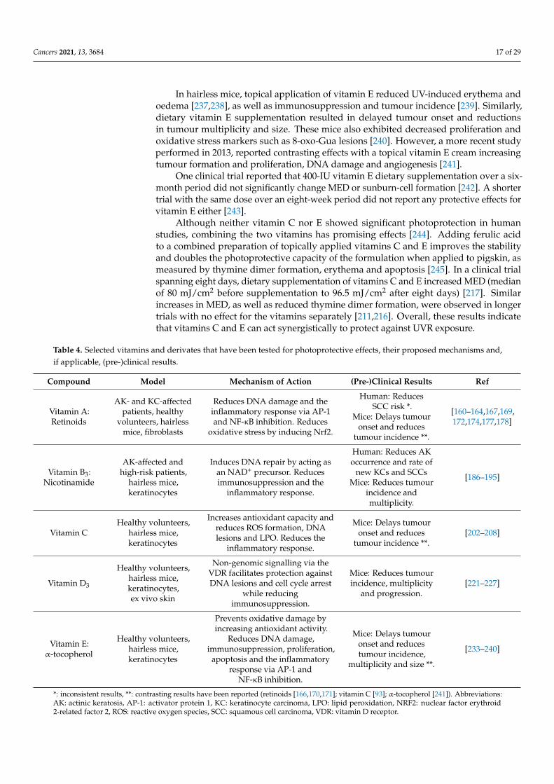
Table 4. Selected vitamins and derivates that have been tested for photoprotective effects, their proposed mechanisms and,if applicable, (pre-)clinical results.
Compound Model Mechanism of Action (Pre-)Clinical Results Ref
Vitamin A:Retinoids
AK- and KC-affectedpatients, healthy
volunteers, hairlessmice, fibroblasts
Reduces DNA damage and theinflammatory response via AP-1and NF-κB inhibition. Reduces
oxidative stress by inducing Nrf2.
Human: ReducesSCC risk *.
Mice: Delays tumouronset and reduces
tumour incidence **.
[160–164,167,169,172,174,177,178]
Vitamin B3:Nicotinamide
AK-affected andhigh-risk patients,
hairless mice,keratinocytes
Induces DNA repair by acting asan NAD+ precursor. Reducesimmunosuppression and the
inflammatory response.
Human: Reduces AKoccurrence and rate of
new KCs and SCCsMice: Reduces tumour
incidence andmultiplicity.
[186–195]
Vitamin CHealthy volunteers,
hairless mice,keratinocytes
Increases antioxidant capacity andreduces ROS formation, DNAlesions and LPO. Reduces the
inflammatory response.
Mice: Delays tumouronset and reduces
tumour incidence **.[202–208]
Vitamin D3
Healthy volunteers,hairless mice,keratinocytes,ex vivo skin
Non-genomic signalling via theVDR facilitates protection againstDNA lesions and cell cycle arrest
while reducingimmunosuppression.
Mice: Reduces tumourincidence, multiplicity
and progression.[221–227]
Vitamin E:α-tocopherol
Healthy volunteers,hairless mice,keratinocytes
Prevents oxidative damage byincreasing antioxidant activity.
Reduces DNA damage,immunosuppression, proliferation,
apoptosis and the inflammatoryresponse via AP-1 and
NF-κB inhibition.
Mice: Delays tumouronset and reducestumour incidence,
multiplicity and size **.
[233–240]
*: inconsistent results, **: contrasting results have been reported (retinoids [166,170,171]; vitamin C [93]; α-tocopherol [241]). Abbreviations:AK: actinic keratosis, AP-1: activator protein 1, KC: keratinocyte carcinoma, LPO: lipid peroxidation, NRF2: nuclear factor erythroid2-related factor 2, ROS: reactive oxygen species, SCC: squamous cell carcinoma, VDR: vitamin D receptor.
Cancers 2021, 13, 3684 18 of 29
5. Perspectives and Concluding Remarks
When sun-avoidant behaviour, protective clothing and sunscreens are used insuffi-ciently to prevent KCs, oral photoprotection presents a promising alternative or supplement.Instead of blocking the absorption of UV-rays, dietary intake of protective compounds fo-cuses on preventing and countering UV-induced events that stimulate photocarcinogenesis(Figures 1 and 2). Using oral photoprotectants in addition to sunscreen will increase theprotection against UVR-induced effects.
Drug repurposing provides a promising avenue for systemic photoprotection, withvarying degree of success in pre-clinical trials (Table 2). However, applying these findingsto clinical trials may be difficult as data obtained from studies in unrelated conditions maynot be relevant for evaluating the efficacy and safety of treatments to prevent photocar-cinogenesis in humans. Therefore, new investigations are needed to identify therapeuticwindows in which photoprotection is effective.
Phytochemicals are promising photoprotectants. Their protective effects (Table 4), lowtoxicity and widespread availability make them ideal candidates for systemic photopro-tection. Nevertheless, not all compounds are effective via ingestion, and phytochemicalsmay require extraction, purification and concentration to generate effective photopro-tective products. Furthermore, some phytochemicals may only be effective via topicaladministration if they exert their effects by absorbing UVR, making them unsuitable forsystemic photoprotection.
Because alternatives to conventional treatments are being considered, it is impor-tant to remember that photoprotection requires a degree of individualisation: side effectsthat are unacceptable to some individuals may be tolerable to others, if the treatmentdecreases the risk of carcinogenesis. Thus, phytochemicals with photoprotective propertiescould be administered in safe and effective doses to less affected individuals where milderstrategies may still provide an effect. Whereas patients receiving immunosuppressive treat-ments who have a higher risk of developing KCs should be treated with more aggressivepharmaceutical therapies to ensure efficient treatment of the carcinomas.
Photoprevention in its current stage is focused on preventing the initial steps of KCdevelopment by counteracting the adverse effects induced by UVR exposure. However,increasing evidence has demonstrated that phytochemical supplementation to cancer treat-ments such as chemotherapy may improve treatment outcome. In chemotherapy, natural orsynthetic compounds such as bleomycin, cisplatin and taxol induce cytotoxicity in tumourcells by interfering with DNA replication and mitosis to prevent tumour proliferation. Theaddition of certain phytochemicals to these treatments have demonstrated better outcomes,either by improving treatment efficacy, drug delivery and accumulation [246] or improv-ing management of side effects in in vitro and pre-clinical models [247], which is furtherreviewed here [248].
In general, more studies are needed to clarify whether photoprotectants on their owncan be used as anticarcinogenic therapies, as well as to identify the most promising targetsfor photoprotection. Because these compounds may display weak pharmacological poten-cies, efficient use may require modification either through synthetically edited structuresor the use of adjuvants to improve results. As the compounds presented in this reviewaffect several different targets, optimisation of their cellular effects must also be considered.Signal transduction is a delicate process evolved to respond to different stimuli resulting inunexpected results that can both promote and prevent cellular growth. As such, treatmentwith these compounds must be optimised through comprehensive studies to ensure thatthe induced outcome is predominantly anticarcinogenic.
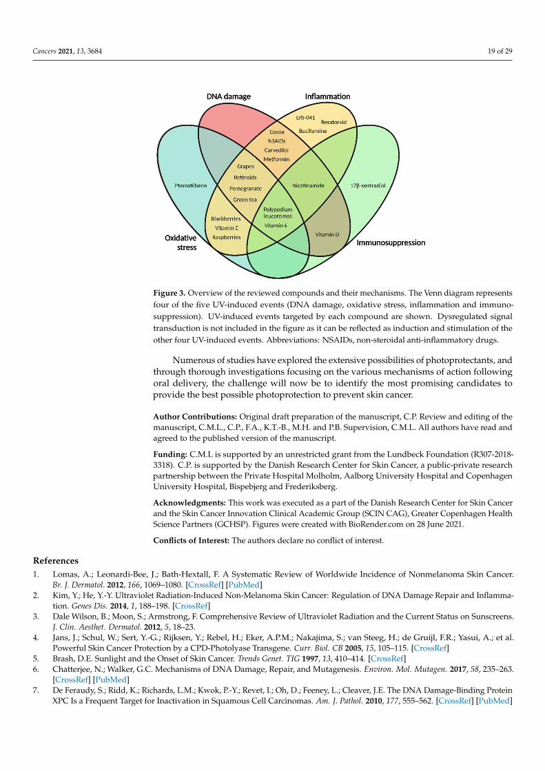
In this review, we have discussed the photoprotective potential of 15 different cate-gories across pharmaceuticals, phytochemicals and vitamins (Tables 2–4). We have alsoprovided an overview of the current understanding of the UV-induced events they targetunder the headings of DNA damage, oxidative stress, inflammation, immunosuppressionand dysregulated signal transduction, as summarised in Figure 3.
Cancers 2021, 13, 3684 19 of 29
Cancers 2021, 13, x FOR PEER REVIEW 20 of 31
Numerous of studies have explored the extensive possibilities of photoprotectants, and through thorough investigations focusing on the various mechanisms of action fol-lowing oral delivery, the challenge will now be to identify the most promising candidates to provide the best possible photoprotection to prevent skin cancer.
Figure 3. Overview of the reviewed compounds and their mechanisms. The Venn diagram repre-sents four of the five UV-induced events (DNA damage, oxidative stress, inflammation and immu-nosuppression). UV-induced events targeted by each compound are shown. Dysregulated signal transduction is not included in the figure as it can be reflected as induction and stimulation of the other four UV-induced events. Abbreviations: NSAIDs, non-steroidal anti-inflammatory drugs.
Author Contributions: Original draft preparation of the manuscript, C.P. Review and editing of the manuscript, C.M.L., C.P., F.A., K.T.-B., M.H. and P.B. Supervision, C.M.L. All authors have read and agreed to the published version of the manuscript.
Funding: C.M.L is supported by an unrestricted grant from the Lundbeck Foundation (R307-2018-3318). C.P. is supported by the Danish Research Center for Skin Cancer, a public-private research partnership between the Private Hospital Molholm, Aalborg University Hospital and Copenhagen University Hospital, Bispebjerg and Frederiksberg.
Acknowledgments: This work was executed as a part of the Danish Research Center for Skin Cancer and the Skin Cancer Innovation Clinical Academic Group (SCIN CAG), Greater Copenhagen Health Science Partners (GCHSP). Figures were created with BioRender.com on June 28th, 2021.
Conflicts of Interest: The authors declare no conflict of interest.
References 1. Lomas, A.; Leonardi-Bee, J.; Bath-Hextall, F. A Systematic Review of Worldwide Incidence of Nonmelanoma Skin Cancer. Br. J.
Dermatol. 2012, 166, 1069–1080, doi:10.1111/j.1365-2133.2012.10830.x. 2. Kim, Y.; He, Y.-Y. Ultraviolet Radiation-Induced Non-Melanoma Skin Cancer: Regulation of DNA Damage Repair and
Inflammation. Genes Dis. 2014, 1, 188–198, doi:10.1016/j.gendis.2014.08.005. 3. Dale Wilson, B.; Moon, S.; Armstrong, F. Comprehensive Review of Ultraviolet Radiation and the Current Status on Sunscreens.
J. Clin. Aesthet. Dermatol. 2012, 5, 18–23. 4. Jans, J.; Schul, W.; Sert, Y.-G.; Rijksen, Y.; Rebel, H.; Eker, A.P.M.; Nakajima, S.; van Steeg, H.; de Gruijl, F.R.; Yasui, A.; et al.
Powerful Skin Cancer Protection by a CPD-Photolyase Transgene. Curr. Biol. CB 2005, 15, 105–115, doi:10.1016/j.cub.2005.01.001. 5. Brash, D.E. Sunlight and the Onset of Skin Cancer. Trends Genet. TIG 1997, 13, 410–414, doi:10.1016/s0168-9525(97)01246-8. 6. Chatterjee, N.; Walker, G.C. Mechanisms of DNA Damage, Repair, and Mutagenesis. Environ. Mol. Mutagen. 2017, 58, 235–263,
doi:10.1002/em.22087.
Figure 3. Overview of the reviewed compounds and their mechanisms. The Venn diagram representsfour of the five UV-induced events (DNA damage, oxidative stress, inflammation and immuno-suppression). UV-induced events targeted by each compound are shown. Dysregulated signaltransduction is not included in the figure as it can be reflected as induction and stimulation of theother four UV-induced events. Abbreviations: NSAIDs, non-steroidal anti-inflammatory drugs.
Numerous of studies have explored the extensive possibilities of photoprotectants, andthrough thorough investigations focusing on the various mechanisms of action followingoral delivery, the challenge will now be to identify the most promising candidates toprovide the best possible photoprotection to prevent skin cancer.
Author Contributions: Original draft preparation of the manuscript, C.P. Review and editing of themanuscript, C.M.L., C.P., F.A., K.T.-B., M.H. and P.B. Supervision, C.M.L. All authors have read andagreed to the published version of the manuscript.
Funding: C.M.L is supported by an unrestricted grant from the Lundbeck Foundation (R307-2018-3318). C.P. is supported by the Danish Research Center for Skin Cancer, a public-private researchpartnership between the Private Hospital Molholm, Aalborg University Hospital and CopenhagenUniversity Hospital, Bispebjerg and Frederiksberg.
Acknowledgments: This work was executed as a part of the Danish Research Center for Skin Cancerand the Skin Cancer Innovation Clinical Academic Group (SCIN CAG), Greater Copenhagen HealthScience Partners (GCHSP). Figures were created with BioRender.com on 28 June 2021.
Conflicts of Interest: The authors declare no conflict of interest.
References1. Lomas, A.; Leonardi-Bee, J.; Bath-Hextall, F. A Systematic Review of Worldwide Incidence of Nonmelanoma Skin Cancer.
Br. J. Dermatol. 2012, 166, 1069–1080. [CrossRef] [PubMed]2. Kim, Y.; He, Y.-Y. Ultraviolet Radiation-Induced Non-Melanoma Skin Cancer: Regulation of DNA Damage Repair and Inflamma-
tion. Genes Dis. 2014, 1, 188–198. [CrossRef]3. Dale Wilson, B.; Moon, S.; Armstrong, F. Comprehensive Review of Ultraviolet Radiation and the Current Status on Sunscreens.
J. Clin. Aesthet. Dermatol. 2012, 5, 18–23.4. Jans, J.; Schul, W.; Sert, Y.-G.; Rijksen, Y.; Rebel, H.; Eker, A.P.M.; Nakajima, S.; van Steeg, H.; de Gruijl, F.R.; Yasui, A.; et al.
Powerful Skin Cancer Protection by a CPD-Photolyase Transgene. Curr. Biol. CB 2005, 15, 105–115. [CrossRef]5. Brash, D.E. Sunlight and the Onset of Skin Cancer. Trends Genet. TIG 1997, 13, 410–414. [CrossRef]6. Chatterjee, N.; Walker, G.C. Mechanisms of DNA Damage, Repair, and Mutagenesis. Environ. Mol. Mutagen. 2017, 58, 235–263.
[CrossRef] [PubMed]7. De Feraudy, S.; Ridd, K.; Richards, L.M.; Kwok, P.-Y.; Revet, I.; Oh, D.; Feeney, L.; Cleaver, J.E. The DNA Damage-Binding Protein
XPC Is a Frequent Target for Inactivation in Squamous Cell Carcinomas. Am. J. Pathol. 2010, 177, 555–562. [CrossRef] [PubMed]
Cancers 2021, 13, 3684 20 of 29
8. Brash, D.E.; Rudolph, J.A.; Simon, J.A.; Lin, A.; McKenna, G.J.; Baden, H.P.; Halperin, A.J.; Pontén, J. A Role for Sunlight inSkin Cancer: UV-Induced P53 Mutations in Squamous Cell Carcinoma. Proc. Natl. Acad. Sci. USA 1991, 88, 10124. [CrossRef][PubMed]
9. Muller, P.A.J.; Vousden, K.H. Mutant P53 in Cancer: New Functions and Therapeutic Opportunities. Cancer Cell 2014, 25, 304–317.[CrossRef]
10. Deliconstantinos, G.; Villiotou, V.; Stavrides, J.C. Alterations of Nitric Oxide Synthase and Xanthine Oxidase Activities of HumanKeratinocytes by Ultraviolet B Radiation. Potential Role for Peroxynitrite in Skin Inflammation. Biochem. Pharmacol. 1996, 51,1727–1738. [CrossRef]
11. Brem, R.; Macpherson, P.; Guven, M.; Karran, P. Oxidative Stress Induced by UVA Photoactivation of the Tryptophan UVBPhotoproduct 6-Formylindolo[3,2-b]Carbazole (FICZ) Inhibits Nucleotide Excision Repair in Human Cells. Sci. Rep. 2017, 7, 4310.[CrossRef]
12. Podda, M.; Traber, M.G.; Weber, C.; Yan, L.-J.; Packer, L. UV-Irradiation Depletes Antioxidants and Causes Oxidative Damage in aModel of Human Skin. Free Radic. Biol. Med. 1998, 24, 55–65. [CrossRef]
13. Kawanishi, S.; Hiraku, Y.; Oikawa, S. Mechanism of Guanine-Specific DNA Damage by Oxidative Stress and Its Role inCarcinogenesis and Aging. Mutat. Res. Mutat. Res. 2001, 488, 65–76. [CrossRef]
14. Hattorinakakuki, Y.; Nishigori, C.; Okamoto, K.; Imamura, S.; Hiai, H.; Toyokuni, S. Formation of 8-Hydroxy-2′-Deoxyguanosinein Epidermis of Hairless Mice Exposed to Near-UV. Biochem. Biophys. Res. Commun. 1994, 201, 1132–1139. [CrossRef] [PubMed]
15. Shibutani, S.; Takeshita, M.; Grollman, A.P. Insertion of Specific Bases during DNA Synthesis Past the Oxidation-Damaged Base8-OxodG. Nature 1991, 349, 431–434. [CrossRef] [PubMed]
16. Yin, H.; Xu, L.; Porter, N.A. Free Radical Lipid Peroxidation: Mechanisms and Analysis. Chem. Rev. 2011, 111, 5944–5972.[CrossRef] [PubMed]
17. Szarka, A.; Tomasskovics, B.; Bánhegyi, G. The Ascorbate-Glutathione-α-Tocopherol Triad in Abiotic Stress Response.Int. J. Mol. Sci. 2012, 13, 4458–4483. [CrossRef]
18. Ramana, K.V.; Srivastava, S.; Singhal, S.S. Lipid Peroxidation Products in Human Health and Disease 2014. Oxid. Med. Cell. Longev.2014, 2014, 162414. [CrossRef] [PubMed]
19. Schaur, R.J. Basic Aspects of the Biochemical Reactivity of 4-Hydroxynonenal. Mol. Asp. Med. 2003, 24, 149–159. [CrossRef]20. Davies, M.J. Singlet Oxygen-Mediated Damage to Proteins and Its Consequences. Biochem. Biophys. Res. Commun. 2003, 305,
761–770. [CrossRef]21. Dalle-Donne, I.; Giustarini, D.; Colombo, R.; Rossi, R.; Milzani, A. Protein Carbonylation in Human Diseases. Trends Mol. Med.
2003, 9, 169–176. [CrossRef]22. Petrov, D.; Zagrovic, B. Microscopic Analysis of Protein Oxidative Damage: Effect of Carbonylation on Structure, Dynamics, and
Aggregability of Villin Headpiece. J. Am. Chem. Soc. 2011, 133, 7016–7024. [CrossRef]23. Zimmerman, R.; Cerutti, P. Active Oxygen Acts as a Promoter of Transformation in Mouse Embryo C3H/10T1/2/C18 Fibroblasts.
Proc. Natl. Acad. Sci. USA 1984, 81, 2085. [CrossRef]24. Zawrotniak, M.; Bartnicka, D.; Rapala-Kozik, M. UVA and UVB Radiation Induce the Formation of Neutrophil Extracellular
Traps by Human Polymorphonuclear Cells. J. Photochem. Photobiol. B 2019, 196, 111511. [CrossRef]25. Kato, T.; Delhase, M.; Hoffmann, A.; Karin, M. CK2 Is a C-Terminal IκB Kinase Responsible for NF-KB Activation during the UV
Response. Mol. Cell 2003, 12, 829–839. [CrossRef]26. May, M.J.; Ghosh, S. Rel/NF-KB and IκB Proteins: An Overview. Semin. Cancer Biol. 1997, 8, 63–73. [CrossRef]27. Abeyama, K.; Eng, W.; Jester, J.V.; Vink, A.A.; Edelbaum, D.; Cockerell, C.J.; Bergstresser, P.R.; Takashima, A. A Role for
NF-KB–Dependent Gene Transactivation in Sunburn. J. Clin. Investig. 2000, 105, 1751–1759. [CrossRef] [PubMed]28. Bachelor, M.A.; Cooper, S.J.; Sikorski, E.T.; Bowden, G.T. Inhibition of P38 Mitogen-Activated Protein Kinase and Phos-
phatidylinositol 3-Kinase Decreases UVB-Induced Activator Protein-1 and Cyclooxygenase-2 in a SKH-1 Hairless Mouse Model.Mol. Cancer Res. MCR 2005, 3, 90–99. [CrossRef] [PubMed]
29. Kabashima, K.; Nagamachi, M.; Honda, T.; Nishigori, C.; Miyachi, Y.; Tokura, Y.; Narumiya, S. Prostaglandin E2 Is Required forUltraviolet B-Induced Skin Inflammation via EP2 and EP4 Receptors. Lab. Investig. 2007, 87, 49–55. [CrossRef]
30. Narumiya, S.; Sugimoto, Y.; Ushikubi, F. Prostanoid Receptors: Structures, Properties, and Functions. Physiol. Rev. 1999, 79,1193–1226. [CrossRef] [PubMed]
31. Stingl, L.A.; Sauder, D.N.; Iijima, M.; Wolff, K.; Pehamberger, H.; Stingl, G. Mechanism of UV-B-Induced Impairment of theAntigen-Presenting Capacity of Murine Epidermal Cells. J. Immunol. Baltim. 1983, 130, 1586–1591.
32. Aberer, W.; Schuler, G.; Stingl, G.; Hönigsmann, H.; Wolff, K. Ultraviolet Light Depletes Surface Markers of Langerhans Cells.J. Investig. Dermatol. 1981, 76, 202–210. [CrossRef]
33. Fukunaga, A.; Khaskhely, N.M.; Ma, Y.; Sreevidya, C.S.; Taguchi, K.; Nishigori, C.; Ullrich, S.E. Langerhans Cells Serve asImmunoregulatory Cells by Activating NKT Cells. J. Immunol. 2010, 185, 4633. [CrossRef] [PubMed]
34. Norval, M.; Simpson, T.J.; Ross, J.A. Urocanic Acid and Immunosuppression. Photochem. Photobiol. 1989, 50, 267–275. [CrossRef][PubMed]
35. Noonan, F.P.; De Fabo, E.C. Immunosuppression by Ultraviolet B Radiation: Initiation by Urocanic Acid. Immunol. Today 1992, 13,250–254. [CrossRef]
Cancers 2021, 13, 3684 21 of 29
36. Kaneko, K.; Smetana-Just, U.; Matsui, M.; Young, A.R.; John, S.; Norval, M.; Walker, S.L. Cis-Urocanic Acid Initiates GeneTranscription in Primary Human Keratinocytes. J. Immunol. 2008, 181, 217. [CrossRef]
37. Shreedhar, V.; Giese, T.; Sung, V.W.; Ullrich, S.E. A Cytokine Cascade Including Prostaglandin E2, IL-4, and IL-10 Is Responsiblefor UV-Induced Systemic Immune Suppression. J. Immunol. 1998, 160, 3783.
38. Rivas, J.M.; Ullrich, S.E. Systemic Suppression of Delayed-Type Hypersensitivity by Supernatants from UV-Irradiated Ker-atinocytes. An Essential Role for Keratinocyte-Derived IL-10. J. Immunol. 1992, 149, 3865–3871.
39. Mittal, S.K.; Cho, K.-J.; Ishido, S.; Roche, P.A. Interleukin 10 (IL-10)-Mediated Immunosuppression: March-I Induction RegulatesAntigen Presentation by Macrophages but Not Dendritic Cells. J. Biol. Chem. 2015, 290, 27158–27167. [CrossRef]
40. Noske, K. Secreted Immunoregulatory Proteins in the Skin. J. Dermatol. Sci. 2018, 89, 3–10. [CrossRef]41. Wu, C.L.; Qiang, L.; Han, W.; Ming, M.; Viollet, B.; He, Y.Y. Role of AMPK in UVB-Induced DNA Damage Repair and Growth
Control. Oncogene 2013, 32, 2682–2689. [CrossRef]42. Bode, A.M.; Dong, Z. Mitogen-Activated Protein Kinase Activation in UV-Induced Signal Transduction. Sci. STKE Signal
Transduct. Knowl. Environ. 2003, 2003, RE2. [CrossRef]43. De Gruijl, F.R.; Forbes, P.D. UV-induced Skin Cancer in a Hairless Mouse Model. BioEssays 1995, 17, 651–660. [CrossRef]44. Zarghi, A.; Arfaei, S. Selective COX-2 Inhibitors: A Review of Their Structure-Activity Relationships. Iran. J. Pharm. Res. IJPR
2011, 10, 655–683. [PubMed]45. Tang, X.; Kim, A.L.; Kopelovich, L.; Bickers, D.R.; Athar, M. Cyclooxygenase-2 Inhibitor Nimesulide Blocks Ultraviolet B-Induced
Photocarcinogenesis in SKH-1 Hairless Mice. Photochem. Photobiol. 2008, 84, 522–527. [CrossRef] [PubMed]46. Reeve, V.E.; Matheson, M.J.; Bosnic, M.; Boehm-Wilcox, C. The Protective Effect of Indomethacin on Photocarcinogenesis in
Hairless Mice. Cancer Lett. 1995, 95, 213–219. [CrossRef]47. Rahman, H.; Kumar, D.; Liu, T.; Okwundu, N.; Lum, D.; Florell, S.R.; Burd, C.E.; Boucher, K.M.; VanBrocklin, M.W.; Grossman, D.
Aspirin Protects Melanocytes and Keratinocytes against UVB-Induced DNA Damage In Vivo. J. Investig. Dermatol. 2021, 141,132–141.e3. [CrossRef] [PubMed]
48. Pentland, A.P.; Schoggins, J.W.; Scott, G.A.; Khan, K.N.; Han, R. Reduction of UV-Induced Skin Tumors in Hairless Mice bySelective COX-2 Inhibition. Carcinogenesis 1999, 20, 1939–1944. [CrossRef]
49. Athar, M.; An, K.P.; Tang, X.; Morel, K.D.; Kim, A.L.; Kopelovich, L.; Bickers, D.R. Photoprotective Effects of Sulindac againstUltraviolet B-Induced Phototoxicity in the Skin of SKH-1 Hairless Mice. Toxicol. Appl. Pharmacol. 2004, 195, 370–378. [CrossRef]
50. Elmets, C.A.; Viner, J.L.; Pentland, A.P.; Cantrell, W.; Lin, H.-Y.; Bailey, H.; Kang, S.; Linden, K.G.; Heffernan, M.; Duvic, M.; et al.Chemoprevention of Nonmelanoma Skin Cancer with Celecoxib: A Randomized, Double-Blind, Placebo-Controlled Trial. JNCI J.Natl. Cancer Inst. 2010, 102, 1835–1844. [CrossRef] [PubMed]
51. Campione, E.; Diluvio, L.; Paternò, E.J.; Chimenti, S. Topical Treatment of Actinic Keratoses with Piroxicam 1% Gel: A PreliminaryOpen-Label Study Utilizing a New Clinical Score. Am. J. Clin. Dermatol. 2010, 11, 45–50. [CrossRef]
52. Trelle, S.; Reichenbach, S.; Wandel, S.; Hildebrand, P.; Tschannen, B.; Villiger, P.M.; Egger, M.; Jüni, P. Cardiovascular Safety ofNon-Steroidal Anti-Inflammatory Drugs: Network Meta-Analysis. BMJ 2011, 342, c7086. [CrossRef] [PubMed]
53. Straube, S.; Tramèr, M.R.; Moore, R.A.; Derry, S.; McQuay, H.J. Mortality with Upper Gastrointestinal Bleeding and Perforation:Effects of Time and NSAID Use. BMC Gastroenterol. 2009, 9, 41. [CrossRef] [PubMed]
54. Huerta, C.; Castellsague, J.; Varas-Lorenzo, C.; García Rodríguez, L.A. Nonsteroidal Anti-Inflammatory Drugs and Risk of ARF inthe General Population. Am. J. Kidney Dis. Off. J. Natl. Kidney Found. 2005, 45, 531–539. [CrossRef] [PubMed]
55. Luo, Z.; Zang, M.; Guo, W. AMPK as a Metabolic Tumor Suppressor: Control of Metabolism and Cell Growth. Future Oncol.Lond. Engl. 2010, 6, 457–470. [CrossRef]
56. Jones, R.G.; Plas, D.R.; Kubek, S.; Buzzai, M.; Mu, J.; Xu, Y.; Birnbaum, M.J.; Thompson, C.B. AMP-Activated Protein KinaseInduces a P53-Dependent Metabolic Checkpoint. Mol. Cell 2005, 18, 283–293. [CrossRef] [PubMed]
57. He, G.; Zhang, Y.-W.; Lee, J.-H.; Zeng, S.X.; Wang, Y.V.; Luo, Z.; Dong, X.C.; Viollet, B.; Wahl, G.M.; Lu, H. AMP-Activated ProteinKinase Induces P53 by Phosphorylating MDMX and Inhibiting Its Activity. Mol. Cell. Biol. 2014, 34, 148–157. [CrossRef]
58. Kirpichnikov, D.; McFarlane, S.I.; Sowers, J.R. Metformin: An Update. Ann. Intern. Med. 2002, 137, 25–33. [CrossRef] [PubMed]59. DeCensi, A.; Puntoni, M.; Goodwin, P.; Cazzaniga, M.; Gennari, A.; Bonanni, B.; Gandini, S. Metformin and Cancer Risk in
Diabetic Patients: A Systematic Review and Meta-Analysis. Cancer Prev. Res. 2010, 3, 1451. [CrossRef]60. Ba, W.; Xu, Y.; Yin, G.; Yang, J.; Wang, R.; Chi, S.; Wang, Y.; Li, C. Metformin Inhibits Pro-Inflammatory Responses via Targeting
Nuclear Factor-KB in HaCaT Cells. Cell Biochem. Funct. 2019, 37, 4–10. [CrossRef]61. Chaudhary, S.C.; Kurundkar, D.; Elmets, C.A.; Kopelovich, L.; Athar, M. Metformin, an Antidiabetic Agent Reduces Growth
of Cutaneous Squamous Cell Carcinoma by Targeting MTOR Signaling Pathway. Photochem. Photobiol. 2012, 88, 1149–1156.[CrossRef]
62. Cui, B.; Liu, Q.; Tong, L.; Feng, X. The Effects of the Metformin on Inhibition of UVA-Induced Expression of MMPs and COL-I inHuman Skin Fibroblasts. Eur. J. Inflamm. 2019, 17, 2058739219876423. [CrossRef]
63. Bonnans, C.; Chou, J.; Werb, Z. Remodelling the Extracellular Matrix in Development and Disease. Nat. Rev. Mol. Cell Biol. 2014,15, 786–801. [CrossRef] [PubMed]
64. Reddi, A.; Powers, M.A.; Dellavalle, R.P. Therapeutic Potential of the Anti-Diabetic Agent Metformin in Targeting the Skin CancerStem Cell Diaspora. Exp. Dermatol. 2014, 23, 345–346. [CrossRef] [PubMed]
Cancers 2021, 13, 3684 22 of 29
65. Pienta, K.J.; Robertson, B.A.; Coffey, D.S.; Taichman, R.S. The Cancer Diaspora: Metastasis beyond the Seed and Soil Hypothesis.Clin. Cancer Res. Off. J. Am. Assoc. Cancer Res. 2013, 19, 5849–5855. [CrossRef] [PubMed]
66. Tseng, C.-H. Metformin Is Associated with Decreased Skin Cancer Risk in Taiwanese Patients with Type 2 Diabetes.J. Am. Acad. Dermatol. 2018, 78, 694–700. [CrossRef] [PubMed]
67. Adalsteinsson, J.A.; Muzumdar, S.; Waldman, R.; Wu, R.; Ratner, D.; Feng, H.; Ungar, J.; Silverberg, J.I.; Olafsdottir, G.H.;Kristjansson, A.K.; et al. Metformin Is Associated with Decreased Risk of Basal Cell Carcinoma: A Whole-Population Case-Control Study from Iceland. J. Am. Acad. Dermatol. 2021, 85, 56–61. [CrossRef]
68. Zhou, Q.; Kim, S.H.; Pérez-Lorenzo, R.; Liu, C.; Huang, M.; Dotto, G.P.; Zheng, B.; Wu, X. Phenformin Promotes KeratinocyteDifferentiation via the Calcineurin/NFAT Pathway. J. Investig. Dermatol. 2021, 141, 152–163. [CrossRef]
69. Jung, J.; Bollag, W.B. Phenformin: AMP(K)Ed for Potential Repurposing. J. Investig. Dermatol. 2021, 141, 11–14. [CrossRef]70. Chen, L.; Guo, S.; Ranzer, M.J.; DiPietro, L.A. Toll-like Receptor 4 Has an Essential Role in Early Skin Wound Healing.
J. Investig. Dermatol. 2013, 133, 258–267. [CrossRef]71. Min, W.; Ahmad, I.; Chang, M.E.; Burns, E.M.; Qian, Q.; Yusuf, N. Baicalin Protects Keratinocytes from Toll-like Receptor-4
Mediated DNA Damage and Inflammation Following Ultraviolet Irradiation. Photochem. Photobiol. 2015, 91, 1435–1443. [CrossRef]72. Janda, J.; Burkett, N.B.; Blohm-Mangone, K.; Huang, V.; Curiel-Lewandrowski, C.; Alberts, D.S.; Petricoin, E.F., 3rd; Calvert, V.S.;
Einspahr, J.; Dong, Z.; et al. Resatorvid-Based Pharmacological Antagonism of Cutaneous TLR4 Blocks UV-Induced NF-KB andAP-1 Signaling in Keratinocytes and Mouse Skin. Photochem. Photobiol. 2016, 92, 816–825. [CrossRef] [PubMed]
73. Matsunaga, N.; Tsuchimori, N.; Matsumoto, T.; Ii, M. TAK-242 (Resatorvid), a Small-Molecule Inhibitor of Toll-like Receptor (TLR)4 Signaling, Binds Selectively to TLR4 and Interferes with Interactions between TLR4 and Its Adaptor Molecules. Mol. Pharmacol.2011, 79, 34–41. [CrossRef] [PubMed]
74. Feng, Y.; Gao, J.; Cui, Y.; Li, M.; Li, R.; Cui, C.; Cui, J. Neuroprotective Effects of Resatorvid Against Traumatic Brain Injury in Rat:Involvement of Neuronal Autophagy and TLR4 Signaling Pathway. Cell. Mol. Neurobiol. 2017, 37, 155–168. [CrossRef] [PubMed]
75. Zandi, Z.; Kashani, B.; Poursani, E.M.; Bashash, D.; Kabuli, M.; Momeny, M.; Mousavi-Pak, S.H.; Sheikhsaran, F.; Alimoghaddam,K.; Mousavi, S.A.; et al. TLR4 Blockade Using TAK-242 Suppresses Ovarian and Breast Cancer Cells Invasion through theInhibition of Extracellular Matrix Degradation and Epithelial-Mesenchymal Transition. Eur. J. Pharmacol. 2019, 853, 256–263.[CrossRef] [PubMed]
76. Blohm-Mangone, K.; Burkett, N.B.; Tahsin, S.; Myrdal, P.B.; Aodah, A.; Ho, B.; Janda, J.; McComas, M.; Saboda, K.; Roe, D.J.;et al. Pharmacological TLR4 Antagonism Using Topical Resatorvid Blocks Solar UV-Induced Skin Tumorigenesis in SKH-1 Mice.Cancer Prev. Res. 2018, 11, 265–278. [CrossRef] [PubMed]
77. Rice, T.W.; Wheeler, A.P.; Bernard, G.R.; Vincent, J.-L.; Angus, D.C.; Aikawa, N.; Demeyer, I.; Sainati, S.; Amlot, N.; Cao, C.; et al.A Randomized, Double-Blind, Placebo-Controlled Trial of TAK-242 for the Treatment of Severe Sepsis. Crit. Care Med. 2010, 38,1685–1694. [CrossRef] [PubMed]
78. Zhou, Y.; Liu, X. The Role of Estrogen Receptor Beta in Breast Cancer. Biomark. Res. 2020, 8, 39. [CrossRef]79. Chang, E.C.; Frasor, J.; Komm, B.; Katzenellenbogen, B.S. Impact of Estrogen Receptor β on Gene Networks Regulated by
Estrogen Receptor α in Breast Cancer Cells. Endocrinology 2006, 147, 4831–4842. [CrossRef]80. Malamas, M.S.; Manas, E.S.; McDevitt, R.E.; Gunawan, I.; Xu, Z.B.; Collini, M.D.; Miller, C.P.; Dinh, T.; Henderson, R.A.; Keith,
J.C.; et al. Design and Synthesis of Aryl Diphenolic Azoles as Potent and Selective Estrogen Receptor-β Ligands. J. Med. Chem.2004, 47, 5021–5040. [CrossRef]
81. Roman-Blas, J.A.; Castañeda, S.; Cutolo, M.; Herrero-Beaumont, G. Efficacy and Safety of a Selective Estrogen Receptor β Agonist,ERB-041, in Patients with Rheumatoid Arthritis: A 12-Week, Randomized, Placebo-Controlled, Phase II Study. Arthritis Care Res.2010, 62, 1588–1593. [CrossRef] [PubMed]
82. Chaudhary, S.C.; Singh, T.; Talwelkar, S.S.; Srivastava, R.K.; Arumugam, A.; Weng, Z.; Elmets, C.A.; Afaq, F.; Kopelovich, L.;Athar, M. Erb-041, an Estrogen Receptor-β Agonist, Inhibits Skin Photocarcinogenesis in SKH-1 Hairless Mice by Downregulatingthe WNT Signaling Pathway. Cancer Prev. Res. 2014, 7, 186–198. [CrossRef]
83. Hiramoto, K.; Tanaka, H.; Yanagihara, N.; Sato, E.F.; Inoue, M. Effect of 17β-Estradiol on Immunosuppression Induced byUltraviolet B Irradiation. Arch. Dermatol. Res. 2004, 295, 307–311. [CrossRef] [PubMed]
84. Vitale, D.C.; Piazza, C.; Melilli, B.; Drago, F.; Salomone, S. Isoflavones: Estrogenic Activity, Biological Effect and Bioavailability.Eur. J. Drug Metab. Pharmacokinet. 2013, 38, 15–25. [CrossRef]
85. Torrens-Mas, M.; Roca, P. Phytoestrogens for Cancer Prevention and Treatment. Biology 2020, 9, 427. [CrossRef] [PubMed]86. Hara, M.R.; Kovacs, J.J.; Whalen, E.J.; Rajagopal, S.; Strachan, R.T.; Grant, W.; Towers, A.J.; Williams, B.; Lam, C.M.; Xiao, K.; et al.
A Stress Response Pathway Regulates DNA Damage through B2-Adrenoreceptors and β-Arrestin-1. Nature 2011, 477, 349–353.[CrossRef]
87. Seiffert, K.; Hosoi, J.; Torii, H.; Ozawa, H.; Ding, W.; Campton, K.; Wagner, J.A.; Granstein, R.D. Catecholamines Inhibit theAntigen-Presenting Capability of Epidermal Langerhans Cells. J. Immunol. 2002, 168, 6128. [CrossRef]
88. Steinkraus, V.; Steinfath, M.; Körner, C.; Mensing, H. Binding of Beta-Adrenergic Receptors in Human Skin. J. Investig. Dermatol.1992, 98, 475–480. [CrossRef]
89. Chen, M.; Liang, S.; Shahid, A.; Andresen, B.T.; Huang, Y. The β-Blocker Carvedilol Prevented Ultraviolet-Mediated Damage ofMurine Epidermal Cells and 3D Human Reconstructed Skin. Int. J. Mol. Sci. 2020, 21, 798. [CrossRef] [PubMed]
Cancers 2021, 13, 3684 23 of 29
90. Huang, K.M.; Liang, S.; Yeung, S.; Oiyemhonlan, E.; Cleveland, K.H.; Parsa, C.; Orlando, R.; Meyskens, F.L.; Andresen, B.T.;Huang, Y. Topically Applied Carvedilol Attenuates Solar Ultraviolet Radiation Induced Skin Carcinogenesis. Cancer Prev. Res.2017, 10, 598. [CrossRef]
91. Chen, M.; Shamim, M.A.; Shahid, A.; Yeung, S.; Andresen, B.T.; Wang, J.; Nekkanti, V.; Meyskens, F.L.J.; Kelly, K.M.; Huang,Y. Topical Delivery of Carvedilol Loaded Nano-Transfersomes for Skin Cancer Chemoprevention. Pharmaceutics 2020, 12, 1151.[CrossRef] [PubMed]
92. Suda, A.; Nagaoka, S.; Ohono, S.; Ideguchi, H.; Soga, T.; Ishigatsubo, Y. The Efficacy and Safety of Bucillamine as a Second-LineDMARD in the Treatment of Rheumatoid Arthritis: A Retrospective Cohort Study. Mod. Rheumatol. 2008, 18, 609–614. [CrossRef][PubMed]
93. D’Agostini, F.; Balansky, R.M.; Camoirano, A.; De Flora, S. Modulation of Light-Induced Skin Tumors by N-Acetylcysteineand/or Ascorbic Acid in Hairless Mice. Carcinogenesis 2005, 26, 657–664. [CrossRef] [PubMed]
94. Mazor, D.; Greenberg, L.; Shamir, D.; Meyerstein, D.; Meyerstein, N. Antioxidant Properties of Bucillamine: Possible Mode ofAction. Biochem. Biophys. Res. Commun. 2006, 349, 1171–1175. [CrossRef] [PubMed]
95. Anwar, A.; Anwar, H.; Yamauchi, T.; Tseng, R.; Agarwal, R.; Horwitz, L.D.; Zhai, Z.; Fujita, M. Bucillamine Inhibits UVB-InducedMAPK Activation and Apoptosis in Human HaCaT Keratinocytes and SKH-1 Hairless Mouse Skin. Photochem. Photobiol. 2020,96, 870–876. [CrossRef] [PubMed]
96. Anwar, A.; Gu, M.; Brady, S.; Qamar, L.; Behbakht, K.; Shellman, Y.G.; Agarwal, R.; Norris, D.A.; Horwitz, L.D.; Fujita, M.Photoprotective Effects of Bucillamine Against UV-Induced Damage in an SKH-1 Hairless Mouse Model. Photochem. Photobiol.2008, 84, 477–483. [CrossRef] [PubMed]
97. Choudhari, A.S.; Mandave, P.C.; Deshpande, M.; Ranjekar, P.; Prakash, O. Phytochemicals in Cancer Treatment: From PreclinicalStudies to Clinical Practice. Front. Pharmacol. 2020, 10, 1614. [CrossRef]
98. Paiva, L.; Rego, C.; Lima, E.; Marcone, M.; Baptista, J. Comparative Analysis of the Polyphenols, Caffeine, and AntioxidantActivities of Green Tea, White Tea, and Flowers from Azorean Camellia Sinensis Varieties Affected by Different Harvested andProcessing Conditions. Antioxidants 2021, 10, 183. [CrossRef]
99. Abotorabi, Z.; Khorashadizadeh, M.; Arab, M.; Hassanpour Fard, M.; Zarban, A. Jujube and Green Tea Extracts Protect HumanFibroblast Cells against UVB-Mediated Photo Damage and MMP-2 and MMP-9 Production. Avicenna J. Phytomed. 2020, 10,287–296.
100. Katiyar, S.K.; Afaq, F.; Azizuddin, K.; Mukhtar, H. Inhibition of UVB-Induced Oxidative Stress-Mediated Phosphorylation ofMitogen-Activated Protein Kinase Signaling Pathways in Cultured Human Epidermal Keratinocytes by Green Tea Polyphenol(−)-Epigallocatechin-3-Gallate. Toxicol. Appl. Pharmacol. 2001, 176, 110–117. [CrossRef]
101. Song, X.; Bi, Z.; Xu, A. Green Tea Polyphenol Epigallocatechin-3-Gallate Inhibits the Expression of Nitric Oxide Synthase andGeneration of Nitric Oxide Induced by Ultraviolet B in HaCaT Cells. Chin. Med. J. (Engl.) 2006, 119, 282–287. [CrossRef]
102. Mittal, A.; Piyathilake, C.; Hara, Y.; Katiyar, S.K. Exceptionally High Protection of Photocarcinogenesis by Topical Application of(–)-Epigallocatechin-3-Gallate in Hydrophilic Cream in SKH-1 Hairless Mouse Model: Relationship to Inhibition of UVB-InducedGlobal DNA Hypomethylation. Neoplasia 2003, 5, 555–565. [CrossRef]
103. Meeran, S.M.; Akhtar, S.; Katiyar, S.K. Inhibition of UVB-Induced Skin Tumor Development by Drinking Green Tea Polyphenols IsMediated through DNA Repair and Subsequent Inhibition of Inflammation. J. Investig. Dermatol. 2009, 129, 1258–1270. [CrossRef]
104. Wajed, S.A.; Laird, P.W.; DeMeester, T.R. DNA Methylation: An Alternative Pathway to Cancer. Ann. Surg. 2001, 234, 10–20.[CrossRef] [PubMed]
105. Schwarz, A.; Ständer, S.; Berneburg, M.; Böhm, M.; Kulms, D.; van Steeg, H.; Grosse-Heitmeyer, K.; Krutmann, J.; Schwarz,T. Interleukin-12 Suppresses Ultraviolet Radiation-Induced Apoptosis by Inducing DNA Repair. Nat. Cell Biol. 2002, 4, 26–31.[CrossRef] [PubMed]
106. Schwarz, A.; Maeda, A.; Gan, D.; Mammone, T.; Matsui, M.S.; Schwarz, T. Green Tea Phenol Extracts Reduce UVB-Induced DNADamage in Human Cells via Interleukin-12. Photochem. Photobiol. 2008, 84, 350–355. [CrossRef] [PubMed]
107. Katiyar, S.K.; Perez, A.; Mukhtar, H. Green Tea Polyphenol Treatment to Human Skin Prevents Formation of Ultraviolet LightB-Induced Pyrimidine Dimers in DNA. Clin. CANCER Res. 2000, 6, 3864–3869. [PubMed]
108. Farrar, M.D.; Huq, R.; Mason, S.; Nicolaou, A.; Clarke, K.A.; Dew, T.P.; Williamson, G.; Watson, R.E.B.; Rhodes, L.E. Oral GreenTea Catechins Do Not Provide Photoprotection from Direct DNA Damage Induced by Higher Dose Solar Simulated Radiation: ARandomized Controlled Trial. J. Am. Acad. Dermatol. 2018, 78, 414–416. [CrossRef]
109. Chow, H.-H.S.; Cai, Y.; Hakim, I.A.; Crowell, J.A.; Shahi, F.; Brooks, C.A.; Dorr, R.T.; Hara, Y.; Alberts, D.S. Pharmacokinetics andSafety of Green Tea Polyphenols after Multiple-Dose Administration of Epigallocatechin Gallate and Polyphenon E in HealthyIndividuals. Clin. Cancer Res. 2003, 9, 3312.
110. Farrar, M.D.; Nicolaou, A.; Clarke, K.A.; Mason, S.; Massey, K.A.; Dew, T.P.; Watson, R.E.; Williamson, G.; Rhodes, L.E. A Random-ized Controlled Trial of Green Tea Catechins in Protection against Ultraviolet Radiation–Induced Cutaneous Inflammation1,2.Am. J. Clin. Nutr. 2015, 102, 608–615. [CrossRef]
111. Friedman, M.; Levin, C.E.; Lee, S.-U.; Kozukue, N. Stability of Green Tea Catechins in Commercial Tea Leaves during Storage for6 Months. J. Food Sci. 2009, 74, H47–H51. [CrossRef]
112. Yang, C.S.; Chen, L.; Lee, M.J.; Balentine, D.; Kuo, M.C.; Schantz, S.P. Blood and Urine Levels of Tea Catechins after Ingestion ofDifferent Amounts of Green Tea by Human Volunteers. Cancer Epidemiol. Biomark. 1998, 7, 351.
Cancers 2021, 13, 3684 24 of 29
113. Perde-Schrepler, M.; Chereches, G.; Brie, I.; Tatomir, C.; Postescu, I.D.; Soran, L.; Filip, A. Grape Seed Extract as Photochemopre-ventive Agent against UVB-Induced Skin Cancer. J. Photochem. Photobiol. B 2013, 118, 16–21. [CrossRef] [PubMed]
114. Sharma, S.D.; Katiyar, S.K. Dietary Grape Seed Proanthocyanidins Inhibit UVB-Induced Cyclooxygenase-2 Expression and OtherInflammatory Mediators in UVB-Exposed Skin and Skin Tumors of SKH-1 Hairless Mice. Pharm. Res. 2010, 27, 1092–1102.[CrossRef] [PubMed]
115. Mintie, C.A.; Musarra, A.K.; Singh, C.K.; Ndiaye, M.A.; Sullivan, R.; Eickhoff, J.C.; Ahmad, N. Protective Effects of Dietary Grapeon UVB-Mediated Cutaneous Damages and Skin Tumorigenesis in SKH-1 Mice. Cancers 2020, 12, 1751. [CrossRef]
116. Sharma, S.D.; Meeran, S.M.; Katiyar, S.K. Dietary Grape Seed Proanthocyanidins Inhibit UVB-Induced Oxidative Stress and Activa-tion of Mitogen-Activated Protein Kinases and Nuclear Factor-KappaB Signaling in in Vivo SKH-1 Hairless Mice. Mol. Cancer Ther.2007, 6, 995–1005. [CrossRef] [PubMed]
117. Sharma, S.D.; Katiyar, S.K. Dietary Grape-Seed Proanthocyanidin Inhibition of Ultraviolet B-Induced Immune Suppression IsAssociated with Induction of IL-12. Carcinogenesis 2005, 27, 95–102. [CrossRef]
118. Vaid, M.; Sharma, S.D.; Katiyar, S.K. Proanthocyanidins Inhibit Photocarcinogenesis through Enhancement of DNA Repair andXeroderma Pigmentosum Group A-Dependent Mechanism. Cancer Prev. Res. 2010, 3, 1621–1629. [CrossRef]
119. Oak, A.S.W.; Shafi, R.; Elsayed, M.; Bae, S.; Saag, L.; Wang, C.L.; Athar, M.; Elmets, C.A. Dietary Table Grape Protects against UVPhotodamage in Humans: 1. Clinical Evaluation. J. Am. Acad. Dermatol. 2021. [CrossRef]
120. Oak, A.S.W.; Shafi, R.; Elsayed, M.; Mishra, B.; Bae, S.; Barnes, S.; Kashyap, M.P.; Slominski, A.T.; Wilson, L.S.; Athar, M.; et al.Dietary Table Grape Protects against Ultraviolet Photodamage in Humans: 2. Molecular Biomarker Studies. J. Am. Acad. Dermatol.2021. [CrossRef]
121. Kapetanovic, I.M.; Muzzio, M.; Huang, Z.; Thompson, T.N.; McCormick, D.L. Pharmacokinetics, Oral Bioavailability, andMetabolic Profile of Resveratrol and Its Dimethylether Analog, Pterostilbene, in Rats. Cancer Chemother. Pharmacol. 2011, 68,593–601. [CrossRef] [PubMed]
122. Sirerol, J.A.; Feddi, F.; Mena, S.; Rodriguez, M.L.; Sirera, P.; Aupí, M.; Pérez, S.; Asensi, M.; Ortega, A.; Estrela, J.M. TopicalTreatment with Pterostilbene, a Natural Phytoalexin, Effectively Protects Hairless Mice against UVB Radiation-Induced SkinDamage and Carcinogenesis. Free Radic. Biol. Med. 2015, 85, 1–11. [CrossRef]
123. Gombau, L.; García, F.; Lahoz, A.; Fabre, M.; Roda-Navarro, P.; Majano, P.; Alonso-Lebrero, J.L.; Pivel, J.P.; Castell, J.V.; Gómez-Lechon, M.J.; et al. Polypodium Leucotomos Extract: Antioxidant Activity and Disposition. Toxicol. Vitr. 2006, 20, 464–471.[CrossRef]
124. Capote, R.; Alonso-Lebrero, J.L.; García, F.; Brieva, A.; Pivel, J.P.; González, S. Polypodium Leucotomos Extract Inhibits Trans-Urocanic Acid Photoisomerization and Photodecomposition. J. Photochem. Photobiol. B 2006, 82, 173–179. [CrossRef]
125. González, S.; Pathak, M.A. Inhibition of Ultraviolet-Induced Formation of Reactive Oxygen Species, Lipid Peroxidation, Erythemaand Skin Photosensitization by Polypodium Leucotomos. Photodermatol. Photoimmunol. Photomed. 1996, 12, 45–56. [CrossRef]
126. Rodriguez-Yanes, E.; Juarranz, A.; Cuevas, J.; Gonzalez, S.; Mallol, J. Polypodium Leucotomos Decreases UV-Induced EpidermalCell Proliferation and Enhances P53 Expression and Plasma Antioxidant Capacity in Hairless Mice. Exp. Dermatol. 2012, 21,638–640. [CrossRef]
127. Zattra, E.; Coleman, C.; Arad, S.; Helms, E.; Levine, D.; Bord, E.; Guillaume, A.; El-Hajahmad, M.; Zwart, E.; van Steeg, H.;et al. Polypodium Leucotomos Extract Decreases UV-Induced Cox-2 Expression and Inflammation, Enhances DNA Repair, andDecreases Mutagenesis in Hairless Mice. Am. J. Pathol. 2009, 175, 1952–1961. [CrossRef]
128. Janczyk, A.; Garcia-Lopez, M.A.; Fernandez-Peñas, P.; Alonso-Lebrero, J.L.; Benedicto, I.; López-Cabrera, M.; Gonzalez, S. APolypodium Leucotomos Extract Inhibits Solar-Simulated Radiation-Induced TNF-Alpha and INOS Expression, TranscriptionalActivation and Apoptosis. Exp. Dermatol. 2007, 16, 823–829. [CrossRef]
129. Siscovick, J.R.; Zapolanski, T.; Magro, C.; Carrington, K.; Prograis, S.; Nussbaum, M.; Gonzalez, S.; Ding, W.; Granstein, R.D.Polypodium Leucotomos Inhibits Ultraviolet B Radiation-Induced Immunosuppression. Photodermatol. Photoimmunol. Photomed.2008, 24, 134–141. [CrossRef] [PubMed]
130. Mulero, M.; Rodríguez-Yanes, E.; Nogués, M.R.; Giralt, M.; Romeu, M.; González, S.; Mallol, J. Polypodium Leucotomos ExtractInhibits Glutathione Oxidation and Prevents Langerhans Cell Depletion Induced by UVB/UVA Radiation in a Hairless RatModel. Exp. Dermatol. 2008, 17, 653–658. [CrossRef] [PubMed]
131. Rodríguez-Yanes, E.; Cuevas, J.; González, S.; Mallol, J. Oral Administration of Polypodium Leucotomos Delays Skin TumorDevelopment and Increases Epidermal P53 Expression and the Anti-Oxidant Status of UV-Irradiated Hairless Mice. Exp. Dermatol.2014, 23, 526–528. [CrossRef]
132. Alcaraz, M.V.; Pathak, M.A.; Rius, F.; Kollias, N.; González, S. An Extract of Polypodium Leucotomos Appears to MinimizeCertain Photoaging Changes in a Hairless Albino Mouse Animal Model: A Pilot Study. Photodermatol. Photoimmunol. Photomed.1999, 15, 120–126. [CrossRef]
133. González, S.; Pathak, M.A.; Cuevas, J.; Villarrubia, V.G.; Fitzpatrick, T.B. Topical or Oral Administration with an Extract ofPolypodium Leucotomos Prevents Acute Sunburn and Psoralen-Induced Phototoxic Reactions as Well as Depletion of LangerhansCells in Human Skin. Photodermatol. Photoimmunol. Photomed. 1997, 13, 50–60. [CrossRef] [PubMed]
134. Middelkamp-Hup, M.A.; Pathak, M.A.; Parrado, C.; Garcia-Caballero, T.; Rius-Díaz, F.; Fitzpatrick, T.B.; González, S. OrallyAdministered Polypodium Leucotomos Extract Decreases Psoralen-UVA-Induced Phototoxicity, Pigmentation, and Damage ofHuman Skin. J. Am. Acad. Dermatol. 2004, 50, 41–49. [CrossRef]
Cancers 2021, 13, 3684 25 of 29
135. Middelkamp-Hup, M.A.; Pathak, M.A.; Parrado, C.; Goukassian, D.; Rius-Díaz, F.; Mihm, M.C.; Fitzpatrick, T.B.; González, S.Oral Polypodium Leucotomos Extract Decreases Ultraviolet-Induced Damage of Human Skin. J. Am. Acad. Dermatol. 2004, 51,910–918. [CrossRef]
136. Kohli, I.; Shafi, R.; Isedeh, P.; Griffith, J.L.; Al-Jamal, M.S.; Silpa-Archa, N.; Jackson, B.; Athar, M.; Kollias, N.; Elmets, C.A.; et al.The Impact of Oral Polypodium Leucotomos Extract on Ultraviolet B Response: A Human Clinical Study. J. Am. Acad. Dermatol.2017, 77, 33–41.e1. [CrossRef] [PubMed]
137. Nestor, M.S.; Berman, B.; Swenson, N. Safety and Efficacy of Oral Polypodium Leucotomos Extract in Healthy Adult Subjects.J. Clin. Aesthet. Dermatol. 2015, 8, 19–23. [PubMed]
138. Gil, M.I.; Tomás-Barberán, F.A.; Hess-Pierce, B.; Holcroft, D.M.; Kader, A.A. Antioxidant Activity of Pomegranate Juice and ItsRelationship with Phenolic Composition and Processing. J. Agric. Food Chem. 2000, 48, 4581–4589. [CrossRef] [PubMed]
139. Khan, N.; Syed, D.N.; Pal, H.C.; Mukhtar, H.; Afaq, F. Pomegranate Fruit Extract Inhibits UVB-Induced Inflammation andProliferation by Modulating NF-KB and MAPK Signaling Pathways in Mouse Skin. Photochem. Photobiol. 2012, 88, 1126–1134.[CrossRef] [PubMed]
140. Afaq, F.; Khan, N.; Syed, D.N.; Mukhtar, H. Oral Feeding of Pomegranate Fruit Extract Inhibits Early Biomarkers of UVBRadiation-Induced Carcinogenesis in SKH-1 Hairless Mouse Epidermis. Photochem. Photobiol. 2010, 86, 1318–1326. [CrossRef]
141. Gómez-García, F.J.; López López, A.; Guerrero-Sánchez, Y.; Sánchez Siles, M.; Martínez Díaz, F.; Camacho Alonso, F. Chemo-preventive Effect of Pomegranate and Cocoa Extracts on Ultraviolet Radiation-Induced Photocarcinogenesis in SKH-1 Mice.PLoS ONE 2020, 15, e0232009. [CrossRef]
142. Henning, S.M.; Yang, J.; Lee, R.-P.; Huang, J.; Hsu, M.; Thames, G.; Gilbuena, I.; Long, J.; Xu, Y.; Park, E.H.; et al. PomegranateJuice and Extract Consumption Increases the Resistance to UVB-Induced Erythema and Changes the Skin Microbiome in HealthyWomen: A Randomized Controlled Trial. Sci. Rep. 2019, 9, 14528. [CrossRef]
143. Duncan, F.J.; Martin, J.R.; Wulff, B.C.; Stoner, G.D.; Tober, K.L.; Oberyszyn, T.M.; Kusewitt, D.F.; Van Buskirk, A.M. TopicalTreatment with Black Raspberry Extract Reduces Cutaneous UVB-Induced Carcinogenesis and Inflammation. Cancer Prev. Res.2009, 2, 665–672. [CrossRef] [PubMed]
144. Wang, P.-W.; Cheng, Y.-C.; Hung, Y.-C.; Lee, C.-H.; Fang, J.-Y.; Li, W.-T.; Wu, Y.-R.; Pan, T.-L. Red Raspberry Extract Protects theSkin against UVB-Induced Damage with Antioxidative and Anti-Inflammatory Properties. Oxid. Med. Cell. Longev. 2019, 2019.[CrossRef] [PubMed]
145. Gao, W.; Wang, Y.; Hwang, E.; Lin, P.; Bae, J.; Seo, S.A.; Yan, Z.; Yi, T.-H. Rubus Idaeus L. (Red Raspberry) Blocks UVB-Induced MMP Production and Promotes Type I Procollagen Synthesis via Inhibition of MAPK/AP-1, NF-Kβ and Stimulation ofTGF-β/Smad, Nrf2 in Normal Human Dermal Fibroblasts. J. Photochem. Photobiol. B 2018, 185, 241–253. [CrossRef] [PubMed]
146. Divya, S.P.; Wang, X.; Pratheeshkumar, P.; Son, Y.-O.; Roy, R.V.; Kim, D.; Dai, J.; Hitron, J.A.; Wang, L.; Asha, P.; et al. BlackberryExtract Inhibits UVB-Induced Oxidative Damage and Inflammation through MAP Kinases and NF-KB Signaling Pathways inSKH-1 Mice Skin. Toxicol. Appl. Pharmacol. 2015, 284, 92–99. [CrossRef]
147. Pratheeshkumar, P.; Son, Y.-O.; Wang, X.; Divya, S.P.; Joseph, B.; Hitron, J.A.; Wang, L.; Kim, D.; Yin, Y.; Roy, R.V.; et al. Cyanidin-3-Glucoside Inhibits UVB-Induced Oxidative Damage and Inflammation by Regulating MAP Kinase and NF-KB SignalingPathways in SKH-1 Hairless Mice Skin. Toxicol. Appl. Pharmacol. 2014, 280, 127–137. [CrossRef] [PubMed]
148. Lee, K.W.; Kundu, J.K.; Kim, S.O.; Chun, K.-S.; Lee, H.J.; Surh, Y.-J. Cocoa Polyphenols Inhibit Phorbol Ester-Induced SuperoxideAnion Formation in Cultured HL-60 Cells and Expression of Cyclooxygenase-2 and Activation of NF-KappaB and MAPKs inMouse Skin in Vivo. J. Nutr. 2006, 136, 1150–1155. [CrossRef] [PubMed]
149. Scapagnini, G.; Davinelli, S.; Di Renzo, L.; De Lorenzo, A.; Olarte, H.H.; Micali, G.; Cicero, A.F.; Gonzalez, S. Cocoa BioactiveCompounds: Significance and Potential for the Maintenance of Skin Health. Nutrients 2014, 6, 3202–3213. [CrossRef]
150. Kim, J.-E.; Song, D.; Kim, J.; Choi, J.; Kim, J.R.; Yoon, H.-S.; Bae, J.-S.; Han, M.; Lee, S.; Hong, J.S.; et al. Oral Supplementation withCocoa Extract Reduces UVB-Induced Wrinkles in Hairless Mouse Skin. J. Investig. Dermatol. 2016, 136, 1012–1021. [CrossRef]
151. Heinrich, U.; Neukam, K.; Tronnier, H.; Sies, H.; Stahl, W. Long-Term Ingestion of High Flavanol Cocoa Provides Photoprotectionagainst UV-Induced Erythema and Improves Skin Condition in Women. J. Nutr. 2006, 136, 1565–1569. [CrossRef] [PubMed]
152. Calzavara-Pinton, P.; Calzavara-Pinton, I.; Arisi, M.; Rossi, M.T.; Scapagnini, G.; Davinelli, S.; Venturini, M. Cutaneous Photopro-tective Activity of a Short-Term Ingestion of High-Flavanol Cocoa: A Nutritional Intervention Study. Photochem. Photobiol. 2019,95, 1029–1034. [CrossRef]
153. Williams, S.; Tamburic, S.; Lally, C. Eating Chocolate Can Significantly Protect the Skin from UV Light. J. Cosmet. Dermatol. 2009,8, 169–173. [CrossRef] [PubMed]
154. Mogollon, J.A.; Boivin, C.; Lemieux, S.; Blanchet, C.; Claveau, J.; Dodin, S. Chocolate Flavanols and Skin Photoprotection: AParallel, Double-Blind, Randomized Clinical Trial. Nutr. J. 2014, 13, 1–12. [CrossRef] [PubMed]
155. Robbins, R.J. Phenolic Acids in Foods: An Overview of Analytical Methodology. J. Agric. Food Chem. 2003, 51, 2866–2887.[CrossRef] [PubMed]
156. Bertram, J.S.; Vine, A.L. Cancer Prevention by Retinoids and Carotenoids: Independent Action on a Common Target. CarotenoidsDiet. Lipids 2005, 1740, 170–178. [CrossRef]
157. Heinonen, M. Food Groups as the Source of Retinoids, Carotenoids, and Vitamin A in Finland. Int. J. Vitam. Nutr. Res. Int. Z.Vitam. Ernahr. J. Int. Vitaminol. Nutr. 1991, 61, 3–9.
Cancers 2021, 13, 3684 26 of 29
158. Ollilainen, V.; Heinonen, M.; Linkola, E.; Varo, P.; Koivistoinen, P. Carotenoids and Retinoids in Finnish Foods: Dairy Productsand Eggs. J. Dairy Sci. 1989, 72, 2257–2265. [CrossRef]
159. Balic, A.; Mokos, M. Do We Utilize Our Knowledge of the Skin Protective Effects of Carotenoids Enough? Antioxidants 2019, 8,259. [CrossRef]
160. Cheng, X.; Qian, W.; Chen, F.; Jin, Y.; Wang, F.; Lu, X.; Lee, S.R.; Su, D.; Chen, B. ATRA Protects Skin Fibroblasts againstUV-induced Oxidative Damage through Inhibition of E3 Ligase Hrd1. Mol. Med. Rep. 2019, 20, 2294–2302. [CrossRef]
161. Darwiche, N.; Bazzi, H.; El-Touni, L.; Abou-Lteif, G.; Doueiri, R.; Hatoum, A.; Maalouf, S.; Gali-Muhtasib, H. Regulation ofUltraviolet B Radiation-Mediated Activation of AP1 Signaling by Retinoids in Primary Keratinocytes. Radiat. Res. 2005, 163,296–306. [CrossRef]
162. Huang, C.; Ma, W.Y.; Dawson, M.I.; Rincon, M.; Flavell, R.A.; Dong, Z. Blocking Activator Protein-1 Activity, but Not ActivatingRetinoic Acid Response Element, Is Required for the Antitumor Promotion Effect of Retinoic Acid. Proc. Natl. Acad. Sci. USA1997, 94, 5826–5830. [CrossRef]
163. Bécherel, P.-A.; Mossalayi, M.; Goff, L.; Francès, C.; Chosidow, O.; Debré, P.; Arock, M. Mechanism of Anti-Inflammatory Actionof Retinoids on Keratinocytes. Lancet 1994, 344, 1570–1571. [CrossRef]
164. Bécherel, P.A.; Le Goff, L.; Ktorza, S.; Chosidow, O.; Francès, C.; Issaly, F.; Mencia-Huerta, J.M.; Debré, P.; Mossalayi, M.D.; Arock,M. CD23-Mediated Nitric Oxide Synthase Pathway Induction in Human Keratinocytes Is Inhibited by Retinoic Acid Derivatives.J. Investig. Dermatol. 1996, 106, 1182–1186. [CrossRef] [PubMed]
165. Kim, B.-H. Safety Evaluation and Anti-Wrinkle Effects of Retinoids on Skin. Toxicol. Res. 2010, 26, 61–66. [CrossRef] [PubMed]166. Lerche, C.M.; Philipsen, P.A.; Sehested, M.; Wulf, H.C. Photocarcinogenesis of Topical Tazarotene and Isotretinoin Alone and in
Combination with Valproic Acid in Hairless Mice. Exp. Dermatol. 2008, 17, 972–974. [CrossRef] [PubMed]167. Sorg, O.; Iran, C.; Carraux, P.; Grand, D.; Hügin, A.; Didierjean, L.; Saurat, J.-H. Spectral Properties of Topical Retinoids Prevent
DNA Damage and Apoptosis After Acute UV-B Exposure in Hairless Mice. Photochem. Photobiol. 2007, 81, 830–836. [CrossRef][PubMed]
168. Epstein, J.H. Chemicals and Photocarcinogenesis*. Australas. J. Dermatol. 1977, 18, 57. [CrossRef]169. Epstein, J.H.; Grekin, D.A. Inhibition of Ultraviolet-Induced Carcinogenesis by All-Trans Retinoic Acid. J. Investig. Dermatol. 1981,
76, 178–180. [CrossRef]170. Mikkelsen, S.; Berne, B.; Staberg, B.; Vahlquist, A. Potentiating Effect of Dietary Vitamin A on Photocarcinogenesis in Hairless
Mice. Carcinogenesis 1998, 19, 663–666. [CrossRef]171. Halliday, G.M.; Robertson, B.O.; Barnetson, R.S.T.C. Topical Retinoic Acid Enhances, and a Dark Tan Protects, from Subedemal
Solar-Simulated Photocarcinogenesis. J. Investig. Dermatol. 2000, 114, 923–927. [CrossRef]172. Kligman, L.H.; Crosby, M.J. Topical Tretinoin Enhances Corticosteroid-Induced Inhibition of Tumorigenesis in Hairless Mice
Previously Exposed to Solar Simulating Radiation. Cancer Lett. 1996, 107, 217–222. [CrossRef]173. Kligman, L.H.; Kligman, A.M. Lack of Enhancement of Experimental Photocarcinogenesis by Topical Retinoic Acid.
Arch. Dermatol. Res. 1981, 270, 453–462. [CrossRef]174. Antille, C.; Tran, C.; Sorg, O.; Carraux, P.; Didierjean, L.; Saurat, J.-H. Vitamin A Exerts a Photoprotective Action in Skin by
Absorbing Ultraviolet B Radiation. J. Investig. Dermatol. 2003, 121, 1163–1167. [CrossRef]175. Smit, J.V.; de Jong, E.M.; de Jongh, G.J.; van de Kerkhof, P.C. Topical All-Trans Retinoic Acid Does Not Influence Minimal
Erythema Doses for UVB Light in Normal Skin. Acta Derm. Venereol. 2000, 80, 66–67. [CrossRef] [PubMed]176. Kim, J.; Park, M.K.; Li, W.-Q.; Qureshi, A.A.; Cho, E. Association of Vitamin A Intake with Cutaneous Squamous Cell Carcinoma
Risk in the United States. JAMA Dermatol. 2019, 155, 1260–1268. [CrossRef] [PubMed]177. Nijsten, T.E.C.; Stern, R.S. Oral Retinoid Use Reduces Cutaneous Squamous Cell Carcinoma Risk in Patients with Psoriasis
Treated with Psoralen-UVA: A Nested Cohort Study. J. Am. Acad. Dermatol. 2003, 49, 644–650. [CrossRef]178. Moon, T.E.; Levine, N.; Cartmel, B.; Bangert, J.L.; Rodney, S.; Dong, Q.; Peng, Y.M.; Alberts, D.S. Effect of Retinol in Preventing
Squamous Cell Skin Cancer in Moderate-Risk Subjects: A Randomized, Double-Blind, Controlled Trial. Southwest Skin CancerPrevention Study Group. Cancer Epidemiol. Biomark. Prev. Publ. Am. Assoc. Cancer Res. Cosponsored Am. Soc. Prev. Oncol. 1997, 6,949–956.
179. Levine, N.; Moon, T.E.; Cartmel, B.; Bangert, J.L.; Rodney, S.; Dong, Q.; Peng, Y.M.; Alberts, D.S. Trial of Retinol and Isotretinoinin Skin Cancer Prevention: A Randomized, Double-Blind, Controlled Trial. Southwest Skin Cancer Prevention Study Group.Cancer Epidemiol. Biomark. Prev. Publ. Am. Assoc. Cancer Res. Cosponsored Am. Soc. Prev. Oncol. 1997, 6, 957–961.
180. PREISS, J.; HANDLER, P. Biosynthesis of Diphosphopyridine Nucleotide. II. Enzymatic Aspects. J. Biol. Chem. 1958, 233, 493–500.[CrossRef]
181. Bieganowski, P.; Brenner, C. Discoveries of Nicotinamide Riboside as a Nutrient and Conserved NRK Genes Establish aPreiss-Handler Independent Route to NAD+ in Fungi and Humans. Cell 2004, 117, 495–502. [CrossRef]
182. Belenky, P.; Bogan, K.L.; Brenner, C. NAD+ Metabolism in Health and Disease. Trends Biochem. Sci. 2007, 32, 12–19. [CrossRef][PubMed]
183. Surjana, D.; Halliday, G.M.; Damian, D.L. Role of Nicotinamide in DNA Damage, Mutagenesis, and DNA Repair. J. Nucleic Acids2010, 2010, 157591. [CrossRef]
Cancers 2021, 13, 3684 27 of 29
184. Robu, M.; Shah, R.G.; Petitclerc, N.; Brind’Amour, J.; Kandan-Kulangara, F.; Shah, G.M. Role of Poly(ADP-Ribose) Polymerase-1in the Removal of UV-Induced DNA Lesions by Nucleotide Excision Repair. Proc. Natl. Acad. Sci. USA 2013, 110, 1658–1663.[CrossRef] [PubMed]
185. Cao, C.; Lu, S.; Kivlin, R.; Wallin, B.; Card, E.; Bagdasarian, A.; Tamakloe, T.; Wang, W.; Song, X.; Chu, W.; et al. SIRT1Confers Protection against UVB- and H2O2-Induced Cell Death via Modulation of P53 and JNK in Cultured Skin Keratinocytes.J. Cell. Mol. Med. 2009, 13, 3632–3643. [CrossRef] [PubMed]
186. Surjana, D.; Halliday, G.M.; Damian, D.L. Nicotinamide Enhances Repair of Ultraviolet Radiation-Induced DNA Damage inHuman Keratinocytes and Ex Vivo Skin. Carcinogenesis 2013, 34, 1144–1149. [CrossRef]
187. Monfrecola, G.; Gaudiello, F.; Cirillo, T.; Fabbrocini, G.; Balato, A.; Lembo, S. Nicotinamide Downregulates Gene Expression ofInterleukin-6, Interleukin-10, Monocyte Chemoattractant Protein-1, and Tumour Necrosis Factor-α Gene Expression in HaCaTKeratinocytes after Ultraviolet B Irradiation. Clin. Exp. Dermatol. 2013, 38, 185–188. [CrossRef]
188. Monfrecola, G.; Di Caprio, R.; Balato, N.; Bevilacqua, M.A.; Iovine, B.; Lembo, S.; Balato, A. Nicotinamide ReducesCyclooxygenase-2 Expression in HaCaT Keratinocytes after Ultraviolet-B Irradiation. Br. J. Dermatol. 2017, 176, 1402–1404.[CrossRef]
189. Gensler, H.L. Prevention of Photoimmunosuppression and Photocarcinogenesis by Topical Nicotinamide. Nutr. Cancer 1997, 29,157–162. [CrossRef]
190. Gensler, H.L.; Williams, T.; Huang, A.C.; Jacobson, E.L. Oral Niacin Prevents Photocarcinogenesis and Photoimmunosuppressionin Mice. Nutr. Cancer 1999, 34, 36–41. [CrossRef] [PubMed]
191. Damian, D.L.; Patterson, C.R.S.; Stapelberg, M.; Park, J.; Barnetson, R.S.C.; Halliday, G.M. UV Radiation-Induced Immunosup-pression Is Greater in Men and Prevented by Topical Nicotinamide. J. Investig. Dermatol. 2008, 128, 447–454. [CrossRef]
192. Sivapirabu, G.; Yiasemides, E.; Halliday, G.M.; Park, J.; Damian, D.L. Topical Nicotinamide Modulates Cellular Energy Metabolismand Provides Broad-Spectrum Protection against Ultraviolet Radiation-Induced Immunosuppression in Humans. Br. J. Dermatol.2009, 161, 1357–1364. [CrossRef]
193. Yiasemides, E.; Sivapirabu, G.; Halliday, G.M.; Park, J.; Damian, D.L. Oral Nicotinamide Protects against Ultraviolet Radiation-Induced Immunosuppression in Humans. Carcinogenesis 2009, 30, 101–105. [CrossRef]
194. Surjana, D.; Halliday, G.M.; Martin, A.J.; Moloney, F.J.; Damian, D.L. Oral Nicotinamide Reduces Actinic Keratoses in Phase IIDouble-Blinded Randomized Controlled Trials. J. Investig. Dermatol. 2012, 132, 1497–1500. [CrossRef] [PubMed]
195. Chen, A.C.; Martin, A.J.; Choy, B.; Fernández-Peñas, P.; Dalziell, R.A.; McKenzie, C.A.; Scolyer, R.A.; Dhillon, H.M.; Vardy, J.L.;Kricker, A.; et al. A Phase 3 Randomized Trial of Nicotinamide for Skin-Cancer Chemoprevention. N. Engl. J. Med. 2015, 373,1618–1626. [CrossRef] [PubMed]
196. Zhao, Y. Nicotinamide for Skin-Cancer Chemoprevention. N. Engl. J. Med. 2016, 374, 789–790. [CrossRef] [PubMed]197. Kyme, P.; Thoennissen, N.H.; Tseng, C.W.; Thoennissen, G.B.; Wolf, A.J.; Shimada, K.; Krug, U.O.; Lee, K.; Müller-Tidow, C.;
Berdel, W.E.; et al. C/EBPεMediates Nicotinamide-Enhanced Clearance of Staphylococcus Aureus in Mice. J. Clin. Investig. 2012,122, 3316–3329. [CrossRef]
198. Ciebiada-Adamiec, A.; Małafiej, E.; Ciebiada, I. Inhibitory Effect of Nicotinamide on Enzymatic Activity of Selected FungalStrains Causing Skin Infection. Mycoses 2010, 53, 204–207. [CrossRef] [PubMed]
199. Yélamos, O.; Halpern, A.C.; Weinstock, M.A. Reply to ‘A Phase II Randomized Controlled Trial of Nicotinamide for Skin CancerChemoprevention in Renal Transplant Recipients’. Br. J. Dermatol. 2017, 176, 551–552. [CrossRef]
200. Boyce, S.T.; Supp, A.P.; Swope, V.B.; Warden, G.D. Vitamin C Regulates Keratinocyte Viability, Epidermal Barrier, and BasementMembrane In Vitro, and Reduces Wound Contraction After Grafting of Cultured Skin Substitutes. J. Investig. Dermatol. 2002, 118,565–572. [CrossRef]
201. Smirnoff, N.; Conklin, P.L.; Loewus, F.A. Biosynthesis of Ascorbic Acid in Plants: A Renaissance. Annu. Rev. Plant Physiol. PlantMol. Biol. 2001, 52, 437–467. [CrossRef]
202. Kang, J.S.; Kim, H.N.; Jung, D.J.; Kim, J.E.; Mun, G.H.; Kim, Y.S.; Cho, D.; Shin, D.H.; Hwang, Y.-I.; Lee, W.J. Regulation ofUVB-Induced IL-8 and MCP-1 Production in Skin Keratinocytes by Increasing Vitamin C Uptake via the Redistribution of SVCT-1from the Cytosol to the Membrane. J. Investig. Dermatol. 2007, 127, 698–706. [CrossRef] [PubMed]
203. Stewart, M.S.; Cameron, G.S.; Pence, B.C. Antioxidant Nutrients Protect against UVB-Induced Oxidative Damage to DNA ofMouse Keratinocytes in Culture. J. Investig. Dermatol. 1996, 106, 1086–1089. [CrossRef] [PubMed]
204. Tebbe, B.; Wu, S.; Geilen, C.C.; Eberle, J.; Kodelja, V.; Orfanos, C.E. L-Ascorbic Acid Inhibits UVA-Induced Lipid Peroxidation andSecretion of IL-1alpha and IL-6 in Cultured Human Keratinocytes in Vitro. J. Investig. Dermatol. 1997, 108, 302–306. [CrossRef][PubMed]
205. Kawashima, S.; Funakoshi, T.; Sato, Y.; Saito, N.; Ohsawa, H.; Kurita, K.; Nagata, K.; Yoshida, M.; Ishigami, A. Protective Effect ofPre- and Post-Vitamin C Treatments on UVB-Irradiation-Induced Skin Damage. Sci. Rep. 2018, 8, 16199. [CrossRef] [PubMed]
206. Lin, J.; Qin, H.; Wu, W.; He, S.; Xu, J. Vitamin C Protects against UV Irradiation-Induced Apoptosis through Reactivating SilencedTumor Suppressor Genes P21 and P16 in a Tet-Dependent DNA Demethylation Manner in Human Skin Cancer Cells. CancerBiother. Radiopharm. 2014, 29, 257–264. [CrossRef] [PubMed]
207. Dunham, W.B.; Zuckerkandl, E.; Reynolds, R.; Willoughby, R.; Marcuson, R.; Barth, R.; Pauling, L. Effects of Intake of L-AscorbicAcid on the Incidence of Dermal Neoplasms Induced in Mice by Ultraviolet Light. Proc. Natl. Acad. Sci. USA 1982, 79, 7532–7536.[CrossRef] [PubMed]
Cancers 2021, 13, 3684 28 of 29
208. Pauling, L. Effect of Ascorbic Acid on Incidence of Spontaneous Mammary Tumors and UV-Light-Induced Skin Tumors in Mice.Am. J. Clin. Nutr. 1991, 54, 1252S–1255S. [CrossRef]
209. McArdle, F.; Rhodes, L.E.; Parslew, R.; Jack, C.I.A.; Friedmann, P.S.; Jackson, M.J. UVR-Induced Oxidative Stress in Human Skinin Vivo: Effects of Oral Vitamin C Supplementation. Free Radic. Biol. Med. 2002, 33, 1355–1362. [CrossRef]
210. Mireles-Rocha, H.; Galindo, I.; Huerta, M.; Trujillo-Hernández, B.; Elizalde, A.; Cortés-Franco, R. UVB Photoprotectionwith Antioxidants: Effects of Oral Therapy with d-Alpha-Tocopherol and Ascorbic Acid on the Minimal Erythema Dose.Acta Derm. Venereol. 2002, 82, 21–24. [CrossRef]
211. Fuchs, J.; Kern, H. Modulation of UV-Light-Induced Skin Inflammation by D-Alpha-Tocopherol and L-Ascorbic Acid: A ClinicalStudy Using Solar Simulated Radiation. Free Radic. Biol. Med. 1998, 25, 1006–1012. [CrossRef]
212. Dreher, F.; Gabard, B.; Schwindt, D.A.; Maibach, H.I. Topical Melatonin in Combination with Vitamins E and C Protects Skin fromUltraviolet-Induced Erythema: A Human Study in Vivo. Br. J. Dermatol. 1998, 139, 332–339. [CrossRef] [PubMed]
213. Dreher, F.; Denig, N.; Gabard, B.; Schwindt, D.A.; Maibach, H.I. Effect of Topical Antioxidants on UV-Induced ErythemaFormation When Administered after Exposure. Dermatology 1999, 198, 52–55. [CrossRef] [PubMed]
214. Fung, T.T.; Spiegelman, D.; Egan, K.M.; Giovannucci, E.; Hunter, D.J.; Willett, W.C. Vitamin and Carotenoid Intake and Risk ofSquamous Cell Carcinoma of the Skin. Int. J. Cancer 2003, 103, 110–115. [CrossRef]
215. Fung, T.T.; Hunter, D.J.; Spiegelman, D.; Colditz, G.A.; Speizer, F.E.; Willett, W.C. Vitamins and Carotenoids Intake and the Riskof Basal Cell Carcinoma of the Skin in Women (United States). Cancer Causes Control CCC 2002, 13, 221–230. [CrossRef] [PubMed]
216. Placzek, M.; Gaube, S.; Kerkmann, U.; Gilbertz, K.-P.; Herzinger, T.; Haen, E.; Przybilla, B. Ultraviolet B-Induced DNA Damage inHuman Epidermis Is Modified by the Antioxidants Ascorbic Acid and D-α-Tocopherol. J. Investig. Dermatol. 2005, 124, 304–307.[CrossRef]
217. Eberlein-König, B.; Placzek, M.; Przybilla, B. Protective Effect against Sunburn of Combined Systemic Ascorbic Acid (Vitamin C)and d-α-Tocopherol (Vitamin E). J. Am. Acad. Dermatol. 1998, 38, 45–48. [CrossRef]
218. Oresajo, C.; Stephens, T.; Hino, P.D.; Law, R.M.; Yatskayer, M.; Foltis, P.; Pillai, S.; Pinnell, S.R. Protective Effects of a TopicalAntioxidant Mixture Containing Vitamin C, Ferulic Acid, and Phloretin against Ultraviolet-Induced Photodamage in HumanSkin. J. Cosmet. Dermatol. 2008, 7, 290–297. [CrossRef]
219. Bikle, D.D. Vitamin D Metabolism and Function in the Skin. Mol. Cell. Endocrinol. 2011, 347, 80–89. [CrossRef]220. Reichrath, J.; Reichrath, S.; Heyne, K.; Vogt, T.; Roemer, K. Tumor Suppression in Skin and Other Tissues via Cross-Talk between
Vitamin D- and P53-Signaling. Front. Physiol. 2014, 5, 166. [CrossRef]221. Gupta, R.; Dixon, K.M.; Deo, S.S.; Holliday, C.J.; Slater, M.; Halliday, G.M.; Reeve, V.E.; Mason, R.S. Photoprotection by 1,25
Dihydroxyvitamin D3 Is Associated with an Increase in P53 and a Decrease in Nitric Oxide Products. J. Investig. Dermatol. 2007,127, 707–715. [CrossRef]
222. Dixon, K.M.; Norman, A.W.; Sequeira, V.B.; Mohan, R.; Rybchyn, M.S.; Reeve, V.E.; Halliday, G.M.; Mason, R.S. 1α,25(OH)2-Vitamin D and a Nongenomic Vitamin D Analogue Inhibit Ultraviolet Radiation–Induced Skin Carcinogenesis. Cancer Prev. Res.2011, 4, 1485. [CrossRef]
223. Wong, G.; Gupta, R.; Dixon, K.M.; Deo, S.S.; Choong, S.M.; Halliday, G.M.; Bishop, J.E.; Ishizuka, S.; Norman, A.W.; Posner, G.H.;et al. 1,25-Dihydroxyvitamin D and Three Low-Calcemic Analogs Decrease UV-Induced DNA Damage via the Rapid ResponsePathway. J. Steroid Biochem. Mol. Biol. 2004, 89–90, 567–570. [CrossRef] [PubMed]
224. De Haes, P.; Garmyn, M.; Verstuyf, A.; De Clercq, P.; Vandewalle, M.; Degreef, H.; Vantieghem, K.; Bouillon, R.; Segaert,S. 1,25-Dihydroxyvitamin D3 and Analogues Protect Primary Human Keratinocytes against UVB-Induced DNA Damage.J. Photochem. Photobiol. B 2005, 78, 141–148. [CrossRef] [PubMed]
225. Song, E.J.; Gordon-Thomson, C.; Cole, L.; Stern, H.; Halliday, G.M.; Damian, D.L.; Reeve, V.E.; Mason, R.S. 1α,25-Dihydroxyvitamin D-3 Reduces Several Types of UV-Induced DNA Damage and Contributes to Photoprotection. J. STEROIDBiochem. Mol. Biol. 2013, 136, 131–138. [CrossRef] [PubMed]
226. Dixon, K.M.; Sequeira, V.B.; Deo, S.S.; Mohan, R.; Posner, G.H.; Mason, R.S. Differential Photoprotective Effects of 1,25-Dihydroxyvitamin D3 and a Low Calcaemic Deltanoid. Photochem. Photobiol. Sci. Off. J. Eur. Photochem. Assoc. Eur. Soc. Photobiol.2012, 11, 1825–1830. [CrossRef]
227. Kim, J.S.; Jung, M.; Yoo, J.; Choi, E.H.; Park, B.C.; Kim, M.H.; Hong, S.P. Protective Effect of Topical Vitamin D3 againstPhotocarcinogenesis in a Murine Model. Ann Derm. 2016, 28, 304–313. [CrossRef]
228. Scott, J.F.; Das, L.M.; Ahsanuddin, S.; Qiu, Y.; Binko, A.M.; Traylor, Z.P.; Debanne, S.M.; Cooper, K.D.; Boxer, R.; Lu, K.Q. OralVitamin D Rapidly Attenuates Inflammation from Sunburn: An Interventional Study. J. Investig. Dermatol. 2017, 137, 2078–2086.[CrossRef]
229. Ellison, T.I.; Smith, M.K.; Gilliam, A.C.; MacDonald, P.N. Inactivation of the Vitamin D Receptor Enhances Susceptibility ofMurine Skin to UV-Induced Tumorigenesis. J. Investig. Dermatol. 2008, 128, 2508–2517. [CrossRef]
230. Sequeira, V.B.; Rybchyn, M.S.; Tongkao-On, W.; Gordon-Thomson, C.; Malloy, P.J.; Nemere, I.; Norman, A.W.; Reeve, V.E.;Halliday, G.M.; Feldman, D.; et al. The Role of the Vitamin D Receptor and ERp57 in Photoprotection by 1α,25-DihydroxyvitaminD3. Mol. Endocrinol. 2012, 26, 574–582. [CrossRef]
231. Munne-Bosch, S.; Alegre, L. The Function of Tocopherols and Tocotrienols in Plants. Crit. Rev. Plant Sci. 2002, 21, 31–57.[CrossRef]
Cancers 2021, 13, 3684 29 of 29
232. Jurkiewicz, B.A.; Bissett, D.L.; Buettner, G.R. Effect of Topically Applied Tocopherol on Ultraviolet Radiation-Mediated FreeRadical Damage in Skin. J. Investig. Dermatol. 1995, 104, 484–488. [CrossRef] [PubMed]
233. Delinasios, G.J.; Karbaschi, M.; Cooke, M.S.; Young, A.R. Vitamin E Inhibits the UVAI Induction of “Light” and “Dark”Cyclobutane Pyrimidine Dimers, and Oxidatively Generated DNA Damage, in Keratinocytes. Sci. Rep. 2018, 8, 423. [CrossRef][PubMed]
234. Wu, C.; Cheng, Y.; Dai, Y.; Chen, M.; Wang, C. A-Tocopherol Protects Keratinocytes against Ultraviolet A Irradiation bySuppressing Glutathione Depletion, Lipid Peroxidation and Reactive Oxygen Species Generation. Biomed. Rep. 2014, 2, 419–423.[CrossRef]
235. Maalouf, S.; El-Sabban, M.; Darwiche, N.; Gali-Muhtasib, H. Protective Effect of Vitamin E on Ultraviolet B Light-InducedDamage in Keratinocytes. Mol. Carcinog. 2002, 34, 121–130. [CrossRef]
236. Jin, G.-H.; Liu, Y.; Jin, S.-Z.; Liu, X.-D.; Liu, S.-Z. UVB Induced Oxidative Stress in Human Keratinocytes and Protective Effect ofAntioxidant Agents. Radiat. Environ. Biophys. 2007, 46, 61–68. [CrossRef]
237. Trevithick, J.R.; Xiong, H.; Lee, S.; Shum, D.T.; Sanford, S.E.; Karlik, S.J.; Norley, C.; Dilworth, G.R. Topical Tocopherol AcetateReduces Post-UVB, Sunburn-Associated Erythema, Edema, and Skin Sensitivity in Hairless Mice. Arch. Biochem. Biophys. 1992,296, 575–582. [CrossRef]
238. Roshchupkin, D.I.; Pistsov, M.Y.; Potapenko, A.Y. Inhibition of Ultraviolet Light-Induced Erythema by Antioxidants.Arch. Dermatol. Res. 1979, 266, 91–94. [CrossRef] [PubMed]
239. Gensler, H.L.; Magdaleno, M. Topical Vitamin E Inhibition of Immunosuppression and Tumorigenesis Induced by UltravioletIrradiation. Nutr. Cancer 1991, 15, 97–106. [CrossRef] [PubMed]
240. Kuchide, M.; Tokuda, H.; Takayasu, J.; Enjo, F.; Ishikawa, T.; Ichiishi, E.; Naito, Y.; Yoshida, N.; Yoshikawa, T.; Nishino, H. CancerChemopreventive Effects of Oral Feeding Alpha-Tocopherol on Ultraviolet Light B Induced Photocarcinogenesis of HairlessMouse. Cancer Lett. 2003, 196, 169–177. [CrossRef]
241. Burns, E.M.; Tober, K.L.; Riggenbach, J.A.; Kusewitt, D.F.; Young, G.S.; Oberyszyn, T.M. Differential Effects of Topical Vitamin Eand C E Ferulic® Treatments on Ultraviolet Light B-Induced Cutaneous Tumor Development in Skh-1 Mice. PLoS ONE 2013,8, e63809. [CrossRef]
242. Werninghaus, K.; Meydani, M.; Bhawan, J.; Margolis, R.; Blumberg, J.B.; Gilchrest, B.A. Evaluation of the Photoprotective Effectof Oral Vitamin E Supplementation. Arch. Dermatol. 1994, 130, 1257–1261. [CrossRef] [PubMed]
243. McArdle, F.; Rhodes, L.E.; Parslew, R.A.G.; Close, G.L.; Jack, C.I.A.; Friedmann, P.S.; Jackson, M.J. Effects of Oral Vitamin E andBeta-Carotene Supplementation on Ultraviolet Radiation-Induced Oxidative Stress in Human Skin. Am. J. Clin. Nutr. 2004, 80,1270–1275. [CrossRef] [PubMed]
244. Lin, J.-Y.; Selim, M.A.; Shea, C.R.; Grichnik, J.M.; Omar, M.M.; Monteiro-Riviere, N.A.; Pinnell, S.R. UV Photoprotection byCombination Topical Antioxidants Vitamin C and Vitamin E. J. Am. Acad. Dermatol. 2003, 48, 866–874. [CrossRef]
245. Lin, F.-H.; Lin, J.-Y.; Gupta, R.D.; Tournas, J.A.; Burch, J.A.; Angelica Selim, M.; Monteiro-Riviere, N.A.; Grichnik, J.M.; Zielinski, J.;Pinnell, S.R. Ferulic Acid Stabilizes a Solution of Vitamins C and E and Doubles Its Photoprotection of Skin. J. Investig. Dermatol.2005, 125, 826–832. [CrossRef]
246. Sugiyama, T.; Sadzuka, Y. Enhancing Effects of Green Tea Components on the Antitumor Activity of Adriamycin against M5076Ovarian Sarcoma. Cancer Lett. 1998, 133, 19–26. [CrossRef]
247. Mehendale, S.; Aung, H.; Wang, A.; Yin, J.-J.; Wang, C.-Z.; Xie, J.-T.; Yuan, C.-S. American Ginseng Berry Extract and GinsenosideRe Attenuate Cisplatin-Induced Kaolin Intake in Rats. Cancer Chemother. Pharmacol. 2005, 56, 63–69. [CrossRef]
248. Sak, K. Chemotherapy and Dietary Phytochemical Agents. Chemother. Res. Pract. 2012, 2012, 282570. [CrossRef]