Embed Size (px)
Citation preview
Kidney injury molecule-1 is involved in the chemotacticmigration of mesenchymal stem cells
Kyung-Mee Park & Hyun-Suk Nam & Pankaj Kumar Teotia &
Kamal Hany Hussein & Seok-Ho Hong & Jung-Im Yun &
Heung-Myong Woo
Received: 8 October 2013 /Accepted: 31 December 2013 /Published online: 21 March 2014 / Editor: T. Okamoto# The Society for In Vitro Biology 2014
Abstract A better understanding of the organ specific factorsthat regulate the migration of mesenchymal stem cells (MSCs)into the target organ is essential for optimization of strategiesto improve the repair after injury. In the present study, weshowed that the kidney injury molecule-1 (KIM-1), a well-known kidney-specific biomarker, enhanced the in vitro mi-gration capacity of MSCs as a potent kidney-specific chemo-attractant or an inducer. The in vitro roles were verified bymigration assay using KIM1-PK1 cell lines, the mouse prox-imal tubular epithelial cells (mPTEs) and recombinant humanKIM-1 proteins (rhKIM-1). Immunofluorescence stainingdisplayed specific ectodomain binding of KIM-1 on the sur-face of MSCs. Upregulation of chemokine receptor type 4(CXCR4) protein when treated with tumor necrosis factoralpha (TNF-α) was shown. The effect of KIM-1 on migrationof MSCs was augmented by TNF-α pretreatment in a dose-
dependent manner, and reduced by AMD3100, an antagonistof CXCR4. These results suggest that KIM-1 is a potentialchemo-ligand of CXCR4 and may play an important role inkidney-specific migration of MSCs via interaction betweenKIM-1 and CXCR4.
Keywords KIM-1 .MSCs . CXCR4 .Migration . TNF-α
Introduction
MSCs represent an important source of cells for the repair of anumber of damaged tissues. Preclinical models with infusionof MSCs in animals have provided evidence for their localengraftment following systemic infusion into the bloodstream,and/or repair capacity in settings such as brain injury (Ji et al.
In Vitro Cell.Dev.Biol.—Animal (2014) 50:648–655DOI 10.1007/s11626-013-9731-0
K.-M. Park and H.-S. Nam contributed equally to this study.
Electronic supplementary material The online version of this article(doi:10.1007/s11626-013-9731-0) contains supplementary material,which is available to authorized users.
K.<M. Park :H.<S. Nam : P. K. Teotia :K. H. Hussein :S.<H. Hong : J.<I. Yun :H.<M. Woo (*)Stem Cell Institute-KNU, Kangwon National University,Chuncheon 200-701, South Koreae-mail: [email protected]
K.<M. Parke-mail: [email protected]
H.<S. Name-mail: [email protected]
P. K. Teotiae-mail: [email protected]
K. H. Husseine-mail: [email protected]
S.<H. Honge-mail: [email protected]
J.<I. Yune-mail: [email protected]
K.<M. Park :H.<S. Nam : P. K. Teotia :K. H. Hussein :H.<M. WooCollege of Veterinary Medicine, Kangwon National University,Chuncheon, South Korea
S.<H. HongCollege of Medicine, Kangwon National University, Chuncheon,South Korea
H.<M. WooRenal Division, Brigham and Women’s Hospital, Harvard MedicalSchool, Boston, MA 02115, USA
2004), myocardial ischemia/infarction (Cai et al. 2009) andacute kidney injury (Togel et al. 2005). Development of theorgan-specific factors that regulate the migration ofMSCs intothe target organ is essential for optimization of strategies toimprove the repair after injury. Currently, the molecular mech-anisms, including chemokine–chemokine receptor interac-tions and possibly several adhesion receptor–ligand pairs,participate in the migration of transplanted MSCs to sites ofinjury (Ji et al. 2004; Herrera et al. 2007). MSCs migrationwas also regulated by numerous cytokines, growth factors andtheir receptors (Ponte et al. 2007). These mechanisms havebeen studied to various degrees in the kidney but the precisemechanisms of MSCs homing to the site of kidney injury arenot fully understood.
It is known that KIM-1, a recently discovered transmem-brane tubular protein, is undetectable in normal kidneys, but itis markedly upregulated in a variety of conditions includingischemia, nephrotoxic drugs, chronic kidney diseases andacute/chronic renal transplant dysfunction (Han et al. 2002;Ichimura et al. 2004). KIM-1 releasing its heavily glycosylat-ed ectodomain is specifically expressed in injured proximaltubular cells, and such an expression can persist until thedamaged cells have completely recovered (Bonventre et al.2009). Interestingly, several studies have been reported thatexogenous MSCs were identified in the peritubular capillariesand the interstitial space of the cortex in close proximity totubular epithelial cells which express large amounts of KIM-1(Broekema et al. 2005; Lange et al. 2005; Herrera et al. 2007).Therefore, it raises a question of whether the KIM-1 may playessential roles in MSCs migration into the injured area ofkidney. The aim of this study was to determine whetherKIM-1 can enhance the in vitro migration capacity of MSCsas a potent kidney-specific chemo-attractant or an inducer. Wefound the chemotactic transwell migration of MSCs towardsKIM-1 expressing KIM1-PK1 cell lines and mPTEs, andincubation of MSCs with TNF-α upregulates CXCR4 expres-sion, which result in an increase in cellular susceptibility toKIM-1. These results suggest that KIM-1 is a potentialchemo-ligand of CXCR4 and may play a critical role inkidney-specific migration of MSCs via interaction betweenKIM-1 and CXCR4.
Materials and Methods
Cell cultures. KIM-1 expressing KIM1-PK1 cells were usedfor this study were generously provided by Bonventre J. V.(Harvard Medical School, MA, Boston) (Bailly et al. 2002).The LLC-PK1 porcine renal tubular epithelial cell lines (ob-tained from ATCC) were transfected with human KIM-1 full-length cDNA subcloned into a pcDNA3 mammalian expres-sion vector (Invitrogen) to generate a KIM-1 expression con-struct and stable transfectants were selected for G418
resistance. Control (pcDNA-PK1) cells were generated usingan empty pcDNA3 vector (Ichimura et al. 2008).
Primary culture of mouse KIM-1 (Kim-1) expressingmPTEs was generated using established methods with modi-fications (Sheridan et al. 1993). KIM-1 and Kim-1 expressionwas confirmed by immune staining prior to our experiment.
Isolation of MSCs. MSCs were isolated from 10 to 11 weeksold BALB/c mouse bone marrows as previously reported(Sung et al. 2008). The isolated cells were characterized byflow cytometry expressing positive for mesenchymal markerCD44 but negative for primitive hematopoietic progenitormarkers (CD45 and CD86). All animal procedures were con-ducted under guideline approved by our Institutional AnimalCare and Use Committee (Kangwon National University,Chuncheon-si, South Korea).
The established human adipose tissues-derived MSCs(hAD-MSCs, passage 2 to 3) were provided by K-stem cellstem cell bank (Korea) just before the experiment. For isola-tion of hAD-MSCs, human adipose tissues were obtained bysimple liposuction from the abdominal subcutaneous fat, asdescribed previously (Cai et al. 2009). All donors providedinformed consent, and the donor program was approved bythe Institutional Review Board (IRB No. H-0610-015-186,Seoul National University, Seoul, South Korea). We con-firmed no mycoplasma contamination of all cells that we usedin this study by using e-Myco plus Mycoplasma PCR detec-tion kit (iNtron Biotechnology, Seongnam, South Korea) justbefore the experiment in our laboratory.
Cell migration study using KIM1-PK1 cell line and mousebone marrow-MSCs (mBM-MSCs). The migration of mBM-MSCs in response to KIM1-PK1 and the pcDNA-PK1 celllines were assessed using the Transwell Migration System(BD Biosciences, Franklin Lakes, NJ) with 8-μm pore size.KIM1-PK1 or pcDNA-PK1 cells (2×10⁵ cells/well) werecultured in the lower chamber with 500-μl serum-free media(DMEM). Mouse BM-MSCs (5×10⁵ cells/well) in 300-μlsame media were added to the top transwell of the systemprior to the migration assays. To rule out a chemokinetic effectof KIM-1, both upper (300 μl) and lower chambers (500 μl)were filled with conditioned medium (CM) of KIM1-PK1 orpcDNA-PK1 after 18-h conditioning with serum-free medi-um. After 3 h of migration assays through the membrane, cellswere fixed with 4% paraformaldehyde and stained with newmethylene blue for 15 min. For all experiments, migrated cellswere quantized in randomly selected fields (n=5) per condi-tion (triplicate, n=4) using inverted microscopy (IX71, Olym-pus, Tokyo, Japan) and digital image software (CellSens,Tokyo, Japan).
Cell migration study using primary mPTEs and mBM-MSCs. For additional confirmation, we used mPTEs, which
KIM-1 INDUCES THE MIGRATION OF MSCS 649
are mouse primary cells expressing Kim-1, in order to checkthe chemotactic effect on mBM-MSCs. The seeded cell num-ber and amount of media were the same as we describedabove. To examine the effect of antagonists on Kim-1-induced chemotactic activity, mPTEs were pre-incubated withanti-mouse Kim-1 antibody (20 μg/ml, R&D Systems, Min-neapolis, MN) for 12 h, to block the receptor for Kim-1 andthen loaded in the lower chamber.
Studies on chemotactic effects of rhKIM-1 on hAD-MSCs. Todetermine chemotactic activity of hAD-MSCs in response tohuman KIM-1 protein, different concentrations (0, 2, 20 and200 ng/ml) of hrKIM-1 (R&D Systems) were added in thelower chamber with serum-free media. For the migrationstudy, hAD-MSCs (5×10⁵ cells/well) were seeded to theupper chamber of transwell for 3 h with 300 μl of the medium.
The interaction of KIM-1 and CXCR4 on hAD-MSCmigration. To functionally confirm whether the interactionbetween KIM-1 and CXCR4 plays a role in mediating MSCsmigration, hAD-MSCs were pre-incubated with a CXCR4-specific antagonist for 2 h (AMD3100, 10 μg/ml, Sigma, St.Louis, MO).
Additionally, to check the interaction between KIM-1 andCXCR4 in terms of protein, we investigate KIM-1 binding tothe surface of the hAD-MSCs. The hAD-MSCs were pre-incubated or not with human recombinant TNF-α (1 ng/ml)or CXCR4-specific antagonist and co-cultured with condi-tioned medium containing the soluble KIM-1 ectodomainreleased from pcDNA-PK1 or KIM1-PK1 cells for 3 h.
Western blot analysis. Cells were lysed in radioimmunopre-cipitation assay lysis buffer including protease inhibitor cock-tail (Intron Biotechnology, Kyungki-Do, South Korea), andwestern blot analysis was performed as we described previ-ously (Ichimura et al. 2008). Membranes were incubated withthe mouse anti-human KIM-1 antibody (1 μg/ml, R&D Sys-tems) or rabbit anti-CXCR4 (1:2000, abcam, Cambridge,MA) antibodies followed by anti-mouse or anti-rabbit IgG-HRP (1:5000, Santa Cruz, TX) antibodies. Bands were visu-alized by chemiluminescence (GE Healthcare, Pittsburgh,PA). Anti-GAPDH (1:3000, Santa Cruz, TX) antibodies wereused for loading controls on stripped membranes.
Immunofluorescence. KIM-1 expression in both KIM1-PK1cell lines and the hAD-MSCs was stained with goat anti-human KIM-1 antibody (20 μg/ml, R&D Systems) followed
by rhodamine-conjugated anti-goat secondary antibody(1:200, Santa Cruz, TX). Kim-1 expression in mPTEs wasconfirmed by staining with goat anti-Kim-1 antibody(20 μg/ml, R&D Systems) followed by same secondary anti-body. Briefly, the cells were fixed with 2% paraformaldehydeand permeabilized with 0.1% Triton X-100. After blocking,the cells were incubated with primary antibody. After beingwashed three times in TBS containing 0.1% Tween-20, thecells were incubated with second antibody diluted in 1% BSAfor 1 h. The cells were stained with DAPI for nucleus staining(Vector Labs, Burlingame, CA). Fluorescent images werecaptured using fluorescence microscopy (IX71, Olympus).
Flow cytometric analysis. Human AD-MSCs were incubatedfor 3 h at 37°C with KIM-1 PK-1 conditioned medium after18 h of conditioning, washed two times and incubated withanti-KIM-1 antibody (20 μg/ml, R&D Systems) followed byFITC-conjugated anti-mouse secondary antibody (1:200,abcam). After final washing, cells were fixed in 2% parafor-maldehyde, resuspended in FACS buffer and assessed by flowcytometry. Data were acquired using a BD FACSCalibur flowcytometry system (BD Biosciences) and are presented as thepercentage of cells expressing each protein.
Statistical analysis. Results are expressed as mean±SD andanalyzed using SAS. Differences between normalized datawere analyzed using paired ratio t-tests. Dose–response datawere compared as ratios using repeated measures one-wayanalysis of variance (ANOVA). P<0.05 was considered sta-tistically significant.
Results
KIM1 induces migration of MSCs. We hypothesized that shedKIM-1 may induce MSCs migration into the injured region ofkidney to facilitate proximity of paracrine factors, improvingtubular repair. In order to test this hypothesis in vitro, themBM-MSCs were seeded on the top chamber, and KIM1-PK1 and mPTEs were placed in the bottom chamber to see ifMSC’s chemotactic migration was enhanced in a KIM-1-dependent way.
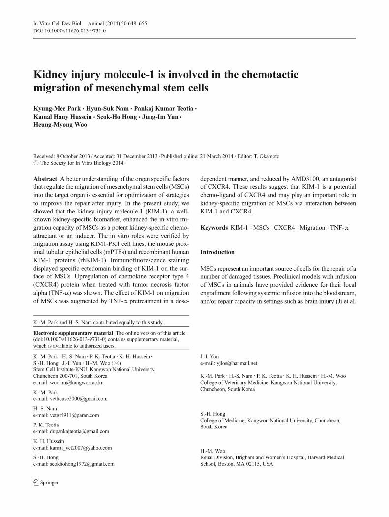
In our result, KIM1-PK1 cells displayed red extracellularstaining for KIM-1, whereas empty vector pcDNA-PK1 cellsdisplayed unstained for KIM-1 (Fig. 1A). Co-culture of themBM-MSCs and KIM1-PK1 significantly enhanced migra-tion of the mBM-MSCs towardKIM-1 expressingKIM1-PK1compared with the pcDNA-PK1 as a control (Fig. 1C, a andb). Next, we analyzed if KIM-1 activated chemotaxis ratherthan chemokinesis. There were no significant differences inthe number of migrated mBM-MSCs between KIM1-PK1and pcDNA-PK1-conditioned medium (Fig. 1C, c and d).
650 PARK ETAL.
TNF-α pretreatment on hAD-MSCs. Diverse concentration ofrhTNF-α (0, 1 and 10 ng/ml, R&D Systems) was added for3 d in the cell culture media to check the upregulation ofCXCR4 by western blotting and to investigate the migrationeffect of hAD-MSCs using KIM1-PK1 CM.
We then investigated if Kim-1 played a role as an inducerfor migration of mBM-MSCs. The migration assay was alsoperformed using the primary cultured-mPTEs and mBM-MSCs. Mouse Kim-1 expression was confirmed by positivityof Kim-1 staining red in extracellular space of primary tubularepithelial cells (Fig. 1D). The mBM-MSCs migration wassignificantly stimulated by a co-culture with mPTEs and thiseffect was markedly inhibited by addition of anti-mouse Kim-1 antibody as a Kim-1 inhibitor (Fig. 1E).
Thus, these results suggested that both human KIM-1 andmouse Kim-1 induced the chemotactic migration of mBM-MSCs. Flow cytometric analysis of mBM-MSCs suggests thatisolated cells exhibited theMSC characteristics (Suppl. Fig.1).
TNF-α regulates CXCR4 expression and hAD-MSCsmigration. The action of KIM-1 induced migration was alsoexamined in hAD-MSCs as a model of human MSCs. ThehAD-MSCswere placed on the top of transwell and themediumcontaining various concentrations (0, 2, 20 and 200 ng/ml) ofhrKIM-1 in the lower chamber was added for migration assay.The hrKIM-1 (2 and 20 ng/ml) added conditions induced hAD-MSCsmigration; however, themigrated cell number was slight-ly decreased in high concentration of KIM-1 (200 ng/ml)(Fig. 2A).
In addition, to investigate the relations between KIM-1 andhAD-MSCs, we assessed binding of soluble KIM-1 to thesurface of hAD-MSCs. It was confirmed that the soluble
KIM-1 INDUCES THE MIGRATION OF MSCS 651
Figure 1. Chemotactic effects of KIM-1 on migration of mBM-MSCs.(A) Immunocytochemistry of human KIM-1 expression (red color) inKIM1-PK1 (left panel) and pcDNA-PK1 cells (right panel). Bar=50 μm. (B) Amount of soluble KIM-1 in conditioned medium ofKIM1-PK1 was checked by western blotting at protein level. Recombi-nant human KIM-1 was used as control. (C) The number of migratedMSCs were significantly increased when they were co-cultured withKIM1-PK1 (a) as compared with pcDNA-PK1(b). However, there wasno difference in the number of migrated MSCs between conditioned
medium of KIM1-PK1 (c) and pcDNA-PK1 (d) in both upper and lowerchamber of transwell. Original magnifications: ×100. *p<0.05. (D) Ex-pression of Kim-1 (red color) in mPTEs was shown in immunocytochem-istry. Bar=50 μm. (E) Effect of Kim-1 and anti-Kim-1 antibodies onmBM-MSCs migration. Migration of mBM-MSCs toward mPTEs wasmarkedly inhibited by anti-Kim-1 antibody (a and b). mBM-MSCs mi-gration rate was not increased in both media only and anti-Kim-1 anti-body conditions (c and d). Original magnifications: ×100. *p<0.05.
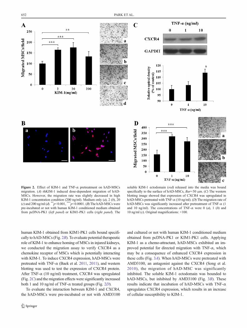
human KIM-1 obtained from KIM1-PK1 cells bound specifi-cally to hAD-MSCs (Fig. 2B). To evaluate potential therapeuticrole of KIM-1 to enhance homing of MSCs in injured kidneys,we conducted the migration assay to verify CXCR4 as achemokine receptor of MSCs which is potentially interactingwith KIM-1. To induce CXCR4 expression, hAD-MSCs werepretreated with TNF-α (Baek et al. 2011, 2011), and westernblotting was used to test the expression of CXCR4 protein.After TNF-α (10 ng/ml) treatment, CXCR4 was upregulated(Fig. 2C) and the migration effects were significantly increasedboth 1 and 10 ng/ml of TNF-α treated groups (Fig. 2D).
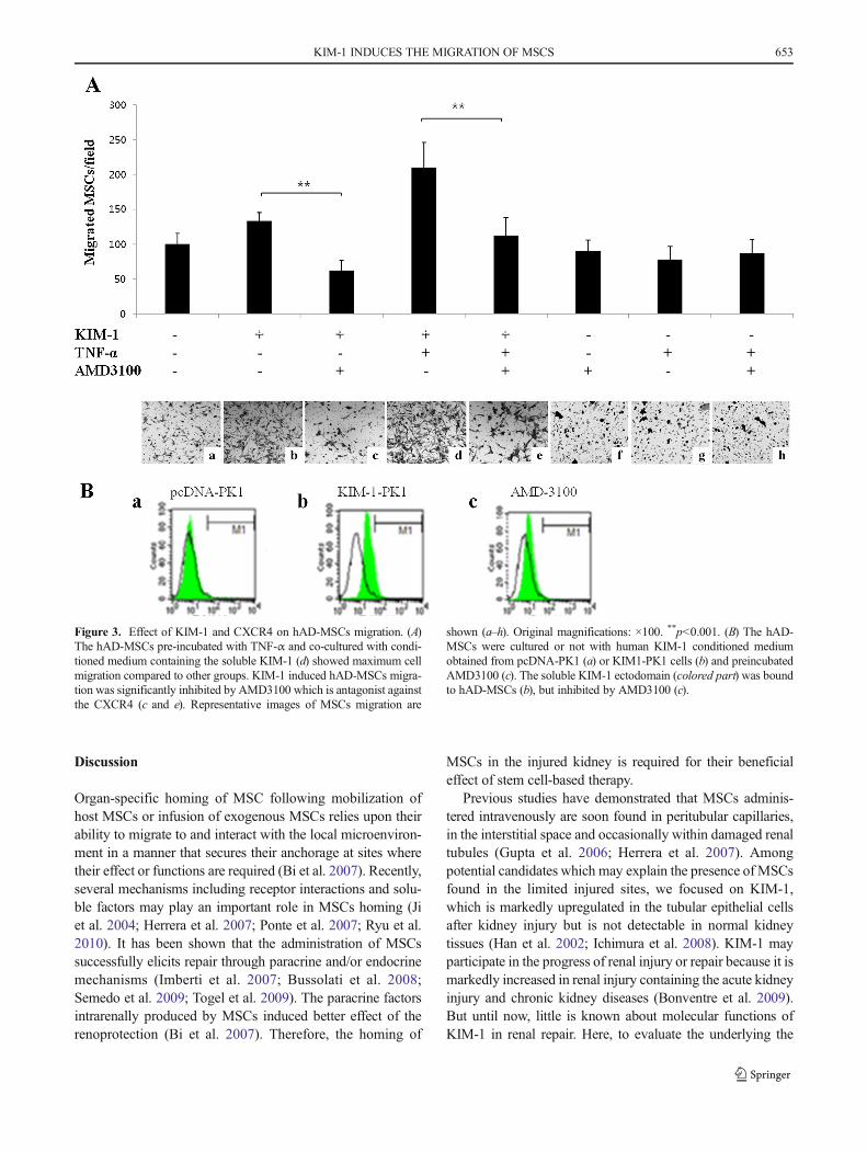
To evaluate the interaction between KIM-1 and CXCR4,the hAD-MSCs were pre-incubated or not with AMD3100
and cultured or not with human KIM-1 conditioned mediumobtained from pcDNA-PK1 or KIM1-PK1 cells. ApplyingKIM-1 as a chemo-attractant, hAD-MSCs exhibited an im-proved potential for directed migration with TNF-α, whichmay be a consequence of enhanced CXCR4 expression inthese cells (Fig. 3A). When hAD-MSCs were pretreated withAMD3100, an antagonist against the CXCR4 (Song et al.2010), the migration of hAD-MSC was significantlyinhibited. The soluble KIM-1 ectodomain was bounded tohAD-MSCs, but inhibited by AMD3100 (Fig. 3B). Theseresults indicate that incubation of hAD-MSCs with TNF-αupregulates CXCR4 expression, which results in an increaseof cellular susceptibility to KIM-1.
Figure 2. Effect of KIM-1 and TNF-α pretreatment on hAD-MSCsmigration. (A) rhKIM-1 induced dose-dependent migration of hAD-MSCs. However, the migration rate was slightly decreased in highKIM-1 concentration condition (200 ng/ml). Medium only (a), 2 (b), 20(c) and 200 ng/ml (d). **p<0.001, ***p<0.0001. (B) The hAD-MSCs werepre-incubated or not with human KIM-1 conditioned medium obtainedfrom pcDNA-PK1 (left panel) or KIM1-PK1 cells (right panel). The
soluble KIM-1 ectodomain (red) released into the media was boundspecifically to the surface of hAD-MSCs, Bar=50 μm. (C) The westernblotting image showed that expression of CXCR4 was upregulated inhAD-MSCs pretreated with TNF-α (10 ng/ml). (D) The migration rate ofhAD-MSCs was significantly increased after pretreatment of TNF-α (1and 10 ng/ml). The concentrations of TNF-α were 0 (a), 1 (b) and10 ng/ml (c). Original magnifications: ×100.
652 PARK ETAL.
Discussion
Organ-specific homing of MSC following mobilization ofhost MSCs or infusion of exogenous MSCs relies upon theirability to migrate to and interact with the local microenviron-ment in a manner that secures their anchorage at sites wheretheir effect or functions are required (Bi et al. 2007). Recently,several mechanisms including receptor interactions and solu-ble factors may play an important role in MSCs homing (Jiet al. 2004; Herrera et al. 2007; Ponte et al. 2007; Ryu et al.2010). It has been shown that the administration of MSCssuccessfully elicits repair through paracrine and/or endocrinemechanisms (Imberti et al. 2007; Bussolati et al. 2008;Semedo et al. 2009; Togel et al. 2009). The paracrine factorsintrarenally produced by MSCs induced better effect of therenoprotection (Bi et al. 2007). Therefore, the homing of
MSCs in the injured kidney is required for their beneficialeffect of stem cell-based therapy.
Previous studies have demonstrated that MSCs adminis-tered intravenously are soon found in peritubular capillaries,in the interstitial space and occasionally within damaged renaltubules (Gupta et al. 2006; Herrera et al. 2007). Amongpotential candidates which may explain the presence of MSCsfound in the limited injured sites, we focused on KIM-1,which is markedly upregulated in the tubular epithelial cellsafter kidney injury but is not detectable in normal kidneytissues (Han et al. 2002; Ichimura et al. 2008). KIM-1 mayparticipate in the progress of renal injury or repair because it ismarkedly increased in renal injury containing the acute kidneyinjury and chronic kidney diseases (Bonventre et al. 2009).But until now, little is known about molecular functions ofKIM-1 in renal repair. Here, to evaluate the underlying the
Figure 3. Effect of KIM-1 and CXCR4 on hAD-MSCs migration. (A)The hAD-MSCs pre-incubated with TNF-α and co-cultured with condi-tioned medium containing the soluble KIM-1 (d) showed maximum cellmigration compared to other groups. KIM-1 induced hAD-MSCs migra-tion was significantly inhibited by AMD3100 which is antagonist againstthe CXCR4 (c and e). Representative images of MSCs migration are
shown (a–h). Original magnifications: ×100. **p<0.001. (B) The hAD-MSCs were cultured or not with human KIM-1 conditioned mediumobtained from pcDNA-PK1 (a) or KIM1-PK1 cells (b) and preincubatedAMD3100 (c). The soluble KIM-1 ectodomain (colored part) was boundto hAD-MSCs (b), but inhibited by AMD3100 (c).
KIM-1 INDUCES THE MIGRATION OF MSCS 653
effect of KIM-1 on MSCs migration, we first verified thatKIM-1 was related to the in vitro migration capacity ofMSCs.Transwell migration assay depicts the chemotactic migrationof MSCs rather than chemokinesis towards KIM-1 expressingKIM1-PK1 cells. In the present study, it was clearly shownthat KIM-1 had a definitively chemotactic effect on bothhuman and mouse MSCs migration, using KIM1-PK1, pri-mary cultured-mPTEs and recombinant KIM-1 proteins.
However, following systemic infusion of exogenousMSCs, very few MSCs were found in the kidney and wereno longer detectable (Togel et al. 2005; Burst et al. 2010).Several studies demonstrated that the enhancing effect ofTNF-α pretreatment could potentially be mediated by modu-lating the expression of CXCR4 and chemokines and/or li-gand binding activity (Ponte et al. 2007; Baek et al. 2011;Egea et al. 2011). It has been shown that specific interactionbetween chemokines and their receptors mediates the migra-tion of MSCs in vitro and in vivo (Ji et al. 2004; Honczarenkoet al. 2006), and the effect of preincubation with TNF-α isassociated with increase of CXCR4 expression, which plays amost important role in MSCs homing as a major chemokine(Egea et al. 2011). In this study, we showed that KIM-1/CXCR4 plays an important role in migration of hAD-MSCsand overexpression of CXCR4 on hAD-MSCs enhancesMSCs migratory capacity toward KIM-1. By using specificfunctional blocking antibodies, we evidenced a determinantrole of KIM-1 in MSCs migration induced by interactionbetween CXCR4 and KIM-1. Presence of the CXCR4 antag-onist AMD 3100 significantly inhibited the chemotaxis ofhAD-MSCs toward KIM-1, supporting a key role of KIM-1/CXCR4 in the migration of hAD-MSCs. Additionally, weconfirmed the interaction between KIM-1 and CXCR4 interms of protein, suggesting that the soluble human KIM1-ectodomain bound specifically to the surface of hAD-MSCs.Future studies are needed to reveal the role of KIM-1 in themigration of endogenous and exogenous MSCs to sites ofkidney injury using in vivo model.
Conclusions
In conclusion, despite the preliminary nature of this study, ourfindings indicate that KIM-1 could be a potential kidney-specific factor to achieve a sufficient quantity of therapeuticMSCs that are localized within injured kidney.
Acknowledgments This work was supported by a Korean ResearchFoundation Grant funded by the Korean government (Grant No. KRF-2010-0025387). This research was also supported by a grant from theNext-Generation BioGreen 21 program (No. P J009627), Rural Develop-ment Administration, Republic of Korea. This study was also supported bya grant (Project Code No. Z-1541745-2013-14-01, Z-1541745-2013-14-02) from Animal and Plant Quarantine Agency, Ministry of Agriculture,Food and Rural Affairs (MAFRA), Republic of Korea in 2013.
References
Baek SJ, Kang SK, Ra JC (2011) In vitro migration capacity of humanadipose tissue-derived mesenchymal stem cells reflects their expres-sion of receptors for chemokines and growth factors. Exp Mol Med43:596–603
Bailly V, Zhang Z, Meier W, Cate R, Sanicola M, Bonventre JV (2002)Shedding of kidney injury molecule-1, a putative adhesion proteininvolved in renal regeneration. J Biol Chem 277:39739–39748
Bi B, Schmitt R, Israilova M, Nishio H, Cantley LG (2007) Stromal cellsprotect against acute tubular injury via an endocrine effect. J AmSocNephrol 18:2486–2496
Bonventre JV (2009) Kidney injury molecule-1 (KIM-1): a urinarybiomarker and much more. Nephrol Dial Transplant 24:3265–3268
Broekema M, Harmsen MC, Koerts JA, Petersen AH, van Luyn MJ,Navis G, Popa ER (2005) Determinants of tubular bone marrow-derived cell engraftment after renal ischemia/reperfusion in rats.Kidney Int 68:2572–2581
Burst VR, Gillis M, Putsch F, Herzog R, Fischer JH, Heid P, Muller-Ehmsen J, Schenk K, Fries JW, Baldamus CA, Benzing T (2010)Poor cell survival limits the beneficial impact of mesenchymal stemcell transplantation on acute kidney injury. Nephron Exp Nephrol114:e107–e116
Bussolati B, Tetta C, Camussi G (2008) Contribution of stem cells tokidney repair. Am J Nephrol 28:813–822
Cai L, Johnstone BH, Cook TG, Tan J, Fishbein MC, Chen PS, MarchKL (2009) IFATS collection: human adipose tissue-derived stemcells induce angiogenesis and nerve sprouting following myocardialinfarction, in conjunction with potent preservation of cardiac func-tion. Stem Cells 27:230–237
Egea V, von Baumgarten L, Schichor C, Berninger B, Popp T, Neth P,Goldbrunner R, Kienast Y, Winkler F, Jochum M, Ries C (2011)TNFalpha respecifies humanmesenchymal stemcells to a neural fateand promotes migration toward experimental glioma. Cell DeathDiffer 18:853–863
Gupta S, Verfaillie C, Chmielewski D, Kren S, Eidman K, Connaire J,Heremans Y, Lund T, Blackstad M, Jiang Y, Luttun A, RosenbergME (2006) Isolation and characterization of kidney-derived stemcells. J Am Soc Nephrol 17:3028–3040
Han WK, Bailly V, Abichandani R, Thadhani R, Bonventre JV (2002)Kidney injury molecule-1 (KIM-1): a novel biomarker for humanrenal proximal tubule injury. Kidney Int 62:237–244
Herrera MB, Bussolati B, Bruno S, Morando L, Mauriello-RomanazziG, Sanavio F, Stamenkovic I, Biancone L, Camussi G (2007)Exogenous mesenchymal stem cells localize to the kidney bymeans of CD44 following acute tubular injury. Kidney Int 72:430–441
Honczarenko M, Le Y, Swierkowski M, Ghiran I, Glodek AM,Silberstein LE (2006) Human bone marrow stromal cells express adistinct set of biologically functional chemokine receptors. StemCells 24:1030–1041
Ichimura T, Asseldonk EJ, Humphreys BD, Gunaratnam L, Duffield JS,Bonventre JV (2008) Kidney injury molecule-1 is aphosphatidylserine receptor that confers a phagocytic phenotypeon epithelial cells. J Clin Invest 118:1657–1668
Ichimura T, Hung CC, Yang SA, Stevens JL, Bonventre JV (2004)Kidney injury molecule-1: a tissue and urinary biomarker fornephrotoxicant-induced renal injury. Am J Physiol Ren Physiol286:F552–F563
Imberti B, Morigi M, Tomasoni S, Rota C, Corna D, Longaretti L, RottoliD, Valsecchi F, Benigni A, Wang J, Abbate M, Zoja C, Remuzzi G(2007) Insulin-like growth factor-1 sustains stem cell mediated renalrepair. J Am Soc Nephrol 18:2921–2928
Ji JF, He BP, Dheen ST, Tay SS (2004) Interactions of chemokines andchemokine receptors mediate the migration of mesenchymal stem
654 PARK ETAL.
cells to the impaired site in the brain after hypoglossal nerve injury.Stem Cells 22:415–427
Lange C, Togel F, Ittrich H, Clayton F, Nolte-Ernsting C, Zander AR,Westenfelder C (2005) Administered mesenchymal stem cells en-hance recovery from ischemia/reperfusion-induced acute renal fail-ure in rats. Kidney Int 68:1613–1617
Ponte AL, Marais E, Gallay N, Langonne A, Delorme B, Herault O,Charbord P, Domenech J (2007) The in vitro migration capacity ofhuman bonemarrowmesenchymal stemcells: comparison of chemokineand growth factor chemotactic activities. Stem Cells 25:1737–1745
Ryu CH, Park SA, Kim SM, Lim JY, Jeong CH, Jun JA, Oh JH, Park SH,Oh WI, Jeun SS (2010) Migration of human umbilical cordbloodmesenchymal stem cells mediated by stromal cell-derivedfactor-1/CXCR4 axis via akt, ERK, and p38 signal transductionpathways. Biochem Biophys Res Commun 398:105–110
Semedo P, Palasio CG, Oliveira CD, Feitoza CQ, Goncalves GM,Cenedeze MA, Wang PM, Teixeira VP, Reis MA, Pacheco-SilvaA, Camara NO (2009) Early modulation of inflammation by mes-enchymal stem cell after acute kidney injury. Int Immunopharmacol9:677–682
Sheridan AM, Schwartz JH, Kroshian VM, Tercyak AM, Laraia J,Masino S, Lieberthal W (1993) Renal mouse proximal tubular cellsare more susceptible thanMDCK cells to chemical anoxia. Am JPhysiol 265:F342–F350
Song JS, Kang CM, Kang HH, Yoon HK, Kim YK, Kim KH, Moon HS,Park SH (2010) Inhibitory effect of CXC chemokine receptor 4antagonist AMD3100 on bleomycin induced murine pulmonaryfibrosis. Exp Mol Med 42:465–472
Sung JH, Yang HM, Park JB, Choi GS, Joh JW, Kwon CH,Chun JM, Lee SK, Kim SJ (2008) Isolation and characteri-zation of mouse mesenchymal stem cells. Transplant Proc40:2649–2654
Togel F, Cohen A, Zhang P, Yang Y, Hu Z, Westenfelder C (2009)Autologous and allogeneic marrow stromal cells are safe and effec-tive for the treatment of acute kidney injury. Stem Cells Dev 18:475–485
Togel F, Hu Z, Weiss K, Isaac J, Lange C, Westenfelder C (2005)Administered mesenchymal stem cells protect against ischemicacute renal failure through differentiation-independent mechanisms.Am J Physiol Ren Physiol 289:F31–F42
KIM-1 INDUCES THE MIGRATION OF MSCS 655

Reproduced with permission of the copyright owner. Further reproduction prohibited withoutpermission.


![Monocyte chemotactic protein-1 provokes mast cell aggregation and [3H]5HT release](https://img.pdfslide.net/doc/110x75/634811a4031992cdcf01d95c/monocyte-chemotactic-protein-1-provokes-mast-cell-aggregation-and-3h5ht-release.jpg)