Embed Size (px)
Citation preview
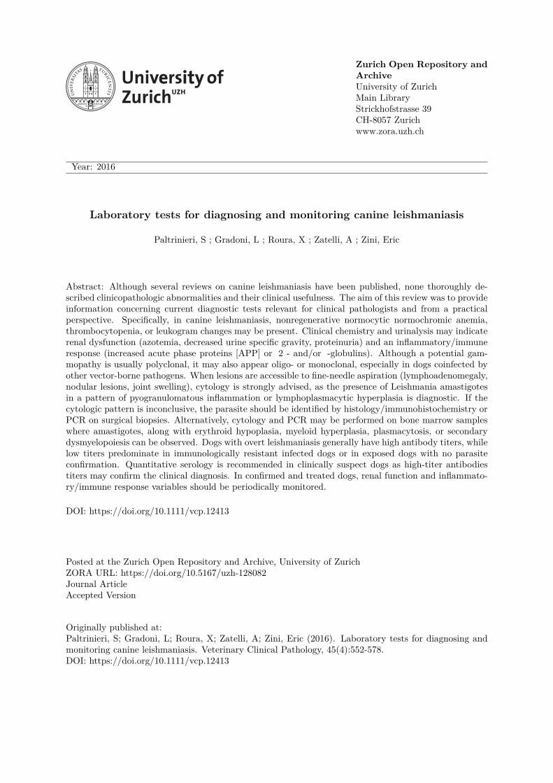
Zurich Open Repository andArchiveUniversity of ZurichMain LibraryStrickhofstrasse 39CH-8057 Zurichwww.zora.uzh.ch
Year: 2016
Laboratory tests for diagnosing and monitoring canine leishmaniasis
Paltrinieri, S ; Gradoni, L ; Roura, X ; Zatelli, A ; Zini, Eric
Abstract: Although several reviews on canine leishmaniasis have been published, none thoroughly de-scribed clinicopathologic abnormalities and their clinical usefulness. The aim of this review was to provideinformation concerning current diagnostic tests relevant for clinical pathologists and from a practicalperspective. Specifically, in canine leishmaniasis, nonregenerative normocytic normochromic anemia,thrombocytopenia, or leukogram changes may be present. Clinical chemistry and urinalysis may indicaterenal dysfunction (azotemia, decreased urine specific gravity, proteinuria) and an inflammatory/immuneresponse (increased acute phase proteins [APP] or 2 - and/or -globulins). Although a potential gam-mopathy is usually polyclonal, it may also appear oligo- or monoclonal, especially in dogs coinfected byother vector-borne pathogens. When lesions are accessible to fine-needle aspiration (lymphoadenomegaly,nodular lesions, joint swelling), cytology is strongly advised, as the presence of Leishmania amastigotesin a pattern of pyogranulomatous inflammation or lymphoplasmacytic hyperplasia is diagnostic. If thecytologic pattern is inconclusive, the parasite should be identified by histology/immunohistochemistry orPCR on surgical biopsies. Alternatively, cytology and PCR may be performed on bone marrow sampleswhere amastigotes, along with erythroid hypoplasia, myeloid hyperplasia, plasmacytosis, or secondarydysmyelopoiesis can be observed. Dogs with overt leishmaniasis generally have high antibody titers, whilelow titers predominate in immunologically resistant infected dogs or in exposed dogs with no parasiteconfirmation. Quantitative serology is recommended in clinically suspect dogs as high-titer antibodiestiters may confirm the clinical diagnosis. In confirmed and treated dogs, renal function and inflammato-ry/immune response variables should be periodically monitored.
DOI: https://doi.org/10.1111/vcp.12413
Posted at the Zurich Open Repository and Archive, University of ZurichZORA URL: https://doi.org/10.5167/uzh-128082Journal ArticleAccepted Version
Originally published at:Paltrinieri, S; Gradoni, L; Roura, X; Zatelli, A; Zini, Eric (2016). Laboratory tests for diagnosing andmonitoring canine leishmaniasis. Veterinary Clinical Pathology, 45(4):552-578.DOI: https://doi.org/10.1111/vcp.12413
1
LABORATORY TESTS FOR DIAGNOSING AND MONITORING CANINE LEISHMANIASIS 1
2
3
Running header: laboratory diagnosis of leishmaniasis 4
5
Saverio Paltrinieri, DVM, PhD, Dipl ECVCP, Department of Veterinary Sciences and Public 6
Health – University of Milan, Italy 7
Luigi Gradoni, Unit of Vector-borne Diseases & International Health, Department of Infectious 8
Diseases, Istituto Superiore di Sanità, Rome, Italy 9
Xavier Roura, Hospital Clínic Veterinari, Universitat Autònoma de Barcelona, Bellaterra, Spain; 10
Andrea Zatelli, Medical Consultancy Services, TàXbiex, Malta 11
Eric Zini, Clinic for Small Animal Internal Medicine, University of Zurich, Switzerland; 12
Department of Animal Medicine, Production and Health, University of Padova, Italy; Istituto 13
Veterinario di Novara, Granozzo con Monticello (NO), Italy. 14
15
Corresponding author: 16
Prof. Saverio Paltrinieri 17
Dipartimento di Scienze Veterinarie e Sanità Pubblica, Università di Milano 18
Via Celoria 10, 20133, Milano, Italy 19
Tel 0039-02-50318103 20
Fax 0039-02-50318095 21
e-mail: [email protected] 22
23
2
Abstract 24
Despite several reviews on canine leishmaniasis, none throughly described laboratory abnormalities 25
and diagnostic tests in light of their clinical usefulness. Aim was to summarize this information 26
from a practical perspective. Canine leishmaniasis often leads to profound clinicopathological 27
abnormalities. CBC reveals non-regenerative normocytic normochromic anemia and 28
thrombocytopenia, but other types of anemia or leukogram changes are also observed. Clinical 29
chemistry and urinalysis frequently show abnormalities suggestive of renal dysfunction (increased 30
creatinine and urea, decreased urine specific gravity, proteinuria) and inflammatory/immune 31
activation (increased acute phase proteins or α2- and/or γ-globulins). The gammopathy is typically 32
polyclonal but can also be oligo- or monoclonal. When lesions accessible to fine needle aspiration 33
or other sampling techniques (lymhpoadenomegaly, nodular or ulcerated skin lesions, joint 34
swelling) are present, cytology is strongly advised; the disease is confirmed if Leishmania 35
amastigotes are identified and pyogranulmatous inflammation or lymphoplasmocytic hyperplasia is 36
present. If results are inconclusive, the parasite should be identified through 37
histology/immunohistochemistry or PCR on surgical biopsies. Alternatively, cytology and PCR 38
may be performed on bone marrow, where amastigotes and erythroid hypoplasia, myeloid 39
hyperplasia, plasmocytosis, or secondary dysmyelopoiesis can be observed. Dogs with overt disease 40
generally have high antibody titers at ELISA or immunofluorescence. Low antibody titers may be 41
found in infected dogs that are immunologically resistant, or in exposed dogs (dogs not harboring 42
the parasite). If leishmaniasis is suspected but cytological evidence of the parasite is negative, 43
serology is recommended for confirmation. In treated dogs, renal function and the magnitude of the 44
inflammatory/immune reaction should be periodically monitored. 45
46
47
Keywords: Dog; Leishmania infantum; clinical usefulness; diagnosis; follow-up 48
49

3
50
4
Leishmaniasis is a frequent infectious disease in dogs living in endemic areas, is associated with 51
important morbidity and, although appropriate treatment, it can cause death. Despite several 52
reviews have been published so far, none has fully described the diagnostic role of laboratory 53
findings that are observed in dogs with leishmaniasis and of available tests. Therefore, the aim of 54
the present review was to provide information concerning laboratory abnormalities and current 55
diagnostic tests, from a practical perspective. 56
57
Etiology and pathogenesis of canine leishmaniasis 58
Canine leishmaniasis is a disease caused by the protozoan parasite Leishmania infantum 59
(Kinetoplastida: Trypanosomatidae), or its New World synonym Leishmania chagasi.1 Although 60
non-vectorial transmission is occasionally reported (e.g. by transplacental, transfusional or venereal 61
route),2-4
it is assumed that canine leishmaniasis is mainly acquired by the bite of infected 62
phlebotomine sand flies (Diptera: Psychodidae). Therefore, the geographical distribution and 63
prevalence of the disease largely depends on the presence and abundance of competent vector 64
species, including members of the Phlebotomus (Larroussius) subgenus in the Old World 65
(Mediterranean Basin and Central Asia) and Luztomyia longipalpis and L. evansi in the New World 66
(from Mexico through Argentina).5 Blood-sucking females ingest the intracellular, non-flagellated 67
form of Leishmania (amastigote) during the blood meal on infected hosts. After multiplication, 68
flagellated forms (promastigote) and transform into non-replicative, infectious metacyclic forms 69
that are inoculated into the host’s dermis when the fly takes another blood meal. Parasites are 70
phagocytosed by local and recruited host cells, and within phagolysosomes of resident macrophages 71
promastigotes transform and replicate as amastigotes.6 72
The pathogenesis of the disease starts with the ingestion of recently inoculated promastigotes by 73
neutrophils that are unable to kill the protozoa but activate dermal macrophages and other 74
mechanisms of innate immunity (e.g. acute phase response). Macrophages initially activate their 75
antimicrobial defenses, which are based on the release of enzymes and on the production of free 76
5
radicals within the phagolysosome. Leishmania organisms are able to interfere with the oxidative 77
activity of the phagocytes7,8
and therefore amastigotes can survive and actively replicate in 78
macrophages, leading to cell destruction and infecting progressively more and more phagocytes. 79
In longitudinal field studies employing naïve dogs, it has been demonstrated that Leishmania can be 80
detected by PCR in bone marrow samples starting about 6 months from natural exposure to 81
phlebotomine vectors.9 Once bone marrow has been colonized it is generally accepted that the dog 82
is persistently infected. However, the same studies showed that dogs being PCR positive in bone 83
marrow may become negative in the following months without any treatment; it is unknown 84
whether the parasite density fluctuates below the threshold limit of the test, the infection persists in 85
organs other than bone marrow, or the host defenses successfully eradicate the infection.9 Despite 86
dogs can mount an antibody response after the first contact with the parasites, resistance or 87
susceptibility to progressive infection (leading to clinical disease) depend on the balance between 88
Th1 (cell-mediated) and Th2 (humoral) immune responses of the host: those with prevailing Th2 89
response are more likely to have uncontrolled dissemination of parasites to all body tissues and to 90
show lesions and clinical signs of leishmaniasis.10-13
91
Of note, depending on the host-parasite interactions, the simple detection of circulating antibodies 92
does not necessarily imply that the dog is actually infected and detection of parasites in tissues does 93
not suggest that the infected dog is actually sick. Therefore, the two existing guidelines that deal 94
with the diagnosis and staging of canine leishmaniasis, recently released by the Leishvet group14
95
and the Canine Leishmaniasis Working Group (CLWG),15
classify dogs as exposed, infected or sick 96
based on a combination of clinical findings and clinicopathological tests or tests for etiological 97
diagnosis. Specifically, both guidelines include two main categories of dogs: 98
- Infected dogs: dogs clinically unremarkable, without abnormalities in hematology, clinical 99
chemistry and urinalysis, that test positive to PCR or cytology in tissues such as bone 100
marrow, lymph node, spleen, skin or peripheral blood; 101
6
- Sick dogs: infected dogs that have clinical signs or clinicopathological changes consistent 102
with leishmaniasis. 103
The classification of the CLWG15
includes two additional categories of dogs at the extremes of the 104
spectrum: 105
- Exposed dogs: dogs clinically unremarkable with positive serology, in which PCR or 106
cytology fail to demonstrate the presence of the parasite; 107
- Severely sick dogs: sick dogs that do not respond to treatments and/or are affected by 108
concurrent diseases. 109
The Leishvet group classifies sick dogs in four stages according to the severity of clinical signs and 110
magnitude of antileishmanial antibody titers.14
111
112
Clinical signs of canine leishmaniasis 113
The interpretation of clinicopathological, serological and molecular tests should be done in light of 114
history, signalment and clinical presentation. History should focus on the risk of exposure to 115
phlebotomine vectors. Signalment should focus on gender, breed and age; clinically overt 116
leishmaniasis is more frequent in male dogs, Boxer and German shepherd, and in those younger 117
than 3 years or older than 8 years, 16,17
although recent reports suggests that any dog above 2 years 118
is at risk.18
119
The spectrum of clinical presentations is wide and ranges from infections characterized by the 120
absence of obvious clinical findings but detectable laboratory abnormalities, to overt clinical 121
infections characterized by the presence of clinical and laboratory abnormalities that require or not 122
hospitalization. However, some signs are more frequent than others. The most typical clinical 123
findings include:14,15,19-22
generalized lymphoadenomegaly, cutaneous lesions, pale mucous 124
membranes, weight loss or cachexia, polyuria/polydipsia, epistaxis, onicogriphosis. Ocular 125
lesions,15,23,24
lameness, lethargy and fever are also quite common.25,26
In a longitudinal study,27
it 126
was shown that lymph node enlargement appears first (approximately 1 year after exposure to sand 127
7
flies), followed by cutaneous lesions (after 18 months) and ocular signs (after 22 months). 128
Furthermore, atypical forms have been described, especially in highly endemic areas, including 129
gastrointestinal, neurological, musculoskeletal, cardiopulmonary, lower urinary tract or genital tract 130
signs.28-36
131
132
Laboratory abnormalities that may support or confirm leishmaniasis 133
In addition to clinical findings, laboratory abnormalities detectable by routine hematology, clinical 134
chemistry or urinalysis may further increase the clinical suspicion of canine leishmaniasis. 135
Moreover, especially in the early phases of the disease or with atypical presentations, laboratory 136
changes may occur in the absence of obvious abnormalities at physical examination. Therefore, a 137
basic panel is mandatory when canine leishmaniasis is suspected or when a dog with positive result 138
tests for etiological diagnosis needs to be classified as “exposed”, “infected” or “sick”. Table 1 139
summarizes the clinicopathological changes that are found in dogs with leishmaniasis (i.e. “sick” 140
dogs). 141
142
1) Hematologic abnormalities 143
Hematological changes in canine leishmaniasis are non specific.37,38
Neutrophilia, due to the 144
systemic inflammatory response associated with the infectious diseases, may be particularly 145
prominent if ulcerative cutaneous lesions with secondary bacterial infection are present.37,39,40
146
Conversely, numerical or morphological changes in the other leukocyte populations are less 147
common, although lymphopenia, lymphocytosis or eosinophilia are occasionally described39-42
148
Amastigotes may be rarely documented in circulating leukocytes of infected dogs (0.5% of cases), 149
within neutrophils, lymphocytes and monocytes (figure 1);41,43
the percentage of infected cells is so 150
low that their search is generally not rewarding. When a systemic disease and blood dissemination 151
is suspected, more sensitive tests such as PCR or quantitative PCR should be preferred to the simple 152
microscopical examination of blood smears (see below). When parasites are found on smears, 153
8
however, it is very likely that the parasite burden is high: therefore, although, as stated above, the 154
simple detection of the parasite does not automatically mean that the infected dog is sick, it is more 155
likely that dogs with a high parasite burden (and parasites detected on smears) are sick than simply 156
infected. 157
The most common hematological changes in leishmaniotic dogs is anemia,37,38,44,45
that may appear 158
6 months after exposure to infection.27
Anemia is usually mild to moderate and has the normocytic 159
normochromic non regenerative pattern40,44,45
typical of the anemia of inflammatory disease, whose 160
pathogenesis include a depression of metabolic activity of bone marrow, in this case sustained also 161
by the infection of stromal cells by the parasite, and a decreased iron availability for 162
erythropoiesis.46
However the pathogenesis of anemia in leishmaniotic dogs include additional 163
mechanisms such renal failure leading to reduced erythropoietin synthesis and secondary 164
dismyelopoiesis associated with the production of antibodies against erythroid precursors.47
165
Moreover, it is very likely that anemia also has a hemolytic component,48
likely due to the presence 166
of anti-RBC antibodies; in a few cases, the hemolysis may be prevalent,49
or associated with a 167
“lupus-like” reaction along with other dermatological or systemic signs or laboratory change, 168
including positive ANA-test50
or perinuclear antineutrophil cytoplasmic autoantibodies.51
169
Thrombocytopenia is fairly frequent in leishmaniotic dogs without concurrent infections. It is 170
usually mild to moderate but if severe, co-infections with other vector-borne pathogens (e.g. 171
Ehrlichia canis, Anaplasma phagocytophilum or A. platys) or other possible causes of reduced 172
platelet concentration should be suspected. The most likely mechanism responsible for 173
thrombocytopenia in leishmaniasis is a peripheral consumption of circulating platelets. In turn, this 174
mechanism may be due to immune-mediated mechanisms, since the presence of anti-Plt antibodies 175
has been demonstrated in leishmaniotic dogs.52-54
176
Moreover, platelet loss may be associated to hypercoagulability caused by a decreased 177
concentration of anti-thrombin III, as in any other protein losing nephropathy55
(see below), or to 178
disseminated intravascular coagulation (DIC) that may be occasionally suspected in leishmaniotic 179
9
dogs.56
However, the mechanism of thrombocytopenia in leishmaniotic dogs includes also a 180
decreased production due to the depressed bone marrow activity and to the secondary 181
dysmyelopoiesis cited above. Even in the absence of reduced platelet counts, however, platelet may 182
be hypofunctional in dogs with leishmaniasis57
although this reduced function is rarely responsible 183
for hemostatic abnormalities, as described below. 184
One additional hematological test that may be run in dogs with leishmaniasis is the flow-cytometric 185
evaluation of the CD4/CD8 ratio. The rationale for performing this test is that as soon as Th1 186
responses decreases, thus increasing the susceptibility to the disease and favoring the shift from 187
latent infection to overt disease, the number of CD4+ lymphocytes decreases causing reduction of 188
the CD4/CD8 ratio.58,59
Therefore, a seropositive or PCR-positive dog with a low CD4/CD8 ratio is 189
more predisposed to develop clinical signs than a similar dog with normal CD4/CD8 ratio. The 190
practical applicability of this test, however, is limited by the high individual variability and by the 191
difficulty to determine a clear cut-off for staging the disease. Hence, this test may be useful for 192
monitoring the post-treatment follow-up, rather than to stage a dog at first diagnosis of 193
leishmaniasis; the authors do not suggest the use of this test for diagnostic purposes in dogs 194
suspected to have leishmaniasis. 195
Finally, the hematological profile of leishmaniotic dogs may be completed by bone marrow 196
cytology.27,42,44,60
This analysis may be useful in leishmaniotic dogs to confirm the infection 197
through the detection of infected macrophages, as better specified below, but it may be also used to 198
differentiate a simple infection from systemic disease (i.e. “infected” vs. “sick” dog).15
Despite 199
some histological studies demonstrated that parasite density can be high despite few clinical signs,61
200
generally the parasite load and the presence of cytological alterations increase as soon as the dogs 201
show clinical signs.62
Therefore, rare infected macrophages may be occasionally seen in the absence 202
of other pathological findings in dogs that are simply infected, whereas dogs in which the parasite 203
induces an inflammatory reaction and/or a systemic disease, are characterized by a higher number 204
of parasites detected cytologically and by a series of morphological changes. Specifically, in the 205
10
latter case cytology of the bone marrow usually reveals an erythroid hypoplasia,40
without 206
abnormalities in the ratio between maturative and proliferative pools of erythroid precursors, 207
occasionally associated with myeloid hyperplasia (and thus with an increased M:E ratio). Moreover, 208
signs of bone marrow inflammation, generically defined by Stockham and Scott as “myelitis”,63
, are 209
usually found (figure 2). These include a proliferation of either infected or non-infected 210
macrophages often with signs of erythrophagia or, more generically, cytophagia, associated with an 211
increase of neutrophils in the framework of the myeloid hyperplasia, and a moderate to severe 212
plasmocytosis characterized by a higher number of plasma cells with signs of activation (e.g. mott 213
cells), and lymphocytes.64,65
Megakaryocyte hyperplasia may also be present, especially when 214
peripheral consumption of platelets occurs. 215
Secondary dismyelopoiesis is another common finding, although less frequent than the typical 216
pattern listed above (figure 3). This condition is characterized by multiple peripheral cytopenias 217
(e.g. the anemia and thrombocytopenia cited above) associated with hypercellular bone marrow on 218
which one or more cell lineages show dysplastic features. In canine leishmaniasis, these mostly 219
include dyserythropoiesis and dysmegakariopoiesis, while dismyelopoiesis (and in particular 220
dysgranulopoiesis, mostly characterized by abnormal maturation and ring forms) is only 221
occasionally found: more specifically, erythroid precursors with abnormal mitoses, asynchronous 222
nucleo-cytoplasmic maturation, nuclear fragmentation, and/or late stage maturation arrest, 223
especially when the pathogenesis includes an immune-mediated reaction against RBC precursors. 224
Dwarf megakaryocytes associated or not with an increased number of megakariobkasts and with 225
emperipolesis may be typical aspects of dysmegakariopoiesis in leishmaniotic dogs. The detection 226
of secondary dysmyelopoiesis however, is not per se diagnostic for leishmaniasis, unless 227
macrophages with intracytoplasmic amastigotes and other signs of reactivity (e.g. 228
lymphoplasmocytic infiltration) are found. Therefore, the cause-effect association between 229
secondary dysmyelopoiesis and the simple seropositivity or PCR-positivity should be carefully 230
considered. Ultimately, in this case the diagnosis of leishmaniasis should be based on the exclusion 231
11
of other infectious, toxic or metabolic causes of secondary dysmyelopoiesis or of primary 232
myelodysplastic syndromes (MDS) as recommended in hematology textbooks.45,47,64
233
The last and less common finding in bone marrow cytology leishmaniotic dogs may be the presence 234
of erythroid hyperplasia when peripheral signs are consistent with the diagnosis of immune-235
mediated hemolytic anemia (IMHA). Also in this case, however, IMHA can be associated with 236
leishmaniasis only if the parasite if found in cytological specimens characterized by erythroid 237
hyperplasia. If not, the diagnostic approach should be taken into account only if any other possible 238
cause of IMHA has been excluded. In brief, bone marrow cytology may be useful for diagnostic 239
purposes in some dogs, by detecting amastigotes and compatible cytological abnormalities, or to 240
differentiate between infected dogs from those that are sick due to leishmaniasis. 241
242
2) Hemostatic abnormalities 243
Hemostatic abnormalities are uncommon in leishmaniotic dogs. Activated partial thromboplastin 244
time (aPTT) and prothrombin time (PT) may be increased; however, in most cases this is due to 245
preanalytical factors, since their prolongation may occur when the concentration of total globulins 246
increases, which is frequent in dogs with leihsmaniasis.66
Alternatively, prolonged coagulation 247
times may result from DIC, although this complication is uncommon in leishmaniotic dogs.56,67
248
Conversely, hypercoagulability may be common in leishmaniotic dogs if affected by severe protein 249
losing nephropathy.68
This is mostly due to glomerular loss of antithrombin III (ATIII), a protease 250
inhibitor involved in the regulation of blood coagulation that prevents the conversion of fibrinogen 251
into fibrin. The lack of this physiologic anticoagulant may induce hypercoagulability that in turn 252
promotes thrombosis and subsequent consumption coagulopathy. Hypercoagulability is also 253
favored by the hyperviscosity syndrome due to increased circulating globulins.69
As in any other 254
hypercoagulable state associated with protein losing nephropathy, hypercoagulability of 255
leishmaniotic dogs was also demonstrated through a decreased clot formation time and an increased 256
global clot strength using thromboelastography (TEG);70
differently, in another study the 257
12
coagulation profile of leishmaniotic dogs assessed by thromboelastometry (TEM, a technique 258
similar to TEG) was within normal limits.71
However, it is worth noting that TEM and TEG are 259
affected by the RBC mass, possibly explaining the different results obtained with TEM and 260
TEG.72,73
In brief, to assess hypercoagulability in dogs with protein losing nephropathy associated 261
with leishmaniasis the authors currently suggest including only ATIII measurement. 262
263
3) Biochemical abnormalities 264
Because the clinical presentation of dogs with leishmaniasis is variable, also the type of 265
biochemical abnormalities varies accordingly. Renal dysfunction and inflammation/immune 266
reactions are frequently observed and their presence should be evaluated in each dog with suspected 267
or confirmed leishmaniasis (see below). Regarding other routine biochemical analytes that may be 268
altered in leishmaniotic dogs, there are enzymes of hepatobiliary or pancreatic damage that may 269
increase in case of pyogranulomatous infiltrates affecting these organs,15,16
muscular enzymes, 270
including LDH and CK, that may increase with musculoskeletal lesion.74
Nevertheless, increased 271
CK may also be due to the increased CK-BB isoenzyme if neurological signs are present,75
since 272
Leishmania has been found in the brain of some affected dogs with cerebrovascular alterations,76-78
273
or to increased CK-MB if leishmaniotic dogs have cardiomyopathy (i.e. increased tropoinin I and 274
cardiopulmonary lesions have been reported in many leishmaniotic dogs).79-81
Biochemical 275
abnormalities consistent with alteration of endocrine organs are rare, despite amastigotes associated 276
with inflammatory lesions have been found in the adrenal cortex of leishmaniotic dogs.82,83
277
278
Assessment of renal function 279
The systemic immune complex disease that characterizes leishmaniasis induces the deposition of 280
circulating immune complexes at the glomerular level. This induces a series of inflammatory 281
glomerular changes detectable histologically and ultramicroscopically,36,84,85,86,87
that are primarily 282
responsible for a proteinuric nephropathy.87
The evolution of this condition, as for any other 283
13
protein-losing nephropathy, is represented by the development of a chronic kidney disease (CKD) 284
due to a series of factors that include progressive glomerulosclerosis, renal hypertension, overload 285
of protein reabsorption in tubular cells, with subsequent tubulointerstitial nephritis85,87
In turn, 286
advanced stages of CKD are characterized by hyperazotemia and may be associated with systemic 287
hypertension, both factors contributing to comorbidity in dogs with leishmaniasis.87,88
Therefore, as 288
for any other renal disease, the early detection of proteinuria and correct classification of CKD is 289
mandatory in the diagnostic workup. To this aim, the clinical and laboratory approach is the same 290
recommended by the International Renal Interest Society (IRIS)89
for any type of CKD. This 291
approach is based on a thorough clinical evaluation of the dog, based on physical examination and 292
diagnostic imaging, on the measurement of arterial pressure according to the guidelines 293
recommended by the American College of Veterinary Internal Medicine (ACVIM)90
and on the 294
quantification of urinary proteins (described in the section of this article regarding urinalysis) and 295
markers of renal function. According to the IRIS guidelines, these include mostly the urine specific 296
gravity and the serum concentration of creatinine.89
This latter has been shown to increase 297
frequently in leishmaniotic dogs14,15,16,19,20,91
and early (from 12 to 18 months) following infection.27
298
Creatinine is a good indicator of a decreased glomerular filtration rate (GFR), and it more specific 299
than other markers such as urea92,93
but unfortunately they are not enough sensitive to detect the 300
earliest stages of renal insufficiency. Due to the compensatory activity of residual nephrons, in fact, 301
an increase of serum creatinine occur only when the GFR dramatically decreases.93
Therefore, 302
although creatinine is commonly used to detect overt CKD, a huge research activity is currently 303
running in veterinary nephrology and clinical pathology to identify earlier markers of decreased 304
GFR, either in leishmaniotic dogs or in dogs affected by other types of CKD. The direct 305
measurement of GFR trough clearance tests (e.g. clearance of inulin, exogenous creatinine or, more 306
recently, iohexol) would be the best method to assess in real time the functionality of the 307
kidneys94,95
Unfortunately these tests, although currently validated for use in dogs, are not widely 308
available in clinical practices or in veterinary laboratories. Therefore, the diagnostic potential of 309
14
indirect markers other than creatinine has been recently investigated. Cystatin C (Cys C) has been 310
proposed as surrogate marker of creatinine, especially using advanced measurement techniques 311
such as particle-enhanced turbidimetric immunoassay.96-99
The serum concentration of Cys C has 312
been assessed also in dogs with leishmaniasis.100
However, there is no evidence that serum Cys C, 313
that is less specific than creatinine, is more sensitive that creatinine in detecting early CKD.94
314
Promising results have been obtained by the measurement of urinary Cys C101
but this seems not to 315
be true in canine leishmaniasis, where urinary Cys C increases only in proteinuric dogs with 316
increased creatinine.102
Recently, a promising biomarker for early detection of renal dysfunction 317
seems the symmetric dymethilarginine (SDMA), a catabolite of methylated proteins that is mainly 318
excreted by the glomeruli and not reabsorbed by tubuli ,103,104
that in published report has been 319
measured in serum using chromatographic techniques.104
Studies in cats, where the early diagnosis 320
of CKD may be more challenging than in dogs, demonstrated that SDMA increases earlier than 321
creatinine,105,106
although it does not provide additional advantages in animals with overt CKD (i.e. 322
with CKD on which serum creatinine is already increased). Preliminary results indicate that SDMA 323
may have the same role of early indicator of renal dysfunction also in dogs,104,107
although a few 324
extrarenal variables may affect the results.108
Therefore, although no studies specifically focused on 325
the use of SDMA in canine leishmaniasis exist, it is very likely that SDMA may be used, in the 326
future, to assess renal function in leishmaniotic dogs that are proteinuric but still have normal 327
creatinine concentration. 328
Other blood markers may provide additional information in leishmaniotic patients with CKD. For 329
example in people the increased serum concentration of homocysteine (Hcy), endothelin-1 (ET-1) 330
or C-reactive protein (CRP) may predict, hypertension and/or inflammation, especially in 331
association with CKD.109-113
In dogs, Hcy increases in those with CKD114
but seems to be less 332
associated with hypertension than in humans.115
Conversely, ET-1 increases in serum of dogs with 333
hypertension associated with early stages of CKD115
and is also associated with inflammation, as 334
demonstrated by its correlation with CRP levels, which in turn increases mostly in CKD associated 335
15
with inflammatory conditions.113
However, the potential of these additional markers as 336
complementary tests for the management of leishmaniotic dogs with CKD has not been 337
investigated. Conversely, inflammatory markers such as CRP, ferritin and adiponectin have been 338
measured in the urine of dogs with leishmaniasis and were found increased in dogs with proteinuria, 339
sometime in the absence of elevated serum creatinine.102,116,117
These analytes might therefore work 340
as early markers of CKD in leishmaniotic dogs, although it is more likely that their increase depend 341
on the elevated serum concentration, thus reflecting the inflammatory state typical of leishmaniasis, 342
rather than a direct consequence of CKD. 343
Finally, in leishmaniotic dogs, tubulointerstitial lesions may occur secondarily to proteinuria caused 344
by glomerular damages. Therefore, the availability of markers to identify dogs with tubular lesions 345
would allow to early differentiate dogs with pure glomerular lesions from dogs with more advanced 346
renal damage. Tubular markers, however, are usually measured in urine and not in serum and are 347
described in the section on urinalysis. It is also worth mentioning that some dogs with CKD may 348
have acute deterioration of their renal dynsfunction due to factors related or not to leishmaniasis 349
(e.g. vomiting, diarrhea). 350
351
Assessment of inflammatory/immune reactions 352
Based on the pathophysiology above described, it is clear that leishmaniotic dogs with overt disease 353
have an intense inflammatory reaction and produces high amount of molecules involved in the 354
immune response, including antibodies. Both these phenomena may be investigated using tests such 355
as protein analysis and serum protein electrophoresis or measurement of acute phase proteins 356
(APPs). 357
358
Protein analysis and serum protein electrophoresis are considered the most accurate tests to 359
diagnose canine leishmaniasis. Protein changes appear 12-18 months after exposure to the 360
parasite.27
Total proteins and total globulin are frequently increased, especially in the acute phase of 361
16
the disease.14,15,16,21,91,118,119
The increase of total protein can correlate with the severity of the 362
clinical score, according to Proverbio et al.120
Moreover albumin decreases both because of its role 363
as negative APPs (see below) and of the renal loss associated with proteinuric nephropathy leading 364
to decreased albumin:globulin (A/G) ratio91,118,119
The decrease of the A/G ratio is so frequent that it 365
has been considered by some Authors to be one of the more sensitive tests for canine 366
leishmaniasis118
and hypoalbuminemia is considered a negative prognostic factor in leishmaniotic 367
dogs since the magnitude of hypoalbuminemia negatively correlates with survival times.121
The 368
typical electrophoretogram of leishmaniotic dogs with overt clinical signs (figure 4) displays 369
hypoalbuminemia, an increase of α2-globulin, where most of the positive APPs migrate, and 370
especially a strong increase of γ-globulins, due to the huge amount of circulating antibodies, 371
immunecomplexes, and other molecules with γ motility involved in immune-mediated 372
inflammatory reactions.119
Occasionally, peaks due to circulating antibodies are found in the 373
β region, where IgM and some APPs migrates. The gammopaty is typically polyclonal but 374
sometime the peak may be narrower (oligoclonal), biclonal122
or definitely monocloncal.123
The 375
interpretation of the peak must take into account the electrophoretic method, since peaks are usually 376
narrower using capillary zonal electrophoresis (CZE) than using agarose gel or cellulose acetate.124
377
Therefore, CZE may lead to falsely interpret as monoclonal a peak that is ultimately oligo- or 378
polyclonal (figure 5). Moreover, although monoclonal peaks associated exclusively with 379
leishmaniasis have been described, the detection of monoclonal peaks should induce to consider the 380
possible presence of concurrent diseases (e.g. other vector-borne diseases or multiple 381
myeloma).125,126
382
383
Acute phase proteins are powerful indicators of inflammation: the pro-inflammatory cytokines 384
produced in inflammatory sites induce the so called “acute phase response”, characterized by the 385
release of neutrophils from storage pools, by an activation of myelopoiesis (see above), and by a 386
17
modulation of protein synthesis in the liver.119
This latter phenomenon leads to a decreased serum 387
concentration of the “negative APPs”, and to an increased concentration of the “positive APPs” that 388
includes a series of immunomodulators, scavenger or transport proteins, antiproteases, and other 389
proteins involved in host defenses.112
Therefore it is not surprising that the serum concentration of 390
positive APPs in dogs with overt canine leishmaniasis is high. The list of APPs whose 391
concentration increases in serum of leishmaniotic dogs is long and includes CRP, Haptoglobin 392
(Hp), Ceruloplasmin (Cp) Serum Amyloid A (SAA) and ferritin,127-132
with CRP and ferritin that, as 393
previously reported, increase also in urine. Similarly, a decrease of negative APPs other than 394
albumin has also been reported; these are transferrin, indirectly measured as total iron binding 395
capacity (TIBC), that induces also a reduction in the concentration of iron, and a decreased activity 396
of the enzyme paraoxonase (PON-1).129,130,133
PON-1 is a negative APP that is bound to high 397
density lipoproteins (HDL) and represents a link between inflammation and oxidative stress. 398
Therefore its decrease is not constantly seen in leishmaniotic dogs but it may become evident when 399
oxidative stress is particularly severe.133
Interestingly, in these cases also the concentration of HDL, 400
that is converted into low density lipoprotein (LDL) after detachment of PON-1, decreases134
and 401
may be a cheap marker of inflammation and oxidative stress associated with leishmaniasis. 402
Recently a reduced serum activity of adenosine deaminase (ADA) and butyrylcholinesterase 403
(BChE), two enzymes involved in modulating immune responses, has also been reported in dogs 404
with leishmaniasis.135
405
The APP changes summarized above are not diagnostic per se since mild increases of positive 406
APPs have been reported also in infected dogs without clinical signs127
and severely increased 407
levels may occur in diseases other than leishmaniasis.112
In a dog in which leishmaniasis has been 408
diagnosed by other clinical or laboratory findings, however, the magnitude of these changes may 409
reflect the magnitude of inflammation and thus provide prognostic information. In particular, the 410
decrease of PON-1 is particularly evident in severe diseases and may therefore be a negative 411
prognostic marker. Moreover, APPs may be useful to monitor the follow up. 412
18
413
4) Abnormalities at urinalysis 414
The analysis of urine from dogs with suspected or confirmed leishmaniasis must be focused on 415
three main aspects: confirmation of CKD, evaluation of proteinuria, and investigation of possible 416
tubular damage. Urine samples should be analyzed using the usual approach followed in any case of 417
CKD, that starts with the physico-chemical analysis and continues with sediment analysis and 418
evaluation of proteinuria, eventually followed by the measurement of markers of tubular damages. 419
420
Physico-chemical analysis 421
According to standardized procedures,136
after centrifugation of urine to separate supernatants from 422
the sediment, the routine physicochemical analysis must be addressed to evaluate with a 423
refractometer the urine specific gravity (USG), that tends to decrease in dogs with tubulo-interstitial 424
damage.93
Then, the supernatant can be used for a dipstick analysis, that should be focused on 425
assessing: 426
- the pH that, although not extremely accurate137
may be useful to properly interpret other 427
dipstick results: for example the semiquantification of proteins may provide erroneously 428
positive results in alkaline urine (pH>8); 93
429
- the concentration of proteins, to be interpreted as described below; 430
- the presence of glucose, that, as specified below, may be an indicator of tubular damage. 431
Sediment analysis is another important step of urinalysis of leishmaniotic dogs: the presence of an 432
active sediment (e.g. a sediment with high numbers of leukocytes, erythrocytes or bacteria) is 433
important either because may indicate a lower urinary tract infection superimposed on the primary 434
disease (leishmaniasis) or because in the presence of an active sediment, results regarding 435
proteinuria should be carefully considered;138
conversely, the presence of granular or cellular casts 436
may be consistent with tubular damage.93
437
438
19
Evaluation of proteinuria 439
The evaluation of proteinuria is an essential step, since the presence and magnitude of proteinuria is 440
not only a marker of kidney disease, but also a risk factor for the progression of nephropathy.139
441
According to the ACVIM guidelines,140
proteinuria should be assessed in any dog with diseases 442
potentially inducing this condition, such as leishmaniasis. The ACVIM guidelines recommend to 443
collect urines by cystocentesis, to avoid contamination from the lower urinary tract, however, to 444
improve the possibility to frequently monitor dogs at risk to develop proteinuria, a first evaluation 445
may be done on voided samples, since results recorded with the two methods of collection overlap 446
when the sediment is inactive.141
Additionally, it may be appropriate to screen the possible presence 447
of proteinuria using a dipstick, since it has been demonstrated that if the dipstick is negative or the 448
dogs is likely non proteinuric according to the IRIS classification89
and any additional evaluation of 449
proteinuria is therefore not necessary.142
Conversely, if the dipstick is positive it is very likely that 450
the dog is proteinuric and the protein to creatinine (UPC) ratio must be run to quantify the level of 451
proteinuria. Similarly the UPC ratio must be measured in dogs with a weakly positive dipstick 452
result but with a low USG, in order to understand if the weak positive result really reflect the 453
presence of proteinuria or not. 142
In all the cases above, the UPC ratio must be measured to 454
definitely classify the dog as proteinuric (UPC >0.5), borderline proteinuric (UPC= 0.2-0.5) or non 455
proteinuric (UPC <0.2) according to the IRIS classification, recently revised for the diagnosis of 456
glomerular disease.89,143
In the interpretation of data, particular attention should be paid on results 457
close to these threshold since results may be affected by analytical variability or by the type of 458
reagent or laboratory procedures used to measure urinary proteins.144-146
Then, especially if the dogs 459
is borderline proteinuric or proteinuric, quantification of proteinuria must be repeatedly assessed (at 460
least 3 times in 2 weeks) because, according to the ACVIM guidelines,140
additional investigations 461
or treatments should be performed only if persistent proteinuria is confirmed by repeated urinalysis. 462
Alternatively, the analysis of proteinuria can be done on pooled urine samples, in order to reduce 463
the possible influence of circadian variations in renal protein excretion.147
Finally, according to the 464
20
ACVIM guidelines140
the origin of urinary protein should be assessed through a renal biopsy. 465
However, according to the recent IRIS guidelines143
renal biopsy is recommended only in the case 466
of rapid progression of CKD or in dogs not responding to conventional treatments. Alternatively, 467
the origin of proteinuria can be argued on the basis of surrogate methods such as qualitative analysis 468
of urinary proteins (see below). However, in the case of very high UPC ratio, as usually occur in 469
leishmaniotic dogs, proteinuria is mostly of glomerular origin and tubular proteinuria, if present, 470
contributes only partially to the concentration of urinary proteins. 471
472
Markers of tubular injury 473
Although the gold standard method to identify the origin of proteinuria is the histological analysis 474
of renal biopsies, due to its invasiveness this technique is rarely applied in leishmaniotic dogs. 475
However, in order to differentiate the dogs with a tubular component of proteinuria, that are in a 476
more advanced stage of renal disease, urinary markers may be used.148
Some rough markers such as 477
granular or cellular casts and glycosuria in normoglycemic dogs are very specific indicators of 478
tubular damage, but are not enough sensitive and therefore do not detect dogs with early tubular 479
damage. Additionally, tubular damage so severe to induce glycosuria are rarely observed in 480
leishmaniotic dogs. Early information about the presence of tubular damage may be achieved using 481
sodium dodecylsulphate (SDS) electrophoresis of urinary proteins or measuring the concentration 482
or some urinary analyte or the activity of some urinary enzymes. The SDS denaturates and charges 483
negatively the urinary proteins. Therefore, after migration on polyacrylamide gel (SDS-PAGE) or 484
agarose gel (SDS-AGE), proteins migrate according their molecular mass.149
This allows to 485
differentiate large proteins, that are of glomerular origin, from small proteins, that are of tubular 486
origin. Results of SDS-PAGE have been shown to well correlate with results of renal biopsies, 487
especially for the identification of glomerular damage or of severe tubule-interstitial damages.150-151
488
while SDS-AGE, that is more diffuse in veterinary laboratories because it may be fully automated 489
and is more rapid, cheap and less toxic that SDS-PAGE.151
However, SDS-AGE has some 490
21
analytical limitations:152
it is more susceptible to the concentration of urine thus potentially 491
providing false positive or false negative results in very concentrated or very diluted urine, 492
respectively, and may suffer from pre-analytical artifacts such as bands due to storage artifacts or to 493
proteins originating in the male genital tract.153,154
However, using SDS-AGE it has been 494
demonstrated that most of the leishmaniotic dogs have a mixed (glomerular and tubular) pattern and 495
only a minority of dogs, likely those with less “chronic” CKD, have a pure glomerular 496
proteinuria.84,155
Occasionally, some leishmaniotic dogs has low molecular weight proteinuria with 497
no signs of glomerular disease, likely due to a free light chain proteinuria (i.e. a pre-renal 498
proteinuria associated with the intense antibody production) rather than to a tubular damage.156
499
Enzymuria is considered a good marker of tubular damage, since the enzymes of interest are usually 500
located in the cytoplasms of tubular cells. Therefore, they may be found in urine when tubular cells 501
are damaged. Several enzymes may be used for this purpose but the two most popular are γ-502
glutamyl transferase (GGT) and N-acetyl-β-N-glucosaminidase (NAG)93,157
that must be measured 503
just after sampling since their activity decreases after refrigeration or freezing.158
Increases of these 504
enzymes, as well as of other enzyme such as alkaline phosphatase (ALP) or β-glucuronidase, have 505
been reported in dog with leishmaniasis159
and the increase of GGT activity has been showed to 506
correlate with the presence of tubular bands in SDS.155
On the contrary, no information is available 507
on the utility in leishmaniotic dogs of the measurement of other urinary analytes such as clusterin, 508
kidney injury molecule 1 (Kim-1), neutrophil gelatinase-associated lipocalin (NGAL) or retinol 509
binding protein (RBP), used in pharmacology to assess the presence of renal tubular damages and 510
that have been shown to be potentially useful markers in dogs with CKD.148,160-162
511
512
Tests for etiological diagnosis that may support or confirm leishmaniasis 513
Tests for etiological diagnosis are used to identify the presence of the parasite or its components 514
(direct tests) or the host’s response to the parasite (indirect tests). As previously mentioned, positive 515
22
indirect tests (serology) may or may not indicate a current infection. Conversely, positive direct 516
tests (cytology, histology, immunohistochemistry, PCR, culture and xenodiagnosis) demonstrate 517
that the dog is actually harboring Leishmania and it is therefore infected. However, as stated above, 518
the relationship between infection and disease should be based on the evaluation of clinical findings 519
and clinicopathologic tests. The most common tests for etiological diagnosis are described below. 520
521
1) Serology 522
Methods 523
Apart from some techniques such as Western blotting, that is highly accurate but not available in 524
routine practice, or other methods that have been proposed but are not extensively used, such as 525
latex agglutination or methods based on the detection of antibodies through immunosensors or flow 526
cytometry,163-166
the most common techniques used to detect antibodies are based on three 527
analytical principles: immunofluorescent antibody test (IFAT), enzyme-linked immunosorbent 528
assay (ELISA) and immunochromatographic test (ICT). ICT is the basis of all-rapid “in clinic” 529
assays, which have a major limitation being that they provide results in a qualitative manner (i.e. 530
presence/absence of specific reactive bands).167
Several commercial ICT kits are available, which 531
employ single or multiple recombinant Leishmania antigens to be used on serum, plasma, whole 532
blood or blood spots dried onto filter paper.168
Several studies have evaluated their diagnostic 533
performances and, in general, they reach similar conclusions: while specificity is quite acceptable, 534
ICT sensitivity is usually low (in the approximate range of 30-70%).169
Lowest sensitivities are 535
found in infected dogs without clinical signs, the highest ones in those with overt disease.170
536
Therefore, ICT may be used as a first “in clinic” test to complete the laboratory evaluation of 537
clinically suspected dogs and, in case of positivity, serology should be repeated by ELISA or IFAT, 538
which provide quantitative results (see below). However, due to the low sensitivity of the ICT test, 539
a negative result may be falsely negative and therefore, if the clinical suspicion persists, tests with 540
higher sensitivity (i.e. IFAT or ELISA) should be performed, Recently, a ICT kit claiming detection 541
23
of antibodies developed after natural infection but not those elicited by vaccination with the LiESP-542
based vaccine, has been proposed as a tool to differentiate vaccinated from infected dogs.171
The 543
principle of the test is sound, and the first studies reported an elevated sensitivity of this ICT 544
format;172
however, other studies reported a low sensitivity also for this test.173
545
IFAT is recognized as the reference method to perform anti-Leishmania serology in dogs,169,174
as it 546
is very sensitive and also highly specific except in areas endemic for the New World parasite 547
Trypanosoma cruzi, that may give false positive results; values approach 100% for both the 548
parameters. ELISA is also very sensitive and specific when a combination of immunodominant, 549
recombinant proteins are used as antigen, whereas it has slightly lower specificity when crude 550
parasite lysates are employed instead.167,173-175
Compared to IFAT, that is based on the evaluation of 551
promastigote fluorescence at UV microscope and is therefore operator-dependent), ELISA is easier 552
to standardize since results are read by an automated spectrophotometer. Both IFAT and ELISA 553
have the advantage to provide quantitative results that are based, respectively, on the final antibody 554
titer (the last sample dilution providing positive result) and optical density values that can be 555
compared with reference titred samples using conversion factors. Based on the unavoidable 556
variability due to operator-dependent or analytical (antigen stability, antiserum or equipment 557
performances) parameters, precise anti-Leishmania antibody titers are not universally available. 558
Hence, a titer is considered “high” if it is 4 fold higher than the threshold value of the laboratory.15
559
Similarly, 4 fold variations in titers of sequential samples of the same dog should be expected in 560
seroconversions, or in the outcome of therapy. Hence, sequential samples must be analyzed by the 561
same method in the same laboratory. 562
563
Interpretation 564
Serological tests detect and quantify the presence of antibodies in serum or plasma. Antibodies can 565
be found in blood as soon as 1 month after exposure to infected phlebotomines, although the 566
median time for seroconversion is about 5 months in natural conditions and 3 in experimental 567
24
studies).176
Therefore is very likely that dogs living in highly endemic regions seroconvert during 568
the sand fly activity period (from late spring to early autumn in temperate zones, all over the year in 569
tropical ones),9 If the infection is efficiently controlled by the host’s immune responses, the 570
antibody titers, when present, tend to remain low and therefore these clinically-healthy dogs are 571
classified as exposed or infected.15
Conversely, the uncontrolled parasite dissemination is associated 572
with an exaggerated humoral response and therefore antibody titers are high when the disease is 573
evident. This condition is classified as “sick dog” or “severely sick dog” by CLWG classification, 574
and stage III or IV (severe or very severe disease) by Leishvet classification.14
Furthermore, a direct 575
relationship between the clinical score and antibody titers exists.120,177
However, dogs with clinical 576
signs (sick) and low-medium antibody titers may also be detected. These have been classified as 577
stage I or II (mild or moderate disease) according to the Leishvet classification.14
578
Therefore, quantitative serology should always be performed when, despite strong clinical suspicion 579
of leishmaniasis, lesions approachable by fine needle aspiration are not present or when cytological 580
analysis of lesions (including lymphoid organs and bone marrow) does not reveal the typical pattern 581
associated with leishmaniasis, despite a possible PCR positivity. In this case a high antibody titer is 582
often consistent with the disease, while, if the antibody titer is low, leishmaniasis should be 583
considered only if any other disease potentially responsible of the clinical presentation is ruled 584
out.14,15
585
586
2) PCR 587
Methods 588
As for serology, several molecular methods have been proposed to detect the presence of the 589
parasite DNA in various biological samples. Some of these methods are not commonly used or 590
recently validated, such as the non-amplification assay based on the use of probes labelled with gold 591
nanoparticles178
or the loop-mediated isothermal amplification (LAMP).179,180
Conversely, 592
conventional PCR, nested PCR and quantitative (real time) PCR are widely used in routine 593
25
practice.14,15,169,174
PCR sensitivity and specificity varies according to the method and especially 594
according to the target DNA sequence. Most of the PCR tests currently used are targeting multicopy 595
DNA sequences, such as the small subunit ribosomal RNA genes or the kinetoplast DNA 596
minicircles, thus increasing the sensitivity of the test.181
Compared with conventional and nested 597
PCR, the quantitative PCR techniques offer two main advantages:182
they are usually run in close 598
systems and are therefore less prone to contamination, and provide information about the copies of 599
DNA that are present in the sample. This latter aspect may be relevant during the follow up to 600
monitor the efficacy of leishmanicidal treatments and therefore it may advisable to use quantitative 601
PCR at first diagnosis (before any treatment), in order to have a baseline value for further analyses 602
during the follow up182,183
However, it does not seem that quantitative PCR techniques are more 603
sensitive than conventional or nested PCR to diagnose leishmaniasis in dogs.184
One additional 604
limitation of quantitative PCR is that standardized methods to accurately quantify the copies of 605
DNA may not be offered by some laboratories. 606
607
Samples 608
PCR techniques may be applied virtually on any tissue or biological fluids. Theoretically, it may be 609
superfluous to use molecular tests in affected tissues in which amastigotes have been visualized by 610
cytology or histology (see below). However, cytology and histology are less sensitive than PCR 611
(see below) and therefore, a negative cytological result does not exclude that amastigotes are 612
present. Therefore, when a fine needle aspirate or a tissue biopsy is performed, it may be advisable 613
to use part of the material to prepare cytological or histological specimens and to store the 614
remaining sample as recommended by the laboratory (usually frozen, fixed in 95% ethyl alcohol or 615
in appropriate preservatives) to run PCR in case amastigotes cannot be visualized despite the 616
cytological or histological pattern is highly consistent with leishmaniasis. If needed, PCR may also 617
be performed on cytological material already fixed on glass slides185
or on formalin fixed and 618
paraffin embedded material.186,187
619
26
In routine practice PCR is rarely run on injured tissues, for which cytology and histology are 620
preferred. When lesions are not present, or they are not approachable by fine needle aspiration or 621
biopsy (for example when the prevalent clinical presentation is anemia or proteinuric nephropathy), 622
other samples are preferred. Several studies demonstrated that bone marrow and/or lymph nodes 623
and spleen provide the highest sensitivity in detecting Leishmania by PCR, especially in sick 624
dogs,14,188-192
pending that the quality of the sample is adequate, especially for the lymph node. 625
Parasite DNA can be easily found in the skin, including intact tissue especially in the facial region 626
or in the ears. Recent studies demonstrated that conjunctival and, to a lesser extent, oral and nasal 627
swab material is very sensitive for the detection of Leishmania DNA and, in addition, can provide 628
positive results earlier than other tissues188,190,193-196
Buffy coat or whole blood may also be used for 629
conventional or quantitative PCR analysis. Although its sensitivity is lower than that of the other 630
tissues cited above, it can be collected without invasive methods and in the case of positive results 631
provide a diagnostic information in a rapid and cheap way14,15
632
633
Interpretation 634
When interpreting PCR results it must be kept in mind the difference between infected and sick 635
dogs. Ultimately, the detection of the parasite’s DNA indicates that the dog is infected. The 636
correlation between infection and disease should be based on the presence of clinical signs and 637
laboratory abnormalities. From this perspective, the detection of Leishmania DNA in lesions with 638
cytological or histological patterns highly consistent with leishmaniasis (see below) supports the 639
diagnosis of disease and, similarly, a positive PCR in blood or bone marrow of a dog with systemic 640
signs of leishmaniasis can support the diagnosis of disease. Conversely, positive results in dogs 641
without signs clearly referable to leishmaniasis do not support the hypothesis that the infected dog 642
is also affected by clinical leishmaniasis, unless any other possible disease is excluded. For 643
example, a transient PCR-positivity in bone marrow may be found a few months from the natural 644
exposure to sand fly bites, without necessarily meaning that the dogs is infected or sick.9 Similarly, 645
27
the PCR positivity in intact skin of a dog living in an endemic area does not necessarily suggest that 646
the presence of parasites in the skin will be followed by parasite dissemination throughout other 647
body tissues.10-13
Skin positive PCR results may in fact be dependent on the presence of recently-648
inoculated promastigotes, or of amastigotes recently phagocytosed by resident macrophages that, in 649
“resistant” dogs, may efficiently control the infection at local level.188,192,197
650
651
3) Cytology 652
Samples and methods 653
Fine needle aspiration should be performed in all cases showing cutaneous papular or nodular 654
lesions and/or lymph node enlargement.15
Ulcerative cutaneous lesions can be sampled by scraping 655
the lesion or using less invasive methods such as imprint smears. Additionally reports describing 656
the presence of amastigotes and associated lesions in nodular masses with atypical localization, 657
such as the tongue,29,33
the testis,198-199
and oral or nasal masses200
have been reported and therefore 658
any nodular lesion in dogs with clinical or laboratory signs potentially consistent with leishmaniasis 659
(e.g. anemia, CKD, alterations of the electrophoretograms, positive serology) should be sampled by 660
fine needle aspiration. Nasal lesions may also be sampled using brush cytology201
Similarly, when 661
clinical or clinicopathological pattern is consistent with leishmaniasis, the possible presence of 662
Leishmania should be investigated also in pathological body fluids such as joint fluids,25,26
663
effusions,41
or cerebrospinal fluid although in this latter sample, cellularity is usually so low that 664
PCR may identify the parasite better than cytology.77
In all these cases, fluid samples must be 665
collected by centesis, following the procedures routinely recommended for each site. 666
When cutaneous lesions or nodular lesions in other organs, lymph node enlargement, abnormal 667
accumulation of fluids are absent but the clinical suspicion of leishmaniasis is high, the possible 668
presence of parasites should be investigated in organs rich of cells of the monocyte-macrophage 669
system, such as bone marrow, lymph nodes or spleen14,15,60
670
671
28
Interpretation 672
Cytology aims to demonstrate the presence of amastigotes that are usually found within the 673
macrophages and, when the parasite burden is high and cell lysis occurs, also on the background 674
(figure 6). The detection of amastigotes may be difficult in cutaneous ulcerative lesions, where the 675
presence of necrosis and cellular debris or of contaminating bacteria may mask the presence of 676
amastigotes. Attention should be paid to misinterpret as amastigotes cellular or granular debris that 677
may be present in these lesions. 678
Additionally, cytology may allow to detect the typical inflammatory patterns associated with 679
leishmaniasis, that are usually characterized by pyogranulomatous inflammation associated with a 680
moderate to severe lymphoplasmocytic infiltration in skin or nodular lesions with atypical 681
localization (figue 5) and, in lymph nodes, by a reactive hyperplasia of variable severity, 682
characterized by lymphoplasmocytic and macrophagic infiltration, usually associated with 683
numerous neutrophils.60,202,203
Similarly, cytologic patterns typically associated with leishmaniasis 684
may be found in the bone marrow, as described in details in the section dedicated to hematology. 685
Neutrophils, lymphocytes and macrophages can be found also in body fluids of dogs affected by 686
leishmaniasis. Additionally, the fluids may show the features typical of inflammation such as high 687
cell counts and protein contents and, in joint fluids, decreased viscosity and negative mucin clot 688
test. 25,26,41,77,204,205
689
The diagnosis of leishmaniasis is easy when amastigotes are detected in samples that shows the 690
cytologic patterns described above. However, when cytologic patterns consistent with leishmaniasis 691
but no amastigotes are seen, leishmaniasis should not be ruled out, since it is known that the 692
diagnostic sensitivity of cytology is low.169,174
In these cases, tests that have higher analytical and 693
diagnostic sensitivity, such as PCR, must be run. Alternatively, affected tissues can be biopsied to 694
perform histology and immunohistochemistry, as described below. Conversely, when amastigotes 695
are seen in the absence of cytological abnormalities, or cytology is done on bone marrow, lymph 696
node or spleen, positive results must be interpreted carefully, as systemic signs may be due to 697
29
diseases other than leishmaniasis.15
Similarly, a diagnostic workup to confirm or exclude 698
leishmaniasis (i.e. the clinical disease associated with infection) should be run when Leishmania is 699
incidentally found in lesions that clearly have a different origin. For example, several reports 700
describe the association between the presence of amastigotes and tumors such as lymphoma, 701
transmissible veneral tumors and other types of neoplasia.206-211
On a practical standpoint in these 702
cases it is important to understand if the dog is affected by both diseases or affected by a neoplastic 703
disease and simply infected with Leishmania. 704
705
4) Histology 706
Histology can demonstrate the presence of Leishmania in routinely hematoxylin and eosin stained 707
sections when cytology provides parasite-negative results in tissues having a cytological pattern 708
highly consistent with leishmaniasis. Compared with PCR, histology has two main disadvantages: it 709
can be more laborious and time consuming, and the identification of amastigotes may be more 710
difficult than in cytological samples. As for the latter, amastigote presence can be confirmed by 711
immunohistochemistry (figure 7),36,212
in situ hybridization213,214
or PCR on formalin-fixed and 712
paraffin embedded samples.186,187
On the other hand, histology has the advantage to provide 713
additional information on the cytoarchitectural pattern of the lesions. This is a great advantage since 714
it may allow to discriminate dogs in which the parasite is associated with typical lesions from those 715
in which the infection does not seem to be associated with the disease. Therefore, according to some 716
guidelines,22
histology should always be performed, especially in the case of cutaneous lesions. The 717
interpretation of histological results is facilitated by the elevated number of papers describing the 718
distribution of parasites and the lesions associated with active disease, mostly characterized by 719
lymphoplasmacytic or granulomatous-pyogranulomatous inflammations and/or by vasculitis either 720
in organs usually affected by Leishmania (bone marrow, spleen, skin, lymph nodes, kidney, etc) but 721
also in unusual sites such as heart, lung, adrenal gland, genital tract, central nervous system, skeletal 722
30
muscle, gastrointestinal tract, nails, lacrimal glands and ocular muscles.23,24,29-31,33,36,74,76-78,80-82,84-
723
86,156,202,215-221 724
725
5) Parasite culture and biological test for infectiousness (xenodiagnosis) 726
Conclusive diagnosis of active infection should be based on tissue cultures, which not only confirm 727
whether dogs harbor parasites, but also demonstrate that the protozoa is viable and multiplies 728
actively. A diagnostic Leishmania culture, which differs from maintenance culture of laboratory-729
adapted strains, requires biphasic blood-agar media that need fresh components.75
A conclusive test 730
for infectiousness (xenodiagnosis) requires that naive (laboratory-reared) sand flies are induced to 731
feed on infected dogs and are examined thereafter for the presence of promastigotes in the gut.222
732
However both tests are unpractical, since on one hand specific blood-agar media are unavailable 733
commercially for diagnostic Leishmania culture and must be prepared in laboratory, on the other 734
hand only a few centers breed sand flies for diagnostic purposes. Therefore these tests are mainly 735
intended for research and cannot be recommended for routine practice. 736
737
Future perspectives 738
Several studies investigated the diagnostic potential of innovative markers in dogs with 739
leishmaniasis: for example, the measurement of iron superoxide dismutase (Fe-SODe) secreted by 740
the parasite has been evaluated as a possible marker of infection;223
proteomic analysis revealed a 741
series of proteins that are over- or under-represented in leishmaniotic dogs;224
the analysis of the 742
expression of cytokines or molecules such as leptin or inducible nitric oxide synthetase (iNOS) in 743
blood or tissues revealed different profiles in leishmaniotic dogs compared to controls11,13,225-227
744
high levels of matrix metalloprotieinases (MMP), and especially of MMP9 and pro-MMP2, have 745
been reported in serum or CSF of leishmaniotic dogs.228,229
However, none of the studies provided, 746
to date, exhaustive information on the possible utility in practice of these markers. However, the 747
31
information provided in these investigations is useful to design future research and explore their 748
potential clinical application. 749
Similarly, the attention of researchers has been recently focused on markers of oxidative stress; 750
inflammation is characterized by the release of reactive oxygen metabolites from phagocytes 751
recruited in inflammatory sites and this leads to a consumption of antioxidant compounds.230
752
Increases of oxidants or oxidized molecules (e.g. reactive oxygen metabolites, malonyldialdeide, 753
lipoperoxides, thiobarbituric acid reacting substances) and decreases of antioxidant compounds 754
(total antioxidant capacity, trace elements, paraoxonase) have been reported in leishmaniotic 755
dogs130,133,134,231-234
Unfortunately, only a few studies assessed the diagnostic potential of oxidative 756
markers. for example, the diagnostic performance of ROS were not excellent, likely because 757
oxidative phenomena are intense when inflammation is superimposed to Leishmania infection 758
during the clinically overt phase, but depressed if macrophages have been recently infected, due to 759
the ability of the parasite to inhibit the production of oxygen radicals by phagocytes.7,8,234
760
761
Tests for monitoring the post-treatment follow up 762
Laboratory tests during the follow up should be focused in monitoring possible toxic effect of 763
treatment as well as the clinical and the parasitological status of the patient following administration 764
of drugs according to conventional treatments protocols. These mainly include the administration of 765
antimonials and allopurinol or of miltefosine and allopurinol. Treatments different from those listed 766
above should be carefully considered only when conventional treatments are not effective.235
767
768
Monitoring the possible toxic effect of treatment 769
Theoretically, the possible toxic effects of treatment should be monitored. However, despite some 770
studies reported possible nephrotoxicity of antimonials,85,236
others did not confirm this finding,237
771
and recent investigations demonstrated that no toxic effects on heart or pancreas are induced by 772
these drug category in dogs, differently from what is observed in humans.238,239
Therefore, toxic 773
32
effects should be monitored only in selected dogs, particularly when peculiar clinical findings are 774
present or history might lead to hypothesize any drug adversity. The only possible adverse effect of 775
allopurinol is the formation of xanthine crystals, and possibly urolithes, in urine.240
These occur 776
very frequently and may be sometime abundant although associated clinical signs and urolith 777
formation are less common and suspension of treatment is unusual. Therefore, the analysis of urine 778
sediment should be always included in the laboratory workup when allopurinol is included in the 779
treatment protocol or when urine appears macroscopically turbid or forms an evident pellet after 780
centrifugation (figure 8). 781
782
Monitoring the clinical status 783
The clinical presentation of leishmaniotic dogs may be extremely variable. Therefore, it is not 784
possible to define, a priori, a common and standardized laboratory work up to be used during the 785
follow up. However, two main aspects need to be monitored each time, namely the presence of 786
renal disease and inflammation. 787
Renal function should be evaluated through the analysis of serum concentrations of creatinine and, 788
especially, through sequential quantification of proteinuria, due to its role as a risk factor for the 789
progression of CKD.139
Proteinuria has been recently shown to be a negative prognostic factor in 790
leishmaniotic dogs.121
After conventional leishmanicidal treatment, the degree of proteinuria 791
decreases in 4-8 weeks,241
thus, additional pharmacological treatments for proteinuria should be 792
decided thereafter. The possibility to restore normal renal function depends on the severity of renal 793
damage at the time of first diagnosis. Therefore, creatinine and proteinuria should be repeatedly 794
assessed during the follow up. The frequency of testing depends on the severity of CKD: dogs in 795
IRIS stages 3 or 489
should be frequently tested also during the treatment period. Conversely, dogs 796
in IRIS stages 1 or 289
should be tested at the end of the first treatment cycle and then every 6-12 797
months.242
This latter approach is also recommended by the IRIS Glomerular Disease Study 798
Group,243
that suggests to evaluate renal functions one month after the end of leishmanicidal 799
33
treatment, and to decide the frequency of further testing depending on the post-treatment IRIS 800
stage: specifically, tests should be run after 12 months in stage I dogs, every 6 months in dogs in 801
stage 2, every 3 months in dogs in stage 3 and avery 6 weeks in dogs in stage 4. 802
The inflammatory status may be monitored through sequential analysis of electrophoretograms and 803
of acute phase proteins, whereas the simple evaluation of total protein, albumin or A/G ratio, may 804
not be helpful because it is very likely that, despite treatment decreases globulin concentrations, 805
albumin concentrations remain low in dogs with persistent glomerular damage and proteinuria, in 806
turn leading to only minor changes in the A/G ratio. Differently, serum protein electrophoresis 807
allow to detect a progressive decrease of α-and γ-globulins. These decreases start to become evident 808
after 2-3 weeks and 4-6 weeks, respectively, following treatment with antimonials.244
Therefore, the 809
first useful electrophoretogram to monitor the efficiency of treatment should be run not earlier than 810
one month after treatment begin.242
The complete normalization of electrophoretograms, however, 811
requires at least 90-120 days240
If after 2-3 months the electrophoretograms still show abnormal 812
profiles, the possible presence of concurrent diseases such as other vector-borne diseases should be 813
considered, especially if the gammopathy tends to be characterized by narrower peaks (see figure 814
4). Treatments with miltefosine or with other drugs may require longer times to be beneficial (more 815
than 2 months to observe a decrease in γ-globulins) and are also characterized by more frequent of 816
relapses after transient normalization of laboratory profiles.245,246
Compared with serum protein 817
electrophoresis, monitoring the concentration of APPs provides earlier information regarding the 818
success of treatments with antimonials. CRP and SAA start to decrease in two weeks after treatment 819
and may return within the reference intervals in about one month.131,132,244
The normalization of 820
PON-1 and HDL is even more rapid: significant increases may be observed 3-7 days after treatment 821
and values return within the reference intervals in two weeks.112,244
Therefore, to assess the efficacy 822
of treatment, it may be advisable to measure the serum activity of PON-1 or the concentration of 823
HDLs or APPs 1-2 weeks after the first administration of drugs, when other clinical or 824
clinicopathological changes are likely still abnormal. 825
34
826
Monitoring the parasitological status 827
As at first diagnosis, the parasitological status can be monitored indirectly, through the assessment 828
of antibody titers, or by direct evaluation of the parasite presence. 829
In case of successful treatment, a decrease of antibody titers may be expected over time; serology 830
can be repeated during the follow up. However, antibody titers tend to decrease at least 6 month 831
after treatment with antimonials combined with continuous allopurinol administration.240
Hence, 832
serology should not be included in the first laboratory tests performed shortly after treatment to 833
monitor the clinical status. Antibody titers should be determined during subsequent evaluations.242
834
With regard to serological results, it should be kept in mind that a complete negativization of 835
antileishmanial antibodies is unlikely, especially for dogs living in endemic areas that may be 836
repeatedly exposed to the parasite, boosting the antibody response. Therefore, sequential serological 837
tests during the follow up should aim to assess whether antibody titers decrease to values consistent 838
with the simple exposure (i.e. to less than 4 folds the threshold value of the laboratory).15
839
In order to assess whether treatment completely eradicates the infection, ideally the presence of 840
parasites should be assessed in the tissues in which the parasite may establish a latent infection and 841
using very sensitive techniques. For this purpose the residual parasites burden should be evaluated 842
with repeated quantitative PCR analyses on bone marrow, spleen or lymph nodes, if still palpable.15
843
However this procedure is invasive and it is difficult that owners accept the analysis, especially if 844
treatment has been successful and the dog is clinically healthy. Therefore, in routine practice the 845
evaluation of treatment efficacy is usually assessed with serology or quantitative PCR analysis in 846
blood. If treatment is successful, the latter test may show a clear decrease of the Leishmania DNA 847
copies after 3 to 6 months of therapy, with complete negativization occurring between 6 and 12 848
months.182
849
850
Conclusive remarks and recommended protocols 851
35
Diagnosing leishmaniasis in dogs may be difficult due to the complex pathogenesis and broad 852
spectrum of clinical and clinicopathological findings. Hence tests that need to be included in the 853
diagnostic protocol vary in each case or epidemiological scenario.247
854
In clinically healthy dogs living or having travelled to an endemic area serology may be the test of 855
choice to assess any possible exposure to parasites. Based on the median time to achieve 856
seroconversion,176
serology should be performed at least 6 months after exposure, or periodically 857
(e.g. every 6 months-1 year) in dogs living in endemic areas. If serology is positive, it is important 858
to quantify the antibody response: a low antibody titer may be consistent with exposure or an early 859
phase of infection, while a high antibody titer may sugges infection or disease.14,15
Therefore, the 860
subsequent diagnostic steps should confirm the suspected infection through cytological and PCR 861
analysis of sensitive tissues, and/or identification of possible clinical or laboratory alterations, 862
especially in dogs with high antibody titers. If serology or PCR is positive and tests have been 863
performed beyond the transmission season, the laboratory workup should identify the most common 864
abnormalities of dogs with leishmaniasis but no clinical sign (e.g. anemia, abnormal serum protein 865
electrophoresis, proteinuria). If changes are detected, additional clinical or laboratory tests must be 866
performed in order to stage the disease (e.g. tests recommended by the IRIS guidelines for CKD,89
867
tests to quantify the acute phase response of inflammation). 868
If the dog is examined because clinical abnormalities are evident, the veterinarian should try to 869
sample any accessible lesion to obtain cytological smears.15
If amastigotes are documented and the 870
cytological pattern is consistent with leishmaniasis the dog is considered sick; thus, next diagnostic 871
steps should clarify whether a systemic involvement is also present (e.g. hematological disorder, 872
inflammation, nephropathy) and quantify the antibody titer or parasite burden with quantitative 873
PCR to obtain baseline values useful during treatment follow-up. Conversely, if amastigotes are not 874
observed but cytological patterns are consistent with leishmaniasis, the lesion can be further 875
analyzed by histology combined with immunohistochemistry, in situ hybridization or PCR.14,15
A 876
positive result with one of these additional tests should lead to investigate further the general health 877
36
status of the sick dog; conversely, if tests are negative, the presence of infection should be assessed 878
in the bone marrow through cytology and/or PCR and, in case of positive results, further 879
clincopathological tests should be performed as above.14,15
Regarding serology, its use is advisable 880
to confirm the disease in dogs suspected to have leishmaniasis but yielding no parasites at 881
cytological examination. Higher titers are more likely to occur in dogs with leishmaniasis and a 882
more severe clinical presentation. 883
884
Conflict of interest statement 885
The authors are members of the Canine Leishmaniasis Working Group (CLWG). None of 886
the authors of this paper has a financial or personal relationship with other people or organisations 887
that could inappropriately influence or bias the content of the paper. 888
889
Acknowledgements: 890
The authors are grateful to Dr. Alessia Giordano for the collaboration in revising the manuscript. 891
892
References 893
1. Kuhls K, Alam MZ, Cupolillo E, Ferreira GE, et al. Comparative microsatellite typing of 894
New World Leishmania infantum reveals low heterogeneity among populations and its 895
recent Old World origin. PLoS Negl Trop Dis. 2011;5:e1155. 896
2. Rosypal AC, Troy GC, Zajac AM, Frank G, Lindsay DS. Transplacental transmission of a 897
North American isolate of Leishmania infantum in an experimentally infected beagle. J 898
Parasitol. 200;91:970-972. 899
3. De Freitas E, Melo MN, da Costa-Val AP, Michalick MSM. Transmission of Leishmania 900
infantum via blood transfusion in dogs: potential for infection and importance of clinical 901
factors. Vet Parasitol. 2006;137:159-167. 902
37
4. Silva FL, Oliveira RG, Silva TM, Xavier MN, Nascimento EF, Santos RL. Venereal 903
transmission of canine visceral leishmaniasis. Vet Parasitol. 2009;160:55-59. 904
5. Maroli M, Feliciangeli MD, Bichaud L, Charrel RN, Gradoni L. Phlebotomine sandflies and 905
the spreading of leishmaniases and other diseases of public health concern. Med Vet 906
Entomol. 2013;27:123-147. 907
6. Bates PA. Transmission of Leishmania metacyclic promastigotes by phlebotomine sand 908
flies. Int J Parasitol. 2007;37:1097-1106. 909
7. Sacks D, Noben-Trauth N. The immunology of susceptibility and resistance to Leishmania 910
major in mice. Nat Rev Immunol. 2002;2:845–858; 911
8. Engwerda CR, Ato M, Kaye PM. Macrophages, pathology and parasite persistence in 912
experimental visceral leishmaniasis. Trends Parasitol. 2004;20:524–530. 913
9. Oliva G, Scalone A, Foglia Manzillo V, et al. Incidence and time course of Leishmania 914
infantum infections examined by parasitological, serologic, and nested-PCR techniques in a 915
cohort of naive dogs exposed to three consecutive transmission seasons. J Clin Microbiol. 916
2006;44:1318-1322. 917
10. Pinelli E, Killick-Kendrick R, Wagenaar J, Bernadina W, del Real G, Ruitenberg J. Cellular 918
and humoral immune responses in dogs experimentally and naturally infected with 919
Leishmania infantum. Infect Immun. 1994;62:229-235. 920
11. Santos-Gomes GM, Rosa R, Leandro C, Cortes S, Romão P, Silveira H. Cytokine 921
expression during the outcome of canine experimental infection by Leishmania infantum. 922
Vet Immunol Immunopathol. 2002;88:21-30. 923
12. Brachelente C, Muller N, Doherr MG, Sattler U, Welle M. Cutaneous leishmaniasis in 924
naturally infected dogs is associated with a T helper-2-biased immune response. Vet Pathol. 925
200;42:166-175. 926
13. Chamizo C, Moreno J, Alvar J. Semi-quantitative analysis of cytokine expression in 927
asymptomatic canine leishmaniasis. Vet Immunol Immunopathol. 2005;103:67-75. 928
38
14. Solano-Gallego L, Koutinas A, Miró G, et al. Directions for the diagnosis, clinical staging, 929
treatment and prevention of canine leishmaniosis. Vet Parasitol. 2009;165:1-18. 930
15. Paltrinieri S, Solano-Gallego L, Fondati A, et al. Guidelines for diagnosis and clinical 931
classification of leishmaniasis in dogs. J Am Vet Med Assoc. 2010;236:1184-1191. 932
16. Alvar J, Canavate C, Molina R, Moreno J, Nieto J. Canine leishmaniasis. Adv Parasitol. 933
2004;57:1–88. 934
17. Fisa R, Gallego M, Castillejo S, et al. Epidemiology of canine leishmaniosis in Catalonia 935
(Spain) the example of the Priorat focus. Vet Parasitol. 1999;83:87-97. 936
18. Cortes S, Vaz Y, Neves R, Maia C, Cardoso L, Campino L. Risk factors for canine 937
leishmaniasis in an endemic Mediterranean region. Vet Parasitol. 2012;189:189-96. 938
19. Slappendel RJ: Canine leishmaniasis. A review based on 95 cases in The Netherlands. Vet 939
Q. 1988;10:1-16. 940
20. Ciaramella P, Oliva G, Luna RD, et al. A retrospective clinical study of canine leishmaniasis 941
in 150 dogs naturally infected by Leishmania infantum. Vet Rec. 1997;141:539-543. 942
21. Koutinas AF, Polizopoulou ZS, Saridomichelakis MN, Argyriadis D, Fytianou A, Plevraki 943
KG. Clinical consideration on canine leishmaniasis in Greece: a retrospective study of 158 944
cases (1989-1996). J Am Anim Hosp Assoc. 1999;35:376-383. 945
22. Noli C, Saridomichelakis MN. An update on the diagnosis and treatment of canine 946
leishmaniosis caused by Leishmania infantum (syn. L. chagasi). Vet J. 2014;202:425-435. 947
23. Naranjo C, Fondevila D, Leiva M, Roura X, Peña T. Detection of Leishmania spp. and 948
associated inflammation in ocular-associated smooth and striated muscles in dogs with 949
patent leishmaniosis. Vet Ophthalmol. 2010;13:139-143. 950
24. Peña MT, Naranjo C, Klauss G, et al. Histopathological features of ocular leishmaniosis in 951
the dog. J Comp Pathol. 2008;138:32-39. 952
25. Sbrana S, Marchetti V, Mancianti F, Guidi G, Bennett D. Retrospective study of 14 cases of 953
canine arthritis secondary to Leishmania infection. J Small Anim Pract. 2014;55:309-13. 954
39
26. Santos M, Marcos R, Assuncao M, Matos AJ. Polyarthritis associated with visceral 955
leishmaniasis in a juvenile dog, Vet Parasitol. 2006;141:340-344. 956
27. Foglia Manzillo V, Di Muccio T, Cappiello S, et al. Prospective study on the incidence and 957
progression of clinical signs in naïve dogs naturally infected by Leishmania infantum. PLoS 958
Negl Trop Dis. 2013;;7:e2225. 959
28. Adamama-Moraitou KK, Rallis TS, Koytinas AF, Tontis D, Plevraki K, Kritsepi M. 960
Asymptomatic colitis in naturally infected dogs with Leishmania infantum: a prospective 961
study. Am J Trop Med Hyg. 2007;76:53-57. 962
29. Parpaglia ML, Vercelli A, Cocco R, Zobba R, Manunta ML. Nodular lesions of the tongue 963
in canine leishmaniosis. J Vet Med A Physiol Pathol Clin Med. 2007;54:414-417. 964
30. Silva FL, Rodrigues AA, Rego IO, et al. Genital lesions and distribution of amastigotes in 965
bitches naturally infected with Leishmania chagasi. Vet Parasitol. 2008;151:86-90. 966
31. Silva JF, Guimarães LB, Ribeiro LR, Moreira MV, Serakides R, Ocarino NM. Acute 967
oesophageal necrosis concurrent with Leishmania chagasi infection in a dog. J Comp 968
Pathol. 2014;;150:148-150. 969
32. Blavier A, Keroack S, Denerolle P, et al. Atypical forms of canine leishmaniosis. Vet J. 970
2001;162:108-120. 971
33. Viegas C, Requicha J, Albuquerque C, et al. Tongue nodules in canine leishmaniosis--a case 972
report. Parasit Vectors. 2012;5:120. 973
34. Mir F, Fontaine E, Reyes-Gomez E, Carlus M, Fontbonne A. Subclinical leishmaniasis 974
associated with infertility and chronic prostatitis in a dog. J Small Anim Pract. 2012;53:419-975
422. 976
35. Ruiz G, Laloy E, Benchekroun G. Chronic gastritis and enterocolitis associated with 977
Leishmania infection in an 18-month-old, intact female dog. Vet Q. 2015; June 8:1-4 [Epub 978
ahead of print]. 979
40
36. dos Santos JP, Alves LC, Ramos RA, et al. Histological changes and immunolabeling of 980
Leishmania infantum in kidneys and urinary bladder of dogs. Rev Bras Parasitol Vet. 981
2013;22:420-423. 982
37. Groulade P . Canine leishmaniasis. Clinical haematology and biology of leishmaniasis . 983
Anim Compagn 1977;12:121-128. 984
38. Kiral, FK , Seyrek, K, Pasa, S, Ertabaklar, H, Unsal, C. Some haematological, biochemical 985
and electrophoretic findings in dogs with visceral leishmaniasis. Revue Med Vet. 986
2004;155:226-229. 987
39. Schultze AE. Interpretation of Canine Leukocyte Response.In: Weiss DJ, Wardrop KY, eds. 988
Schalm’s Veterinary Hematology, 6th
ed. Ames, IA: Wiley-Blackwell; 2010:321-324 989
40. Nicolato R de C, de Abreu RT, Roatt MB et al. Clinical forms of canine visceral 990
Leishmaniasis in naturally Leishmania infantum-infected dogs and related myelogram and 991
hemogram changes. PLoS One. 2013;8:e82947. 992
41. Ruiz de Gopegui, R, Espada Y. Peripheral Blood and Abdominal Fluid from a Dog with 993
Abdominal Distention. Vet Clin Pathol. 1998;27:64-67. 994
42. De Tommasi AS, Otranto D, Furlanello T, et al. Evaluation of blood and bone marrow in 995
selected canine vector-borne diseases. Parasit Vectors. 2014;7:534 996
43. Giudice E, Passantino A Detection of Leishmania amastigotes in peripheral blood from four 997
dogs. Acta Vet Hung. 2011;59:205-13. 998
44. Gavazza A , Lubas G , Gugliucci B , Pasquini A, Mancianti F. Hemogram and bone marrow 999
patterns in canine leishmaniasis. Vet Clin Pathol. 2002;31:198. 1000
45. Wardrop KJ, Infectious injury to bone marrow In: Weiss DJ, Wardrop KY, eds. Schalm’s 1001
Veterinary Hematology, 6th
ed. Ames, IA: Wiley-Blackwell; 2010:118-120. 1002
46. Fry MM. Anemia of nflammatory, neoplastic, renal, and endocrine diseases In: Weiss DJ, 1003
Wardrop KY, eds. Schalm’s Veterinary Hematology, 6th
ed. Ames, IA: Wiley-Blackwell; 1004
2010:246-250. 1005
41
47. Weiss DJ. Myelodysplastic syndromes In: Weiss DJ, Wardrop KY, eds. Schalm’s Veterinary 1006
Hematology, 6th
ed. Ames, IA: Wiley-Blackwell; 2010:467-474. 1007
48. Day MJ. Immune - mediated anemias in the dog In: Weiss DJ, Wardrop KY, eds. Schalm’s 1008
Veterinary Hematology, 6th
ed. Ames, IA: Wiley-Blackwell; 2010:216-225 1009
49. Tvedten H. Laboratory and clinical diagnosis of anemia In: Weiss DJ, Wardrop KY, eds. 1010
Schalm’s Veterinary Hematology, 6th
ed. Ames, IA: Wiley-Blackwell; 2010:152-161. 1011
50. Smith BE, Tompkins MB, Breitschwerdt EB. Antinuclear antibodies can be detected in dog 1012
sera reactive to Bartonella vinsonii subsp. berkhoffii, Ehrlichia canis, or Leishmania 1013
infantum antigens. J Vet Intern Med. 2004;18:47-51. 1014
51. Karagianni AE, Solano-Gallego L, Breitschwerdt EB, et al. Perinuclear antineutrophil 1015
cytoplasmic autoantibodies in dogs infected with various vector-borne pathogens and in 1016
dogs with immune-mediated hemolytic anemia. Am J Vet Res. 2012;73:1403-1409. 1017
52. Terrazzano G, Cortese L, Piantedosi D, et al. Presence of anti - platelet IgM and IgG 1018
antibodies in dogs naturally infected by Leishmania infantum. Vet Immunol Immunopathol. 1019
2006;110:331-337. 1020
53. Cortese L, Sica M, Piantedosi D, et al.: Secondary immune-mediated thrombocytopenia in 1021
dogs naturally infected by Leishmania infantum. Vet Rec. 2009;164:778-782. 1022
54. Cortese L, Terrazzano G, Piantedosi D, et al. Prevalence of anti-platelet antibodies in dogs 1023
naturally co-infected by Leishmania infantum and Ehrlichia canis. Vet J. 2011;188:118-21. 1024
55. Green RA, Kabal AL. Hypercoagulable state in three dogs with nephrotic syndrome: role of 1025
acquired antithrombin III deficiency. J Am Vet Med Assoc. 1982;181:914-917. 1026
56. Honse CO, Figueiredo FB, de Alencar NX, Madeira M de F, Gremião ID, Schubach TM. 1027
Disseminated intravascular coagulation in a dog naturally infected by Leishmania 1028
(Leishmania) chagasi from Rio de Janeiro - Brazil. BMC Vet Res. 2013;9:43. 1029
57. Abid M, Kalbantner K, Mischke R. Platelet function in dogs with bacterial infections and 1030
leishmaniasis. Berl Munch Tierarztl Wochenschr. 2015;128:289-296. 1031
42
58. Papadogiannakis E, Andritsos G, Kontos V, Spanakos G, Koutis C, Velonakis E. 1032
Determination of CD4+ and CD8+ T cells in the peripheral blood of dogs with 1033
leishmaniosis before and after prolonged allopurinol monotherapy. Vet J. 2010;186:262-1034
263. 1035
59. Rosypal AC, Gogal RM Jr, Zajac AM, Troy GC, Lindsay DS. Flow cytometric analysis of 1036
cellular immune responses in dogs experimentally infected with a North American isolate of 1037
Leishmania infantum. Vet Parasitol. 2005;131:45-51. 1038
60. Saridomichelakis MN, Mylonakis ME, Leontides LS, Koutinas AF, Billinis C, Kontos VI. 1039
Evaluation of lymph node and bone marrow cytology in the diagnosis of canine 1040
leishmaniasis (Leishmania infantum) in symptomatic and asymptomatic dogs. Am J Trop 1041
Med Hyg. 2005;73:82-86. 1042
61. Momo C, Jacintho AP, Moreira PR, Munari DP, Machado GF, Vasconcelos Rde O. 1043
Morphological changes in the bone marrow of the dogs with visceral leishmaniasis. Vet Med 1044
Int. 2014;2014:150582. 1045
62. Trópia de Abreu R, Carvalho Md, Carneiro CM, et al. Influence of clinical status and 1046
parasite load on erythropoiesis and leucopoiesis in dogs naturally infected with leishmania 1047
(Leishmania) chagasi. PLoS One. 2011;6:e18873. 1048
63. Stockham SL, Scott MA. Bone marrow and lymph node. In: Stockham SL, Scott MA, eds. 1049
Fundamentals of veterinary clinical pathologyy, 2nd
ed. Ames, IA: Blackwell Publishing; 1050
2008:323-368. 1051
64. Weiss DJ . Chronic Inflammation and secondary myelofibrosis In: Weiss DJ, Wardrop KY, 1052
eds. Schalm’s Veterinary Hematology, 6th
ed. Ames, IA: Wiley-Blackwell; 2010:112-117 1053
65. Foglia Manzillo V , Restucci B , Pagano A , Gradoni L, Oliva G. Pathological changes in 1054
the bone marrow of dogs with leishmaniasis. Vet Rec. 2006;158:690-694. 1055
43
66. Lubas G., Caldin M., Wiinberg B., Kristensen A.M. Laboratory testing of coagulation 1056
disorders In: Weiss DJ, Wardrop KY, eds. Schalm’s Veterinary Hematology, 6th
ed. Ames, 1057
IA: Wiley-Blackwell; 2010:1082-1100. 1058
67. Okano S, Yoshida M, Fukushima U, Higuchi S, Takase K, Hagio M. Usefulness of systemic 1059
inflammatory response syndrome criteria as an index for prognosis judgement. Vet Rec. 1060
2002;150:245-246. 1061
68. Braun JP, Lefevbre, HP. Kidney function and damage. In: Kaneko JJ, Harvey JW, Bruss 1062
ML.eds. Clinical Biochemistry of Domestic Animals, 6th
ed. San Diego, CA: Elsevier; 1063
2008:457-528. 1064
69. Borgatti A. Plasma cell tumors In: Weiss DJ, Wardrop KY, eds. Schalm’s Veterinary 1065
Hematology, 6th
ed. Ames, IA: Wiley-Blackwell; 2010:511-519 1066
70. Lennon EM, Hanel RM, Walker JM, Vaden SL. Hypercoagulability in dogs with protein-1067
losing nephropathy as assessed by thromboelastography. J Vet Intern Med. 2013;27:462-1068
468. 1069
71. Bruno B, Maurella C, Falco S, et al. Thromboelastometric evaluation of hemostasis in dogs 1070
infected with Leishmania infantum. J Vet Emerg Crit Care (San Antonio). 2015;25:502-511. 1071
72. Brooks AC, Guillaumin J, Cooper ES, Couto CG. Effects of hematocrit and red blood cell-1072
independent viscosity on canine thromboelastographic tracings. Transfusion. 2014;54:727-1073
734. 1074
73. McMichael MA, Smith SA, Galligan A, Swanson KS. In vitro hypercoagulability on whole 1075
blood thromboelastometry associated with in vivo reduction of circulating red cell mass in 1076
dogs. Vet Clin Pathol. 2014;43:154-163. 1077
74. Vamvakidis CD1, Koutinas AF, Kanakoudis G, Georgiadis G, Saridomichelakis M. 1078
Masticatory and skeletal muscle myositis in canine leishmaniasis (Leishmania infantum). 1079
Vet Rec. 2000;146:698-703. 1080
44
75. Paltrinieri S., Cazzaniga S., Pinto da Cunha N., Giordano A. Electrophoretic fractionation of 1081
creatine kinase isoenzymes and macroenzymes in clinically healthy dogs and cats and 1082
preliminary evaluation in central neurologic diseases. Vet Clin Pathol. 2010;39:329-333. 1083
76. Grano FG, Melo GD, Belinchón-Lorenzo S, Gómez-Nieto LC, Machado GF. First detection 1084
of Leishmania infantum DNA within the brain of naturally infected dogs. Vet Parasitol. 1085
2014;204:376-380. 1086
77. Márquez M1, Pedregosa JR, López J, Marco-Salazar P, Fondevila D, Pumarola M. 1087
Leishmania amastigotes in the central nervous system of a naturally infected dog. J Vet 1088
Diagn Invest. 2013;25:142-146. 1089
78. José-López R1, la Fuente CD, Añor S. Presumed brain infarctions in two dogs with systemic 1090
leishmaniasis. J Small Anim Pract. 2012;53:554-557. 1091
79. Silvestrini P, Piviani M, Alberola J, et al. Serum cardiac troponin I concentrations in dogs 1092
with leishmaniasis: correlation with age and clinicopathologic abnormalities. Vet Clin 1093
Pathol. 2012;41:568–574. 1094
80. Rosa FA1, Leite JH, Braga ET, et al. Cardiac lesions in 30 dogs naturally infected with 1095
Leishmania infantum chagasi. Vet Pathol. 2014;51:603-606. 1096
81. Alves GB, Pinho FA, Silva SM, Cruz MS, Costa FA. Cardiac and pulmonary alterations in 1097
symptomatic and asymptomatic dogs infected naturally with Leishmania (Leishmania) 1098
chagasi. Braz J Med Biol Res. 2010;43:310-315. 1099
82. Momo C, Rocha NA, Moreira PR, et al. Morphological changes and parasite load of the 1100
adrenal from dogs with visceral leishmaniasis. Rev Bras Parasitol Vet. 2014;23:30-35. 1101
83. Saridomichelakis MN, Xenoulis PG, Chatzis MK, et al. Thyroid function in 36 dogs with 1102
leishmaniosis due to Leishmania infantum before and during treatment with allopurinol with 1103
or without meglumine antimonate. Vet Parasitol. 2013;197:22-28. 1104
84. Zatelli A, Borgarelli M, Santilli R, et al. Glomerular lesions in dogs infected with 1105
Leishmania organisms. Am J Vet Res. 2003;64:558-561. 1106
45
85. Aresu L, Benali S, Ferro S, et al. Light and electron microscopic analysis of consecutive 1107
renal biopsy specimens from Leishmania-seropositive dogs. Vet Pathol. 2013;50:753-760. 1108
86. Rigo RS, Carvalho CM, Honer MR, et al. Renal histopathological findings in dogs with 1109
visceral leishmaniasis. Rev Inst Med Trop Sao Paulo. 2013;55:113-116. 1110
87. Koutinas AF, Koutinas CK. Pathologic mechanisms underlying the clinical findings in 1111
canine leishmaniasis due to Leishmania infantum/chagasi. Vet Pathol. 2014;51:527-538. 1112
88. Braga ET, Leite JH, Rosa FA, et al. Hypertension and its correlation with renal lesions in 1113
dogs with leishmaniosis. Rev Bras Parasitol Vet. 2015;24:45-51. 1114
89. International renal Interest Society (IRIS): Guidelines for staging chronic kidney disease 1115
(CKD). Available at: www.iris-kidney.com. Accessed, October 20, 2015 1116
90. Brown S, Atkins C, Bagley R, et al. Guidelines for the identification, evaluation, and 1117
management of systemic hypertension in dogs and cats. J Vet Intern Med. 2007;21:542-558. 1118
91. Ribeiro RR, da Silva SM, Fulgêncio Gde O, Michalick MS, Frézard FJ. Relationship 1119
between clinical and pathological signs and severity of canine leishmaniasis. Rev Bras 1120
Parasitol Vet. 2013;22:373-378. 1121
92. Braun JP, Lefebvre HP, Watson AD. Creatinine in the dog: a review. Vet Clin Pathol. 1122
2003;32:162-179. 1123
93. Stockham SL, Scott MA. Urinary system. In: Stockham SL, Scott MA, eds. Fundamentals 1124
of veterinary clinical pathologyy, 2nd
ed. Ames, IA: Blackwell Publishing; 2008:415-494. 1125
94. Von Hendy-Willson VE, Pressler BM. An overview of glomerular filtration rate testing in 1126
dogs and cats. Vet J. 2011;188:156-165. 1127
95. Sasaki Y, Iwama R, Shimamura S, et al. Estimation of glomerular filtration rate in dogs by a 1128
single-blood sample method involving iodixanol. Am J Vet Res. 2015;76:828-835. 1129
96. Jensen AL, Bomholt M, Moe L. Preliminary evaluation of a particle-enhanced turbidimetric 1130
immunoassay (PETIA) for the determination of serum cystatin C-like immunoreactivity in 1131
dogs. Vet Clin Pathol. 2001;30:86-90 1132
46
97. Almy FS, Christopher MM, King DP, Brown SA . Evaluation of cystatin C as an 1133
endogenous marker of glomerular filtration rate in dogs. J Vet Intern Med. 2002;16:45-51. 1134
98. Miyagawa Y, Takemura N, Hirose H. Evaluation of the measurement of serum cystatin C by 1135
an enzyme-linked immunosorbent assay for humans as a marker of the glomerular filtration 1136
rate in dogs. J Vet Med Sci. 2009;71:1169-1176. 1137
99. Ghys L, Paepe D, Smets P, Lefebvre H, Delanghe J, Daminet S. Cystatin C: a new renal 1138
marker and its potential use in small animal medicine. J Vet Intern Med. 2014;28:1152-1139
1164. 1140
100. Pasa S, Bayramli G, Atasoy A, Karul A, Ertug S, Ozensoy Toz S. Evaluation of 1141
serum cystatin-C in dogs with visceral leishmaniasis. Vet Res Commun. 2009;33:529-534. 1142
101. Monti P, Benchekroun G, Berlato D, Archer J. Initial evaluation of canine urinary 1143
cystatin C as a marker of renal tubular function. J Small Anim Pract. 2012;53:254-259. 1144
102. García-Martínez JD, Martinez-Subiela S, Tvarijonaviciute A, Caldin M, Ceron JJ. 1145
Urinary ferritin and cystatin C concentrations at different stages of kidney disease in 1146
leishmaniotic dogs. Res Vet Sci. 2015;99:204-207. 1147
103. Jepson RE, Syme HM, Vallance C, Elliott J., Plasma asymmetric dimethylarginine, 1148
symmetric dimethylarginine, l-arginine, and nitrite/nitrate concentrations in cats with 1149
chronic kidney disease and hypertension. J Vet Intern Med. 2008;22:317-324. 1150
104. Nabity MB, Lees GE, Boggess MM, et al. Symmetric Dimethylarginine Assay 1151
Validation, Stability, and Evaluation as a Marker for the Early Detection of Chronic Kidney 1152
Disease in Dogs. J Vet Intern Med. 2015;29:1036-1044. 1153
105. Braff J, Obare E, Yerramilli M, Elliott J, Yerramilli M. Relationship between serum 1154
symmetric dimethylarginine concentration and glomerular filtration rate in cats. J Vet Intern 1155
Med. 2014;28:1699-1701. 1156
106. Hall JA, Yerramilli M, Obare E, Yerramilli M, Yu S, Jewell DE. Comparison of 1157
serum concentrations of symmetric dimethylarginine and creatinine as kidney function 1158
47
biomarkers in healthy geriatric cats fed reduced protein foods enriched with fish oil, L-1159
carnitine, and medium-chain triglycerides. Vet J. 2014;202:588-596. 1160
107. Yerramilli M, Yerramilli M, Obare E, Jewell DE, Hall JA. Symmetric 1161
dimethylarginine (SDMA) increases earlier than serum creatinine in dogs with chronic 1162
kidney disease (CKD). J Vet Intern Med. 2014;28:1084–1085. 1163
108. Hall JA, Yerramilli M, Obare E, Yerramilli M, Melendez LD, Jewell DE. 1164
Relationship between lean body mass and serum renal biomarkers in healthy dogs. J Vet 1165
Intern Med. 2015;29:808-814. 1166
109. Stanger, O., Herrmann, W., Pietrzik, K., et al. DACH-LIGA Homocystein (German, 1167
Austrian and Swiss Homocysteine Society): Consensus paper on the rational clinical use of 1168
homocysteine, folic acid and B-vitamins in cardiovascular and thrombotic diseases: 1169
Guidelines and recommendations. Clin Chem Lab Med. 2003;41:1392-1403. 1170
110. Schiffrin EL. Role of endothelin-1 in hypertension. J Am Heart Assoc. 1999;34:876-1171
881. 1172
111. Barton M, Yanagisawa M. Endothelin: 20 years from discovery to therapy. Can J 1173
Physiol Pharmacol. 2008;86:485-498. 1174
112. Ceron JJ, Eckersall PD, Martinez-Subiela S. Acute phase proteins in dogs and cats: 1175
Current knowledge and future perspectives. Vet Clin Pathol. 2005;34:85-99. 1176
113. Raila J, Schweigert FJ, Kohn B. C-reactive protein concentrations in serum of dogs 1177
with naturally occurring renal disease. J Vet Diagn Invest. 2011;23:710-715. 1178
114. Rossi S, Rossi G, Giordano A, Paltrinieri S. Homocysteine measurement by an 1179
enzymatic method and potential role of homocysteine as a biomarker in dogs. J Vet Diagn 1180
Invest. 2008;20:644-649. 1181
115. Rossi G, Giordano A, Breda S, et al. Big-endothelin 1 (Big ET-1) and homocysteine 1182
in the serum of dogs with chronic kidney disease. Vet J. 2013;198:109-115. 1183
48
116. Martínez-Subiela S, García-Martínez JD, Tvarijonaviciute A, et al. Urinary C 1184
reactive protein levels in dogs with leishmaniasis at different stages of renal damage. Res 1185
Vet Sci. 2013;95:924-929. 1186
117. Tvarijonaviciute A, Ceron JJ, Martinez-Subiela S, García-Martinez JD. Serum and 1187
urinary adiponectin in dogs with renal disease from leishmaniasis. Vet Rec. 2012;171:297. 1188
118. Almeida MA, Jesus EE, Sousa-Atta ML, Alves LC, Berne ME, Atta AM. Clinical 1189
and serological aspects of visceral leishmaniasis in northeast Brazilian dogs naturally 1190
infected with Leishmania chagasi. Vet Parasitol. 2005;127:227-232. 1191
119. Eckersall PD. Proteins, proteomics, and the dysproteinemias. In: Kaneko JJ, Harvey 1192
JW, Bruss ML. eds. Clinical Biochemistry of Domestic Animals, 6th
ed. San Diego, CA: 1193
Elsevier; 2008:117-156 1194
120. Proverbio D, Spada E, Bagnagatti de Giorgi G, Perego R, Valena E. Relationship 1195
between Leishmania IFAT titer and clinicopathological manifestations (clinical score) in 1196
dogs. Biomed Res Int. 2014;2014:412808. 1197
121. Geisweid K, Mueller R, Sauter-Louis C, Hartmann K. Prognostic analytes in dogs 1198
with Leishmania infantum infection living in a non-endemic area. Vet Rec. 2012;171:399. 1199
122. Vailati Facchini R, Bertazzolo W, Zuliani D, et al. Detection of biclonal 1200
gammopathy by capillary zone electrophoresis in a cat and a dog with plasma cell neoplasia. 1201
Vet Clin Pathol. 2010;39:440-446. 1202
123. Font A , Closa JM , Mascort J . Monoclonal gammopathy in a dog with visceral 1203
leishmaniasis. J Vet Int Med. 1994;8:233-235. 1204
124. Giordano A, Paltrinieri S. Interpretation of capillary zone electrophoresis compared 1205
with cellulose acetate and agarose gel electrophoresis: reference intervals and diagnostic 1206
efficiency in dogs and cats. Vet Clin Pathol. 2010;39:464-473. 1207
49
125. Antognoni MT, Birettoni F, Miglio A, Lalli P, Porciello F, Mangili Pecci V. 1208
Monoclonal gammopathy associated with multiple myeloma and visceral leishmaniasis in 1209
the dog: a comparison of two cases. Vet Res Commun. 2010;34 Suppl 1:S97-101. 1210
126. Geigy C, Riond B, Bley CR, Grest P, Kircher P, Lutz H. Multiple myeloma in a dog 1211
with multiple concurrent infectious diseases and persistent polyclonal gammopathy. Vet Clin 1212
Pathol. 2013;42:47-54. 1213
127. Martínez-Subiela S, Tecles F, Eckersall PD, Cerón JJ. Serum concentrations of acute 1214
phase proteins in dogs with leishmaniasis. Vet Rec. 2002;150:241-244. 1215
128. Martinez-Subiela S, Strauss-Ayali D, Cerón JJ, Baneth G. Acute phase protein 1216
response in experimental canine leishmaniasis. Vet Parasitol. 2011;180:197-202. 1217
129. Silvestrini P, Zoia A, Planellas M, et al. Iron status and C-reactive protein in canine 1218
leishmaniasis. J Small Anim Pract. 2014;55:95-101. 1219
130. Martinez-Subiela S, Cerón JJ, Strauss-Ayali D, et al. Serum ferritin and paraoxonase-1220
1 in canine leishmaniosis. Comp Immunol Microbiol Infect Dis. 2014;37:23-29. 1221
131. Martínez-Subiela S, Bernal LJ, Cerón JJ. Serum concentrations of acute-phase 1222
proteins in dogs with leishmaniosis during short-term treatment. Am J Vet Res. 1223
2003;64:1021-1026. 1224
132. Sasanelli M, Paradies P, de Caprariis D, et al. Acute-phase proteins in dogs naturally 1225
infected with Leishmania infantum during and after long-term therapy with allopurinol. Vet 1226
Res Commun. 2007;31 Suppl 1:335-338. 1227
133. Rossi G, Giordano A, Pezzia F, Kjelgaard-Hansen M, Paltrinieri S. Serum 1228
paraoxonase activity (PON1) in dogs: preanalytical and analytical factors and correlation 1229
with C-reactive protein and alpha-2-globulin. Vet Clin Pathol. 2013;42:329-341. 1230
134. Ibba F, Rossi G, Meazzi S, Giordano A, Paltrinieri S. Serum concentration of high 1231
density lipoproteins (HDLs) in leishmaniotic dogs. Res Vet Sci. 2015;98:89-91. 1232
50
135. Tonin AA, Calado AM, Bottari NB, et al. Novel markers of inflammatory response 1233
and hepatic dysfunction in canine leishmaniasis. Comp Immunol Microbiol Infect Dis. 2015 1234
pii: S0147-9571(15)00064-8. doi: 10.1016/j.cimid.2015.09.004. [Epub ahead of print] 1235
136. Osborne CA, Stevens J. Urine sediment. In: Osborne CA, Stevens J. Urinalysis: a 1236
clinical guide to compassionate patient care. Shawnee, KS; Bayer Corporation: 1999;125-1237
150. 1238
137. Heuter KJ, Buffington CA, Chew DJ. Agreement between two methods for 1239
measuring urine pH in cats and dogs. J Am Vet Med Assoc. 1998;213:996-998. 1240
138. Vaden SL, Pressler BM, Lappin MR, Jensen WA. Effects of urinary tract 1241
inflammation and sample blood contamination on urine albumin and total protein 1242
concentrations in canine urine samples. Vet Clin Pathol. 2004;33:14-19. 1243
139. Jacob F, Polzin DJ, Osborne CA, et al. Evaluation of the association between initial 1244
proteinuria and morbidity rate or death in dogs with naturally occurring chronic renal 1245
failure. J Am Vet Med Assoc. 2005;226:393-400. 1246
140. Lees GE, Brown SA, Elliott J, Grauer GE, Vaden SL; American College of 1247
Veterinary Internal Medicine. Assessment and management of proteinuria in dogs and cats: 1248
2004 ACVIM Forum Consensus Statement (small animal). J Vet Intern Med. 2005;19:377-1249
385. 1250
141. Beatrice L, Nizi F, Callegari D, et al. Comparison of urine protein-to-creatinine ratio 1251
in urine samples collected by cystocentesis versus free catch in dogs. J Am Vet Med Assoc. 1252
2010;236:1221-1224. 1253
142. Zatelli A, Paltrineiri S, Nizi F, Roura X, Zini E. Evaluation of a urine dipstick test for 1254
confirmation or exclusion of proteinuria in dogs. Am J Vet Res. 2010;71:235-240. 1255
143. IRIS Canine GN Study Group Diagnosis Subgroup, Littman MP, Daminet S, Grauer 1256
GF, Lees GE, van Dongen AM. Consensus recommendations for the diagnostic 1257
51
investigation of dogs with suspected glomerular disease. J Vet Intern Med. 2013;;27 Suppl 1258
1:S19-526. 1259
144. Rossi G, Giori L, Campagnola S, Zatelli A, Zini E, Paltrinieri S. Evaluation of 1260
factors that affect analytic variability of urine protein-to-creatinine ratio determination in 1261
dogs. Am J Vet Res. 2012;73:779-788. 1262
145. Rossi G, Bertazzolo W, Dondi F, et al. Inter-laboratory variability in 1263
protein:creatinine (UPC) ratio on canine urine. Vet J. 2015;204:66-72. 1264
146. Rossi G, Bertazzolo W, Binnella M, Scarpa P, Paltrinieri S. Measurement of 1265
proteinuria in dogs: analytical and diagnostic differences using two laboratory methods Vet 1266
Clin Pathol. In press 1267
147. LeVine DN, Zhang D, Harris T, Vaden SL. The use of pooled vs serial urine samples 1268
to measure urine protein:creatinine ratios. Vet Clin Pathol. 2010;39:53-56. 1269
148. Smets PM, Meyer E, Maddens BE, Duchateau L, Daminet S. Urinary markers in 1270
healthy young and aged dogs and dogs with chronic kidney disease. J Vet Intern Med. 1271
2010;24:65-72. 1272
149. Schultze,AE, Jensen RK. Sodium dodecyl sulfate polyacrylamide gel electrophoresis 1273
of canine urinary proteins for the analysis and differentiation of tubular and glomerular 1274
diseases. Vet Clin Pathol. 1998;18:93-97. 1275
150. Brown JS, Nabity MB, Brock R, Cianciolo R, Lees GE. Comparison of urine sodium 1276
dodecyl sulphate polyacrylamide gel electrophoresis (SDS-PAGE) with renal histological 1277
findings and clinicopathologic data in dogs with renal diseases. Vet Clin Pathol. 1278
2010;39:556. 1279
151. Zini E, Bonfanti U, Zatelli A. Diagnostic relevance of qualitative proteinuria 1280
evaluated by use of sodium dodecyl sulfate-agarose renal histologic findings in dogs. Am J 1281
Vet Res. 2004;65:964–971. 1282
52
152. Giori L, Tricomi FM, Zatelli A, Roura X, Paltrinieri S. High-resolution gel 1283
electrophoresis and sodium dodecyl sulphate–agarose gel electrophoresis on urine samples 1284
for qualitative analysis of proteinuria in dogs. J Vet Diagn Invest. 2011;23:682-690. 1285
153. Nadaud C, Piane L, Theron ML, Palanche F, Trumel C, Lavoue R. 2014. Urine 1286
electrophoretic protein profile of healthy male dogs. Vet Clin Pathol. 2005;44:E8–E9 1287
154. Theron ML, Trumel C, Piane L, Lucarelli L, Henrion R, Lavoue R. Effects of storage 1288
conditions on canine urine sodium dodecyl sulfate-agarose gel electrophoresis (SDS-AGE). 1289
Vet Clin Pathol. 2013;42:34. 1290
155. Ibba F, Mangiagalli G, Paltrinieri S. Urinary gamma-glutamyl transferase (GGT) as a 1291
marker of tubular damage in dogs with canine leishmaniasis, using sodium dodecylsulphate 1292
(SDS) electrophoresis as a reference method. Vet J. in press 1293
156. Bonfanti U, Zini E, Minetti E, Zatelli A. Free light-chain proteinuria and normal 1294
renal histopathology and function in 11 dogs exposed to Leishmania infantum, Ehrlichia 1295
canis, and Babesia canis. J Vet Intern Med. 2004;18:618-624. 1296
157. Brunker JD, Ponzio NM, Payton ME. Indices of urine N-acetyl-beta-D-1297
glucosaminidase and gamma-glutamyl transpeptidase activities in clinically normal adult 1298
dogs. Am J Vet Res. 2009;70:297-301. 1299
158. Matteucci E, Giampietro O. To store urinary enzymes: how and how long? Kidney 1300
Int. Suppl. 1994;47:S58-559. 1301
159. Palacio J, Liste F, Gascón M. Enzymuria as an index of renal damage in canine 1302
leishmaniasis. Vet Rec. 1997;140:477-480. 1303
160. García-Martínez JD, Tvarijonaviciute A, Cerón JJ, Caldin M, Martínez-Subiela S. 1304
Urinary clusterin as a renal marker in dogs. J Vet Diagn Invest. 2012;24:301-306. 1305
161. Nabity MB, Lees GE, Cianciolo R, Boggess MM, Steiner JM, Suchodolski JS. 1306
Urinary biomarkers of renal disease in dogs with X-linked hereditary nephropathy. J Vet 1307
Intern Med. 2012;26:282-293. 1308
53
162. Fuchs TC, Hewitt P. Biomarkers for drug-induced renal damage and nephrotoxicity-1309
an overview for applied toxicology. AAPS J. 2011;13:615-631. 1310
163. Sousa S, Cardoso L, Reed SG, et al. Development of a fluorescent based 1311
immunosensor for the serodiagnosis of canine leishmaniasis combining immunomagnetic 1312
separation and flow cytometry. PLoS Negl Trop Dis. 2013;7:e2371. 1313
164. Ker HG, Coura-Vital W, Aguiar-Soares RD, et al. Evaluation of a prototype flow 1314
cytometry test for serodiagnosis of canine visceral leishmaniasis. Clin Vaccine Immunol. 1315
2013;20:1792-1798. 1316
165. Ramos-Jesus J, Carvalho KA, Fonseca RA, et al. A piezoelectric immunosensor for 1317
Leishmania chagasi antibodies in canine serum. Anal Bioanal Chem. 2011;401:917-925. 1318
166. Akhoundi B, Mohebali M, Shojaee S, et al. Rapid detection of human and canine 1319
visceral leishmaniasis: assessment of a latex agglutination test based on the A2 antigen from 1320
amastigote forms of Leishmania infantum. Exp Parasitol. 2013;133:307-313. 1321
167. Maia C, Campino L. Methods for diagnosis of canine leishmaniasis and immune 1322
response to infection. Vet Parasitol. 2008;158:274-287. 1323
168. Rosypal AC, Pick LD, Hernandez JO, Lindsay DS. Evaluation of a novel dried blood 1324
spot collection device (HemaSpot™) to test blood samples collected from dogs for 1325
antibodies to Leishmania infantum. Vet Parasitol. 2014;205:338-342. 1326
169. EFSA AHAW Panel (EFSA Panel on Animal Health and Welfare), 2015. Scientific 1327
Opinion on canine leishmaniosis. EFSA Journal 2015;13:4075, 77 pp. 1328
doi:10.2903/j.efsa.2015.4075 1329
170. Grimaldi G Jr, Teva A, Ferreira AL, et al. Evaluation of a novel chromatographic 1330
immunoassay based on Dual-Path Platform technology (DPP® CVL rapid test) for the 1331
serodiagnosis of canine visceral leishmaniasis. Trans R Soc Trop Med Hyg. 2012;106:54-59. 1332
171. Sagols E, Martin V, Claret E, McGahie D, Ciusinier AM, Gueguen S. Evaluation of 1333
the humoral immune response after vaccination with LiESP/QA21 (CaniLeish): interest of 1334
54
Leishmania specific anti-kinesin antibodies detection. Proceedings of the 2012 BSAVA 1335
Congress, Birmingham, United Kingdom, April 12th
-15th
, 2012 1336
172. Ferroglio E, Zanet S, Mignone W, Poggi M, Trisciuoglio A, Bianciardi P. Evaluation 1337
of a rapid device for serological diagnosis of Leishmania infantum infection in dogs as an 1338
alternative to immunofluorescence assay and Western blotting. Clin Vaccine Immunol. 1339
2013;20:657-659. 1340
173. Solano-Gallego L, Villanueva-Saz S, Carbonell M, Trotta M, Furlanello T, Natale 1341
Aserological diagnosis of canine leishmaniosis: comparison of three commercial ELISA 1342
tests (Leiscan, ID Screen and Leishmania 96), a rapid test (Speed Leish K) and an in-house 1343
IFAT. . Parasit Vectors. 2014;7:111. 1344
174. Gradoni L, Gramiccia M. Leishmaniosis. In Manual of diagnostic tests and vaccines 1345
for terrestrial animals. World Organisation for Animal Health (OIE); 2008:240-250. (2014 1346
update available at: 1347
http://www.oie.int/fileadmin/Home/eng/Health_standards/tahm/2.01.08_LEISHMANIOSIS.1348
pdf; accessed, October 20, 2015) 1349
175. Rodríguez-Cortés A, Ojeda A, Francino O, López-Fuertes L, Timón M, Alberola J. 1350
Leishmania infection: laboratory diagnosing in the absence of a "gold standard". Am J Trop 1351
Med Hyg. 2010;82:251-256. 1352
176. Moreno J, Alvar J. Canine leishmaniasis: epidemiological risk and the experimental 1353
model. Trends Parasitol. 2002;18:399-405. 1354
177. dos-Santos WL, Jesus EE, Paranhos-Silva M, et al. Associations among 1355
immunological, parasitological and clinical parameters in canine visceral leishmaniasis: 1356
emaciation, spleen parasitism, specific antibodies and leishmanin skin test reaction. Vet 1357
Immunol Immunopathol. 2008;5:3-4. 1358
55
178. Andreadou M, Liandris E, Gazouli M, et al. A novel non-amplification assay for the 1359
detection of Leishmania spp. in clinical samples using gold nanoparticles. J Microbiol 1360
Methods. 2014;96:56-61. 1361
179. Chaouch M, Mhadhbi M, Adams ER, et al. Development and evaluation of a loop-1362
mediated isothermal amplification assay for rapid detection of Leishmania infantum in 1363
canine leishmaniasis based on cysteine protease B genes. Vet Parasitol. 2013;198:78-84. 1364
180. Gao CH, Ding D, Wang JY, et al. Development of a LAMP assay for detection of 1365
Leishmania infantum infection in dogs using conjunctival swab samples. Parasit Vectors. 1366
2015;8:370. 1367
181. Cortes S, Rolao N, Ramada J, Campino L. PCR as a rapid and sensitive tool in the 1368
diagnosis of human and canine leishmaniasis using Leishmania donovani s.l.-specific 1369
kinetoplastid primers. Trans R Soc Trop Med Hyg. 2004;98:12-17. 1370
182. Francino O, Altet L, Sanchez-Robert E, et al.: Advantages of real-time PCR assay for 1371
diagnosis and monitoring of canine leishmaniosis. Vet Parasitol. 2006;137:214-221. 1372
183. Martínez V, Quilez J, Sanchez A, Roura X, Francino O, Altet L. Canine 1373
leishmaniasis: the key points for qPCR result interpretation. Parasit Vectors. 2011;4:57. 1374
184. Solcà Mda S, Guedes CE, Nascimento EG, et al. Qualitative and quantitative 1375
polymerase chain reaction (PCR) for detection of Leishmania in spleen samples from 1376
naturally infected dogs. Vet Parasitol. 2012;184:133-140. 1377
185. Santos TR, Carreira VS, Ferrari HF, Moreira MA, Luvizotto MC. Comparison of 1378
PCR with stained slides of bone marrow and lymph nodes aspirates with suspect diagnosis 1379
for leishmaniasis. Acta Trop. 2014;140:137-140. 1380
186. Roura X, Fondevila D, Sanchez A, Ferrer L. Detection of Leishmania infection in 1381
paraffin-embedded skin biopsies of dogs using polymerase chain reaction. J Vet Diagn 1382
Invest. 1999;11:385-387. 1383
56
187. Muller N, Zimmermann V, Forster U, Bienz M, Gottstein B, Welle M. PCR-based 1384
detection of canine Leishmania infections in formalin-fixed and paraffinembedded skin 1385
biopsies: elaboration of a protocol for quality assessment of the diagnostic amplification 1386
reaction. Vet Parasitol. 2003;114:223-229. 1387
188. Gradoni, L. The diagnosis of canine leishmaniasis. In: Killick-Kendrick R, ed. 1388
Canine leishmaniasis: moving towards a solution. Proceedings of the 2nd
International 1389
leishmaniasis Forum, Sevilla, Spain. Boxmeer, NL. Intervet International. 2002:7-14. 1390
189. Ramos RA, Ramos CA, Santos EM, et al. Quantification of Leishmania infantum 1391
DNA in the bone marrow, lymph node and spleen of dogs. Rev Bras Parasitol Vet. 1392
2013;22:346-350. 1393
190. Carvalho Ferreira AL, Carregal VM, de Almeida Ferreira S, Leite RS, de Andrade 1394
AS. Detection of Leishmania infantum in 4 different dog samples by real-time PCR and 1395
ITS-1 nested PCR. Diagn Microbiol Infect Dis. 2014;78:418-421. 1396
191. Almeida AB, Sousa VR, Gasparetto ND, et al. Canine visceral leishmaniasis: 1397
diagnostic approaches based on polymerase chain reaction employing different biological 1398
samples. Diagn Microbiol Infect Dis. 2013;76:321-324. 1399
192. Reis LE, Coura-Vital W, Roatt BM, et al. Molecular diagnosis of canine visceral 1400
leishmaniasis: a comparative study of three methods using skin and spleen from dogs with 1401
natural Leishmania infantum infection. Vet Parasitol. 2013;197:498-503. 1402
193. Ferreira Sde A, Ituassu LT, de Melo MN, de Andrade AS: Evaluation of the 1403
conjunctival swab for canine visceral leishmaniasis diagnosis by PCR-hybridization in 1404
Minas Gerais State, Brazil. Vet Parasitol. 2008;152:257-263. 1405
194. Gramiccia M, Di Muccio T, Fiorentino E, et al.: Longitudinal study on the detection 1406
of canine Leishmania infections by conjunctival swab analysis and correlation with 1407
entomological parameters. Vet Parasitol. 2010;171:223-228. 1408
57
195. Di Muccio T, Veronesi F, Antognoni MT, Onofri A, Piergili Fioretti D, Gramiccia 1409
M. Diagnostic value of conjunctival swab sampling associated with nested PCR for different 1410
categories of dogs naturally exposed to Leishmania infantum infection. J Clin Microbiol. 1411
2012;50:2651-2659. 1412
196. Ferreira Sde A, Almeida GG, Silva Sde O, et al. Nasal, oral and ear swabs for canine 1413
visceral leishmaniasis diagnosis: new practical approaches for detection of Leishmania 1414
infantum DNA. PLoS Negl Trop Dis. 2013;7:e2150. 1415
197. Madeira MF, Figueiredo FB, Pinto AG, et al.: Parasitological diagnosis of canine 1416
visceral leishmaniasis: is intact skin a good target? Res Vet Sci. 2009;87:260-262. 1417
198. Diniz SA, Melo MS, Borges AM, et al. Genital lesions associated with visceral 1418
leishmaniasis and shedding of Leishmania spp. in the semen of naturally infected dogs. Vet 1419
Pathol. 2005;42:650-658 1420
199. Manna L, Paciello O, Morte RD, Gravino AE. Detection of Leishmania parasites in 1421
the testis of a dog affected by orchitis: case report. Parasit Vectors. 2012;5:216. 1422
200. Levy E, Mylonakis ME, Saridomichelakis MN, Polizopoulou ZS, Psychogios V, 1423
Koutinas AF. Nasal and oral masses in a dog. Vet Clin Pathol. 2006;35:115-118 1424
201. Caniatti M, da Cunha NP, Avallone G, et al. Diagnostic accuracy of brush cytology 1425
in canine chronic intranasal disease. Vet Clin Pathol. 2012;41:133-140. 1426
202. Costa MM, Lima WG, Figueiredo MM, Michalick MS, Tafuri WL, Tafuri WL. 1427
Cervical, mandibular, and parotid lymph nodes of dogs naturally infected with Leishmania 1428
infantum: a histopathologic and immunohistochemistry study and its correlation with facial 1429
skin lesions. Vet Pathol. 2008;45:613-616. 1430
203. Mylonakis ME, Papaioannou N, Saridomichelakis MN, Koutinas AF, Billinis C, 1431
Kontos VI. Cytologic patterns of lymphadenopathy in dogs infected with Leishmania 1432
infantum. Vet Clin Pathol. 2005;34:243-247. 1433
58
204. Fernandes PJ. Synovial fluid analysis. In: Cowell RL, Tyler RD, Meinkoth JH, 1434
DeNicola DB,eds. Diagnostic cytology and hematology of the dog and cat. 3rd
ed. St. Louis, 1435
MO: Mosby Elsevier; 2008:193-209. 1436
205. Desnoyers M, Bedard C, Meinkoth JH, Crystal MA. Cerebrospinal fluid analysis In: 1437
Cowell RL, Tyler RD, Meinkoth JH, DeNicola DB,eds. Diagnostic cytology and 1438
hematology of the dog and cat. 3rd
ed. St. Louis, MO: Mosby Elsevier; 2008:215-234. 1439
206. Ferro S, Palmieri C, Cavicchioli L, De Zan G, Aresu L, Benali SL. Leishmania 1440
amastigotes in neoplastic cells of 3 nonhistiocytic canine tumors. Vet Pathol. 2013;50:749-1441
752. 1442
207. Kegler K, Habierski A, Hahn K, Amarilla SP, Seehusen F, Baumgärtner W. Vaginal 1443
canine transmissible venereal tumour associated with intra-tumoural Leishmania spp. 1444
amastigotes in an asymptomatic female dog. J Comp Pathol. 2013;149:156-161. 1445
208. Marino G, Gaglio G, Zanghì A. Clinicopathological study of canine transmissible 1446
venereal tumour in leishmaniotic dogs. J Small Anim Pract. 2012;53:323-327. 1447
209. Foglia Manzillo V, Pagano A, Guglielmino R, Gradoni L, Restucci B, Oliva G. 1448
Extranodal gammadelta-T-cell lymphoma in a dog with leishmaniasis. Vet Clin Pathol. 1449
2008;37:298-301. 1450
210. Catone G, Marino G, Poglayen G, Gramiccia M, Ludovisi A, Zanghì A. Canine 1451
transmissible venereal tumour parasitized by Leishmania infantum. Vet Res Commun. 1452
2003;27:549-553. 1453
211. Albanese F, Poli A, Millanta F, Abramo F. Primary cutaneous extragenital canine 1454
transmissible venereal tumour with Leishmania-laden neoplastic cells: a further suggestion 1455
of histiocytic origin? Vet Dermatol. 2002;13:243-246. 1456
212. Toplu N, Aydogan A. An immunohistochemical study in cases with usual and 1457
unusual clinicopathological findings of canine visceral leishmaniosis. Parasitol Res. 1458
2011;109:1051-1057. 1459
59
213. Menezes RC, Figueiredo FB, Wise AG, et al. Sensitivity and specificity of in situ 1460
hybridization for diagnosis of cutaneous infection by Leishmania infantum in dogs. J Clin 1461
Microbiol. 2013;51:206-211. 1462
214. Dinhopl N, Mostegl MM, Richter B, et al. In situ hybridisation for the detection of 1463
Leishmania species in paraffin wax-embedded canine tissues using a digoxigenin-labelled 1464
oligonucleotide probe. Vet Rec. 2011;169:525. 1465
215. Nieto CG, Navarrete I, Habela MA, Serrano F, Redondo E. Pathological changes in 1466
kidneys of dogs with natural Leishmania infection. Vet Parasitol. 1992;45:33-47. 1467
216. Koutinas AF, Carlotti DN, Koutinas C, Papadogiannakis EI, Spanakos GK, 1468
Saridomichelakis MN. Claw histopathology and parasitic load in natural cases of canine 1469
leishmaniosis associated with Leishmania infantum. Vet Dermatol. 2010;21:572-577 1470
217. Naranjo C, Fondevila D, Altet L, et al. Evaluation of the presence of Leishmania spp. 1471
by real-time PCR in the lacrimal glands of dogs with leishmaniosis. Vet J. 2012;193:168-1472
173. 1473
218. Figueiredo MM, Moura EP, Costa MM, et al. Histopathological and parasitological 1474
investigations of ear healthy skin of dogs naturally and experimentally infected with 1475
Leishmania (Leishmania) chagasi. Histol Histopathol. 2010;25:877-887. 1476
219. Moreira PR, Vieira LM, de Andrade MM, et al. Immune response pattern of the 1477
popliteal lymph nodes of dogs with visceral leishmaniasis. Parasitol Res. 2010;107:605-1478
613. 1479
220. Pinto AJ, Figueiredo MM, Silva FL, et al. Histopathological and parasitological 1480
study of the gastrointestinal tract of dogs naturally infected with Leishmania infantum. Acta 1481
Vet Scand. 2011;53:67. 1482
221. Bardagí M, Fondevila D, Zanna G, Ferrer L. Histopathological differences between 1483
canine idiopathic sebaceous adenitis and canine leishmaniosis with sebaceous adenitis. Vet 1484
Dermatol. 2010;21:159-165. 1485
60
222. Bongiorno G, Paparcone R, Foglia Manzillo V, Oliva G, Cuisinier AM, Gradoni L. 1486
Vaccination with LiESP/QA-21 (CaniLeish®) reduces the intensity of infection in 1487
Phlebotomus perniciosus fed on Leishmania infantum infected dogs--a preliminary 1488
xenodiagnosis study. Vet Parasitol. 2013;197:691-695. 1489
223. Longoni SS, Sánchez-Moreno M, López JE, Marín C. Leishmania infantum secreted 1490
iron superoxide dismutase purification and its application to the diagnosis of canine 1491
Leishmaniasis. Comp Immunol Microbiol Infect Dis. 2013;36:499-506. 1492
224. Britti D, Gaspari M, Massimini G, Casalinuovo F, Morittu VM, Cuda G. Proteomic 1493
analysis in canine leishmaniasis. Vet Res Commun. 2010;34 Suppl 1:S91-596. 1494
225. Menezes-Souza D, Corrêa-Oliveira R, Guerra-Sá R, et al. Cytokine and transcription 1495
factor profiles in the skin of dogs naturally infected by Leishmania (Leishmania) chagasi 1496
presenting distinct cutaneous parasite density and clinical status. Vet Parasitol. 1497
2011;177:39-49. 1498
226. Di Loria A, Squillacioti C, De Luca A, et al. Increased leptin mRNA expression in 1499
the blood of dogs naturally infected by Leishmania infantum. Vet J. 2014;202:634-636. 1500
227. dos Santos FR, Vieira PM, Correa-Oliveira R, et al. Qualitative and quantitative 1501
immunohistochemical evaluation of iNOS expression in the spleen of dogs naturally 1502
infected with Leishmania chagasi. Parasitol Res. 2011;108:1397-1403. 1503
228. Melo GD, Marangoni NR, Marcondes M, Lima VM, Machado GF. High levels of 1504
serum matrix metalloproteinases in dogs with natural visceral leishmaniosis: a preliminary 1505
report. Vet J. 2011;188:243-245. 1506
229. Marangoni NR, Melo GD, Moraes OC, Souza MS, Perri SH, Machado GF. Levels of 1507
matrix metalloproteinase-2 and metalloproteinase-9 in the cerebrospinal fluid of dogs with 1508
visceral leishmaniasis. Parasite Immunol. 2011;33:330-334. 1509
230. Lugrin J, Rosenblatt-Velin N, Parapanov R, Liaudet L. The role of oxidative stress 1510
during inflammatory processes. Biol Chem. 2014;395:203-230. 1511
61
231. Heidarpour M, Soltani S, Mohri M, Khoshnegah J. Canine visceral leishmaniasis: 1512
relationships between oxidative stress, liver and kidney variables, trace elements, and 1513
clinical status. Parasitol Res. 2012;111:1491-1496. 1514
232. Almeida BF, Narciso LG, Melo LM, et al. Leishmaniasis causes oxidative stress and 1515
alteration of oxidative metabolism and viability of neutrophils in dogs. Vet J. 2013;198:599-1516
605. 1517
233. Souza CC, Barreto Tde O, da Silva SM, et al. A potential link among antioxidant 1518
enzymes, histopathology and trace elements in canine visceral leishmaniasis. Int J Exp 1519
Pathol. 2014;95:260-270. 1520
234. Paltrinieri S, Ravicini S, Rossi G, Roura X Serum concentration of the derivative of 1521
reactive oxygen metabolites (d-ROMs) in dogs with leishmaniosis. Vet J. 2010;186:393-1522
395. 1523
235. Oliva G, Roura X, Crotti A, et al. Guidelines for treatment of leishmaniasis in dogs. J 1524
Am Vet Med Assoc. 2010;236:1192-1199. 1525
236. Bianciardi P, Brovida C, Valente M, et al. Administration of miltefosine and 1526
meglumine antimoniate in healthy dogs: clinicopathological evaluation of the impact on the 1527
kidneys. Toxicol Pathol. 2009;37:770-775. 1528
237. Miró G, Oliva G, Cruz I, et al. Multicentric, controlled clinical study to evaluate 1529
effectiveness and safety of miltefosine and allopurinol for canine leishmaniosis. Vet 1530
Dermatol. 2009;20:397-404. 1531
238. Xenoulis PG, Saridomichelakis MN, Chatzis MK, et al. Prospective evaluation of 1532
serum pancreatic lipase immunoreactivity and troponin I concentrations in Leishmania 1533
infantum-infected dogs treated with meglumine antimonate. Vet Parasitol. 2014;203:326-1534
330. 1535
62
239. Luciani A, Sconza S, Civitella C, Guglielmini C. Evaluation of the cardiac toxicity of 1536
N-methyl-glucamine antimoniate in dogs with naturally occurring leishmaniasis. Vet J. 1537
2013;196:119-121. 1538
240. Torres M, Bardagí M, Roura X, Zanna G, Ravera I, Ferrer L. Long term follow-up of 1539
dogs diagnosed with leishmaniosis (clinical stage II) and treated with meglumine 1540
antimoniate and allopurinol. Vet J. 2011;188:346-351. 1541
241. Pierantozzi M, Roura X, Paltrinieri S, Poggi M, Zatelli A. Variation of proteinuria in 1542
dogs with leishmaniasis treated with meglumine antimoniate and allopurinol: 53 cases 1543
(2006-2010). J Am Anim Hosp Assoc. 2013;49:231-236. 1544
242. Roura X, Fondati A, Lubas G, et al. Prognosis and monitoring of leishmaniasis in 1545
dogs: A working group report. Vet J. 2013;198:43-47. 1546
243. Goldstein RE, Brovida C, Fernández-Del Palacio MJ, et al, Consensus 1547
recommendations for treatment for dogs with serology positive glomerular disease. J Vet 1548
Intern Med. 2013;27 Suppl 1:S60-6. 1549
244. Rossi G, Ibba F, Meazzi S, Giordano A, Paltrinieri S. Paraoxonase activity as a tool 1550
for clinical monitoring of dogs treated for canine leishmaniasis. Vet J. 2014:199:143-149. 1551
245. Rougier S, Hasseine L, Delaunay P, Michel G, Marty P. One-year clinical and 1552
parasitological follow-up of dogs treated with marbofloxacin for canine leishmaniosis. Vet 1553
Parasitol. 2012;186:245-253. 1554
246. Manna L, Corso R, Galiero G, Cerrone A, Muzj P, Gravino AE. Long-term follow-1555
up of dogs with leishmaniosis treated with meglumine antimoniate plus allopurinol versus 1556
miltefosine plus allopurinol. Parasit Vectors. 2015;8:289. 1557
247. Morales-Yuste M, Morillas-Márquez F, Díaz-Sáez V, Barón-López S, Acedo-1558
Sánchez C, Martín-Sánchez J. Epidemiological implications of the use of various methods 1559
for the diagnosis of canine leishmaniasis in dogs with different characteristics and in 1560
differing prevalence scenarios. Parasitol Res. 2012;111:155-164. 1561