Embed Size (px)
Citation preview

Acta Derm Venereol 89
INVESTIGATIVE REPORT
Acta Derm Venereol 2009; 89: 115–121
© 2009 The Authors. doi: 10.2340/00015555-0549Journal Compilation © 2009 Acta Dermato-Venereologica. ISSN 0001-5555
Eczema is often associated with development of allergic asthma. The Neuropeptide S Receptor 1 (NPSR1) gene has previously been associated with asthma and elevated serum IgE levels. The aim of this study was to investigate a potential association between the NPSR1 gene and eczema in patients and healthy individuals from five different populations in Western Europe, in total 6275 individuals. Seven single nucleotide polymorphisms previously associated with allergic asthma were genotyped. The protein expression of NPSR1 in the skin was studied using immunohistochemistry in six eczema patients and eight healthy individuals. No association was found between eczema and the seven single nucleotide polymorphisms in NPSR1 in any of the populations, either independently or in combinations. In addition, no difference was detected in epidermal NPSR1 expression between eczema patients and healthy individuals. These results strongly suggest that NPSR1 is not involved in the pathogenesis of eczema. Key words: atopic dermatitis; eczema; genetic association; G protein-coupled receptor for asthma susceptibility; neuropeptide S receptor 1.
(Accepted May 27, 2008.)
Acta Derm Venereol 2009; 89: 115–121.
Elisabeth Ekelund, Department of Molecular Medicine and Surgery, Karolinska Institutet, Karolinska University Hospital, CMM L8:02, SE-17176 Stockholm, Sweden. E-mail: [email protected]
Eczema, also known as atopic dermatitis (OMIM 603165) (1), commonly begins in infancy or early childhood, and is characterized by itchy, inflamed skin. It affects 10–20% of children in Western societies and shows strong familial aggregation (2). Eczema is associated with later development of allergic asthma and rhino-conjunctivitis in approximately 30–70% of the affected
individuals (3). Eczema and other atopic manifestations share patho genic and immunological features, such as elevated total serum IgE and/or allergen-specific IgE levels, even though the role of IgE in eczema still remains unclear (4). A candidate gene for asthma susceptibility and elevated total serum IgE levels was identified on chromosome 7p through positional cloning (5). Laitinen et al. (5) reported four risk haplotypes and three non-risk haplotypes in Neuropeptide S Receptor 1 (NPSR1) (also called GPRA, GPR154, GENE ID 387129) that showed significant association with asthma and/or high levels of total serum IgE in three independent populations. Later, other studies have partially replicated these observa-tions (6, 7). In Chinese asthmatic patients, the amino acid changing single nucleotide polymorphism (SNP) (rs324981, Asn107Ile) showed association with airway hyper-responsiveness (8).
The NPSR1 gene, coding for a G-protein-coupled receptor, undergoes alternative splicing resulting in two isoforms, A and B (5). Among multiple splice variants, only A and B produce receptors that reside on the cell surface (9). In situ hybridization with a NPSR1 specific antisense probe showed expression in the bronchus, the gastrointestinal tract and the skin (9) and polymor phisms in the NPSR1 gene might therefore be a risk factor for different atopic phenotypes, such as eczema.
To investigate the role of NPSR1 in eczema, we performed genetic association and expression studies. In total, we genotyped seven haplotype-tagging SNPs in the NPSR1 gene in 6275 individuals (1848 eczema patients) from five Western European countries in or-der to determine if we could find any association with eczema. We also wanted to elucidate if we among our patients who were primarily included to have eczema we could find any association with other phenotypes often found together with eczema, such as asthma, rhino-conjunctivitis, allergen-specific serum IgE or elevated
Lack of Association between Neuropeptide S Receptor 1 Gene (NPSR1) and Eczema in Five European PopulationsElisabeth EKELUND1*, Maria BrADLEy1,2*, Stephan WEIDINGEr3, Dragan L. JOVANOVIC4, Catharina JOHANSSON4, Cecilia M. LINDGrEN5, Antonia TODOrOVA3, Thilo JAKOB3, Thomas ILLIG6, Erika von MUTIUS7, Charlotte BrAUN-FAHrLäNDEr8, Gert DOEKES9, Josef rIEDLEr10, Annika SCHEyNIUS4, Göran PErSHAGEN11, Ingrid KOCKUM12 and Juha KErE5, 13
1Department of Molecular Medicine and Surgery, Karolinska Institutet, 2Dermatology Unit, Department of Medicine Solna, Karolinska University Hospital and Karolinska Institutet, Stockholm, Sweden, 3Department of Dermatology and Allergy, and Division of Environmental Dermatology and Allergy GSF/TUM, GSF National Research Center for Environment and Health & ZAUM Center for Allergy and Environment, Technical University Munich, Germany, 4Clinical Allergy Research Unit, Department of Medicine Solna, Karolinska University Hospital and Karolinska Institutet, 5Department of Biosciences and Nutrition, Karolinska Institutet, Stockholm, and Clinical Research Centre, Karolinska University Hospital, Huddinge, Sweden, 6Institute of Epidemiology, GSF National Research Center for Environment and Health, Neuherberg, 7University Children’s Hospital, Ludwig Maximilian’s University, Munich, Germany, 8Department of Environment and Health, Institute of Social and Preventive Medicine, University of Basel, Basel, Switzerland, 9Institute for Risk Assessment Sciences (IRAS), University Utrecht, Utrecht, The Netherlands, 10Paediatric Pulmonology and Allergology, Children’s Hospital Salzburg, Salzburg, Austria, 11Institute of Environmental Medicine, 12Department of Clinical Neurosciences, Neuroimmunology Unit, Karolinska Institutet, Stockholm, Sweden and 13Department of Medical Genetics, University of Helsinki, Helsinki, Finland. *The first two authors have contributed equally to this paper and should be considered as first authors.

116 E. Ekelund et al.
total serum IgE. We also studied the expression pattern of the isoforms A and B in skin biopsies from eczema patients and healthy individuals.
MATErIALS AND METHODS
Study populationsFive independent samples with eczema patients and healthy controls from European populations were used for the genetic association analysis (Table I).
From Sweden, we used 406 multiplex families, with 572 sibling pairs and 30 half-sibling pairs affected with eczema, recruited during 1995 to 1997 in Stockholm, Sweden (in total 1440 individuals with successful genotyping in at least one marker) (10). Subjects were all classified as having eczema on the basis of clinical examination by dermatologists applying the UK Working Party’s Diagnostic Criteria (11). Based on an interview by a dermatologist, subjects were classified as having asthma and/or rhinoconjunctivitis or neither.
From the cross-sectional PArSIFAL study (Prevention of Allergy risk factors for Sensitisation In children related to Farming and Anthroposophic Lifestyle), samples from 3113 schoolchildren aged 5–13 years from five Western European countries were in-cluded (6). The PArSIFAL study was designed to investigate the role of different lifestyles and environmental exposures in farm children, children from Steiner schools (mainly from families with anthroposophic lifestyle) and two corresponding reference groups, to identify protective factors against the development of asthma and allergic disorders. In Austria, Germany, the Netherlands, and Switzerland farm children were recruited from schools in rural areas known to have a high percentage of farmers and in Sweden through the Farming registry at the National Bureau of statis-tics. Children with anthroposophic lifestyle were recruited from classes in Steiner schools. The respective reference groups were recruited with similar methods from the same geographical areas. Information on environmental exposures and health endpoints were reported by the parents of the children, except for atopic sensitization (see below). Children ever diagnosed with asthma, or obstructive bronchitis more than once, were considered to have
asthma. Children diagnosed with hay fever and who ever had had symptoms of hay fever, were considered to have rhinoconjunctivi-tis. The subjects were classified as having eczema when reporting ever having an intermittent itchy rash lasting at least 6 months and having a doctors’ diagnosis of atopic eczema. The study design is described in detail elsewhere (12).
From Germany we used 224 families of Caucasian origin for eczema (complete trios with father, mother and child, and extended families) that had been recruited between January 2001 and December 2003 through an offspring with eczema (689 individ uals) (13). Diagnosis of eczema was made on the basis of clinical examination by dermatologists applying the UK Working Party’s Diagnostic Criteria (11). The subjects were classified as having asthma or rhinoconjunctivitis when they reported a physician’s diagnosis.
The KOrA (Cooperative Health research in the Augsburg re-gion) S3 and S4 surveys are large population-based cross-sectional studies on adults performed from 1994 to 1995 and from 1999 to 2001 in the city and region of Augsburg, South Germany. KOrA C represents an enriched sample of 1502 subjects out of the 4178 KORA S3 individuals, who had valid allergen-specific serum IgE results. Subjects were selected, stratified by age and sex, to pro-vide 50% with and 50% without allergen-specific IgE to at least one of the allergens tested, and furthermore, so that within these groups 50% had reported symptoms of atopic diseases such as rhinoconjunctivitis, asthma or eczema. DNA was available from 1420 individuals. From KOrA S4 (n = 4261), 227 individuals with eczema and 227 matched controls were selected. The subjects were classified as having asthma, rhinoconjunctivitis or eczema when they reported a physician’s diagnosis of asthma, rhinoconjunctivitis or eczema, respectively. The sampling frames and study designs have been described previously (13, 14).
IgE serologyIn all populations in the study, allergen-specific IgE antibod-ies against a mixture of inhalant allergens were measured using ImmunoCAP System PhadiatopFEIA (Phadia AB, Uppsala, Sweden). Additionally, in the PArSIFAL study and in the Swed-ish family material, antibodies against a mix of six common food allergens were measured using ImmunoCAP System™ Fx5 and
Table I. Study populations (for reference to the studies, see Materials and Methods)
Swedish familiesn = 1440b
PArSIFALn = 2272b
German familiesn = 689b
KOrA Cn = 1420b
KOrA S4n = 454b
Totaln = 6275b
Age at inclusion (years), mean 29 9 18 49 44Country of origin (%)Germany a 100 100 100Sweden 100 33.3Austria 30.5The Netherlands 22.0Switzerland 14.2
Eczema (n) 1071 218 302 30 227 1848Dermatologist’s diagnosis of eczema (n) 1071 302 1373reported diagnosis of eczema (n) 218 30 227 475
Asthma (n) 450 202 117 136 42rhinoconjunctivitis (n) 814 75 240 388 129Atopic sensitizationc (n) 564 635 356 772 154Elevated total serum IgEd (n) 279 – 225 504 145Healthy individuals (n) 1365 348 58 1771aExcluded from analysis due to Hardy-Weinberg equilibrium deviation.bDenotes number of individuals with successful genotype in at least one marker.cDefined as allergen-specific IgE ≥ 0.35 kU/l in Phadiatop and/or in Fx5.dIndividuals with total serum IgE levels above the 66th percentile in each population were defined as having an elevated level.KOrA: Cooperative Health research in the Augsburg region; PArSIFAL: Prevention of Allergy risk factors for Sensitisation In children related to Farming and Anthroposophic Lifestyle.
Acta Derm Venereol 89

117Lack of association between NPSR1 and eczema
ImmunoCAP System rASTFEIA, respectively (Phadia AB). Atopic sensitization was defined as IgE ≥ 0.35 kU/l in Phadiatop and/or in Fx5.
Total serum IgE was measured in all subjects in the different cohorts (except in the PArSIFAL study) using the ImmunoCAP System™ IgE FEIA. Due to differences in age and reference va-lues between the populations we analysed the trait "elevated total serum IgE" as a qualitative trait. Individuals with total serum IgE levels above the 66th percentile in each population were defined as having an elevated level.
Since PArSIFAL and KOrA C participants were not primarily recruited as eczema patients we excluded them when we analysed the phenotypes asthma, rhinoconjunctivitis, atopic sensitization and elevated total serum IgE levels in the context of eczema patients.
Ethics approval and informed consent were obtained in each participating study centre.
As healthy controls in the case-controls studies (n = 1771), indi-viduals were selected who did not suffer from eczema, asthma, or rhinoconjunctivitis. Furthermore, the controls did not have elevated total serum IgE or atopic sensitization.
Genetic analysisSeven SNPs between exons 2 and 3 within the NPSR1 gene were genotyped: rs323917, rs323922, rs324377, SNP546333, rs324384, rs324396 and rs740347. These SNPs were selected on the basis of their risk or non-risk haplotype tagging properties and significant associations in previous studies (5–7). Genoty-pes were analysed using matrix-assisted laser desorption/ioni-zation-time-of-flight (MALDI-TOF) mass spectrometry (SE-QUENOM Inc., San Diego, CA, USA) as described elsewhere (6, 15). Both the minor allele frequency of the markers and the haplotype frequencies were comparable to each other in the five different studies (Table II).
The rate of successful genotyping was above 90% for all study populations. rs324384 could not successfully be genotyped in any of the three German patient materials (KOrA C, KOrA S4, or the family material from Germany).
Statistical analysisHardy-Weinberg equilibrium (HWE) was evaluated for each SNP in all five populations using a χ2-test as implemented in PLINK v1.01 (16). Association between genetic markers and disease status was analysed using the UNPHASED 3.0.10 program (17, 18) with the following analysis options; full model option, rare frequency threshold of 3%, uncertain haplotypes option (for the joint analysis and the three case-control studies) and uncertain and missing genotypes option for the two family materials. With individual haplotype option in the haplotype analyses, a score test of the difference in risk between haplotypes and all the others pooled together was performed, although the odds ratio (Or) is shown relative to a reference haplotype (the most frequent haplotype, H1). In the analysis of associated phenotypes in the context of eczema, the zero frequency threshold was set to 0.01, whereas in all the other analysis the default value was used. Significant p-values were corrected with permutation tests. Power analysis was performed using the Genetic Power Calculator (19) with model parameters set to calculate the power to detect a dominant effect.
Skin biopsy specimens and immunohistochemistrySix adult eczema patients with positive atopy patch test (APT) reactivity to house dust mites (Dermatophagoides pteronyssinus) were included (two males, four females, age range 37–65 years). Four had elevated total serum IgE levels (498–13900 kU/l) and three of these had allergen-specific Ta
ble
II. N
PSR1
hap
loty
pes a
nd fr
eque
ncie
s
SNP
rs32
3917
[C/G
]rs
3239
22[G
/C]
rs32
4377
[C/A
]SN
P546
333
[G/A
]rs
3243
84[T
/C]
rs32
4396
[C/T
]rs
7403
7[G
/C]
Hap
loty
pe fr
eque
ncie
s
NT-
0003
8051
5224
5223
6352
9556
5463
3355
5608
5637
0458
5883
Join
tSw
edis
h fa
mili
esPA
rSI
FAL
Ger
man
fa
mili
esK
Or
A C
KO
rA
S4
H1
CG
CG
CT
G0.
320
0.30
50.
325
0.33
20.
295
0.32
4H
2C
CA
GT
CG
0.21
40.
208
0.21
40.
191
0.24
20.
232
H3
CG
CG
CC
G0.
240a
0.15
50.
133
0.22
8a0.
244a
0.25
9a
H4
CC
AA
TC
C0.
077
0.07
90.
082
0.07
10.
065
0H
5C
CA
GT
CC
0.05
80.
055
0.04
90.
089
0.05
50.
046
H6
CG
CG
TC
G0.
240a
0.09
20.
102
0.22
8a0.
244a
0.25
9a
H7
GC
AG
TC
G0.
061
0.07
00.
062
0.04
60.
058
0.04
6M
AF
S0.
072
0.45
70.
483
0.08
00.
434
0.31
40.
141
MA
F P
0.06
20.
418
0.44
70.
084
0.44
10.
309
0.14
1M
AF
T0.
045
0.39
60.
420
0.07
1b
0.33
80.
158
MA
F K
C0.
061
0.40
90.
444
0.07
7b
0.31
70.
141
MA
F K
S0.
054
0.40
40.
429
0.06
6b
0.30
80.
128
a Join
t hap
loty
pe fr
eque
ncy
for H
3 an
d H
6, d
ue to
no
geno
typi
ng o
f rs3
2438
4.b N
ot g
enot
yped
in th
is p
atie
nt m
ater
ial.
KO
rA
: Coo
pera
tive
Hea
lth r
esea
rch
in th
e Aug
sbur
g r
egio
n; P
Ar
SIFA
L: P
reve
ntio
n of
Alle
rgy
ris
k fa
ctor
s for
Sen
sitis
atio
n In
chi
ldre
n re
late
d to
Far
min
g an
d A
nthr
opos
ophi
c Li
fest
yle;
SN
P: si
ngle
nu
cleo
tide
poly
mor
phis
m.
Acta Derm Venereol 89
118 E. Ekelund et al.
serum IgE to D. pteronyssinus (ImmunoCAP, Phadia AB). Skin biopsies were taken from the APT sites (D. pteronyssinus) at 6, 24, 48 and 72 h after application and at 72 h after application with the vehicle alone and were snap-frozen (20). In addition, skin biopsies were obtained from 8 healthy donors. The protocol was approved by the regional ethics committee of Karolinska University Hospital, Stockholm, Sweden.
Immunohistochemical staining was performed on acetone-fixed, 6 µm thick, vertical cryostat sections. Affinity purified rabbit anti-bodies directed against NPSr1 isoforms A and B (5), respectively, were obtained from GeneOS Oy, Helsinki, Finland, and used at an IgG concentration of 5 µg/ml. We employed the avidin-biotin complex (ABC) method, using biotinylated secondary goat-anti-rabbit antibody and Vectastain elite ABC kit (both from Vector Laboratories Inc., Burlingame, CA, USA) and developed with 3-amino-9-ethylcarbazol. Endogenous peroxidase activity was blocked with hydrogen peroxidase, and non-specific binding of the antibodies was reduced by incubating the sections with normal goat serum (Dako Cytomation A/S, Glostrup, Denmark)
and avidin/biotin blocking kit (Vector Laboratories Inc.) prior to staining. Pre-immune rabbit serum and omission of the primary antibody were used as negative controls (Fig. 1). Positive staining with the rabbit antibodies directed against NPSr1 was blocked by pre-incubation of the antibodies with their corresponding peptide (GeneOS Oy, 25 µg/ml peptide to 5 µg/ml IgG) overnight, before it was added to the skin sections (Fig. 1).
rESULTS
Genetic analysis
All analysed markers were in HWE in the patient materials, except in the PArSIFAL group where two SNPs (rs324377 and rs323922) deviated from HWE (p < 0.00007 and p < 0.0003, respectively). As PArSI-FAL contains individuals from five different countries
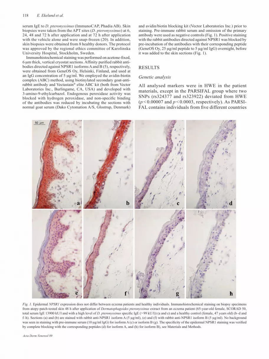
Fig. 1. Epidermal NPSR1 expression does not differ between eczema patients and healthy individuals. Immunohistochemical staining on biopsy specimens from atopy-patch-tested skin 48 h after application of Dermatophagoides pteronyssinus extract from an eczema patient (65-year-old female, SCOrAD 50, total serum IgE 13900 kU/l and with a high level of D. pteronyssinus specific IgE (> 99 kU/l)) (a and e) and a healthy control (female, 47 years old) (b–d and f–h). Sections (a) and (b) are stained with rabbit anti-NPSr1 isoform A (5 µg/ml), (e) and (f) with rabbit anti-NPSr1 isoform B (5 µg/ml). No background was seen in staining with pre-immune serum (10 µg/ml IgG) for isoform A (c) or isoform B (g). The specificity of the epidermal NPSR1 staining was verified by complete blocking with the corresponding peptides (d) for isoform A, and (h) for isoform B), see Materials and Methods.
Acta Derm Venereol 89

119Lack of association between NPSR1 and eczema
in Europe, we tested HWE in the different subgroups and found that the non-atopic individuals in the German subgroup in PArSIFAL was not in HWE regarding these SNPs (p < 1.2 × 10⁻⁷ and p < 2.4 × 10⁻⁷, respecti-vely). We therefore excluded the German subgroup in the PArSIFAL material from further analysis.
We found no association with eczema for any of the seven SNPs in any of the five different populations or in a joint analysis with all materials pooled together (Table III). There was also lack of association with any of the seven common haplotypes of NPSR1 in the dif-ferent patient materials (data not shown) and also when pooling the materials into a joint analysis (Table IV). In this joint analysis the power was more than 93% for detecting a factor with an allele frequency > 0.13 (H4 or H5) and an Or of 1.4.
In the analysis of atopic phenotypes in the context of eczema, we found rhinoconjunctivitis to be signifi-cantly associated with the minor allele of polymorphism rs324396 in NPSR1 (Or 1.25 (1.06–1.47, p < 0.03 corrected after 1000 permutations). No significant as-sociation was found with NPSR1 and any of the other phenotypes in the context of eczema even if the power to detect these was over 80% in our material.
Immunohistochemistry
In both eczema patients and healthy controls, a strong epidermal expression of isoform A was found, although this was weaker in the basal proliferating keratinocytes. Isoform B showed a weak but homogenous epidermal ex-pression in all individuals (Fig. 1). None of the isoforms
showed an appreciable difference in epidermal NPSR1 expression between eczema patients and healthy controls, between eczema patients with high or low total serum IgE levels, or between different time-points after atopy patch testing with allergen or vehicle application.
DISCUSSIONAsthma and eczema can both be manifestations of atopic diseases and share some pathogenic and im-munological features, such as the tendency to develop allergen-specific IgE against common allergens in our environment. The association of NPSR1 and asthma and raised IgE originally described by Laitinen et al. has been replicated in several studies (6–8). Genetic asso-ciation has also been found to other allergic phenotypes, such as atopic sensitization and allergic rhinoconjunc-tivitis (6). The NPSr1 protein is found to be expressed in the bronchus, the gut and the skin (5, 9). recently, inflammatory bowel disease has also been shown to associate with one haplotype in NPSR1 (H2) and with the functional SNP (rs324981, Asn107Ile) (21). In this study we wanted to elucidate whether any of the SNPs in the NPSR1 gene also were associated with eczema, and if the NPSr1 protein expression was altered in skin with eczema compared with healthy skin.
We found no association with eczema for any of the analysed SNPs in NPSR1 or the common haplotypes. Our results suggest that genetic variants in NPSR1 do not influence the susceptibility for eczema, thus confirming the results found by a German study, that found no association of polymorphism in rs232922,
Table III. Lack of association to eczema for 7 SNPs in the NPSR1 gene
SNP Minor allele
Swedish families PArSIFAL German families KOrA C KOrA S4 Joint
P P P P P P
rs323917 G 0.11 0.72 0.22 0.09 0.68 0.06rs323922 C 0.53 0.20 0.40 0.91 0.60 0.75rs324377 A 0.92 0.17 0.48 0.64 0.61 0.60SNP546333 A 0.32 0.65 0.15 0.39 0.96 1.00rs324384 C 0.42 0.16 a a a a
rs324396 T 0.07 0.08 0.76 0.64 0.75 0.45rs74037 C 0.91 0.75 0.45 0.09 0.76 0.44aNot genotyped in this patient materialP: global p-value; SNP: single nucleotide polymorphism; KOrA: Cooperative Health research in the Augsburg region; PArSIFAL: Prevention of Allergy risk factors for Sensitisation In children related to Farming and Anthroposophic Lifestyle.
Table IV. Lack of association of NPSR1 haplotypes with eczema in all five materials. No values are significant.
Haplotype Case Control Case frequency Control frequency Or 95% CI
H1 549 1287 0.325 0.317 1.0H2 370 871 0.219 0.215 1.05 0.89–1.23H3/H6 415 982 0.246 0.242 0.99 0.85–1.16H4 123 314 0.073 0.077 0.95 0.76–1.20H5 96 224 0.057 0.055 0.91 0.71–1.17H7 95 255 0.056 0.063 0.87 0.67–1.13
OR: odds ratio; CI: confidence interval.
Acta Derm Venereol 89

120 E. Ekelund et al.
with eczema (22) and a group from UK who reported lack of association with any of the haplotypes H1–H7 in NPSR1 and adult eczema (23).
Considering the number of eczema patients in this study (1848) and the estimated power of 93%, it is pro-bable that a true association with eczema would have been found, assuming that the impact of NPSR1 gene variants would be of a similar magnitude in eczema and asthma. However, we could not discriminate the haplo-type H6 from H3 in the joint analysis and can therefore not exclude an association of the haplotype H6 with eczema. Furthermore, the impact of the functional SNP rs324981 on eczema susceptibility was not evaluated in this study, but its association is unlikely given its strong association with the risk/non-risk haplotypes.
We also showed that the two isoforms, NPSr1-A and NPSr1-B, are expressed in the epidermis of both healthy individuals and eczema patients, but with no apparent difference between patients and controls. The APT-induced eczema was used here as an experimental model of eczema (20, 24), and in addition lesional and non-lesional eczema skin was stained with comparable results (data not shown). The ligand identified to interact with NPSr1 is a 20-residue peptide called Neuropeptide S (NPS). Studies have shown strong expression of NPS in the brain (25) and also in bronchial and colonial epi-thelia (9), but not in the skin (23). Absence of expression of the ligand in the skin might help explain the lesser importance of NPSr1 in the eczema pathogenesis, but as NPS is co-expressed in all other tissue with NPSr1, its absence may also be a technical artefact.
Regarding the atopic phenotypes, the only signifi-cant association with NPSR1 we found in the context of eczema was with rs324396 and rhinoconjunctivitis. No other phenotype was associated with NPSR1 in the context of eczema.
In conclusion, we report a lack of genetic association of seven NPSR1 polymorphisms with eczema in five European eczema patient materials. In addition, we found the NPSr1 isoforms A and B to be expressed in the epidermis of healthy individuals and eczema patients, but with no difference in expression. Taken together, these findings suggest that the NPSR1 gene is not a susceptibility gene for eczema.
ACKNOWLEDGEMENTSThe authors would like to thank all individuals and families who were a part of this study, and whose co-operation was essential for it. We also thank Dr Lena Holm for taking skin biopsy specimens.
This study was supported by: The Swedish research Council, the Swedish Asthma and Allergy Association research Foun-dation, the Edward Welander-Finsen Foundation, the Swedish Society for Medical research and the Juvenile Diabetes Founda-tion International. Grants were received from BMBF – Federal Ministry of Education and research (NGFN) and research grant KKF-07/04 from the University Hospital “rechts der Isar”,
Technical University Munich. Grants were also received from Ulla och Gustaf af Ugglas Foundation, Alex and Eva Wallströms Foundation, Academy of Finland, Sigrid Jusélius Foundation, Päivikki and Sakari Sohlberg Foundation. The PArSIFAL Study was supported by a main research grant from the European Union QLrT 1999-01391 and the Swedish Foundation for Health Care Science and Allergy research.
rEFErENCES
1. Johansson SG, Bieber T, Dahl r, Friedmann PS, Lanier BQ, Lockey rF, et al. revised nomenclature for allergy for global use: report of the Nomenclature review Committee of the World Allergy Organization, October 2003. J Allergy Clin Immunol 2004; 113: 832–836.
2. Cookson WO, Ubhi B, Lawrence r, Abecasis Gr, Walley AJ, Cox HE, et al. Genetic linkage of childhood atopic dermatitis to psoriasis susceptibility loci. Nat Genet 2001; 27: 372–373.
3. Williams HC, Wüthrich B. The natural history of atopic dermatitis. In: Williams HC, editor. Atopic dermatitis. The epidemiology, causes, and prevention of atopic eczema. Cambridge: Cambridge University Press, 2000: p. 41–59.
4. Spergel JM, Paller AS. Atopic dermatitis and the atopic march. J Allergy Clin Immunol 2003; 112 Suppl 6: 118–127.
5. Laitinen T, Polvi A, rydman P, Vendelin J, Pulkkinen V, Salmikangas P, et al. Characterization of a common susceptibility locus for asthma-related traits. Science 2004; 304: 300–304.
6. Melen E, Bruce S, Doekes G, Kabesch M, Laitinen T, Lauener r, et al. Haplotypes of G-protein-coupled receptor 154 are associated with childhood allergy and asthma. Am J respir Crit Care Med 2005; 171: 1089–1095.
7. Kormann MS, Carr D, Klopp N, Illig T, Leupold W, Fritzsch C, et al. G-Protein coupled receptor polymorphisms are associated with asthma in a large German population. Am J respir Crit Care Med 2005; 171: 1358–1362.
8. Feng y, Hong X, Wang L, Jiang S, Chen C, Wang B, et al. G protein-coupled receptor 154 gene polymorphism is as-sociated with airway hyperresponsiveness to methacholine in a Chinese population. J Allergy Clin Immunol 2006; 117: 612–617.
9. Vendelin J, Pulkkinen V, rehn M, Pirskanen A, raisanen-Sokolowski A, Laitinen A, et al. Characterization of GPrA, a novel G protein-coupled receptor related to asthma. Am J respir Cell Mol Biol 2005; 33: 262–270.
10. Bradley M, Kockum I, Soderhall C, Van Hage-Hamsten M, Luthman H, Nordenskjold M, et al. Characterization by phenotype of families with atopic dermatitis. Acta Derm Venereol 2000; 80: 106–110.
11. Williams HC, Burney PG, Hay rJ, Archer CB, Shipley MJ, Hunter JJ, et al. The UK Working Party’s Diagnostic Criteria for Atopic Dermatitis. I. Derivation of a minimum set of discriminators for atopic dermatitis. Br J Dermatol 1994; 131: 383–396.
12. Alfven T, Braun-Fahrlander C, Brunekreef B, von Mutius E, riedler J, Scheynius A, et al. Allergic diseases and atopic sensitization in children related to farming and anthropo-sophic lifestyle – the PArSIFAL study. Allergy 2006; 61: 414–421.
13. Weidinger S, Klopp N, Wagenpfeil S, rummler L, Schedel M, Kabesch M, et al. Association of a STAT 6 haplotype with elevated serum IgE levels in a population based cohort of white adults. J Med Genet 2004; 41: 658–663.
14. Illig T, Bongardt F, Schopfer A, Holle r, Muller S, rathmann
Acta Derm Venereol 89

121Lack of association between NPSR1 and eczema
W, et al. The endotoxin receptor TLr4 polymorphism is not associated with diabetes or components of the metabolic syndrome. Diabetes 2003; 52: 2861–2864.
15. Weidinger S, Klopp N, rummler L, Wagenpfeil S, Novak N, Baurecht HJ, et al. Association of NOD1 polymorphisms with atopic eczema and related phenotypes. J Allergy Clin Immunol 2005; 116: 177–184.
16. Purcell S, Neale B, Todd-Brown K, Thomas L, Ferreira MA, Bender D, et al. PLINK: a tool set for whole-genome association and population-based linkage analyses. Am J Hum Genet 2007; 81: 559–575.
17. Dudbridge F. Pedigree disequilibrium tests for multilocus haplotypes. Genet Epidemiol 2003; 25: 115–121.
18. Dudbridge F, editor. UNPHASED user guide. Cambridge: MrC Biostatistics Unit; 2006.
19. Purcell S, Cherny SS, Sham PC. Genetic Power Calculator: design of linkage and association genetic mapping studies of complex traits. Bioinformatics 2003; 19: 149–150.
20. Holm L, Matuseviciene G, Scheynius A, Tengvall Linder M. Atopy patch test with house dust mite allergen – an IgE-mediated reaction? Allergy 2004; 59: 874–882.
21. D’Amato M, Bruce S, Bresso F, Zucchelli M, Ezer S,
Pulkkinen V, et al. Neuropeptide s receptor 1 gene poly-morphism is associated with susceptibility to inflammatory bowel disease. Gastroenterology 2007; 133: 808–817.
22. Soderhall C, Marenholz I, Nickel r, Gruber C, Kehrt r, rohde K, et al. Lack of association of the G protein-coupled receptor for asthma susceptibility gene with atopic derma-titis. J Allergy Clin Immunol 2005; 116: 220–221.
23. Veal CD, reynolds NJ, Meggitt SJ, Allen MH, Lindgren CM, Kere J, et al. Absence of association between asthma and high serum immunoglobulin E associated GPrA haplo-types and adult atopic dermatitis. J Invest Dermatol 2005; 125: 399–401.
24. Langeveld-Wildschut EG, Thepen T, Bihari IC, ven reijsen FC, de Vries IJ, Bruijnzeel PL, et al. Evaluation of the atopy patch test and the cutaneous late-phase reaction as relevant models for the study of allergic inflammation in patients with atopic eczema. J Allergy Clin Immunol 1996; 98: 1019–1027.
25. Xu yL, reinscheid rK, Huitron-resendiz S, Clark SD, Wang Z, Lin SH, et al. Neuropeptide S: a neuropeptide promoting arousal and anxiolytic-like effects. Neuron 2004; 43: 487–497.
Acta Derm Venereol 89


