Embed Size (px)
Citation preview

Light induced hydrophilicity and osteoblast adhesion promotion onamorphous TiO2
Antonia Terriza,1 Ar�anzazu Dıaz-Cuenca,1 Francisco Yubero,1 Angel Barranco,1
Agustın R. Gonz�alez-Elipe,1 Juan Luis Gonzalez Caballero,2 Jos�e Vilches,2 Mercedes Salido2
1Instituto de Ciencia de Materiales de Sevilla (CSIC - Univ. Sevilla). Avda. Am�erico Vespucio 49, E-41092 Seville, Spain2Department of Histology, School of Medicine, Lab 57 Servicios Centrales de investigaci�on en Ciencias de la Salud,
University of Cadiz, c/Dr. Mara~non 3, 11002 Cadiz, Spain
Received 17 March 2012; revised 31 July 2012; accepted 1 August 2012
Published online 11 September 2012 in Wiley Online Library (wileyonlinelibrary.com). DOI: 10.1002/jbm.a.34405
Abstract: We have studied the effect of the UV induced
superhydrophilic wetting of TiO2 thin films on the osteoblasts
cell adhesion and cytoskeletal organization on its surface. To
assess any effect of the photo-catalytic removal of adventi-
tious carbon as a factor for the enhancement of the osteo-
blast development, 100 nm amorphous TiO2 thin layers were
deposited on polyethylene terephthalate (PET), a substrate
well known for its poor adhesion and limited wettability and
biocompatibility. The TiO2/PET materials were characterized
by X-ray photoelectron spectroscopy, and atomic force mi-
croscopy and their wetting behavior under light illumination
studied by the sessile drop method. The amorphous TiO2
thin films showed a very poor photo-catalytic activity even if
becoming superhydrophilic after illumination. The illumi-
nated samples recovered partially its initial hydrophobic state
only after their storage in the dark for more than 20 days.
Osteoblasts (HOB) were seeded both on bare PET and on
TiO2/PET samples immediately after illumination and also af-
ter four weeks storage in darkness. Cell attachment was
much more efficient on the immediately illuminated TiO2/PET
samples, with development of focal adhesions and cell trac-
tion forces. Although we cannot completely discard some
photo-catalytic carbon removal as a factor contributing to
this cell enhanced attachment, our photodegradation experi-
ments on amorphous TiO2 are conclusive to dismiss this
effect as the major cause for this behavior. VC 2012 Wiley Period-
icals, Inc. J Biomed Mater Res Part A: 101A: 1026–1035, 2013.
Key Words: adhesion, bone in growth, cell culture, cell-mate-
rial interactions, polyethylene-terephthalate
How to cite this article: Terriza A, Dıaz-Cuenca A, Yubero F, Barranco A, Gonzalez-Elipe AR, Caballero JLG, Vilches J, Salido M.2013. Light induced hydrophilicity and osteoblast adhesion promotion on amorphous TiO2. J Biomed Mater Res Part A2013:101A:1026–1035.
INTRODUCTION
The biological performance of implantable titanium devicesin medicine and dentistry can be enhanced by appropriatesurface treatments intended to favor the osseointegration.1–3
A common effect of these surface treatments and also ofthe air handling of any titanium specimen is the formationof stable passive layers of titanium oxide (TiO2), which pro-vide superior biocompatibility and promote osseointegra-tion.1 UV light irradiation of TiO2 oxide has been reportedto positively influence protein adsorption and osteogeniccells migration, attachment, and proliferation in vitro4,5 andin vivo.6 These effects differ from those induced by othermicro/nano topography surface modifications also influenc-ing cell growth and adhesion and providing some synergyto enhance the biological activity of TiO2.
3,5 The UVenhancement of the bioactivity of TiO2 has been relatedwith the photo-catalytic activity of this oxide that makes it
capable of photo-oxidizing organic molecules and carbona-ceous rests deposited or contaminating its surface andtherefore with the efficiency of the hydrocarbon removalfrom the TiO2 layer formed on commercially pure Ti(TiCP).6 An interesting phenomenon observed on illumi-nated TiO2 surfaces is their conversion from partially hydro-phobic to superhydrophilic.7 The reversal into the originalhydrophobic situation may take minutes or days, dependingon the microstructure and crystallographic structure of theoxide and can, therefore, additionally influence cellularattachment and proliferation at the initial stages. Previousstudies dealing with the osteoblast growth on TiO2 haveused crystalline anatase layers (i.e., one of the crystallinestructures of this compound) covering metallic titanium.4,6
Anatase is characterized by both an intense photo-catalyticactivity and an efficient superhydrophilic conversion asrelated expressions of its photo-activity.7 By contrast,
Correspondence to: M. Salido; e-mail: [email protected]
Contract grant sponsors: Junta de Andalucıa (P09CTS5189, FQM-6900, TEP 5283), Spanish Ministry of Economy and Competitiveness
(CONSOLIDER FUNCOAT CSD2008-00023, MAT2010-18447, MAT2010-21228), and Instituto de Salud Carlos III (FIS PI 0900508)
1026 VC 2012 WILEY PERIODICALS, INC.

amorphous TiO2 subjected to UV illumination is capable ofchanging its surface wettability from hydrophobic to superhy-drophilic, but its photo-catalytic activity and therefore itscapacity to get rid of the spurious carbon contaminating itssurface is very small or negligible.7 To study the effect of thewetting surface properties of TiO2 on human osteoblasts, wehave used amorphous TiO2 proving that its small photo-cata-lytic activity is not an obstacle for its superhydrophilic trans-formation and the attachment of osteoblasts after illumina-tion. To avoid crystallization, TiO2 was deposited at lowtemperature on the surface of conventional polyethylene ter-ephthalate (PET) by plasma enhanced chemical vapor deposi-tion (PECVD), a method well suited for the fabrication of oxidelayers on very sensitive substrates materials.8 To our knowl-edge this approach has not been previously used in this do-main, although PET has been used clinically for several appli-cations including vascular grafts, suture threads, artificialjoint prostheses,9 and more recently, as cell culture substratesfor tissue engineering.10 Although PET cannot be consideredbioactive because of its low wettability and poor cell adhe-sion,10,11 this inertness makes it very suitable for the presentstudy, focused on the regulatory factors for cellular growthand development on the surface of illuminated amorphousTiO2. Herein, we report on the preparation and characteriza-tion of thin films of amorphous TiO2 deposited on a PET sub-strate, about the evolution of the wetting contact angle (CA) ofPET and TiO2/PET upon UV irradiation, as well as on the re-covery of the wetting state by keeping the illuminated samplesstored in the dark. The photo-catalytic activity of the surfaceswas verified by dye photo-degradation tests.7 We have studiedthe influence of the UV induced hydrophobic/superhydro-philic transformation of the surfaces on osteoblasts morphol-ogy and adhesion. Osteoblasts are sensitive to a number ofinput signals from the surrounding microenvironment that actas mechanical, chemical, and topographical cues. In conse-quence, a wide number of responses, ranging from cell growthto apoptosis, including cell differentiation, migration, adhe-sion, gene expression, and signal transduction resulting intophenotypic changes, can be assessed in vitro.12,13 The molecu-lar events that occur in the cytoskeleton are princeps for theunderstanding the process by which the environment inducesthe biochemical cascade of signals, thus building the molecu-lar basis of mechanotransduction.14,15 We believe that theherein developed in vitro investigation proves the effect of UVillumination in activating the surface of amorphous TiO2 andTiO2 supported layers to enhance their osteoconductivitythrough the conversion into a superhydrophilic state.
MATERIALS AND METHODS
Deposition and characterization of TiO2 thin film layersTiO2 was deposited on PET substrate plates (Goodfellow) byPECVD in a plasma reactor with a remote configuration. Thesystem, supplied with an external microwave plasma source(SLAN, Plasma Consult, GMbh, Germany) in a downstreamconfiguration, was coupled to the reaction chamber and sep-arated from it by a grounded grid to avoid the microwaveheating of the substrates. Distance from substrate and gridwas 10 cm. The source was operated at 400 W with pure
O2 as plasma gas, and the synthesis of the films was carriedout at room temperature by using titanium tetrakis isoprop-oxide (TTIP) as precursor. For dosing the TTIP in a con-trolled way, it was placed in a stainless steel receptaclethrough which oxygen was bubbled while heating at 305 K.The dosing line was heated at 373 K to prevent condensa-tion in the tube walls. Total pressure during deposition was4 � 10�3 Torr.16,17 The deposited TiO2 was controlled by aquartz crystal monitor and calibrated by comparison withthe direct electron microscopic observation of the film thick-ness for films deposited on a flat silicon substrate. Beforedepositing TiO2, the polymeric substrate was exposed topure plasma of oxygen for 1 min, a pretreatment that favorsthe adhesion of the oxide layer onto the polymer surface. Atypical thin layer of TiO2 of 100 nm was deposited on thepolymeric substrate. These samples will be designated asTiO2/PET. The samples were characterized by X-ray photo-electron spectroscopy (XPS) to determine their surfacechemical state and composition, atomic force microscopy(AFM) to ascertain their surface topography and by waterCA measurements to check the wetting characteristics oftheir surfaces. XPS spectra were recorded with a SPECSPHOIBOS-100 spectrometer working in the constant passenergy mode fixed at a value of 20 eV. The Mg Ka radiationwas used as excitation source. For calibration of the bindingenergy scale, a value of 284.6 eV was considered for theC1s component attributed to CAH and CAC bonds. Surfacecomposition was estimated by calculating the area behindthe C1s, Ti2p, and O1s peaks and by correcting the obtainedvalues with the sensitivity factors of these elements. Sam-ples were illuminated for 40 min through a quartz windowin the prechamber of the XPS spectrometer under a pres-sure of 20 torr O2 to assess the efficiency of the UV lightillumination for removing part of the spurious carbonaceousrests accumulated on the surface of the TiO2 layer. AFMimages were taken with a Cervantes AFM microscope drivenwith a Dulcinea control system (Nanotec, Madrid, Spain)working in tapping mode and using high frequency cantile-vers, and processed with Nanotec WSxM software. StaticCAs of water were determined by the Young method with aCAM100 instrument (KSV Instruments Ltd, Finland), withan estimated error of 10%. In the experiments where theCA variation was determined as a function of the illumina-tion time, a metal foil was used as a shutter for the lampoutput. All wetting angle measurements were taken afterUV illumination for successive periods of time, thus thetime scale in the plots refers to the accumulative illumina-tion of the samples, which was carried out with a Xe dis-charge lamp with photon intensity at the position of thesamples of 2W cm�2 for the complete spectrum. An infraredfilter was kept between the lamp and the samples to pre-vent heating. The photo-catalytic activity of the amorphousTiO2 thin layers was compared with that of anatase TiO2 de-posited on silicon at 523 K by PECVD and a silicon wafer asblank control experiment. Photocatalytic tests were carriedout in a self-designed set-up,18 provided with automatic re-cording of the spectra from the solution while the filmswere irradiated. The system consisted of a quartz cell (total
ORIGINAL ARTICLE
JOURNAL OF BIOMEDICAL MATERIALS RESEARCH A | APR 2013 VOL 101A, ISSUE 4 1027

volume 3 cm3) where 2 cm3 of a 3.5 � 10�5 M solution ofmethyl orange dye was placed together with a piece of a sil-icon substrate (1 � 0.8 cm2) covered with the studied thinfilm deposited onto its surface. Irradiation was carried outfrom a frontal position while recording spectra from thesolution by means of two optical fibers connected to a UV–visible spectrometer placed in opposite sides of the vessel.The solution was bubbled with oxygen and the vessel cov-ered with teflon to avoid evaporation. The whole set-up wasrefrigerated. Reference experiments were carried out with-out the thin films substrates. The intensity of the UV þ visradiation at the position of the cell was 1.8 W. The illumina-tion area of the samples was defined with a slit of 1 cm2.
Cell cultureHOBVR human osteoblasts (Promocell, Heidelberg, Germany)were seeded at a density of 5000 cells/cm2 and incubatedin osteoblast growing medium (Promocell) supplementedwith 10% fetal calf serum (Promocell) at 37�C and 5% CO2
on test surfaces and immunolabeled after 24, 48 h and 7days. Growth medium was changed every three days. HOBcells did not exceed 10 population doublings. Test surfaceswere exposed to UV light for 20 min each side, in a laminarflow chamber under sterile conditions, in order to achieveoptimal sterilization, prior to cell seeding. Then, prior to cellseeding, half the number of the samples was stored underdark ambient conditions for 4 weeks, according to previ-ously described protocols.23 The remaining half set of sam-ples were deposited on sterile petri dishes and immediatelyseeded. Additional UV exposure conditions to analyze themost suitable treatment for photoactivation are describedearlier. At least five samples of each type were seeded andanalyzed in each experiment.
Cell morphology and spreadingCells were daily examined with the phase contrast micro-scope in order to evaluate cell morphology, alignment, andinitial adhesion phase to surfaces. Phenotypic changes, celldistribution, and spreading were assessed under light mi-croscopy after staining with toluidine blue, prior to fluores-cence and CLSM examination.
Actin cytoskeletal organization and focal adhesionexpressionAt the end of each experiment, cells were washed with pre-warmed phosphate buffered saline (PBS), pH 7.4, and fixed
with 3.7% paraformaldehyde at room temperature, washed,and then permeabilized with 0.1% Triton X-100 (Sigma, StLouis, MO). After washing, cells were preincubated with 1%bovine serum albumin (Sigma) in PBS for 20 min prior tocell immunolabeling for actin cytoskeleton with rhodaminephalloidin (Sigma) and for focal adhesion identification withmonoclonal antivinculin FITC conjugate (Sigma, MO). After20 min samples were rinsed with prewarmed PBS prior tomounting with Vectashield VR (Vector. Burlingame, CA).
Confocal examinationSamples were visualized using a Leica TCS-SL confocalmicroscope. At least five samples were analyzed for each ex-perimental group to assess surface influence on cytoskeletalorganization, focal adhesion number and development, andcell morphology. To analyze the differences in focal adhesionnumber between different sample groups, images were col-lected as frames obtained at 40� magnification and proc-essed using Leica imaging software. Sample size is deter-mined by the number of frames in each group (Table II).Samples were exposed to the lowest laser power that wasable to produce a fluorescent signal for a time interval nothigher than 5 min to avoid photobleaching. A pinhole of 1Airy unit was used. Images were acquired at a resolution of512 � 512, mean voxel size of 209.20 nm.
Statistical analysisAfter the descriptive analysis, and owing to the type of analyzedvariables, the sample size in each group and the results of theLevene contrast used to ascertain the equivalence of variances,two nonparametric statistical methods were chosen in order tocontrast the basic (null) hypothesis on nonsample treatmentinduced differences (H0 : s1 ¼ s2 ¼ s3 ¼ s4, where s ¼ popu-lation median), that is, the four samples can be thought of as asingle (combined) sample from one population. We have usedtwo alternatives hypothesis: (a) the Kruskal–Wallis (K–W) uni-lateral test, that contrast the general alternative of that the s’sare not all equal, and (b) the Jonckheere–Terpstra (J–T) unilat-eral test that can be used when the treatments are ordered, andcontrast alternatives of the form H1 : s1 � s2 � s3 � s4, whereleast one of the inequalities is strict.
RESULTS
XPS and AFM analysis of TiO2/PET samplesThe TiO2 layers deposited by PECVD under the conditionsdescribed above were amorphous, as confirmed by the
FIGURE 1. From left to right C1s, O1s, and Ti2p photoelectron spectra of PET and TiO2/PET samples.
1028 TERRIZA ET AL. OSTEOBLAST ADHESION PROMOTION ON TiO2

absence of any signal when 100 nm or even thicker films ofdeposited TiO2 were examined by XRD or Raman spectros-copy.19 The Ti2p spectrum in the TiO2/PET samples wascharacterized by a Ti2p3/2 binding energy of 458.4 eV, typi-cal of the Ti4þ oxidation state of this element. This sustainsthat titanium is deposited in the form of TiO2 (Fig. 1). Theatomic percentages (Table I) revealed that in the TiO2/PETsample there is some carbon contamination on the surface.A similar conclusion can be figured out by looking at theO1s and C1s spectra. This latter is attributed to spuriouscarbon contaminating the films surface because of themanipulation of samples in air and a small shoulder at� 289.5 eV due to carbonate and related species. Mean-while, the O1s spectrum of this sample is characterized by amain peak at 529.8 eV due to the oxygen ions of TiO2 and ashoulder at � 531.8 eV due to either AOH surface groupsand/or to oxygen of the carbonate surface species. Thesespectral features differ from those of the PET substrates(Fig. 1) where the O1s spectrum is characterized by bandsat 531.9 and 532.9 eV attributed to oxygen bonded to car-bon through double and single bonds.20 The C1s spectrumof PET was characterized by a series of peaks and shouldersattributed to CAC and CAH species (peak at 284.6 eV),CAOH (shoulder at 285.4 eV), C¼¼O (shoulder at 286.2 eV)and ACOO (small band at 288.6 eV) species. Therefore, theanalysis of all previous spectra of the TiO2/PET samplesconfirms a complete coverage of the polymeric substrate bythe TiO2 amorphous layer.
The surface composition of the TiO2/PET sample wasalso examined just after 40 min UV irradiation in oxygen inthe prechamber of the XPS spectrometer and after a pro-longed storage of the irradiated samples for four weeks. Thesurface compositions of the irradiated and stored samplesare also reported in Table I. The obtained atomic percen-tages of the irradiated sample indicate that UV is effective inremoving a small part of the spurious carbon contaminatingthe surface of TiO2, although a significant amount of the ini-tial carbon contamination remains on the surface after thisprolonged treatment (i.e., no completely ‘‘clean’’ TiO2 surfacewas obtained by UV irradiation of our amorphous thin filmprepared by PECVD). Meanwhile, after storage, additionalcarbon becomes incorporated to the surface although to anextent that does not reach the initial situation of this sample.
The surface morphology of the PET and TiO2/PET sam-ples examined by AFM (Fig. 2) and the roughness values,0.94 and 2.46 nm for PET and TiO2/PET, respectively show
that the relatively flat surface of PET becomes rougher afterdeposition of the TiO2 and depict a granular topographylikely resulting from the aggregation of the small grains ofthe plasma deposited oxide completely covering the poly-mer surface.
Wetting behavior and photoactivity ofilluminated samplesFigure 3 (left) shows the evolution of the water CAs deter-mined for the PET, and TiO2/PET samples subjected to UVillumination for increasing periods of time. For TiO2/PETthe decrease in wetting angle converts its surface intosuperhydrophilic (water CA close to zero). When the sam-ples were stored in the dark, the wetting angle increasedaccording to a slow kinetic and required more than onemonth to partially restore its original state (Fig. 3, right). Asit has been reported for UV illuminated PET,21 the CA ofthis material decreased up to a value of � 50� andremained almost invariable after prolonged storage in thedark. The limited CA decrease observed in PET must beattributed to the activation processes induced in polymersirradiated with UV light. This activation implies the breakingof some polymer chains and the formation of new oxygen-ated functional groups by reaction of light induced radicalswith the oxygen and/or water of the atmosphere. Thesuperhydrophilic transformation process of the surface ofTiO2/PET after UV irradiation has a completely differentcause. To account for this transformation both the hydroxy-lation of some domains leading to the formation of anamphiphilic surface and the photo-catalytic removal of car-bonaceous rests have been proposed.21 Most important forour hypothesis of a bioactivity induced by the hydrophilictransformation of TiO2 is the fact that amorphous TiO2 onlypresents a limited photo-catalytic activity when comparedwith the anatase phase of this oxide. However, both the illu-minated amorphous and the anatase phases transform theirsurface from partially hydrophobic into superhydrophilicunder the effect of UV light. The photo-catalytic activitytests (Fig. 4), confirm the comparatively low photo-catalyticactivity of amorphous TiO2. Although the test is carried outin solution, the low activity depicted by the amorphouslayer suggests that it will be difficult to achieve a significantremoval of the spurious carbon contaminating its surface
TABLE I. Atomic Percentages (Absolute Values) Determined
by XPS for PET and TiO2/PET Samples, this Latter in the
‘‘As Prepared’’, After UV Irradiation for 40 min (i.e., TiO2/PET-
UV) and Its Subsequent Storage for Four Weeks in the Dark
(i.e., TiO2/PET-UV-Dark)
Sample Ti (%) O (%) C (%)
PET – 23 77TiO2/PET 25 62 13TiO2/PET-UV 27 64 9TiO2/PET-UV-dark 26 63 11
FIGURE 2. AFM images of PET (left) and TiO2/PET (right) samples taken
for two observation areas as indicated. [Color figure can be viewed in
the online issue, which is available at wileyonlinelibrary.com.]
ORIGINAL ARTICLE
JOURNAL OF BIOMEDICAL MATERIALS RESEARCH A | APR 2013 VOL 101A, ISSUE 4 1029

after irradiation for 40 min, time approximately requiredto render its surface superhydrophilic (Fig. 3).
Cell morphology and spreadingLiving osteoblasts examination 24 h after seeding revealed asuccessful cell attachment with marked phenotypic changes,like filopodial and lamellopodial emission, mainly oriented toelongation and alignment towards the ‘‘new’’ photoactivatedand functionalized TiO2 PET surfaces. After 48 h in culture,cell spreading had evolved to a near confluence stage, wherephenotypic changes turned to osteoblasts well adhered tothe surface and to the neighboring cells. After one week,confluence was observed in most samples. Cells grown on‘‘old’’ photoactivated functionalized TiO2/PET surfacesappeared to be mostly rounded in shape and showed no filo-podial or lamelopodial emission. Although cells adhered tosurface, no significant cell spreading was observed either af-ter 48 h or one week in culture. Osteoblasts grown on non-functionalized PET showed elongated morphology in bothgroups, with no significant lamelopodial or filopodial emis-sion after 24 or 48 h. After one week in culture cell spread-ing increased, although without confluence.
Cytoskeletal organization and focal adhesionsActin cytoskeleton immunolabeling of growing cells revealeda clear polarization of actin cytoskeleton with stress fibersdevelopment and a more defined osteoblast orientationwith a higher number of well developed focal adhesions(FAs) [Fig. 5(A,C)] in those cells that were seeded at any ex-perimental time point on TiO2/PET immediately after UVirradiation, (Figs. 5–7), when compared with cells grown on‘‘old’’ UV photoactivated/PET or nonfunctionalized PET[Table II, Figs. 5(B,D), 6, and 7] Cytoskeletal arrangementand development in cells grown on nonfunctionalized PETsamples was quite similar both in cells seeded immediatelyafter UV irradiation of surfaces or in those seeded on storedPET samples (Figs. 6 and 7).
Statistical analysisFor each group, Table II shows the number of consideredframes (N), as well as the essential statistic parameters forthe variable ‘‘FAs’’ (Table II). The Box–Wisker graphic of theFAs detected in the different groups clearly revealed that
the number of well developed FAs in cells immediatelyseeded on the ‘‘new’’ TiO2/PET is higher than on cellsseeded on any other PET sample or on the stored TiO2/PETsamples (Table II, Fig. 8). The Levene statistics (L ¼ 4.472)reveals a significance level smaller than 0.05 (p ¼ 0.007),thus implying that the variances are significantly differentand that nonparametric methods have to be used. Once thecontrast of the K–W test has been used for the four studiedsamples, the statistic H ¼ 24.092 provides a high signifi-cance (p ¼ 0.000; gl ¼ 3) and indicates that the effects ofthe surface treatments are different. Moreover, using the J–Ttest with the four samples, the standardized statistic index J¼ 4.396 also reveals a high significance (p ¼ 0.000; gl ¼ 3).This indicates that the median values of the variable ‘‘num-ber of FAs’’ obtained for each group (from one to four) canbe deemed ordered from lower to higher values (Table II,Fig. 8). Taking into account the results of the descriptiveanalysis, we have used the two previous tests with the threefirst specimens. With the K–W test we obtained H ¼ 2.740(p ¼ 0.254; gl ¼ 2), while with the J–T test we got J ¼1.604 (p ¼ 0.055). Since, the J–T tests yields a significancelevel close to 0.05, we can infer a certain order between thethree first samples. When the tests were used with the first
FIGURE 3. Evolution of the water contact angle (CA) on the surface of PET, and TiO2/PET samples. Left irradiated with UV light for increasing
periods of time; right stored in the dark after illumination.
FIGURE 4. Evolution of the normalized concentration of dye mole-
cules during photodegradation experiments by using amorphous or
anatase thin films. The results for a blank control experiment are
included for comparison.
1030 TERRIZA ET AL. OSTEOBLAST ADHESION PROMOTION ON TiO2

two specimens, with the K–W test the obtained value was H¼ 1.052 (p ¼ 0.305; p ¼ 1), while with the J–T test we got J¼ 1.026 (p ¼ 0.161), thus showing that nonsignificant dif-ferences were found between PET samples.
DISCUSSION
Our results have shown clear differences in osteoblasts de-velopment when they are grown on the illuminated TiO2/PET. Up to now, similar results obtained on illuminated TiO2
grown on metallic titanium implants or bulk pieces, havebeen correlated with the photo-oxidation removal of carbonimpurities always present on the surface of any oxide sur-face exposed to air.4–6 Although not always reported, themost typical form of TiO2 over pure metal bulk titanium isthe crystalline anatase, particularly if handled in air at ele-vated temperatures. We have used PET as substrate andperformed the deposition of an amorphous TiO2 layer byPECVD at room temperature. Owing to the poor photo-cata-lytic activity of this form of TiO2, evidenced here by a much
smaller photo-catalytic activity than for anatase (Fig. 4), andthe fact that only a limited amount of the spurious carboncontaminating the TiO2/PET surface could be removed dur-ing the photo-oxidation experiment in the XPS spectrometer,we think that photo-catalytic removal of carbon is not themain cause modifying the osteoblast behavior on illumi-nated TiO2. In a previous work by Aita et al.,6 these authorsclaim that photo-catalytic carbon removal rather thanhydrophilicity changes is the main factor accounting for anenhanced protein adsorption and osteoblast development ofthe surface of illuminated TiO2. However, both the contami-nation level of their titanium oxide surfaces with a carbonconcentration higher than 50% atomic and the fact thattheir titanium oxide was crystalline and not amorphous pre-clude a direct extrapolation and application of their conclu-sions to our working conditions.
Tissue engineering offers a promising solution for thedevelopment of synthetic and/or natural scaffolds, uponwhich osteoprogenitor cells can be seeded and cultured
FIGURE 5. HOB cells immunolabeled, 48 h after seeding, with rhodamine-phalloidine for actin cytoskeleton (red) and antivinculin antibody
(green) for focal adhesion sites. Both a higher polarization in actin cytoskeleton, with strongly labeled stress fibers, and focal adhesion develop-
ment was found on osteoblasts immediately seeded on UV irradiated PECVD TiO2 activated PET samples (A, C) than in osteoblasts seeded on
samples stored in the dark for 4 weeks after UV irradiation (B, D).
ORIGINAL ARTICLE
JOURNAL OF BIOMEDICAL MATERIALS RESEARCH A | APR 2013 VOL 101A, ISSUE 4 1031

under optimal conditions and ultimately implanted to pro-mote osteogenesis in vivo.22 Ideally, a bone scaffold shouldbe biocompatible and osteoconductive.22 Our group has pre-viously described how scaffolds activation by PECVD of TiO2
appears to be an alternative to wet chemical treatments inbone tissue engineering.23 To evaluate biocompatibility andthe osteoconductive properties of the PECVD TiO2 activatedPET surfaces, we have carried out an in vitro study withhuman normal osteogenic cells. Phenotypic changes, cytos-keletal arrangement, and focal contact development wereevaluated in the initial phases of cell growth, by far, themost critical issue for tissue regeneration in vitro. UV light-mediated photofunctionalization has been described toimprove osteoconductivity of titanium implants,24,25 but theprecise molecular mechanism of why and how chemical sur-face modification and increased hydrophilicity may affectthe tissue materials is not yet well understood. In the pres-ent study, time related changes in bioactivity induced by UVirradiation of TiO2/PET surfaces have been assessed in vitro.The sterilization of scaffolds, including the UV sterilization,is an imperative step in the development of cell culture pro-tocols for tissue engineering, although their application mayalter the surface properties of biomaterials and conse-
quently affect the cell behavior.23,24 According to somegroups, UV irradiation can dramatically enhance the bioac-tivity of titanium by inducing alterations of its surfacechemistry and wettability.4,6,25,26 In the present work, afteranalyzing the surface properties of illuminated TiO2, we ten-tatively propose that hydrophilicity changes rather than re-moval of carbon is the main factor favoring the osteoblastsdevelopment. For the present study, we have assessed theHOB cells response to UV sterilized PET scaffolds, clean orcovered with TiO2 nanolayers, and also the possible influ-ence on the cell growing processes of the time-dependentbioactivation of the samples by UV irradiation. The resultsobtained on the ‘‘new’’ TiO2/PET activated samples, that isthose which were seeded immediately after irradiation,show cells with marked phenotypic changes and a substan-tial improvement in cell polarization, cytoskeletal organiza-tion, stress fibers development, and in the number of FAs ascompared with TiO2/PET samples that were kept in darkthat, at some time-points, showed similar results thanuncovered PET samples. Thus, the combination of PECVDTiO2 activated samples with an adequate UV exposureappears to effectively stimulate an efficient osteoblastspreading and attachment. Fluorescent images obtained
FIGURE 6. HOB cells grown on PET samples 48 h after culture. In A, C cells seeded immediately after UV irradiation. In B, D cells seeded on
samples previously stored in dark for 4 weeks after UV irradiation. In both cases, small cells with slight polarization of actin cytoskeleton (red,
immunolabeled with rhodamine–phalloidine) showing a few stress fibers are observed. [Color figure can be viewed in the online issue, which is
available at wileyonlinelibrary.com.]
1032 TERRIZA ET AL. OSTEOBLAST ADHESION PROMOTION ON TiO2
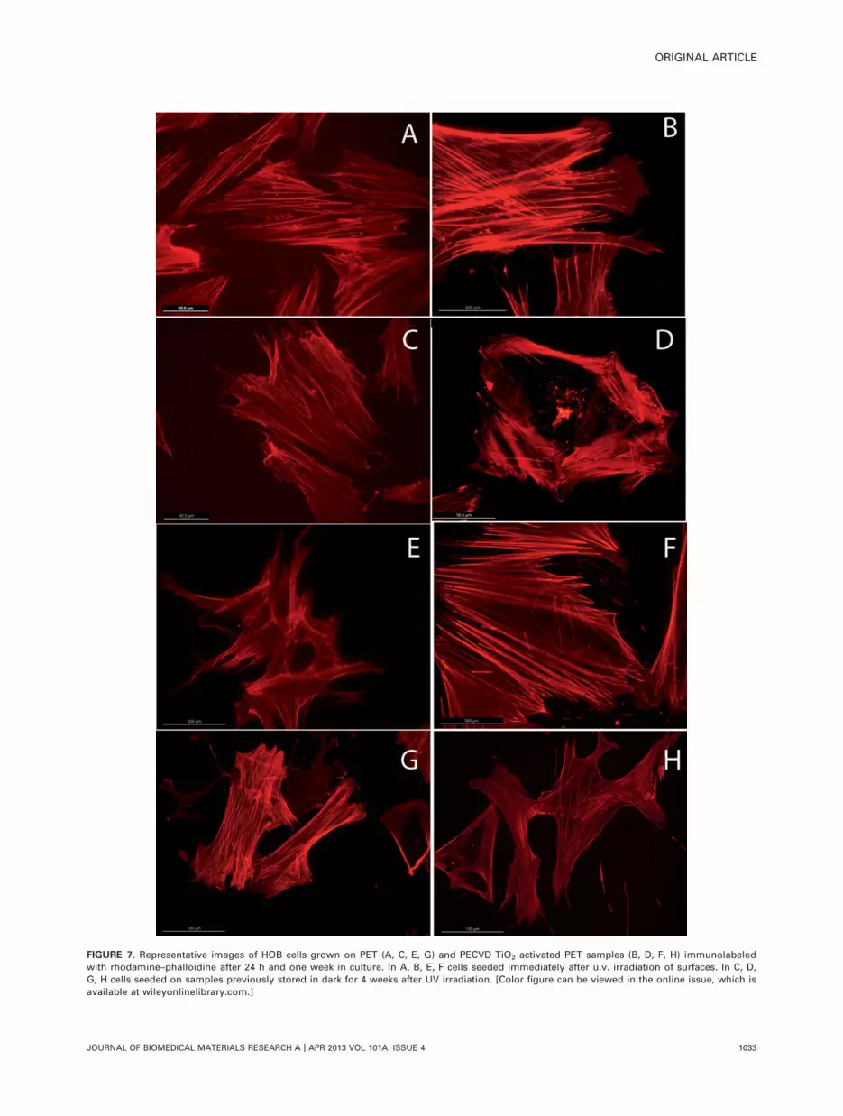
FIGURE 7. Representative images of HOB cells grown on PET (A, C, E, G) and PECVD TiO2 activated PET samples (B, D, F, H) immunolabeled
with rhodamine–phalloidine after 24 h and one week in culture. In A, B, E, F cells seeded immediately after u.v. irradiation of surfaces. In C, D,
G, H cells seeded on samples previously stored in dark for 4 weeks after UV irradiation. [Color figure can be viewed in the online issue, which is
available at wileyonlinelibrary.com.]
ORIGINAL ARTICLE
JOURNAL OF BIOMEDICAL MATERIALS RESEARCH A | APR 2013 VOL 101A, ISSUE 4 1033

during the whole experiment, from 24 h to one week ofincubation, confirmed that cells were evidently larger andinitiated phenotypic changes with the emission of filopodialand lamellopodial processes. Immunolabeling for actinrevealed a subsequent osteoblast orientation and polariza-tion associated with a higher expression of stress fibers.This occurred in close association with an intensive andextensive cytoplasmic localization of the focal adhesion pro-tein vinculin in this experimental group, in contrast with itsfaint expression on nonirradiated samples or samples storedfor 4 weeks after irradiation. It is important to stress thatfocal adhesion development is also substantially increasedin the ‘‘new’’ irradiated TiO2/PET samples. Cell internal or-ganization and orientation are controlled by FAs that medi-ate the regulatory effects of extracellular matrix (ECM) ad-hesion and the variation of actin-myosin stress fibersdistribution in response to surface properties.27,28 As wehave described in a previous work, this behavior can bealso associated with a substantial increase in osteoblasticmitochondrial bioenergetics polarized to focal adhesionsites.29 FAs are formed during initial cell adhesion andthereafter constantly assembled and disassembled duringcell movement. In addition, FAs serve as mechanosensors torecognize both the biochemical and biophysical characteris-tics of surfaces.27,30,31 Maturation of FAs is essential for theestablishment of a firm adhesion to a surface and, upon amaturation of FA-complexes, also for the transduction of sig-nals by outside-in and inside-out signaling leading an effec-tive control of transcription factors.32,33 The HOB osteoblastresponse in our design supports that the proposed UV irra-diation protocol induces wettability changes of PECVD TiO2
activated surfaces that result in a superhydrophilic transfor-mation. To our knowledge this is the first time that theinfluence of UV photoactivation of PCEVD TiO2 nanolayersdeposited on PET is described for human osteoblasts,although similar results have recently been reported forbone marrow rat cells grown on Ti sputtered glass and UVtreated plates5 and onto titanium disks that were previouslymachined, acid-etched or sandblasted.4,33,34 To our knowl-edge, this is also the first time that this photoactivation ofcell development has not been associated with the photo-catalytic removal of carbon as the major cause for thatbehavior.
CONCLUSIONS
The study reported here on the osteoblast development onUV illuminated amorphous TiO2 has shown that theobserved positive effect on cell development must be attrib-uted to the transformations undergone by the surface of the
oxide subjected to irradiation. Trying to go a step forwardin the analysis of the causes leading to this improvement incell development, we have studied the UV induced changesin the hydrophilic state of the TiO2 surface and found thatits superhydrophilic conversion is the most likely factor con-tributing to the attachment and proliferation of osteoblasts.Although we cannot completely discard that the removal ofsome carbon residues from the surface of the illuminatedTiO2 could be a factors contributing to this enhanced devel-opment of cells, our experiments on the photo-catalytic car-bon removal from amorphous TiO2 surfaces tend to discardphoto-catalysis as the main cause for this behavior. Anotherinteresting consequence of our work is that relatively inac-tive polymeric substrates like PET can be made bioactive bymodifying their surface with the deposition of a very thinlayer of photo-active TiO2. For this purpose, a main requisiteto protect the integrity of the polymeric structure is the useof mild deposition conditions like in the PECVD processes.The TiO2 PECVD deposition carried out in this work pro-duces the formation of a well adhered TiO2 layer while pre-serving the integrity of sensitive substrates such as PET.
Finally, from the perspective of bone tissue engineering,the main results of this work can be summarized into threemain conclusions: (i) we have developed a reproducible cellculture protocol including the successful sterilization ofTiO2 activated PET samples; (ii) both the polymeric sub-strate (scaffold) integrity and the titanium oxide layer are
FIGURE 8. Box–Wisker graphics reporting the variable number of
focal adhesions quantified on each sample.
TABLE II. Descriptive Parameters of the Statistical Analysis Performed for the Variable ‘‘Number of Focal Adhesions’’
Samples N Minimum MaximumRangeMean Median Mean St. Dev.
1 PET ‘‘old’’ 12 56 98 18.38 90.50 87.08 12.0342 PET ‘‘new’’ 15 39 153 23.97 96.00 96.40 32.1223 TiO2/PET ‘‘old’’ 19 59 170 26.92 96.00 104.00 29.9574 TiO2/PET ‘‘new’’ 10 120 348 50.45 214.00 217.30 62.363
1034 TERRIZA ET AL. OSTEOBLAST ADHESION PROMOTION ON TiO2

preserved, thus reinforcing the methodology proposed as avaluable tool, which could be applied to different polymericsubstrates and (iii) the observed major time dependent var-iations on the cellular response to the surface changes aredue to the UV photofunctionalization. This supports that acareful observance of time dependent changes on bioactivityis an important factor for improving UV sterilized samplessuitability for clinical purposes.
ACKNOWLEDGMENT
The authors thank the technician Emilio de la Orden, fromCadiz labs, for his collaboration.
REFERENCES1. Das K, Balla VK, Bandyopadhyay M, Bose S. Surface modification
of laser-processed porous titanium for load-bearing implants.
Scripta Materialia 2008;59:822–825.
2. Geetha M, Singh AK, Asokamani R, Gogia AK. Ti based biomateri-
als, the ultimate choice for orthopaedic implants—A review. Prog
Mater Sci 2009;54:397–425.
3. Tsukimura N, Yamada M, Iwasa F, Minamikawa H, Att W, Ueno T,
Saruwatari L, Aita H, Chiou WA, Ogawa T. Synergistic effects of
UV photofunctionalization and micro-nano hybrid topography on
the biological properties of titanium. Biomaterials 2011;32:
4358–4368.
4. Att W, Hori N, Iwasa F, Yamada M, Ueno T, Ogawa T. The effect
of UV photo functionalization on the time-related bioactivity of ti-
tanium and chromium–cobalt alloys. Biomaterials 2009;30:
4268–4276.
5. Miyauchi T, Yamada M, Yamamoto A, Iwasa F, Suzawa T, Kamijo
R, Baba K, Ogawa T. The enhanced characteristics of osteoblast
adhesion to photo functionalized nanoscale TiO2 layers on bioma-
terials surfaces. Biomaterials 2010;31:3827–3839.
6. Aita H, Hori N, Takeuchi M, Suzuki T, Yamada M, Anpo M, Ogawa
T. The effect of ultraviolet functionalization of titanium on integra-
tion with bone. Biomaterials 2009;30:1015–1025.
7. Rico V, Romero P, Hueso JL, Espinos JP, Gonzalez-Elipe AR. Wet-
ting angles and photocatalytic activities of illuminated TiO2 thin
films. Catal Today 2009;143:347–354.
8. Yun J, Lee S, Bae TS, Yun Y, Lee S, Kwon JD, Lee GH. Adhesive
and structural failures of oxide costaings on plasma treated poly-
mers. Plasma Proc Polym 2011;8:815–831.
9. Dimitrievska S, Petit A, Doillon CJ, Epure L, Ajji A, Yahia L, Bu-
reau MN. Effect of sterilization on non-woven polyethylene ter-
ephthalate fiber structures for vascular grafts. Macromol Biosci
2011;11:13–21.
10. Sasmazel HT, Manolache S, Gumus�derelioglu. Functionalization
of nonwoven PET fabrics by water/O2 plasma for biomolecule
mediated cell cultivation. Plasma Process Polym 2010;7:588–600.
11. Reisinger B, Fahrner M, Frischauf I, Yakunin S, Svorcik V, Fiedoro-
wicz H, Bartnik A, Romanin C, Heitz J. EUV micropatterning for
biocompatibility control of PET. Appl Phys A 2010;100:511–516.
12. Gittens RA, McLachlan T, Olivares-Navarrete R, Cai Y, Berner S,
Tannenbaum R, Schwartz Z, Sandhage KH, Boyan BD. The effects
of combined micron/submicron-scale surface roughness and
nanoscale features on cell proliferation and differentiation. Bio-
materials 2011;32:3395–3403.
13. Rape AD, Guo WH, Wang YL. The regulation of traction force in
relation to cell shape and focal adhesions. Biomaterials 2011;32:
2043–2051.
14. Dalby MJ. Topographically induced direct cell mechanotransduc-
tion. Med Eng Phys 2005;27:730–742.
15. Hata K, Ikebe K, Wada M, Nokubi T. Osteoblast response to tita-
nium regulates transcriptional activity of Runx2 through MAPK
pathway. J Biomed Mater Res A 2007;81:446–452.
16. Sanchez-Valencia JR, Borras A, Barranco A, Rico VJ, Espinos JP,
Gonzalez-Elipe AR. Preillumination of TiO2 and Ta2O5 photoactive
thin films as a tool to tailor the synthesis of composite materials.
Langmuir 2008;24:9460–9469.
17. Borr�as A, Cotrino J, Gonz�alez-Elipe AR. Type of plasmas and
microstructures of TiO2 thin films prepared by plasma
enhanced chemical vapor deposition. J Electrochem Soc 2007;
154:152–157.
18. Romero-Gomez P, Hamad S, Gonzalez JC, Barranco A, Espinos
JP, Cotrino J, Gonzalez-Elipe AR. Band gap narrowing versus for-
mation of electronic states in the gap in N-TiO2 thin films. J Phys
Chem C 2010;114:22546–22557.
19. Romero-G�omez P, Rico V, Borr�as A, Barranco A, Espin�os JP,
Cotrino J, Gonz�alez-Elipe AR. Chemical state of nitrogen and visi-
ble surface and schottky barrier driven photoactivities of N-doped
TiO2 thin films. J Phys Chem C 2009;113:13341–13351.
20. L�opez-Santos C, Yubero F, Cotrino J, Barranco A, Gonz�alez-Elipe
AR. Plasmas and atom beam activation of the surface of poly-
mers. J Phys D: Appl Phys 2008;41:225209–225221.
21. Yasukawa A, Kobayashi Y. Wettability characteristics of PET films
treated by atmospheric pressure plasma and ultraviolet excimer
light. Polym J 2011;43:545–551.
22. Whited BM, Whitney JR, Hofmann MC, Xu Y, Rylander MN. Pre-
osteoblast infiltration and differentiation in highly porous apatite-
coated PLLA electrospun scaffolds. Biomaterials 2011;32:
2294–2304.
23. Salido M, Terriza A, Torres D, de la Orden E, Barranco A, Diaz
Cuenca MA, Vilches J. Nanolayers of PECVD TiO2 suitability for
human osteoblasts growth for tissue engineering. Histol Histopa-
thol 2011;26(Supp1):55–56.
24. Zhao L, Mei S, Wang W, Chu PK, Wu Z, Zhang Y. The role of ster-
ilization in the cytocompatibility of titania nanotubes. Biomaterials
2010;31:2055–2063.
25. Han Y, Chen D, Sun J, Zhang Y, Xu K. UV-enhanced bioactivity
and cell response of micro-arc oxidized titania coatings. Acta Bio-
mater 2008;4:1518–1529.
26. Gallardo-Moreno AM, Pacha-Olivenza MA, Salda~na L, Perez-Gir-
aldo C, Bruque JM, Vilaboa N, Gonz�alez-Martin ML. In vitro bio-
compatibility and bacterial adhesion of physico-chemically
modified Ti6Al4V surface by means of UV irradiation. Acta Bio-
mater 2009;5:181–192.
27. Diener A, Nebe B, Luthen F, Becker P, Beck U, Neumann HG,
Rychly J. Control of focal adhesion dynamics by material surface
characteristics. Biomaterials 2005;26:383–392.
28. Salido M, Vilches JI, Guti�errez JL, Vilches J. Actin cytoskeletal
organization in human osteoblasts grown on different dental
titanium implant surfaces. Histol Histopathol 2007;22:
1355–1364.
29. Salido M, Vilches-Perez JI, Gonzalez JL, Vilches J. Mitochondrial
bioenergetics and distribution in living human osteoblasts grown
on implant surfaces. Histol Histopathol 2009;24:1275–1286.
30. Geiger B, Spatz JP, Bershadsky AD. Environmental sensing
through focal adhesions. Nat Rev 2009;10:21–33.
31. Pathak A, Deshpande VS, McMeeking RM, Evans AG. The simula-
tion of stress fibers and focal adhesion development in cells on
patterned substrates. J R Soc Interface 2008;6:507–524.
32. Lamers E, van Horssen R, te Riet J, van Delft FC, Luttge R, Wal-
boomers XF, Jansen JA. The influence of nanoscale topographi-
cal cues on initial osteoblast morphology and migration. Eur Cell
Mater 2010;20:329–343.
33. Vlacic-Zischke J, Hamlet SM, Friis T, Tonetti MS, Ivanovski S. The
influence of surface microroughness and hydrophilicity of tita-
nium on the up-regulation of TGFb/BMP signalling in osteoblasts.
Biomaterials 2011;32:665–671.
34. Aita H, Att W, Ueno T, Yamada M, Hori N, Iwasa F, Tsukimura N,
Ogawa T. Ultraviolet light-mediated photofunctionalization of tita-
nium to promote human mesenchymal stem cell migration,
attachment, proliferation and differentiation. Acta Biomater 2009;
5:3247–3257.
ORIGINAL ARTICLE
JOURNAL OF BIOMEDICAL MATERIALS RESEARCH A | APR 2013 VOL 101A, ISSUE 4 1035


