Embed Size (px)
Citation preview

Clin. exp. Immunol. (1991) 84, 501-507
Limiting-dilution analysis of the HLA restriction ofanti-Epstein-Barr virus-specific cytolytic T lymphocytes
I. BOURGAULT, A. GOMEZ*, E. GOMARD & J. P. LEVY Institut Cochin de Genetique Moleculaire,INSERM U152, H6pital Cochin, Paris, France, and * Unidad de Genetica Clinica, Hospital San Ignacio, Universidad Javeriana,
Bogota, Colombia
(Acceptedfor publication 19 December 1990)
SUMMARY
Human Epstein-Barr virus (EBV) specific cytotoxic T lymphocytes (CTL) play an important role inmaintaining the virus/host equilibrium during persistent infections. We analysed precursors of anti-EBV CTL by the limiting-dilution technique. Seven healthy EBV-seropositive and two EBV-seronegative donors were tested. All the donors seropositive for EBV gave clear-cut positive results,and it was remarkable that the frequency ofCTL precursors (CTLp) observed was much higher thanthat reported for other viruses. In contrast, in the seronegative donors the frequency of CTLp wasundetectable. The CTLp were derived from the CD4-CD8+ population only, although EBV-specificCD4+ cytolytic T cell clones have been described. A study of the HLA restriction showed that someHLA-A or HLA-B antigens can function as preferential restricting molecules, but that CTLprestricted by the other HLA-A or HLA-B molecules also exist. However, the dominant population ofCTL present in primary responses is sometimes different from that of long term cell lines establishedfrom the same donor.
Keywords Epstein-Barr virus liniting-dilution analysis cytotoxic T lymphocytes
INTRODUCTION
Following the primary infection with Epstein-Barr virus (EBV),whether clinically silent or manifest as infectious mononucleo-sis, EBV-specific memory T cells with cytotoxic potential persistin individuals. We have studied the number of cytotoxic Tlymphocyte precursors (CTLp) in healthy seropositive donorsby limiting-dilution analysis (LDA). This approach has twomajor interests. It allows quantification of the CTL responseand characterization of the immune repertory of each donorwith much more accuracy than any other technique; and its usemakes possible the study of different subpopulations of CTLwith a common target antigen and/or common restrictingmolecule and the following of their evolution without cloning.To our knowledge, the quantification of CTLp by LDA incirculating peripheral blood mononuclear cells (PBMC) hasbeen applied to five human viruses: mumps virus (Enssle,Wagner & Fleischer, 1987) varicella-zoster virus (VZV) (Hic-kling, Borysiewicz & Sissons, 1987), human cytomegalovirus(CMV) (Borysiewicz et al., 1988), herpes simplex virus (HSV)(Schmid, 1988), and influenza A virus (Bourgault et al., 1989),but never to EBV, although recently the frequency of anti-EBVCTL in bulk culture after two in vitro stimulations was
Correspondence: Dr I. Bourgault, ICGM, INSERM U152, H6pitalCochin, 27 rue du Faubourg Saint-Jacques, 75014 Paris, France.
determined by Chen et al. (1989). The EBV system is remarkablefor at least two reasons: it corresponds to a chronic viralinfection persisting throughout life; and anti-EBV CTL arealmost found after a secondary in vitro stimulation by autolo-gous EBV-transformed lymphoblastoid cell lines (LCL), where-as only a small proportion of healthy donors produce CTL inthe other viral systems. In this study, we aimed to determinewhether: (i) all seropositive donors tested provided clear cutpositive results in LDA; (ii) the number ofCTLp was in the samerange as that in other viral systems; (iii) the response was specificof EBV; (iv) the role of different HLA class I or class IImolecules was the same in the presentation of EBV-associatedantigens; and (v) the same populations ofCTLp were present inprimary response and in the course of the in vitro stimulations.
MATERIALS AND METHODS
Human blood cellsPBMC were isolated by density gradient centrifugation onlymphocyte separation medium (MSL, Eurobio, France), andwere used either immediately or stored at - 180'C in liquidnitrogen. Seven healthy seropositive and two seronegativedonors were tested. The HLA typing of the donors wasperformed either at the Centre National de Transfusion San-guine (Paris and Poitiers) or at the Saint-Louis and CochinHospitals (Paris), using a standardized complement-mediatedmicrocytotoxicity assay.
501
ADONIS 000991049100178Q

I. Bourgault et al.
In vitro induction of anti-EBV cell linesHuman EBV LCL were obtained as previously described(Toubert et al., 1984) with EBV produced by the B-95-8-E cellline. For the induction of anti-EBV CTL, primary cultures wereperformed by incubation of 2 x 10⁶ PBMC with 5 x 10⁴ irra-diated (100 Gy) autologous EBV LCL in 2 ml ofculture medium(RPMI 1640) supplemented with penicillin, streptomycin, gluta-mine, sodium pyruvate, non-essential amino acids, HEPESbuffer (Flow Laboratories) and 10% pooled heat-inactivatedhuman AB serum (SAB). The cultures were incubated in 24-wellplates for 10 days. A 7-day secondary culture was performed in
the same conditions using a 5: 1 responder-to-stimulator cellratio. Thereafter, continuously growing interleukin-2 (IL-2)dependent cell lines were established by weekly re-stimulationsof 5 x 10⁵/ml responder cells by 5 x 10⁵/ml stimulator cells inculture medium supplemented with IL-2-containing super-
natants (IL-2-SN) (5% final dilution) prepared as previouslydescribed (Healy et al., 1988).
Cells fractionationPBMC were incubated at a concentration of 107 cells/ml witheither anti-CD3 monoclonal antibody (MoAb) (1/50) or anti-CD8 MoAb (1/50) (OKT3 or OKT8 , Ortho Diagnostics) for 30min at 40C. Rabbit complement was then added to a finaldilution of 1/7. After incubation for 30 min at 370C, the cellswere washed. T cells were purified by rosette formation withneuraminidase-treated sheep erythrocytes and subsequent serialcentrifugations over MSL.
Cultures for limiting-dilution analysis (LDA)Limiting dilution was performed as described by Langhorne &Fischer-Lindahl (1981) in the murine system, with some modifi-cations for the human system. PBMC were seeded in limitingnumbers in round-bottomed microwells (Titertek, Flow Labor-atories) containing 2 5 x 10⁴ irradiated (100 Gy) autologousEBV LCL as stimulator cells, and 103 irradiated (40 Gy)autologous PBMC as feeder cells in 0-2 ml of culture medium.Twenty-four replicates were set up for each responder concen-
tration. Subsequently, 5% IL-2-SN was added to each micro-
well at days 3 and 7. In preliminary experiments recombinantIL-2 was also used, but IL-2-SN was generally preferred sincethe results appeared either identical or slightly better with IL-2-SN.
Chromium release testEBV LCL were incubated with 100 pCi of Na251CrO4 (CEA,France) for 1 h, washed twice and then used as target cells. Insome experiments, Burkitt cell lines (a gift from Dr G. Lenoir)were also used as target cells. The cytolytic activity of LDAcultures was assayed after 10-14 days of culture, when prolifer-ation (determined by optic microscope) was judged satisfactory.The cells were resuspended with a micropipette, then divided intwo 100-tl aliquots and transferred to V-bottomed wells ofmicrotitre trays. 5'Cr-labelled target cells (5 x 10⁵) were thenadded (the final volume per well was 200 pl). Spontaneousrelease was determined in 12 control wells where the target cellswere incubated with medium alone. Plates were incubated for
4 h at 37°C, after which 100 p1 of supernatant were harvestedfrom each well and analysed by a gamma counter. Results were
expressed as specific chromium release (ct/min): 100 x (Ex-perimental - Spontaneous release)/(Maximum - Spontaneous
release). Cultures in which 5"Cr release exceeded the mean
spontaneous release, usually less than 15N, of total 5"Crincorporated, by 8-10 (Yo (i.e. always above 3 s.d.) were
considered positive for cytolytic activity.
Determination of the frequency or cytolytic T lymphocyteprecursors
Estimates ofprecursor frequency were obtained by the weightedmean method from the Poisson distribution relationshipbetween the responding cell number and the logarithm of thepercentage of non-responding (negative) culture as described byTaswell (1981). The 95% confidence limit for each frequencywas calculated. We verified, using the X2 test, that the points werealigned and that the kinetics were in line with the single hitmodel. The straight lines were traced only when the calculated X2
was inferior to the theorical X2 value for each degree of freedom.To determine whether two frequencies were significantly differ-ent, we used the x2 test.
RESULTS
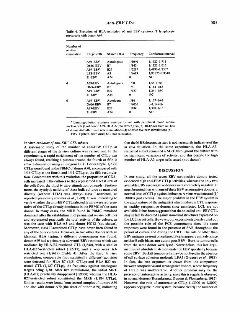
Frequency and nature ofanti-EBV CTLp in PBMCFigure 1 illustrates the results of a typical LDA of anti-EBVCTLp, showing a frequency of 1/1000 cells. In all the seven
donors tested, it appeared that the cytolytic activity was
restricted by HLA molecules, since heterologous HLA-incom-patible target cells were not lysed (Fig. la). Furthermore, the Tcell nature of the effectors was confirmed by three additionalobservations: (i) the cytolytic activity was increased (1/220)when E+ cells purified by rosetting were used as effectors (Fig.lb); (ii) treatment of PBMC with anti-CD3 plus complementsuppressed the response (Fig. Ic); and (iii) anti-CD8 pluscomplement treatment ofPBMC greatly reduced the number ofCTLp, indicating that the majority of these CTLp were CD8+cells (Fig Id). The total number of anti-EBV CTLp found in thedonors was approximately the same and always very high, from1/400 to 1/3000 with unseparated PBMC.
Frequencies of CTLp in PBMC restricted by different HLAmoleculesTo separate CTLp subsets restricted by different HLA mol-ecules, experiments were performed with effector and target cellscompatible for only one class I or one class II molecule. It isnoteworthy that in all experiments, the activity seemed to be duemostly to a single subset of CTL restricted by a single HLAspecificity. This major restricting element (MRE) was respon-sible for 90-100% of the response. As shown in Table I fordonor HC12 (HLA-A2/3, B7/60, Cw2/7, DR4/8), HLA-A3-restricted anti-EBV CTLp, identified by the use of target cellssharing only HLA-A3 with the CTL, represented 1/550 cells.This was almost identical to the frequency observed with
autologous target cells.However, the MRE varied from donor to donor, as shown in
Table I with donor A69 (HLA-A3/24, B7/27, Cw2/7, DR4/5),where HLA-B7 acted as a MRE (f= 1/840), despite the presenceofHLA-A3. In that case, HLA-A3 functioned at a very low level
(f< 1/8000), as a minor restricting element (mRE), and HLA-A24 did not function at all. Other experiments (not shown)verified that A69 share the same HLA-A3 specificity as HC12and the HLA-A3 target cells. With some donors, mRE appearedat intermediate levels, such as HLA-B27 for donor A69, since
502

Anti-EBV LDA
(a)
37!
lo0
Cells/culture125 250 500 1000
* 249-EBV m
M35-EBV e
HC12-EBV -
Targets HLA shared f Confidence interval
HC1 2-EBV Autologous 1/1000 1/1586-1/730M35-EBV DR4/8 1/9050 1/125 273-1/4700249-EBV Heterologous <1/9000 NC
(c) Cells/culture125 250 500 1000
100 |7 -I * HC12-EBVA-26-EBV u249-EBV -
37
Targets HLA shared f Confidence interval
HC12-EBV Autologous 0 NC26-EBV A3 0 NC249-EBV Heterologous 0 NC
Ina)
._
a)
a)
a)z
(b)125 250 500rnn
371
101
Cells/culture1000
\* * ~ 249-EBVv
A HC12-EBV -
Targets IHLA shared L f Confidence interval
HC12-EBV Autologous 11/220 1/370-1/156
249-EBV Heterologous <1/8000 NC
(d) Cells/culture-0 125 250 500 1000
HC12-EBVM35-EBV-249-EBV.
a)37-
Targets HLA shared f Confidence interval
() HC12-EBV Autologous 1/17 622 0-1/7718M35-EBV DR4/8 1/16 640 0-1/7550
249-EBV Heterologous 1/4950 1/28 907-1/2700
Fig. 1. Frequency analysis ofanti-EBV cytotoxic T lymphocyte precursors from donor HC 12. Responder cells were: (a) unfractionatedperipheral blood mononuclear cells (PBMC); (b) E+ cells purified by resetting; (c) PBMC depleted ofCD3+ cells by treatment with ananti-CD3 MoAb plus complement at the beginning of the culture; (d) PBMC depleted of CD8 + cells by treatment with an anti-CD8MoAb plus complement at the beginning ofthe culture. Target cells were autologous, HLA-A3 compatible, or HLA-DR4/8 compatibleor heterologous EBV lymphoblastoid cell lines.
Table 1. HLA-restriction of anti-EBV CTL induced in limiting dilution cultures
Effector cells* Target cells Shared HLA Frequency Confidence interval
HC12 anti-EBV HC12-EBVA65-EBVDI-EBV9-EBVD81-EBVM13-EBVM35-EBV249-EBV
A69 anti-EBV A69-EBVD666-EBVA19-EBVLES-EBV21-EBV
AutologousA3A2B60B7Cw7DR4/8Heterologous
AutologousB7B27A3A24
1/5401/5501/33251/39301/44361/69301/115801/5360
1/10401/8401/22171/86190
1/790-1/4071/800-1/4131/8910-1/20451/12 768-1/23251/14330-1/26241/67 830-1/36500-1/54301/21 730-1/3060
1/1922-1/7111/1320-1/6151/4190-1/15071/93 275-1/4518NC
* Effector cells were derived from peripheral blood mononuclear cells ofdonor HC 12(HLA-A2/3, B7/60, Cw5/7, DR4/8) or A69 (HLA-A3/24, B7/27, Cw2/7, DR4/5) afterstimulation with autologous EBV LCL as described in Materials and Methods.
EBV, Epstein-Bart virus; CTL, cytotoxic T lymphocytes; NC, not calculable.
target cells sharing only HLA-B27 were lysed with a frequencyofCTLp of 1/2200. The frequency of anti-EBV CTLp restrictedby HLA-A or HLA-B mRE was often too slight to be calcu-lated and HLA-Cw-restricted CTLp were never detected inthese seven donors.
In the experiment illustrated in Table 1 with donor HC12,the number of class I-restricted CTLp was 1/550 for the MREHLA-A3, and there appeared to be no class II-restricted CTLp.Similar results were found in repeated experiments usingdifferent donors, suggesting that class II-restricted CTLp are
503
.-O
C.)a)
a1)
4-co
a)z
9-a)
U)
a)
a)z
I DUllinnIuu .
i i~~~~~~~~~

I. Bourgault et al.
Table 2. Role of fetal calf serum (FCS) in frequency analysis anti-EBV cytotoxic T lymphocyteprecursors in donor HC12
Stimulator cells* Target cells Shared HLA Frequency Confidence interval
HC12-EBV-SAB HC12-EBV-SAB Autologous 1/1185 1/1915- 1/858HC12-EBV-FCS Autologous 1/1170 1/1952- 1/83626-EBV-FCS A3 1/653 1/1060- 1/472249-EBV-FCS Heterologous 0 NC
HC12-EBV-FCS HC12-EBV-SAB Autologous 1/830 1/1400-1/590HC12-EBV-FCS Autologous 1/830 1/1400- 1/59026-EBV-FCS A3 1/760 1/1160- 1/560249-EBV-FCS Heterologous 1/11 750 0- 1/3970
* Effector cells were derived from peripheral blood mononuclear cells ofdonor HC 12 (HLA-A2/3, B7/60), Cw5/7, DR4/8) after stimulation with autologous EBV lymphoblastoid cell linescultivated in human AB serum (SAB) or in FCS. Chromium release test was performed withtarget cells cultivated in SAB or in FCS.EBV, Epstein-Barr virus; NC, not calculable.
Table 3. Specificity of anti-EBV cytotoxic T lymphocyte precursors in donor HC12
Effector cells* Target cellst Shared HLA Frequency Confidence interval
HC12-anti-EBV HC12-EBV Autologous 1/2933 1/7447-1/1826A65-EBV A3 1/3624 1/9338-1/2248LCL-49 A3 1/1998 1/3626-1/1379BL-49 A3 0 NC02-AU-EBV A2, B7 0 NC249-EBV Heterologous 0 NC
* Effector cells were derived from peripheral blood mononuclear cells of donor HC 12(HLA-A2/3, B7/60, Cw5/7, DR4/8).
t Target cells were EBV lymphoblastoid cell lines or Burkitt tumour cell lines whichshare only HLA-A3 with donor HC12.
EBV, Epstein-Barr virus; NC, not calculable.
not present in PBMC, or that they are too scarce to bedetectable. Furthermore, when CD8+ cells were eliminated atthe beginning of LDA in an attempt to overcome a possibledown-regulation of CD4+ CTL, detection of class II-restrictedCTLp was still impossible in any of the experiments, as shown inFig. ld.
Specificity ofanti-EBV CTLpOne of the major problems in the EBV system is that EBV non-infected LCL are not available as a control. It has beensuggested that non-viral antigens, especially components of theFCS used in the culture medium could be involved in the CTLresponse (Misko, Kane & Hope, 1982; Torsteinsdottin et al.,1986a). Experiments were therefore performed in which thestimulator and target cells were established and maintained inhuman SAB before beginning the LDA also in the absence ofFCS. As shown in Table 2, the number of anti-EBV CTLpcalculated in these conditions was 1/830 with stimulator cellsmaintained in FCS, and about 1/1180 with stimulator cellsmaintained in SAB. No statistically significant difference wastherefore observed, whatever the serum used, for the mainte-nance of the target cells. It must be emphasized that in both
cases the cytolytic activity was restricted by the same HLA classI molecule (HLA-A3), supporting the idea that the same antigenwas involved.
Another possibility could be that part of the CTL popula-tion is directed against B cell-specific markers (Torsteinsdottinet al., 1986b). In order to study further the specificity of thesupposed anti-EBV-CTL, experiments were therefore carriedout using autologous pokeweed mitogen-induced blasts ascontrol target cells: LDA never revealed significant levels ofCTLp with these targets (not shown). Furthermore, whenEBV+ LCL and autologous EBV- Burkitt tumour cell linescompatible with donor HC12 only through HLA-A3 weretested as targets, a strong activity (1/2000) was found againstLCL (LCL-49) but not against the Burkitt cells of the samepatients (BL-49) (Table 3). The same results were found withdonor Al9 and HLA-A 11 compatible target cells (not shown).These experiments therefore suggest that the target antigens ofCTLp are not B-specific antigens or any other autologousantigen expressed in cultured B lymphocytes.
Furthermore, the comparison between donors seropositiveand seronegative for EBV revealed very clear-cut differencessince the two EBV seronegative donors tested were totallydevoid of any CTLp anti-EBV reactivity (not shown).
504
Anti-EBV LDA
Table 4. Evolution of HLA-restriction of anti EBV cytotoxic T lymphocyteprecursors with donor A69
Number ofin vitrostimulation Target cells Shared HLA Frequency Confidence interval
1 A69-EBV Autologous 1/1040 1/1922-1/711D666-EBV B7 1/840 1/1320- 1/615A19-EBV B27 1/2217 1/4190-1/1507LES-EBV A3 1/8619 1/93 275-1/451821-EBV A24 0 NC
4 A69-EBV Autologous 1/30 1/58-1/20D666-EBV B7 1/81 1/114-1/63A19-EBV B27 1/127 1/241-1/9021-EBV A24 0 NC
6 A69-EBV Autologus 1/86 1/137-1/62D666-EBV B7 1/9030 0-1/14466A19-EBV B27 1/184 1/308-1/13121-EBV A24 0 NC
* Limiting-dilution analyses were performed with peripheral blood mono-nuclear cells (1) ofdonor A69 (HLA-A3/24, B7/27, Cw2/7, DR4/5) or from cell lineof donor A69 after three new stimulations (4) or after five new stimulations (6).
EBV, Epstein-Barr virus; NC, not calculable.
In vitro evolution ofanti-EBV CTL subsetsA systematic study of the number of anti-EBV CTLp atdifferent stages of the in vitro culture was carried out. In theexperiments, a rapid enrichment of the number of CTLp wasalways found, reaching a plateau around the fourth or fifth invitro restimulation using autologous LCL. For example, 1/2330CTLp were found in the PBMC ofdonor A70, as compared with1/16 CTLp at the fourth and 1/11 CTLp at the fifth restimula-tion. Concomitant with this evolution, the proportion ofCD8 +
cells increased in the cultures so they represented at least 90% ofthe cells from the third in vitro stimulation onwards. Further-more, the cytolytic activity of these bulk cultures as measureddirectly (without LDA) was always class I-restricted, asreported previously (Gomez et al., 1989). It was interesting toverify whether the anti-EBV CTL selected in vitro were represen-tative of the CTLp already dominant in the PBMC of the samedonor. In many cases, the MRE found in PBMC remaineddominant after the establishment ofpermanent in vitro cell linesand represented practically the total activity of the culture, aswas the case with HLA-A3 and donor HC12 (not shown).Moreover, class II-restricted CTLp have never been found inany of the bulk cultures. However, in two other donors with anidentical HLA typing, a different phenomenon was found:donor A69 had a primary in vitro anti-EBV response which wasmediated by HLA-B7-restricted CTL (1/840), with a smallerHLA-B27-restricted subset (1/2217), and a very weak A3-restricted one (1/8619) (Table 4). After the third in vitrostimulation, comparable (not statistically different) activitieswere detected for HLA-B7 (1/81 CTLp) and HLA-B27-res-tricted CTL (1/127 CTLp), the frequency against autologoustargets being 1/30. After five stimulations, the initial MRE(HLA-B7) practically disappeared (1/9030) whereas the HLA-B27-restricted subset constituted the MRE (1/184 CTLp).Similar results were found from several samples of donors A69and also with donor A70 (the sister of donor A69), indicating
that the MRE detected in vitro is not necessarily indicative ofthein vivo situation. In the same experiments, the HLA-A3-restricted subset remained a MRE throughout the culture withno significant variations of activity, and this despite the highnumber of HLA-A3 target cells tested (not shown).
DISCUSSION
In our study, all the seven EBV seropositive donors testedevidenced high anti-EBV CTLp activities, whereas the only twoavailable EBV seronegative donors were completely negative. Itmust be noted that with one ofthese EBV seronegative donors, anormal level ofCTLp against influenza A virus was detected (1/18 000) (not shown). The major problem in the EBV system isthe exact nature of the antigen(s) which induce a CTL responsein healthy seropositive donors since uninfected LCL are notavailable. It has been suggested that the so-called anti-EBV CTLmay in fact be directed against non-viral structures expressed onthe LCL target cells. However, our experiments clearly ruled outthe possible role of the FCS components since the sameresponses were found in the presence of SAB throughout theperiod of culture and during the CRT. The role of other thanEBV antigens present on cultured B cells appears unlikely, sinceneither B cells blasts, nor autologous EBV- Burkitt tumour cellsfrom the same donor were lysed. Nevertheless, this last argu-ment is not absolute to demonstrate the EBV specificity becausesome EBV- Burkitt tumour cells may be not lysed in the absenceof cell surface adhesion molecule LFA3 (Gregory et al., 1988).In fact, the best argument is drawn from the comparisonbetween seropositive and seronegative donors, whose frequencyof CTLp was undetectable. Another problem may be thepresence of autoreactive activity, since this is regularly observedin normal donors (Rosenkrantz, Dupont & Flomenberg, 1985).However, the role of autoreactive CTLp (1/3000 to 1/8000)appears negligible in our system, because clearly the number of
505
506 L Bourgault et al.
autoreactive CTLp is lower than the number of anti-EBVCTLp. Altogether, we can conclude that the measures of CTLpwere EBV specific but we do not know what specific antigen isinvolved. In other experiments from our laboratory a peptidederived from the EBV-encoded latent membrane protein (LMP)of the virus, reported to be able to restimulate anti-EBV CTL(Thorley-Lawson & Israelsohn, 1987), did not function as atarget antigen for the CTL (not shown). However, murine CTLhave been found able to recognize LMP after transfection inmurine cells (Reiss et al., 1987) and this protein could beinvolved in the human system (A.B. Rickinson, personalcommunication). Furthermore, a peptide of LMP was recentlyidentified as target ofhuman CTL which show cross-recognitionof the same peptide in the context of a mouse H-2Kd molecule(Murray et al., 1990a). The role of a product of the EBNA-2gene (Moss et al., 1988) as well as that of a new gene created bycircularization of the linear viral genome (Laux, Perricaudet &Farrell, 1988) must be also considered. Recently, a peptide fromEBNA-3 protein (Murray et al., 1990b, Burrows et al., 1990a)and one from EBNA-6 protein (Burrows et al., 1990b) wereidentified as target epitopes of anti-EBV T cell clones.
Previously data have shown that CTLp represent 1/540-1/8300 E+ cells in response to mumps virus infection (Enssle et al.,1987); 0-1/30 000 in the VZV system (Hickling et al., 1987); 1/5000-1/20 000 in HCMV system (Borysiewicz et al., 1988); and1/3500-1/20000 in the influenza A virus system (Bourgault etal., 1989; and our unpublished data). The same situation existswith HSV-1 for which lower numbers of CTLp, (1/8000) havebeen found (Schmid, 1988). However, these experiments withHSV were performed using inactivated virus as the source ofantigen, which could account for the lower responses and for thefact that CTLp detected were only CD4+ class II-restrictedCTLp. The differential stimulation of CD4+ or CD8+ subsets,depending on whether the virus was inactivated or not, has beenpreviously demonstrated with VZV (Hickling et al., 1987).Similar numbers of CTLp were found in BALB/c mice for anti-influenza virus responses (about 1/13000 CTLp) (Wysocka &Bennink, 1988). Higher levels ofCTLp were however describedin mice for acute infection with herpes simplex (1/170-1/500 inCBA mice) (Hengel et al., 1987) or influenza A virus (1/1200 inBALB/c mice) (Owen et al., 1988), but in these two reports theLDA were performed with spleen or lymph node cells harvesteda very short time (5 days) after hyper-immunization.
The strong anti-EBV CTL response from PBMC that weobserved in LDA (1/550-1/2000) is therefore much greater thanthat reported in any other viral system in humans. This resultwas also obtained by Rickinson et al. (1981) using a differentmethod (regression assay), and agrees with the observation thatvirtually 100% of donors gave a clear EBV-specific response,whereas no more than 25-30% ofdonors responded to influenzavirus. We cannot exclude the role of different stimulating cells,since LCL produce by themselves many growth factors. Never-theless, Chen et al. (1989), working with anti-EBV cell lines,have recently detected a frequency of CTL identical as thatfound in our experiments (1/25-1/120 ). The most likelyhypothesis to explain the high frequency of anti-EBV CTLp inseropositive donors would be related to the chronic stimulationby viral antigens since, in contrast with mumps or influenza Avirus, EBV persists in most people, with frequent endogenousreactivations. Similar in vivo stimulation could exist for anti-HIVl CTLp. However, in this system LDA activity was not
determine in PBMC but only at the level ofcultured effector cellsand the frequency was in the range from 1/150 to 1/ 1500(Hoffenbach et al. 1988).
It must be emphasized that our effector cells are CD8+. Wedid not find any class II-restricted anti-EBV CTL in these LDAexperiments. This is in agreement with other experiments, wherewe have shown that class II-restricted antiviral CTL appear invitro only after several antigenic stimulations and can bedetected only after the elimination of CD8 + cells (Gomez et al.,1989). Furthermore, in most ifnot all cases, a single HLA class Imolecule acts as a MRE in the LDA. Among MRE, we foundonly HLA-A and HLA-B molecules. Nevertheless, Chen et al.(1989) have recently suggested that CTL could be restricted byHLA-C molecules in LDA of bulk culture from a single donor.This confirms the results obtained from other methods in bulkcultures and suggests that only one, or at most a small number ofantigens, could be involved in our experiments since it is clearthat various peptide antigens are most often presented to T cellsby different HLA molecules. Similar results using LDA fromPBMC have been reported in the mumps virus system (Enssle etal., 1987). However, subsets restricted by the mRE still persistedafter several restimulations, demonstrating that CTL cell linesremained polyclonal in vitro. Interestingly, in two of our casesthe dominant subset in an established cell line was different fromthat found in the primary in vitro response, suggesting that the invivo dominant subset was not selected in culture. It wouldtherefore be unwise to conclude from the observed dominantsubset in permanent cell lines that the same subset is alsodominant in vivo. Nevertheless, it must be emphasized thatCTLp restricted by mRE can be evidenced among PBMC byLDA and not bulk culture. They are present in vivo and can beprobably selected in vitro after several stimulations by theappropriate peptide, as demonstrated recently in the influenzasystem (Martinon et al., 1990).
In conclusion, LDA is a valuable method to assess the wholein vivo immune repertory ofeach individual, and the evolution invitro of CTL subsets restricted by different HLA molecules, aswell as the respective strengths of the responses directed againstvarious viral peptide antigens. It may also be of value as a test ofefficiency of vaccination procedures.
ACKNOWLEDGMENTS
We would like to thank Dr Jean-Yves Muller and Dr Virginia Lepagefor the HLA typing of donors, Dr Gilbert Lenoir for the gift of theBurkitt cell lines, Dr Franck Healy for his help and comments during theediting of the manuscript, Miss Bernadette Begue for expert technicalassistance, and Miss Sandrine Vitteaud for typing the manuscript.
REFERENCES
BORYSIEWICZ, L.K., GRAHAM, S., HICKLING, J.K., MASON, P.D. &SISSONS, J.G.P. (1988) Human cytomegalovirus-specific cytotoxic Tcells: their precursor frequency and stage specificity. Eur. J. Immunol.18, 269.
BOURGAULT, I., GOMEZ, A., GOMARD, E., PICARD, F. & LEVY, J.P. (1989)A virus-specific CD4+ cell-mediated cytolytic activity revealed byCD8 + cell elimination regularly develops in uncloned human anti-viral cell lines. J. Immunol. 142, 252.
BURROWS, S.R., SCULLEY, T.B., MISKO, I.S., SCHMIDT, C. & Moss, D.J.(1990a) An Epstein-Barr virus-specific cytotoxic T cell epitope inEBV nuclear antigen 3 (EBNA 3). J. exp. Med. 171, 345.

Anti-EBV LDA 507
BURROWS, S.R., MISKO, I.S., SCULLEY, T.B., SCHMIDT, C. & MOSS, D.J.(1990b) An Epstein-Barr virus-specific cytotoxic T-cell present on A-and B-type transformants. J. Virol. 64, 3974.
CHEN, B.P., LAM, V., KRAUS, E.E., DEMARS, R & SONDEL, P.M. (1989)Restriction of Epstein-Barr virus-specific cytotoxic T cells by HLA-A, -B and -C molecules. Hum. Immunol. 26, 137.
ENSSLE, K.H., WAGNER, H. & FLEISCHER, B. (1987) Human mumpsvirus-specific cytotoxic T lymphocytes: quantitative analysis ofHLArestriction. Hum. Immunol. 18, 135.
GoMEZ, A., BOURGAULT, I., GoMARD, E., PICARD, F. & LEVY, J.P. (1989)Role of different lymphocyte subsets in human antiviral T cellcultures. Cell Immunol. 118, 312.
GREGORY, C.D., MURRAY, R.J., EDWARDS, C.F. & RICKINSON, A.B.(1988), Downregulation of cell adhesion molecules LFA-3 andICAM-1 in Epstein-Barr virus-positive Burkitt's lymphoma under-lies tumor cell escape from virus-specific T cell surveillance. J. exp.Med. 167, 1811.
HEALY, F., SIRE, J., GOMARD, E., YSSEL, H., JORDAN, B.R. & LEVY, J.P.(1988) A study of functionally active amino acids involved in theinteraction of HLA-A2 or HLA-A3 molecules with cytolytic Tlymphocytes. J. Immunol. 141, 2487.
HENGEL, H., LINDNER, M., WAGNER, H. & HEEG, K. (1987) Frequencyof herpes simplex virus-specific murine cytotoxic T lymphocyteprecursors in mitogen- and antigen-driven primary in vitro T cellresponses. J. Immunol. 139, 4196.
HICKLING, J.K., BORYSIEWICZ, L.K. & SISSONS, J.G.P. (1987) Varicella-Zoster virus-specific cytotoxic T lymphocytes (Tc): detection andfrequency analysis of HLA class I-restricted Tc in human peripheralblood. J. Virol. 61, 3463.
HOFFENBACH, A., LANGLADE-DEMOYEN, P., DADAGLIO, G., VILMER, E.,MICHEL, F., MAYAUD, C. AUTRAN, B. & PLATA, F. (1989) Unusuallyhigh frequencies of HIV-specific cytotoxic T lymphocytes in humans.J. Immunol. 142, 452.
LANGHORNE, J. & FISHER-LINDAHL, K. (198 1) Limiting dilution analysisof precursors of cytotoxic T lymphocytes. J. immunol. Methods. ii,221.
LAUX, G., PERRICAUDET, M. & FARRELL, P.J. (1988) A spliced Epstein-Barr virus gene expressed in immortalized lymphocytes is created bycircularization of the linear viral genome. EMBO J. 7, 769.
MARTINON, F., GOMARD, E., HANNOUN, C. & LEVY, J.P. (1990) In vitrohuman T cell responses against influenza A virus can be induced andselected by synthetic peptides. Eur. J. Immunol. 20, 2171.
MISKO, I.S., KANE, R.G. & HOPE, J.H. (1982) Generation in vitro ofHLA-restricted EB virus-specific cytotoxic human T cells by autolo-gous lymphoblastoid cell lines: the roles ofprevious EB virus infectionand foetal calf serum. Int. J. Cancer 29, 41.
Moss, D.J., MISKO, I.S., BURROWS, S.R., BURMAN, K., MCCARTHY, R.& SCULLEY,, T. B. (1988) Cytotoxic T-cell clones discriminate betweenA- and B-type Epstein-Barr virus transformants. Nature, 331, 719.
MURRAY, R.J., BROOKS, J.M., RICKINSON, A.B. & ROWE, M. (1990a).
Cross-recognition of a mouse H-2-peptide complex by human HLA-restricted cytotoxic T cells. Eur. J. Immunol. 20, 659.
MURRAY, R.J., KURILLA, M.G., GRIFFIN, H.M., BROOKS, J.M., MACK-Err, M., ARRAND, J.R., RowE, M., BURROWS, S.R., Moss, D.J.,KIEFF, E. & RJCKINSON, A.B. (1990b) Human cytotoxic T-cellresponses against Epstein-Barr virus nuclear antigens demonstratedby using recombinant vaccinia viruses. Proc. nat!. Acad. Sci USA, 87,2906.
OWEN, J.A., DUDZIK, K.I., KLEIN, L. & DORER, D.R. (1988) Thekinetics of generation of influenza-specific cytotoxic T-lymphocyteprecursor cells. Cell Immunol. 111, 247.
REISS, C.S., WANG, D., GHOSH, D., GAPOSCHKIN, C. & KIEFF, E. (1987)Recognition ofEBV plasma membrane protein expressed on murinecells after gene transfer. J. Immunol. 139, 711.
RICKINSON, A.B., Moss, D.J., WALLACE, L.E. RowE, M., MISKO, I.S.,EPSTEIN, M.A. & POPE, J.H. (1981) Long-term T-cell-mediatedimmunity to Epstein-Barr virus. Cancer Res. 41, 4216.
ROSENKRANTZ, K., DUPONT, B. & FLOMENBERG, N. (1985) Generationand regulation of autocytotoxicity in mixed lymphocyte cultures:evidence for active suppression of autocytotoxic cells. Proc. nat!.Acad. Sci. USA, 82, 4508.
SCHMID, D.S. (1988) The human MHC-restricted cellular response toherpes simplex virus type 1 is mediated by CD4 +, CD8 - T cells and isrestricted to the DR region of the MHC complex. J. Immunol. 140,3610.
TASWELL, C. (1981) Limiting dilution assays for the determination ofimmunocompetent cell frequencies. I. Data analysis. J. Immunol. 126,1614.
THORLEY-LAWSON, D.A. & ISRAELSOHN, E.S. (1987) Generation ofspecific cytotoxic T cells with a fragment of the Epstein-Barr virus-encoded p63/latent membrane protein. Proc. nadl Acad. Sci. USA, 84,5384.
TORSTEINSDOTTIR, S., MASUCCI, M.G., BRAUTBAR, C., LENOIR, G.,KLEIN, G. & KLEIN, E. (1986a) Differential recognition of tumor-derived and in vitro Epstein-Barr virus-transformed B cells lines byfetal calf serum-specific T4-positive cytotoxic T-lymphocyte clones.Cell Immunol. 98, 453.
TORSTEINSDOrrIR, S., MASUCCI, M.G., EHLIN-HENRIKSON, B., BRAUT-BAR, G., BEN BASSAT, H., KLEIN, G. & KLEIN, E. (1986b) Differencia-tion-dependent sensitivity of human B-cell-derived lines to majorhistocompatibility complex restricted T-cell cytotoxicity. Proc. NatlAcad. Sci. USA, 83, 5620.
TOUBERT, A., GOMARD, E., GRUMET, F.C., AMOR, B., MULLER, J.Y. &LEVY, J.P. (1984) Identification of several functional subgroups ofHLA-B27 by restriction of the activity of antiviral T killer lympho-cytes. Immunogenetics, 20, 513.
WYSOCKA, M. & BENNINK, J.R. (1988) Limiting dilution analysis ofmemory cytotoxic T lymphocytes specific for individual influenzavirus gene products. Cell Immunol. 112, 425.


