Embed Size (px)
Citation preview

Long-term changes of prostacyclin secretion in radiation--induced myelopathy
Seied Rabie Mahdavi1, Alireza Shirazi2, Bagher Minaee3, Alireza Nikoofar4,
Hamid Reza Mirzaee5
1 Department of Biophysics and Biochemistry, Babol University of Medical Sciences, Iran2 Department of Medical Physics, Tehran University of Medical Sciences, Tehran, Iran3 Department of Anatomy and Histology, Tehran University of Medical Sciences, Iran4 Department of Radiation Oncology, Iran University of Medical Sciences Iran5 Department of Radiation Oncology, Sh-Beheshti University of Medical Sciences, Iran
This research was granted by the offi ce of Vice-Chancellor for research of Tehran University of Medical Sciences
Summary
Background We have previously reported the short-term changes in prostacyclin profi le after irradiation of rat cervical cord.
Aim Present research investigated the long-term changes of prostacyclin content.
Materials/Methods Wistar rats in groups of fi ve were irradiated with doses of 2, 4, 6, 15, 25, 30Gy and a single group of 25 with 35Gy X-rays. After 26 and 39 weeks, prostacyclin con-tent was quantifi ed by 6-keto-prostaglandin-F1a (prostacyclin stabilized metabo-lite). Specimens were stained routinely for histological studies.
Results The 50% latent period and effective dose were 14.86±1.16 weeks and 25.66±0.54Gy (p<0.0001), respectively. Average ratios of 6-keto-PG-F1a for doses of 2–30Gy were between 78.33–12.93% and 79.48–99.96% for 26 and 39 weeks, respective-ly. Prostacyclin level after 35Gy shows approximately a 7:1 ratio in comparison to the control group (p<0.002). Histopathological changes in glial and vascular tis-sues were diagnosed and scored. Prostacyclin bimodal profi le was observed.
Conclusions Radiation can cause complex fl uctuations of prostacyclin in association with marked histopathological changes.
Key words spinal cord • radiation myelopathy • prostacyclin • histopathology • rat
Full-text PDF: http:/www.rpor.pl/pdf.php?MAN=9890
Word count: 2297 Tables: 1 Figures: 5 References: 22
Author’s address: Seied Rabie M. Mahdavi, P.O. Box 14665-584 Tehran-Iran, e-mail: [email protected]
Received: 2006.10.01Accepted: 2006.11.20Published: 2006.12.22
Authors’ Contribution:
A Study Design B Data Collection C Statistical Analysis D Data Interpretation E Manuscript Preparation F Literature Search G Funds Collection
Original Paper
273
Rep Pract Oncol Radiother, 2006; 11(6): 273-279

BACKGROUND
Radiation myelopathy is known as one of the most important complications of radiation in radiother-apy of patients. Destructive changes of the white matter and other nervous tissues are the major his-topathological aspects. Dose response curves for treatment of malignancies distinctly show a strict margin between virtual 100% tumoricidal dose and neurotoxicity occurrence in spinal cord as a normal tissue [1]. It was shown that a single dose of 35Gy X-ray to the spinal cord can cause limb paralysis in rat models with a latency of 19.0±0.3 weeks [2].
Different types and methods of irradiation have been listed for different strains of animals, indi-cating dose dependency, latency, relative biologi-cal effectiveness (RBE), histopathology and patho-genesis of radiation myelopathy [3]. Recently we reported the short-term changes of prostacyclin secretory profi le after various irradiation doses [4]. Histological observations showed that radi-ation with doses of a few Gy or more in excess of the tolerance level induces more or less uniform histological changes [1,5,6].
AIM
Based on vascular theory it is postulated that en-dothelial morphologic changes are associated with changes in prostaglandins, including pros-tacyclin, which plays a major role in haemody-namic alterations. Our short-term study showed fl uctuations in the concentration of prostacyc-lin early after irradiation [4]. The authors stud-ied late histopathology and enzymatic changes after doses of 2–35Gy.
MATERIALS AND METHODS
Animals and irradiation
Male Wistar rats weighing 150–200g were fi xed on a jig under gentle anaesthesia. The C1–T2 segment of the spine was irradiated through two parallel opposed ports with an orthovoltage ma-chine (200kVp, 1.5mmCu HVL, focal skin dis-tance 25cm) at a dose rate of 1.53Gy/min at the depth of the spinal cord. Doses of 2, 4, 6, 15, 25 and 30Gy were administered using a circular 3.0cm diameter localizator [7]. One group of an-imals (25 rats) was irradiated with a dose of 35Gy for evaluation of survival rate and to produce the clinical manifestation of radiation myelopathy. Five rats were included in each dose group in addition to the age-matched control group. The
animals were sacrifi ced chronologically at fi ve time points of 24 hours, 2, 13, 26 and 39 weeks post-irradiation. Rats irradiated with 35Gy were followed for symptoms of radiation myelopathy to get radiobiological data on latent symptoms and lethal dose.
Sample preparations
Following decapitation under ether anaesthe-sia, rats’ neck regions were opened posteriorly and the spinal cord was removed after laminec-tomy. Then the spinal cord was cut rapidly and segmented into three pieces for enzyme immu-noassay (EIA) and histopathological studies with transmission electron and light microscopes.
Measurement of spinal cord PGI2
Spinal cord samples were analyzed for prostag-landin I2 (PGI2 or prostacyclin) concentration by enzyme immunoassay kit (Cayman Chemical Co.). For this purpose, samples were homoge-nized with weight proportion of 0.1M perchlo-ric acid and centrifuged at 4°C for 10 minutes at 3000rpm. Then 100–150μl supernatant was re-moved for dilution (1:5 and 1:10) and poured into a 96 well EIA kit. This kit measures 6-keto prostaglandin F1a (6-keto PGF1a), the main metabolic product of PGI2. The concentration of 6-keto PGF1a is directly proportional to PGI2. Data of 6-keto PGF1a concentrations were nor-malized in the form of the percent of the com-parative control groups. Changes in PGI2 concen-tration were plotted against time and irradiation dose using Sigma-Plot version 8.
Histopathologic study
The samples were studied histopathologically for cellular and subcellular changes using electron (TEM) and light microscopes. Preparation of sam-ples for the light microscope followed the routine procedure of fi xation, embedding, cutting (4μm thick) and staining. TEM preparation consisted of fi xation, embedding, semithin formation, griding/grinding and toluiden blue staining. Examination was done under appropriate magnifi cation to ob-serve morphological changes in endothelial cells.
Actuarial analysis was carried out on survival data. Quantifi cation of histologic changes was scored based on one to three pluses in compari-son to the age-matched control group. We used Student’s t-test for comparison of histopathologic and PGI2 concentrations between irradiated and
Original Paper Rep Pract Oncol Radiother, 2006; 11(6): 273-279
274

control groups. Spearman’s test was also used to evaluate the correlativity between PGI2 and his-topathological changes.
RESULTS
Survival data on the irradiated rats are shown in Figure 1. For the 35Gy group, the 50% latent period (LP50) and mean latency period were 14.9±1.2 and 14.2±0.9 weeks (95% confi dence limit 12.3–16.1 weeks), respectively. The ob-tained LP100 was 20 weeks post-irradiation. Dose response curve based on these fi ndings is plot-ted in Figure 2. Effective dose of 50% (ED50), which is the radiation dose that produces radia-tion myelopathy in 50% of rats, was found to be 25.7±2Gy within the 95% confi dence interval of 24.6 and 26.7Gy.
Prostacyclin changes
Prostacyclin level in animals irradiated with 35Gy at the time of induction of paralysis and just prior to death showed a steep increase in comparison to corresponding control rats (approx. 7:1 with p<0.002, not shown). After 26 weeks of irradia-tion, overall prostacyclin concentrations changed little with radiation dose. In contrast to a signifi -cant decrease in PGI2 levels at 4 and 6Gy, a signifi -cant increase was observed at 25 and 30Gy. Details of PGI2 changes after different time points are shown in Figure 3A. Initially a positive part of this
1.2
1.0
0.8
0.6
0.4
0.2
0.0
Cum
ulat
ive
surv
ival
15Gy, n=25
25Gy, n=25
LP50=14.86±1.16weeks
35Gy, n=25
0 10 20 30 40 50
Time post-irradiation (weeks)
Figure 1. Kaplan-Meier analysis of Survival Function.
100
80
60
40
20
0
Dea
th %
10 15 20 25 30 35
X-irradiation (Gy)
40
ED50=25.66±2.13Gy
Figure 2. Dose-response curve for incidence of death due to radiation myelopathy (error bar shows mean ±SE and N=25).
Figure 3. Profi le of PGI2 changes in long-term study: (A) Detailed presentation of time-wise profi le of prostacyclin changes (not scaled); this diagram shows concentration ratios as defi ned by PGI2 (irradiated)/PGI2 (age-matched control), and means ±SE are shown as a percentage at confi dence limit of 95%. Dots indicate mean value of PGI2 at each time point. (B) 3D profi le of prostacyclin changes as a function of time and dose.
700
600
500
400
300
200
100
0
6-ke
to c
once
ntra
tion
(% o
f Ctr
l)
15Gy25Gy30GyProfile
Ctrl2Gy4Gy6Gy
24hours
2weeks
13weeks
26weeks
39weeks
Time after irradiation
6kPG
F1a
conc
. % o
f Ctr
l700
600
500
400
300
200
100
0
2520
1510
5
2520
1510
5Irradiation dose (Gy)Post-
irradiatio
n
time (w
eeks)
A
B
Rep Pract Oncol Radiother, 2006; 11(6): 273-279 Mahdavi SR et al – Prostacyclin changes in radiation myelopathy
275
Gaussian curve is seen at dose of 2Gy (p=0.07), decreasing at dose of 4Gy (p=0.01). At dose of 6Gy it shows a concentration not very different from control (p=0.007). In the high dose region there is an increase after 25 and 30Gy (p=0.004 and 0.05, respectively). After 39 weeks of irradi-ation, prostacyclin concentration decreased sig-nifi cantly in the low dose region of 2, 4 and 6Gy from 78.4% to 93.3% of control (p=0.001, 0.002 and 0.04, respectively). In the high dose region the decrease was signifi cant after 15Gy (p=0.004) but it was not signifi cant in the 25 and 30Gy ir-radiated groups (Figure 3A). Overall changes in PGI2 concentration in different time and irradia-tion dose are shown in Figure 3B. A double-peak action of prostacyclin response to irradiation was seen after 13 weeks in both low and high dose re-gions, which may indicate bi-modal parameters contributing to radiation myelopathy.
Histopathological changes
Different kinds of pathologic evidence were ob-served which are well established as signs of radi-ation-induced myelopathy. They included severe demyelination, white and grey matter atrophy, necrosis accompanied by atrophy, gliosis, and vasculopathy consisting of vascular rupture and haemorrhage. The incidence of histopathological lesions was both dose- and time-dependent. Sub-structural changes were observed even at a dose lower than 6Gy. However, we observed rats with clinical manifestation of radiation myelopathy af-ter 25Gy. They showed paralysis of limbs and se-vere urine discontinuity and were sacrifi ced just prior to death after 26–28 weeks post-irradiation. In these animals necrotic and atrophic changes in spinal cord specimens were observed. The most apparent pattern after 26 weeks was an increased density of vessels mimicking revascularization at the irradiated volume (Figure 4A).
Findings after 39 weeks were similar to the fi nd-ings after 26 weeks but more intense. Severe multiple necrotic areas were seen in sections ob-tained from irradiated rat cervical spinal cord af-ter 25Gy. Fibrin deposition and replaced colla-gen fi bres as fi brosis were seen in the interstitial spaces (Figure 4A).
Animals irradiated with a dose of 35Gy were ex-amined histologically just prior to sacrifi ce for symptoms of radiation myelopathy. Marked path-ological changes both in vascular and white mat-ter parenchyma consisting of vascular rupture, wall thickening, severe necrosis, demyelination
and glial proliferation were observed and are shown in Figure 4C.
Histopathological changes revealed with both light microscope and TEM are listed in Table 1. Changes encountered with TEM are at the level of the vascular endothelial cells. Photomicrographs were obtained at different times post-irradiation with different doses (Figure 5). They show chang-es at the subcellular level including nuclear mem-brane, cytoplasmic and basal cell membranes, and also alterations in endothelial cell organelles.
DISCUSSION
Animals exposed to single doses ≤15Gy (i.e. 2, 4, 6 and 15Gy) did not reveal any specifi c clini-cal manifestation of radiation myelopathy. After doses >15Gy the probability and also the severi-ty of radiation myelopathy increases. Our results are similar to the results of other reported exper-imental studies which have used different types of radiation [8–11]. The latency observed in our study is about 4 weeks shorter than the report of Hopewell et al. 1987 [12]. A similar discrepan-cy was also reported by Okeda et al. 1998 [10]. It may have been caused by the greater volume of irradiated spinal cord which may decrease the latency [13,14].
Similar results of histopathology and radiobiology were shown in the study on the effects of a carbon ion beam on the thoracic spinal cord by Shinobu and Okeda 2001 [6]. The ED50 for hind-limb pa-ralysis derived from the dose-response curve was 18.5Gy and the RBE was 1.38. The equivalent dose of the X-ray beam was 25.5Gy and they found that 67% of animals at 20Gy and 100% of animals at 25 and 30Gy showed hind-limb paralysis, which is identical to our results, but at 16–20 weeks af-ter irradiation [10].
Substantial data suggest that the prostanoids are particularly important in inhibiting smooth mus-cle cell proliferation, preserving tissue viabili-ty and organ function in experimental models of ischaemia. Ischaemic tissues are subjected to decreased oxygen tension (hypoxia), decreased blood fl ow, accumulation of metabolites, and pa-renchymal cell damage. The changes of prostacy-clin against various damaging agents were stud-ied in vitro and in vivo. There are two target cell populations involved in radiation myelopathy: I) vascular endothelial cells and II) glial cells (es-pecially oligodendrocytes) and prostacyclin may originate from both cell types [3,15,16].
Original Paper Rep Pract Oncol Radiother, 2006; 11(6): 273-279
276

According to the present fi ndings, low-dose irra-diation that is insuffi cient for the induction of a white matter lesion may still induce a vascular lesion, possibly because of the difference in ra-diosensitivity between vascular endothelium and neuro-glial cells [6]. It was reported that vascular
changes are not necessarily accompanied by mor-phological endothelial alterations. In fact, some of the changes may be degenerative changes re-lated to injury from infl ammation induced by radiation such as rupture of vessel walls, dilata-tion and infi ltration of blood cells. Intra-vascular
Figure 4. Some of the histopathological changes of white matter showing after H&E staining: (A) vascularization and/or increase in vascular density was prominent after 15Gy 26 weeks post-irradiation (×400), (B) congestion and gliosis after dose of 4Gy 13 weeks after irradiation (×100), (C) malacia after 25Gy 39 weeks after irradiation (×400), same manifestation of myelopathy was also seen in the group of 35Gy, and (D) spinal cord with deformed pattern and huge glioma after 35Gy of X-ray dose (×40).
A
B
C
D
White matter parenchyma Vascular changesUltra-microscopic changes
in endothelial cells
– White matter necrosis,– Gliosis,– Myelin sheath irregularities– Demyelination of nerve fi bre– Atrophy
– Vascular rupture and collapse,– Vascular density variation,– Thrombo-emboli formation,– Blood cell infi ltration (haemorrhage)
– Cytoplasmic and basal cell membrane irregularities
– Organelle morphological pattern– Vesiculation– Increase in number of lysosomes– Auto lysosome formation– Degeneration of mitochondria (Ballooned
cristae)– Dilatation of endoplasmic reticulum
Table 1. Microscopic and ultra-microscopic histopathological changes in white matter parenchyma and vascularity in rat cervical spinal cord observed (24 hours–39 weeks) after irradiation.
Rep Pract Oncol Radiother, 2006; 11(6): 273-279 Mahdavi SR et al – Prostacyclin changes in radiation myelopathy
277

haemodynamic load can be deteriorated second-ary to infl ammation [17,18]. However, recently a dose-dependent loss of endothelial cells in rat cervical spinal cord at 24 hours after X-ray inter-action was reported [19].
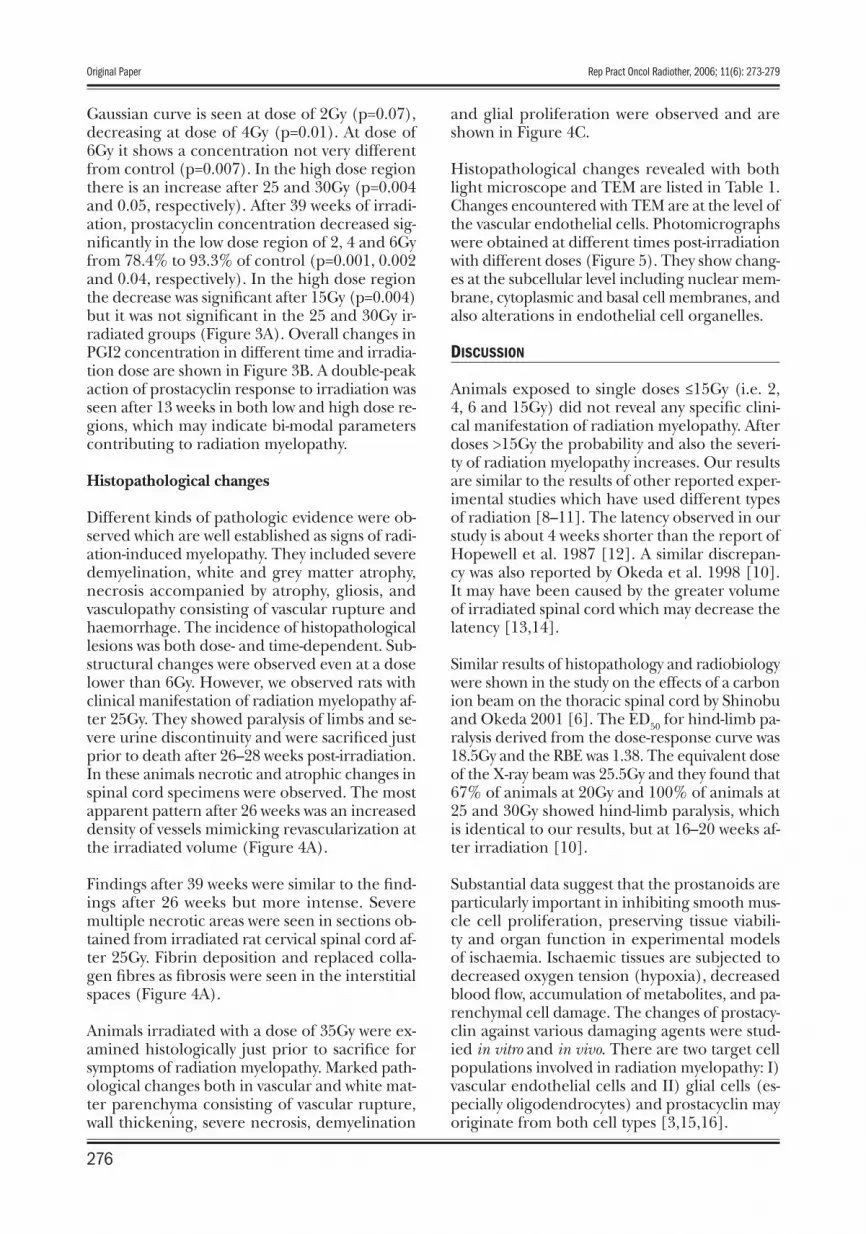
We found related injury from radiation-induced myelopathy such as blood vessels plugged with erythrocytes in and around the destroyed vessels, which may support the idea of changes in blood fl ow and endothelial surfaces. We also observed irregularities in the thickness of endothelial cell membrane (basement and cytoplasmic) with in-creased numbers of intracellular organelles, par-ticularly vesicles early after irradiation, endoplas-mic reticulum and degeneration of mitochondrial architecture (Figure 5).
Our chronological short- and long-term results further suggest that the response of the vascular tissues to radiation occurs prior to that of the nervous tissues of the spinal cord. Tissue dam-age is not the sum of cellular damage but an in-dependent response of a highly structured tissue architecture and the most responsive substruc-ture in the endothelial cell components. In addi-tion, the response of the vascular system to radi-ation can be determined by the response of the endothelial cells, because the endothelial cells are introduced as the most radiosensitive among the cells constituting the vessel wall [6,18].
The pathological fi ndings of radiation myelopa-thy are almost always confi ned to the damage of white matter and its vasculature [3,20]. Recently it has been suggested that radiation may alter the
secretory profi le of mediators, which can cause an infl ammatory response, cellular proliferation, and cellular injury in the CNS [17,21]. Response of prostacyclin secretion to radiation should be considered in terms of its sensitivity to irradiation dose and kinetics. Finding out the existence of any relation between prostacyclin changes and induction of radiation toxicity needs to be based on knowledge of the physiologic effect of pros-tacyclin on the spinal cord [22].
Our previous report indicated that prostacyclin content decreased at 24 hours and 2 weeks post-ir-radiation followed by an increase at 13 weeks post irradiation [4]. In the present study we observed that the level of prostacyclin again decreased at 26 and 39 weeks after irradiation. Overall change in the profi le of prostacyclin as a function of time has a bell shape. Change of prostacyclin as a func-tion of time and dose is double-peaked (Figure 3A). This may be explained at the vascular level, since ionizing radiation stimulates the release of free arachidonic acid from membrane phosphol-ipids through activation of phospholipases. This can cause an abrupt increase in the level of free arachidonic acid in the extra-cellular space and may induce a transient disruption of microvessel permeability measured in the spinal cord of irra-diated rats. This may also confi rm an earlier study of the effects of free radicals on inactivation of cy-clo-oxygenase and prostacyclin synthase enzymes [21,22]. The steep down fl uctuations in our study may be either secondary to exhaustion of the en-dogenous substrate or due to reduction in the activ-ity of enzymes involved in prostaglandin synthesis. Late effects after low and high doses of irradiation showed a decrease in the concentration of prosta-cyclin compared to the age-matched control val-ues. This type of unsteady dose dependency may be due to responses of different cell populations with different radiation responsiveness.
CONCLUSIONS
In summary, prostacyclin synthesis shows fl uctu-ation with radiation dose and time. Very low ra-diation doses mainly affect endothelial cell func-tions, while neuroglial tissues are more resistant. High radiation doses affect mostly neuroglial tis-sues, inhibiting normal proliferation and differ-entiation. Low radiation doses of 2–6Gy increase production of prostacyclin at early times after ir-radiation. Single high doses of radiation show more complex fl uctuations of prostacyclin in as-sociation with marked histopathological changes in neuroglial tissues of the spinal cord. Clearly it
Figure 5. Transmission electron microscope images from rat spinal cord specimens: (A) Basal cell membrane detachment from the vessel wall (arrow) and deformed intracellular organelles (arrow head) are seen at 2Gy irradiation dose after 2 weeks (×12000), and (B) Mitochondrial destructive changes such as swelling and ballooned internal cristae (irreversible injury; arrows) with association of endoplasmic reticulum dilatation (arrows) at 30Gy (×30000) after 2 weeks are shown.
A B
Original Paper Rep Pract Oncol Radiother, 2006; 11(6): 273-279
278

can be postulated that induction of prostacyclin at certain times may assist the body to overcome late effects of radiation.
ACKNOWLEDGMENTS
We are thankful to the staff of the Toxicology Department, Faculty of Pharmacy, and Radiotherapy Department of the Cancer Research Institute and Pharmacology Department, Faculty of Medicine, Tehran University of Medical Sciences.
REFERENCES:
1. Liang BC: Radiation-associated neurotoxicity. Hospital Physician April, 1999; 54–8
2. Calvo W, Hopewell JW, Reinhold HS, Yeung TK: Time – and dose related changes in the white mat-ter of the rat brain after single doses of x-rays. BJR, 1988; 61: 1043–52
3. Shirazi A, Mahdavi SR, Trott KR: Radiation mye-lopathy: a radiobiological review. Rep Pract Oncol Radiother, 2004; 9: 119–27
4. Shirazi A, Mahdavi SR, Minaee B et al: Short-term changes in prostacyclin secretory profi le of irra-diated rat cervical spinal cord. Prostaglandins, Leukotrienes and Essential Fatty Acids, 2005; 72: 373–8
5. Schulteiss TE, Kun LE, Ang KK et al: Radiation re-sponse of the central nervous system. Int J Radiat Oncol Biol Phys, 1995; 31: 1093–112
6. Shinobu O, Okeda R: Review article: Pathology of radiation myelopathy. Neuropathology, 2001; 21: 247–65
7. Ma CM, Caffey CW, De Werd LA et al: AAPM pro-tocol for 40–300kV x-ray beam dosimetry. AAPM TG61, 2001
8. Calvo W, Hopewell JW, Reinhold HS, Yeung TK: Time – and dose related changes in the white mat-ter of the rat brain after single doses of x-rays. BJR, 1988; 61: 1043–52
9. Morris GM, Coderre JA, Whitehouse EM et al: Boron neutron capture therapy: a guide to the un-derstanding of the pathogenesis of late radiation damage to the rat spinal cord. Int J Radiat Oncol Biol Phys, 1994; 28: 1107–12
10. Okada S, Okeda R, Matsushita S, Kawano A:
Histopathological and morphometric study of the late effects of heavy-ion irradiation on the spinal cord of the rat. Radiat Res, 1998; 150: 299–304
11. Reinhold HS, Hopwell JW: Late changes in the ar-chitecture of blood vessels of the rat brain after ir-radiation. BJR, 1980; 53: 693–6
12. Hopwell JW, Morris AD, Dixon-Brown: The infl u-ence of fi eld size on the late tolerance of the rat spinal cord to single doses of x-rays. Br J Radiol, 1987; 60: 1099–108
13. Hopewell JW, Trott KR: Volume effects in radiobi-ology as applied to radiotherapy. Radiother Oncol, 2000; 56: 283–88
14. Van der Kogel AJ, Barendsen GW: Late effects of spinal cord irradiation with 300 kv xray and 15 Mev meutrons. Br J Radiol, 1974; 47: 393–8
15. De Wit C, Bolz SS, Pohl U: Interaction of endothe-lial autacoids in microvascular control. Z Kardiol, 2000; 9(Ssuppl.): 113–6
16. Vanegas H, Schaible HG: Prostagalndins and cycloxygenases in the spinal cord. Progress in Neurobiol, 2001; 64: 327–63
17. Siegal T, Pfeffer MR, Meltzer A et al: Cellular and secretory mechanisms related to delayed radia-tion-induced microvessel dysfunction in the spinal cord of rats. Int J Radiat Oncol Biol Phys, 1996; 36: 649–59
18. Estrada-Garcia L, Carrrea-Rotllan J, Puig-Parrellada P: Effects of oxidative stress and antioxidant treat-ments on eicosanoid synthesis and lipid peroxida-tion in long term human umbilical vein endothe-lial cells culture. Prostaglandin & other Lipid Mediators, 2002; 67: 13–25
19. Li YQ, Ballinger JR, Nordal RA et al: Hypoxia in radiation-induced blood-spinal cord barrier break-down. Cancer Research, 2001; 61: 3348–54
20. Schultheiss TE, Stephens LC: Invited review: Permanent radiation myelopathy. Brit J Radiol, 1992; 65: 737–53
21. Siegal T, Pfeffer: Radiation-induced changes in the profi le of spinal cord serotonin, prostagland-in synthesis, and vascular permeability. Int J Radiat Oncol Biol Phys, 1995; 31: 57–64
22. Lindemann S, Gierer C, Darius H: Prostacyclin in-hibits adhesion of polymorphonuclear leukocytes to human vascular endothelial cells due to adhe-sion molecule independent regulatory mecha-nisms. Basis Res Cardiol, 2003; 98: 8–15
Rep Pract Oncol Radiother, 2006; 11(6): 273-279 Mahdavi SR et al – Prostacyclin changes in radiation myelopathy
279


