Embed Size (px)
Citation preview

Li
VHAa
Sb
c
d
a
AA
KAARESL
1
aspiCtiasLiCIf
vJ
1h
Respiratory Physiology & Neurobiology 194 (2014) 37–48
Contents lists available at ScienceDirect
Respiratory Physiology & Neurobiology
jou rn al h om epa ge: www.elsev ier .com/ locate / resphys io l
ow-level laser therapy inhibits bronchoconstriction, Th2nflammation and airway remodeling in allergic asthma
anessa R. Silvaa, P. Marcondesb, M. Silvaa, Antonio B. Villaverdec,ugo C. Castro-Faria-Netod, Rodolfo P. Vieiraa, Flavio Aimbireb,∗,na Paula L. de Oliveiraa
Nove de Julho University – UNINOVE, Laboratory of Pulmonary and Exercise Immunology – LABPEI, Rua Vergueiro 239/245, PO Box 01504-001, São Paulo,P, BrazilDepartment of Science and Technology, Federal University of São Paulo, PO Box 12231-280, São José dos Campos, SP, BrazilInstitute of Biomedical Engineering, Unicastelo, PO Box 12247-016, São José dos Campos, SP, BrazilLaboratory of Immunopharmacology, IOC, FIOCRUZ, PO Box 21045-900, Rio de Janeiro, RJ, Brazil
r t i c l e i n f o
rticle history:ccepted 8 January 2014
eywords:llergyirway hyperresponsiveness
a b s t r a c t
Low-level laser therapy (LLLT) controls bronchial hyperresponsiveness (BHR) associated with increasedRhoA expression as well as pro-inflammatory mediators associated with NF-kB in acute lung inflam-mation. Herein, we explore if LLLT can reduce both BHR and Th2 cytokines in allergic asthma. Micewere studied for bronchial reactivity and lung inflammation after antigen challenge. BHR was measuredthrough dose–response curves to acetylcholine. Some animals were pretreated with a RhoA inhibitor
hoAosinophilsTAT6aser therapy
before the antigen. LLLT (660 nm, 30 mW and 5.4 J) was applied on the skin over the right upper bronchusand two irradiation protocols were used. Reduction of BHR post LLLT coincided with lower RhoA expres-sion in bronchial muscle as well as reduction in eosinophils and eotaxin. LLLT also diminished ICAMexpression and Th2 cytokines as well as signal transducer and activator of transduction 6 (STAT6) levelsin lungs from challenged mice. Our results demonstrated that LLLT reduced BHR via RhoA and lessened
n via
allergic lung inflammatio. Introduction
Asthma is one of the most common diseases characterized byirway obstruction, airway inflammation and airway hyperrespon-iveness (AHR) to a variety of stimuli (Burney, 1997). Current viewsropose airway inflammation as the underlying process, resulting
n airway hyperresponsiveness and variable airflow obstruction.ytokines have been increasingly recognized as playing an impor-ant role in the chronic inflammation response in asthma. Manynflammatory cells, mainly eosinophils, and structural cells, suchs epithelial, smooth muscle, and endothelial cells, are capable ofynthesizing and releasing these proteins (Barnes, 1999; Busse andemanske, 2001). Cytokines of particular importance for asthmanclude the lymphokines secreted by the T helper 2 (Th2)-type
D4+ T lymphocytes. These cells produce and secrete interleukinsL-3, IL-4, IL-5, and IL-13. Moreover, IL-4 and IL-13 are essentialor IgE switching of B lymphocytes, and the IL-5 selectively acts
∗ Corresponding author at: Department of Science and Technology, Federal Uni-ersity of São Paulo – UNIFESP, Rua Talim, 330 – Vila Nair, PO Box 12231-280, Sãoosé dos Campos, SP, Brazil. Tel.: +55 12 3309 9578; fax: +55 12 3921 9512.
E-mail address: [email protected] (F. Aimbire).
569-9048/$ – see front matter © 2014 Elsevier B.V. All rights reserved.ttp://dx.doi.org/10.1016/j.resp.2014.01.008
STAT6.© 2014 Elsevier B.V. All rights reserved.
on eosinophil maturation, survival and activation. The presence ofthese cells and their products in the lung often correlate with dis-ease severity and the degree of AHR (Azzawi et al., 1990; Broideet al., 1991; Ohashi et al., 1992).
The signal transducer and activator of transduction 6 (STAT6)is an essential transcription factor for IL-4 and IL-13 signaling.Using STAT-deficient mice, it was shown that STAT6 is essentialfor the induction of allergic asthma. Failure of these animals todevelop allergic asthma has been considered to be due to a defectin IL-4- and/or IL-13-dependent Th2 differentiation and Ig classswitching to IgE (Takeda et al., 1996; Kaplan et al., 1996; Emsonet al., 1998). This finding reveals the importance of STAT6 forthe initial phase induction of the Th2-dependent allergic response(Kuperman et al., 1998; Akimoto et al., 1998). Likewise, severalresearch groups have reported that STAT6 also plays a critical rolein the late phase of allergic asthma in mice. They demonstrated thatairway hyperresponsiveness, IgE production and eosinophilia werefully dependent on STAT6 signaling in mice (Kuperman et al., 1998;Akimoto et al., 1998).
The AHR is an important symptom of disease, although thepathophysiological variations resulting in the hyperresponsivenessstill remain unclear. Several mechanisms have been suggested toexplain airway hyperresponsiveness, such as alterations in the

3 logy &
nitactS
iri(nmttttarpoFkaa
ttLimdheatad2rwa
gta
2
2
l(mdl
2
((
8 V.R. Silva et al. / Respiratory Physio
eural control of airway smooth muscle (Boushey et al., 1980),ncreased mucosal secretions (Jeffery, 1993) and mechanical fac-ors related to remodeling of the airways (Martin et al., 2000). Inddition, it has also been suggested that one of the factors thatontribute to the exaggerated airway narrowing in asthmatics ishe abnormal nature of airway smooth muscle (Wiggs et al., 1990;eow et al., 1998).
Smooth muscle contraction is mainly regulated by an increasen cytosolic Ca+2 concentrations in myocytes. Some studies haveeported an additional mechanism, known as Ca+2 sensitization,n agonist-induced smooth muscle contraction, including airwaysChiba et al., 2001, 2003, 2005a,b). Although the detailed mecha-ism is not fully understood, there is increasing evidence that theonomeric GTP-binding protein RhoA and its downstream target,
he Rho-kinase, are involved in the agonist-induced Ca+2 sensitiza-ion of airway smooth muscle contraction (Chiba et al., 2003). Whenhe RhoA/Rho-kinase system is activated by contractile agonists,he activity of myosin light chain (MLC) phosphatase is reducednd the level of phosphorylated MLC in subsequently increased,esulting in an enhancement of smooth muscle contraction. It isossible that an increase in the RhoA/RhoA-kinase system is onef the causes for bronchial smooth muscle hyperresponsiveness.urthermore, the importance of RhoA and its downstream RhoA-inases on the contraction of human bronchial smooth muscle haslso been proposed as a new target for the treatment of AHR insthma (Gosens et al., 2006).
Low-level laser therapy (LLLT) has been used in experimen-al (Pires et al., 2011) and clinical (Chow et al., 2006) studies forhe treatment of inflammatory diseases. It has been reported thatLLT can relieve the late and early symptoms of airway and lungnflammations by reducing the mRNA expression of inflammatory
ediators, such as TNF-� and IL-1� (Aimbire et al., 2008; Mafrae Lima et al., 2009a,b). Moreover, LLLT reduces the cholinergicyperreactivity and TNF-� mRNA expression in rat BSM segmentxposure to lipopolysaccharide via a NF-kappaB-dependent mech-nism (Mafra de Lima et al., 2009a,b). Our group has demonstratedhat low-level laser irradiation is effectively beneficial in relievingirway hyperresponsiveness by reducing RhoA expression and theecrease in calcium sensitivity (Aimbire et al., 2009; de Lima et al.,010). Thus, it would be relevant to determine whether LLLT canelieve bronchial hyperresponsiveness via a RhoA-dependent path-ay, as well as to control lung inflammation via STAT6 signaling in
nimals with allergic asthma.In this context, the present study was designed to investi-
ate whether LLLT can modulate bronchial hyperresponsivenesso cholinergic stimulation and lung inflammation in a model ofllergic asthma in rat.
. Materials and methods
.1. Animals
All experiments were performed according to the guide-ines of Vale do Paraíba University (Univap) for animal careA06/CEP/2008). The assays were performed on eight-week-old
ale BALB/c mice, which were maintained under standard con-itions of temperature (22–25 ◦C), relative humidity (40–60%) and
ight/dark cycle with access to food and water ad libitum.
.2. Anesthesia
The animals were pre-anesthetized with Acepromazine0.1 mg kg−1) and anesthetized with Zolazepam Chloride2.5 mg kg−1) + Tiletamine Chloride (2.5 mg kg−1). For euthanasia,
Neurobiology 194 (2014) 37–48
the mice received an excessive dose of chloral hydrate(>400 mg kg−1, i.p.) under anesthesia.
2.3. Sensitization and antigen challenge
Mice were sensitized via an intraperitoneal injection of asuspension of 10 �g of ovalbumin (OVA) with 10 mg aluminumhydroxide. One week later, the rats were boosted subcutaneouslywith 10 �g of OVA dissolved in phosphate buffer solution (PBS).Two weeks after the first sensitization, the mice were subjectedto a single 15-min exposure to aerosolized OVA (1% in PBS) usingan ultrasonic nebulizer device (ICEL, São Paulo, Brazil) coupledto a plastic inhalation 7 chamber (18.5 cm × 18.5 cm × 13.5 cm).The mice were euthanized 24 h after challenge by section-ing the abdominal aorta under deep chloral hydrate anesthesia(>400 mg kg−1, i.p.), and then the measurements were performed.
2.4. Low-level laser therapy (LLLT)
Animals challenged with OVA were irradiated using a CW laserdiode module (MM Optics, São Paulo, Brazil) with the followingparameters: output power of 30 mW, 660 nm wavelength, spot sizeof 0.08 cm2, resulting in an optical power density of 375 mW cm−2.The optical power was calibrated using a Newport 1835 C multi-function optical power meter (Equipland, Oklahoma Road, Sao Jose,CA, USA). The laser power was monitored during laser irradiation bycollecting laser light with a partial reflection (4%) mirror. The laserirradiation dose was set at 5.4 J for 3 min. Two series of laser irradia-tion protocols were performed: Protocol I – the mice were dividedinto four different groups, where each mouse received a uniqueirradiation dose of low-level laser at 5 min, 1 h, 6 h or 12 h afterantigenic response, respectively; Protocol II – all mice from onegroup were irradiated on the skin over the right upper bronchusat four consecutive times 5 min, 1 h, 6 h and 12 h from the anti-gen challenge. Twenty-four hours after the antigenic challenge, thebronchial reactivity, RhoA mRNA expression in BSM, IgE level insera and lung inflammation were analyzed.
2.5. Experimental groups
The animals were provided by the Central Animal House of theUniversity of São Paulo – USP. All mice were placed in a commonbox and divided randomly into nine groups consisting of seven ani-mals: (1) control group, which consisted of non-manipulated mice;(2) laser group, where animals were non-challenged and treatedwith laser irradiation; (3) the challenged group, which consisted ofmice subjected to antigen exposure, without treatment; (4) ani-mals that received inhalation of Y-27632 for 3 min, 1 h prior tothe antigenic challenge; (5–8) challenged + laser groups (I): sen-sitized and challenged mice, which were treated with laser oncefor 3 min at the following irradiation times after the antigenic chal-lenge: at 6 min, 1 h, 6 h, or 12 h, respectively (irradiation protocol I)and (9) the challenged + laser group (II), sensitized and challengedanimals, treated with laser for 3 min, and then at four consecutivetimes: 6 min, 1 h, 6 h and 12 h after antigenic challenge (irradiationprotocol II). Table 1 summarizes the experimental groups.
2.6. Pharmacological intervention
As a positive control for the contribution of RhoA in the hyper-
responsiveness of BSM, the mice received inhalation of Y-27632(Chiba et al., 2005b) (RhoA inhibitor 10 �M; Tocris Cookson, Bris-tol, UK) for 3 min, 1 h prior to the antigenic challenge. Twenty-fourhours after antigenic exposure, BSM segments from mice were
V.R. Silva et al. / Respiratory Physiology & Neurobiology 194 (2014) 37–48 39
Table 1Description of the experimental groups.
Group Description
(1) Control Non-sensitized and non-challenged animals(2) Laser Non-sensitized animals, treated with laser for 3 min at the four consecutive times: 5 min, 1 h, 6 h and 12 h after antigenic
challenge (irradiation protocol II)(3) Challenged Sensitized and challenged animals(4) Y-27632 + challenged Animals received inhalation of Y-27632 for 3 min, 1 h before the antigenic challenge(5–8) Challenged + laser (I) Sensitized and challenged animals, treated with laser once for 3 min at the following irradiation times after antigenic
challenge (irradiation protocol I)5 min1 h6 h
ted wl II)
pe
2f
ttemsc(twssMiuT10ccta1t
2c
ecwpoccpUa(fic
12 h(9) Challenged + laser (II) Sensitized and challenged animals, trea
antigenic challenge (irradiation protoco
repared for functional studies of contractility, and RhoA mRNAxpression analyses.
.7. Preparation of bronchial smooth muscle (BSM) andunctional study
To determine the change in bronchial smooth muscle contrac-ility, the isometric contraction of the circular smooth muscle ofhe main bronchus was measure as previously reported (Aimbiret al., 2009). Briefly, 24 h after the last antigen challenged, theice were sacrificed under anesthesia. After thoracotomy, blood
amples were collected from the hearts to obtain sera, and bron-hoalveolar lavage (BAL) was performed as previously describedAimbire et al., 2009). The airway tissues from the larynx up tohe lungs were then immediately removed. The left main bronchusas isolated and then suspended in an organ bath at a resting ten-
ion of 1 g. The organ bath contained modified Krebs–Henseleitolution consisting of (mM): 118.0 NaCl, 4.7 KCl, 2.5 CaCl2, 1.2gSO4, 25.0 NaHCO3, 1.2 KH2PO4 and 10.0 glucose (pH 7.4). The
sometric contraction of the circular smooth muscle was measuredsing a force-displacement transducer (MP100, BioPac, CA, USA).he tissues were washed three or four times at time intervals of5–20 min, and then slowly equilibrated to a baseline tension of.5 g. After the equilibration period, the concentration–responseurve to acetylcholine (ACh: 10−7–10−3 M final concentration) wasumulatively constructed. In another series of experiments, iso-onic K+ solution (10–60 mM final concentration) was cumulativelydministered in the presence of atropine and indomethacin (both0−6 M) to determine the bronchial smooth muscle responsivenesso high-K+ depolarization (Table 2).
.8. Quantification of bronchoalveolar lavage fluid (BALF)ellularity
Twenty-four hours after antigenic challenge, the animals wereuthanized under anesthesia and the trachea was cannulated. BALFells were collected from the airway lumen by flushing the airwaysith a 10 mg kg−1RPMI 1640 through the tracheal cannula. Thisrocedure was repeated and a pool of samples for each animal wasbtained. The samples were refrigerated for subsequent use. Totalell counts were obtained in the BALF samples using an automatedell counter (Sellex, USA). The cytospins of these samples were pre-ared by centrifugation of 100 �L aliquots in a cytospin (Sellex,SA) at 700 rpm for 5 min at low acceleration and room temper-
ture. The slides were fixed and stained using a Hema-tek 2000BioRad, USA) with Wright–Giemsa stain. The cells were quanti-ed according to the standard morphological criteria. The BALFellularity data were expressed as cells mL−1.ith laser for 3 min, at the four consecutive times: 5 min, 1 h, 6 h and 12 h after
2.9. Histology and histomorphometric analysis of inflammation,collagen and mucus
2.9.1. Histology and image analysisFive micrometer slides were stained with hematoxylin and
eosin, picrossirius, and periodic acid Shiff and Alcian blue to iden-tify and quantify the density of the eosinophils in the airway walls,the density of collagen fibers in the airway walls and the neutraland acid mucus in the airway epithelium, respectively. Five air-ways of all animals were imaged at 400× magnification using aNikon Eclipse E-200 microscope, a camera infinity and the softwareImage Pro-Plus 4.0. The color threshold for collagen, periodic acidShiff (PAS) and Alcian blue (AB) were determined and the analyseswere performed as follows:
2.9.2. Eosinophil density in airway wallThe area between the airway basal membrane and the adventitia
was quantified using the software Image Pro-Plus and the numberof eosinophils was quantified in this area according to the mor-phological criteria. These results were expressed as the number ofeosinophils per square millimeter.
2.9.3. Percentage of neutral and acid mucusThe airway epithelium area was determined. Next, using the
previously determined color threshold for neutral mucus (PAS pos-itive) and for acid mucus (AB positive), the amount of PAS and ABarea was determined. These results were expressed as the % of PASand AB area compared to the total epithelium.
2.9.4. Percentage of collagen fibers deposition in the airway wallFirst, the area between the airway basal membrane until the
airway adventitia was determined. Next, using the previouslydetermined color threshold for picrossirius and the picrossirius-positive area was quantified. These results were expressed as the %of collagen (picrossirius positive) area compared to the total mea-sured area.
2.9.5. Peribronchial edemaTo measure the amount of peribronchial edema, transversely
sectioned noncartilaginous airways were selected and magnified1000×. The number of points of the interacting eyepiece falling onareas of edema was quantified in three randomly selected areas ofeach airway wall.
2.9.6. Index of bronchoconstrictionThe airway bronchoconstriction index was assessed as the num-
ber of points hitting the airway lumen divided by the root squareof the number of intercepts between the lines of the grid and theairway basal membrane. Measurements were performed in fiveairways from each animal at 400× magnification.

40 V.R. Silva et al. / Respiratory Physiology & Neurobiology 194 (2014) 37–48
Table 2Description of the histology.
2
imasiUw(t3hfgZ
2
crtpSsmpTiMjceStuRpCnbaE�w5qoswmt�
.10. Immunohistochemical of ICAM-1 in lung
Paraffin sections of lung tissue were processed for standardmmunohistological staining using the labeled streptavidin-biotin
ethod and polyclonal rabbit anti-mice ICAM-1 antibody dilutedt 1:100 (Santa Cruz Biotechnology, Santa Cruz, CA, USA). Theecondary antibody consisted of biotinylated sheep anti-rabbitmmunoglobulin (Santa Cruz Biotechnology, Santa Cruz, CA,SA) containing 10% normal rat serum. The tertiary antibodyas streptavidin–horseradish peroxidases conjugated antibodies
Santa Cruz Biotechnology, Santa Cruz, CA, USA). The positive reac-ion was visualized as a brown stain following treatment with,3-diaminobenzidine. Sections were counterstained with Mayer’sematoxylin solution. Immunohistochemical images (five images
rom each sample collected from all mice in each experimentalroup) were assessed using the Imaging Densitometer (AxioVision,eiss, USA) and a computer program (AxioVision).
.11. Real time-PCR for RhoA and ICAM-1 mRNA expression
Twenty-four hours after the antigenic challenge, the thoracicavity of the mice was exposed and the heart and lung wereemoved in bloc. The pulmonary artery was cannulated and thenhe pulmonary vasculature was perfused with ice-cold sterile phos-hate buffer solution (PBS) using a peristaltic pump (Thermo Fishercientific, Suwannee, GA) to remove the intravascular blood. BSMegments (RhoA mRNA expression) or lung fragments (ICAM-1RNA expression) were cut into 5-mm pieces using a tissue chop-
er, flash-frozen in liquid nitrogen and stored at −80 ◦C for Realime-PCR (RT-PCR) analysis of gene expression. Next, total RNA wassolated from lung using TRIzol reagent (GibcoBRL, Gaithersburg,
D), according to the manufacturer’s protocol. RNA was sub-ected to DNase I digestion, followed by reverse transcription toDNA, as previously described by Cayla and colleagues (Caylat al., 2002). PCR was performed in a 7000 Sequence Detectionystem (ABI Prism, Applied Biosystems, Foster City, CA) usinghe SYBRGreencore reaction kit (Applied Biosystems). Primerssed for RhoA and ICAM-1 mRNA quantification were mousehoA 195–305 (GenBankTMaccession number X66539) forwardrimer 5′-CTGGTTGGGAACAAGAAGGA-3′ and reverse primer 5′-AAAAACCTCCCTCACTCCA-3′; mouse Exon (GenBankTMaccessionumber D00475), ICAM-1 793–871 (GenBankTM accession num-erM98820) forward primer 5′-CACCTCTCAAGCAGAGCACAG-3′
nd reverse primer 5′-GGGTTCCATGGTGAAGTCAAC-3′; mousexon (GenBankTM accession number NW047658) and mouse-actin-3474–3570 (GenBankTM accession number J00691) for-ard primer 5′-TTCAACGGCACAGTCAAGG-3′ and reverse primer
′-ACATACTCAGCACCAGCATCAC-3′ were used as control. Theuantitative values for RhoA and �-actin mRNA transcription werebtained from the threshold cycle number, where an increase in theignal growth of PCR products could be detected. Melting curves
ere generated at the end of every run to ensure product unifor-ity. The relative target gene expression level was normalized onhe basis of �-actin expression as endogenous RNA control. TheCt values of the samples were determined by subtracting the
average Ct value of RhoA mRNA from the average Ct value of theinternal control �-actin. Because it is uncommon to use �Ct as arelative data due to the logarithmic characteristic, the 2−�Ct param-eter was used to express the relative expression data (Livak et al.,1999). These results are expressed as the ratio relative to the sumof �-actin transcript level as an internal control.
2.12. Lung tissue sampling and processing mediators
After BALF was performed, the thoracic cavity was exposed andthe heart and lung were removed in bloc. The two major lung lobeswere dissected and the pulmonary vasculature of the lobes was per-fused with ice-cold sterile phosphate buffer solution (PBS) using aperistaltic pump (Sellex, USA) to remove the blood poll of cells.Next, the lobes were cut into 5-mm pieces using a tissue chop-per, flash-frozen in liquid nitrogen and stored at −80 ◦C for furtheranalyses using an enzyme-linked immunosorbent assay (ELISA) ofinflammatory protein.
2.13. BALF cytokines and Lung ICAM-1
The pro-inflammatory mediator levels in BALF were deter-mined using the Enzyme Linked Immuno Sorbent Assay (ELISA)with commercially available kits according to the manufacturer’sinstructions. Interleukins-4, -5, -13 and eotaxin concentrationswere determined using a solid phase sandwich ELISA kit, usingperoxidase and tetramethylbenzidine as a detection method (Bio-Rad, USA). The detection limit of these assays was within the rangeof 1–5 pg mL−1. For lung tissue, ICAM-1 levels were further cor-rected for protein content using the Lowry assay. The protein datain BALF and lung tissue were expressed as ng mL−1 and pg mg−1,respectively.
2.14. Quantification STAT6 protein in lung
STAT6 in lung tissue was determined using the ELISA kit, whichquantifies this transcription factor independently of phosphory-lation status and enables normalization of phosphorylated STAT6to total STAT6. Briefly, the STAT6 antigen binds to the immobi-lized (capture) antibody during the first incubation. After washing,a rabbit antibody specific for STAT6 phosphorylated at tyrosine641 is added to the wells. During the second incubation, this anti-body serves as a detection antibody by binding to the immobilizedSTAT6 protein, which was captured during the first incubation.After removal of the excess detection antibody, a horseradishperoxidase-labeled anti-rabbit IgG (anti-rabbit IgG-HRP) is added.This binds to the detection antibody to complete the four-membersandwich. After the third incubation and washing to remove all theexcess of anti-rabbit IgG-HRP, a substrate solution is added, whichis activated by the bound enzyme to produce a color precipitate. The
intensity of this colored product is analyzed using a spectropho-tometer (450 nm) and is directly proportional to the concentrationof STAT6 present in the lung tissue. The analytical sensitivity of thisassay is <1.25 units mL−1 of STAT6.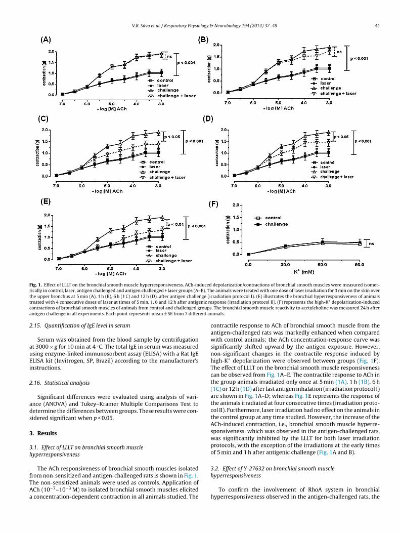
V.R. Silva et al. / Respiratory Physiology & Neurobiology 194 (2014) 37–48 41
Fig. 1. Effect of LLLT on the bronchial smooth muscle hyperresponsiveness. ACh-induced depolarization/contractions of bronchial smooth muscles were measured isomet-rically in control, laser, antigen challenged and antigen challenged + laser groups (A–E). The animals were treated with one dose of laser irradiation for 3 min on the skin overt nge (it enic rec roupsa ent an
2
auEi
2
ads
3
3h
fTAa
he upper bronchus at 5 min (A), 1 h (B), 6 h (1 C) and 12 h (D), after antigen challereated with 4 consecutive doses of laser at times of 5 min, 1, 6 and 12 h after antigontractions of bronchial smooth muscles of animals from control and challenged gntigen challenge in all experiments. Each point represents mean ± SE from 7 differ
.15. Quantification of IgE level in serum
Serum was obtained from the blood sample by centrifugationt 3000 × g for 10 min at 4 ◦C. The total IgE in serum was measuredsing enzyme-linked immunosorbent assay (ELISA) with a Rat IgELISA kit (Invitrogen, SP, Brazil) according to the manufacturer’snstructions.
.16. Statistical analysis
Significant differences were evaluated using analysis of vari-nce (ANOVA) and Tukey–Kramer Multiple Comparisons Test toetermine the differences between groups. These results were con-idered significant when p < 0.05.
. Results
.1. Effect of LLLT on bronchial smooth muscleyperresponsiveness
The ACh responsiveness of bronchial smooth muscles isolated
rom non-sensitized and antigen-challenged rats is shown in Fig. 1.he non-sensitized animals were used as controls. Application ofCh (10−7–10−3 M) to isolated bronchial smooth muscles elicitedconcentration-dependent contraction in all animals studied. The
rradiation protocol I). (E) illustrates the bronchial hyperresponsiveness of animalssponse (irradiation protocol II). (F) represents the high-K+ depolarization-induced. The bronchial smooth muscle reactivity to acetylcholine was measured 24 h afterimals.
contractile response to ACh of bronchial smooth muscle from theantigen-challenged rats was markedly enhanced when comparedwith control animals: the ACh concentration-response curve wassignificantly shifted upward by the antigen exposure. However,non-significant changes in the contractile response induced byhigh-K+ depolarization were observed between groups (Fig. 1F).The effect of LLLT on the bronchial smooth muscle responsivenesscan be observed from Fig. 1A–E. The contractile response to ACh inthe group animals irradiated only once at 5 min (1A), 1 h (1B), 6 h(1C) or 12 h (1D) after last antigen inhalation (irradiation protocol I)are shown in Fig. 1A–D; whereas Fig. 1E represents the response ofthe animals irradiated at four consecutive times (irradiation proto-col II). Furthermore, laser irradiation had no effect on the animals inthe control group at any time studied. However, the increase of theACh-induced contraction, i.e., bronchial smooth muscle hyperre-sponsiveness, which was observed in the antigen-challenged rats,was significantly inhibited by the LLLT for both laser irradiationprotocols, with the exception of the irradiations at the early timesof 5 min and 1 h after antigenic challenge (Fig. 1A and B).
3.2. Effect of Y-27632 on bronchial smooth muscle
hyperresponsivenessTo confirm the involvement of RhoA system in bronchialhyperresponsiveness observed in the antigen-challenged rats, the

42 V.R. Silva et al. / Respiratory Physiology &
Fig. 2. Effect of RhoA inhibitor on bronchial hyperresponsiveness. To confirm theRhoA participation in bronchial hyperresponsiveness in antigen challenged mice,animals were treated with RhoA inhibitor, Y-27632 (10 �M) 1 h before antigenchallenge. Bronchial smooth muscles obtained from animals not challenged wereuwd
atorec
3
aasterltRatteRs
3l
iqts(DeaHiprc(poo
sed as control. The bronchial smooth muscle hyperresponsiveness to acetylcholineas measured 24 h after antigen challenge. Each bar represents mean ± SE from 7ifferent animals.
nimals were treated with Y-27632, a RhoA inhibitor, by inhala-ion 1 h prior to the antigenic challenge (Fig. 2). After treatmentf Y-27632, the bronchial hyperresponsiveness to ACh completelyeturned to control levels. In addition, the RhoA inhibitor had noffect on the contractility of bronchial smooth muscle to ACh inontrol animals.
.3. Effect of LLLT or Y-27632 on RhoA mRNA expression
The levels of total RhoA mRNA expression from all animal groupsre shown in Fig. 3: animals that were challenged or not challengednd laser-treated or non-treated, were examined for mRNA expres-ion of the main and intrapulmonary bronchial smooth muscles inhe absence of epithelia and lung parenchyma. The RhoA mRNAxpression in bronchial smooth muscle of the antigen-challengedat was significantly increased compared to the control group. Theaser treatment had no effect on the RhoA expression in the con-rol group. However, LLLT markedly blocked the upregulation ofhoA expression in BSM from antigen-challenged rats. Moreover,n enhanced inhibitory effect on expression was obtained whenhe rats subjected to antigenic challenge were initially treated withhe Y-27632 RhoA inhibitor. This inhibitor decreases RhoA mRNAxpression in BSM segments from allergic rats to control levels. ThehoA inhibitor had not effect on RhoA mRNA expression in BSMegments from the control and laser-treated groups.
.4. Effect of LLLT on allergic inflammation in the lung and IgEevels in serum
To determine the effect of LLLT on changes in airway biologynduced by antigen exposure, the total cell number in BAL wasuantified and histological analysis was performed. We found thathe total number of cells in BAL of antigen-challenged rats wasignificantly increased when compared with the control groupFig. 4A). Moreover, this increase was significantly reduced by LLLT.iff-Quik cell staining revealed that most of the new cells wereosinophils and that the number of eosinophils in BAL from thentigen-challenged mice was largely reduced by LLLT (Fig. 4B).istological examinations also revealed increased eosinophilic
nflammation in the airway walls of antigen-challenged mice com-ared with the control group (Fig. 4C). Furthermore, laser treatmenteduced eosinophilic inflammation induced by antigenic challengeompared with antigen-challenged mice, but not in treated mice
Fig. 4C and D). Histological alterations in lung parenchyma anderibronchiolar region were also characterized by the appearancef peribronchial edema and bronchoconstriction with metaplasiaf bronchiolar smooth muscle cells (Fig. 4E and F). The levels ofNeurobiology 194 (2014) 37–48
total IgE in sera of rats are shown in Fig. 4G. A marked increasein total IgE levels in animals that were sensitized and challengedwith antigen was observed and this increase was not altered whenthese animals were laser-treated. Moreover, laser irradiation of thecontrol group showed no effect on the total levels of IgE.
3.5. Effect of LLLT on ICAM-1
The LLLT effect on ICAM-1 mRNA (5A) and protein (5B) expres-sion is shown in Fig. 5. Lung localization of ICAM-1 was markedwith immunohistochemical staining (5C). Both ICAM-1 mRNA andprotein expression in the lung was significantly higher in the chal-lenge group compared with the control group (Fig. 5A and B,respectively); however, there was a significant decrease in ICAM-1 expression and protein in lungs harvested from animals treatedwith laser irradiation (Fig. 5A and B). The presence of ICAM-1 inlungs from challenged animals is confirmed by immunohistochem-ical staining in Fig. 5C and is also evidenced by the low-level lasertherapy effect directly on ICAM-1. Laser irradiation on animals fromthe control group showed no effect on ICAM-1 expression.
3.6. Effect of LLLT on the deposition of mucus and collagen
These results demonstrated that OVA sensitization and LLLTtreatment significantly increased and decreased neutral and acidmucus production, respectively (Fig. 6A and B, respectively). Anincrease in secretion in the lungs of challenged mice and the low-level laser therapy effect directly on mucus are shown in Fig. 6C.Moreover, OVA sensitization significantly increased secretion aswell as the deposition of collagen in the airway wall, while LLLTsignificantly reduced the production and deposition of collagen(Fig. 7A and B, respectively).
3.7. Effect of LLLT on IL-4, IL-5, IL-13 and eotaxin
As shown in Fig. 8, the levels of IL-4 (8A), IL-5 (8B), IL-13 (8C)and eotaxin (8D) in BAL fluids of antigen-challenged rats were sig-nificantly increased compared with those from the non-sensitizedanimals (control group). The LLLT of animals from control grouphad no effect on the levels of these parameters. However, the lev-els of IL-4 and IL-5 in BAL from the antigen-challenge rats werereduced after laser therapy when compared with animals chal-lenged, but not treated with laser, where the levels remained higherthan the control group. In addition, LLLT markedly reduced boththe IL-13 and eotaxin levels in BAL in the antigen-challenged ratscompared with those in the laser-untreated animals. No significantdifferences were observed between the control and laser groups.
3.8. Effect of LLLT on STAT6
There was a significant change in the STAT6 protein levels in thechallenge group compared with the control group, where the levelswere strongly reduced when the animals were treated with laser(Fig. 9). Moreover, laser irradiation on animals not challenged withantigen (control group) had no effect on the level of STAT6 proteinin lung tissue.
4. Discussion
Currently, available therapies for asthma are non-curative.These treatments only control inflammation, without altering the
underlying immune reactivity, which predisposes the individual toatopic airway inflammation (Virchow and Barnes, 2012). Inhaledglucocorticoids are currently the mainstay of asthma therapy.Although this treatment is generally effective and well tolerated,
V.R. Silva et al. / Respiratory Physiology & Neurobiology 194 (2014) 37–48 43
Fig. 3. Effect of RhoA inhibitor on upregulation of RhoA mRNA in bronchial smooth muscles. Bronchial smooth muscles obtained from control and antigen challenged micew were
a challa
stracb
F(eoib
ere assayed for RhoA mRNA expression by Real Time-PCR technique. The animalsfter antigen challenge or RhoA inhibitor, Y-27632 (10 �M) 1 h before the antigenicllergic challenge. Each bar represents mean ± SE from 7 different animals.
everal side effects, such as an impairment of growth, abnormali-ies in the metabolism of glucose, adrenal suppression, increased
isk of fracture and the formation of cataracts may occur when theyre used at high doses or for an extended period of time. Topi-al adverse effects, such as candidiasis and dysphonia have alsoeen previously described with inhaled glucocorticoids. Sometimesig. 4. Effect of LLLT on lung inflammation and on levels of total immunoglobulin E (IgE)Fig. 4B) were carried out in BALF obtained from all studied groups. Sections (5 �m) oxamination (C) aiming to identify and quantify the density of eosinophils in airways wall (f peribronchiolar edema (F) were measured as described in Material and Methods sectiommunosorbent assay (ELISA). The IgE levels in serum were measured 24 h after the anronchus at 5 min, 1, 6 and 12 h after antigen challenge. Each bar represents mean ± SE fr
treated with laser for 3 min on skin over the upper bronchus at 5 min, 1, 6 and 12 henge. The PCR primer efficiencies were calculated using standard curves 24 h after
resistance to glucocorticoids treatment occurs due to the usu-ally higher dose requirements of glucocorticoids in these patients,
which results in an increased risk of systemic side effects (Louiset al., 2012). For these patients, there is an urgent need to developnew anti-inflammatory therapies with immunomodulatory activ-ity to provide alternative strategies to treat asthma.in serum. Total, mononuclear and neutrophil cells count (Fig. 4A) and eosinophilsf formalin-fixed lungs were stained with hematoxylin and eosin for histologicalD) (original magnification, ×200). Bronchoconstriction index (E) and the percentagen. Blood serum obtained from all mice was assayed for IgE by using enzyme-linkedtigen challenge (G). The animals were treated with laser on skin over the upperom 7 different animals.

44 V.R. Silva et al. / Respiratory Physiology & Neurobiology 194 (2014) 37–48
Fig. 5. Effect of LLLT on ICAM-1 protein expression in lung. The changes in ICAM-1 expression for all groups, control, laser, antigen challenge and antigen challenged + laser,a NA exl (C). Ta imals
pdlsall2aa
Fmr
re illustrated in the figure. The changes in ICAM-1 expression were assessed by mRocalization of ICAM-1 in lung, the positive reaction was visualized as a brown stainnd 12 h after antigen challenge. Each bar represents mean ± SE from 7 different an
Since 1967, over 100 phase III, randomized, double-blind,lacebo-controlled, clinical trials (RCTs) with low-level laser irra-iation have been published and supported by more than 1000
aboratory studies investigating the primary mechanism and theignaling cascade of secondary effects that contribute to therrangement of local tissue, as well as the systemic effects ofaser treatment. RCTs with positive outcomes have been pub-ished on pathologies as diverse as osteoarthritis (Bjordal et al.,
006), back pain (Chow et al., 2009), stroke (Lampl et al., 2007)nd pulmonary disorders, such as chronic obstructive bronchitisnd asthma (Nedeljkovic et al., 2008; Kashanskaia and Fedorov,ig. 6. Effect of LLLT on mucus production in lung. Sections of formalin-fixed lungs wereicroscopy (original magnification, ×200). The animals were treated with laser on skin
epresents mean ± SE from 7 different animals.
pression (A) and by protein concentration by ELISA (B). For immunohistochemicalhe animals were treated with laser on skin over the upper bronchus at 5 min, 1, 6
.
2009). Nevertheless, these results have not been always been pos-itive. This failure can be attributed to several factors, such asdosimetry (too low or too much energy delivered, too low ortoo much irradiance, inappropriate pulse structure, irradiation ofinsufficient area of the pathology), inappropriate anatomical treat-ment location and concurrent patient medication (such as steroidaland non-steroidal anti-inflammatory which can inhibit healing)(Huang et al., 2009). Due to the many existing physical parame-
ters, which can directly affect the laser activity and its beneficialanti-inflammatory effects on diverse diseases, we performed aseries of experimental studies within the last 6 years in orderstained with Periodic Acid Schif (PAS) before histological examination using light over the upper bronchus at 5 min, 1, 6 and 12 h after antigen challenge. Each bar

V.R. Silva et al. / Respiratory Physiology & Neurobiology 194 (2014) 37–48 45
F ngs wm broncf
tLiap
dt
F(a
ig. 7. Effect of LLLT on collagen production in lung. Sections of formalin-fixed luagnification, ×200). The animals were treated with laser on skin over the upper
rom 7 different animals.
o clarify which of the inflammatory mediators is the target forLLT in pulmonary disorders, thereby initiating an understand-ng of the cellular signaling pathways responsible for the lasernti-inflammatory effect in inflammatory conditions, which com-
romise the lung and airway.In clinical practice, the beneficial effects of LLLT have beenemonstrated in patients with compromised airway and lung, par-icularly asthmatics (Esaulenko et al., 2009; Nikitin et al., 2008;
ig. 8. Effect of LLLT on cytokines levels in BALF. BALF obtained from control, laser, antigB), IL-13 (C) and eotaxin (D) by enzyme-linked immunosorbent assay (ELISA). The animfter antigen challenge. The cytokines levels in BALF were assayed 24 h after the antigen.
ere stained with Picrossirius before examination with light microscopy (originalhus at 5 min, 1, 6 and 12 h after antigen challenge. Each bar represents mean ± SE
Nedeljkovic et al., 2008; Farkhudtinov, 2007). In fact, several studieshave demonstrated that LLLT provides a significant improvement inthe course of bronchial asthma, enabling for outpatient treatmentand rehabilitation, recovery of bronchial sensitivity to sympath-
omimetics and xanthine derivatives, lower glucocorticoid doses,shorter hospital stays, and reduced bronchial asthma-related dis-ability (Nikitin et al., 2008; Ostronosova, 2006; Milojevic and Kuruc,2003; Landyshev et al., 2002). Thus, additional studies are neededen challenge and antigen challenged + laser groups were assayed for IL-4 (A), IL-5als were treated with laser on skin over the upper bronchus at 5 min, 1, 6 and 12 h
Each bar represents mean ± SE from 7 different animals.

46 V.R. Silva et al. / Respiratory Physiology &
Fig. 9. Effect of LLLT on signal transducer and activator of transduction 6 (STAT6) inlung. The STAT6 concentration in lung tissue from control, laser, antigen challengeand antigen challenged + laser groups was determined by ELISA. The intensity ofcoor
ta
tRodittcfis
mpcsrposl
eltftRepite
rpstiric
ti
olored product analyzed in 450 nm is directly proportional to the concentrationf STAT6 present in the lung tissue. The animals were treated with laser on skinver the upper bronchus at 5 min, 1, 6 and 12 h after antigen challenge. Each barepresents mean ± SE from 7 different animals.
o understand the beneficial effect of LLLT before LLLT can be useds a noninvasive therapy for the treatment of chronic asthma.
We have demonstrated that LLLT is an effective and beneficialreatment in relieving airway hyperresponsiveness by reducing thehoA expression, and can reduce the calcium sensitivity in a modelf acute lung inflammation induced by TNF (Aimbire et al., 2009;e Lima et al., 2010). In addition, acute lung inflammation is also
nfluenced by LLLT, which down-regulates pro-inflammatory pro-eins and simultaneously up-regulates anti-inflammatory proteins,hereby promoting a counterbalance of Th1/Th2 cytokines, whichontrols the disease (de Lima et al., 2011a,b). On the basis of thisnding, we proposed that LLLT could modulate bronchial hyperre-ponsiveness and lung inflammation in a rat allergic asthma model.
Our findings in the current study demonstrated that LLLT couldodulate bronchial hyperresponsiveness via a mechanism inde-
endent of Ca+2 releases, which was potentially due to a G proteinoupled receptor activation response. This was with the currenttudy because LLLT attenuated BSM hyperresponsiveness due to aeduction in RhoA mRNA expression, which was attributed to MLChosphatase inhibition. This inhibition resulted in the promotionf a contractile state, which was caused by Ca+2 sensitization ofmooth muscle contraction. In addition, the late beneficial effect ofaser, i.e., 24 h after last antigenic challenge was observed.
Thus, new therapies used to prevent an increase in RhoA proteinxpression in bronchial smooth muscle could modulate intracellu-ar signaling, resulting in decreased hyperresponsiveness. Althoughhe LLLT was efficient in reducing RhoA mRNA expression in BSMrom antigen-challenged rats, we could not affirm that laser irradia-ion specifically affected RhoA protein expression at the membrane.eal Time-PCR was used in this study, which only detects mRNAxpression. However, it is known that after translocation, RhoArotein is expressed at the BSM membrane. Thus, the effect of LLLT
n reducing the RhoA mRNA expression in BSM is a strong indica-or that the low-level laser is able to interfere with protein RhoAxpression at the membrane.
In the present study, our results also suggested that LLLT couldeduce inflammatory cell infiltration, increase BALF eosinophils androduced histological changes in lungs induced by antigen expo-ure, which were both attenuated by laser therapy. It is likely thathe inhibitory effects of laser treatment on the inflammatory cellnfiltration were due to an inhibition of pro-inflammatory cytokineelease or IgE production because LLLT significantly altered thencreased expression of IL-4, IL-5 and eotaxin in BAL after antigenic
hallenge.Our results also suggested that the LLLT effect in reducinghe bronchial hyperreactivity via RhoA was associated to its anti-nflammatory effect on IL-13 in BALF from allergic mice. It has been
Neurobiology 194 (2014) 37–48
well established that levels of IL-13 were increased in bronchiallavage fluid of asthmatics, which induces specific significant fea-tures of bronchial asthma, including airway hyperresponsiveness(Horiba et al., 2011). Moreover, some studies have demonstrateda cross-talk between IL-13 and RhoA in the pulmonary system(Chiba et al., 2009). It was also been reported that IL-13 gener-ated in the lung can induce an increase in RhoA expression inbronchial smooth muscle in asthmatic mice via STAT-6 (Goto et al.,2010). Thus, our findings suggested that the pronounced effect ofthe laser irradiation on bronchial hyperresponsiveness also reflectsits action on the IL-13 pro-inflammatory cytokine. Moreover, itmay also explain the marked laser anti-inflammatory effect on thenumber of eosinophils in BALF from asthmatic mice as an indi-rect response of the laser irradiation on IL-13. The laser effect onSTAT6 reinforces the ability of this therapy to suppress the activityof the principal transcription factor involved in regulating IL-4, IL-5,IL-13 and eotaxin, which are pro-inflammatory mediators respon-sible by exacerbation and perpetuation of the allergic inflammatoryresponse.
The total IgE in sera from antigen-challenged mice was not mod-ified by LLLT. Since the first publication of the LLLT effect on acuteairway and lung inflammation, we demonstrated that laser treat-ment did not present a systemic effect (Aimbire et al., 2005). Incontrast, other studies have shown a LLLT systemic effect in dif-ferent experimental models (Samoilova et al., 2008; Moreira et al.,2009). This negative result of the LLLT in the current study maybe because the irradiation dose was too low to cause any signif-icant change of IgE level in serum after antigen challenge or thatthe irradiated area was only surrounding the bronchi of the mice.However, local treatment with laser appeared to be advantageousbecause only the airway and lung was affected; thus, a potentiallytoxic effect of laser irradiation could be better controlled or evenavoided. Our results reinforced the idea of using local treatment forallergic asthma.
Several studies have reported that STAT6 plays a pivotal role inthe development of pulmonary eosinophilia, airway hyperrespon-siveness and mucus hypersecretion (Hoshino et al., 2004). In thepresent study, we found that LLLT significantly reduced both themRNA expression for STAT6 in lung tissue in animals challengedwith antigen, and the concentration of cytokines, which function aschemoattractants for inflammatory cells, specifically eosinophils.On the basis of these results, it is reasonable to suggest that theanti-inflammatory effect of the laser irradiation on cytokines IL-4,IL-5, IL-13 and eotaxin in lung could be due to a reduction of STAT6mRNA expression.
Some studies have demonstrated that the LLLT effect in vivo isnot evidenced if the biological sample was analyzed just a few min-utes after irradiation (Mafra de Lima et al., 2009a,b; de Lima et al.,2011a,b). Our results confirmed this observation. Although theinteraction of the laser light with biological tissue occurred almostimmediately, the laser effect at the cellular level required moretime after the absorption of light by tissue to stimulate transcrip-tion factors to induce the generation or reduction of inflammatoryproteins responsible for promoting the beneficial effect. Red or nearinfra-red laser fluencies as low as 3 or 5 J cm−2 will be beneficial invivo, but large doses such as 50 or 100 J cm−2 will lose the beneficialeffect and may even become detrimental (Chen et al., 2011).
It was observed in the present study that when laser irradiationwas applied four times in a period of 24 h after antigenic chal-lenged, a 5.4 J/cm2 laser dose produced an in vivo beneficial effecton bronchial reactivity dysfunction and lung inflammation due tothe reduction of pro-inflammatory cytokines release involved in
lung allergic response. Accumulated laser doses in a short timeperiod between the irradiations could cause some deleterious effectof LLLT or even eliminate its beneficial effect (Huang et al., 2009). Toavoid this effect, the animals were irradiated four times at intervals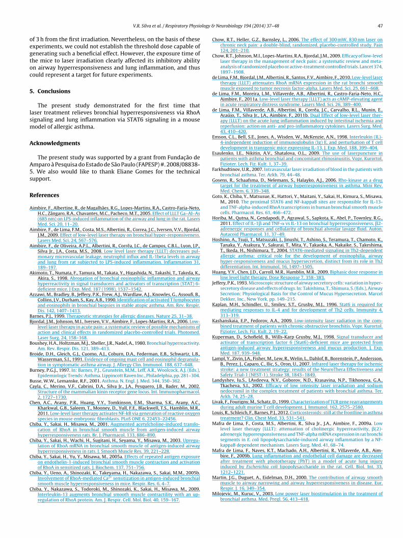
logy &
oegtoc
5
lsm
A
A5s
R
A
A
A
A
A
BB
B
B
B
BC
C
C
C
C
C
C
V.R. Silva et al. / Respiratory Physio
f 3 h from the first irradiation. Nevertheless, on the basis of thesexperiments, we could not establish the threshold dose capable ofenerating such a beneficial effect. However, the exposure time ofhe mice to laser irradiation clearly affected its inhibitory abilityn airway hyperresponsiveness and lung inflammation, and thusould represent a target for future experiments.
. Conclusions
The present study demonstrated for the first time thataser treatment relieves bronchial hyperresponsiveness via RhoAignaling and lung inflammation via STAT6 signaling in a mouseodel of allergic asthma.
cknowledgments
The present study was supported by a grant from Fundac ão demparo à Pesquisa do Estado de São Paulo (FAPESP) #. 2008/08838-. We also would like to thank Eliane Gomes for the technicalupport.
eferences
imbire, F., Albertine, R., de Magalhães, R.G., Lopes-Martins, R.A., Castro-Faria-Neto,H.C., Zângaro, R.A., Chavantes, M.C., Pacheco, M.T., 2005. Effect of LLLT Ga–Al–As(685 nm) on LPS-induced inflammation of the airway and lung in the rat. LasersMed. Sci. 20, 11–20.
imbire, F., de Lima, F.M., Costa, M.S., Albertini, R., Correa, J.C., Iversen, V.V., Bjordal,J.M., 2009. Effect of low-level laser therapy on bronchial hyper-responsiveness.Lasers Med. Sci. 24, 567–576.
imbire, F., de Oliveira, A.P.L., Albertini, R., Corrêa, J.C., de Campos, C.B.L., Lyon, J.P.,Silva Jr., J.A., Costa, M.S., 2008. Low level laser therapy (LLLT) decreases pul-monary microvascular leakage, neutrophil influx and IL-1beta levels in airwayand lung from rat subjected to LPS-induced inflammation. Inflammation 31,189–197.
kimoto, T., Numata, F., Tamura, M., Takata, Y., Higashida, N., Takashi, T., Takeda, K.,Akira, S., 1998. Abrogation of bronchial eosinophilic inflammation and airwayhyperreactivity in signal transducers and activators of transcription (STAT) 6-deficient mice. J Exp. Med. 187 (1998), 1537–1542.
zzawi, M., Bradley, B., Jeffery, P.K., Frew, A.J., Wardlaw, A.J., Knowles, G., Assoufi, B.,Collins, J.V., Durham, S., Kay, A.B., 1990. Identification of activated T lymphocytesand eosinophils in bronchial biopsies in stable atopic asthma. Am. Rev. Respir.Dis. 142, 1407–1413.
arnes, P.J., 1999. Therapeutic strategies for allergic diseases. Nature 25, 31–38.jordal, J.M., Johnson, M.I., Iversen, V.V., Aimbire, F., Lopes-Martins, R.A., 2006. Low-
level laser therapy in acute pain: a systematic review of possible mechanisms ofaction and clinical effects in randomized placebo-controlled trials. Photomed.Laser Surg. 24, 158–168.
oushey, H.A., Holtzman, M.J., Sheller, J.R., Nadel, A., 1980. Bronchial hyperreactivity.Am. Rev. Respir. Dis. 121, 389–413.
roide, D.H., Gleich, G.J., Cuomo, A.J., Coburn, D.A., Federman, E.B., Schwartz, L.B.,Wasserman, S.I., 1991. Evidence of ongoing mast cell and eosinophil degranula-tion in symptomatic asthma airway. J. Allergy Clin. Immunol. 188, 637–648.
urney, P.G.J., 1997. In: Barnes, P.J., Grunstein, M.M., Leff, A.R., Woolcock, A.J. (Eds.),Epidemiologic Trends: Asthma. Lippincott Raven Inc., Philadelphia, pp. 281–304.
usse, W.W., Lemanske, R.F., 2001. Asthma. N. Engl. J. Med. 344, 350–362.ayla, C., Merino, V.F., Cabrini, D.A., Silva Jr., J.A., Pesquero, J.B., Bader, M., 2002.
Structure of the mammalian kinin receptor gene locus. Int. Immunopharmacol.2, 1727–1730.
hen, A.C., Arany, P.R., Huang, Y.Y., Tomkinson, E.M., Sharma, S.K., Arany, A.C.,Kharkwal, G.B., Saleem, T., Mooney, D., Yull, F.E., Blackwell, T.S., Hamblin, M.R.,2011. Low-level laser therapy activates NF-kB via generation of reactive oxygenspecies in mouse embryonic fibroblasts. PLoS ONE 6, 22453–22456.
hiba, Y., Sakai, H., Misawa, M., 2001. Augmented acetylcholine-induced translo-cation of RhoA in bronchial smooth muscle from antigen-induced airwayhyperresponsiveness rats. Br. J. Pharmacol. 133, 886–890.
hiba, Y., Sakai, H., Wachi, H., Sugitani, H., Seyama, Y., Misawa, M., 2003. Upregu-lation of RhoA mRNA in bronchial smooth muscle of antigen-induced airwayhyperresponsiveness in rats. J. Smooth Muscle Res. 39, 221–228.
hiba, Y., Sakai, H., Yu, Y., Misawa, M., 2005a. Effects of repeated antigen exposureon endothelin-1-induced bronchial smooth muscle contraction and activationof RhoA in sensitized rats. J. Biochem. 137, 751–756.
hiba, Y., Ueno, A., Shinozaki, K., Takeyama, H., Nakazawa, S., Sakai, M.M., 2005b.
Involvement of RhoA-mediated Ca2+ sensitization in antigen-induced bronchialsmooth muscle hyperresponsiveness in mice. Respir. Res. 6, 4–7.hiba, Y., Nakazawa, S., Todoroki, M., Shinozaki, K., Sakai, H., Misawa, M., 2009.Interleukin-13 augments bronchial smooth muscle contractility with an up-regulation of RhoA protein. Am. J. Respir. Cell. Mol. Biol. 40, 159–167.
Neurobiology 194 (2014) 37–48 47
Chow, R.T., Heller, G.Z., Barnsley, L., 2006. The effect of 300 mW, 830 nm laser onchronic neck pain: a double-blind, randomized, placebo-controlled study. Pain124, 201–210.
Chow, R.T., Johnson, M.I., Lopes-Martins, R.A., Bjordal, J.M., 2009. Efficacy of low-levellaser therapy in the management of neck pain: a systematic review and meta-analysis of randomized placebo or active-treatment controlled trials. Lancet 374,1897–1908.
de Lima, F.M., Bjordal, J.M., Albertini, R., Santos, F.V., Aimbire, F., 2010. Low-level lasertherapy (LLLT) attenuates RhoA mRNA expression in the rat bronchi smoothmuscle exposed to tumor necrosis factor-alpha. Lasers Med. Sci. 25, 661–668.
de Lima, F.M., Moreira, L.M., Villaverde, A.B., Albertini, R., Castro-Faria-Neto, H.C.,Aimbire, F., 2011a. Low-level laser therapy (LLLT) acts as cAMP-elevating agentin acute respiratory distress syndrome. Lasers Med. Sci. 26, 389–400.
de Lima, F.M., Villaverde, A.B., Albertini, R., Corrêa, J.C., Carvalho, R.L., Munin, E.,Araújo, T., Silva Jr., J.A., Aimbire, F., 2011b. Dual Effect of low-level laser ther-apy (LLLT) on the acute lung inflammation induced by intestinal ischemia andreperfusion: action on anti- and pro-inflammatory cytokines. Lasers Surg. Med.43, 410–420.
Emson, C.L., Bell, S.E., Jones, A., Wisden, W., McKenzie, A.N., 1998. Interleukin (IL)-4-independent induction of immunoglobulin (Ig) E, and perturbation of T celldevelopment in transgenic mice expressing IL-13. J. Exp. Med. 188, 399–404.
Esaulenko, I.E., Nikitin, A.V., Shatalova, O.L., 2009. The use of laserpuncture inpatients with asthma bronchial and concomitant rhinosinusitis. Vopr. Kurortol.Fizioter. Lech. Fiz. Kult. 1, 37–39.
Farkhudtinov, U.R., 2007. Intravascular laser irradiation of blood in the patients withbronchial asthma. Ter. Arkh. 79, 44–48.
Gosens, R., Schaafsma, D., Nelemans, S., Halayko, A.J., 2006. Rho-kinase as a drugtarget for the treatment of airway hyperresponsiveness in asthma. Mini Rev.Med. Chem. 6, 339–348.
Goto, K., Chiba, Y., Matsusue, K., Hattori, Y., Maitani, Y., Sakai, H., Kimura, S., Misawa,M., 2010. The proximal STAT6 and NF-kappaB sites are responsible for IL-13-and TNF-alpha-induced RhoA transcriptions in human bronchial smooth musclecells. Pharmacol. Res. 61, 466–472.
Horiba, M., Qutna, N., Gendapodi, P., Agrawal, S., Sapkota, K., Abel, P., Townley, R.G.,2011. Effect of IL-1� and TNF-� vs IL-13 on bronchial hyperresponsiveness, �2-adrenergic responses and cellularity of bronchial alveolar lavage fluid. Auton.Autacoid Pharmacol. 31, 37–49.
Hoshino, A., Tsuji, T., Matsuzaki, J., Jinushi, T., Ashino, S., Teramura, T., Chamoto, K.,Tanaka, Y., Asakura, Y., Sakurai, T., Mita, Y., Takaoka, A., Nakaike, S., Takeshima,T., Ikeda, H., Nishimura, T., 2004. STAT6-mediated signaling in Th2-dependentallergic asthma: critical role for the development of eosinophilia, airwayhyper-responsiveness and mucus hypersecretion, distinct from its role in Th2differentiation. Int. Immunol. 16, 1497–1505.
Huang, Y.Y., Chen, J.D., Carroll, M.R., Hamblin, M.R., 2009. Biphasic dose response inlow level light therapy. Dose Response 7, 358–383.
Jeffery, P.K., 1993. Microscopic structure of airway secretory cells: variation in hyper-secretory disease and effects of drugs. In: Takishima, T., Shimura, S. (Eds.), AirwaySecretion: Physiological Basis for the Control of Mucus Hypersecretion. MarcelDekker, Inc., New York, pp. 149–215.
Kaplan, M.H., Schindler, U., Smiley, S.T., Grusby, M.J., 1996. Stat6 is required formediating responses to IL-4 and for development of Th2 cells. Immunity 4,313–319.
Kashanskaia, E.P., Fedorov, A.A., 2009. Low-intensity laser radiation in the com-bined treatment of patients with chronic obstructive bronchitis. Vopr. Kurortol.Fizioter. Lech. Fiz. Kult. 2, 19–22.
Kuperman, D., Schofield, B., Wills-Karp Grusby, M.J., 1998. Signal transducer andactivator of transcription factor 6 (Stat6)-deficient mice are protected fromantigen-induced airway hyperresponsiveness and mucus production. J. Exp.Med. 187, 939–948.
Lampl, Y., Zivin, J.A., Fisher, M., Lew, R., Welin, L., Dahlof, B., Borenstein, P., Andersson,B., Perez, J., Caparo, C., Ilic, S., Oron, U., 2007. Infrared laser therapy for ischemicstroke: a new treatment strategy: results of the NeuroThera Effectiveness andSafety Trial-1 (NEST-1). Stroke 38, 1843–1849.
Landyshev, Iu.S., LAvdeeva, N.V., Goborov, N.D., Krasavina, N.P., Tikhonova, G.A.,Tkacheva, S.I., 2002. Efficacy of low intensity laser irradiation and sodiumnedocromil in the complex treatment of patients with bronchial asthma. Ter.Arkh. 74, 25–28.
Livak, F., Fourigny, M., Schatz, D., 1999. Characterization of TCR gene rearrangementsduring adult murine T cell development. J. Immunol. 162, 2575–2580.
Louis, R., Schleich, F., Barnes, P.J., 2012. Corticosteroids: still at the frontline in asthmatreatment? Clin. Chest Med. 33, 531–541.
Mafra de Lima, F., Costa, M.S., Albertini, R., Silva Jr., J.A., Aimbire, F., 2009a. Lowlevel laser therapy (LLLT): attenuation of cholinergic hyperreactivity, �(2)-adrenergic hyporresponsiveness and TNF-alpha mRNA expression in rat bronchisegments in E. coli lipopolysaccharide-induced airway inflammation by a NF-kappaB dependent mechanism. Lasers Surg. Med. 41, 68–74.
Mafra de Lima, F., Naves, K.T., Machado, A.H., Albertini, R., Villaverde, A.B., Aim-bire, F., 2009b. Lung inflammation and endothelial cell damage are decreasedafter treatment with phototherapy (PhT) in a model of acute lung injuryinduced by Escherichia coli lipopolysaccharide in the rat. Cell. Biol. Int. 33,1212–1221.
Martin, J.G., Duguet, A., Eidelman, D.H., 2000. The contribution of airway smoothmuscle to airway narrowing and airway hyperresponsiveness in disease. Eur.Respir. J. 16, 349–354.
Milojevic, M., Kuruc, V., 2003. Low power laser biostimulation in the treatment ofbronchial asthma. Med. Pregl. 56, 413–418.
4 logy &
M
N
N
O
O
P
ishi, K., Yoshida, N., Kishimoto, T., Akira, S., 1996. Essential role of Stat6 in IL-4signaling. Nature 380, 627–630.
8 V.R. Silva et al. / Respiratory Physio
oreira, M.S., Velasco, I.T., Ferreira, L.S., Ariga, S.H., Barbeiro, D.F., Meneguzzo, D.T.,Abatepaulo, F., Marques, M.M., 2009. Effect of phototherapy with low intensitylaser on local and systemic immunomodulation following focal brain damage inrat. J. Photochem. Photobiol. B 97, 145–151.
edeljkovic, M., Ljustina-Pribic, R., Savic, K., 2008. Innovative approach to laseracupuncture therapy of acute obstruction in asthmatic children. Med. Pregl. 61,123–130.
ikitin, A.V., Eusalenko, I.E., Shatalova, O.L., 2008. Effectiveness of laser puncture inelderly patients with bronchial asthma. Vopr. Kurortol. Lech. Fiz. Kult. 6, 38–39.
hashi, Y., Motojima, S., Fukuda, T., Makino, S., 1992. Airway hyperresponsiveness,increased intracellular spaces of bronchial epithelium, and increased infiltrationof eosinophils and lymphocytes in bronchial mucosa in asthma. Am. Rev. Respir.
Dis. 145, 1469–1476.stronosova, N., 2006. Outpatient use of laser therapy in bronchial asthma. Ter. Arkh.78, 41–44.
ires, D., Xavier, M., Araújo, T., Silva Jr., J.A., Aimbire, F., Albertini, R., 2011. Low-levellaser therapy (LLLT; 780 nm) acts differently on mRNA expression of anti- and
Neurobiology 194 (2014) 37–48
pro-inflammatory mediators in an experimental model of collagenase-inducedtendinitis in rat. Lasers Med. Sci. 26, 85–94.
Samoilova, K.A., Zhevago, N.A., Petrishchev, N.N., Zimin, A.A., 2008. Role of nitricoxide in the visible light-induced rapid increase of human skin microcirculationat the local and systemic levels: II. Healthy volunteers. Photomed. Laser Surg.26, 443–449.
Seow, C.Y., Schellenberg, R.R., Paré, P.D., 1998. Structural and functional changes inthe airway smooth muscle of asthmatic subjects. Am. J. Respir. Crit. Care Med.158, 179–186.
Takeda, K., Tanaka, T., Shi, W., Matsumoto, M., Minami, M., Kashiwamura, S., Nakan-
Virchow, J.C., Barnes, P.J., 2012. Asthma Semin. Respir. Crit. Care Med. 33, 577–585.Wiggs, B.R., Moreno, R., Hogg, J.C., Hilliam, C., Paré, P.D., 1990. A model of the mechan-
ics of airway narrowing. J. Appl. Physiol. 69, 849–860.


