Embed Size (px)
Citation preview

T-cell infiltration in autosomal dominant neovascularinflammatory vitreoretinopathy
Vinit B. Mahajan,1,2 John G. Vallone,4 Jonathan H. Lin,3 Robert F. Mullins,1 Audrey C. Ko,1 James C. Folk,1Edwin M. Stone1,5
1Vitreoretinal Service, Department of Ophthalmology and Visual Sciences, The University of Iowa Hospitals & Clinics; Iowa City,IA; 2Omics Laboratory, Iowa City, IA; 3Department of Pathology University of California San Diego; San Diego, CA; 4Departmentof Pathology, University of California Irvine; Irvine, CA; 5Howard Hughes Medical Institute, Iowa City, IA
Purpose: Autosomal dominant neovascular inflammatory vitreoretinopathy (ADNIV) is a familial blinding disease ofunknown pathophysiology. The eyes and sera from patients with ADNIV were studied to understand the immune responsein this condition.Methods: The clinical case of an ADNIV patient was reviewed. Eye specimens from two donors with ADNIV wereexamined with a panel of standard histopathological stains and immunohistochemical markers. These findings werecompared to specimens of noninflammatory eye disease. Sera from twelve patients were also tested against retinal proteinwestern blots for the presence of autoretinal antibodies.Results: Each of the ADNIV and control eyes showed degenerative features of phthisis bulbi. Immunohistological stainsrevealed a supraciliary T-cell infiltrate in ADNIV eyes composed of cluster of differentiation-4 (CD4) positive and clusterof differentiation-8 (CD8)-positive cells. No immunoglobulin or B cells were detected in these eyes. Inflammatory cellswere absent from the control eyes. No specific autoretinal antibodies were detected in ADNIV sera.Conclusions: Aberrant T-cell-mediated processes may underlie ADNIV, and therapeutics directed at T cells may bettermanage inflammation in these patients. Genes related to T-cell function are high priority screening candidates.
The eye is an immune-privileged site where the localimmunological mechanisms are poorly understood.Autosomal dominant neovascular inflammatoryvitreoretinopathy (ADNIV) is an autoimmune disease of theeye without systemic features [1,2]. ADNIV shares severalfeatures with more common vitreoretinal diseases, includingdiabetic retinopathy, idiopathic uveitis, proliferativevitreoretinopathy, and retinitis pigmentosa.
ADNIV is an eye-specific inflammatory conditioncharacterized by pigmentary retinal degeneration, loss of theelectroretinogram b-wave, and peripheral field loss [1]. Thisprogressive degeneration is complicated by anterior segmentand vitreous inflammation, retinal neovascularization, retinaldetachment, and eventual phthisis. Cellular infiltrates in thevitreous are among the earliest detectable signs of ADNIV andcontinue throughout the course of the disease. The nature ofthe cells is not known.
Despite photoreceptor degenerative changes, onehypothesis suggests that ocular autoimmunity is the primarypathogenic cause of ADNIV and that the cells are either of B-cell or T-cell origin. Despite organ atrophy in the late stagesof disease, antigens that instigate autoimmune reactions maystill be active. The ADNIV autoimmune reaction continues
Correspondence to: Edwin M. Stone, Department of Ophthalmology,The University of Iowa Carver College of Medicine, 200 HawkinsDrive, Iowa City, IA, 52242; Phone: (319) 335-8270; FAX: (319)335-7142; email: [email protected]
through end-stage disease when the eye becomes shrunkenand blind, and studies in these eyes may still be relevant toearlier stages of ADNIV. To better understand ADNIVpathogenesis, we performed studies to detect autoretinalantibodies and in the case of ADNIV autopsy eyes detect thepresence of B-cell and T-cell infiltration.
METHODSInformed consent was obtained to review the case history ofa 80-year-old ADNIV patient examined in the University ofIowa Department of Ophthalmology clinic. The case historywas reviewed for a first-generation ADNIV patient. We usedsix postmortem eyes (University of Iowa, Department ofPathology archived tissue collection), that had been receivedin formalin and post fixed in Pen-fix (Thermoscientific,Waltham, MA). After the eye was opened by pupil–opticnerve section, it was decalcified. Histological staining withMasson's Trichrome stain was performed according to themanufacturer’s protocol (Sigma-Aldrich, St. Louis, MO).Immunohistochemical staining was performed as follows. Allslides were stained on the DAKO Autostainer+ (Carpinteria,CA), using heat pretreatment with a pressure cooker. Allantibodies used Targert Retrieval pH 6.0 (#S1699; DAKO),except cluster of differentiation-4 (CD4), which used high-pHretrieval (#S3308; DAKO). The following antibodies wereused: anti-CD3 (#A0452; DAKO) diluted to 1:200; anti-CD4(#NCL-L-CD4–1F6; LeicaSystems, Bannockburn, IL)diluted to 1:40; anti-CD8 (#M7103; DAKO) diluted to
Molecular Vision 2010; 16:1034-1040 <http://www.molvis.org/molvis/v16/a114>Received 24 February 2010 | Accepted 3 June 2010 | Published 8 June 2010
© 2010 Molecular Vision
1034
1:1,000; anti-CD20 (#M0755; DAKO) diluted to 1:400; anti-CD68 (#M0814; DAKO) diluted to 1:400; and anti-immunoglobulin G (IgG; #A0424; DAKO) diluted to1:40,000. All antibodies were incubated for 30 min. A dualendogenous enzyme block (DAKO #S2003, Carpinteria, CA)was used for 5 min. Detection was for 30 min and DAB+(DAKO #K3467, Carpinteria, CA) was used for 5 min. DAKOEnvision+ Dual-Link labeled polymer (#K4061) was used fordetection.
Autoretinal antibody assay: Following informed consent,serum was collected from 12 patients with ADNIV (2 males,and 9 females; age range 7–68) and 12 unaffected, healthycontrols (3 males, and 8 females; age range 18–74). Thesamples were screened on human retinal lysate to determinewhether these sera contained autoantibodies against retinalantigens. Methods for western blot were performed,essentially as described previously [3]. Briefly, human retinallysate was pooled from three donor eyes, separated by sodiumdodecyl sulfate PAGE, and transferred to polyvinyldifluoridemembrane. Serum from ADNIV patients or unaffected controlpatients without retinal disease was incubated with membranestrips to detect retinal antigens and visualized usinghorseradish peroxidase-conjugated antihuman secondaryantibody. Results were compared between ADNIV patientsand controls.
RESULTS
Case report: An 80-year-old female originally presented withidiopathic posterior uveitis and retinitis pigmentosa. Sheunderwent intracapsular cataract extractions 11 years priorand had postoperative visual acuity in the 20/200 range. Herright eye became phthisical 2 years before presentation, atwhich time she had a severe uveitis and vitreous hemorrhagein her left eye. She had no systemic inflammatory diseases,and a posterior uveitis workup was negative. Her familyhistory suggested a genetic etiology, and she was found to berelated to the original ADNIV pedigree that we first describedin 1990 with similar clinical findings and genetic linkage tochromosome 11q13 [1,2].
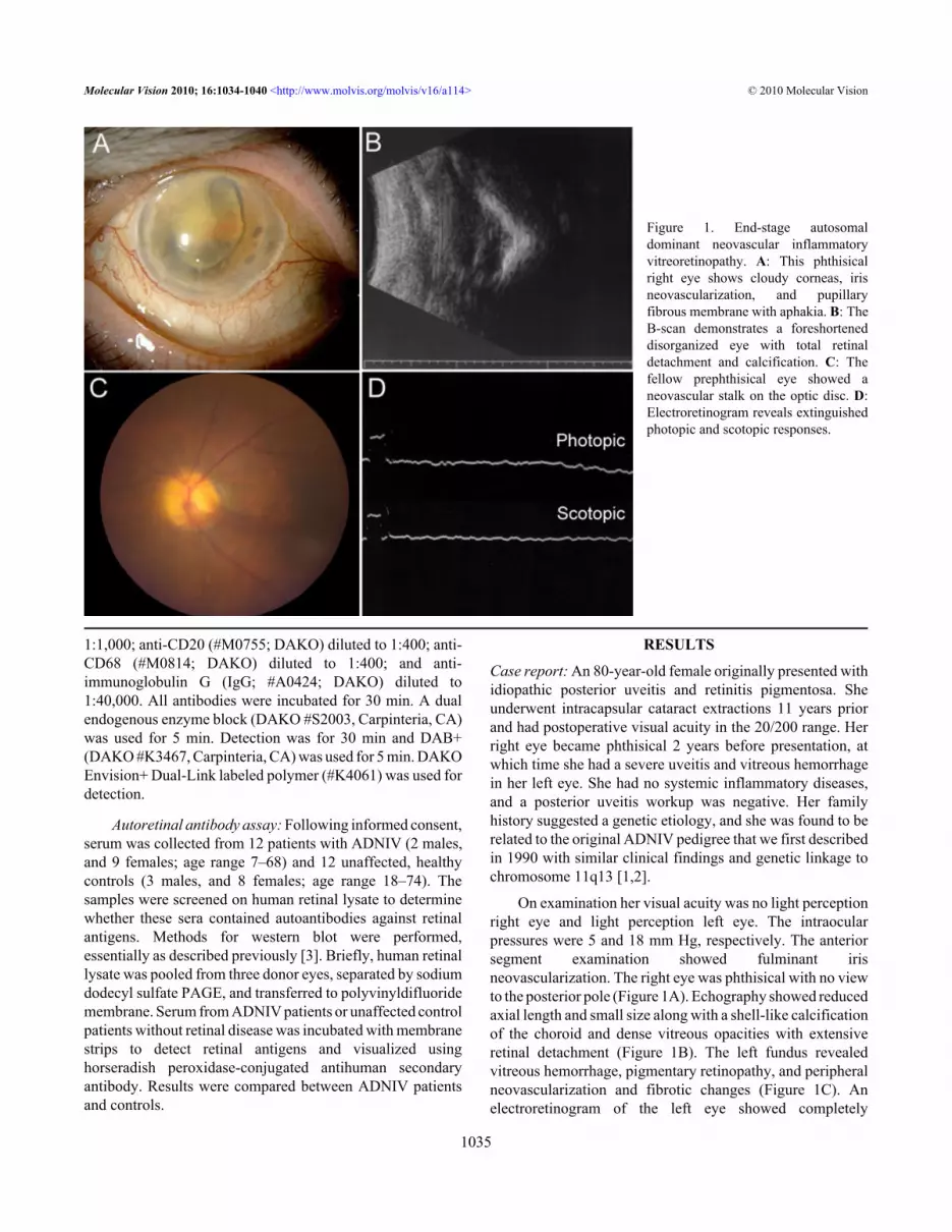
On examination her visual acuity was no light perceptionright eye and light perception left eye. The intraocularpressures were 5 and 18 mm Hg, respectively. The anteriorsegment examination showed fulminant irisneovascularization. The right eye was phthisical with no viewto the posterior pole (Figure 1A). Echography showed reducedaxial length and small size along with a shell-like calcificationof the choroid and dense vitreous opacities with extensiveretinal detachment (Figure 1B). The left fundus revealedvitreous hemorrhage, pigmentary retinopathy, and peripheralneovascularization and fibrotic changes (Figure 1C). Anelectroretinogram of the left eye showed completely
Figure 1. End-stage autosomaldominant neovascular inflammatoryvitreoretinopathy. A: This phthisicalright eye shows cloudy corneas, irisneovascularization, and pupillaryfibrous membrane with aphakia. B: TheB-scan demonstrates a foreshorteneddisorganized eye with total retinaldetachment and calcification. C: Thefellow prephthisical eye showed aneovascular stalk on the optic disc. D:Electroretinogram reveals extinguishedphotopic and scotopic responses.
Molecular Vision 2010; 16:1034-1040 <http://www.molvis.org/molvis/v16/a114> © 2010 Molecular Vision
1035

extinguished photopic and scotopic response (Figure 1D).Two years later, the left eye went into phthisis, andechography revealed tractional retinal detachment andposterior calcification. These clinical findings werecharacteristic of end-stage disease in several members of theADNIV pedigree. When the patient expired, a pathologicalexamination of the eyes was performed.
Pathology findings: Gross examination of the eyes revealedshrunken, firm, grossly distorted globes. There weresignificant corneal opacities, collapsed anterior chambers, and
a gray-brown, irregular, firm material within the vitreouscavities. The retinas and the choroids were detached, withaccumulation of suprachoroidal fluid.
Typical features of phthisis bulbi were apparent on pupil–optic nerve sections (Figure 2). The cornea and sclera werethick, forming a shrunken square outline. The intraocularcontents were grossly disorganized. An anterior hyaloidfibrovascular connective tissue detached the ciliary body, withunderlying suprachoroidal effusions. The membraneextended over the iris and into a collapsed anterior chamber.
Figure 2. Masson’s trichrome stain ofautosomal dominant neovascularinflammatory vitreoretinopathy(ADNIV) eye demonstratesfibrovascular proliferation and featuresof phthisis. A: There was massivefibrovascular connective tissue withinthe pupil, overlying the iris, and causingdetachment of the ciliary body. B: Asupraciliary effusion was presentoutside the detached ciliary body. C:The sclera was thickened. D: There werea few lens remnants present. E: Afibrovascular stalk extended from theoptic nerve to the peripheral retina. F:The retina was detached, atrophic, andosseous metaplasia was present.
Figure 3. Antiretinal autoantibodies. A:Control serum from 12 subjects wasapplied to human retinal proteinsseparated by sodium dodecyl sulfatePAGE. Nonspecific banding patternswere detected. B: Serum from 12autosomal dominant neovascularinflammatory vitreoretinopathy patientswas applied to similar retinal blots, andno specific bands or unique patternswere detected. The major band in allsamples indicates cross-reaction ofsecondary antibody with endogenoushuman immunoglobulin G (IgG) chains.
Molecular Vision 2010; 16:1034-1040 <http://www.molvis.org/molvis/v16/a114> © 2010 Molecular Vision
1036

The retinas were atrophic, cystic, and completely detached. Afibrovascular stalk extended from the disc anteriorly througha vitreous filled with proteinacious exudates (congo rednegative, not shown). Osseous metaplasia was present alongthe retinal pigment epithelium and around atrophic optic
nerves. These features were also present in control eyes thatwere phthisical from glaucoma, a noninflammatory conditionwithout continuous autoimmunity found in ADNIV.
Autoretinal antibodies: To determine whether systemicautoretinal antibodies were present in ADNIV patients, serum
Figure 4. B-cell markers were absent in autosomal dominant neovascular inflammatory vitreoretinopathy (ADNIV) eyes. A: Lymph nodetissue is a positive control for the B-cell marker, cluster of differentiation-20 (CD20), which was shown by the brown color reaction. B-C:ADNIV supraciliary space and vitreous showed only rare CD20 positive cells. D: Placental tissue is a positive control for immunoglobulin-G (IgG), which was shown by the brown color reaction. E-F: IgG was rarely detected in ADNIV eyes. The scale bar represents 50 µm.
Molecular Vision 2010; 16:1034-1040 <http://www.molvis.org/molvis/v16/a114> © 2010 Molecular Vision
1037

Figure 5. T-cell markers were detected in autosomal dominant neovascular inflammatory vitreoretinopathy (ADNIV) eyes. A: Lymph nodetissue is a positive control for the T-cell marker, cluster of differentiation-3 (CD3), which was shown by the brown color reaction. B-C: InADNIV eyes, the supraciliary space and vitreous showed CD3 positive cells. D-E: In control (non-ADNIV) phthisical eyes, CD3 positivecells were absent. F: Lymph node tissue is a positive control for the T-cell marker, cluster of differentiation-4 (CD4), which was shown bythe brown color reaction. G-H: In ADNIV eyes, the supraciliary space and vitreous both showed CD4 positive cells. I-J: In control (non-ADNIV) phthisical eyes, CD4 positive cells were absent. K: Lymph node tissue is a positive control for the T-cell marker, cluster ofdifferentiation-8 (CD8), which was shown by the brown color reaction. L: In ADNIV eyes, the supraciliary space showed CD8 positive cells.M: In the ADNIV vitreous, CD8 positive cells were absent. N-O: In control (non-ADNIV) phthisical eyes, CD8 positive cells were absent.The scale bar represents 50 μm.
Molecular Vision 2010; 16:1034-1040 <http://www.molvis.org/molvis/v16/a114> © 2010 Molecular Vision
1038

antibodies were applied to retinal protein blots. Sera from 12control patients without ADNIV and 12 patients with ADNIVwere compared (Figure 3). Although some immunoreactivebands were present in both the ADNIV and unaffected sera,no consistent distinct protein band or pattern of bands waspresent in the ADNIV subjects when compared to controlsamples.B-cell and immunoglobulin G immunohistopathology: Aseries of immunohistochemical markers for immune cells wasapplied to the ADNIV eyes. Antibody markers for both CD20(a marker for B lymphocytes) and IgG were applied to theADNIV eyes, but no CD20 cells or clusters of IgG weredetected (Figure 4). These markers were also not seen in thecontrol phthisis eyes.T-cell immunohistopathology: In the ADNIV eyes, CD3-positive (CD3+) cells were detected within a supraciliaryeffusion (Figure 5). Such an effusion was not present in thecontrol eyes. CD3+ cells were also present in the vitreous. Todistinguish these T cells, antibody markers for CD8 and CD4were applied. Both CD8+ and CD4+ cells were present in thesupraciliary effusion, but only CD4+ cells were found in thevitreous.
The macrophage marker CD68 was detected infrequentlyand did not show expression differences between ADNIV andcontrol eyes (data not shown).
DISCUSSIONThese findings support the hypothesis that an underlyingocular immune dysfunction is present in ADNIV. CD4expression, which is found predominantly on the surface ofhelper T cells, was found in both the vitreous and supraciliaryspace. CD8 expression, which is found predominantly on thesurface of cytotoxic T cells, was mostly restricted to thesupraciliary space. By comparison, there were few B cells,IgG, and no specific antiretinal autoantibodies. This suggeststhat cell-mediated rather than antibody-mediatedautoimmunity may be the primary inflammatory mechanism.
The genetic locus for ADNIV was mapped tochromosome 11q13 in 1992 [2], but the causative generemains unknown. These findings suggest genes that functionin T-cell activation could be prioritized in candidate genesequencing. For example, one gene in the interval is tumornecrosis factor (TNF) receptor superfamily member 19(TNFRSF19L), a member of the TNF receptor superfamily,which can stimulate T-cell proliferation in the presence ofCD3 signaling [4]. Until the causative gene is found andhighly specific therapy can be applied, current managementof ADNIV involves local immunosuppression with periocularor intraocular Kenalog. This nonspecific therapy might beoptimized by medications directed at the specific mediatorsof ADNIV. Our findings suggest that therapeutic strategiestargeting T cells may be more effective than nonspecifictherapies or those that target B cells. For example, drugs, such
as cyclosporin A, FK06, anti-CD3, and rapamycin, that targetT-cell activation and downstream T-cell pathways might havegreater effect than B-cell drugs, such as cyclophosphamide,mycophenylate mofetil, anti-inducible costimulatorymolecule (ICOS), anti-CD20, or anti-CD52 [5]. Like someother uveitic conditions, ADNIV eventually becomesresistant to conventional steroid immunosuppression. It ispossible that steroid refractory T cells are present, andtherapies directed at steroid-independent mechanisms mayovercome this limited response [6].
The main limitation of this study is the few number ofeyes, but ADNIV is rare and there are no previoushistopathological reports. The eyes in this study werephthisical and represent late stage ADNIV. Nevertheless,there is an ongoing autoimmune reaction in this disease, andphthisis in the ADNIV eye seems to be immunologicallydifferent from that observed in control eyes with phthisis fromother conditions [7,8]. This study provides a basis for futurestudies that will test vitreous samples from early stage ADNIVto confirm the presence of T cells [9].
ACKNOWLEDGMENTSSupported by an unrestricted grant from Research to PreventBlindness.
REFERENCES1. Bennett SR, Folk JC, Kimura AE, Russell SR, Stone EM,
Raphtis EM. Autosomal dominant neovascular inflammatoryvitreoretinopathy. Ophthalmology 1990; 97:1125-35.[PMID: 2234842]discussion 35–6
2. Stone EM, Kimura AE, Folk JC, Bennett SR, Nichols BE, StrebLM, Sheffield VC. Genetic linkage of autosomal dominantneovascular inflammatory vitreoretinopathy to chromosome11q13. Hum Mol Genet 1992; 1:685-9. [PMID: 1284594]
3. Mullins RF, Skeie JM, Malone EA, Kuehn MH. Macular andperipheral distribution of ICAM-1 in the humanchoriocapillaris and retina. Mol Vis 2006; 12:224-35. [PMID:16604055]
4. Sica GL, Zhu G, Tamada K, Liu D, Ni J, Chen L. RELT, a newmember of the tumor necrosis factor receptor superfamily, isselectively expressed in hematopoietic tissues and activatestranscription factor NF-kappaB. Blood 2001; 97:2702-7.[PMID: 11313261]
5. Miao CH. Recent advances in immune modulation. Curr GeneTher 2007; 7:391-402. [PMID: 17979685]
6. Lee RW, Schewitz LP, Nicholson LB, Dayan CM, Dick AD.Steroid refractory CD4+ T cells in patients with sight-threatening uveitis. Invest Ophthalmol Vis Sci 2009;50:4273-8. [PMID: 19339737]
7. Apple DJ, Rabb MF. Phthsis bulbi. Ocular Pathology: ClinicalApplications and Self-Assessment. 5th ed. St. Louis: Mosby;1998. p. 304–6.
8. Spencer WH. Atrophy of the Eye and Phthisis Bulbi. (Chapter4, Sclera). In: Spencer WH, editor. Ophthalmic Pathology: anatlas and textbook. 4th ed. Philadelphia: W.B. Saunders;1996. p. 342–5.
Molecular Vision 2010; 16:1034-1040 <http://www.molvis.org/molvis/v16/a114> © 2010 Molecular Vision
1039

9. Davis JL, Miller DM, Ruiz P. Diagnostic testing of vitrectomyspecimens. Am J Ophthalmol 2005; 140:822-9. [PMID:16310459]
Molecular Vision 2010; 16:1034-1040 <http://www.molvis.org/molvis/v16/a114> © 2010 Molecular Vision
The print version of this article was created on 4 June 2010. This reflects all typographical corrections and errata to the articlethrough that date. Details of any changes may be found in the online version of the article.
1040


