Embed Size (px)
Citation preview

Diabetic Retinopathy and Diabetic Macular Edema
Gandorfer A (ed): Pharmacology and Vitreoretinal Surgery.Dev Ophthalmol. Basel, Karger, 2009, vol 44, pp 1–13
VEGF Inhibitors and Vitrectomy for Diabetic VitreoretinopathyCarlos E. Cury Jr � Eduardo B. Rodrigues � Carsten H. Meyer
Department of Ophthalmology, University of Bonn, Bonn, Germany
Abstract
Diabetic retinopathy (DR) is one of the leading causes of blindness worldwide. Breakdown of the blood-retinal barrier in DR promotes accumulation of a high concentration of pre-retinal serum-derived chemoattractants, thereby stimulating cellular migration on the attached posterior hyaloid. This review assesses the role of intravitreal application of vascular endothelial growth factor (VEGF) inhibitors in combination with surgical removal of the vitreous. Vitrectomy with removal of the pos-terior hyaloids wash out the pocket of preretinal growth factors and enhance the diffusion of macro-molecules, including VEGF, insulin-like growth factor 1 or histamines, from the retinal into the vitreous cavity for further absorption through the anterior segment outflow pathways. The release of tractional forces induced by the vitreomacular traction or epiretinal membranes demonstrated a strong correlation to the reduction of retinal thickness in DME. Three techniques are described to remove the pre-retinal thickened and adherent vitreous: (1) delamination, (2) segmentation, and (3) en block dissection. A better visualization of remaining cortex vitreous or adjacent epiretinal mem-brane and safer removal may be achieved by a better intraoperative visualization using a variety of vital dyes to satin retinal tissue (chromovitrectomy). Anti-VEGF treatments may represent an alterna-tive adjunctive treatment for DR. Copyright © 2009 S. Karger AG, Basel
Diabetic retinopathy (DR) is one of the leading causes of blindness worldwide and the most common microvascular complication of diabetes [1]. During the first two decades of the disease, nearly all patients with type 1 diabetes and over 60% with type 2 diabetes develop retinopathy. In the Wisconsin Epidemiologic Study of Diabetic Retinopathy, also named WESDR, 3.6% of younger-onset patients and 1.6% of older-onset patients were blind [1]. Duration of diabetes and severity of hyperglycemia are the major risk factors for DR, while others include age, type of diabetes, clotting factors, and renal dis-ease [2]. Timely application and reapplication of laser photocoagulation is the mainstay of treatment to reduce visual loss and to avoid the need for vitrectomy in patients with more advanced complications of diabetic retinopathy [3]. However, despite preventive regimens and timely treatment, substantial numbers of eyes will develop complications of proliferative retinopathy and may become candidates for vitrectomy [4].
DOP009-044.indd 1DOP009-044.indd 1 2009-02-26 12:452009-02-26 12:45

2 Cury Jr · Rodrigues · Meyer
Due to the limitations of current treatment approaches, new pharmacological therapies have being developed. The novel drugs target specific biochemical path-ways that cause DR through involvement of protein kinase C (PKC) activation, oxida-tive stress, the angiogenesis pathway, and the glycation and sorbital pathway. These treatments aim to prevent diabetes-induced damage to the retinal microvasculature [5]. In this chapter the role of intravitreal application of vascular endothelial growth factor (VEGF) inhibitors in combination with surgical removal of the vitreous will be discussed.
Role of the Vitreous and Various Factors Including VEGF in Diabetic
Vitreoretinopathy
In healthy eyes ultrastructural components of the vitreous can be observed histologi-cally or histochemically, and basically they are mainly collagen type II, hyaluronan acid and hyalocytes [6]. In diabetic eyes, consecutive studies have demonstrated elevated levels of various cytokines such as VEGF, insulin-like growth factor-1 and histamines [7]. Pars plana vitrectomy with consecutive removal of the posterior hyaloids may resolve the diabetic macular edema (DME) and improve the central vision of patients who previously failed to respond to conventional laser photocoagulation treatment.
Four theories may support this hypothesis [7]: Mechanical Release Theory. Breakdown of the BRB in DR promotes accumula-
tion of a high concentration of preretinal serum-derived chemoattractants, thereby stimulating cellular migration on the attached posterior hyaloid. In addition, molecu-lar changes in the vitreous induced by hyperglycemia may thicken and enhance its adherence to the retinal surface. Sebag et al. [9] suggested that the abnormal cross-linking might affect the collagen architecture structure in diabetic vitreous. The con-traction of retinal cells, preretinal elements and the vitreous may induce tangential and anteroposterior traction and exacerbation of DME with vitreomacular traction (VMT) [8–10]. Release of tractional forces induced by the vitreous or epiretinal membranes demonstrated a strong correlation to the reduction of retinal thickness in DME. Moreover, several retrospective clinical studies have shown that vitrectomy and removal of the posterior hyaloid may lead to visual improvement of 2 lines in 38–92% of the eyes. Recently, Tachi and Ogino [11] observed the resolution of DME eyes within 3 months after vitrectomy without inner limiting membrane (ILM) peel-ing and questioned whether there is an additional beneficial effect of ILM removal.
Chemical Diffusion Theory. In eyes with diabetic retinopathy there may be an abnor-mal concentration of growth factors under the abnormally attached premacular pos-terior hyaloid thereby exacerbating DME. Vitrectomy with removal of the posterior hyaloids and vitreous gel may release and wash out the pocket of preretinal growth factors and enhance the diffusion of macromolecules, including VEGF, insulin-like growth factor 1 or histamines, from the retinal into the vitreous cavity for further
DOP009-044.indd 2DOP009-044.indd 2 2009-02-26 12:462009-02-26 12:46

VEGF Inhibitors and Vitrectomy for Diabetic Vitreoretinopathy 3
absorption through the anterior segment outflow pathways. In 1992, Lewis et al. [12] described a positive effect on visual acuity of pars plana vitrectomy on diabetic patients who had a thickened and taut posterior hyaloid traction on the macula. In the follow-ing years, numerous retrospective studies have shown the advantage of vitrectomy and removal of the posterior hyaloid on morphologic and functional results [13, 14].
Decrease or Wash-Out of Pro-Angiogenic Growth Factors – VEGF. Previous reports by Funatsu’s group have associated the aqueous VEGF concentration or determined its vitreous concentration in patients with DR and DME. A clear relationship has been established between structural and molecular parameters demonstrating that there are differences in the concentration of cytokines between DME and normal eyes. VEGF, a vascular endothelial cell mitogen and potent permeability factor, is produced by glial cells, retinal pigment epithelial cells, and vascular endothelial cells and is normally pres-ent in the retina and vitreous in low levels. Retinal hypoxia upregulates VEGF produc-tion, which results in abnormal angiogenesis, may increase in vascular permeability. Chronic hyperglycemia of uncontrolled diabetes leads to increased cellular levels of dia-cylglycerol, which in turn increases the synthesis of VEGF and also contributes to the microvascular abnormalities in DR. Inhibition of either VEGF moderates the microvas-cular complications seen in experimental animal models. Recent work also disclosed elevated levels of VEGF in ocular fluids of patients with proliferative diabetic retinopa-thy (PDR) [15]. These studies also point out the growth of new vessels from the retina or optic nerve to occur as a result of VEGF release into the vitreous cavity as a response to ischemia. Furthermore, injection of VEGF into normal primate eyes induces the same pathologic processes seen in diabetic retinopathy, including microaneurysm forma-tion and increased vascular permeability [16]. In addition, experiments in animals have suggested a central role for the 165 isoform of VEGF specifically in the pathogenesis of DME, and increased retinal VEGF164 (the rodent equivalent to primate VEGF165) levels in this model coincide temporally with breakdown of the BRB [17]. Clinical stud-ies demonstrate that intravitreal VEGF concentration increased as subjects progressed from early diabetic retinopathy to active PDR [16]. Gandorfer et al. [13] reported on the resolution of DME after vitrectomy in eyes without any evidence of VMT. The charac-terization of molecular and cellular events causing retinal microvascular abnormalities in DR has provided new targets for pharmacologic manipulation.
Oxygenation Hypothesis. Experimental data suggest that both scatter laser treat-ment and vitrectomy have a positive effect on the improvement of retinal oxygen-ation. Stefansson et al. [18], performed pars plana vitrectomy on one eye in cats and created a branch retinal vein occlusion in both eyes while the retinal oxygen tension was measured simultaneously in both. In the nonvitrectomized eye the branch reti-nal vein occlusion led to severe hypoxia of the retina. In the eye where vitrectomy had been performed there was insignificant change of the retinal oxygen tension with the vein occlusion. The beneficial effect of vitrectomy on DME can be further understood through improved oxygenation of the retina. Following vitrectomy (or a posterior vit-reous detachment) the ischemic retina receives oxygen from the vitreous cavity and
DOP009-044.indd 3DOP009-044.indd 3 2009-02-26 12:462009-02-26 12:46

4 Cury Jr · Rodrigues · Meyer
the oxygenation of the tissue is increased. The increased oxygen level leads to reduced VEGF production and decreased vascular permeability as well as arteriolar vasocon-striction and reduced blood flow, with decreased hydrostatic pressure in the capillaries and venules and reduced edema formation. Vitrectomy alleviates hypoxia in ischemic areas of the retina which may reduce VEGF production in these areas and decreases new vessel formation [18].
Anti-VEGF Agents
A key molecule being targeted in current clinical trials is VEGF. Anti-VEGF treat-ments may represent as an alternative adjunctive treatment for DR and three pres-ently available anti-VEGF agents are pegaptanib, bevacizumab and ranibizumab [19, 20].
a
b
c
Fig. 1. a Color retinography, angiography and optical coherence tomography of a patient with type 2, submitted to prior laser photocoagulation and triancinolone acetonide, demonstrating clinical significant macular edema (CSME), VA 20/50. b Images of the 30th post-PPV plus avastin 1.25 mg showing worsening of the CSME. c Images of 1-year follow-up, after another two avastin injections, demonstrating regression of the edema, VA 20/25.
DOP009-044.indd 4DOP009-044.indd 4 2009-02-26 12:462009-02-26 12:46

VEGF Inhibitors and Vitrectomy for Diabetic Vitreoretinopathy 5
Pegaptanib
An inhibition of VEGF165 can be accomplished with an aptamer, a new therapeutic class of highly selective nonbiologic agents. Pegaptanib sodium is the first FDA-approved ophthalmologic anti-VEGF agent for the treatment of choroidal neovascularization (CNV) from age-related macular degeneration (AMD). Animal model studies have shown that intravitreal injection of pegaptanib can decrease the breakdown of the BRB characteristic of diabetes and can even reverse this damage to some degree. A recent phase II study reported that intravitreal pegaptanib has promising results for DME when given every 6 weeks for 30 weeks. The results showed better visual acuity at week 36 with 0.3 mg injection (20/50) as compared with sham injection (20/63) (p = 0.04); a mean central retinal thickness decrease of 68 μm with 0.3 mg, versus an increase of 4 μm with sham (p = 0.02); and reduction of subjects requiring photocoagulation
a
c
b
d
Fig. 2. Color retinography of a 32-year-old type 1 DM patient. a, b Right and left eye demonstrating a TRD affecting the macula. c, d 30th postoperative day view of right and left eye submitted to PPV plus membranes dissection and avastin 1.25 mg.
DOP009-044.indd 5DOP009-044.indd 5 2009-02-26 12:462009-02-26 12:46

6 Cury Jr · Rodrigues · Meyer
in each pegaptanib arm (0.3, 1, 3 mg) [21]. Prospective clinical investigation should disclose the role of pegaptanib for the management of DME. Retrospective analysis of these data demonstrated some efficacy on retinal neovascularization as well. Phase 3 trials of pegaptanib for DME are currently being conducted.
Bevacizumab
Bevacizumab is a full-length recombinant humanized antibody active against all iso-forms of VEGF-A. The US FDA approved bevacizumab in February 2004 for treatment for first-line metastatic colorectal cancer [22]. Case reports and small observational series show the efficacy of off-label intravitreal bevacizumab to treat exudative AMD [23], macular edema from nonischemic central retinal vein occlusion [24], iris neovas-cularization [25] and pseudophakic cystoid macular edema [26]. Haritoglou et al. [27] disclosed a prospective consecutive noncomparative case series of 51 patients treated with bevacizumab for diffuse refractory DME followed for up to 12 weeks. Their results showed a significant reduction in mean retinal thickness, from 425 μm at 2 weeks to 377 μm at 12 weeks (p = 0.001), although no significant changes in ETDRS letters were
Fig. 3a–c. Color retinography and angiography of a only eye patient with very severe PDR. d, e Color retinography and late phase angiography 15 days after avastin injection, demonstrating involution of the fibrovascular complex. f 15th day post-PPV plus membrane dissection color retinography.
a b c
d e f
DOP009-044.indd 6DOP009-044.indd 6 2009-02-26 12:462009-02-26 12:46
VEGF Inhibitors and Vitrectomy for Diabetic Vitreoretinopathy 7
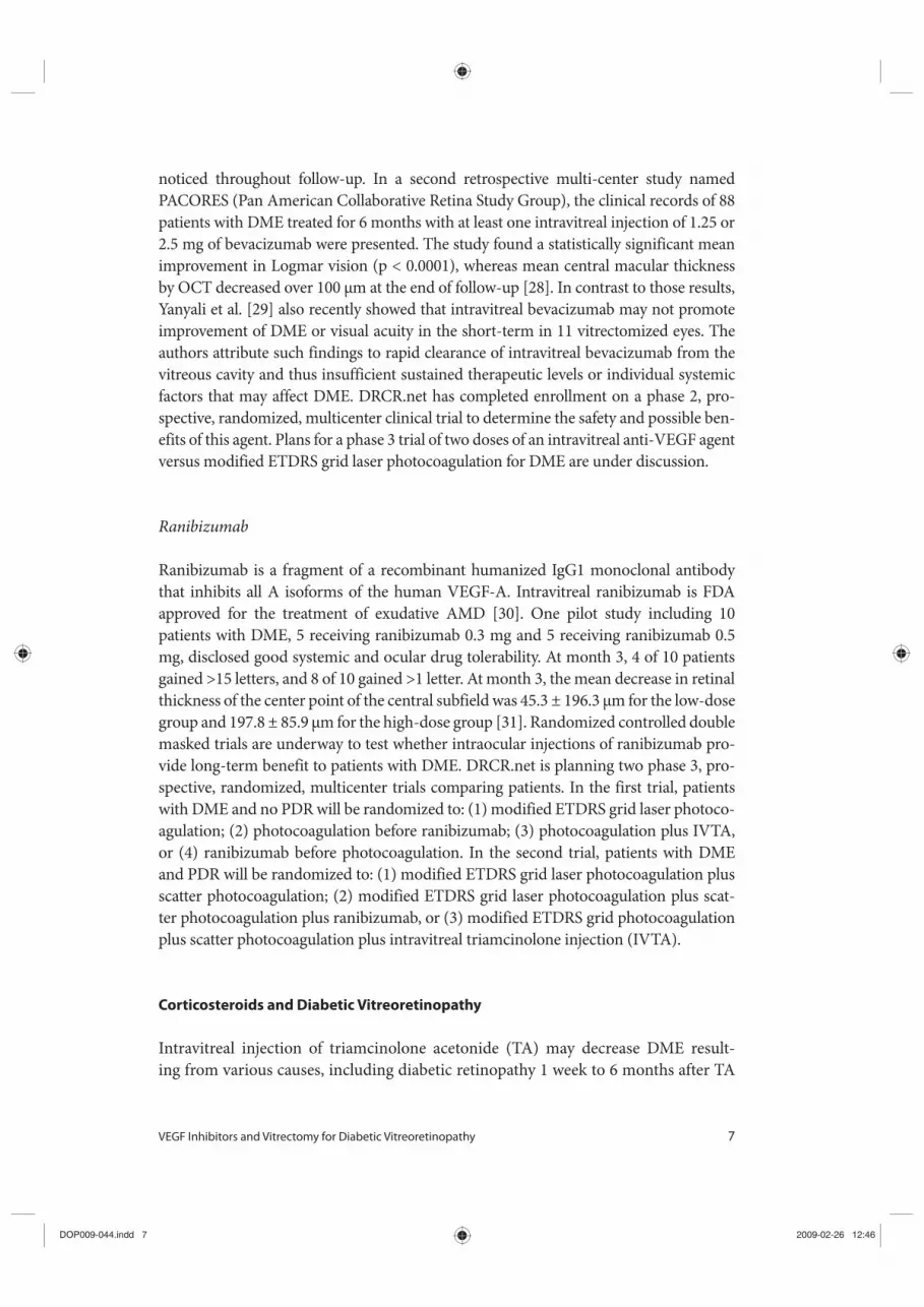
noticed throughout follow-up. In a second retrospective multi-center study named PACORES (Pan American Collaborative Retina Study Group), the clinical records of 88 patients with DME treated for 6 months with at least one intravitreal injection of 1.25 or 2.5 mg of bevacizumab were presented. The study found a statistically significant mean improvement in Logmar vision (p < 0.0001), whereas mean central macular thickness by OCT decreased over 100 μm at the end of follow-up [28]. In contrast to those results, Yanyali et al. [29] also recently showed that intravitreal bevacizumab may not promote improvement of DME or visual acuity in the short-term in 11 vitrectomized eyes. The authors attribute such findings to rapid clearance of intravitreal bevacizumab from the vitreous cavity and thus insufficient sustained therapeutic levels or individual systemic factors that may affect DME. DRCR.net has completed enrollment on a phase 2, pro-spective, randomized, multicenter clinical trial to determine the safety and possible ben-efits of this agent. Plans for a phase 3 trial of two doses of an intravitreal anti-VEGF agent versus modified ETDRS grid laser photocoagulation for DME are under discussion.
Ranibizumab
Ranibizumab is a fragment of a recombinant humanized IgG1 monoclonal antibody that inhibits all A isoforms of the human VEGF-A. Intravitreal ranibizumab is FDA approved for the treatment of exudative AMD [30]. One pilot study including 10 patients with DME, 5 receiving ranibizumab 0.3 mg and 5 receiving ranibizumab 0.5 mg, disclosed good systemic and ocular drug tolerability. At month 3, 4 of 10 patients gained >15 letters, and 8 of 10 gained >1 letter. At month 3, the mean decrease in retinal thickness of the center point of the central subfield was 45.3 ± 196.3 μm for the low-dose group and 197.8 ± 85.9 μm for the high-dose group [31]. Randomized controlled double masked trials are underway to test whether intraocular injections of ranibizumab pro-vide long-term benefit to patients with DME. DRCR.net is planning two phase 3, pro-spective, randomized, multicenter trials comparing patients. In the first trial, patients with DME and no PDR will be randomized to: (1) modified ETDRS grid laser photoco-agulation; (2) photocoagulation before ranibizumab; (3) photocoagulation plus IVTA, or (4) ranibizumab before photocoagulation. In the second trial, patients with DME and PDR will be randomized to: (1) modified ETDRS grid laser photocoagulation plus scatter photocoagulation; (2) modified ETDRS grid laser photocoagulation plus scat-ter photocoagulation plus ranibizumab, or (3) modified ETDRS grid photocoagulation plus scatter photocoagulation plus intravitreal triamcinolone injection (IVTA).
Corticosteroids and Diabetic Vitreoretinopathy
Intravitreal injection of triamcinolone acetonide (TA) may decrease DME result-ing from various causes, including diabetic retinopathy 1 week to 6 months after TA
DOP009-044.indd 7DOP009-044.indd 7 2009-02-26 12:462009-02-26 12:46

8 Cury Jr · Rodrigues · Meyer
injection through inhibition of VEGF cytokines. Recent prospective, randomized clin-ical trials have demonstrated generally favorable outcomes, the Diabetic Retinopathy Clinical Research network (DRCR.net) has completed enrollment on a three-year, randomized, prospective, multicenter clinical trial comparing two doses (1 mg and 4 mg) of preservative-free IVTA with modified early treatment diabetic retinopathy study (ETDRS) photocoagulation for DME. Long-term efficiency and safety reports of repeated intravitreal injections remain uncertain, but encouraging reports promote the development of long-term sustained release devices for corticosteroid therapy. The most important complication of IVTA is increased intraocular pressure resulting in secondary open-angle glaucoma, which sometimes may be severe and intractable. Elevation of IOP up to 24 mm Hg may occur in about 40% of patients, usually within about 3 months. The second most important complication of IVTA is cataract forma-tion, which may become visually significant in about half of eyes within 1 year. Other reported complications of IVTA include endophthalmitis, pseudoendophthalmitis, retinal detachment, lens trauma, and vitreous hemorrhage.
Dexamethasone is theoretically a more potent corticosteroid than TA, and intra-vitreal injections of dexamethasone have been shown to produce high intravitreal drug levels without toxic effects. Unfortunately, the short intraocular half-life of dex-amethasone after intravitreal injection (approximately 3 h) makes this approach less useful for clinical therapy. A phase 6-month study randomized 315 patients with per-sistent DME with visual acuity of 20/40 to 20/200 in the study eye to observation or a single treatment with a dexamethasone implant called DDS, 350 or 700 μg. At day 90 (primary end point), an improvement in VA of 10 letters or more was achieved by a greater proportion of patients treated with dexamethasone-DDS. Results were similar in patients with diabetic retinopathy, vein occlusion, or uveitis or Irvine-Gass syndrome. During 3 months of observation, 11% of treated patients and 2% of observed patients had intraocular pressure increases of 10 mm Hg or higher. The study reported that Dexamethasone-DDS, 700 μg, may have potential as a treatment for persistent macular edema.
PPV for DR: Surgical Techniques
The diabetic retinopathy vitrectomy study (DRVS) demonstrated the efficacy of vit-rectomy for severe proliferative diabetic retinopathy with non-clearing hemorrhage and progression despite laser treatment [20, 32]. At the time of the DRVS up to 20% of eyes progressed to no light perception. Today many advances have improved our success rates, including the advent of high-speed vitreous cutters, endolaser, wide-field viewing systems and pharmacological management of fibrin, retinal neovascu-larization and retinal edema.
The main objectives of vitrectomy are to remove media opacities, for control of active progressive PDR completely relieve all tractional adhesions, and manage
DOP009-044.indd 8DOP009-044.indd 8 2009-02-26 12:462009-02-26 12:46

VEGF Inhibitors and Vitrectomy for Diabetic Vitreoretinopathy 9
recurrent complications from previous vitrectomy [33]. There are now reports in the literature suggesting that vitrectomy surgery may be helpful in eyes with refractory macular edema in eyes not responsive to laser photocoagulation.
Techniques for Membrane Dissection: Delamination, Segmentation and en-bloc Removal
The three techniques to remove the pre-retinal thickened and adherent vitreous are (1) delamination, (2) segmentation, and (3) en block dissection. (1) Delamination starts with the removal of the partially detached posterior vitreous surface between the vitreous base and the edge of the fibrovascular adhesions. Using bimanual tech-niques, from anterior to posterior, the edge of the fibrovascular membrane is reflected and then it can be grasped with a forceps and avulsed. (2) Segmentation is a tech-nique used to release retinal traction caused by preretinal fibrovascular proliferation. Anteroposterior traction then is released by circumferentially cutting the posterior vitreous surface around the area of epiretinal proliferation followed by cutting of the posterior vitreous surface between epicenters of fibrovascular adhesion, leaving islands of fibrovascular tissue. (3) During an en bloc technique, the anteroposterior vitreous traction may be used to elevate the edge of the fibrovascular membrane. Initially, the vitrectomy cutter is used to create a tunnel through the formed vitreous from the sclerotomy site to an area of vitreoretinal separation. The remaining poste-rior vitreous surface is left attached and assists in visualizing epicenters of adhesion between fibrovascular tissue and the retina. Horizontal scissors are used to amputate epicenters of fibrovascular vitreoretinal adhesion. A better visualization of remaining cortex vitreous or adjacent epiretinal membrane may be visualized by intraoperative staining with a variety of vital dyes (Chromovitrectomy). While fluorescein stains the vitreous collagen, trypan blue may assist to visualize cellular structures of epiretinal membrane. The ILM may be stained with indocyanine green or brilliant blue [34]. After the posterior vitreous surface and the entire fibrovascular membrane are freed from the retina, they may be removed using the vitrectomy probe.
High-Speed Cutters
Several high-speed vitreous cutters are now available for diabetic vitrectomy and pro-vide the advantage of more controlled vitreous removal and working on the retinal surface to remove complex diabetic membranes using the technique of indentation and delamination for removal of diabetic membranes and the concept of ‘one-step’ diabetic vitrectomy. The high-speed cutters can be used to delaminate membranes from the retinal surface and, in the majority of cases, eliminate the need for forceps, scissors, endodiathermy, healon dissection of membranes and other maneuvers
DOP009-044.indd 9DOP009-044.indd 9 2009-02-26 12:462009-02-26 12:46

10 Cury Jr · Rodrigues · Meyer
traditionally used to manage these complex patients. Once one becomes experienced with this approach, cautery and most other steps may be usually eliminated.
Vitreous Base Cleaning
Scleral depression is performed while viewing through the wide-field lens system to check the retinal periphery for tears. The vitreous cutter should perform a thorough cleaning of the vitreous base to assure that: (1) postoperative retina oxygenation will be efficacious; (2) diffusion of the cytokines from ciliary body to the retina and from retina to aqueous humor will be allowed, and (3) postoperative hemorrhage will be prevented.
Small-Gauge Vitrectomy
Vitreoretinal surgeons have been developing new and innovative ways to operate with less invasive techniques. Small gauge vitrectomy instrumentation is now avail-able allowing no-stitch surgery. 23- and 25-gauge vitrectomy instruments are now available from Bausch and Lomb, Alcon and others. These systems allow no-stitch transconjuntival insertion of vitreoretinal instrumentation, minimizing trauma to the eye and reducing the number of surgical steps. Each 25-gauge system utilizes a trocar-cannula system placed directly through the displaced conjunctiva as ports for instru-ments and infusion. Advantages of 25-gauge vitrectomy include a quieter eye with minimal patient discomfort and potentially less postoperative inflammation with more rapid visual recovery.
Pharmacologicaly-Assisted PPV: Anti-VEGF Agents and Corticosteroids
Diabetic Macular Edema
For DME pharmacologic agents including anti-VEGF or corticosteroids may be used intra- or postoperatively. Before surgery there is currently no rationale for the use of pharmacologic agents in DME. During surgery, the white corticosteroid triamcino-lone may be injected for better visualization of the semi-transparent vitreous thereby enabling a complete vitrectomy, and this procedure has been recently called chro-movitrectomy. Most patients with DR show a multilayered preretinal vitreoschisis. Therefore, when the vitreous is not stained, one or more layers of vitreous may remain, which may hamper postoperative vision improvement in patients with DME. In addi-tion to chromovitrectomy, at the end of vitrectomy either anti-VEGF or TA may be injected into the vitreous cavity as powerful adjuvants for vitrectomy for DME.
DOP009-044.indd 10DOP009-044.indd 10 2009-02-26 12:462009-02-26 12:46

VEGF Inhibitors and Vitrectomy for Diabetic Vitreoretinopathy 11
Proliferative Diabetic Retinopathy
There is recent evidence for a benefit of preoperative anti-VEGF agents as an adjuvant to vitrectomy for the management of severe PDR [35]. Bevacizumab can potentially inhibit early neovascularization, which might be one of the causes of early recurrent hemorrhage. Then, the chances of postoperative complications such as rebleeding or fibrinoid syndrome may be decreased. It has also been hypothesized that suppression of intraocular VEGF theoretically could reduce the risk of intraoperative hemorrhage during membrane dissection facilitating the surgery. With a better visibility the sur-geon may be less likely to create an iatrogenic retinal break. However, others recently proposed that even though bevacizumab may promote new vessel regression, it is unlikely to prevent early postoperative bleeding from vessels that have been injured mechanically during surgery. They suggest that the use of anti-VEGF at the end of surgery might delay the repairing process of injured blood vessels and potentially induce more recurrent hemorrhage during the early postoperative period.
New vessel formation and evolution may be affected and controlled by various growth factors and cytokines. Some factors may stimulate active vessel proliferation, while others induce fibrosis. When anti-VEGF is used, the balance among differ-ent growth factors may tip toward fibrosis inducing factors, causing contraction of the fibrovascular tissue. The manifested result may be more vitreoretinal traction or frank traction RD or even combined detachment. Arevalo et al. [36] concluded that tractional retinal detachment may occur or progress very shortly following admin-istration of intravitreal bevacizumab in patients with severe PDR. The development or progression of tractional retinal detachment (TRD) in PDR following intravitreal bevacizumab could have happened by rapid neovascular involution with accelerated fibrosis and posterior hyaloidal contraction as a response to decreased levels of VEGF. Based on that case series and their hypothesis, the PPV should be conducted no more than seven days after the injection of anti-VEGF agents to avoid the fearful complica-tion of TRD.
Conclusions
In DR, the gathering of preretinal serum-derived chemoattractants promotes stim-ulation of cellular migration on the attached posterior hyaloid. VEGF inhibitors in combination with surgical removal of the vitreous may be a reasonable approach to remove the pocket of pre-retinal growth factors and enhance the diffusion of macromolecules, including VEGF, insulin-like growth factor 1 or histamines from the retinal into the vitreous. In addition, the release of tractional forces induced by the vitreomacular traction promotes additional vision improvement and reduc-tion of macular thickness in DME. Sutureless 23–25 gauge instruments allowed the surgeon to be less aggressive to the tissues, reducing the surgical trauma and
DOP009-044.indd 11DOP009-044.indd 11 2009-02-26 12:462009-02-26 12:46

12 Cury Jr · Rodrigues · Meyer
consequently less inflammation. Preoperative drug-induced fibrovascular complex regression brought the possibility to a less bleeding procedure, facilitating the dis-section and reducing post-operative hemorrhage. In conclusion, new surgical tech-niques and pharmacological agents improved the therapeutic arsenal against this harmful disease.
References
1 Fong DS, Aiello LP, Gardner TW, et al: Retinopathy in diabetes. Diabetes Care 2004;27(suppl 1):84S-87S.
2 Preferred Practice Pattern: Diabetic Retinopathy. San Francisco, American Academy of Ophthalmology, 2003.
3 Aaberg TM, Abrams GW: Changing indications and techniques for vitrectomy in management of com-plications of diabetic retinopathy. Ophthalmology 1987;94:775–779.
4 Aaberg TM: Results of 100 consecutive vitrectomy procedures; in McPherson A (ed): Controversial Aspects of Vitreoretinal Surgery. St. Louis, Mosby, 1977, pp 245–249.
5 Yam JCS, Kwok AKH: Update on the treatment of diabetic retinopathy. Hong Kong Med J 2007;13:46–60.
6 Ryan SJ: Retina. St Louis, Elsevier Mosby, 2006, vol 3, pp 1–1923.
7 Meyer CH: Current treatment approaches in dia-betic macular edema. Ophthalmologica 2007;221: 118–131.
8 Lewis H, Abrams GW, Blumenkranz MS, Campo RV: Vitrectomy for diabetic macular traction and edema associated with posterior hyaloidal traction. Ophthalmology 1992;99:753–759.
9 Sebag J, Buckingham B, Charles MA, Reiser K: Biochemical abnormalities of vitreous in humans with proliferative diabetic retinopathy. Arch Ophthalmol 1992;110:1472–476.
10 Pendergast SD: Vitrectomy for diabetic macular edema associated with a taut premacular posterior hyaloid. Curr Opin Ophthalmol 1998;9:71–75.
11 Tachi N, Ogino N: Vitrectomy for diffuse macular edema in cases of diabetic retinopathy. Am J Ophthalmol 1996;122:258–260.
12 Lewis H: The role of vitrectomy in the treatment of diabetic macular edema. Am J Ophthalmol 2001; 131:123–125.
13 Gandorfer A, Messmer EM, Ulbig MW, Kampik A: Resolution of diabetic macular edema after surgical removal of the posterior hyaloid and the inner lim-iting membrane. Retina 2000;20:126–133.
14 Gandorfer A, Kampik A: Surgical removal of the internal limiting membrane in diabetic cystoid macular edema. Retina 2006;26:485–4856.
15 Aiello LP, Avery RL, Arrigg PG, et al: Vascular endothelial growth factor in ocular fluid of patients with diabetic retinopathy and other retinal disor-ders. N Engl J Med 1994;331:1480–1487.
16 Tolentino MJ, Mcleod DS, Taomoto M, et al: Pathologic features of vascular endothelial growth factor-induced retinopathy in the nonhuman pri-mate. Am J Ophthalmol 2002;133:373–385.
17 Qaum T, Xu Q, Joussen AM, et al: VEGF-initiated bloodretinal barrier breakdown in early diabetes. Invest Ophthalmol Vis Sci 2001;42:2408–2413.
18 Stefánsson E: Ocular oxygenation and the treatment of diabetic retinopathy. Surv Ophthalmol 2006;51: 364–380.
19 Emerson MV, Lauer AK: Emerging therapies for the treatment of neovascular age-related macular degeneration and diabetic macular edema. BioDrugs 2007;21:245–257.
20 Furlani BA, Meyer CH, Rodrigues EB, Maia M, Farah ME, Penha FM, Holz FG: Emerging pharma-cotherapies for diabetic macular edema. Expert Opin Emerg Drugs 2007;12:591–603.
21 Cunninghan ET Jr, Adamis AP, Altawell M, et al: Macugen diabetic retinopathy study group: a phase II randomized double-masked trial of pegaptanib, an antivascular endothelial growth factor aptamer, for diabetic macular edema. Ophthalmology 2005; 112:1747–1757.
22 Hurwitz H, Fehrenbacher L, Novotny W, et al: Bevacizumab plus irinotecan, fluorouracil, and leu-covorin for metastatic colorectal cancer. N Engl J Med 2004;350:2335–2342.
23 Rosenfeld PJ, Moshfeghi AA, Puliafito CA: Optical coherence tomography findings after an intravitreal injection of bevacizumab (Avastin) for neovascular age-related macular degeneration. Ophthal Surg Lasers Imaging 2005;36:331–335.
DOP009-044.indd 12DOP009-044.indd 12 2009-02-26 12:462009-02-26 12:46

VEGF Inhibitors and Vitrectomy for Diabetic Vitreoretinopathy 13
24 Rosenfeld PJ, Fung AE, Puliafito CA: Optical coher-ence tomography findings after an intravitreal injec-tion of bevacizumab (Avastin) for macular edema from central retinal vein occlusion. Ophthal Surg Lasers Imaging 2005;36:336–339.
25 Avery RL: Regression of retinal and iris neovascu-larization after intravitreal bevacizumab (Avastin) treatment. Retina 2006;26:352–354.
26 Mason JO, Albert MA, Vail R: Intravitreal bevaci-zumab (Avastin) for refractory pseudophakic cys-toid macular edema. Retina 2006;26:356–357.
27 Haritoglou C, Kook D, Neubauer A: Intravitreal bevacizumab (Avastin) therapy for persistent dif-fuse diabetic macular edema. Retina 2006;26:999–1005.
28 Arevalo JF, Fromow-Guerra J, Quiroz-Mercado H, Pan-American Collaborative Retina Study Group: Primary intravitreal bevacizumab (Avastin) for dia-betic macular edema: results from the Pan-American collaborative retina study group at 6-month follow-up. Ophthalmology 2007;114:743–750.
29 Yanyali A, Aytug B, Horozoglu F, Nohutcu AF: Bevacizumab (Avastin) for diabetic macular edema in previously vitrectomized eyes. Am J Ophthalmol 2007;144:124–126.
30 Heier JS, Atoszik AN, Pavan PR, et al: Ranibizumab for treatment of neovascular age-related macular degen-eration: a phase I/II multicenter, controlled, multi dose study. Ophthalmology 2006;113:642.E1–E4.
31 Chun DW, Heier JS, Topping TM, et al: A pilot study of multiple intravitreal injections of ranibizumab in patients with center-involving clinically significant diabetic macular edema. Ophthalmology 2006;113: 1706–1712.
32 Kroll P, Rodrigues EB, Hoerle S: Pathogenesis and classification of proliferative diabetic vitreoretinop-athy. Ophthalmologica 2007;221:78–94.
33 Smiddy WE, Flynn HW Jr: Vitrectomy in the man-agement of diabetic retinopathy. Surv Ophthalmol 1999;43:491–507.
34 Rodrigues EB, Maia M, Meyer CH, Penha FM, Dib E, Farah ME: Vital dyes for chromovitrectomy. Curr Opin Ophthalmol 2007;18:179–187.
35 Chen E, Park CH: Use of intravitreal bevacizumab as a preoperative adjunct for tractional retinal detachment repair in severe proliferative diabetic retinopathy. Retina 2006;26:699–700.
36 Arevalo F, Maia M, Flynn Jr HW, Saravia M, Avery RL, Wu L, Farah ME, Pieramici DJ, Berrocal MH, Sanchez JG: Tractional retinal detachment follow-ing intravitreal bevacizumab (Avastin) in patients with severe proliferative diabetic retinopathy Br J Ophthalmol 2008;92:213–216.
Carsten H. Meyer, MDDepartment of Ophthalmology, University of BonnErnst-Abbe-Strasse 2DE–53127 Bonn (Germany)Tel. +49 228 2871 11515, Fax +49 228 2871 5603, E-Mail [email protected]
DOP009-044.indd 13DOP009-044.indd 13 2009-02-26 12:462009-02-26 12:46


