Embed Size (px)
Citation preview
Dounomycin in the Treatment of ExperimentalProliferative Vitreoretinopathy
Effective Doses In Vitro and In Vivo
Peter Wiedemann,* Nino Sorgente,t Clara Dekhor,j- Randi Patterson,* Tai Tran,* and Stephen J. Ryan"
In previous studies the authors have shown that daunomycin, an anthracycline antibiotic, wheninjected into the vitreous effectively controls experimental proliferative vitreoretinopathy. Here weshow that by administering daunomycin intravitreally it is possible to achieve in vivo concentrationsthat prevent fibroblast proliferation in vitro. The authors have also determined that the half-life ofdaunomycin in the vitreous is 131 min, indicating that a critical concentration is maintained in theeye for longer than 4 hr after a single injection. Using 3H-daunomycin, the authors have found thatthe drug is eliminated across the retina; no significant binding of the drug to vitreous componentsoccurs. These studies demonstrate that it is possible to define the kinetics of drugs injected into thevitreous; and a knowledge of the distribution of any drug in ocular tissues is necessary to effectivelydetermine whether such drug is of therapeutic value. Invest Ophthalmol Vis Sci 26:719-725, 1985
Proliferative vitreoretinopathy (PVR) is the mostcommon complication of retinal reattachment sur-gery.1 The condition is characterized by the prolifer-ation of cells on both surfaces of the retina, resultingin membrane formation and traction on the retina.2
If the proliferation of cells in the vitreous that resultsin re-detachment of the retina could be inhibited,more favorable long-term results of surgery would beobtained. Control of cellular proliferation would pre-vent the secondary processes of contraction and col-lagen deposition, which lead to traction detachment.
A drug appropriate for the control of PVR mustpossess two qualities: it has to inhibit cell proliferationeffectively, and it must do this without intolerabletoxicity to the retina or other ocular structures. Theeffectiveness depends on the pharmacodynamic andpharmacokinetic characteristics of the drug. The de-sired pharmacokinetic properties include suitabilityfor local injection into the vitreous, as this results inthe highest possible concentration at the target site,thus eliminating or at least reducing systemic toxicity.
From the Department of Ophthalmology,* University of SouthernCalifornia, and the Estelle Doheny Eye Foundation,! Los Angeles,California.
Presented in part at the ARVO meeting, May 1983, Sarasota,Florida.
Supported in part by grant EY-03040 awarded by the NationalInstitutes of Health (National Eye Institute).
Submitted for publication: September 6, 1983.Reprint requests: Stephen J. Rvan. MD, Estelle Doheny Eye
Foundation, 1355 San Pablo Street, Los Angeles, CA 90033.
The drug must remain in the vitreous long enoughto affect the proliferation of cells during a sensitiveperiod of their cycle.
Previous studies have shown the anthracyclineantibiotic daunomycin to be effective in the controlof experimental PVR. A dose response study in therabbit showed that the detachment score, which is ameasure of the severity of membrane formation andtraction detachment following intravitreal injectionof cultured fibroblasts, was significantly lower intreated animals than in controls. At high doses,however, there was marked toxicity to the retina,causing detachment and degeneration.3
Obviously, it is very important to determinewhether it is possible to achieve useful concentrationsof the drug at the target site and whether suchconcentrations last long enough to be of value intreating clinical PVR. To determine what minimaldose has to be maintained for what length of time inthe vitreous to prevent proliferation of fibroblastswithout toxicity to the retina, we determined cellsurvival into vitro after exposure to daunomycin andperformed pharmacokinetic studies in the rabbit todetermine the concentrations of daunomycin and itsmetabolite daunomycinol in the vitreous and otherocular tissues.
Materials and MethodsMaterials
Amphotericin B was purchased from Sauibb(Princeton, NJ); Ciaramycin, from Schering Corpo-
719
720 INVESTIGATIVE OPHTHALMOLOGY & VISUAL SCIENCE / May 1985 Vol. 26
ration (Kenilworth, NJ); daunomycin and Sigmacote,from Sigma Chemical Company (St. Louis, MO.);[3H(G)]-daunomycin (specific activity, 2.5 Ci/mmol)and Protosol, from New England Nuclear (Boston,MA); fetal bovine serum and RPMI 1640 culturemedium, from Irvine Scientific (Santa Ana, CA).Daunomycinol was the generous gift of Dr. Arcamone(Farmitalla; Milano, Italy).
Animals
The investigations utilizing animals, as describedin this article, conform to the ARVO Resolution onthe Use of Animals in Research. The animals usedin this study were maintained in animal care facilitiesfully accredited by the American Association of Lab-oratory Animal Sciences.
The Effect of Daunomycin on Colony Forming Units
Rabbit dermal fibroblasts (3rd to 5th passage) wereseeded in 35-mm petri dishes (30,000 cells/dish) inRPMI 1640 medium containing 20% fetal bovineserum, 50 /xg/ml Garamycin, and 5 /*g/ml amphoter-icin B and incubated at 37°C in a humidified atmo-sphere of 5% CO2 and 95% air.
Seventy-two hours after seeding, the cells weretreated for 1 hr with various concentrations of dau-nomycin at final concentrations ranging from 100 to1000 nM. The fibroblasts were then washed two timeswith media, trypsinized, and harvested as a single cellsuspension. For each dose point, three dishes werepooled, and the suspension was counted in an elec-tronic particle counter (Coulter counter). For eachconcentration of daunomycin, six petri dishes (60-mm) were seeded at a density of 500 cells/dish.Control cells underwent all manipulations as thetreated cells except exposure to drug and received anamount of BSS (vehicle) equal to that of the experi-mental group.
After 2 weeks, the cells were fixed with methanolfor 10 min, and stained with Giemsa, and the numberof colonies containing more than 30 cells was deter-mined. The number of cells in a colony was deter-mined using a stereomicroscope. We considered col-onies of 30 or more cells to originate from normallyreproducing cells. The percentage survival was cal-culated in reference to the controls, and the logarithmof the percentage was plotted against increasing drugconcentration. The experiment was repeated threetimes.
The extent of cell damage as a function of exposuretime was examined by exposing cells to daunomycin
(500 nM) for different times between 30 min and5 hr.
Spectrofluorimetric Assay of Daunomycin
Three sets of six pigmented rabbits of either sex,weighing 2.5 to 3.5 kg, were anesthetized3 and injectedwith 10 nmol daumocyin into one eye only using aStepper pipette (Tridak; Highland, NY) and killedafter 30 min, 1 hr, 2 hr, 3 hr, 4 hr, 5 hr, and 6 hr.No paracentesis was performed after the injection.After enucleation, the cornea and lens were removed,and the vitreous was carefully expressed and homog-enized on ice. The volume of the vitreous was between1.3 and 1.5 ml. Daunomycin was extracted from thevitreous by the addition of an equal volume of a 4:1mixture of 0.1 M phosphoric acid/acetonitrile.4 Thesamples were centrifuged for 1 hr at 1000 X g in anIEC Centra 7R centrifuge. The total fluorescence ofthe supernatants was determined with an AmincoBowman spectrophotofluorometer (Binney, MA) usingan excitation wavelength of 470 nm and an emissionwavelength of 585 nm.5 Daunomycin standards car-ried through the extraction procedure were used tocalculate the amount of daunomycin in the super-natant. The extraction efficiency as determined bymixing 1-6 nmol daunomycin with 1 ml vitreouswas 104 ± 6%. Siliconized glassware was used for allprocedures.
Light Microscopic Autoradiography
Twenty-five yuCi of 3H-daunomycin in a volume of100 n\ (= 10 nmol daunomycin) were injected intoone eye of four rabbits to determine whether therewas loss of drug across the retina after intravitrealinjection. Two rabbits were killed after 1 hr; theremaining two rabbits were killed after 2 hr. Acorneal button was removed, and the eyes were fixedby immersion in 2% paraformaldehyde and 2.5%glutaraldehyde in 0.1 M phosphate buffer (pH 7.4)for several days and then processed for glycol meth-acrylate embedding. Three-micrometer sections wereprepared on a Reichert/Jung Model 1140 Autocut(AO) (Chatsworth, CA), and sections were mountedon acid-cleaned glass slides. Under safelite conditions,slides were dipped in NTB-2 Nuclear Track emulsion(Kodak; Rochester, NY), mixed 1:1 with distilledwater, and heated to 42°C, dried for 2 hr and storedin a light-tight box with drierite (Ted Pella; Tustin,CA) at 4°C. After 1 week, the slides were developedfor 3 min in D-19 developer (Kodak) at 20°C withconstant agitation, rinsed briefly, and then fixed inRapid Fix (Kodak) for 3 min. After rinsing for several

No. 5 TREATMENT OF EXPERIMENTAL PVR WITH DAUNOMYCIN / Wiedemonn er ol. 721
minutes, the sections were stained with 2% Richard-son's stain, dried, coverslipped, and viewed with theZeiss photomicroscope.
Scintillation Counting of Radioactive Daunomycin inOcular Tissues
Daunomycin and 3H-daunomycin were mixed, and100 n\ of a BSS solution containing 100 nmol/mland approximately 2 nCi/ml daunomycin was injectedinto one eye of four rabbits. The animals were killedafter 1 and 2 hr, respectively. The eyes were enucle-ated, kept on ice, and processed immediately. Adher-ent episclera was cleaned off. The aqueous was re-moved with a tuberculin syringe and a 27-gaugeneedle, then the cornea was excised at the limbus,the lens removed, and the vitreous carefully expressed.The eye was opened, and retina and choroid togetherwere scraped from the sclera with a scalpel blade.Each tissue was weighed and placed in a scintillationvial. Protosol (2 ml) was added to each vial, and thevials were incubated at 55°C for 24 hr to solubilizethe tissues. H2O2 (200 ix\) and acetic acid (50 /x\) wereadded to suppress quenching and chemoluminescence.Scintiverse II (Fisher Scientific; Tustin, CA) (10 ml)was added. A control eye, injected with BSS only,was processed in the same way as the experimentaleye. The samples were counted in a Beckman LS9000liquid scintillation counter (Beckman; Fullerton, CA).For the evaluation of the data, the mean counts ofthe two experimental animals for each time pointwere corrected for tissue weight (to a weight that wascalculated as the mean weight of the respective tissueof all 5 animals), and the blank value (untreated eye)was subtracted.
Vitreous and Lenticular Binding of Daunomycin
In siliconized vials, dilutions of daunomycin wereprepared at a final concentration of 1, 4, 7, 10, 15,20, 25, 50, 100, 200, 250, 500, and 1000 nmol/ml if100 JU.1 was added to 2 ml homogenized vitreous. Tothis mixture of vitreous and daunomycin we added2 nCi tritiated daunomycin. After mixing with aVortex mixer the vitreous was placed in a Spectrapordialysis membrane (2000 molecular weight cut off)(VWR) and placed into 50 ml cold BSS; it wasdialyzed and stirred in the cold. After 20 hr, theradioactivity of the vitreous in the dialysis bag andof 1 ml of the dialysate was determined. The experi-ment was repeated four times, each time in triplicate.
To determine whether the lens-bound daunomycin,freshly removed rabbit lenses were stirred for 24 hrin 4.5 ml ice-cold BSS containing 10 nCi 3H-dauno-
Cytotoxicity assay of daunomycin
Exponential decrease in number ofcolony forming units (CFU)
y=2.0-1.9xCorrelation:-0.86
O.1 0.2 O.3 0.4 O.S 0.6 O.7 O.8 0.9 1.0
0.2 _ pM
Fig. 1. Cytotoxicity assay of daunomycin. The logarithm of thenumber of colony forming units (CFU) in percent of control isplotted against the final concentration of daunomycin. The exper-iment is representative of three similar experiments.
mycin and unlabelled daunomycin at final concen-trations of 1 /xM, 10 ixM, and 50 /JLM daunomycin.After 24 hr, radioactivity was determined in 1 ml ofsupernatant. All determinations were done on tripli-cate samples.
High Performance Liquid Chromatography (HPLC)
We adapted a method for the quantitative mea-surement of daunomycin4 in the vitreous, using aBeckman 344 HPLC system equipped with an Altexultrasphere C8 column and a UV detector (Beckmanmodel 165). The column was developed with aceto-nitrile/H3PO4 0.01 M (50:50) and 0.005 M Hexanesulfonic acid. The flow rate was 1.5 ml/min and theabsorption wave length was 500 nm. The sample sizeapplied to the column was 20 ^\.
Results
Colony Formation Assay
Killing of asynchronous cells as a function ofincreasing daunomycin concentration was shown byan exponential decrease of the number of colony

722 INVESTIGATIVE OPHTHALMOLOGY G VISUAL SCIENCE / Moy 1985 Vol. 26
DAUNOMYCIN 500nM
Control normalized to a lOOpercent CFU
Fig. 2. Colony formation assay. Exposure to 500 nM daunomycinfor various times. CFU expressed in percent of control.
forming units (CFU) after the cells were exposed toconcentrations of daunomycin varying between 100and 1000 nM (Fig. 1). The concentration of dauno-mycin that caused a 50% inhibition of CFU after 1hr exposure was 700 nM. Prolongation of exposuretime to 5 hr resulted in complete inhibition offibroblast reproduction at a daunomycin concentra-tion of 500 nM (Fig. 2). Staining of cells with trypanblue is often used as a criterion of cell death. By thiscriterion, immediately after the cells were exposed toany daunomycin concentration for 1 hr, cell viabilitywas 70-95%.
Half-Life of Daunomycin in the Vitreous
As shown in Figure 3, the drug was cleared fromthe vitreous following a first order kinetic with a half-life of approximately 2 hr. The highest measuredconcentration at 30 min after injection of 10 nmolwas 4.4 nM. When 10 nmol daunomycin were injectedinto an enucleated eye, concentrations between 7.3and 8.5 pM were measured; this was in agreementwith values obtained when the drug was added toexpressed vitreous in vitro, where we measured aconcentration of 7.5 nM, suggesting that the collectionof vitreous was complete.
Autoradiography
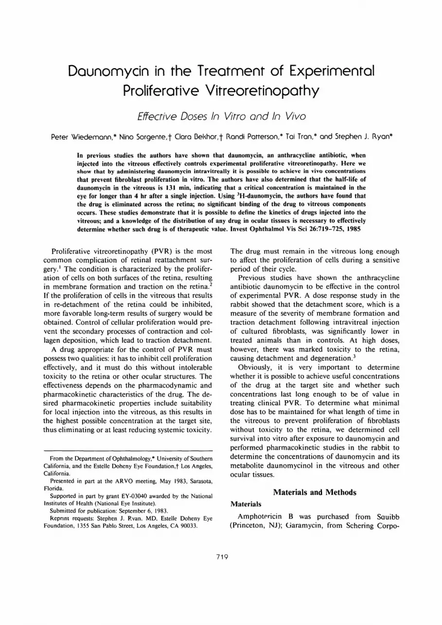
Autoradiography showed drug in all retinal layersaround the posterior pole (Fig. 4). The peripheralretina contained fewer grains; radioactive daunomycinwas found in the ciliary body and a small number ofgrains was evident in the lens and sclera. In the areaof the retina that contained most of the grains, weobserved unspecific retinal damage, such as slight
disruption of inner and outer nuclear layers, andshortening and thickening of the photoreceptor outersegments; the photoreceptor outer segments appearto be the most sensitive structure in the eye withregard to daunomycin toxicity.6
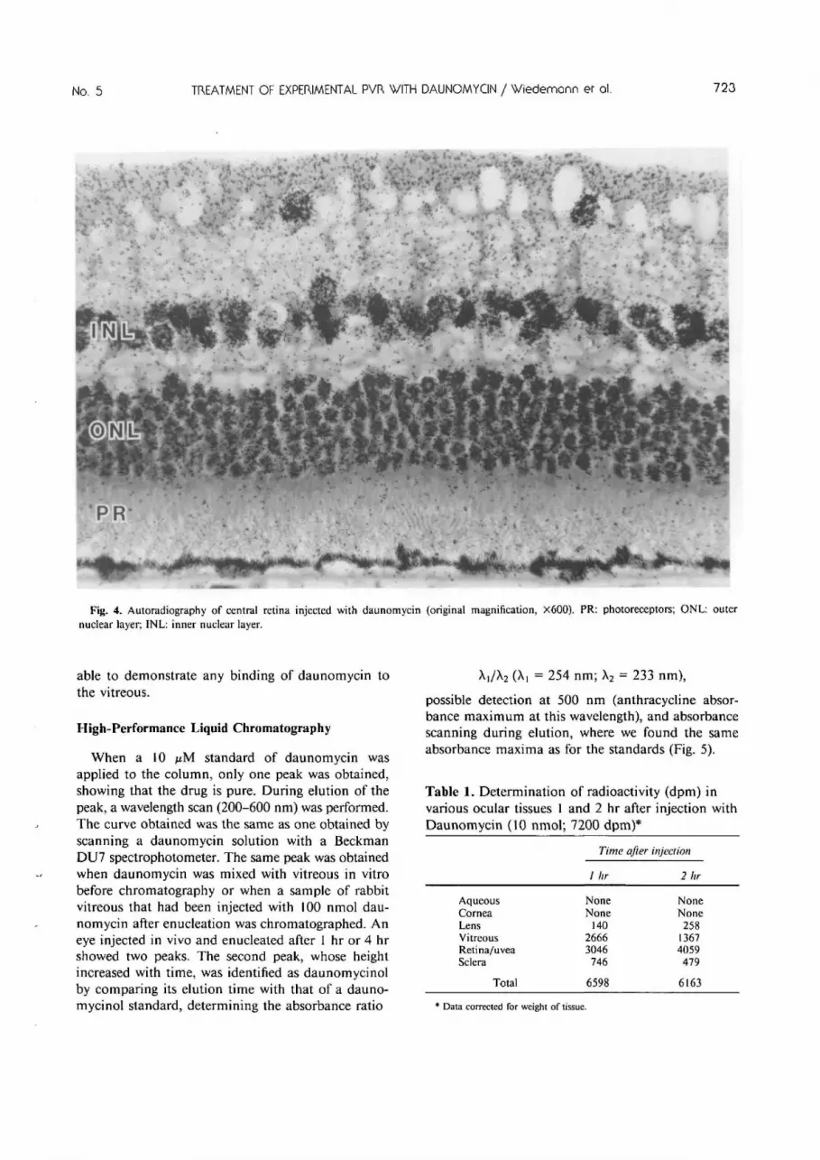
Daunomycin in Ocular Tissues and Vitreous andLenticular Binding
Direct measurement of radioactivity in ocular tis-sues (Table 1) after the injection of 10 nmol dauno-mycin demonstrated that most of the drug was ineither the vitreous or retina; practically none wasfound in the aqueous and cornea. When lenses wereincubated in vitro with daunomycin, we could notdemonstrate any loss of radioactivity from the incu-bation medium indicating that very likely the lensdoes not bind daunomycin. Likewise, we were not
A Elimination of daunomycin from the vitreous
3 _
2 _
1 _
D
y=3.57-0.002x
Correlation:-0.93
Elimination Half-life:t
o o
a • \ ^ »
D °
£=131 min.
Elimination rate constant:k=0.005min.~
First order elimination
I I I
C*Coe-M
I I I
60 120 180 240Minutes
300 360
Fig. 3. Elimination of daunomycin from the vitreous (spectro-fluorometric assay).
No. 5 TREATMENT OF EXPERIMENTAL PVR WITH DAUNOMYCIN / Wiedemonn er ol. 723
Fig. 4. Autoradiography of central retina injected with daunomycin (original magnification, X600). PR: photoreceptors; ONL: outernuclear layer; INL: inner nuclear layer.
able to demonstrate any binding of daunomycin tothe vitreous.
High-Performance Liquid Chromatography
When a 10 pM standard of daunomycin wasapplied to the column, only one peak was obtained,showing that the drug is pure. During elution of thepeak, a wavelength scan (200-600 nm) was performed.The curve obtained was the same as one obtained byscanning a daunomycin solution with a BeckmanDU7 spectrophotometer. The same peak was obtainedwhen daunomycin was mixed with vitreous in vitrobefore chromatography or when a sample of rabbitvitreous that had been injected with 100 nmol dau-nomycin after enucleation was chromatographed. Aneye injected in vivo and enucleated after 1 hr or 4 hrshowed two peaks. The second peak, whose heightincreased with time, was identified as daunomycinolby comparing its elution time with that of a dauno-mycinol standard, determining the absorbance ratio
A]/X2 (Xi = 254 nm; X2 = 233 nm),
possible detection at 500 nm (anthracycline absor-bance maximum at this wavelength), and absorbancescanning during elution, where we found the sameabsorbance maxima as for the standards (Fig. 5).
Table 1. Determination of radioactivity (dpm) invarious ocular tissues 1 and 2 hr after injection withDaunomycin (10 nmol; 7200 dpm)*
AqueousCorneaLensVitreousRetina/uveaSclera
Total
Time after injection
1 hr
NoneNone
14026663046
74§
6598
2 hr
NoneNone
25813674059479
6163
* Data corrected for weight of tissue.

724 INVESTIGATIVE OPHTHALMOLOGY & VISUAL SCIENCE / Moy 1985 Vol. 26
A B C D E
Fig. 5. High-performance liquid chromatography of daunomycinin the vitreous (Detection wavelength 500 nm, 0.02 AUFS). A:vitreous blank; B: vitreous with daunomycinol standard (elutiontime 2.79 min) and daunomycin standard (elution time 3.69 min);C: vitreous from eye injected with daunomycin after enucleation;D: vitreous from eye injected with daunomycin in vivo andenucleated after 1 hr; E: Vitreous from eye injected with daunomycinin vivo and enucleated after 4 hr.
Discussion
Selection of the proper daunomycin concentrationsand exposure times in vitro and in vivo is a complextask. Essentially the effectiveness of a drug dependson the concentration of the drug at the target site,the time during which an effective concentration ispresent at the target site, as well as on the kinetics ofthe cell population at the time it is exposed to thedrug. With regard to the effective concentration,probably the time of exposure to superthresholdconcentrations is more important than peak concen-trations.
Cell death is a multistage process and manifestsitself in morphologic degeneration, metabolic death,or reproductive death. The inability to reproduceshould be the only relevant criterion to assess cell killafter use of an antiproliferative drug, since metabolicdeath does not represent a valid measure of the viablecell population.7 The goal of this study was to quan-titatively determine loss of reproductive integrity invitro and correlate this with drug concentrations andexposure times achievable in vivo. Our results withfibroblasts, in agreement with the literature,8 show alogarithmic decrease of CFUs as a function of increas-ing daunomycin concentration. The efficacy of differ-ent agents on a single cell type and the activity ofone drug on different cell classes can be determinedand compared by using the colony formation test.
This is important, as different cell lines may showdifferent sensitivity to various drugs.9 We showed theinability of fibroblasts to proliferate, i.e., form colonies,when they were exposed to 1 fiM daunomycin for 1hr or 500 nM for 5 hr.
Depending on the vitreous volume (1-1.5 ml), andassuming an even distribution of daunomycin in thevitreous, a dose of 10 nmol, the therapeutic dose inexperimental PVR in the rabbit, will result in amaximal concentration of 6.6-10 /zM daunomycin.Even though there is loss of drug from the injectionsite, our results show that a dose that is effective invitro can be achieved and maintained in vivo for atleast 4 hr. The close correlation between the inhibitoryconcentrations obtained in vitro and in vivo indicatethat the colony formation assay can be used to predicteffective drug concentrations in vivo, thus sparing thelife of experimental animals.
We have shown that there is no significant bindingof daunomycin to the vitreous or the lens. We alsowere able to show that the drug is metabolized to lesseffective daunomycinol,10 but from our data it is notpossible to determine the site of metabolism. It seemsgenerally accepted that the diffusion of solutes in thevitreous, even if they have a large molecular weight,appears to be unrestricted." We were therefore sur-prised when we found the drug concentrated aroundthe optic disc in the autoradiographs, whereas theperipheral retina was essentially free of drug. FromFigure 4 it is obvious that there is more drug in theinner and outer nuclear layer than in the photorecep-tor outer segment layer. As the 1- and 2-hr autora-diographs and the determination of radioactive drugin the ocular tissues (Table 1) show only the imme-diate deposition of daunomycin and not its finalplace of action, we did not further quantitate thedistribution of daunomycin using grain counts.
We have previously noted that a dose of 9 nmol/eye daunomycin resulted in alteration of the photo-receptor outer segments. However, no other clinicaland histologic signs of toxicity were evident.6 Sincethere was a time period of 28 days between theinjection of drug and enucleation in that study,6 thedamage to the retina observed here may only betransient due to retinal edema. If more extensivestudies on the toxicity of low doses of daunomycinwill show that the structural changes we have notedare not permanent, daunomycin may be useful as atherapeutic drug for PVR.
Key words: colony formation assay, proliferative vitreoret-inopathy, rabbit, autoradiography, scintillation counting,spectrofluorometry, daunomycin

No. 5 TREATMENT OF EXPERIMENTAL PVR WITH DAUNOMYCIN / Wiedemann er ol. 725
Acknowledgment
The authors thank E. Cogan, PhD, of Beckman Instru-ments for his advice and for the generous use of an HPLC.
References
1. Hilton G, Machemer R, Michels R, Okun E, Schepens C, andSchwartz A: The classification of retinal detachment withproliferative vitreoretinopathy. Ophthalmology 90:121, 1983.
2. Machemer R: Pathogenesis and classification of massive peri-retinal proliferation. Br J Ophthalmol 62:737, 1978.
3. Wiedemann P, Kirmani M, Santana M, Sorgente N, and RyanSJ: Control of experimental massive periretinal proliferationby daunomycin: dose-response relation. Graefes Arch Clin ExpOphthalmol 220:233, 1983.
4. Brown JE, Wilkinson PA, and Brown JR: Rapid high-perfor-mance liquid chromatographic assay for the anthracyclinesdaunorubicin and 7-con-O-methylnogarol in plasma. J Chro-matogr 226:521, 1981.
5. Alberts DS, Bachur NR, Holtzman JL: The pharmacokineticsof daunomycin in man. Clin Pharmacol Ther 12:96, 1971.
6. Santana M, Wiedemann P, KJrmani M, Minckler DS, PattersonR, Sorgente N, and Ryan SJ: Daunomycin in the treatment ofexperiment proliferative vitreoretinopathy: retinal toxicity ofintravitreal daunomycin in the rabbit. Graefes Arch Clin ExpOphthalmol 221:210, 1984.
7. Roper PR and Drewinko B: Comparison of in vitro methodsto determine drug-induced cell lethality. Cancer Res 36:2182,1976.
8. Drewinko B, Roper PR, and Barlogie B: Patterns of cellsurvival following treatment with antitumor agents in vitro.Eur J Cancer 15:93, 1979.
9. Blumenkranz M, Hajek A, Sossi N, and Sossi G: Evidence forcell specificity in proliferative vitreoretinopathy. ARVO Ab-stracts. Invest Ophthalmol Vis Sci 25(Suppl):272, 1984.
10. Yesair DW, Thayer PS, McNitt S, and Teague K: Comparativeuptake, metabolism and retention of anthracyclines by tumorsgrowing in vitro and in vivo. Eur J Cancer 16:901, 1980.
11. Maurice DM and Mishima S: Ocular pharmacokinetics. InPharmacology of the Eye, Sears ML, editor. New York, Springer-Verlag, 1984, pp. 19-116.


