Embed Size (px)
Citation preview

Mannose-specific lectin from the mushroomHygrophorus russula
Tomohiro Suzuki2, Kozue Sugiyama3, Hirofumi Hirai3,Hiroyuki Ito3, TatsuyaMorita3, Hideo Dohra4,TakeomiMurata3, Taichi Usui3, Hiroaki Tateno5,Jun Hirabayashi5, Yuka Kobayashi1,6, andHirokazu Kawagishi1,3,7
2Inovation and Joint Research Center; 3Department of Applied BiologicalChemistry, Faculty of Agriculture and 4Genetic Research and Biotechnology,Shizuoka University, 836 Ohya, Suruga-ku, Shizuoka 422-8529, Japan;5Research Center for Glycoscience, National Institute of Advanced IndustrialScience and Technology, Tsukuba Central 2, Tsukuba, Ibaraki 305-8566,Japan; 6J-Oil Mills, Inc., 11, Kagetoricho, Totsuka-ku, Yokohama, Kanagawa245-0064, Japan; and 7Graduate School of Science and Technology, ShizuokaUniversity, 836 Ohya, Suruga-ku, Shizuoka 422-8529, Japan
Received on April 14, 2011; revised on December 14, 2011; accepted onDecember 14, 2011
A lectin was purified from the mushroom Hygrophorusrussula by affinity chromatography on a Sephadex G-50column and BioAssist S cation exchange chromatographyand designated H. russula lectin (HRL). The results ofsodium dodecyl sulfate–polyaclylamidegel electrophoresis,gel filtration and matrix-assisted laser desorption ioniza-tion time-of-flight mass spectrometry of HRL indicatedthat it was composed of four identical 18.5 kDa subunitswith no S-S linkage. Isoelectric focusing of the lectinshowed bands near pI 6.40. The complete sequence of 175amino acid residues was determined by amino acid se-quencing of intact or enzyme-digested HRL. The sequenceshowed homology with Grifola frondosa lectin. The cDNAof HRL was cloned from RNA extracted from the mush-room. The open reading frame of the cDNA consisted of528 bp encoding 176 amino acids. In hemagglutination in-hibition assay, α1-6 mannobiose was the strongest inhibi-tor and isomaltose, Glcα1-6Glc, was the second strongestone, among mono- and oligosaccharides tested. Frontal af-finity chromatography indicated that HRL had the highestaffinity for Manα1-6(Manα1-3)Manβ1-4GlcNAcβ1-4GlcNAc, and non-reducing terminal Manα1-6 was essen-tial for the binding of HRL to carbohydrate chains. Thesugar-binding specificity of HRL was also analyzed by
using BIAcore. The result from the analysis exhibited posi-tive correlations with that of the hemagglutination inhib-ition assay. All the results suggested that HRL recognizedthe α1-6 linkage of mannose and glucose, especially theManα1-6 bond. HRL showed a mitogenic activity againstspleen lymph cells of an F344 rat. Furthermore, anenzyme-linked immunosorbent assay showed strongbinding of HRL to human immunodeficiency virus type-1gp120.
Keywords: fungal lectin /Hygrophorus russula / mannose-binding lectin / mushroom
Introduction
Lectins are powerful tools for the sugar-chain structure deter-mination. Historically, their sugar-binding specificities havebeen used for qualitative analysis of sugar chains. Recently,new technology such as lectin array is becoming popular;therefore, lectins with specific sugar-binding properties arerequired (Hirabayashi 2004; Kuno et al. 2005; Tateno et al.2009).Many species produce mannose (Man)-recognizing lectins.
Concanavalin A (ConA) is specific to Man and has been usedas a biochemical or biological agent for a long time (Loriset al. 1998). This lectin recognizes not only Man but alsoglucose (Glc); therefore, it is categorized as a Man/Glc-specific lectin. On the other hand, there are Man-specificlectins that bind to Man but not to Glc. GNA (snowdrop;Galanthus nivalis agglutinin) and HHA (amaryllis;Hippeastrum hybrid agglutinin) are produced by plants(Howard and Sage 1969; Van Damme et al. 1987; Kaku et al.1990). FimH proteins from Escherichia coli and Man-bindinglectin from mammalians are also specific to Man amongmonosaccharides (Krogfelt et al. 1990; Worthley et al. 2005).In the course of our continuing screening for new mushroomlectins, we found lectin activity in the extract of the mush-room Hygrophorus russula, and the binding affinity to sugarwas specifically potent to Man. This specificity was notablynew and unusual compared with characteristics of the knownmushroom lectins. In particular, further study showed that thislectin activity was α1,6-Man specific and its sugar-bindingspecificity has not yet been reported. Here, we describe theisolation and characterization of the lectin from thismushroom.
1To whom correspondence should be addressed: Tel: +81-54-238-4885;Fax: +81-54-238-4885; e-mail [email protected] (H. Kawagishi);Tel: +81-45-852-4001; Fax: +81-45-852-6357; e-mail [email protected] (Y. Kobayashi)
Glycobiology vol. 22 no. 5 pp. 616–629, 2012doi:10.1093/glycob/cwr187Advance Access publication on December 22, 2011
© The Author 2011. Published by Oxford University Press. All rights reserved. For permissions, please e-mail: [email protected] 616
at Takeda Pharm
aceutical Com
pany Lim
ited on July 14, 2015http://glycob.oxfordjournals.org/
Dow
nloaded from

ResultsPurification of H. russula lectinThe purification procedure is summarized in Table I. In thepresent study, Sephadex G-50 was chosen as the affinitysupport since the hemagglutination activity of the crude
extract from the mushroom was inhibited by Man and Glc.Initially, the extract of H. russula was applied to SephadexG-50. Almost all the lectin activity from the extract wasadsorbed to the affinity column and eluted with 0.3 M Man.The eluate was further purified by BioAssist S cation ex-change chromatography, and a pure lectin, which was namedH. russula lectin (HRL), was obtained.
Molecular properties of HRLHRL gave a single band with an apparent mass of 18.5 kDaon sodium dodecyl sulfate–polyaclylamidegel electrophoresis(SDS–PAGE; Figure 1A) regardless of the presence (lane 1)or the absence (lane 2) of 2-mercaptoethanol. The isoelectricfocusing of HRL gave a band with a pI of 6.4 (Figure 1B).High-performance liquid chromatography (HPLC) gel filtra-tion of the protein gave a single symmetrical peak at anelution volume corresponding to a molecular mass of 74 kDa(Supplementary data, Figure S1). The matrix-assisted laser de-sorption ionization time-of-flight (MALDI-TOF) mass spec-trum gave a main peak at m/z 18,511 and a small peak at m/z9267 (Figure 1C). The results of SDS–PAGE, gel filtrationand MALDI-TOF mass analysis indicated that the molecular
Table I. Purification of HRL from 100 g of fruiting bodies of H. russula
Fraction Totalprotein(mg)
Totalagglutination(titer)a
Specificagglutination(titer/mgprotein)
Recoveryof activity(%)
Supernatant afterhomogenization
8.7 × 10² 4.4 × 10⁵ 5.1 × 10² 100
Sephadex G-50affinitychromatography
6.0 9.8 × 10⁴ 1.6 × 10⁴ 22
BioAssist S cationexchangechromatography
2.7 8.8 × 10⁴ 3.2 × 10⁴ 20
aTiter was defined as the reciprocal of the end-point dilution exhibitinghemagglutination.
Fig. 1. The molecular structure of HRL. (A) SDS–PAGE of HRL. Lane M, marker proteins; lane 1, HRL, non-reduced; lane 2, HRL, reduced with2-mercaptoethanol. (B) Isoelectric focusing of HRL. Lane M, marker proteins; lane 1, HRL. (C) MALDI-TOF MS of HRL.
Man-specific lectin from Hygrophorus russula
617
at Takeda Pharm
aceutical Com
pany Lim
ited on July 14, 2015http://glycob.oxfordjournals.org/
Dow
nloaded from

mass of HRL was 74 kDa and it was composed of four identi-cal 18.5 kDa subunits with no disulfide bonds.The protein content of HRL was �94.4% (Supplementary
data, Figure S2A). The amino acid composition revealed ahigh content of Gly, Asx and Thr (Supplementary data,Table S1). N-terminal amino acid sequence analysis ofintact HRL gave a sequence of 30 amino acids from theterminal (Figure 2, lane 1). The protein was digested withLysobacter enzymogenes protease (Lys-C), Clostridium histo-lyticum proteinase (Arg-C) or Pseudomonas fragi mutantprotease (Asp-N), and the resulting peptides were isolatedby reverse-phase HPLC. Each purified peptide sequencewas determined by N-terminal amino acid sequence analysis(Figure 2, lanes 2–7). As a result, the complete amino acidsequence of HRL was determined as shown in Figure 2(lane 8). This lectin had heterogeneity in the sequence atposition 52 (Gln or Trp) from the N-terminus. The neutralcarbohydrate content amounted to 3.5% (Supplementarydata, Figure S2B), and the components of the sugars in theglycoprotein were identified as L-Fuc:Glc:Man:Gal:GlcN:GalN:Xyl in a 3.4:3.3:2.9:2. 2:1.3:1.0:0.3 molar ratio. HRLdid not contain any sialic acid.
cDNA cloning and structural analysis of HRLThe cDNA of HRL was cloned from total RNA extractedfrom fruiting bodies of the fungus using reverse transcriptase–polymerase chain reaction (RT–PCR) coupled with RACE–PCR (GenBank™ accession no. AB586742). The openreading frame of the cDNA consisted of 528 bp encoding 176amino acids (Figure 3). No signal sequence was present in thecDNA. Two amino acids, Asp111 and Cys111, were detected inthe cDNA.The amino acid sequence of HRL was analyzed by the
BLAST program, and the sequence showed homology to thelectin from the mushroom Grifola frondosa (GFL; 38%) andthe lectin from Japanese sago palm Cycas revoluta (CRLL;27%) (Figure 4).
Properties of HRL as a lectinHRL was stable below 60°C, but rapidly inactivated at tem-peratures above 60°C (Supplementary data, Figure S3A).HRL was relatively stable over a wide range of pH values, es-pecially between pH 2.0 and 9.5 (Supplementary data,Figure S3B). Ethylenediaminetetraacetic acid (EDTA) treat-ment and metal cations addition to the pre-treated lectin didnot affect the lectin activity. The lectin agglutinated Pronase-and neuraminidase-treated human erythrocytes (Table II).Table III shows the inhibition of hemagglutination activity ofHRL by various monosaccharides, oligosaccharides and gly-copeptides. Among the mono- and oligosaccharides tested,the most potent inhibitor was α1-6 mannobiose [minimum in-hibitory concentration (MIC), 39.1 μM]. Isomaltose, isomalto-triose, isomaltotetraose isomaltopentaose and isomaltohexaosewere also inhibitory at higher concentrations 156, 316, 316,316 and 316 μM, respectively, than α1-6 mannobiose.Thyroglobulin, ribonuclease B, asialo-PSM (porcine stomachmucin) and PSM inhibited agglutination. Comparison of thesugar-binding specificity of HRL with those of other Man/Glc-specific lectins, ConA, Pisum sativum agglutinin (PSA),banana lectin (BanLec), GNA, HHA and Tulipa gesnerianalectin (TxLc-I), is shown in Table IV. ConA, PSA, BanLec,GNA, HHA and TxLc-I bound to α-mannosides regardless oftheir linkage positions and some of them bound to Glcα1-6linkage, too. However, the binding specificity to mannosidesand glucosides of HRL was strictly limited to α1-6 linkage,especially Manα1-6 one.The sugar-binding specificity of HRL was also investigated
by surface plasmon resonance (SPR) analysis. HRL wasimmobilized on the sensor chip CM-5 by amine coupling and9 glycoproteins were used as analytes. The sensorgrams andthe kinetic data of the binding are shown in Supplementarydata, Figure S4 and Table V, respectively. The binding of allthe glycoproteins to the immobilized lectin fitted best the 1:1binding model among various models in the evaluating soft-ware (Supplementary data, Figures S4 and S5). Asialo-PSM,PSM and thyroglobulin, which inhibited the HRL-mediated
Fig. 2. Summary of sequence determination of HRL. Lane 8 shows the complete sequence of HRL. The sequence in lane 1 was determined by the Edmandegradation of intact HRL. The sequences of the peptides in lanes 2–7 were determined by amino acid sequence analysis and MALDI-TOF MS. The peptidesobtained with L. enzymogenes proteinase, C. histolyticum proteinase and P. fragi mutant protease were designated Lys, Arg and Asp, respectively. Isoforms areboxed in black.
T Suzuki et al.
618
at Takeda Pharm
aceutical Com
pany Lim
ited on July 14, 2015http://glycob.oxfordjournals.org/
Dow
nloaded from

hemagglutination, showed similar kinetic parameters to eachother. Asialo-bovine submaxillary mucin (BSM), asialo-fetuin, BSM and fetuin that showed weaker inhibitory activityin the hemagglutination assay also bound to the immobilizedlectin. On the other hand, α1-acid glycoprotein and transferrin,
which were not inhibitory in the hemagglutination assay, didnot bind to the immobilized lectin at all. The binding specifi-city of the immobilized HRL toward α1-6 mannobiose orother α-mannobioses was analyzed by the co-injectionmethod (Kobayashi et al. 2005). After binding of PSM,
Fig. 3. cDNA sequence and deduced amino acid sequence of HRL. Nucleotide numbers are shown above the nucleotide sequence, and the deduced amino acidsequence is shown using alphabets below the nucleotide sequence. The asterisk denotes the termination codon. A possible N-glycosylation site is boxed.Isoforms are boxed in black.
Fig. 4. Alignment of the amino acid sequences of HRL, GFL and CRLL. The amino acid sequence of HRL was compared with those of GFL and CRLL.Identical (colon) and similar (dot) residues are indicated.
Man-specific lectin from Hygrophorus russula
619
at Takeda Pharm
aceutical Com
pany Lim
ited on July 14, 2015http://glycob.oxfordjournals.org/
Dow
nloaded from

dissociation was initiated by α1-6 mannobiose at various con-centrations. The dissociation of the glycoprotein by the disac-charide was promoted dose-dependently (Figure 5A).Furthermore, disassociation of the glycoprotein caused byα1-6 mannobiose and other α-mannobioses was comparedwith one another. As shown in Figure 5B, the binding ofPSM was abruptly reversed by the injection of α1-6 manno-biose, whose MIC in the hemagglutination inhibition assaywas 39.1 μM (Table III). On the other hand, the effects of theother mannosides, α1-3 mannobiose (MIC, 2.50 mM), α1-2mannobiose (10.0 mM) and α1-4 mannobiose (10.0 mM)were very small.The sugar-binding specificity of HRL was further eluci-
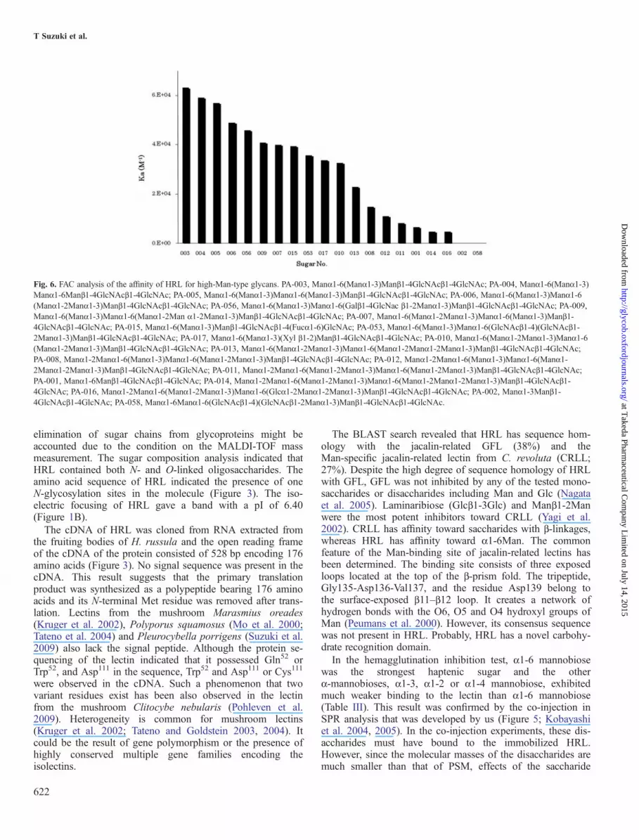
dated by frontal affinity chromatography (FAC) analysis using125 kinds of pyridylaminated (PA)-glycans (Supplementarydata, Figure S6). The amounts of immobilized HRL weredetermined to be 10 μg/mL gel. For evaluation of the preparedcolumns, it was necessary to determine the effectiveligand content (Bt) based on the concentration dependenceanalysis. Concentration dependence analysis was performedwith methotrexate derivatized Manα1-3(Manα1-6)Manβ1-4GlcNAcβ1-4GlcNAc (Nakamura et al. 2005) at various con-centrations ranging from 4.2 to 33.3 μL. As a result, Bt, Kd
and Ka values were determined to be 1.84 nmol, 1.63 × 10⁻⁵
M and 6.13 × 10⁻⁴ M−1, respectively (Supplementary data,Figure S7). The strength of affinity of each PA-glycans for theimmobilized lectin was shown as the V − V0 value (μL). HRLbound to high-Man-type sugar chains (Figure 6). The lectinshowed the highest affinity for Manα1-6(Manα1-3)Manβ1-4GlcNAcβ1-4GlcNAc (PA-003). Manα1-6(Manα1-3)Manα1-6Manβ1-4GlcNAcβ1-4GlcNAc (PA-004) andManα1-6(Manα1-3)Manα1-6(Manα1-3)Manβ1-4GlcNAcβ1-4GlcNAc (PA-005) were also very stronghaptenic sugars.
Mitogen activity of HRLHRL stimulated the production of interleukin (IL)-6, inter-feron (IFN)-γ and IL-10 dose-dependently when rat spleencells were treated with the lectin (Figure 7 and Supplementarydata, Figure S8). The stimulatory effect of HRL on both IL-6and IFN-γ production was suppressed by addition of α1-6mannobiose. The IL-6 production by HRL is much strongerthan that by ConA.
Table II. Agglutination profiles of HRL
Group of erythrocytes Titera
Untreated Pronase-treatedb Trypsin-treatedc Neuraminidase-treatedd
Human A 0 211 0 24
Human B 0 29 0 0Human O 0 211 0 24
Rabbit 25 28 NT NT
NT, not tested.aTiter was defined as the reciprocal of the end-point dilution exhibiting the hemagglutination.b10% suspension of erythrocytes in PBS (10 mL) was treated with Pronase (5.0 mg) for 30 min at 45°C.c10% suspension of erythrocytes in PBS (10 mL) was treated with trypsin (1.0 mg) for 180 min at 37°C.d10% suspension of erythrocytes in PBS (10 mL) was treated with neuraminidase (1 U/mL) for 60 min at 37°C.
Table III. Inhibition of HRL-mediated hemagglutination by mono- andoligosaccharides and glycoproteins
Inhibitora MICb
α1-6 mannobiose 0.0391 mMIsomaltose (Glcα1→ 6Glc) 0.156 mMIsomaltotriose 0.313 mMIsomaltotetraose 0.313 mMIsomaltopentaose 0.313 mMIsomaltohexaose 0.313 mMMethyl α-mannoside 1.56 mMα1-3 mannobiose 2.50 mMMethyl β-mannoside 3.13 mMα1-2 mannobiose 10.0 mMα1-4 mannobiose 10.0 mMMethyl α-glucoside 12.5 mMMan 12.5 mMLacturose 12.5 mMN-Acetylglucosamine 50 mMFructose 50 mM2-deoxy-2-fluoro-D-Man 50 mMGlc 100 mM2-deoxy-Glc 100 mMThyroglobulin 3.91 μg/mLRibonuclease B 7.81 μg/mLAsialo-PSM 7.81 μg/mLPSM 15.6 μg/mLImmunoglobulin A 31.3 μg/mLMannan 31.3 μg/mLImmunoglobulin M 62.5 μg/mLAsialo-BSM 62.5 μg/mLAsialo-fetuin 125 μg/mLBSM 125 μg/mLImmunoglobulin G 250 μg/mLAlbumin (chicken egg) 250 μg/mLFetuin 250 μg/mLLactoferrin 250 μg/mL
PSM, porcine stomach mucin; BSM, bovine submaxillary glands mucin.aGalactose, fucose, L-fucose, arabinose, L-arabinose, ribose, L-rhamnose,xylose, raffinose, saccharose, lactose, lactitol, ribulose, N-acetylgalactosamine,N-acetyllactoosamine, methyl β-glucoside, methyl α-galactoside, melibiose,galacturonic acid, glucosamine hydrochloride, galactosamine hydrochloride,mannosamine hydrochloride, gluconic acid, chitobiose, maltose,2-deoxy-galactose, 2-deoxy-ribose and 2-deoxy-2-fluoro-D-Glc did not inhibitat all at concentrations up to 400 mM. N-Acetylneuraminic acid andN-glycolylneuraminic acid did not inhibit at concentrations up to 40 mM.α1-Acid glycoprotein (human), transferrin (human), hyaluronic acid andalbumin (bovine) did not inhibit at all at concentrations up to 1 mg/mL.bMICs required for the inhibition of four hemagglutination doses of the lectin.
T Suzuki et al.
620
at Takeda Pharm
aceutical Com
pany Lim
ited on July 14, 2015http://glycob.oxfordjournals.org/
Dow
nloaded from

Binding of HRL to gp120In order to examine anti-HIV (human immunodeficiencyvirus) of HRL, interaction of gp120 and HRL was analyzedby plate assay using immobilized gp120 on the plate and wascompared with other lectins (Figure 8). HRL and BanLecshowed strong affinity to gp120.
Discussion
A novel Man-specific lectin, HRL, was purified from theedible mushroom H. russula by repeated chromatography.The complete amino acid sequence of HRL was determinedand this lectin had heterogeneity in the sequence at the pos-ition of 52 (Gln or Trp) from the N-terminus (Figure 2).HRL was composed of 175 amino acid residues and its cal-culated molecular mass was 18,484 Da (Gln52) or 18,542
Da (Trp52). The theoretical molecular mass was in goodagreement with the value (18,511 Da) obtained byMALDI-TOF mass spectrometry (MS) (Figure 1C). The
Table IV. Comparison of sugar-binding specificity of HRL with those of other Man/Glc-specific lectins
Inhibitors MICa
HRL ConA PSA BanLec GNA HHA TxLc-I
Glc (mM) 100 50 25 12.5 >100 >100 >100Man (mM) 12.5 12.5 12.5 3.13 25 25 >100Methyl α-mannoside (mM) 3.13 3.13 12.5 3.13 12.5 12.5 >100Isomaltose (mM) 0.156 12.5 25 1.25 >40 >40 >40Isomaltotriose (mM) 0.313 25 25 2.5 >40 >40 >40Isomaltotetraose (mM) 0.313 25 25 5.0 >40 >40 >40Isomaltohexaose (mM) 0.313 25 25 12.5 >40 >40 >40Isomaltoheptaose (mM) 0.313 25 25 12.5 >40 >40 >40α1-2 mannobiose (mM) 10.0 1.25 1.25 1.25 10.0 10.0 >30α1-3 mannobiose (mM) 2.50 2.5 5.0 1.25 10.0 10.0 >30α1-4 mannobiose (mM) 10.0 2.5 5.0 5.0 20.0 20.0 >30α1-6 mannobiose (mM) 0.0391 2.5 5.0 2.5 10.0 10.0 >302-deoxy-Glc (mM) 100 25 50 >100 >100 >100 >1002-deoxy-2-fluoro-D-Glc (mM) >100 50 100 100 >100 >100 >1002-deoxy-2-fluoro-D-Man (mM) 50 50 50 >100 100 >100 >100Thyroglobulin (μg/mL) 3.91 3.91 15.6 7.81 125 125 125Asialo-PSM (μg/mL) 7.81 125 125 >500 62.5 62.5 62.5PSM (μg/mL) 15.6 250 250 >500 250 250 250Immunoglobulin A (μg/mL) 31.3 31.3 125 125 125 125 125Immunoglobulin G (μg/mL) 250 250 >500 >500 >500 >500 >500Asialo-fetuin (μg/mL) 125 250 125 >500 250 250 125Fetuin (μg/mL) 250 250 250 >500 125 125 >500
aMinimum inhibitor concentration required for the inhibition of four hemagglutination doses of the lectins.
Table V. Binding kinetics of interaction between immobilized HRL andglycoproteins
Analyte ka (M–1 S–1) kd (S
–1) KD (M)
PSM 4.13 × 10⁵ 4.54 × 10⁻³ 1.10 × 10⁻⁸
Asialo-PSM 1.95 × 10⁵ 5.39 × 10⁻³ 2.76 × 10⁻⁸
Thyroglobulin 4.75 × 10⁴ 5.19 × 10⁻³ 1.09 × 10⁻⁷
Asialo-BSM 2.58 × 10³ 7.89 × 10⁻³ 3.05 × 10⁻⁶
Asialo-fetuin 3.18 × 10² 3.97 × 10⁻³ 1.25 × 10⁻⁵
BSM 1.32 × 10³ 8.77 × 10⁻³ 6.64 × 10⁻⁶
Fetuin 2.99 × 10² 4.01 × 10⁻³ 1.34 × 10⁻⁵
α1-Acid glycoprotein NI NI NITransferrin NI NI NI
NI, no interaction.
Fig. 5. Sensorgrams showing interaction between immobilized HRL and PSMby co-injection with various mannobioses. (A) α1-6 mannobiose at variousconcentrations. (B) Various α-mannobioses at 5 mM.
Man-specific lectin from Hygrophorus russula
621
at Takeda Pharm
aceutical Com
pany Lim
ited on July 14, 2015http://glycob.oxfordjournals.org/
Dow
nloaded from
elimination of sugar chains from glycoproteins might beaccounted due to the condition on the MALDI-TOF massmeasurement. The sugar composition analysis indicated thatHRL contained both N- and O-linked oligosaccharides. Theamino acid sequence of HRL indicated the presence of oneN-glycosylation sites in the molecule (Figure 3). The iso-electric focusing of HRL gave a band with a pI of 6.40(Figure 1B).The cDNA of HRL was cloned from RNA extracted from
the fruiting bodies of H. russula and the open reading frameof the cDNA of the protein consisted of 528 bp encoding 176amino acids (Figure 3). No signal sequence was present in thecDNA. This result suggests that the primary translationproduct was synthesized as a polypeptide bearing 176 aminoacids and its N-terminal Met residue was removed after trans-lation. Lectins from the mushroom Marasmius oreades(Kruger et al. 2002), Polyporus squamosus (Mo et al. 2000;Tateno et al. 2004) and Pleurocybella porrigens (Suzuki et al.2009) also lack the signal peptide. Although the protein se-quencing of the lectin indicated that it possessed Gln52 orTrp52, and Asp111 in the sequence, Trp52 and Asp111 or Cys111
were observed in the cDNA. Such a phenomenon that twovariant residues exist has been also observed in the lectinfrom the mushroom Clitocybe nebularis (Pohleven et al.2009). Heterogeneity is common for mushroom lectins(Kruger et al. 2002; Tateno and Goldstein 2003, 2004). Itcould be the result of gene polymorphism or the presence ofhighly conserved multiple gene families encoding theisolectins.
The BLAST search revealed that HRL has sequence hom-ology with the jacalin-related GFL (38%) and theMan-specific jacalin-related lectin from C. revoluta (CRLL;27%). Despite the high degree of sequence homology of HRLwith GFL, GFL was not inhibited by any of the tested mono-saccharides or disaccharides including Man and Glc (Nagataet al. 2005). Laminaribiose (Glcβ1-3Glc) and Manβ1-2Manwere the most potent inhibitors toward CRLL (Yagi et al.2002). CRLL has affinity toward saccharides with β-linkages,whereas HRL has affinity toward α1-6Man. The commonfeature of the Man-binding site of jacalin-related lectins hasbeen determined. The binding site consists of three exposedloops located at the top of the β-prism fold. The tripeptide,Gly135-Asp136-Val137, and the residue Asp139 belong tothe surface-exposed β11–β12 loop. It creates a network ofhydrogen bonds with the O6, O5 and O4 hydroxyl groups ofMan (Peumans et al. 2000). However, its consensus sequencewas not present in HRL. Probably, HRL has a novel carbohy-drate recognition domain.In the hemagglutination inhibition test, α1-6 mannobiose
was the strongest haptenic sugar and the otherα-mannobioses, α1-3, α1-2 or α1-4 mannobiose, exhibitedmuch weaker binding to the lectin than α1-6 mannobiose(Table III). This result was confirmed by the co-injection inSPR analysis that was developed by us (Figure 5; Kobayashiet al. 2004, 2005). In the co-injection experiments, these dis-accharides must have bound to the immobilized HRL.However, since the molecular masses of the disaccharides aremuch smaller than that of PSM, effects of the saccharide
Fig. 6. FAC analysis of the affinity of HRL for high-Man-type glycans. PA-003, Manα1-6(Manα1-3)Manβ1-4GlcNAcβ1-4GlcNAc; PA-004, Manα1-6(Manα1-3)Manα1-6Manβ1-4GlcNAcβ1-4GlcNAc; PA-005, Manα1-6(Manα1-3)Manα1-6(Manα1-3)Manβ1-4GlcNAcβ1-4GlcNAc; PA-006, Manα1-6(Manα1-3)Manα1-6(Manα1-2Manα1-3)Manβ1-4GlcNAcβ1-4GlcNAc; PA-056, Manα1-6(Manα1-3)Manα1-6(Galβ1-4GlcNac β1-2Manα1-3)Manβ1-4GlcNAcβ1-4GlcNAc; PA-009,Manα1-6(Manα1-3)Manα1-6(Manα1-2Man α1-2Manα1-3)Manβ1-4GlcNAcβ1-4GlcNAc; PA-007, Manα1-6(Manα1-2Manα1-3)Manα1-6(Manα1-3)Manβ1-4GlcNAcβ1-4GlcNAc; PA-015, Manα1-6(Manα1-3)Manβ1-4GlcNAcβ1-4(Fucα1-6)GlcNAc; PA-053, Manα1-6(Manα1-3)Manα1-6(GlcNAcβ1-4)(GlcNAcβ1-2Manα1-3)Manβ1-4GlcNAcβ1-4GlcNAc; PA-017, Manα1-6(Manα1-3)(Xyl β1-2)Manβ1-4GlcNAcβ1-4GlcNAc; PA-010, Manα1-6(Manα1-2Manα1-3)Manα1-6(Manα1-2Manα1-3)Manβ1-4GlcNAcβ1-4GlcNAc; PA-013, Manα1-6(Manα1-2Manα1-3)Manα1-6(Manα1-2Manα1-2Manα1-3)Manβ1-4GlcNAcβ1-4GlcNAc;PA-008, Manα1-2Manα1-6(Manα1-3)Manα1-6(Manα1-2Manα1-3)Manβ1-4GlcNAcβ1-4GlcNAc; PA-012, Manα1-2Manα1-6(Manα1-3)Manα1-6(Manα1-2Manα1-2Manα1-3)Manβ1-4GlcNAcβ1-4GlcNAc; PA-011, Manα1-2Manα1-6(Manα1-2Manα1-3)Manα1-6(Manα1-2Manα1-3)Manβ1-4GlcNAcβ1-4GlcNAc;PA-001, Manα1-6Manβ1-4GlcNAcβ1-4GlcNAc; PA-014, Manα1-2Manα1-6(Manα1-2Manα1-3)Manα1-6(Manα1-2Manα1-2Manα1-3)Manβ1-4GlcNAcβ1-4GlcNAc; PA-016, Manα1-2Manα1-6(Manα1-2Manα1-3)Manα1-6(Glcα1-2Manα1-2Manα1-3)Manβ1-4GlcNAcβ1-4GlcNAc; PA-002, Manα1-3Manβ1-4GlcNAcβ1-4GlcNAc; PA-058, Manα1-6Manα1-6(GlcNAcβ1-4)(GlcNAcβ1-2Manα1-3)Manβ1-4GlcNAcβ1-4GlcNAc.
T Suzuki et al.
622
at Takeda Pharm
aceutical Com
pany Lim
ited on July 14, 2015http://glycob.oxfordjournals.org/
Dow
nloaded from

binding on the sensorgrams are negligible. The oligosacchar-ides possessing Glcα1-6Glc were also strong inhibitors com-pared with α1-2, α1-3 or α1-4 mannobiose in the
hemagglutination inhibition test (Table III). The other knownManα-specific lectins tested, ConA, PSA, BanLec, GNA andHHA, were not able to distinguish among α1-2, α1-3, α1-4and α1-6 mannobioses (Table IV). To our knowledge, this isthe first lectin that binds to the α1-6 linkage of Man or Glcexclusively. Among the glycoproteins, thyroglobulin containshigh-Man-type sugar chains exhibited potent binding affinity(Tsuji et al. 1981). α1-Acid glycoprotein and transferrin didnot have Man at non-reducing end and were not able to bindto the lectin at all (Spik et al. 1975; Yoshima et al. 1981). Thesugar-binding specificity of HRL was further analyzed indetail by using FAC (Figure 6). The lectin showed the largestaffinity for Manα1-6(Manα1-3)Manβ1-4GlcNAcβ1-4GlcNAc(PA-003). A partial structure of PA-003, Manα1-6Manβ1-4GlcNAcβ1-4GlcNAc (PA-001), bound to the lectin veryweakly, whereas other partial structure, Manα1-3Manβ1-4GlcNAcβ1-4GlcNAc (PA-002), was not able to bind to thelectin at all. The addition of the Fucα1-6 residue (PA-015) tothe best haptenic sugar (PA-003) weakened the affinity to thelectin. All the results of the hemagglutination inhibition test,SPR and FAC allowed us to conclude that (i) HRL recognizedManα1-6 or Glcα1-6 linkage, especially Manα1-6, and hasthe highest affinity for Manα1-6(Manα1-3)Manβ1-4GlcNAcβ1-4GlcNAc (PA-003) among the sugar chains used,(ii) the non-reducing terminal Manα1-6 in sugar chains wasessential for the binding, (iii) the branched Manα1-3 residueenhanced the binding of HRL to carbohydrate chains and (iv)the branched Fucα1-6 reduced the affinity of the lectin forsugar chains.The results of bioassay using rat spleen cells showed that
HRL had mitogenic activity through the production of IL-6,IFN-γ and IL-10 (Figure 7 and Supplementary data,Figure S8). IL-6 is a multifunctional cytokine that is producedby a range of cells including T and B cells (Hirano et al.1990), whereas IFN-γ is produced by T cell (Jonasch andHaluska 2001). Previous studies demonstrate that IL-10 exhi-bits key regulatory effects on immune activation, includingTh2 cell, mast cell and eosinophil activation (Takanaski et al.1994; Arock et al. 1996; Royer et al. 2001). IL-10 also serves
Fig. 7. Mitogen activity of HRL. Spleens were added to flat-bottom 96-wellplates and incubated at 37°C in 5% CO2 with either various samples. Cont,medium as a control; ConA, 0.15 μM concanavalin A; C + H, 0.15 μM ConAcontaining 0.15 μM HRL; HRL, 0.075, 0.015, 0.3, 0.6, 1.2, 2.4 and 4.8 μMHRL; HRL + α1-6 mannobiose, 0.3, 0.6, 1.2 and 2.4 μM HRL containing0.075 μM α1-6 mannobiose; α1-6 mannobiose, 0.038, 0.075, 0.15 and0.29 μM α1-6 mannobiose.
Fig. 8. Binding of HRL and other lectins to gp120. HRL (filled diamond), BanLec (filled square), ConA (filled triangle), PSA (cross), GNA (asterisk), HHA(hyphen) and TxLc-I (vertical line) were interacted with immobilized gp120 (10 nM) on 96-well ELISA plates. Serial dilutions of biotinylated lectins were addedto the wells.
Man-specific lectin from Hygrophorus russula
623
at Takeda Pharm
aceutical Com
pany Lim
ited on July 14, 2015http://glycob.oxfordjournals.org/
Dow
nloaded from

as a key effecter of regulatory T cell activity (Asseman et al.1999). The obtained result indicated that HRL activated theproliferation of both T and B cells. Furthermore, α1-6 manno-biose inhibited the mitogenic activity, suggesting that theMan-binding site of HRL was related to the activity.HRL strongly binds to the gp120 protein present on the
surface of viruses and surface sugar chains on host cells,which plays an important role as one of the entry methods forvirus infection. In the case of HIV, there are two kinds of gly-coproteins, gp120 and gp41, in the envelope of the virus,which bind to the receptors on the surface of host cell andlead to the infection of virus to the host. gp120 possessesmore than 20 N-glycosidically linked sugar chains withvarying structural variety. Among the chains, three chainspossessing 7–9 Man residues play a crucial role in the infec-tion. Therefore, drugs or molecules that can specifically bindto these glycans can hinder the interactions between the pro-teins of the viral envelope and the cells of the host andprevent further interactions with the co-receptors (Botos andWlodawer 2005; Balzarini 2006). Sugar-binding proteins cancross-link glycans on the viral surface (Sacchettini et al. 2001;Shenoy et al. 2002; Ziolkowska and Wlodawer 2006).Therefore, lectins that can bind to gp120 are attracting atten-tion recently. Anti-HIV activity of cyanovilin-N from thecyanobacterium (blue-green algae) Nostoc ellipsosporum hasbeen reported and the lectin showed strong affinity for Man8-and Man9-type glycans (Botos et al. 2002; Helle et al. 2006;Ziolkowska and Wlodawer 2006). The binding probably wasdue to the recognition of Manα1-2 residues in the glycans bythe lectin. The anti-HIV activities of Manα1-2Man-specificlectins, actinohivin and Oscillatoria agardhii agglutinin, havebeen also reported (Sato et al. 2007; Hoorelbeke et al. 2010;Matoba et al. 2010; Takahashi et al. 2010). Recently,anti-HIV activity of a Man-specific jacalin-related lectin,BanLec, was also reported (Swanson et al. 2009). BanLec isunique in its specificity for internal α1-3 linkages of Man/Glcas well as β1-3 linkages Man/Glc at the reducing termini(Koshte et al. 1990; Winter et al. 2005). The binding strengthof HRL to gp120 was the same as or more than BanLec. FACanalysis revealed that HRL also bound to Man7 (PA-008,PA-009 and PA-010), Man8 (PA-011, PA-012 and PA-013)and Man9 (PA-014 and PA-016) (Figure 6).The mushroom H. russula from which HRL was isolated is
edible. This mushroom might become a candidate for a func-tional food that can prevent HIV infection in future.Furthermore, HRL has a promising potential for a probe thatis able to distinguish Manα1-6 linkage from the other lin-kages of Man.
Materials and methodsMaterialsFruiting bodies of H. russula were collected at Narusawavillage, Yamanashi Prefecture, Japan, frozen upon collectionand stored at −20°C. Bananas were purchased from a localgrocery store. Snowdrop, amaryllis and tulip bulbs were pur-chased from local markets. Canavalia ensiformis agglutinin(ConA) and PSA were products of J-Oil Mills, Inc. (Tokyo,Japan). Sephadex G-50 was a product of GE Healthcare UK
Ltd. Man-agarose was purchased from Sigma (St Louis, MO).BioAssist S and TSK-GEL G3000SWXL were products ofTosoh (Tokyo, Japan). The MALDI-TOF MS spectrum wasacquired on an AutoFlex (Bruker Daltonics, Billerica, MA).The partial N-terminal sequence of HRL has been determinedby a PPSQ-21A protein sequencer (Shimazu, Kyoto, Japan).Lysyl endopeptidase, endopeptidase Asp-C and Arg-C wereproducts of Wako Pure Chemicals (Osaka, Japan), Sigma andTakara Bio Inc. (Shiga, Japan), respectively. An ABEE(p-aminobenzoic ethyl ether) reagent and a Wakosil-II columnwere obtained from Wako Pure Chemicals. All the sugars andglycoproteins for the hemagglutinating inhibition tests and theSPR analyses were purchased from Nacalai tesque (Tokyo,Japan), Wako Pure Chemicals, Calbiochem (La Jolla, CA) orSigma. BIAcore 2000 was a product of GE Healthcare UKLtd. PA-oligosaccharides for FAC analysis were purchasedfrom Takara Bio Inc. HiTrap NHS-activated Sepharose (acti-vated agarose gel) was purchased from GE Healthcare UKLtd. Stainless steel empty miniature column (inner diameter,2 mm; length, 10 mm; bed volume, 31.4 μL) was obtainedfrom Shimadzu (Kyoto, Japan).
AnimalsMale Fisher 344 rats were purchased from the ShizuokaLaboratory Animal Center (Shizuoka, Japan). They werehoused in individual cages of screen-bottomed stainless inroom with controlled temperature (23 ± 2°C) and lighting(light on from 8:00 to 20:00).
Purification of the lectinAll the procedures were carried out at 4°C. After defrosting,the fruiting bodies of H. russula were homogenized andextracted with 10 mM phosphate-buffered saline (PBS), pH7.4, overnight. The homogenate was centrifuged at 8500 × gfor 15 min and the resulting supernatant was applied to acolumn of Sephadex G-50 (5 × 15 cm) equilibrated with thebuffer. After unbound materials were washed with the buffer,the bound fraction was desorbed with 0.3 M Man in thebuffer. The eluates were concentrated and equilibrated with0.1 M acetate buffer, pH 4.7, by ultrafiltration and furtherseparated by cation-exchange chromatography on a BioAssistS column (1.0 × 10 cm) equilibrated with the buffer. Thecolumn was exhaustively washed with the same buffer, andthe adsorbed lectin was eluted by a linear gradient of NaCl(0–1 M) in the buffer. The eluant was dialyzed extensivelyagainst distilled water, ultrafiltered and lyophilized.Approximately 2.7 mg of HRL was obtained from 100 g ofthe fresh fruiting bodies.
Sodium dodecyl sulfate–PAGESDS–PAGE was done by the method of Laemmli (1970).Samples were heated in the presence or the absence of 2-mercaptoethanol for 10 min at 100°C. Gels were stained withCoomassie Brilliant Blue. The molecular masses on SDS–PAGE were estimated using recombinant ladder markers (10–250 kDa; XL-Ladder Broad; APRO Life Science Institute,Tokushima, Japan).
T Suzuki et al.
624
at Takeda Pharm
aceutical Com
pany Lim
ited on July 14, 2015http://glycob.oxfordjournals.org/
Dow
nloaded from

Isoelectric focusingIsoelectric focusing on a gel (PhastGel IEF, pH 3–9) wasdone in a Phastsystem (GE Healthcare Bio-Sciences). The pIstandards (GE Healthcare Bio-Sciences) used were trypsino-gen (pI 9.30), lentil lectin-basic band (8.65), lentil lectin-middle band (8.45), lentil lectin-acidic band (8.15),myoglobin-basic band (7.35), myoglobin-acidic band (6.85),human carbonic anhydrase B (6.55), bovine carbonic anhy-drase B (5.85), β-lactoglobulin A (5.20), soybean trypsin in-hibitor (4.55) and amyloglucosidase (3.50).
Gel filtration for estimation of molecular massGel filtration by HPLC was carried out on a TSK-gelG3000SWXL column (7.8 × 300 mm) operating at room tem-perature in PBS at a flow rate of 0.5 mL/min. Fractions werecollected by monitoring absorbance at 280 nm. The molecularmass was calibrated with the following standard proteins(Sigma); bovine thyroglobulin (669 kDa), horse spleenapoferritin (443 kDa), sweet potato β-amylase (200 kDa),yeast alcohol dehydrogenase (150 kDa), bovine serumalbumin (67 kDa), carbonic anhydrase (29 kDa) and cyto-chrome c (12.4 kDa).
MALDI-TOF mass spectrometryMALDI-TOF mass spectra were acquired on an AutoFlex(Bruker Daltonics). The spectrum was measured in linearmode using 20 kV ion acceleration without post-acceleration.The spectrum was recorded at a detector voltage of 1.65 kVand was expressed as the averaged results of at least 300 lasershots. Sinapinic acid was used as the matrix. Each samplewas dissolved in 0.1% trifluoroacetic acid (TFA)-CH3CN (2:1v/v) and mixed with the matrix solution (1:1 or 1:4 v/v). Themixture (1 μL) was put on a stainless target and crystallized atroom temperature. A mass calibration procedure wasemployed prior to the analysis of a sample using protein cali-bration standards (Bruker Daltonics).
Protein content estimationProtein concentrations were determined by the method ofLowry et al. (1951), using immunoglobulin G as the standard.
Amino acid composition analysisEach sample was hydrolyzed with 6 M HCl at 110°C for 24 hin a sealed evacuated tube and analyzed on a Hitachi L-8900amino acid analyzer. The cysteine content was determined bycarboxymethylation of the protein with iodoacetic acid fol-lowed by hydrolysis under the same conditions as that of theintact protein (Gurd and Hirs 1967). The content of trypto-phan was estimated by the spectrometric method of Edelhoch(1967).
N-terminal sequence analysisThe N-terminal amino acid of the intact protein was analyzedon a PPSQ-21A protein sequencer (Shimadzu).
Enzymatic digestion and peptide sequence analysisEach sample (0.5 mg) was reduced with dithiothreitol (0.5mg) at room temperature for 5 h, S-carboxymethylated withICH2COOH (1.25 mg) at room temperature for 30 min, dia-lyzed against 0.1% TFA in H2O and digested with an endo-proteinase Asp-N from a P. fragi mutant (Roche AppliedScience, Mannheim, Germany) [enzyme (E)/substrate (S) =1:150 w/w], in 50 mM sodium phosphate buffer (pH 7.8) for6 h at 37°C. The S-carboxymethylated HRL was also digestedwith an endoproteinase Arg-C from C. histolyticum (RocheApplied Science; E/S = 1:100 w/w), in 90 mM Tris–HClbuffer (pH 7.6) containing 8.5 mM CaCl2, 5 mM dithiothrei-tol and 0.5 mM EDTA at 37°C for 6 h, or an endoproteinaseLys-C, Staphylococcus aureus V8 (E/S = 1:60 w/w), in 25mM Tris–HCl buffer (pH 7.6) containing 1 mM EDTA for 6h at 37°C. The resulting peptides were separated byreverse-phase HPLC using a Cadenza CD-C18 column(250 × 4.6 mm) with a linear gradient of 10–90% acetonitrile/0.1% TFA in H2O at a flow rate of 1 mL/min. The effluentwas monitored at 215 nm. After the isolation, each peptidewas analyzed with an AutoFlex MALDI-TOF mass spectrom-eter (Bruker Daltonics) and a PPSQ-21A sequencer. The hom-ology of the sequences with other proteins was searched byBLAST service.
Isolation of RNAThe fruiting bodies collected at Narusawa village, YamanashiPrefecture, Japan, were frozen and stored at −80°C. Thefrozen tissue was ground to a fine powder under liquid nitro-gen using a mortar and pestle. Total RNA from the frozenpowder was purified with the RNeasy Mini Kit (Qiagen,Hilden, Germany).
Cloning of HRL cDNART–PCR was performed with a PrimeScript™ RT-PCR Kit(Takara Bio Inc.). The total RNA was used as the template.Two degenerate oligonucleotide primers (sense, 5′-TGGCCNGCNAARYTNGARAT-3′; antisense, 5′-TCNCCRTTNCCRAADATRTT-3′) were designed from the amino acidsequence of HRL. The PCR product (314 bp) was purifiedand cloned with a Mighty TA-cloning Kit (Takara Bio Inc.)for sequencing. The PCR product was extended by RACEusing 5′-Full RACE Core set (Takara Bio Inc.) and 3′-FullRACE Core set (Takara Bio Inc.) with a gene-specific primer(5′-RACE-specific primers, 5′-CGTTCACCATCCTT-3′, 5′-TCACTAGTGTTCAGGGTAGG-3′, 5′-ACAGGTCTTATAACAGGCCC-3′, 5′-CGCCACGGATGGATCATTAA-3′, 5′-CCCTATTACTGGGATCACTG-3′; 3′-RACE-specific primer, 5′-AAGACTTCCCTATTACTGGG-3′) following the manufacturer’sinstructions. The full-length cDNA sequence was obtainedthrough RT–PCR using primer F1 (5′-ATGACTATTGGAACCGCAAAG-3′) and R1 (5′-TATGCATTGGTGATCACCTTG-3′). The cDNA fragments obtained was cloned witha Mighty TA-cloning Kit for sequencing.
Bioinformatics analysisThe peptide sequence was characterized using the ComputepI/Mw and ProtParam algorithms (http://www.expasy.ch). The
Man-specific lectin from Hygrophorus russula
625
at Takeda Pharm
aceutical Com
pany Lim
ited on July 14, 2015http://glycob.oxfordjournals.org/
Dow
nloaded from

sequence was also analyzed for homologues using theBLAST program (http://www.ncbi.nlm.nih.gov/BLAST/).
Neutral sugar content estimationThe sugar content of HRL was measured by the phenol-sulfuric acid method with reference to Glc (Hodge andHofreiter 1962).
Sugar composition analysisSugar compositions were determined as described previously(Yasuno et al. 1997). Briefly, HRL (0.2 mg) was dissolved in20 µL of distilled water in a test tube to which 4 M TFA (20µL for neutral sugars) or 8 M HCl (20 µL for amino sugars)was added. The test tube was incubated at 100°C in a hotblock bath. After 4 h (neutral sugars) or 6 h (amino sugars),the tube was cooled to room temperature, and the acid wasremoved using a centrifugal concentrator at 35°C. The driedsample was derivatized with ABEE in the presence of borane-pyridine complex at 80°C. After 1 h, the reaction mixture wascooled to room temperature. Distilled water (200 µL) and anequal volume of chloroform were added to the reactionmixture. After vigorous vortexing, the sample was centrifuged(6000 × g, 1 min). The upper aqueous layer was analyzed byreversed-phase HPLC under the following conditions:column, Wakosil-II 5C18HG (4.6 × 150 mm); solvent A,0.02% TFA:acetonitrile (90:10), solvent B, 0.02% TFA:aceto-nitrile (50:50); program, 0–45 min (solvent B concentration0%), 45–55 min (solvent B concentration 100%), 55–70 min(solvent B concentration 0%); flow rate, 1 mL/min; tempera-ture, 45°C; detection, absorbance at 305 nm. GlcNAc,GalNAc, Glc, Gal, Man, Xyl and L-Fuc were used as themonosaccharide and amino monosaccharide standards.Sialic acid composition was determined according to the
method of Hara et al. (1986). Briefly, the protein (10 μg) wasdissolved in 10 μL distilled water in a test tube to which 25mM HCl (400 μL) was added. The test tube was incubated at80°C in a hot block bath. After 1 h, the tube was cooled toroom temperature and the acid was removed by using a centri-fugal concentrator at 35°C. The dried sample was derivatizedwith 1,2-diamino-4,5-methylenedioxybenzene at 65°C. After2.5 h, the reaction mixture was cooled to room temperature.The sample was analyzed by reversed-phase HPLC under thefollowing conditions: column, Wakosil-II 5C18HG (4.6 × 150mm); solvent A MeOH/CH3CN/H2O (3/1/10 v/v/v) andsolvent B MeOH/CH3CN/H2O (1/1/1 v/v/v); program, 0–35min (B conc. 0%), 35–45 min (B conc. 100%), 45–60 min (Bconc. 0%); flow rate, 1 mL/min; temperature, 35°C; detection,fluorescence at 373 nm (excitation) and 448 nm (emission).The sialic acid standards used were NeuAc and NeuGc.
Thermostability, pH stability and metal cation requirementsThe thermostability and pH stability of the lectin were exam-ined as described previously (Kawagishi et al. 1994). Briefly,samples in PBS were heated for 30 min at the temperaturesindicated, cooled on ice and titrated. The pH stability of thelectin was measured by incubating the samples in the follow-ing buffers for 12 h at 4°C, dialyzing against PBS and titrat-ing in PBS: 50 mM glycine–HCl buffer (pH 2.0–3.0), 50 mM
sodium acetate buffer (pH 4.0–5.5), 50 mM sodium phos-phate buffer (pH 6.0–7.5), 50 mM Tris–HCl buffer (pH 8.0–8.5) and 50 mM glycine–NaOH buffer (pH 9.0–11.0). Toexamine metal cation requirements of the hemagglutinationby the lectin, the sample (0.1 mg/mL) was incubated in 10mM EDTA for 1 h at room temperature, dialyzed against PBSand titrated. To the demetalized lectin, 0.1 M metal cation(CaCl2, FeCl2, MgCl2, MnCl2 or ZnCl2) was added, and thesolution was incubated for 1 h at room temperature andtitrated.
ErythrocytesIntact, Pronase-, trypsin- and neuraminidase-treated erythro-cytes were prepared as described previously (Kawagishi et al.2001).
Preparation of PSM and asialo-PSMPSM or asialo-PSM was treated with phenol in order toremove impurities as follows. 50% phenol was added toaqueous solution of each sample (5 g), and the resultingmixture was stirred overnight. The mixture was centrifuged(15,000 × g, 30 min) and the phenol was removed. After thisprocedure was repeated twice, the water layer was dialyzedextensively against distilled water and lyophilized.
Hemagglutination and inhibition assayThe hemagglutinating activity of the lectin was determined bya 2-fold serial dilution procedure using intact, Pronase-,trypsin- and neuraminidase-treated erythrocytes. The hem-agglutination titer was defined as the reciprocal of the highestdilution exhibiting hemagglutination. Inhibition was expressedas the minimum concentration of each sugar or glycoproteinrequired for inhibition of hemagglutination of titer 4 of thelectin.
SPR analysisReal-time detection of the lectin binding to glycoproteins wasrecorded by using a BIAcore 2000. Intact lectin was immobi-lized covalently via its primary amines to carboxyl groupswithin a dextran layer on the sensor chip CM-5 according tothe manufacture’s specifications. After chip activation with0.4 M 1-ethyl-3-(3-dimethylaminopropyl) carbodiimidehydrochloride and 10 mM N-hydroxysuccinimide, the lectin(in 10 mM sodium acetate buffer, pH 5.0) at a concentrationof 10 μg/mL was passed through the flow cell at a rate of5 μL/min. After immobilization, the chip was capped by ex-posure to 1 M ethanolamine. All analyses were performed at aflow rate of 20 μL/min. Before loading of analytes, the chipwas equilibrated with 10 mM Hepes containing 0.15 M NaCl,3 mM EDTA and 0.005% surfactant P-20, pH 7.4 (HBS-EP).Each analyte at various concentrations in the same buffer wasinjected over the immobilized ligand. After injection of theanalyte, HBS-EP was introduced onto the sensor surface tostart dissociation. In the case of the co-injection method, eachanalyte in HBS-EP was injected over immobilized ligand, andthen the other analyte in the buffer was introduced onto thesensor surface. After each measurement, the sensor chip wasregenerated by flowing 3 M NaCl, for 2 min.
T Suzuki et al.
626
at Takeda Pharm
aceutical Com
pany Lim
ited on July 14, 2015http://glycob.oxfordjournals.org/
Dow
nloaded from
The experimental sensorgrams were fitted to various kineticmodels in BIAevaluation 3.2 software. Association and dis-sociation rate constants (ka and kd) were calculated by usingBIAevaluation 3.2 software. The affinity constant (KD) wascalculated from the ka and kd. For the calculation of rate con-stants, samples were appropriately diluted in HBS-EP atvarious concentrations.
FAC analysisFAC using fluorescence detection of the lectin binding tofluorescent-labeled glycans was performed by using FAC-1(Tateno et al. 2007). The lectin was dissolved in 0.2 MNaHCO3 containing 0.5 M NaCl (pH 8.3) and coupled toHiTrap NHS-activated Sepharose by following the manufac-turer’s instructions. After washing and deactivation of excessactive groups by 0.5 M Tris containing 0.5 M NaCl (pH 8.3),the lectin-immobilized Sepharose beads were suspended in10 mM Tris–HCl buffer, pH 7.4, containing 0.8% NaCl tris-buffered saline (TBS) and the slurry was packed into a stain-less steel column (2.0 × 10 mm) and connected to the FAC-1machine, which had been specially designed and manufac-tured by Shimadzu. The amount of immobilized protein wasdetermined by measuring the amount of uncoupled protein inthe washing solutions by the method of Bradford (1976). Theflow rate and the column temperature were kept at 125 μL/minand 25°C, respectively. After equilibration with TBS, anexcess volume (0.5–0.8 mL) of PA-glycans (2.5 or 5.0 nM)was successively injected into the columns by an auto-sampling system. Elution of PA-glycans was monitored bymeasuring fluorescence (excitation and emission wavelengths, 310 and 380 nm, respectively). The elution front rela-tive to that of a standard oligosaccharide (PA-701,PA-lactose), i.e. V − V0, was then determined. V is the elutionvolume of each PA-sugar. For the determination of V0,PA-701, which has no affinity to the lectin, was used.
Splenocyte isolation and culture conditionSpleens from rats (n = 2) were harvested into ice-coldRPMI-1640 containing 0.5% heat-inactivated fetal calf serum(FCS), mashed between frost ends of the glass slides and fil-tered through a sterile 70-μm nylon cell strainer (BDBiosciences, Franklin Lakes, NJ). After centrifugation at300 × g for 5 min, the pellets were suspended in IOtest3lysing solution (Beckman Coulter, Brea, CA) for lysis of redblood cells followed by one wash with PBS (pH 7.2) contain-ing 2% FCS. After centrifugation at 300 × g for 5 min, thepellets were resuspended in complete medium (RPMI-1640containing 10% FCS, 50 μM 2-mercaptethanol, 100 U/mLpenicillin and 100 μg/mL streptomycin) and viable mono-nuclear cells were counted using Trypan Blue dye exclusion.Then, the cells (5 × 10⁶ cells/mL) were added to flat-bottom96-well plates at a volume of 200 μL and incubated at 37°Cin 5% CO2 with either various concentration (0.5, 1, 2, 4, 8or 25 μg/mL) of HRL, 4 μg/mL of ConA or medium as acontrol. After 38 h, the cell culture supernatants were col-lected and stored at −80°C until assayed for cytokine analysis.
Cytokine analysisCytokines (IFN-γ, IL-4, IL-6 and IL-10) were analyzed bycommercial enzyme-linked immunosorbent assay (ELISA) set(OptEIATM, BD Biosciences).
Biotinylation of lectins and ELISABanLec, GNA, HHA and TxLc-I were isolated from banana,snowdrop, amaryllis and Tulipa by modification of previouslydescribed methods using Man-agarose (Oda and Minami1986; Van Damme et al. 1987; Van Damme and Peumans1988; Shibuya et al. 1988; Kaku et al. 1990; Koshte et al.1990).Each lectin was incubated with biotin amidocaproate
N-hydroxysuccinimide ester (Sigma) in 0.1 M NaHCO3 withits haptenic sugar for 12 h at 4°C, desalted with SephadexG-25 (GE Healthcare UK Ltd) and lyophilized. The degree ofbiotinylation was determined by using the4′-hydroxyazobenzene-2-carboxylic acid (HABA) assay(Green et al. 1970). Briefly, the avidin/HABA reagent wasprepared by adding 10 mg of avidin (Funakoshi, Tokyo,Japan) and 600 μL of 10 mM HABA (Sigma) to 19.4 mLPBS (pH 7.4). One hundred microliters of serially dilutedlectin solution was added to 900 μL of the avidin/HABA so-lution, and the absorbance at 500 nm was measured. The con-jugation ratios of biotin to HRL, BanLec, ConA, PSA, GNA,HHA and TxLc-I were estimated to be 1.7:1, 2.7:1, 3.9:1,2.0:1, 2.6:1, 1.1:1 and 2.0:1, respectively.Interaction between glycosylated HIV-1 gp120 and the bio-
tinylated Man-specific lectins was detected by ELISA.96-well ELISA plates (Greiner, Frickenhausen, Germany)were coated by adding 25 μL of gp120 at a concentration of10 nM containing 0.1 M carbonate buffer (pH 9.5) per welland incubated overnight at 4°C. The plates were blocked for1.5 h at room temperature with PBS containing 1% bovineserum albumin, and then rinsed with wash buffer (PBS con-taining 0.05% Tween-20, pH 7.4) three times before the add-ition of each biotinylated lectins at various concentrationsranging from 0 to 100 nM in blocking buffer. After incubationfor 1 h at room temperature, the plates were washed threetimes before the addition of the horseradish peroxidase strep-tavidin (Vector, Burlingame, CA). After the plate was washed,3,3′,5,5′-tetramethylbenzidine microwell peroxidase substratesystem (KPL, Baltimore, MD) was used for colorimetric ana-lysis, and the absorbance was measured at 450 nm.
Funding
This work was partially supported by a Grant-in-aid forScientific Research on priority areas, “Creation ofBiologically Functional Molecules” (17035037) from theMinistry of Education, Culture, Sports, Science, andTechnology of Japan.
Acknowledgement
We thank Dr. V. K. Deo (Shizuoka University) for valuablediscussion.
Man-specific lectin from Hygrophorus russula
627
at Takeda Pharm
aceutical Com
pany Lim
ited on July 14, 2015http://glycob.oxfordjournals.org/
Dow
nloaded from

Conflict of interest
None declared.
Abbreviations
ABEE, p-aminobenzoic ethyl ether; BanLec, Musa acuminatelectin (banana lectin); BSM, bovine submaxillary mucin;ConA, Canavalia ensiformis agglutinin (Concanavalin A);CRLL, Cycas revoluta leaf lectin; EDTA, ethylenediaminete-traacetic acid; ELISA, enzyme-linked immunosorbent assay;FAC, frontal affinity chromatography; FCS, fetal calf serum;Glc, glucose; GFL, Grifola frondosa lectin; GNA, Galanthusnivalis agglutinin (snowdrop lectin); HABA,4′-hydroxyazobenzene-2-carboxylic acid; HBS-EP, 10 mMHepes containing 0.15 M NaCl, 3 mM EDTA, and 0.005%surfactant P20, pH 7.4; HHA, Hippeastrum hybrid agglutinin(amaryllis lectin); HIV-1, human immunodeficiency virustype 1; HPLC, high-performance liquid chromatography;HRL, H. russula lectin; IFN, interferon; IL, interleukin;MALDI-TOF, matrix-assisted laser desorption ionizationtime-of-flight; Man, mannose; MIC, minimum inhibitory con-centration; PA, pyridylaminated; PBS, phosphate-bufferedsaline; PSA, Pisum sativum agglutinin; PSM, porcine stomachmucin; RT–PCR, reverse transcriptase–polymerase chain reac-tion; SDS–PAGE, sodium dodecyl sulfate–polyaclylamidegelelectrophoresis; SPR, surface plasmon resonance; TFA, tri-fluoroacetic acid; TxLc-I, Tulipa gesneriana lectin; All sugarsare of D-configuration unless otherwise stated.
References
Arock M, Zuany-Amorim C, Singer M, Benhamou M, Pretolani M. 1996.Interleukin-10 inhibits cytokine generation from mast cells. Eur JImmunol. 26:166–170.
Asseman C, Mauze S, Leach MW, Coffman RL, Powrie F. 1999. An essentialrole for interleukin 10 in the function of regulatory T cells that inhibit in-testinal inflammation. J Exp Med. 190:995–1004.
Balzarini J. 2006. Inhibition of HIV entry by carbohydrate-binding proteins.Antiviral Res. 71:237–247.
Botos I, O’Keefe BR, Shenoy SR, Cartner LK, Ratner DM, Seeberger PH,Boyd MR, Wlodawer A. 2002. Structures of the complexes of a potentanti-HIV protein cyanovirin-N and high mannose oligosaccharides. J BiolChem. 277:34336–34342.
Botos I, Wlodawer A. 2005. Proteins that bind high-mannose sugars of theHIV envelope. Prog Biophys Mol Biol. 88:233–282.
Bradford MM. 1976. A rapid and sensitive method for the quantitation ofmicrogram quantities of protein utilizing the principle of protein-dyebinding. Anal Biochem. 72:248–254.
Edelhoch H. 1967. Spectroscopic determination of tryptophan and tyrosine inproteins. Biochemistry. 6:1948–1954.
Green NM, Donald BM, Lemuel DW. 1970. Spectrophotometric determin-ation of avidin and biotin. Methods Enzymol. 18-A:418–424.
Gurd FRN, Hirs CHW. 1967. Carboxymethylation. Methods Enzymol.11:532–541.
Hara S, Yamaguchi M, Takemori Y, Nakamura M, Ohkura Y. 1986. Highlysensitive determination of N-acetyl- and N-glycolylneuraminic acids inhuman serum and urine and rat serum by reversed-phase liquid chromatog-raphy with fluorescence detection. J Chromatogr. 377:111–119.
Helle F, Wychowski C, Vu-Dac N, Gustafson KR, Voisset C, Dubuisson J.2006. Cyanovirin-N inhibits hepatitis C virus entry by binding to envelopeprotein glycans. J Biol Chem. 281:25177–25183.
Hirabayashi J. 2004. Lectin-based structural glycomics: Glycoproteomics andglycan profiling. Glycoconj J. 21:35–40.
Hirano T, Akira S, Taga T, Kishimoto T. 1990. Biological and clinical aspectsof interleukin 6. Immunol Today. 11:443–449.
Hodge JE, Hofreiter BT. 1962. Determination of reducing sugars and carbo-hydrates. Methods Carbohydr Chem. 1:338.
Hoorelbeke B, Huskens D, Ferir G, Francois KO, Takahashi A, Van LaethemK, Schols D, Tanaka H, Balzarini J. 2010. Actinohivin, a broadly neutraliz-ing prokaryotic lectin, inhibits HIV-1 infection by specifically targetinghigh-mannose-type glycans on the gp120 envelope. Antimicrob AgentsChemother. 54:3287–3301.
Howard IK, Sage HJ. 1969. Isolation and characterization of a phytohem-agglutinin from the lentil. Biochemistry. 8:2436–2441.
Jonasch E, Haluska FG. 2001. Interferon in oncological practice: Review ofinterferon biology, clinical applications, and toxicities. Oncologist.6:34–55.
Kaku H, Van Damme EJ, Peumans WJ, Goldstein IJ. 1990.Carbohydrate-binding specificity of the daffodil (Narcissus pseudonarcis-sus) and amaryllis (Hippeastrum hybr.) bulb lectins. Arch BiochemBiophys. 279:298–304.
Kawagishi H, Yamawaki M, Isobe S, Usui T, Kimura A, Chiba S. 1994. Twolectins from the marine sponge Halichondria okadai. An N-acetyl- sugar-specific lectin (HOL-I) and an N-acetyllactosamine-specific lectin(HOL-II). J Biol Chem. 269:1375–1379.
Kawagishi H, Yasui M, Uno A, Murata T, Usui T, Furukawa S. 2001.Purification and characterization of two lectins from a toxic moray,Gymnothrax javanicus. Biosci Biotechnol Biochem. 65:2437–2442.
Kobayashi Y, Kobayashi K, Umehara K, Dohra H, Murata T, Usui T,Kawagishi H. 2004. Purification, characterization, and sugar binding speci-ficity of an N-glycolylneuraminic acid-specific lectin from the mushroomChlorophyllum molybdites. J Biol Chem. 279:53048–53055.
Kobayashi Y, Nakamura H, Sekiguchi T, Takanami R, Murata T, Usui T,Kawagishi H. 2005. Analysis of the carbohydrate binding specificity of themushroom Pleurotus ostreatus lectin by surface plasmon resonance. AnalBiochem. 336:87–93.
Koshte VL, van Dijk W, van der Stelt ME, Aalberse RC. 1990. Isolation andcharacterization of BanLec-I, a mannoside-binding lectin from Musa para-disiac (banana). Biochem J. 272:721–726.
Krogfelt KA, Bergmans H, Klemm P. 1990. Direct evidence that the FimHprotein is the mannose-specific adhesion of Escherichia coli type 1 fim-briae. Infect Immun. 58:1995–1998.
Kruger RP, Winter HC, Simonson-Leff N, Stuckey JA, Goldstein IJ, DixonJE. 2002. Cloning, expression, and characterization of the Galα 1,3Galhigh affinity lectin from the mushroom Marasmius oreades. J Biol Chem.277:15002–15005.
Kuno A, Uchiyama N, Koseki-Kuno S, Ebe Y, Takashima S, Yamada M,Hirabayashi J. 2005. Evanescent-field fluorescence-assisted lectinmicroarray: A new strategy for glycan profiling. Nat Methods.227:680–685.
Laemmli UK. 1970. Cleavage of structural proteins during the assembly ofthe head of bacteriophage T4. Nature. 227:680–685.
Loris R, Hamelryck T, Bouckaert J, Wyns L. 1998. Legume lectin structure.Biochim Biophys Acta. 193:265–275.
Lowry OH, Rosebrough NJ, Farr AL, Randall RJ. 1951. Protein measurementwith the folin phenol reagent. J Biol Chem. 193:265–275.
Matoba N, Husk AS, Barnett BW, Pickel MM, Arntzen CJ, Montefiori DC,Takahashi A, Tanno K, Omura S, Cao H, et al. 2010. HIV-1 neutralizationprofile and plant-based recombinant expression of actinohivin, an Envglycan-specific lectin devoid of T-cell mitogenic activity. PLoS One. 5:e11143.
Mo H, Winter HC, Goldstein IJ. 2000. Purification and characterization of aNeu5Acα 2-6Galβ1-4Glc/GlcNAc-specific lectin from the fruiting body ofthe polypore mushroom Polyporus squamosus. J Biol Chem.275:10623–10629.
Nagata Y, Yamashita M, Honda H, Akabane J, Uehara K, Saito A, Sumisa F,Nishibori K, Oodaira Y. 2005. Characterization, occurrence, and molecularcloning of a lectin from Grifola frondosa: Jacalin-related lectin of fungalorigin. Biosci Biotechnol Biochem. 69:2374–2380.
Nakamura S, Yagi F, Totani K, Ito Y, Hirabayashi J. 2005. Comparative ana-lysis of carbohydrate-binding properties of two tandem repeat-type jacalin-related lectins, Castanea crenata agglutinin and Cycas revoluta leaf lectin.FEBS J. 272:2784–2799.
Oda Y, Minami K. 1986. Isolation and characterization of a lectin from tulipbulbs, Tulipa gesneriana. Eur J Biochem. 159:239–245.
Peumans WJ, Barre A, Hao Q, Rougé P, Van Damme EJM. 2000. Higherplants developed structurally different motifs to recognize foreign glycans.Trends Glycosci Glycotech. 12:83–101.
T Suzuki et al.
628
at Takeda Pharm
aceutical Com
pany Lim
ited on July 14, 2015http://glycob.oxfordjournals.org/
Dow
nloaded from

Pohleven J, Obermajer N, Sabotic J, Anzlovar S, Sepcic K, Kos J, Kralj B,Strukelj B, Brzin J. 2009. Purification, characterization and cloning of aricin B-like lectin from mushroom Clitocybe nebularis with antiprolifera-tive activity against human leukemic T cells. Biochim Biophys Acta.1790:173–181.
Royer B, Varadaradjalou S, Saas P, Guillosson JJ, Kantelip JP, Arock M.2001. Inhibition of IgE-induced activation of human mast cells by IL-10.Clin Exp Allergy. 31:694–704.
Sacchettini JC, Baum LG, Brewer CF. 2001. Multivalent protein-carbohydrateinteractions. A new paradigm for supermolecular assembly and signaltransduction. Biochemistry. 40:3009–3015.
Sato Y, Okuyama S, Hori K. 2007. Primary structure andcarbohydrate binding specificity of a potent anti-HIV lectin isolated fromthe filamentous cyanobacterium Oscillatoria agardhii. J Biol Chem.282:11021–11029.
Shenoy SR, Barrientos LG, Ratner DM, O’Keefe BR, Seeberger PH,Gronenborn AM, Boyd MR. 2002. Multisite and multivalent bindingbetween cyanovirin-N and branched oligomannosides: Calorimetric andNMR characterization. Chem Biol. 9:1109–1118.
Shibuya N, Goldstein IJ, Van Damme EJM, Peumans WJ. 1988. Bindingproperties of a mannose-specific lectin from the snowdrop (Galanthusnivalis) bulb. J Biol Chem. 263:728–734.
Spik G, Bayard B, Fournet B, Strecker G, Bouquelet S, Montreuil J. 1975.Studies on glycoconjugates. LXIV. Complete structure of two carbohydrateunits of human serotransferrin. FEBS Lett. 50:296–299.
Suzuki T, Amano Y, Fujita M, Kobayashi Y, Dohra H, Hirai H, Murata T,Usui T, Morita T, Kawagishi H. 2009. Purification, characterization, andcDNA cloning of a lectin from the mushroom Pleurocybella porrigens.Biosci Biotechnol Biochem. 73:702–709.
Swanson MD, Winter HC, Goldstein IJ, Markovitz DM. 2009. A lectin iso-lated from bananas is a potent inhibitor of HIV replication. J Biol Chem.285:8646–8655.
Takahashi A, Inokoshi J, Tsunoda M, Suzuki K, Takenaka A, Sekiguchi T,Omura S, Tanaka H. 2010. Actinohivin: Specific amino acid residues es-sential for anti-HIV activity. J Antibiot. 63:661–665.
Takanaski S, Nonaka R, Xing Z, O’Byrne P, Dolovich J, Jordana M. 1994.Interleukin 10 inhibits lipopolysaccharide-induced survival and cytokine pro-duction by human peripheral blood eosinophils. J Exp Med. 180:711–715.
Tateno H, Goldstein IJ. 2003. Molecular cloning, expression, and character-ization of novel hemolytic lectins from the mushroom Laetiporus sulphur-eus, which show homology to bacterial toxins. J Biol Chem.278:40455–40463.
Tateno H, Nakamura-Tsuruta S, Hirabayashi J. 2007. Frontal affinity chroma-tography: Sugar-protein interactions. Nat Protoc. 2:2529–2537.
Tateno H, Nakamura-Tsuruta S, Hirabayashi J. 2009. Comparative analysis ofcore-fucose-binding lectins from Lens culinaris and Pisum sativum usingfrontal affinity chromatography. Glycobiology. 19:527–536.
Tateno H, Winter HC, Goldstein IJ. 2004. Cloning, expression in Escherichiacoli and characterization of the recombinant Neu5Acα2,6Galβ1,4GlcNAc-specific high-affinity lectin and its mutants from the mushroom Polyporussquamosus. Biochem J. 382:667–675.
Tsuji T, Yamamoto K, Irimura T, Osawa T. 1981. Structure of carbohydrateunit A or porcine thyroglobulin. Biochem J. 195:691–699.
Van Damme EJ, Allen AK, Peumans WJ. 1987. Isolation and characterizationof a lectin with exclusive specificity towards mannose from snowdrop(Galanthus nivalis) bulbs. FEBS Lett. 215:140–144.
Van Damme EJ, Peumans WJ. 1988. Biosynthesis of the snowdrop(Galanthus nivalis) lectin in ripening ovaries. Plant Physiol. 86:922–926.
Winter HC, Oscarson S, Slattegard R, Tian M, Goldstein IJ. 2005. Bananalectin is unique in its recognition of the reducing unit of 3-O-β-glucosyl/mannosyl disaccharides: A calorimetric study. Glycobiology.15:1043–1050.
Worthley DL, Bardy PG, Mullighan CG. 2005. Mannose-binding lectin:Biology and clinical implications. Intern Med J. 35:548–555.
Yagi F, Iwaya T, Haraguchi T, Goldstein IJ. 2002. The lectin from leaves ofJapanese cycad, Cycas revoluta Thunb. (gymnosperm) is a member of thejacalin-related family. Eur J Biochem. 269:4335–4341.
Yasuno S, Murata T, Kokubo K, Yamaguchi T, Kamei M. 1997. Two-modeanalysis by high-performance liquid chromatography of p-aminobenzoicethyl ester-derivatized monosaccharides. Biosci Biotechnol Biochem.61:1944–1946.
Yoshima H, Matsumoto A, Mizuochi T, Kawasaki T, Kobata A. 1981.Comparative study of the carbohydrate moieties of rat and human plasmaα1-acid glycoproteins. J Biol Chem. 256:8476–8484.
Ziolkowska NE, Wlodawer A. 2006. Structural studies of algal lectins withanti-HIV activity. Acta Biochim Pol. 53:617–626.
Man-specific lectin from Hygrophorus russula
629
at Takeda Pharm
aceutical Com
pany Lim
ited on July 14, 2015http://glycob.oxfordjournals.org/
Dow
nloaded from


