Embed Size (px)
Citation preview

Accepted by W. Sterrer: 10 Sept. 2008; published: 24 Oct. 2008 1
ZOOTAXAISSN 1175-5326 (print edition)
ISSN 1175-5334 (online edition)Copyright © 2008 · Magnolia Press
Zootaxa 1914: 1–33 (2008) www.mapress.com/zootaxa/
Marine Rhabdocoela (Platyhelminthes, Rhabditophora) from Uruguay, with the description of eight new species and two new genera
NIELS VAN STEENKISTE1,2,4, ODILE VOLONTERIO3, ERNEST SCHOCKAERT1 & TOM ARTOIS1
1Hasselt University, Centre for Environmental Sciences, Research Group Biodiversity, Phylogeny and Population Studies, Universi-taire Campus Gebouw D, B-3590 Diepenbeek, Belgium2Ghent University, Biology Department, Marine Biology Section, Krijgslaan 281/S8, B-9000 Gent, Belgium3Universidad de la República, Facultad de Ciencias, Laboratorio de Zoología de Invertebrados, Piso 8 Sur, Igua 4225, 11400 Montev-ideo, Uruguay4Corresponding author. E-mail: [email protected]
Abstract
An overview of the marine rhabdocoel fauna of Uruguay is given. Eight species, new to science, are described and dis-cussed. Two of these, Acirrostylus poncedeleoni n.g. n.sp. and Polliculus cochlearis n.g. n.sp. could not be placed in anyexisting genera. A. poncedeleoni n.g. n.sp. can be recognized from other Cicerinidae Meixner, 1928 by the fact that thereis only one ovovitellarium and by the lack of a cirrus in the male atrium. P. cochlearis n.g. n.sp. is characterized by thefact that there is only one testis and vas deferens, a unique situation within the Dalyelliidae Graff, 1905. Apart from thesetwo species, six other new species are described: Cheliplana triductibus n.sp. and C. uruguayensis n.sp. (Karkino-rhynchidae Meixner, 1928), Carcharodorhynchus viridis n.sp. (Schizorhynchidae Graff, 1905), Baicalellia forcipiferan.sp. (Provorticidae Beklemischew, 1928) and Vauclusia multistriata n.sp. and Coronhelmis mimosa n.sp. (Promesosto-midae Den Hartog, 1964). All these species can be recognized from their congeners by the detailed structure of the geni-tal organs in general, and the copulatory organ in particular. For four known species of Dalytyphloplanida Willems, 2006new data are given: Ceratopera axi (Riedl, 1954) Den Hartog, 1964 (Trigonostomidae Graff, 1905), Lurus evelinae Mar-cus, 1950 (Luridae Sterrer & Rieger, 1990), Byrsophlebs caligulachaena (Ehlers & Ehlers, 1981) Karling, 1985 (Byr-sophlebidae Graff, 1905) and Oneppus lacus Marcus, 1954 (Placorhynchidae Meixner, 1938). The occurrence of onenew species of Polycystididae Graff, 1905 and the possible occurrence of Cheliplana firmata Brunet, 1968 andProschizorhynchus atopus Marcus, 1950 are also mentioned.
Key words: turbellaria, Kalyptorhynchia, Dalytyphloplanida, Uruguay, taxonomy, new taxa, new records
Introduction
Although the turbellarian fauna of South America has recently received some attention, very little is known.Moreover, recent literature almost exclusively focuses on the freshwater turbellaria (e.g. Amato et al. 2005,2006; Brusa et al. 2003; Brusa 2006a; Brusa & Damborenea 2000; Curino & Cazzaniga 1993; Damborenea &Cannon 2001; Moretto 1996; Noreña-Janssen 1995; Noreña et al. 2005a, b, 2006a, b; Noreña-Janssen &Faubel 1996; Volonterio 2007). Only Marcus (1945a, b, 1946, 1948, 1949, 1950, 1951, 1952, 1954), du Bois-Reymond Marcus (1958), Brusa (2006b), Brusa et al. (2006), Ponce de León & Mañé-Garzón (1979) andPonce de León (1984) have examined and described a large number of marine species from localities in south-ern Brazil, Uruguay and Argentina. Only a minority of these species (38; see table 1) belong to the rha-bodocoels. Apart from the studies mentioned above, no other work has been done on marine rhabdocoels fromthis region.

VAN STEENKISTE ET AL.2 · Zootaxa 1914 © 2008 Magnolia Press
TABLE 1. Rhabdocoels with known localities from the Atlantic coast of South America.
DALYTYPHLOPLANIDA
Graffillidae Nygulgus evelinae Marcus, 1954 Estuario do rio Itanhaen, Brazil
Paravortex mesodesma Brusa, Ponce de León &Damborenea, 2006
Playa de La Coronilla, La Coronilla, Rocha,Uruguay
Luridae Lurus evelinae Marcus, 1950 Baía de Santos, Brazil
Provorticidae Daelja secuta Marcus, 1951 Cananea, Brazil
Kalyla gabriellae Marcus, 1951 Santos, São Vicente, Guarujá, Brazil
Pogaina suslica (Marcus, 1951) Marcus, 1954 Ilha de São Sebastião, Brazil
Pogaina tifa Marcus, 1954 Ilha de São Sebastião, Brazil
Umagillidae Anoplodium evelinae Marcus, 1949 Baía de Santos, ilha das Palmas, ilha de SãoSebastião, Brazil
Collastoma wahli Ponce de León & Mañé-Garzón,1979
Puerto Deseado, Argentina
Byrsophlebidae Byrsophlebs lutheri (Marcus, 1952) Karling, 1985 Ilha de São Sebastião, São Vicente andCananea, Brazil
Solenopharyngidae Artinga evelinae Marcus, 1948 Baía/perto de Santos, ilha das Palmas,Guarujá, Brazil
Lenopharynx triops Marcus, 1951 Ilha de São Sebastião, Brazil
Trisaccopharynx pusa (Marcus, 1952) Ehlers, 1972 Ilha de São Sebastião, Brazil
Trigonostomidae Brinkmanniella augusti Marcus, 1951 Ilha de São Sebastião, Brazil
Memyla phocanella Marcus, 1952 Porto Novo, perto de Caraguatatuba, Brazil
Promesostoma scylax Marcus, 1952 Ilha de São Sebastião, Brazil
Trigonostomum divae Marcus, 1948 Baía de Santos, ilha das Palmas, Brazil
Typhloplanidae Haloplanella ibla Marcus, 1952 Ilha de São Sebastião, Brazil
Ruanis pandula Marcus, 1952 Ilha de São Sebastião, Brazil
KALYPTORHYNCHIA
Cicerinidae Toia ycia Marcus, 1952 Ilha de São Sebastião, ilha das Palmas, Bra-zil
Koinocystididae Itaipusa divae Marcus, 1949 Praía Grande, Baía/perto de Santos, ilha dasPalmas, Brazil
Itaipusa evelinae (Marcus, 1954) Karling, 1980 Ilha de São Sebastião, Baía de Santos, Brazil
Rhinolasius sartus Marcus, 1951 Cananea, Perto de Caraguatatuba, Brazil
Utelga deina Marcus, 1949 Baía de Santos, ilha das Palmas, Brazil
Placorhynchidae Harsa obnixa Marcus, 1951 Cananea, Brazil; Porto Novo (Marcus,1952), Brazil
Oneppus lacus Marcus, 1954 Itanhaen, Brazil
Oneppus timius Marcus, 1952 Canal de São Sebastião, Brazil
Polycystididae Alcha evelinae Marcus, 1949 Baía de Santos, ilha das Palmas, ilha de SãoSebastião, Brazil
Paraustrorhynchus elixus (Marcus, 1954) Karling& Schockaert, 1977
Ilha de São Sebastião, Baía de Santos, Brazil
......continue

Zootaxa 1914 © 2008 Magnolia Press · 3MARINE RHABDOCOELS FROM URUGUAY
In this contribution we give an overview of the marine rhabdocoels collected during a five week stay atthe coast of Uruguay. Thirteen species are discussed, eight of which are new to science. Apart from these 13species, we collected one new species of Typhlopolycystidinae Evdonin, 1977 (Polycystididae, Kalyptorhyn-chia), which will be described and discussed in a recently submitted monograph of this taxon. Finally, wehave also collected material of two species of Karkinorhynchidae, which is not discussed in this paper. Proba-bly this material is of Cheliplana firmata Brunet, 1968 and Proschizorhynchus atopus Marcus, 1950, but it isin such a poor state that conclusive identification is impossible.
The phylogeny of the Rhabdocoela has been studied recently by Willems et al. (2006), who found that thetwo former rhabdocoel subtaxa “Dalyellioida” Bresslau, 1933 (including the Temnocephalida Blanchard,1847 and some other symbiotic taxa) and “Typhloplanoida s. l.” (including the Kalyptorhynchia) appeared notto be monophyletic. Rather, they determined that the Rhabdocoela consists of two sister-clades: the Kalypto-rhynchia Graff, 1905 and the Dalytyphloplanida Willems et al., 2006 (including the representatives of theformer “Dalyellioida” and the “Typhloplanoida s. s.”). Here we will adopt this new classification.
Material and methods
The material was collected during a sampling campaign in July-August 2004 in Uruguay. Most specimenswere collected in and around Santa Teresa National Park in eastern Uruguay by Tom Artois, Ernest Schock-aert, Niels Van Steenkiste, Rodrigo Ponce de León and Odile Volonterio. Some specimens were collected insouthern Uruguay (Playa Ramírez, Montevideo) by Ernest Schockaert.
The animals were extracted from the sediment and algae using the MgCl2 decantation method (see Schoc-
kaert 1996). The specimens were studied alive and afterwards whole-mounted with lactophenol. Specimensintended for sectioning were fixed in marine Bouin's solution, embedded in paraffin, serially sectioned (4 μmsections), and stained with Heidenhain's iron haematoxylin, using erythrosin as counterstain. Hard parts weredrawn with the aid of a camera lucida, using Nomarski interference. Drawings without a scale are freehand.Measurements of hard parts are taken axially, unless indicated otherwise. The positions of the gonopore andorgans, and the measurements of the pharynx are expressed in percentages of the total body length (distancefrom the anterior tip of the body).
Holotypes will be deposited in the collections of the Swedish Museum of Natural History (Stockholm)(SMNH). All other material, including paratypes, will be deposited in the collections of the research groupBiodiversity, Phylogeny and Population Studies of the Hasselt University (HU).
TABLE 1. (continued)
Paulodora felis (Marcus, 1954) Artois & Schock-aert, 1998
Ilha de São Sebastião, Brazil
Paulodora fredelyna (Marcus, 1948) Artois &Schockaert, 1998
Baía de Santos, ilha das Palmas, Brazil
Paulodora matarazzoi Marcus, 1948 Baía de Santos, ilha das Palmas, Brazil
Polycystis gabriellae (Marcus, 1948) Karling, 1952 Baía/perto de Santos, ilha das Palmas,Guarujá, Brazil
Karkinorhynchidae Cheliplana asica Marcus, 1952 Porto Novo, Perto de Caraguatatuba, Cana-nea, Brazil
Cheliplana targa (Marcus, 1952) Karling, 1983 Baía de Santos, Itanhaen, Brazil
Schizorhynchidae Proschizorhynchus atopus Marcus, 1950 Ilha de São Sebastião, Brazil
Schizorhynchoides martae Marcus, 1950 Cananea, Brazil
Trapichorhynchus tapes Marcus, 1949 Ilha das Palmas, Brazil

VAN STEENKISTE ET AL.4 · Zootaxa 1914 © 2008 Magnolia Press
Abbreviations used in the figuresac: accessory cirrus; acs: accessory stylet; ag: accessory glands; am: ampullae; ap: adhesive papillae; b:
female bursa; bc: bursa copulatrix; bg: basophilic glands; br: brain; bs: seminal bursa; cg: caudal glands; ci:cirrus; co: proboscis cone; di: dilators; de: ejaculatory duct; ds: spermatic duct; e: eye; eg: eosinophilic glands;fd: female duct; fg: female glands; fi: fixators; frg: frontal glands; ga: common genital atrium; gg: prostateglands; gm: glands of Minot; gp: gonopore; i: intestine; ir: integument retractors; m : mouth; ma: male genitalatrium; ms: “Manschette”; od: oviduct; oe: eosophagus; ov: ovary; p: proboscis; pb: postrostral bulb; pc:prepharyngeal cavity; pg: proboscis glands; ph: pharynx; pl: muscular proboscis plates; pr: protractors; ps:proboscis sheath; re: retractors; rg: rhabdite glands; rs: seminal receptacle; sph: sphincter; st: stylet; stc: stato-cyst; t: testis; u: uterus; vd: vas deferens; ve: vagina externa; vg: prostate vesicle; vi: vitellarium; vs: vesiculaseminalis
Taxonomic account
Kalyptorhynchia Graff, 1905
Eukalyptorhynchia Meixner, 1928
Cicerinidae Meixner, 1928
Acirrostylus poncedeleoni n.g. n.sp.(Figs. 1–2)
Locality. La Coronilla, Departamento de Rocha, Uruguay (33°54’18.50”S, 53°30’39.30”W). Beach andmouth of the canal near hotel Parque Océanico: sand covered by a thin green layer of organic material, andsand with organic material near a small pool in open contact with the ocean (01/08/2004): type locality.
Material. Observations on a live animal. Two whole mounts, one of which designated holotype (SMNH7495), and two serially-sectioned specimens, one of which designated paratype (HU no. 400).
Etymology. The genus name refers to the lack of a cirrus and the presence of a stylet. The species is dedi-cated to Prof. Dr. Rodrigo Ponce de León (Montevideo, Uruguay).
Description. The animal is about 0.5 mm long and has two eyes. The syncytial epidermis is strongly cili-ated and ± 2–3 μm thick in the rostral and caudal part of the animal. In the central part it is ± 1–2 μm thick.Cilia are ± 2 μm long. Small round to oval nuclei with a diameter half of the epidermis height occur through-out the epithelium. Round rhabdites of a similar size to the nuclei are present in the apical part of the epider-mis. They are most numerous on the dorsal side of the animal. The basal membrane is very thin. Circular andlongitudinal muscle layers are successively present under the very thin basal membrane.
The proboscis is about 1/6 to 1/7 of the body length long (± 70–80 μm). The proboscis glands run anteri-orly through the muscular bulb to form a girdle of eight coarse-grained eosinophilic glandular ampullae. Inbetween these ampullae, small muscles run from the cone to the peripheral wall of the proboscis. The convexcone is completely filled with a darkly-staining substance, possibly a glandular secretion, although its origincould not be traced. The proboscis bulb is provided with well-developed longitudinal muscles, surrounded bycircular muscles. The latter do not surround the glandular girdle. The epithelium of the proboscis sheath lackscilia. Anteriorly it is membranous and anucleated. Here the sheath is surrounded by small basophilic glands,but whether they empty into the lumen of the proboscis sheath or more frontally is not fully clear. The epithe-lium of the sheath becomes higher and nucleated near the proboscis bulb, where it is surrounded by circularmuscles. These continue for a short distance around the anterior part of the glandular girdle. The different setsof motional muscles are not fully visible in the serial sections. Parts of the protractors run around the probos-cis bulb. Relatively thick muscle fibers, which probably function as dilators, insert at the transition zone

Zootaxa 1914 © 2008 Magnolia Press · 5MARINE RHABDOCOELS FROM URUGUAY
between the sheath and the glandular girdle. Fixators insert on the bulbar septum just below the glandular gir-dle and adhere on the epidermal basal membrane at the same level where the integument retractors insert. Thelatter were observed dorsally as well as ventrally and run posteriorly to the body wall. Posterior to the probos-cis bulb, remnants of the proboscis retractors were visible. Their insertion place could not, however, be veri-fied.
The pharynx rosulatus is situated in the first body half, posterior to the proboscis, brain and eyes. In theserial sections, the pharynx is somewhat shifted rostrally with regard to the position that was observed in thelive animal. The mouth is situated at approximately 35%. The pharynx contains eosinophilic and basophilicglands; the exact location of their opening into the pharynx lumen could not be determined. The pharynx bulbis surrounded by a circular muscle layer. These muscles are more developed around the distal part of the bulb.The tube-shaped lumen is lined with a membranous epithelium surrounded by outer circular and inner longi-tudinal muscles. The membranous epithelium of this lumen is probably ciliated, but this could not be con-firmed. The prepharyngeal cavity is also lined with a membranous epithelium and is only surrounded by acircular muscle layer. Glands of Minot empty into the oesophagus. A bundle of eosinophilic and basophilicglands is situated caudally from the brain.
The paired testes are located at both ventrolateral sides of the animal. In the serial sections they are situ-ated at approximately 50% of the body length. However, in the live animal they were observed more caudally.A vas deferens leaves from each testis, distally broadening to form a seminal vesicle. Just before entering thecopulatory bulb, the vasa deferentia join to form the ejaculatory duct. This ejaculatory duct runs centrallythrough the prostate vesicle (conjuncta-type copulatory organ; terminology of Karling 1956a). The prostatevesicle contains fine- and coarse-grained eosinophilic prostate glands. Only the fine-grained glands are extra-capsular. The prostate vesicle is lined with a membranous, anucleated epithelium and surrounded by an innercircular and an outer longitudinal muscle layer. The outer longitudinal layer continues around the male atrium.Distally the prostate glands and the ejaculatory duct enter the stylet, which is a single-walled, slightly curvedtube, 62 μm long. Its proximal end is funnel-shaped, and somewhat asymmetrical. For approximately 2/3 ofits length, the stylet forms two lateral, wing-like protrusions, which give the impression of forming a sheatharound the stylet. The distal blunt end of the stylet shows a round opening.
The ovary is unpaired and lies in the middle of the body at the dorsal side. The vitellarium runs rostrally tothe proboscis and is caudally connected with the ovary to form an ovovitellarium. In the live animal, two lat-eral branches of this vitellarium were observed. A long oviduct connects the ovary with the female duct. Thisoviduct is lined with a membranous, anucleated epithelium and not surrounded by muscles. The female duct isstrongly widened and surrounded by a longitudinal muscle layer. It is lined with a rather high, degeneratedepithelium. Where the female duct enters the common genital atrium, it is surrounded by a weak circular mus-cle layer.
The elongated bursa enters the common genital atrium. Dorsally, a sphincter divides the bursa into asmall, thin-walled proximal part and a broad, muscular distal part. The latter is very darkly stained in the serialsections and apparently lined with a very high epithelium. Where the bursa enters the common genital atrium,it is surrounded by a circular muscle layer and lined with a high epithelium. The rest of the distal bursal part issurrounded by a longitudinal muscle layer. Proximally from the sphincter, two cuticularized spermatic ductsoriginate. These ducts run towards the ovary and the oviduct, but their exact proximal ends could not be ascer-tained. A possible connection between the ovary and the proximal part of the bursa could not be observed.
The common genital atrium is surrounded by a circular muscle layer and lined with a membranous, anu-cleated epithelium. Ventrocaudally the common genital atrium empties into the gonopore, which is sur-rounded by a sphincter. The caudal body region contains eosinophilic and basophilic caudal glands.

VAN STEENKISTE ET AL.6 · Zootaxa 1914 © 2008 Magnolia Press
FIGURE 1. Acirrostylus poncedeleoni n.g. n.sp. (A) Habitus of a live animal. (B) Stylet from the holotype. (C) Recon-struction of the entire animal from the right side.

Zootaxa 1914 © 2008 Magnolia Press · 7MARINE RHABDOCOELS FROM URUGUAY
FIGURE 2. Acirrostylus poncedeleoni n.g. n.sp. Reconstruction of the proboscis from the right side.
Discussion. The presence of a proboscis without hooks and muscular plates, but with a glandular girdle,places Acirrostylus poncedeleoni n.sp. clearly within the taxon Cicerinidae Meixner, 1928. Traditionally, thefamily Cicerinidae is subdivided into three subfamilies: Cicerininae Meixner, 1928, NannorhynchidinaeEvdonin, 1977, and Xenocicerininae Evdonin, 1977, the last containing a single genus (Xenocicerina Karling,1956). Morphological analyses of sperm (Watson 1998) and proboscis ultrastructure (De Vocht 1992; DeVocht & Schockaert 1999) strongly suggest a monophyletic Nannorhynchidinae. All representatives of Nan-norhynchidinae show a reduction of the axonemata in the sperm, similar to the situation found in the Schizo-rhynchia, which makes them a possible sister group to the Schizorhynchia (Watson 1998, 2001). Moreover, allspecies within Nannorhynchidinae have eyes with lenses, which is clearly not the case for the other species ofCicerinidae.
Most probably the taxon Cicerininae is not monophyletic, and even not related to the Nannorhynchidinae.The presence of sensory organs associated with the distal belt of the sheath epithelium suggests a close rela-tionship of Cicerina remanei Meixner, 1928 with Psammorhynchus tubulipenis Meixner, 1938 (Psammo-rhynchidae Karling, 1956) and Cytocystis clitellatus Karling, 1953 (Cytocystididae Karling, 1964) (De Vocht1990; De Vocht & Schockaert 1999). These taxa possibly form a larger monophyletic group, together withspecies of Ethmorhynchus Meixner, 1928, Ptyalorhynchus Meixner, 1938, Paracicerina Meixner, 1928,Xenocicerina Karling, 1956, Placorhynchidae and Gnathorhynchidae, based on the presence of two sets ofproboscis retractors (De Vocht 1992). Ethmorhynchus, Ptyalorhynchus, Cicerina and Paracicerina differfrom the other taxa within this group by the fact that they all have a nucleo-glandular girdle associated withthe proboscis. Species of Zonorhynchus Karling, 1952 and Didiadema Brunet, 1965 have a nucleo-glandulargirdle, but have only one set of proboscis retractors.
Acirrostylus poncedeleoni n.sp. does not have eyes with lenses, which places it outside the Nanno-rhynchidinae. It clearly differs from the typical species of “Cicerininae” by the lack of nuclei in the glandulargirdle. A typical feature of A. poncedeleoni n.sp. is the presence of a stylet, a feature which it shares only withsome species of Zonorhynchus, a taxon traditionally placed within the Cicerininae. However, species of Zono-rhynchus also have an armed cirrus which is lacking in A. poncedeleoni n.sp. The most typical feature of Acir-rostylus n.g., however, is the fact that there is only one ovovitellarium, a situation that is unique within theCicerinidae. Moreover, a uterus is completely lacking, which is also very unusual within the Cicerinidae.Some species of Nannorhynchidinae have a well-developed uterus, whereas in the other Cicerininae and

VAN STEENKISTE ET AL.8 · Zootaxa 1914 © 2008 Magnolia Press
Xenocicerina, the uterus is weakly differentiated, often being no more than a small protrusion of the commongenital atrium. All Cicerinidae, except for Toia and Zonorhynchus, have cuticularized spermatic ducts of vari-able length, which connect the bursa to the ovovitellaria. Although this connection could not be observed inthe Uruguayan species, this is probably also the case for A. poncedeleoni n.sp.
From the discussion above it is clear that the species from Uruguay cannot be placed in any of the existinggenera, and therefore a new genus is erected. The phylogenetic relationships between the different cicerinidtaxa is far from clear, and should be established in a large phylogenetic study.
Diagnosis. Acirrostylus n.g.: small taxon of Cicerinidae with a girdle of eight separate eosinophilic glan-dular proboscis ampullae and two sets of proboscis retractors. Nuclei absent in the glandular girdle. Eyeswithout lenses. Pharynx in the anterior part of the body. Copulatory organ with stylet, without cirrus. Pairedtestes and seminal vesicles. Unpaired ovovitellarium. Large bursa composed of a distal and a proximal partseparated by a sphincter. Two cuticularized spermatic ducts. Uterus absent. Type species: A. poncedeleonin.sp.
Diagnosis. Acirrostylus poncedeleoni n.sp.: provisionally with the same diagnosis as the genus. The styletis a 62 μm long tube with wing-like, lateral protrusions.
Placorhynchidae Meixner, 1938
Oneppus lacus Marcus, 1954(Fig. 3)
New locality. Playa Ramírez, Departamento de Montevideo, Uruguay (34°54’58.55”S, 56°10’11.70”W).Beach with numerous holes of invertebrates, upper to lower eulittoral, relatively coarse sand with a largeamount of fine fraction (12/08/04); same locality, mid-eulittoral, rather coarse sand from a sheltered area witha large amount of fine fraction (12/08/04); same locality, fine sand with large amounts of detritus from a tidalpool in the lower eulittoral (12/08/04).
Known distribution. Itanhaen, Brazil (Marcus 1954).Material. One drawing of a live animal. Several whole mounts, some in bad condition, and three serially-
sectioned specimens, all of rather poor quality. Original material from Oneppus lacus Marcus, 1954: SMNH95815. Original material from Oneppus timius Marcus, 1952: SMNH 95805, SMNH 95806, SMNH 95807,SMNH 95808, SMNH 95809, SMNH 95810, SMNH 95811, SMNH 95812, SMNH 95813, SMNH 95914.
Additional remarks and discussion. The body length of the studied specimens ranges from 0.7 to 0.8mm. Habitus and internal organisation correspond to those of Oneppus timius Marcus, 1952 and Oneppuslacus Marcus, 1954 (see Marcus 1952, 1954). However, O. timius is normally larger (1.2 mm) than O. lacus(0.6 mm).
The syncytial epidermis is ciliated and has a height of 1–2 μm. The cilia are as long as the epidermis ishigh, and the basal membrane is about ¼ of the epidermis height. As already mentioned by Marcus (1952) inthe description of O. timius, the nuclei in the epithelium of the epidermis are most numerous at both bodyends.
The proboscis and the pharynx of the studied individual also correspond completely to those of the speciesof Oneppus described by Marcus (1952, 1954).
The construction of the male system greatly resembles that of O. lacus as described by Marcus (1952,1954). On the serial sections, the ejaculatory duct seems to be slightly sclerotized. However, this could not beconfirmed in the whole mounts. Although Marcus (1952, 1954) does not mention it, this could also beobserved in one of the original serial sections of O. timius, provided by the SMNH. The cirrus of the Uru-guayan specimens is armed with hundreds of small spines. In some whole mounts and in one of the serial sec-

Zootaxa 1914 © 2008 Magnolia Press · 9MARINE RHABDOCOELS FROM URUGUAY
tions, a proximal crown of larger spines was clearly observed (arrow in fig. 3C). Distally, the cirrus is armedwith very large, curved spines. According to the description given by Marcus (1954) this type of cirrus is alsofound in O. lacus, as opposed to O. timius, which has only one, proximal girdle of large spines. For O. lacus,only one slide of serial sections of rather poor quality is available, which makes verification of the originalmaterial and comparison with the Uruguayan material very difficult.
FIGURE 3. Oneppus lacus Marcus, 1954 (A) Habitus of a live animal. (B) Reconstruction of the proboscis from theright side. (C) Cirrus (arrow indicates the proximal crown of larger spines).
As far as could be ascertained from the sections of the Uruguayan specimens, the female system is con-structed in the same way as in O. lacus and O. timius. It also seems to have a large separate seminal recepta-cle, completely filled with sperm, as in O. timius. Some uncertainties remain, especially concerning the

VAN STEENKISTE ET AL.10 · Zootaxa 1914 © 2008 Magnolia Press
ventral part of the female system, which was impossible to reconstruct from the serial sections.Based on the relative length of the individuals and the construction of the cirrus, the Uruguayan speci-
mens are placed in O. lacus. Future study should resolve the problems within this genus.
Schizorhynchia Meixner, 1928
Karkinorhynchidae Meixner, 1928
Cheliplaninae Schilke, 1970
Cheliplana triductibus n.sp.(Fig. 4)
Locality. Playa Ramírez, Departamento de Montevideo, Uruguay (34°54’58.55”S, 56°10’11.70”W). Mid-eulit-toral, rather coarse sand from a sheltered area with a large amount of fine fraction (12/08/04): type locality.
Material. Observations on live animals. Three whole mounts, one designated holotype (SMNH 7496),the rest paratypes (HU no. 401–402) and seven serially-sectioned specimens, three of which designatedparatypes (HU no. 403–405).
Etymology. The species epithet refers to the three sclerotized spermatic ducts and muscular ducts con-necting the ovary with the bursa. Tres (Lat.): three. Ductus (Lat.): duct.
Description. The body length of the animal varies between 0.7–1 mm. Habitus and internal organisationstrongly resemble those of C. varicauda Brunet, 1971 (see Brunet 1971).
The epidermis is syncytial and ± 2 µm thick, with cilia ± 2 µm long. The basal membrane is ± ½ of theepidermis height thick. Caudally, a girdle of adhesive papillae surrounds the body.
The proboscis is about 1/8 to 1/10 of the body length long. The two small proboscis halves are 10–12 μmlong. Both are armed with a simple, 17–20 μm long hook. Accessory hooks are lacking. The postrostral bulbis ± 2/3 of the length of the proboscis (± 20 μm).
The pharynx is situated in the anterior part of the body. The wall of the prepharyngeal cavity is coveredwith spines. The pharynx is typically cylindrical and its length is about 1/5 of the body length (± 140–220μm). The mouth is situated rostrally, at the ventral side, and is surrounded by circular muscles.
The gonads are unpaired. The testis is situated in the anterior part of the body, ventrally from the pharynx.The ovary lies at about ¾ of the body length at the left-hand side of the body. The vitellarium is situated dor-sally, and extends from just behind the pharynx up to the oviduct. The gonopore lies at 70 %, just behind theexternal opening of the vagina externa, and is surrounded by a weak sphincter. The common genital atrium isslender, tubular and proximally surrounded by a circular muscle layer. Two types of glands empty into thecommon genital atrium: eosinophilic glands and, more distally, basophilic glands. Distally from the openingof the basophilic glands, the common genital atrium is surrounded by longitudinal muscles.
Two vasa deferentia leave the testis, and run caudally until halfway past the copulatory organ. Each ofthem then widens to form a seminal vesicle, which turns 180° and runs back in anterior direction. The seminalvesicles and the vasa deferentia are lined with an anucleated, membranous epithelium, and the seminal vesi-cles are surrounded by a circular muscle layer. Proximally, both vasa deferentia join to form the ejaculatoryduct, which enters the copulatory bulb, and receives prostate secretion (conjuncta-type copulatory organ; ter-minology of Karling 1956a). The prostate vesicle is surrounded by an inner circular and an outer longitudinalmuscle layer. In live animals, the prostate vesicle and ejaculatory duct clearly showed a single winding. Theprostate glands are eosinophilic, with the nuclei outside of the prostate vesicle. The prostate vesicle and theejaculatory duct open into a slender cirrus. The cirrus and the prostate vesicle are surrounded by a muscularseptum, which is lined with an inner circular and an outer longitudinal muscle layer (conjuncta-duplex typecopulatory organ; terminology of Karling 1956a). The cirrus is 52–76 μm long, with a diameter of about 8 μm.
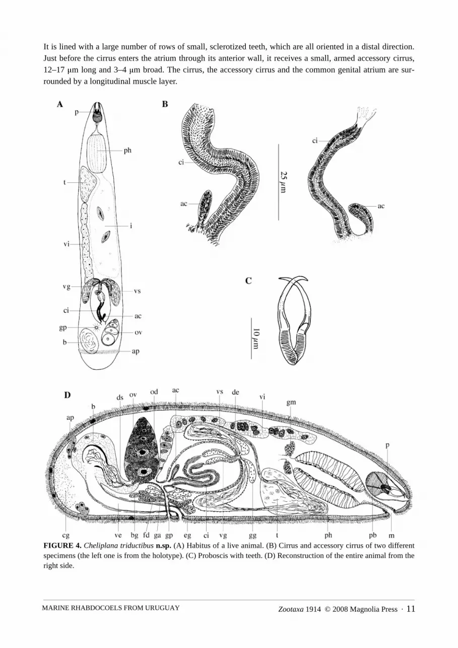
Zootaxa 1914 © 2008 Magnolia Press · 11MARINE RHABDOCOELS FROM URUGUAY
It is lined with a large number of rows of small, sclerotized teeth, which are all oriented in a distal direction.Just before the cirrus enters the atrium through its anterior wall, it receives a small, armed accessory cirrus,12–17 μm long and 3–4 μm broad. The cirrus, the accessory cirrus and the common genital atrium are sur-rounded by a longitudinal muscle layer.
FIGURE 4. Cheliplana triductibus n.sp. (A) Habitus of a live animal. (B) Cirrus and accessory cirrus of two differentspecimens (the left one is from the holotype). (C) Proboscis with teeth. (D) Reconstruction of the entire animal from theright side.

VAN STEENKISTE ET AL.12 · Zootaxa 1914 © 2008 Magnolia Press
The short oviduct receives the vitelloduct through its dorsal wall, and together they form the very shortfemale duct. This female duct enters the common genital atrium a little caudally from the place where themale genital system enters. A circular muscle layer surrounds the female duct, the oviduct and the most distalpart of the vitelloduct. The female duct is lined with an anucleated, membranous epithelium. Caudally fromthe ovary a large bursa occurs, which contains several compartments with sperm. The female bursa is con-nected to an external vagina, which is a relatively long duct, lined with a high, anucleated epithelium. Its prox-imal part is surrounded by circular muscles. This part also contains spermatozoa. The distal part of the vaginais surrounded by longitudinal muscles. The vaginal opening is surrounded by a strongly-developed sphincter.Within the dorso-anterior part of the bursa, three muscular sperm-containing compartments can be observed.From each of these compartments a sclerotized spermatic duct departs towards the ovary. In their middle partsthese three ducts are wound around each other, so that they cannot be discerned separately. Near the ovarythey diverge again, and enter the ovary as three separate ducts near to the place where the oviduct departs. Thespermatic ducts are surrounded by a parenchymatous tissue, which is a continuation of the parenchymatoustissue of the bursa. A uterus is absent.
Discussion. See discussion Cheliplana uruguayensis n.sp.
Cheliplana uruguayensis n.sp.(Fig. 5)
Locality. La Coronilla, Departamento de Rocha, Uruguay (33°54’18.50”S, 53°30’39.30”W). Beach andmouth of the canal near hotel Parque Océanico: flat beach further away from the mouth of the canal, sand withorganic material a couple of meters from a small intertidal pool (01/08/2004): type locality.
Material. Observations on a live animal. Three whole mounts, one of which designated holotype (SMNH7497), another one designated paratype (HU no. 406). Six serially-sectioned specimens, two of which desig-nated paratypes (HU no. 407–408).
Etymology. The species name refers to its occurrence in Uruguay.Description. The body length of the animal varies between 0.6–0.8 mm. Habitus and internal organisation
as in Cheliplana triductibus n.sp., except for the construction of the genital system.From the testis two morphologically dissimilar vasa deferentia depart. One is of the normal construction;
thin-walled and distally widening to a seminal vesicle, which is lined with a membranous, nucleated epithe-lium. The other vas deferens is enlarged and is surrounded by a layer of very thick, longitudinal muscles. It is190–260 μm long. The seminal vesicle and the modified vas deferens join each other distally to form the ejac-ulatory duct, which enters the copulatory bulb. The copulatory organ is of the conjuncta-duplex type, as in theother species of Cheliplana de Beauchamp, 1927. The prostate vesicle is 120–170 μm long, with a diameter of± 35 μm, and is surrounded by strongly-developed longitudinal muscles. The prostate secretion and the ejacu-latory duct distally enter a short, tubular stylet. This stylet is ± 11 μm long.
The male genital atrium is lined with a membranous epithelium and surrounded by a longitudinal musclelayer. In its distal half, it receives a bundle of basophilic glands. Distally it widens before entering the com-mon genital atrium. This common genital atrium is very broad and relatively long. Proximally it receivessome eosinophilic and basophilic glands. The epithelium is high and the common genital atrium is surroundedby longitudinal muscles over its entire length. Distally, it narrows again to broaden a second time just before itreaches the gonopore. This is situated at 80% and surrounded by a sphincter.
The ovary is situated dorsally at the left-hand side and joins the bursa. Proximally it is connected with thevitellarium to form an ovovitellarium. This vitellarium bends from the ovary to the ventral side and extendsrostrally up to the level of the testis. The caudally-situated female bursa is connected to the ovary by a broadduct consisting of bursal tissue. A separate spermatic duct was not observed. The bursa contains numerous

Zootaxa 1914 © 2008 Magnolia Press · 13MARINE RHABDOCOELS FROM URUGUAY
optically-clear vacuoles. An external vagina is connected to the bursa and has a thick, sclerotized wall. It wid-ens to a spherical space just proximally from the vaginal opening. The vaginal opening is situated rostrallyfrom the gonopore. At the place where the external vagina enters the bursa, a separate seminal receptacle isalso connected to the bursa. This seminal receptacle is elliptical and is surrounded by circular muscles.
Discussion. These two new species can easily be recognized as species of Karkinorhynchidae, as theyhave a proboscis provided with symmetrical hooks. In representatives of Diascorhynchidae Meixner, 1929,the only other schizorhynch taxon with representatives with armed probosces, the hooks are asymmetrical. Alarge number of diagnostic features makes it easy to place both new species within the taxon Cheliplaninae(see Karling 1983): presence of a postrostral bulb, presence of only one pair of hooks (also the case in someKarkinorhynchinae), absence of separate lateral glands, lack of eyes (also the case in very few species ofKarkinorhynchinae), presence of a single girdle of adhesive papillae (two such girdles in almost all Karkino-rhynchinae), anteriorly-situated pharynx mostly cylindrical, directed anteriorly and often with a long, spinyprepharyngeal cavity.
By comparing the features of the two species discussed here with the diagnoses of the genera of the taxonCheliplaninae, the Uruguayan species clearly belong to the genus Cheliplana because of the following fea-tures: proboscis hooks without denticles, soft fingerlike side pieces and a cylindrical pharynx with a longspinous cavity. They distinguish themselves from species of Baltoplana Karling, 1949 by the fact that Bal-toplana has paired ovaries and testes, and from species of Cheliplanilla Meixner, 1938 because this taxon hascrutch-shaped cuticular rods between the proboscis side pieces, proboscis hooks with two pairs of inside den-ticles, a blind seminal vesicle and an oviform pharynx with a short unarmed cavity (see Karling 1983). Theyalso differ from Archipelagoplana triplocirro Noldt & Hoxhold, 1984 because this species lacks a longspinous pharynx cavity and because A. triplocirro has two small accessory cirri (see Noldt & Hoxhold 1984).Karling (1983) considered the number of testes as an unreliable generic character and by consequence placedall representatives of the genus Rhinipera Meixner, 1928 (one testis) within the genus Cheliplana (two tes-tes), a view we follow here.
Cheliplana triductibus n.sp. stands unique within the genus because of the combined presence of threesclerotized spermatic ducts and one smaller accessory cirrus. In C. hiemalis Brunet, 1968, C. pacifica Noldt &Hoxhold, 1984, C. piriformis Brunet, 1968, C. pusilla Brunet, 1968, C. schilkei Noldt, 1989 and C. targa(Marcus, 1952) Karling, 1983, there is only one sclerotized spermatic duct between the bursa copulatrix andthe ovary (see Brunet 1968; Karling 1983; Noldt & Hoxhold 1984, Noldt 1989). C. varicauda Brunet, 1971has a comparable female and male system to C. triductibus n.sp., but its three spermatic ducts are onlyslightly sclerotized and the muscular proximal parts contain a spherical nucleus. In the male system, C. vari-cauda lacks the smaller accessory cirrus. Also the extremely long caudal part, posterior to the genital system,is lacking in the new species (see Brunet 1971). Accessory cirri are also present in C. textilis Jouk & DeVocht, 1989, but they have two blind accessory cirri that are lined with small spines in the posterior part andeven on one side (see Jouk & De Vocht 1989). Only very recently, Ax (2008) described a new species of Che-liplana from brackish water habitats along the French Atlantic coast and the Baltic Sea, with a very similarcopulatory organ. This species, C. deverticula Ax, 2008, has also a winding prostate vesicle and ejaculatoryduct, and a slender, armed cirrus (75 μm long) with a single small, armed accessory cirrus (17 μm long). How-ever, nothing is known about the female system of this species. Awaiting more material of C. deverticula anda description of its female system, the Uruguayan specimens are provisionally placed within a new species. Ifthe female system would prove to be identical, C. triductibus n.sp. should be synonymized with C. devertic-ula.
Cheliplana uruguayensis n.sp. differs from all other species of Cheliplana by the presence of two differ-ent seminal vesicles, a character shared with the genus Cheliplanilla, although in the latter the modified semi-nal vesicle ends blindly. This new species mostly resembles C. targa. Similarities are found in the femalesystem as well as in the male genital system: one testis situated ventrally from the pharynx; a seminal vesicle

VAN STEENKISTE ET AL.14 · Zootaxa 1914 © 2008 Magnolia Press
with a well-developed muscular wall; a very muscular prostate vesicle that distally ends in a short, tubularstylet, and a syncytial bursa containing vacuoles. In contrast to C. targa, which has only one seminal vesicle,C. uruguayensis n.sp. has two seminal vesicles. Moreover, C. uruguayensis n.sp. differs from C. targa byhaving a non-muscular vas deferens, eosinophilic and basophilic glands opening into the common genitalatrium, and by lacking a slerotized spermatic duct.
FIGURE 5. Cheliplana uruguayensis n.sp. (A) Habitus of a live animal. (B) Reconstruction of the entire animal.

Zootaxa 1914 © 2008 Magnolia Press · 15MARINE RHABDOCOELS FROM URUGUAY
Diagnosis. Cheliplana triductibus n.sp.: species of Cheliplana with unpaired male and female gonads.Proboscis 30 μm long and consisting of 11 μm-long proboscis hooks, 19 μm-long proboscis halves and a 20μm-long postrostral bulb. Without accessory denticles in the proboscis. Two seminal vesicles. Muscular pros-tate vesicle followed by a 64 μm-long cirrus with uniform teeth (3–4 μm long). Small, armed, lateral accessorycirrus with a length of 15 μm at the distal end of the cirrus. Short female duct. Vagina externa connected witha syncytial bursa. Three cuticularized spermatic ducts and muscular ducts connect the ovary with the bursa.
Diagnosis. Cheliplana uruguayensis n.sp.: species of Cheliplana with unpaired male and female gonads.Proboscis 23 μm long, consisting of 8 μm-long proboscis hooks, 16 μm-long proboscis halves and a 23–25μm-long postrostral bulb. Accessory denticles absent in the proboscis. Two different seminal vesicles, of whichone is a modified vas deferens surrounded by strongly-developed, longitudinal muscles. Both seminal vesiclesare connected with the testis. Long, longitudinally muscular prostate vesicle (± 150 μm long). Short tubularstylet (11 μm long). Vagina externa with cuticularized wall connected with a syncytial bursa containing vacu-oles. Elliptic seminal receptacle filled with spermatozoa.
Schizorhynchidae Karling, 1950 (sensu Graff, 1905)
Carcharodorhynchus viridis n.sp.(Fig. 6)
Locality. La Coronilla, Departamento de Rocha, Uruguay (33°54’18.50”S, 53°30’39.30”W). Beach andmouth of the canal near hotel Parque Océanico: sand covered by a thin green layer of organic material andsand with organic material near a small pool in open contact with the ocean (01/08/2004): type locality.
Playa Cerro Chato, Parque Nacional de Santa Teresa, Departamento de Rocha, Uruguay (33°59’6.34”S,53°31’48.81”W). Coarse superficial sand between rocks (20/07/2004).
Playa las Achiras, Parque Nacional de Santa Teresa, Departamento de Rocha, Uruguay (33°59’5.00”S,53°31’54.79”W). Sand of a steeply declining beach close to the waterline (26/07/2004).
Material. Observations on a live specimen. Three whole mounts, one of which designated holotype(SMNH 7498), another one designated paratype (HU no. 409).
Etymology. The species epithet refers to the colour of the live animal. Viridis (Lat.): greenDescription. Carcharodorhynchus viridis n.sp. is 1–1.4 mm long and has a vivid green colour. The ani-
mal lacks eyes and adhesive papillae were not observed. The pharynx rosulatus has a diameter of ± 75 μm andis at 65% of the body length. The epidermis contains a large number of elliptical rhabdites with an averagelength of 2–3 μm, especially at the rostral side. The proboscis is slightly asymmetrical with one half somewhatlarger than the other one, and measures 55–71 μm. In all whole mounts, only one field of denticles wasobserved on the largest proboscis half. However, in one specimen, denticles were also present on the transitionfrom the larger to the smaller proboscis half. The denticles are not in rows, but randomly distributed in anoblong field. All denticles are uniform.
About eight testes were observed in the live animal, although the precise number could not be determined.They are in one row, which extends rostrally from the pharynx. Two seminal vesicles are situated just behindthe pharynx. The apparently very muscular copulatory apparatus is 55 μm long and contains at least two dif-ferent types of prostate secretion. The hard parts of the male copulatory organ consist of a 7 μm-long stylet,surrounding a 12–15 μm-long cirrus. The stylet is an asymmetrical tube, which is distally backfolded to forma second tube that surrounds the distal part of the cirrus. The teeth of the cirrus are spirally-implanted and arelonger at the proximal and distal ends. The male genital duct is large and very muscular. The female systemcould not be observed in the live animal nor in the whole mounts.
Discussion. Based on the general organisation and the morphology of the proboscis, this species can eas-

VAN STEENKISTE ET AL.16 · Zootaxa 1914 © 2008 Magnolia Press
ily be placed within the genus Carcharodorhynchus Meixner, 1938. All representatives of this genus are slen-der, lack eyes and have a proboscis that is armed with denticles, usuallly in two fields on the sides of theproboscis halves. They have paired gonads (paired ovaries, vitellaria and one to eight pairs of testes). Themale copulatory organ has a cirrus or a stylet. Sometimes both structures are present.
The structure and armament of the proboscis are uncertain as reliable diagnostic characters for the differ-ent species. Fields of denticles on the sides of both the proboscis halves are present in all species. Conse-quently two U-shaped batteries of denticles are formed. Sometimes the denticles are placed in rows,sometimes randomly. Only the form, size and density of the denticles can differ distinctly. Thus the observa-tion of only one field of denticles in the whole mounts of the Uruguayan animals probably gives an incom-plete picture of the armament of the proboscis, especially because in the live animals fields of denticles wereobserved on both proboscis halves. Further, the species belonging to this genus can be grouped into a group ofspecies with asymmetrical proboscis halves and species with symmetrical proboscis halves. Also the con-struction of the male copulatory organs (either a cirrus, a stylet or both) is remarkably similar when the differ-ent species are compared. This may suggest that the number of species currently recognised might be reducedafter a thorough revision of this genus.
Carcharodorhynchus viridis n.sp. differs from all but one of the other species of this genus by the struc-ture of the male copulatory organ. Together with C. involutus Jouk & De Vocht, 1989, it is the only specieswhere the stylet surrounds the cirrus (see Jouk & De Vocht 1989). In other species where both a cirrus and astylet are present, this stylet is always found within the cirrus. C. viridis n.sp. differs from C. involutusbecause the cirrus is longer than the stylet while in C. involutus the reverse is true.
Diagnosis. Carcharodorhynchus viridis n.sp.: species of Carcharodorhynchus with a bright green colour.Slightly asymmetrical, armed proboscis with a length of 55–71 μm. A 7 μm-long stylet consisting of an asym-metrical tube, which distally bends inwards to form a second tube that distally surrounds a 12–15 μm long cir-rus. Spirally-implanted cirrus teeth that are longer distally and proximally.
FIGURE 6. Carcharodorhynchus viridis n.sp. (A) Habitus of a live animal. (B) Proboscis of two different specimens.(C) Copulatory apparatus with cirrus and stylet (from the holotype).

Zootaxa 1914 © 2008 Magnolia Press · 17MARINE RHABDOCOELS FROM URUGUAY
Dalytyphloplanida Willems et al., 2006
Provorticidae Beklemischew, 1928
Kirgisellinae Luther, 1962
Baicalellia forcipifera n.sp.(Figs. 7, 8A)
Locality. La Coronilla, Departamento de Rocha, Uruguay (33°54’18.50”S, 53°30’39.30”W). Beach andmouth of the canal near hotel Parque Océanico: sand covered by a thin green layer of organic material andsand with organic material near a small pool in open contact with the ocean (01/08/2004): type locality.
Material. Observations on a live animal. Three whole mounts, one of which designated holotype (SMNH7499), another one designated paratype (HU no. 410). Nine serially-sectioned specimens, of which seven des-ignated paratypes (HU no. 411–417). Unfortunately one of the better whole mounts, originally intended to bedesignated as the holotype and shown in fig. 7B (lower figure), was lost. Another whole mount was chosen asthe holotype.
Etymology. The species name refers to the pincers-shaped stylet. Forceps (Lat.): pincers. Ferre (Lat.): tocarry.
Description. The body length of the animal varies between 0.5–0.7 mm. The internal organisation doesnot differ from the other species of the taxon Baicalellia Nasonov, 1930 (see Nasonov 1930, 1932; Luther1962; Ax 1995).
The anteriorly-situated pharynx doliiformis lacks tentacle-like structures. The copulatory bulb is sur-rounded by a thick, inner, spirally-running to circular muscle layer and outer longitudinal muscles. The pros-tate vesicle is also surrounded by longitudinal muscles. Distal to the copulatory bulb, a pincers-shaped styletis present. It consists of a common base from which two separate, curved arms depart. The longest of thesearms is 44–49 µm long and hollow. It is filled with the prostate secretion that proximally enters the stylet. Theshortest arm is 20–27 µm long and seems to be an accessory outgrowth of the longer arm's wall. From thebase to the distal apex of the longest arm, the stylet measures 54–59 μm. The base of the stylet has a proximalrim and is connected to the copulatory bulb.
The male atrium fits tightly around the stylet. Distally it opens into the common genital atrium. The bursacopulatrix is a sack-shaped protrusion of the common genital atrium and is filled with spermatozoa. It runsdorsally from the male atrium and has a very broad connection with the common atrium. In consequence, dif-ferentiation between the bursa copulatrix and the common atrium is not always clear. The bursa copulatrix andthe male genital atrium are lined with a relatively high, anucleated epithelium and surrounded by a weakly-developed circular muscle layer. The gonopore is at about 75% and surrounded by a sphincter.
The female duct enters the common genital atrium through its caudal wall. At the place of entry, a strongsphincter is present. This female duct is relatively short and broad, lined with a high, nucleated epithelium andsurrounded by a strongly-developed circular muscle layer. The lumen is filled with spermatozoa. Proximally,both oviducts and a bundle of eosinophilic glands enter the female duct. The oviducts are very short and linedwith a membranous epithelium. Ventrally from the oviducts, a seminal bursa opens into the female ductthrough a sphincter. The seminal bursa is spherical to club-shaped and has a fairly long bursal stalk. It is linedwith a membranous epithelium. The bursa contains sperm, which are thinner and less darkly-stained than thesperm found elsewhere in the animal. A uterus is absent.
Discussion. This species can easily be placed within the taxon Baicalellia Nasonov, 1930, as it showsdiagnostic characters of this taxon: paired gonads, with the testes rostrally connected to each other, and ova-ries fused with vitellaria to form two ovovittelaria.
Almost all of the 17 species of Baicalellia have a tubiform to funnel-shaped prostate stylet, which is

VAN STEENKISTE ET AL.18 · Zootaxa 1914 © 2008 Magnolia Press
straight or curved. Apart from Baicalellia forcipifera n.sp., only two other species have a stylet with a lateraloutgrowth: B. canadensis Ax & Armonies, 1987 and B. anchoragensis Ax & Armonies, 1990. In B. anchor-agensis the stylet is short and broad, and the outgrowth has a little spine of its own. In B. canadensis the styletis somewhat longer and less sturdy, and the outgrowth is a simple, relatively short spine. In B. forcipifera n.sp.both the stylet and the outgrowth are long and very slender, giving it its typical pincer-like shape.
Nasonov (1930) describes the seminal bursa as a seminal receptacle and a phagocytic organ that serves toresorb redundant sperm. Ax (1954, 2008), Luther (1918, 1921) and Marcus (1946) use the name seminalbursa, while Ax & Armonies (1990) keep to syncytial tissue. According to Nasonov (1930) the strongly-thick-ened, syncytial epithelial wall of this organ contains vacuoles with sperm in various degrees of disintegration.
FIGURE 7. Baicalellia forcipifera n.sp. (A) Reconstruction of the anterior body part from the right side. (B) Stylet(upper from the holotype). (C) Reconstruction of the atrial organs from the left side.

Zootaxa 1914 © 2008 Magnolia Press · 19MARINE RHABDOCOELS FROM URUGUAY
The existence of such vacuoles is confirmed by Ax (1954), Ax & Armonies (1990), Luther (1921), Marcus(1946) and Joffe & Selivanova (1988). In Baicalellia forcipifera n.sp. only a very thin epithelium withoutvacuoles was observed. However, the lumen of the seminal bursa contains disintegrating sperm. The base ofthis organ is surrounded by a sphincter. This has also been observed in nearly all species except for B.canadensis, B. evelinae Marcus, 1946 and B. groenlandica Ax, 1995. The opening and position of this organin the genital system vary from species to species, as discussed by Ax & Armonies (1990) for B. posietiNasonov, 1930, B. sewardensis, B. anchoragensis and B. brevituba (Luther, 1921) Nasonov, 1930. In the otherspecies, the seminal bursa is always situated between, ventrally or dorsally from the ovovitellaria. In B.canadensis, a seminal bursa (indicated as seminal receptacle by Ax & Armonies 1987) was only observed asan appendage of the copulatory bursa. This makes the placement of this species within the genus Baicalelliauncertain (see Ax & Armonies 1987).
FIGURE 8. Baicalellia forcipifera n.sp. (A) Habitus of a live animal. Polliculus cochlearis n.g. n.sp. (B) General organ-isation of an animal based on serial sections. (C) Stylet and accessory stylet (from the paratype). (D) Reconstruction ofthe atrial organs from the right side (arrow indicates intracapsular seminal vesicle).

VAN STEENKISTE ET AL.20 · Zootaxa 1914 © 2008 Magnolia Press
In Baicalellia forcipifera n.sp. the female duct is clearly separated from the genital atrium by a strongly-developed sphincter. This is a somewhat unusual situation in Baicalellia, where the female duct is mostly veryshort, even considered part of the common genital atrium. Only Ax & Armonies (1987, 1990) use the term“female duct” as such, but in B. forcipifera n.sp. it is unusually long, even compared with that of B. canaden-sis, where it is also clearly separated from the common genital atrium by a strong sphincter. This feature,together with the construction of the seminal bursa and the form of the stylet, clearly shows that the materialfrom Uruguay belongs to a new species.
Diagnosis. Baicalellia forcipifera n.sp.: species of Baicalellia with a ± 54–59 μm-long, slender stylet, car-rying a ± 20–27 μm-long, slender spine. Stylet and outgrowth together have the shape of pincers. Female ductlong, clearly delimited, with a nucleated epithelium and surrounded by a strongly-developed, circular musclelayer. Seminal bursa without vacuoles and containing disintegrated sperm.
Dalyelliidae Graff, 1908
Polliculus cochlearis n.g. n.sp.(Figs. 8B–8D)
Locality. La Coronilla, Departamento de Rocha, Uruguay (33°54’18.50”S, 53°30’39.30”W). Beach andmouth of the canal near hotel Parque Océanico: sand covered by a thin green layer of organic material andsand with organic material near a small pool in open contact with the ocean (01/08/2004): type locality.
Playa Cerro Chato, Parque Nacional de Santa Teresa, Departamento de Rocha, Uruguay (33°59’6.34”S,53°31’48.81”W). Sand a couple of meters from the waterline (± 30 cm deep) (31/07/2004).
Material. Two whole mounts, one of which designated paratype (HU no. 418). Two serially-sectionedspecimens, of which one designated holotype (SMNH 7500).
Etymology. The genus name refers to the French fairy tale Hop o' My Thumb (French: Le Petit Poucet)written by Charles Perrault. Polliculus (Lat.): little thumb. The species name refers to the screw-shaped acces-sory stylet. Cochlea (Lat.): snail shell.
Description. This species is remarkably small, the entire body length being no more than 0.4 mm. Thesyncytial epidermis is ± 2 μm high, strongly ciliated and contains vacuoles. The cilia are about 1–3 μm high.The diameter of the vacuoles varies from 1–2 μm. The basal membrane is about 1/6 of the epidermis heightthick. Round to oval nuclei, with a diameter of ± 2 μm, are scattered throughout the epithelium. Small baso-philic rhabdites, with a diameter of less than 1 μm, are situated apically in the epidermal epithelium of theventral side and the rostral part.
The mouth lies in the anterior part of the body. It is surrounded by a weak sphincter. The frontal pharynxdoliiformis is exceptionally large and elongated, and measures about 1/3 of the total body length. The distalhalf of the pharynx is narrower than the proximal half. The prepharyngeal cavity is lined with an anucleated,membranous epithelium and surrounded by a longitudinal muscle layer. The oesophagus is surrounded by theglands of Minot. The brain lies dorsally from the pharynx, with two eyes situated in front of it.
The gonads are unpaired. The testis lies just behind the pharynx and is situated at the right-hand side ofthe animal. A broad vas deferens leaves the testis and widens distally to form a seminal vesicle. Both the vasdeferens and the seminal vesicle are lined with a membranous epithelium. There are no muscles surroundingthese structures. The seminal vesicle proximally penetrates the prostate vesicle, which is surrounded bystrongly-developed circular muscles (conjuncta-type copulatory organ; terminology of Karling 1956a). Proxi-mally in the prostate vesicle, the vas deferens remains very broad to form an intracapsular seminal vesicle(arrow in fig. 8D). Basophilic and eosinophilic prostate glands are present. The nuclei-containing parts ofthese glands are situated extracapsularly.

Zootaxa 1914 © 2008 Magnolia Press · 21MARINE RHABDOCOELS FROM URUGUAY
The stylet is a long, hollow tube with a length of 25–27 μm. It bends distally and ends in an asymmetrical,distally-widening apex. The proximal part is funnel-shaped. It receives the ejaculatory duct and the prostatesecretion. Around the tubular stylet, an accessory stylet with an axial length of 41–52 μm is present. Thisaccessory stylet is spirally-wound around the stylet proper. In the holotype and live animals, these proximalwindings are close to each other, while the distance between these two stylets increases distally. The last distalwinding ends in a sharp point at about the same level as the distal end of the stylet proper. In the paratype, thedistal part of the accessory stylet is partly unwound and runs next to the stylet. Proximally, this accessorystylet receives the secretion of about four accessory glands that lie around the narrowed, distal part of the cop-ulatory bulb. Dorsally from the stylets, a membranous epithelium connects to the prostate vesicle; it is lackingventrally. This is probably the epithelium of the male genital atrium. This male genital atrium and the prostatevesicle are in their turn surrounded by another membranous epithelium that is proximally and distally encom-passed by a well-developed, circular muscle layer (conjuncta-duplex type copulatory organ; terminology ofKarling 1956a). The male genital atrium is at the left-hand side of the animal. Rostrally, it opens into the smallcommon genital atrium, which is lined with an anucleated, membranous epithelium.
The female duct enters the caudal wall of the common atrium. It is relatively short, lined with a membra-nous epithelium and surrounded by a circular muscle layer. Small clumps of sperm are observed in the distalpart of the female duct, which proximally receives the oviduct and the vitelloduct. The oviduct is also linedwith an anucleated, membranous epithelium and surrounded by circular muscles. The single ovary is at 60%of the body length and lies roughly in the middle. Laterally, two vitellaria are present. These run rostrally atboth sides of the pharynx and have a widened distal part. Distally, they join and open into the female ductthrough a common vitelloduct. An oviform seminal receptacle enters the common atrium caudally. This organcontains many sperm and is surrounded by a strongly-developed circular muscle layer. Its distal part is nar-rower, lined with a membranous epithelium and surrounded by a weak circular muscle layer.
Between the male atrium and the gonopore a uterus also enters the common atrium. The gonopore is situ-ated at about 75% on the ventral side.
Discussion. The combined presence of the following features: one ovary, paired vitellaria, a frontal phar-ynx and a gonopore in the posterior part of the body, suggests the placement of Polliculus cochlearis n.sp. inthe Dalyelliidae.
Polliculus cochlearis n.sp. clearly differs from every other species of dalyelliids in having only one testisand one vas deferens. In all other dalyelliids there are two. Moreover, the vas deferens of P. cochlearis n.sp.forms a relatively large extracapsular vas deferens. Apart from this species, extracapsular seminal vesicles,one on each vas deferens, only occur in very few species: Gieysztoria dodgei (Graff, 1911) Ruebush & Hayes,1939, Microdalyellia armigera (Schmidt, 1861) Gieysztor, 1938, Microdalyellia fusca (Fuhrmann, 1894)Gieysztor, 1938 and Jensenia angulata (Jensen 1878) Graff, 1882 (see Luther 1955). In Halammovortex Kar-ling, 1943, the vasa deferentia join in an unpaired, extracapsular seminal vesicle. None of these genera, how-ever, has a muscular septum around the conjuncta-duplex type as does P. cochlearis n.sp.
Within the Dalyelliidae, Luther (1955) distinguishes four types of stylet: the Microdalyellia (= armigera)-type, the Gieysztoria (= rubra)-type, the Castrella-type and the Axiola-type (Axia-type in Luther 1955). TheMicrodalyellia-, Gieysztoria- and Castrella-types all consist of a common base on which a group of spines isimplanted. The Axiola-type, in contrast, is a tubular stylet. It is found uniquely within the marine genus AxiolaLuther, 1957. The stylet of Polliculus cochlearis n.sp. thus could be considered as of the Axiola-type. Thepresence of an accessory stylet, however, is unique within the Dalyelliidae.
Polliculus cochlearis n.sp. also differs from all other species of Dalyelliidae by the lack of a so-calledbursa copulatrix. This structure is a well-developed protrusion of the common genital atrium where sperm isalso stored (Luther 1955). Only in Vaillantiella algerica Luther, 1955, is it rather indistinct. Another uniquecharacteristic of P. cochlearis n.sp. is the presence of a distinct uterus, opening in the rostral wall of the com-mon genital atrium and with clear uterine glands. Such a uterus occurs in several rhabdocoels, but is lacking in
VAN STEENKISTE ET AL.22 · Zootaxa 1914 © 2008 Magnolia Press
all other species of Dalyelliidae, where the uterus is formed by a distal widening of the female duct (Luther1955). These considerable differences clearly warrant the erection of a new genus for the species from Uru-guay.
Five genera of dalyelliids are known that are exclusively marine: Alexlutheria Karling, 1956, AxiolaLuther, 1957, Beauchampiola Luther, 1957, Halammovortex Karling, 1943 and Jensenia Graff, 1882. Twoother taxa, Gieysztoria Ruebush & Hayes, 1939 and Microdalyellia Gieysztor, 1938, mainly include freshwater species, but also contain a number of species that occur in brackish habitats. The other known genera,Austrodalyellia Hochberg & Cannon, 2002, Castrella Fuhrmann, 1900, Dalyellia Gieysztor, 1938, Fulin-skiella Gieysztor & Szynal, 1939, Sergia Nasonov, 1923, Vaillantiella Luther, 1955 and Varsoviella Gieysztor& Wiszniewski, 1947, only occur in fresh water.
Diagnosis. Polliculus n.g.: Dalyelliidae with pharynx in the first body half. Unpaired testis and ovary.Paired vitellaria. Vas deferens widened to an extracapsular seminal vesicle. Eosinophilic and basophilic pros-tate glands with extracapsular, nuclei-containing parts. Copulatory bulb with intracapsular seminal vesicle,prostate vesicle and male genital atrium with stylet. Screw-shaped accessory stylet wound around a long tubu-lar stylet. Accessory stylet receives the secretion of four extracapsular accessory glands. Short female ductsurrounded by circular muscles. Oviform seminal receptacle. Copulatory bursa absent. With distinct uterus.Type species: Polliculus cochlearis n.sp.
Diagnosis. Polliculus cochlearis n.sp.: provisionally, the same diagnosis as the genus. Stylet with a lengthof ± 26 μm. Accessory stylet ± 47 μm long.
Luridae Sterrer & Rieger, 1990
Lurus evelinae Marcus, 1950(Figs. 9A–9B)
New locality. Playa del Barco, Parque Nacional de Santa Teresa, Departamento de Rocha, Uruguay(33°59’52.12”S, 53°32’6.51”W). Coarse-grained sand just in between the dunes and the water line, up to 30cm deep (16/07/2004).
Playa Grande, Parque Nacional de Santa Teresa, Departamento de Rocha, Uruguay (34°1’55.17”S,53°32’10.86”W). Between filiform, rather large brown algae from rocks exposed to heavy wave action (18/07/2004).
Known distribution. Baía de Santos, São Paulo, Brazil (Marcus, 1950).Material. Observations on a live individual. Two whole mounts (one without a stylet). Type material
from Lurus evelinae Marcus, 1950: SMNH 95803, SMNH 95804.Discussion. The Uruguayan specimens clearly belong to the taxon Luridae Sterrer & Rieger, 1990. Typi-
cal of this taxon is the fact that the overall organisation resembles that of the Provorticidae and that a polyli-thophoric statocyst is present (Marcus 1950; Faubel et al. 1994; Sterrer & Rieger 1990; Sterrer 1992).
Based on the morphology of the stylet (a spirally-winding tube, two turns), the Uruguayan specimens canbe classified in Lurus evelinae Marcus, 1950 (see Marcus 1950). The specimens measure 0.3 mm. The stylethas an axial length of 165 μm. Since no whole mounts are available in the material from Brazil, the length ofthe stylet is estimated based on the figure drawn by Marcus (1950). Axially they measure 140 μm.
Although Sterrer & Rieger (1990) already mentioned some clear differences between Lurus evelinae Mar-cus, 1950 and all other species of Luridae, it was Faubel et al. (1994) who divided the monogeneric taxonLuridae into Lurus Marcus, 1950 (one species: L. evelinae) and Luriculus Faubel et al., 1994 (four species: L.australiensis Faubel et al., 1994, L. castor Sterrer & Rieger, 1990, L. minos Sterrer, 1992, and L. tyndareusSterrer & Rieger, 1990). Lurus evelinae stands unique because it has paired (sometimes coalescing) testes andvasa deferentia, separate ovaries and vitellaria, and a clear differentiation between the seminal vesicle and the

Zootaxa 1914 © 2008 Magnolia Press · 23MARINE RHABDOCOELS FROM URUGUAY
prostate vesicle. All other species of Luridae have a single testis, one median vas deferens, ovovitellaria and acombined seminal vesicle and prostate vesicle. Therefore they were transferred to the new genus Luriculus(see Faubel et al. 1994; Marcus 1950; Sterrer & Rieger 1990). One species, Lurus pollux Sterrer & Rieger,1990, remains a species inquirenda. Although L. pollux has only a single testis, one medioventral vas deferensand a seminal vesicle inside the copulatory bulb, Faubel et al. (1994) did not place it in Luriculus becausevitellaria have not been observed. Besides the stylet, no other confirmative features could be observed in thelive specimens of L. evelinae from Uruguay. The single testis could well be two coalescing testes and no vasadeferentia were seen. However, ovaries and vitellaria seemed rather separate.
FIGURE 9. Lurus evelinae Marcus, 1950 (A) Habitus of a live animal. (B) Stylet. Vauclusia multistriata n.sp. (C) Habi-tus of a live animal.
As was discussed by Marcus (1950) and Sterrer & Rieger (1990), Lurus evelinae apparently always hastwo statoliths, whereas all other species of Luridae mostly have a variable number of statoliths (see Sterrer &Rieger 1990; Sterrer 1992): two to four in L. castor, two to five in L. pollux and three to four in L. tyndareusand L. minos. L. australiensis always has a constant number of three intracapsular and three extracapsular sta-toliths. In the whole mounts of the specimens from Uruguay, the statoliths were not visible. Only two sta-toliths were observed in the specimen that was studied alive. This seems to confirm the observations byMarcus (1950), which were made on serial sections, and were therefore doubted by Sterrer (1992).
After Luriculus minos (see Sterrer 1997), Lurus evelinae is the second species of Luridae to be reportedfrom outside its type locality.
VAN STEENKISTE ET AL.24 · Zootaxa 1914 © 2008 Magnolia Press
Promesostomidae Den Hartog, 1964
Vauclusia multistriata n.sp.(Figs. 9C, 10)
Locality. La Coronilla, Departamento de Rocha, Uruguay (33°54’18.50”S, 53°30’39.30”W). Beach andmouth of the canal near hotel Parque Océanico: sand covered by a thin green layer of organic material andsand with organic material near a small pool in open contact with the ocean (01/08/2004): type locality.
Material. Observations on one live animal. One whole mount, designated holotype (SMNH 7501). Twoserially-sectioned specimens, of which one designated paratype (HU no. 363).
Etymology. The species name refers to the many internal ridges of the stylet. Multus (Lat.): many. Stria-tus (Lat.): striated.
Description. Slender, 0.5 mm long animal. Eyes absent. The habitus is characterized by division of thebody into three zones: a slender, rostral part where rhabdite glands occur (± 1/5 of the body length), a middlepart where all organs are situated (± 3/5 of the body length), and a caudal part, which consists of a clearly-delimited, small tail with an epidermis containing many more rhabdites than the rest of the body (± 1/5 of thebody length).
The ciliated epidermis is cellular and has a height of about 3 μm. The basal membrane is very thin. Thecilia measure about 3 μm. In the rostral part of the body, two different kinds of rhabdite glands occur. The firsttype produces long, lancet-shaped, basophilic rhabdites with a length of 8–10 μm, the other type producessmaller, oval, eosinophilic rhabdites with a length of about 2–3 μm. Both kinds of glands extend from abovethe brain to the rostral body end, where they empty. Caudally from the pharynx, dispersed eosinophilic rhab-dite glands occur, which empty in the epidermis. The largest concentration of these rhabdite glands is situatedbehind the genital system in the caudal body part. The length of the oval, eosinophilic rhabdites that aresecreted by these glands varies between 2–4 μm. The epidermis of the caudal region is completely filled withthese rhabdites.
The mouth is situated at about 40%. The organisation and structure of the pharynx is identical with that ofVauclusia conica Willems et al., 2004 (see Willems et al. 2004).
The gonads are paired. The two round testes are situated ventrally, just in front of the pharynx and at bothsides of the body. The seminal vesicles are rather small and have an anucleated, membranous epithelium.They are not surrounded by muscle layers. When entering the prostate vesicle, both vasa deferentia join toform a ductus ejaculatorius, which runs centrally through the prostate vesicle. Centrally, this prostate vesiclecontains coarse-grained, eosinophilic glands, which are surrounded by fine-grained eosinophilic glands. Allglands together take up about half of the total volume of the prostate vesicle. Both the fine-grained andcoarse-grained eosinophilic glands have extracapsular nuclei-containing parts, and penetrate the prostate vesi-cle together with the ejaculatory duct. The prostate vesicle is surrounded by thick, inner circular muscles andouter longitudinal muscles. Distally, the prostate vesicle is connected with a conical and thin-walled stylet.This stylet has a length of 45 μm. Proximally, it is 21 μm broad at its broadest, distally up to 4 μm. The innerside of this stylet is ornamented with a large number of ridges that are oriented in three different directions.For the first 17 μm, the ridges run lengthways, the next 7 μm they run transversely and the last 21 μm againlengthways. The male genital atrium is surrounded by circular muscles and is lined with an anucleated, mem-branous epithelium, as is the common genital atrium. The common gonopore is situated at about 70%.
The ovaries lie caudally from the gonopore and together with the more rostrally-situated vitellaria, theyform long ovovitellaria, which stretch dorsolaterally at both sides beyond the testes. The oviducts are veryshort, and open in the long, very muscular female duct, which caudally enters the common genital atrium. It islined with a high, anucleated epithelium and surrounded by a circular muscle layer, which is very well devel-oped in the proximal half. Distally, it is slightly swollen and some nuclei are seen in the somewhat higher epi-

Zootaxa 1914 © 2008 Magnolia Press · 25MARINE RHABDOCOELS FROM URUGUAY
thelium of the transition zone between the female duct and the common genital atrium. Moreover, the distalhalf of the female duct is surrounded by a peripheral, longitudinal muscle layer. The female bursa is sphericaland lined with a nucleated, membranous epithelium. It contains a great deal of sperm in the lumen. A broadbursal stalk connects the bursa with the proximal end of the female duct. Its epithelium is very high and showsmany glands. Where it enters the bursa, a sphincter is present. In the proximal part of the female duct, spermand a glandular secretion were observed. Eosinophilic and basophilic female glands enter this part of thefemale duct. A uterus is lacking.
FIGURE 10. Vauclusia multistriata n.sp. (A) Reconstruction of the entire animal. (B) Reconstruction of the atrialorgans. (C) Stylet with internal ridges (from holotype).
Discussion. The species described here can easily be placed in the taxon Vauclusia Willems et al., 2004because of the combined presence of the following characters (see Willems et al. 2004): pharynx in the middleof the body, paired testes and seminal vesicles, spherical prostate vesicle and two types of glandular secre-tions, conical stylet with different internal ridges, a long male atrium, paired ovovitellaria, a long muscularfemale duct with a swollen distal part, and the presence of a female bursa and female glands. The constructionof the pharynx and the position of the testes are comparable with those of V. conica Willems et al., 2004, theonly other representative of the taxon, although in V. multistriata n.sp. the pharynx is situated somewhat moreanteriorly.
A first important difference between the two species occurs in the size of the stylet and the pattern of itsridges. Although Vauclusia conica is almost three times as long as V. multistriata n.sp., its stylet is smallerthan the stylet of V. multistriata n.sp. The exact structure of the stylet in V. conica could not be ascertained,but it apparently has about eight spirally-running and striated ridges (Willems et al. 2004). In contrast, the

VAN STEENKISTE ET AL.26 · Zootaxa 1914 © 2008 Magnolia Press
stylet of V. multistriata n.sp. has many ridges, transverse as well as longitudinal. Secondly, the prostate glandsof V. conica take up the full volume of the prostate vesicle, which is not the case in V. multistriata n.sp.Another difference is the presence of a bursal stalk with a glandular epithelium between the bursa and thefemale duct in V. multistriata n.sp. In V. conica, the bursa links up directly with the proximal part of thefemale duct through a sphincter. Finally, V. conica has an inversion of the muscle layer in the transition zonebetween the common genital atrium and the male atrium.
Some of the features that are unique to Vauclusia conica were mentioned in the original diagnosis of thisgenus by Willems et al. (2004), but now these features should be removed from this diagnosis.
Amended diagnosis (after Willems et al. 2004). Vauclusia: Promesostomidae with the pharynx in the mid-dle of the body. Paired testes and seminal vesicles. Spherical prostate vesicle with two types of secretions.Conical stylet with several internal ridges. Paired ovovitellaria. Very long, muscular female duct. Distal partof the female duct swollen. Female bursa and female glands present. Male atrium long.
Diagnosis. Vauclusia multistriata n.sp.: species of Vauclusia with a 45 μm-long stylet with many internalridges that run lengthways proximally and distally, and transversely in between. Prostate glands only fill aportion of the prostate vesicle. Duct with glandular epithelium between the bursa and the female duct.
Brinkmaniellinae Luther, 1948
Coronhelmis mimosa n.sp.(Figs. 11A–11B)
Locality. Playa la Moza, Parque Nacional de Santa Teresa, Departamento de Rocha, Uruguay(33°58’30.02”S, 53°31’50.89”W). Beach north of the observation tower: coarse sand near the stairs at about 8m from the rocks (24/07/2004): type locality.
Material. Observations on a live animal. Four whole mounts, one designated as the holotype (SMNH7502) and one designated paratype (HU no. 420), two of poor quality.
Etymology. The species name refers to the opening and closing movements of the stylet as observed onthe live animal. These movements are reminiscent of those of the leaves of the plant genus Mimosa.
Description. The animal is ± 0.5–0.6 mm long. The pharynx lies in the middle of the body. The generalorganisation of the genital system (based on observations on a live animal) does not differ from other speciesof the genus Coronhelmis Luther, 1948 (see Ax 1951, 1994; Ehlers 1974; Luther 1948).
The stylet is 15–17 μm long (m = 16 µm; n = 3) and 12–15 μm (m = 14 µm; n = 3) broad. It consists of aproximal, thin-walled, 7–9 μm-long part [called “Manschette” by Luther (1948)] and a distal ring of 4–5 μm-long spines. The proximal part has a number of grooves of ± 4 μm long that run lengthways. In the central partof the stylet of one of the whole mounts, a reticulate structure was observed, possibly caused by the uneven-ness of the stylet wall. In the live animals, we observed opening and closing movements of the ring of spines.
Discussion. This species has all diagnostic characters of the genus Coronhelmis (see Luther 1948): thepresence of a bursa copulatrix and a copulatory organ with a stylet that consists of a proximal part or “Man-schette” and a distal ring of spines. An overview of the diagnostic characters of all species described wasgiven by Willems et al. (2005b: table 1).
From this table it is immediately clear that Coronhelmis mimosa n.sp. is a relatively small species ofCoronhelmis, of which the representatives are normally around 1 mm long. In C. mimosa, the ratio “Man-schette” to entire stylet length is ± 1/2. Only in C. cuypersi Willems et al., 2005, C. lutheri Ax, 1951, C.novaecaledoniae Willems et al., 2005 and C. subtilis Ax, 2008, is a comparable ratio found. Of these four spe-cies, the stylets of C. cuypersi, C. lutheri and C. subtilis have more than 30 spines. The number of spines inCoronhelmis mimosa could not be ascertained, but presumably amounts to 10–15, a number comparable to

Zootaxa 1914 © 2008 Magnolia Press · 27MARINE RHABDOCOELS FROM URUGUAY
that found in C. novaecaledoniae. However, the stylet is proportionally much wider in C. novaecaledoniae(width/length = 1.5) than in C. mimosa (width/length = 0.9). The proximal grooves and the central, reticulatestructure can not with certainty be considered part of the stylet, but are not mentioned in other species.Because of the above-cited differences from the other species of the genus Coronhelmis, the Uruguayan spec-imens are considered to belong to a new species.
Unfortunately, the whole mount with the reticulate structure in the central part of the stylet was not wellconserved. Another whole mount was chosen as the holotype.
Diagnosis. Coronhelmis mimosa n.sp.: small species of Coronhelmis with a 15–17 μm-long and 12–15μm-broad stylet that consists of a proximal “Manschette” with a length of 7–9 μm and a distal ring of 4–5μm-long spines. Possibly vertical grooves in the proximal part and a reticulate structure in the central part.
Byrsophlebidae Graff, 1905
Byrsophlebs caligulachaena (Ehlers & Ehlers, 1981) Karling, 1985(Fig. 11C)
syn. Maehrenthalia caligulachaena Ehlers & Ehlers, 1981
New locality. Playa Grande, Parque Nacional de Santa Teresa, Departamento de Rocha, Uruguay(34°1’55.17”S, 53°32’14.52”W). On algae (Corallimorpha sp.) in intertidal pools (18/07/2004).
Known distribution. Egedesminde, Greenland (Levinsen 1879); White Sea, Solovki Islands, Russia(Sabussow 1900); Plymouth Sound, United Kingdom (Gamble 1893); Port Erin, Man Island (Jameson 1896);Jekaterinhafen and Bergen, Norway (Graff 1905; Luther 1936); Tomales Bay, California, USA (Karling1985).
Material. Observations on a live animal. One whole mount.Discussion. The examined animal is very small and measures only 0.3 mm in the whole mount. The
parenchym has a vivid red colour. Although the form of the stylet is not completely clear in the whole mount,it seems to be a funnel-shaped, oblong tube with a length of ± 25–30 μm and a distally-bent apex. The vasadeferentia are very broad near the pharynx and narrow caudally from the pharynx to strongly widen again asseminal vesicles laterally from the copulatory apparatus. These broad seminal vesicles fuse before penetratingthe copulatory apparatus as one single broad seminal vesicle. In the live animal, two larger organs wereobserved caudally from the copulatory bulb. These are possibly a bursa and seminal receptacle.
Karling (1985) reviewed the taxonomy and systematics of the Byrsophlebidae, in which he recognizedfour genera. These four genera are separated based on clear characters, but in practice sections are oftenneeded to asses these characters. All species of Maehrenthalia Graff, 1905 and Byrsophlebs Jensen, 1878have a female bursa, a structure that can normally be seen in the live animal. Such a bursa is lacking in speciesof Maehrenthaliella Karling, 1985 and Parabyrsophlebs Karling, 1985. Byrsophlebs differs from Mae-hrenthalia by the following features: a globular-oviform copulatory bulb with two layers of strong spiral mus-cle fibres, a female duct with a seminal receptacle and bursa without terminal resorptive vesicle.Maehrenthalia, on the contrary, has a cylindrical copulatory bulb with a single layer of strong spiral musclefibres, a female duct without a seminal receptacle and a bipartite bursa consisting of a uterus and terminalresorptive vesicle (Karling 1985).
The stylet observed in the live specimen from Uruguay mostly resembles that of Byrsophlebscaligulachaena. Although the red colour is only found in Maehrenthalia agilis (Levinsen, 1879) Graff, 1905,the tip of the stylet of M. agilis is more pointed and curved, than that of B. caligulachaena, which is shoe-shaped as in the Uruguayan specimen (see Ehlers & Ehlers 1981; Karling 1985). Therefore the Uruguayan

VAN STEENKISTE ET AL.28 · Zootaxa 1914 © 2008 Magnolia Press
specimens are provisionally considered as Byrsophlebs caligulachaena. Also the possible presence of a semi-nal receptacle (see above) suggests that the species found belongs to the taxon Byrsophlebs rather than Mae-hrenthalia.
FIGURE 11. Coronhelmis mimosa n.sp. (A) Habitus of live animal. (B) Stylet (right one from holotype). Byrsophlebscaligulachaena (Ehlers & Ehlers, 1981) Karling, 1985 (C) Habitus of live animal. Ceratopera axi (Riedl, 1954) DenHartog, 1964 (D) Bursal appendage. (E) Stylet.

Zootaxa 1914 © 2008 Magnolia Press · 29MARINE RHABDOCOELS FROM URUGUAY
Trigonostomidae Den Hartog, 1964 (sensu Graff, 1905)
Ceratopera axi (Riedl, 1954) Den Hartog, 1964(Figs. 11D–11E)
syn. Proxenetes axi Riedl, 1954syn. Ceratopera bifida Ehlers & Ax, 1974
New locality. Playa Cerro Chato, Parque Nacional de Santa Teresa, Departamento de Rocha, Uruguay(33°59’6.34”S, 53°31’48.99”W). Between algae (mostly filiform brown algae) on exposed rocks, betweensmall brown algae on top of very flat, isolated rocks, partly embedded in sand (20/07/2004).
Known distribution. Gulf of Napels and Sicily (Riedl 1954); Galapagos Islands (Ehlers & Ax 1974);Falkland Islands and California (Karling 1986); Weddell Sea and La Réunion (Artois et al. 2000), New SouthWales, Australia (Willems et al. 2004); Kerguelen (Willems et al. 2005a); New Caledonia (Willems et al.2005b).
Material. Observations on a live specimen. Two whole mounts.Discussion. The stylets of the Uruguayan specimens measure 86–90 μm, when measured axially and the
bursal appendage is 56–64 μm long. Both structures, especially the stylet, are small compared to known mea-surements of this species: stylet between 70–180 μm, bursal appendage between 67–117 μm (see Willems etal. 2004, table 1; Willems et al. 2005a, b). Only in the specimens of the Galapagos Islands (Ehlers & Ax1974), is the bursal appendage also less than 70 μm. Because the variation in the size of the stylet is relativelylarge, it is possible that we are dealing with a species complex (see Willems et al. 2005a, b). However, thisshould be examined in the future by a thorough morphological (and molecular) analysis.
Acknowledgements
The following people are gratefully acknowledged: Prof. Dr. Rodrigo Ponce de León (Universidad de laRepública) for logistic support during our stay in Uruguay; the Uruguayan military authorities in Santa TeresaNational Park for providing us with a sleeping and working place inside the park; Prof. Dr. Magda Vincx(Ghent University) for use of the laboratory facilities in the marine biology section in Ghent; Dr. Sven Bos-tröm (Swedish Museum of Natural History) for sending us the material of Lurus evelinae, Oneppus lacus andOneppus timius; Dr. Wim Willems for his help with the figures and advice in interpreting the serial sections;Dr. Nikki Watson for her helpful comments on the manuscript and correction of the English text; Mrs. Nat-ascha Steffanie for sectioning and staining the specimens. The first author is supported by a grant (aspirant) ofthe Research Foundation-Flanders (FWO-Vlaanderen).
References
Amato, J.F.R., Amato, S.B. & Seixas, S.A. (2005) Temnocephala lutzi Monticelli (Platyhelminthes, Temnocephalida)ectosymbiont on two species of Trichodactylus Latreille (Crustacea, Decapoda, Trichodactylidae) from southernBrazil. Revista Brasileira de Zoologia, 22, 1085–1094.
Amato, J.F.R., Amato, S.B. & Seixas, S.A. (2006) A new species of Temnocephala Blanchard (Platyhelminthes, Temno-cephalida) ectosymbiont on Trichodactylus fluviatilis Latreille (Crustacea, Decapoda, Trichodactylidae) from south-ern Brazil. Revista Brasileira de Zoologia, 23, 796–806.
Artois, T., Vermin, W. & Schockaert, E.R. (2000) Rhabdocoela (Platyhelminthes) from the Weddell Sea (Antarctica)with the description of eight new species. Belgian Journal of Zoology, 130, 103–110.
Ax, P. (1951) Die Turbellarien des Eulitorals der Kieler Bucht. Zoologische Jahrbücher-Abteilung für Systematik, Ökol-ogie und Geographie der Tiere, 80, 277–378.

VAN STEENKISTE ET AL.30 · Zootaxa 1914 © 2008 Magnolia Press
Ax, P. (1954) Marine Turbellaria (Dalyellioida) von den deutschen Küsten. Zoologische Jahrbücher-Abteilung für Sys-tematik, Ökologie und Geographie der Tiere, 82, 481–496.
Ax, P. (1994) Coronhelmis-species (Rhabdocoela, Platyhelminthes) from Greenland, Iceland and the Faroes. MicrofaunaMarina, 9, 221–237.
Ax, P. (1995) Plathelminthes aus dem Eulitoral von Godhavn (Disko, Grönland). Microfauna Marina, 10, 249–294.Ax, P. (2008) Plathelminthes aus Brackgewässern der Nordhalbkugel. Abhandlungen der Mathematisch-naturwissen-
schaftligen Klasse, Jahrgang 2008, 1. Akademie der Wissenschaften und der Literatur, Mainz, Franz Steiner Verlag,Stuttgart, 696 pp.
Ax, P. & Armonies, W. (1987) Amphiatlantic identities in the composition of the boreal brackish water community ofPlathelminthes. Microfauna Marina, 3, 7–80.
Ax, P. & Armonies, W. (1990) Brackish water Plathelminthes from Alaska as evidence for the existence of a borealbrackish water community with circumpolar distribution. Microfauna Marina, 6, 7–109.
Beklemischew, W.N. (1928) Ueber die Turbellarienfauna des Aralsees. Zugleich ein Beitrag zur Morphologie und zumSystem der Dalyelliida. Zoologische Jahrbücher-Abteilung für Systematik, Geographie und Biologie der Tiere, 54,87-138.
Blanchard, E. (1847) Recherches sur l’organisation des vers. Annales des Sciences Naturelles Zoologie, 3, 271–341.Bresslau, E. (1933) Turbellaria. In : Kükenthal, W. & Krumbach, T. (Eds.), Handbuch der Zoölogie. Eine Naturge-
schichte der Stämme der Tierreiches. Band II.Walter De Gruyter & Co., Leipzig, pp. 52-304.Brunet, M. (1965) Turbellariés Calyptorhynques de substrats meubles de la région de Marseille. Recueil des Travaux de
la Station Marine d’Endoume, 39, 127–219.Brunet, M. (1968) Turbellariés Karkinorhynchidae de la région de Marseille. Les genres Cheliplana et Cheliplanilla.
Cahiers de Biologie Marine, 9, 421–440.Brunet, M. (1971) Cheliplana varicauda n. sp., nouveau turbellarié calyptorhynque de la famille des Karkinorhynchidae.
Annales de l’Université de Provence, 45, 25–28.Brusa, F. (2006a) Macrostomidae (Platyhelminthes: Rhabditophora) from Argentina, with description of two new species
of Macrostomum and of stylet ultrastructure. Zoological Science, 23, 853–862.Brusa, F. (2006b) First record of Prolecithophora (Platyhelminthes) from Argentina: Plagiostomum cilioejaculator sp. n.
Zootaxa, 1223, 47–53.Brusa, F. & Damborenea, M.C. (2000) First report of Temnocephala brevicornis Monticelli 1889 (Temnocephalidae:
Platyhelminthes) in Argentina. Memorias do Instituto Oswaldo Cruz, 95, 81–82.Brusa, F., Damborenea, M.C. & Noreña, C. (2003) A new species of Gieysztoria (Platyhelminthes, Rhabdocoela) from
Argentina and a kinship analysis of South American species in the genus. Zoologica Scripta, 32: 449–457.Brusa, F., Ponce de León, R. & Damborenea, C. (2006) A new Paravortex (Platyhelminthes, Dalyellioida) endoparasite
of Mesodesma mactroides (Bivalvia, Mesodesmatidae) from Uruguay. Parasitology Research, 99, 566–571.Curino, A.C. & Cazzaniga, N.J. (1993) A new species of freshwater planarian from Chile (Platyhelminthes, Tricladida),
with a nomenclatural note on Girardia festae (Borelli, 1898). Proceedings of the Biological Society of Washington,106, 633–644.
Damborenea, C.M. & Cannon, L.R.G. (2001) On neotropical Temnocephala (Platyhelminthes). Journal of Natural His-tory, 35, 1103–1118.
de Beauchamp, P. (1927) Rhabdocoeles des sables à diatomées d’Arcachon. I. Coup d’oeil sur l’association Schizo-rhynchidae. Bulletin de la Societé de Zoologie de France, 52, 351–359.
Den Hartog, C. (1964) A preliminary revision of the Proxenetes group (Trigonostomidae: Turbellaria). I–III. KoninklijkeNederlandse Akademie van Wetenschappen Amsterdam, 67, 331–407.
De Vocht, A.J-P. (1990) The ultrastructure of the proboscis in Psammorhynchus tubulipenis Karling, 1964 and Cytocystisclitellatus Karling, 1953 (Platyhelminthes, Rhabdocoela). Acta Zoologica, 71, 113–124.
De Vocht, A.J-P. (1992) Anatomy and ultrastructure of the proboscis in Eukalyptorhynchia (Plathelminthes, Rhab-docoela). PhD, Limburgs Universitair Centrum, Belgium, 260 pp.
De Vocht, A.J-P. & Schockaert, E.R. (1999) The anatomy and ultrastructure of the proboscis in Zonorhynchus-speciesand implications for phylogenetic relationships within the Eukalyptorhynchia Meixner, 1928 (Platyhelminthes,Rhabdocoela). Belgian Journal of Zoology, 129, 219–234.
du Bois-Reymond Marcus, E. (1958) On South American Turbellaria. Anais da Academia Brasileira de Ciências, 30,391–417.
Ehlers, U. (1974): Interstitielle Typhloplanoida (Turbellaria) aus dem Litoral der Nordseeinsel Sylt. Mikrofauna desMeeresbodens, 49, 427–526.
Ehlers, U. & Ax, P. (1974) Interstitielle Fauna von Galapagos VIII. Trigonostominae (Turbellaria, Typhloplanoida). Mik-rofauna des Meeresbodens, 30, 641–671.
Ehlers, U. & Ehlers, B. (1981) Interstitielle Fauna von Galapagos XXVII. Byrsophlebidae, Promesostomidae Brinkman-niellinae, Kytorhynchidae (Turbellaria, Typhloplanoida). Microfauna des Meeresbodens, 83, 83–116.
Evdonin, L.A. (1977) Turbellaria Kalyptorhynchia in the fauna of USSR and adjacent areas. Fauna USSR (N. S.), 115, 1–

Zootaxa 1914 © 2008 Magnolia Press · 31MARINE RHABDOCOELS FROM URUGUAY
400 (In Russian).Faubel, A., Rohde, K. & Watson, N.A. (1994) Sandy Beach Meiofauna of Eastern Australia (Southern Queensland and
New South Wales). II. Luriculus australiensis, gen. et sp. nov. (Luridae: Dalyeliida: Platyhelminthes). InvertebrateTaxonomy, 8, 1009–1015.
Fuhrmann, O. (1894) Die Turbellarien der Umgebung von Basel. Revue suisse de Zoologie, 2, 213–292.Fuhrmann, O. (1900) Note sur les Turbellariés des environs de Genève. Revue suisse de Zoologie, 7, 717–731.Gamble, F.W. (1893) Contributions to a knowledge of British Marine Turbellaria. Quarterly Journal of Microscopic Sci-
ence, 34, 433–528.Gieysztor, M. (1938) Systematisch-anatomische Untersuchungen an Turbellarien Polens. Zoologica Poloniae, 2, 215–
248.Gieysztor, M. & Szynal, E. (1939) Beiträge zur Kenntnis der Turbellarienfauna des Czarnohora-Gebirges (Ostkarpa-
then). Zoologica Poloniae, 3, 267–282.Gieysztor, M. & Wiszniewski, J. (1947) Sur un Turbellarié vivant sur les branchies de Gammarus ischnus G.O.Sars
(Rhabdocoela, Dalyelliidae). Annales Musei Zoologici Polonici, 14, 1–5.Graff, L. von (1882) Monographie der Turbellarien. I. Rhabdocoelida. Verlag von Wilhelm Engelmann, Leipzig, 454 pp.Graff, L. von (1904–1908) Turbellaria, Acoela und Rhabdocoelida. Bronn’s Klassen und Ordnungen des Tierreichs, vol-
ume IV.Graff, L. von (1905) Marine Turbellarien Orotavas und der Küsten Europas. Ergebnisse einiger, mit Unterstützung der
kais. Akademie der Wissenschaften in Wien (aus dem Legate Wedl) in den Jahren 1902 und 1903 unternommen Stu-dienreise. II. Rhabdocoela. Zeitschrift für Wissenschaftlichen Zoologie, 83, 68–148.
Graff, L. von (1911) Acoela, Rhabdocoela und Alloeocoela des Ostens der Vereinigten Staaten von Amerika. Zeitschriftfür Wissenschaftlichen Zoologie, 99, 1–108.
Hochberg, R. & Cannon, L.R.G. (2002) Two new freshwater rhabdocoels, Austrodalyellia gen. nov. and Haplodidymosgen. nov. (Platyhelminthes), from Queensland, Australia. Zootaxa, 44, 1–15.
Jameson, H.L. (1896) Additional notes on the Turbellaria of the L. M. B. C. District. Transactions of the Liverpool Bio-logical Society XI, p. 166, tab. V, fig. 1.
Jensen, O.S. (1878) Turbellaria ad litora Norvegiae occidentalia. Turbellaria ved Norges vestkyst. JW Eided Bogtryk-keri, Bergen, 97 pp.
Joffe, B.I. & Selivanova, R.V. (1988) New species of the genus Baicalellia (Turbellaria, Dalyellioida) from the BarentsSea. Zoolichesky Zhurnal, 67, 1109–1116.
Jouk, P.E.H. & De Vocht, A.J-P. (1989) Kalyptorhynchia (Plathelminthes, Rhabdocoela) from de Kenyan Coast, withdescriptions of four new species. Tropical Zoology, 2, 145–157.
Karling, T.G. (1943) Studien an Halammovortex nigrifrons (Karling) (Turbellaria, Neorhabdocoela). Acta ZoologicaFennica, 37, 1–23.
Karling, T.G. (1949) Studien über Kalyptorhynchien (Turbellaria). II. Die Familien Karkinorhynchidae und Diasco-rhynchidae. Acta Zoologica Fennica, 58, 1–42.
Karling, T.G. (1950) Studien über Kalyptorhynchia (Turbellaria). III. Die Familie Schizorhynchidae. Acta ZoologicaFennica, 59, 1–33.
Karling, T.G. (1952) Studien über Kalyptorhynchia (Turbellaria). IV. Einige Eukalyptorhynchia. Acta Zoologica Fen-nica, 69, 1–49.
Karling, T.G. (1953) Cytocystis clitellatus n.gen., n.sp., ein neuer Eukalyptorhynchien-Typus (Turbellaria). Arkiv förZoologi, 4, 493–505.
Karling, T.G. (1956a) Morphologisch-histologische Untersuchungen an den mänlichen Atrialorganen der Kalyptorhyn-chia (Turbellaria). Arkiv för Zoologi, 9, 187–277.
Karling, T.G. (1956b) Alexlutheria acrosiphoniae n.gen., n.sp., ein bemerkenswerter mariner Vertreter der Familie Daly-elliidae (Turbellaria). Arkiv för Zoologi, 10, 331–345.
Karling, T.G. (1964) Über einige neue und ungügend bekannte Turbellaria Eukalyptorhynchia. Zoologischer Anzeiger,172, 159–183.
Karling, T.G. (1983) Structural and systematic studies on Turbellaria Schizorhynchia (Plathelminthes). ZoologicaScripta, 12, 77–89.
Karling, T.G. (1985) Revision of Byrsophlebidae (Turbellaria, Typhloplanoida). Annales Zoologici Fennici, 22, 105–116.Karling, T.G. (1986) Free-living marine Rhabdocoela (Platyhelminthes) from the N. American Pacific coast. With
remarks on species from other areas. Zoologica Scripta, 15, 201–219.Levinsen, G.M.R. (1879) Bidrag til kundskap om Grönlands Turbellaria-Fauna. Videnskabelige Meddelelser Naturhis-
torisk Forening i København 1879–1880, III, 165–204.Luther, A. (1918) Vorläufiges Verzeichnis der rhabdocölen und alloeocölen Turbellarien Finnlands. Meddelanden af
Societas pro Fauna et Flora Fennica, 44, 47–52. Luther, A. (1921) Untersuchungen an rhabdocoelen Turbellarien -I- Über Phaenocora typhlops (Vejd.) und Ph. subsalina
n. subsp. II. Über Provortex brevitubus. Acta Societatis pro Fauna et Flora Fennica, 48, 1–59.

VAN STEENKISTE ET AL.32 · Zootaxa 1914 © 2008 Magnolia Press
Luther, A. (1936) Studien an Rhabdocoelen Turbellarien -III- Die Gattung Maehrenthalia v. Graff. Acta Zoologica Fen-nica, 18, 3–24.
Luther, A. (1948) Untersuchungen an Rhabdocoelen Turbellarien -VII- Ueber einige marine Dalyellioida -VIII- Beiträgezur Kenntnis der Typhloplanoida. Acta Zoologica Fennica, 55, 3–122.
Luther, A. (1955) Die Dalyelliiden (Turbellaria, Neorhabdocoela): eine Monographie. Acta Zoologica Fennica, 87, 1–337.
Luther, A. (1957) Die Namen der Turbellariengattungen Axia und Beauchampiella, Luther, 1955, sind durch neue zuersetzen. Memoranda Societatis pro Fauna et Flora Fennica, 32, 121.
Luther, A. (1962) Die Turbellarien Ostfenoskandiens III. Neorhabdocoela 1. Dalyellioida, Typhloplanoida: Byrsophle-bidae und Trigonostomidae. Fauna Fennica, 12, 1–71.
Marcus, E. (1945a) Sobre Turbellaria Brasileiros. Boletins da Faculdade de Filosofia, Ciências e Letras Universidade deSao Paulo, Zoologia, 11, 4–133
Marcus, E. (1945b) Sobre Microturbelarios do Brasil. Comunicaciones Zoolgicas del Museo de Historia Natural deMontevideo, 25, 1–74.
Marcus, E. (1946) Sobre Turbellaria Brasileiros. Boletins da Faculdade de Filosofia, Ciências e Letras Universidade deSao Paulo, Zoologia, 11, 5–254.
Marcus, E. (1948) Sobre Turbellaria Brasileiros. Boletins da Faculdade de Filosofia, Ciências e Letras Universidade deSao Paulo, Zoologia, 13, 111–243.
Marcus, E. (1949) Sobre Turbellaria Brasileiros. Boletins da Faculdade de Filosofia, Ciências e Letras Universidade deSao Paulo, Zoologia, 14, 7–155.
Marcus, E. (1950) Turbellaria Brasileiros (8). Boletins da Faculdade de Filosofia, Ciências e Letras Universidade deSao Paulo, Zoologia, 15, 5–191.
Marcus, E. (1951) Turbellaria Brasileiros (9). Boletins da Faculdade de Filosofia, Ciências e Letras Universidade deSao Paulo, Zoologia, 16, 5–215.
Marcus, E. (1952) Turbellaria brasileiros (10). Boletins da Faculdade de Filosofia, Ciências e Letras Universidade deSao Paulo, Zoologia, 17, 5–187.
Marcus, E. (1954) Turbellaria brasileiros (11). Boletins da Faculdade de Filosofia, Ciências e Letras Universidade deSao Paulo, Zoologia, 24, 419–489.
Meixner, J. (1928) Aberrante Kalyptorhynchia (Turbellaria : Rhabdocoela) aus dem Sande der Kieler Bucht. Zoologis-cher Anzeiger, 77, 229–253.
Meixner, J. (1929) Morphologisch-ökologische Studien an neuen Turbellarien aus dem Meeressande der Kieler Bucht.Zeitschrift für Morphologie und Ökologie der Tiere, 14, 765-791.
Meixner, J. (1938) Turbellaria (Strudelwürmer). Die Tierwelt der Nord- und Ostsee, 33, 1–146.Moretto, H.J.A. (1996) The Chilean planaria Dugesia sanchezi (Platyhelminthes: Turbellaria) and Dugesia bonaerensis
n.sp., planaria from the damp pampas of Buenos Aires, Argentina. Revista Chilena de Historia Natural, 69, 213–230.
Nasonov, N. (1923) Sur la distribution géographique des Turbellaria rhabdocoelida dans la Russie de l’Europe (In Rus-sian). Bulletin de l’Académie des Sciences de Russie, 1923, 2–3.
Nasonov, N. (1930) Vertreter der fam. Graffillidae (Turbellaria) des Baikalsees. Bulletin de l’Académie des Sciences del’USSR, 727–737.
Nasonov, N. (1932) Zur Morphologie der Turbellaria Rhabdocoelida des japanischen Meeres. Akademie der Wissen-schaften der CCCP, 1932, 65–113.
Noldt, U. (1989) Kalyptorhynchia (Platyhelminthes) from sublittoral coastal areas near the island of Sylt (North Sea). I.Schizorhynchia. Microfauna Marina 5, 7–85.
Noldt, U. & Hoxhold, S. (1984) Interstitielle Fauna von Galapagos. XXXIV. Schizorhynchia (Plathelminthes, Kalypto-rhynchia). Microfauna Marina, 1, 199–256.
Noreña, C., Brusa, F., Ponce de León, R. & Damborenea, C. (2005a) Mesophaenocora polyova, gen. nov., sp. nov. fromUruguay and establishment of the new subfamily Mesophaenocorinae (Platyhelminthes: Rhabdocoela: Typhlo-planidae). Invertrebrate Systematics, 19, 577–584.
Noreña, C., Damborenea, C. & Brusa, F. (2005b) New freshwater interstitial Otoplanidae (Platyhelminthes: Proseriata)from the Paraná and Uruguay rivers, South America. Journal of Natural History, 39, 1457–1468.
Noreña, C., Damborenea, C., Brusa, F. & Escobedo, M. (2006a) Free-living Platyhelminthes of the Pacaya-SamiriaNational Reserve, a Peruvian Amazon floodplain. Zootaxa, 1313, 39–55.
Noreña, C., Damborenea, C. & Escobedo, M. (2006b) Two new Rhabdocoels (Platyhelminthes) from the Peruvian Ama-zon floodplain. Biodiversity and Conservation, 15, 1609–1620.
Noreña-Janssen, C. (1995) Studies on the taxonomy and ecology of the Turbellaria (Plathelminthes) in the floodplain ofthe Paraná river (Argentina). II. Taxonomy and ecology of the Turbellaria. Archiv für Hydrobiologie, 107, 211–262.
Noreña-Janssen, C., Faubel, A. (1996) Myoretronectes paranaensis n. gen. et sp., a new freshwater genus of the familyRetronectidae (Turbellaria, Catenulida) from the Paraná, Argentina. Hydrobiologia, 330, 111–118.
Zootaxa 1914 © 2008 Magnolia Press · 33MARINE RHABDOCOELS FROM URUGUAY
Ponce de León, R. (1984) Turbellaria del Uruguay II. Sobre Mesostoma ehrenbergii (Focke) (Rhabdocoela, Typhlo-planidae). Revista de la Facultad de Humanidades y Ciencias, Serie Ciencias Biologicas, 1, 381–391.
Ponce de León, R. & Mañé-Garzón, F. (1979) Collastoma wahli n. sp. (Turbellaria, Rhabdocoela), simbionte del intes-tino de un sipuncula del atlantico sur. Revista de Biologia del Uruguay, 7, 89–99.
Riedl, R.J. (1954) Neue Turbellarien aus dem mediterranen Felslittoral. Ergebnisse der “Unterwasser-Expedition Austria1948–1949.” Zoologische Jahrbücher-Abteilung für Systematik, Geographie und Biologie der Tiere, 82, 157–244.
Ruebush, T.K. & Hayes, W.J. (1939) The genus Dalyellia in America II. Zoologischer Anzeiger, 128, 136–152.Sabussow, H.P. (1900) Beobachtungen über die Turbellarien der Inseln von Solowetzk. Trudy Obshchestva Est-
estvoispytatelei pri imperatorkom Kazanskom Universitetie, 34, 1–208.Schilke, K. (1970) Kalyptorhynchia (Turbellaria) aus dem Eulitoral der deutschen Nordseeküste. Helgoländer wissen-
schaftliche Meeresuntersuchungen, 21, 143–265.Schmidt, O. (1861) Untersuchungen über Turbellarien von Corfu und Cephalonia. Zeitschrift für Wissenschaftlichen
Zoologie, 11, 1–31.Schockaert, E.R. (1996) The Importance of Turbellarians in Ecosystems. In: Hall, G.S. (Ed.) Methods for the Examina-
tion of Organismal Diversity in Soils and Sediments. CAB International, Wallingford, pp. 211–225.Sterrer, W. (1992) Lurus minos, the first species of Luridae (Turbellaria: Rhabdocoela) from the old world. Proceedings
of the Biological Society of Washington, 105, 636–639.Sterrer, W. (1997) Luriculus minos (Platyhelminthes: Rhabdocoela: Luridae) from the Canary Islands. Proceedings of the
Biological Society of Washington, 110, 150–153.Sterrer, W. & Rieger, M.R. (1990) New species of the statocyst-bearing marine dalyellioid genus Lurus Marcus (Luridae
nov. fam., Turbellaria-Rhabdocoela). Cahiers de Biologie marine, 31, 485–500.Volonterio, O. (2007) A new species of Temnocephala (Platyhelminthes, Temnocephalida) and a description of T-axenos
from Uruguay. Journal of Natural History, 41, 1245–1257.Watson, N.A. (1998) Spermiogenesis and eyes with lenses in two kalyptorhynch flatworm species, Toia calceformis and
Nannorhynchides herdlaensis (Eukalyptorhynchia, Platyhelminthes). Invertebrate Biologie, 117, 9–19.Watson, N.A. (2001) Insights from comparative spermatology in the ‘turbellarian’ Rhabdocoela. In: Littlewood, D.T. &
Bray, R.A. (Ed.) Interrelationships of the Platyhelminthes. Taylor and Francis, London-New York, pp. 217–230.Willems, W.R., Artois, T.J., Vermin, W., Backeljau, T., Schockaert, E.R. (2004) Reports on the free-living Plathelminthes
from Australia: Typhloplanoida, with description of three new taxa. Zoological Science, 21, 333–341.Willems, W.R., Artois, T.J., Vermin, W., Backeljau, T., Schockaert, E.R. (2005a) Typhloplanoida (Platyhelminthes:
Rhabdocoela) from the Indian Ocean, with the description of six new taxa. Journal of Natural History, 39, 1561–1582.
Willems, W.R., Artois, T.J., Backeljau, T., Schockaert, E.R. (2005b) Typhloplanoida (Platyhelminthes, Rhabdocoela)from New Caledonia and eastern Australia, with the description of six nex taxa. New Zealand Journal of Zoology,32, 79–98.
Willems, W.R., Wallberg, A., Jondelius, U., Littlewood, D.T., Backeljau, T., Schockaert, E.R., Artois, T.J. (2006) Fillinga gap in the phylogeny of flatworms: relationships within the Rhabdocoela (Platyhelminthes), inferred from 18Sribosomal DNA sequences. Zoologica Scripta, 35, 1–17.


