Embed Size (px)
Citation preview

THE MEDICAL COLLEGE OF VIRGINIA QUARTERLY
The publication of this journal begins at a time when most physicians already are unable to cope with the fast-growing medical literature. Our aim is not to burden them further, but to serve, at least some, in a different way.
First, the QUARTERLY will publish selected proceedings of symposiums, seminars, and guest lectures held at the medical school; material presented at such occasions is often not printed elsewhere. Second, the QUARTERLY will report on new and exciting developments in research and teaching at this school. This feature of the journal is directed primarily to our alumni and friends everywhere, but we trust will occasionally interest and enlighten others as well. Third, the QUARTERLY will provide a means for the relatively quick dissemination of scientific information from all sources, including results of work that is still in progress or has not been completely accepted. Finally, believing that no journal-even a scientific one-need be dull or drab, we will attempt to present the material in an attractive, if at times unconventional, format.
In a recent article (New Engl. J. Med. 271: 1249-1251, 1964), Dr. Joseph Garland discusses the special functions that medical journals can fulfill in the continuing education of the student and physician. We hope the QUARTERLY will contribute a small share.
SIS
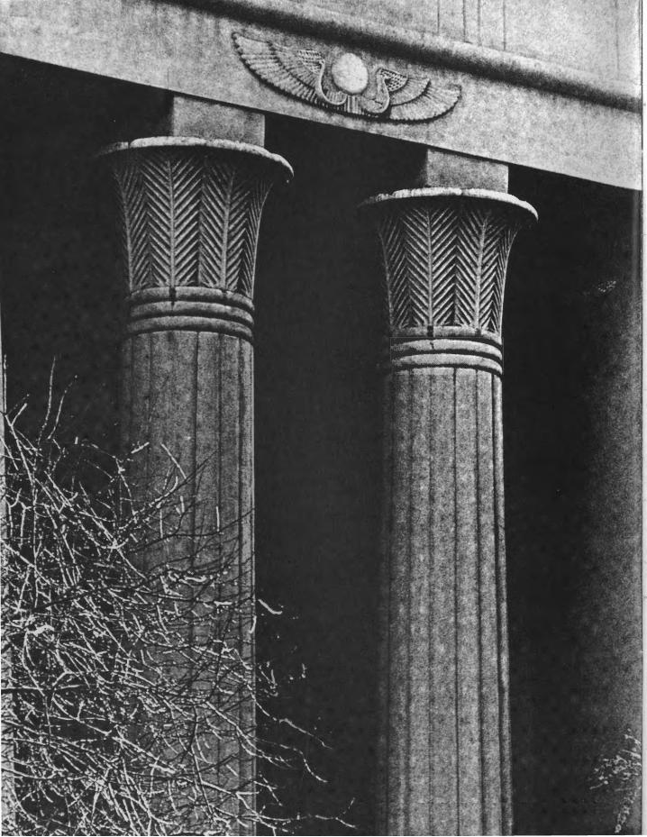
COVER Detail ofpalm leaf design at base and head of columns flanking entrance to Egyptian Building.
OPPOSITE The Egyptian Building, first permanent building of the Medical College of Virginia. Architect: Thomas S. Stewart, Philadelphia. Completed, 1845 and remodeled, 1939. Stewart was in Richmond to plan and erect St. Paul's Church. Dr. A . L. Warner, dean and professor of surgery and surgical anatomy (1807-1847), asked him to build an anatomy laboratory-a "house of the dead." The Richmond Times and Compiler (1845) commented, "there is a mystery in the spirit of the Egyptian style of Architecture, which makes it to our taste singularly appropriate for this temple of the medical science." Perhaps another factor in Stewart's choice of Egyptian style was the influence of Napoleon's campaigns in Egypt on the French school of architecture and, in turn, on young American architects.
The Egyptian Building was reconstructed and restored through the generosity of Mr. Bernard M. Baruch, and its auditorium was dedicated to the memory of his father, Dr. Simon Baruch (1840- 1921), an alumnus, class of 1862.
MCV/Q MEDICAL COLLEGE OF VIRGINIA QUARTERLY
A Scientific Publication of the School of Medicine
SPRING 1965 •VOLUME ONE• NUMBER ONE
CONTENTS
Editorial : The Medical College of Virginia Quarterly -y-Guanidinobutyric Acid : An Inhibitor of Clot Formation and of Clot Lysis PHYLLIS S. ROBERTS 2
The Effect of -y-Guanidinobutyric Acid on the Clotting Time of Normal Plasma and on the Euglobulin Lysis Time of Fibrinolytically Active Plasma LYMAN M. FISHER, PHYLLIS S. ROBERTS, and WARNER E. BRAXTON 4
Electron Microscopic Observations of Human Leucocytes. II. Appearance in Naturally Occurring Fevers FAIRFIELD GOODALE, JR., ELIZABETH A. HILL-MAN, and RALSTON FILLMORE 6
Oxygen Affinities and Electrophoretic Patterns of Hemoglobins in Trout and Basses from Virginia JACK D. BURKE 16
On the Mathematical Basis of Medical Diagnosis MALCOLM E. TURNER 22
Gastric Secretion Mediated by Extravagal Neural Influences CHARLES H. HOCKMAN and EBBE C. HOFF 28
The Effect of ldoxuridine (IDU) on Corneal Strama! Cells in Tissue Culture WALTER J. GEERAETS, GUY WONG, and DUPONT GUERRY, III 30
The Challenge of Pulmonary Emphysema DAVID Y. BATES 34
The Modern Treatment of Respiratory Fail-ure DAVID Y. BATES 42
The Interdependence of Pulmonary Structure and Function: A Synopsis NORMAN C. STAUB 46
Energy Requirements of Breathing W. T. THOMPSON, JR. 48 -
ReJative Incidence of Pulmonary Emphysema Among Negroes and Whites of Both Sexes EDWARDS. RAY 50
Pulmonary Surfactant and its Relation to . Disease SAMI I. SAID 52
Books: Body Fluids and the Acid-Base Balance, by Halvor N. Christensen; Respiratory Function in Disease: An Introduction to the Integrated Study of the Lung, by David V. Bates and Ronald V. Christie; Curiosities of Medicine: An Assembly of Medical Diver-sions, 1552- 1962, edited by Berton Roueche 56
Contributors to this issue 58
Postgraduate Seminars 62
')'-Guanidinobutyric Acid: An Inhibitor of
Clot Formation and of Clot Lysis*
PHYLLIS S. ROBERTS
Department of Medicine, Medical College of Virginia, Richmond
During a study of the effects of a series of guanidine compounds on the esterolytic activities of thrombin, plasmin, and streptokinase plus plasmin or plasminogen, it was found that one of these compounds, y-guanidinobutyric acid (GGBA), acted in several ways like e-aminocaproic acid (EACA). Neither compound had any inhibiting effects on the rate of hydrolysis of TAMe (p-toluenesulfonyl-L-arginine methyl ester), but both inhibited the activation of plasminogen by streptokinase. EACA was the more potent inhibitor. Since EACA has been shown to inhibit the lysis of fibrin, primarily because it inhibits the activation of plasminogen (Ablondi et al. , 1959, Alkjaersig, Fletcher, and Sherry, 1959), GGBA was tested to see if it, too, would inhibit the lysis of blood clots. It was found to do so. In addition, it was found that GGBA also inhibits the formation of blood clots, which EACA does not do. These preliminary results are reported here.
Materials and Methods
Fresh citrated blood from normal donors was used. Plasma was obtained by centrifuging the blood for 20 minutes at 2,500 rpm and 5°C. The euglobulin fraction of the plasma was precipitated by diluting the plasma with distilled water (1 part plasma, 14 parts water) and was brought to pH 5.35 with 0.1 N HCI. After centrifugation for 3 minutes at 1,500 rpm and 5°C, the supernatant was discarded and the precipitate was dissolved in a barbital-saline buffer, pH 7.35 (Wintrobe, 1961). It was tested immediately for clot formation and clot lysis.
The contents of a vial of thrombin, (Thrombin Topical, bovine, Parke, Davis & Co., 1,000 NIH units) were
* Supported by U. S. Public Health Service Research Grant HE-04016, from the National Heart Institute.
2
dissolved in 12.5 ml of glycerol and 12.5 ml of 1.8% NaCl and refrigerated. Just before use the stock solution was diluted with 0.9% NaCl to contain 2 or 4 NIH units per ml. Distilled water (4.0 ml) was added to a vial containing 100,000 units of streptokinase (Varidase). Immediately before use it was diluted with 0.9% NaCl to contain either 1,000 or 2,000 units per ml. EACA was purchased from Mann Research Laboratories, and GGBA from Calbiochem. A 0.1 M solution of each compound was prepared in 0.9% NaCl.
Results
With the blood and plasma from four donors, inhibition of clot lysis was shown in the following way. To test tubes containing 0.1 ml of blood or plasma from a single donor, 0.1 ml of thrombin (4 NIH units per ml) was added and the contents were mixed. A clot formed immediately in each tube. After 15 minutes at room temperature (22-25°C), the following solutions were added to duplicate tubes: (1) 0.3 ml of saline (0.9% NaCl), (2) 0.2 ml of saline plus 0.1 ml of EACA (0.1 M), (3) 0.2 ml of saline plus 0.1 ml of GGBA (0.1 M), (4) 0.2 ml of saline, 15 minutes later 0.1 ml of streptokinase (100 units per ml), (5) 0.1 ml of saline plus 0.1 ml of EACA (0.1 M), 15 minutes later 0.1 ml of streptokinase (1,000 units per ml), and (6) 0.1 ml of saline plus 0.1 ml of GGBA (0.1 M), 15 minutes later 0.1 ml of streptokinase (1,000 units per ml).
None of the clots in the first three sets of tubes lysed, even after remaining at room temperature overnight. The clots in tubes (4) were completely or partially lysed in a few hours, and were completely lysed the next morning. The clots in tubes (5) and (6) we;e not lysed and apparently were completely intact when examined the next morning.
When the experiments, as outlined in (4), (5), and (6), were repeated with either double the concentration of streptokinase or double the concentration of EACA or GGBA, the same results were obtained. There was lysis in tubes (4) and no lysis in tubes (5) or (6).
Inhibition of clot formation was shown, with whole blood from two donors. To test tubes containing 0.1 ml of blood from a single donor were added: (1) 0.3 ml of saline, (2) 0.2 ml of saline plus 0.1 ml of EACA (0.1 M), or (3) 0.2 ml of saline plus 0.1 ml of GGBA (0.1 M). After 15 minutes, 0.1 ml of thrombin (4 NIH units per ml) was added to each tube. Four minutes later firm clots were present in all tubes containing saline or saline plus EACA. No clots were observed in any of the tubes containing GGBA. After 20 minutes, an additional 0.1 ml of thrombin was added to the tubes containing GGBA. Clots formed immediately. Firm clots were still present the following day in all tubes.
A combined clotting and lysing experiment was performed on the euglobulin fraction of the plasma from two donors. The euglobulin precipitate was dissolved in 0.3 ml of buffered saline. To duplicate tubes (controls), 0.1 ml of saline was added. To other tubes, 0.1 ml EACA (0.1 M) or GGBA (0.1 M) was added. Thrombin, 0.1 ml (2 units per ml) was added to each tube and the tubes were placed in a 37°C bath. After 15 minutes, firm clots were present in the control tubes and in the tubes containing EACA, but no clots were present in the tubes containing GGBA. After 60 minutes, however, the latter tubes also contained clots. No evidence of lysis of any of the clots was seen even after the tubes had remained at 37°C for 5 hours. The tubes were then left at room temperature. The next morning no clots were found in the control tubes, but clots
still remained in the tubes containing either EACA or GGBA.
Discussion
Low concentrations of GGBA are widely distributed in mammalian urine, brain, liver, and other tissues (Pisano, Abraham, and Udenfriend, 1963). These authors estimate that 0.05 and 0.09 µ,moles of GGBA, respectively, are present in 1 gm, fresh weight, of rat brain and liver. GGBA has been reported to be of very low toxicity in rabbits, rats, and guinea pigs (Kamiya, Kiyota, and Kita, 1962). The finding that it inhibits the formation as well as the lysis of clots in the test tube suggests that a guanidine-containing compound may be involved in the physiological regulation of blood clotting and lysing. GGBA itself is probably not the compound because it is not potent enough, judged from the preliminary experiments in vitro. Possibly a peptide (or peptides), which is released when fibrinogen is changed to fibrin, may be the physiological regulating compound.
From these and other data, it seems that GGBA may inhibit the hydrolysis of fibrinogen by thrombin, and in this way inhibit clot formation. EACA, on the other hand, has no effect on the action of thrombin and therefore does not inhibit clot formation. In addition, both GGBA and EACA may react reversibly with plasminogen, changing it to a compound that cannot be activated to plasmin. In this way both compounds inhibit the lysis of clots.
Experiments are now being done both in vitro and in vivo to establish quantitatively the extent of the inhibition of clot formation and lysis due to GGBA. Nagamatsu et al. (1963) have shown that esters of EACA are more potent inhibitors of clot lysis than is EACA itself. It is possible that esters of GGBA may also be more potent than GGBA as inhibitors of clot lysis
and clot formation. These compounds or related ones may prove to be of value in preventing the formation of blood clots as well as controlling excessive fibrinolytic activity in vivo.
Summary
y-Guanidinobutyric acid inhibited the formation and the lysis of clots made from whole blood, plasma, or the euglobulin fraction of the plasma from several donors. e-Amino-caproic acid inhibited only the lysis of these clots. not their formation.
Acknowledgments
I thank Dr. Ali A. Hossaini, director of the Blood Bank, Medical College of Virginia, for the blood, and Dr. V. A. Place, Lederle Laboratories, American Cyanamid Company, for the streptokinase used in this work.
References
ABLONDI, F. B., J. J. HAGAN, M. PHILIPS, AND E. c. DE RENZO. Inhibition of plasmin, trypsin and the streptokinase-activated fibrinolytic system by •-aminocaproic acid. Arch. Biochem. Biophys. 82: 153-160, 1959.
ALKJAERSIG, N., A. P. FLETCHER, AND s. SHERRY. •-Aminocaproic acid: an inhibitor of plasminogen activation. J. Biol. Chem. 234: 832-837, 1959.
KAMIYA, H., C. KIYOTA, AND T. KITA. Central depressants V. pharmacological actions and toxicities of 'Y-aminobutyric acid derivatives. Chem. Abst. 57: 2813g, 1962.
NAGAMATSU, A., T. 0KUMA, M. WATANABE, AND Y. y AMAMURA. The inhibition of plasmin by some amino acid derivatives. J. Biochem. 54: 491-496, 1963.
PISANO, J. J ., D. ABRAHAM, ANDS. UDENFRIEND. Biosynthesis and disposition of 'Y-guanidinobutyric acid in mammalian tissues. Arch. Biochem. Biophys. 100: 323-329, 1963.
WINTROBE, M. M. Clinical Hematology (5th ed.). Philadelphia: Lea and Febiger, 1961, p. 300.
3
The Effect of ')"-Guanidinobutyric Acid on
the Clotting Time of Normal Plasma
and on the Euglobulin Lysis Time
of Fibrinolytica lly Active Plasma*
LYMAN M. FISHER, PHYLLIS S. ROBERTS, AND WARNER E. BRAXTON
Division of Clinical Pathology, Department of Pathology, and Department of Medicine, Medical College of Virginia , Richmond
It has been established that e-aminocaproic acid (BACA) inhibits the activation of human plasminogen (Ablondi et al., 1959; Alkjaersig, Fletcher, and Sherry, 1959). Because of this observation, this compound has been used extensively to inhibit the pathologically occurring fibrinolytic system in patients. Recently Roberts (1965) reported that another compound, y-guanidinobutyric acid (GGBA), like BACA, inhibits the lysis of human blood clots. Furthermore, GGBA, unlike BACA, retards the formation of these clots.
The present investigation was undertaken to determine whether GGBA inhibits clot formation in the onestage prothrombin and in the partial thromboplastin time tests. In addition, the ability of GGBA to inhibit clot lysis was tested using blood from a patient showing active fibrinolysis.
Materials and Methods
Blood was collected in a 3.8 % sodium citrate anticoagulant (9 parts blood, 1 part citrate) from 15 individuals. The blood was centrifuged at 2,500 rpm in an angle centrifuge for 10 minutes to obtain the plasma. Clotting tests were performed in duplicate in glass test tubes (10 X 75 mm. at 37° C. Solutions of BACA (Mann Research Laboratories, New York, N.Y.) and GGBA (Calbiochem, Los Angeles, Calif.) were prepared, each having a concentration of 0.1 M in physiological saline. The pH of the BACA solution was 7.02 and that of the GGBA solution was 7.03.
* Supported in part by U. S. Public Health Service Research Grant HE 05868:3640 and HE 04016 from the National Institutes of Health, and by the Charlotte County (Va.) United Fund, Inc.
4
The clotting tests were performed in the following manner.
1. One-stage prothrombin time: 0.1 ml of plasma; 0.1 ml of either physiological saline (control), 0.1 M BACA, or 0.1 M GGBA; 0.2 ml of equal volumes of thromboplastin and CaCl2 (0.025 M). The time taken for clot formation was measured.
2. Partial thromboplastin time: 0.1 ml of plasma; 0.1 ml of either physiological saline (control), 0.1 M BACA, or 0.1 M GGBA; 0.1 ml of Thrombofax (Ortho Research Foundation, Raritan, N.J.). The mixture was incubated for 30 seconds, and then 0.1 ml of CaCl2 (0.025 M) was added. The tubes were removed from the water bath after incubation for 60 seconds, wiped dry, tilted, and the time taken for clot formation was measured.
The euglobulin lysis time was performed as outlined by von Kaulla and Schultz (1958), except that 0.1 ml of either physiological saline, 0.1 M BACA, or 0.1 M GGBA was added to the euglobulin precipitate.
Results and Discussion
The effects of BACA and GGBA on the clotting systems are shown in table 1.
There was no significant difference between the values obtained with the one-stage test for the samples containing saline or BACA. The values obtained when GGBA was added, however, were significantly prolonged (p < 0.001) when compared with the saline control.
The findings for the partial thromboplastin test were similar to those obtained for the one-stage test. GGBA significantly prolonged the clotting time when compared with the samples containing either saline or BACA (p < 0.01).
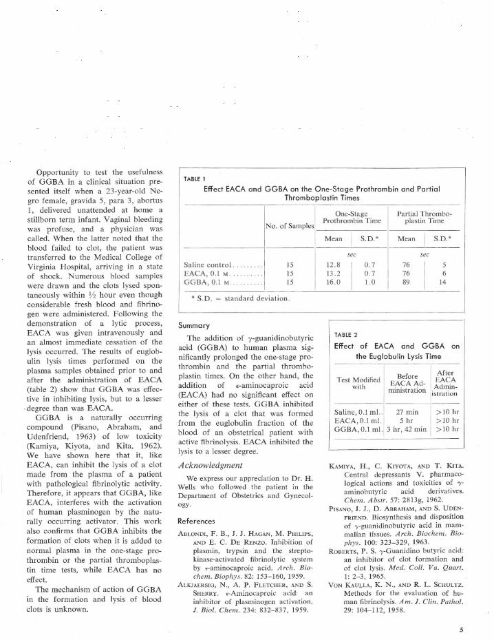
Opportunity to test the usefulness of GGBA in a clinical situation presented itself when a 23-year-old Negro female, gravida 5, para 3, abortus l, delivered unattended at home a stillborn term infant. Vaginal bleeding was profuse, and a physician was called. When the latter noted that the blood failed to clot, the patient was transferred to the Medical College of Virginia Hospital, arriving in a state of shock. Numerous blood samples were drawn and the clots Jysed spontaneously within Y2 hour even though considerable fresh blood and fibrinogen were administered. Following the demonstration of a lytic process, EACA was given intravenously and an almost immediate cessation of the lysis occurred. The results of euglobulin lysis times performed on the plasma samples obtained prior to and after the administration of EACA (table 2) show that GGBA was effective in inhibiting lysis, but to a lesser
· degree than was EACA. GGBA is a naturally occurring
compound (Pisano, Abraham, and Udenfriend, 1963) of low toxicity (Kamiya, Kiyota, and Kita, 1962). We have shown here that it, like EACA, can inhibit the lysis of a clot made from the plasma of a patient with pathological fibrinolytic activity. Therefore, it appears that GGBA, like EACA, interferes with the activation of human plasminogen by the naturally occurring activator. This work also confirms that GGBA inhibits the formation of clots when it is added to normal plasma in the one-stage prothrombin or the partial thromboplastin time tests, while EACA has no effect.
The mechanism of action of GGBA in the formation and lysis of blood clots is unknown.
TABLE 1
Effect EACA and GGBA on the One-Stage Prothrombin and Partial Thromboplastin Times
No. of Samples
Saline control . 15 EACA, 0.1 M . . . 15 GGBA , 0.1 M . 15
* S.D. standard deviation.
Summary
The addition of y-guanidinobutyric acid (GGBA) to human plasma significantly prolonged the one-stage prothrombin and the partial thromboplastin times. On the other hand, the addition of e-aminocaproic acid (EACA) had no significant effect on either of these tests. GGBA inhibited the lysis of a clot that was formed from the euglobulin fraction of the blood of an obstetrical patient with active fibrinolysis. EACA inhibited the lysis to a lesser degree.
Acknowledgment
We express our appreciation to Dr. H. Wells who followed the patient in the Department of Obstetrics and Gynecology.
References
ABLONDI, F . B., J . J. HAGAN, M. PHILIPS, AND E. c. DE RENZO. Inhibition of plasmin, trypsin and the streptokinase-activated fibrinolytic system by <-aminocaproic acid. Arch. Biochem. Biophys. 82: 153-160, 1959.
ALKJAERSIG, N., A. P. FLETCHER, AND S. SHERRY. <-Aminocaproic acid: an inhibitor of plasminogen activation. J. Biol. Chem. 234: 832-837, 1959.
One-Stage Partial Thrombo-Prothrombin Time plastin Time
Mean I
S.D.* Mean I
S.D.*
sec sec
12.8
I
0.7 76 5 13 .2 0.7 76 6 16.0 1.0 89 14
TABLE 2
Effect of EACA and GGBA on
the Euglobulin Lysis Time
Test Modified with
Before After EACA
EACA Ad- Admin-ministration istration
Saline , 0.1 ml. . 27 min > 10 hr EACA, 0.1 ml. 5 hr > 10 hr GGBA , 0.1 ml. 3 hr , 42 min >10 hr
KAMIYA, H ., C. KIYOTA, AND T. KITA. Central depressants V. pharmacological actions and toxicities of 'Yaminobutyric acid derivatives. Chem. Abstr. 57: 2813g, 1962.
PISANO, J. J., D. ABRAHAM, ANDS. UDENFRIEND. Biosynthesis and disposition of 'Y-guanidinobutyric acid in mammalian tissues. Arch. Biochem. Biophys. 100: 323-329, 1963.
ROBERTS, P. S. 'Y-Guanidino butyric acid: an inhibitor of clot formation and of clot lysis. Med. Coll . Va. Quart. 1: 2-3, 1965.
VON KAULLA, K. N., AND R. L. SCHULTZ. Methods for the evaluation of human fibrinolysis. Am. J. Clin. Pathol. 29: 104-112, 1958.
5
Believing as I do in the continuity of nature, I cannot stop abruptly where our microscopes cease to be of use. Here the vision of the mind authoritatively supplements the vision of the eye.
John Tyndall Address at Belfast, 1874
Electron Microscopic Observations of
Human Leucocytes II. Appearance in Naturally Occurring Fevers*
F. GOODALE,t E. A. HILLMAN,t AND R. FILLMOREt
Departments of Pathology, Albany Medical College, Albany, New York, and Medical College of Virginia, Richmond
When human leucocytes are artificially stimulated in vivo or in vitro by a bacterial pyrogen, they release, without destroying themselves, a pyrogenic substance that differs chemically and biologically from the original bacterial pyrogen (Snell et al., 1956; Cranston et al., 1956). Although leucocytic pyrogen has not been seen, or at least recognized, by light or phase microscope, a finely granular extracellular material (fig. 1) is consistently visible by electron microscope in artificially stimulated leucocyte preparations that we know to be pyrogenic (Goodale, Fillmore, and Hillman, 1962).
We are reasonably certain that the granular material is a genuine cellular product in response to the stimulation and, although definite proof is lacking, that it represents, at least in part, leucocytic pyrogen. If the artificially induced granular material does indeed represent leucocytic pyrogen, and if leucocytic pyrogen is responsible for naturally occurring human fevers, it would follow that the same granular material should also be visible in leucocyte preparations from febrile patients with a variety of diseases. The purpose of this study is to report the findings by electron microscope in preparations of human leucocytes that have been "naturally" stim-
* Presented in part at the Fifth International Congress for Electron Microscopy, Philadelphia, August 29 to September 5, 1962. This work was supported by Public Health Service Grant E-3720 from the National Institute of Allergy and Infectious Diseases, by a grant-in-aid from Eli Lily & Company, and by a grant from the John A. Hartford Foundation.
t Dr. Goodale and Miss Hillman are now at the Medical College of Virginia; Dr. Fillmore is at St. Vincent's Hospital, New York, N.Y.
6
ulated in vivo by pathological processes ranging from infections to terminal carcinomas.
Materials and Methods
All equipment was sterilized and made pyrogen-free by heating at 180° for 2 hours. Glassware was siliconized. A 3 % dextran solution (molecular weight 186,000 in 0.9% saline, from Pharmachemical Corporation, Bethlehem, Pa.), was usually used to sediment the erythrocytes. This solution was tested in rabbits to ensure its freedom from pyrogens, as was the autoclaved 0.9% saline that was used for control purposes. The patients (table 1) were drawn from the medical and surgical wards of the Albany Medical Center Hospital, without selection as to age, type of disease, or treatment but with regard only to the height and duration of the fever.
Blood (20 ml) was drawn from an antecubital vein of each febrile patient into a glass syringe with the use of heparin (10,000 U.S.P. units per 10 ml) as the anticoagulant. When the patients had been afebrile for at least 48 hours (except for patient # 17 in table 1, who had been afebrile 20 hours) 20 ml of blood were again drawn and a control leucocyte preparation made by exactly the same method as that used for the febrile leucocyte preparation. Many patients died before becoming afebrile, so that from them control samples could not be obtained. The blood was divided into two equal parts and placed in centrifuge tubes. In most cases a volume of 3 % dextran equal to the volume of blood was added to sediment the erythrocytes. (In a few instances erythrocyte sedimentation was accomplished over a longer period without the use of dextran.) After sedimentation for 20 minutes in a 37°C water bath, the leucocyte-rich plasma from each sample was aspi-
rated and centrifuged at 4 °C for 5 minutes at 1,000 rpm (250 X g) in an International Refrigerated Centrifuge, model PR-2. Unless otherwise stated, all subsequent steps were carried out at 4°C.
The supernatant fluids were discarded and one of the two cell buttons thus obtained were processed as follows. The cells were washed twice, with 10 ml of 0.9% saline for each wash and then incubated in 10 ml of saline for 30 minutes at 37°C. They were then fixed and embedded exactly as to be described for the cells in the second button. The cells in the second button were fixed immediately for 1 hour with 2 % osmium tetroxide in Dalton's buffer (pH 7.4). The osmiumleucocyte mixture was centrifuged at 1,000 rpm for 5 minutes and the supernatant discarded. After suspension and thorough mixing in 10% ethanol, the cells were centrifuged at 1,000 rpm for 3 minutes. In this manner the cells were dehydrated through a series of graded ethanol solutions, washed three times in absolute ethanol, once in equal parts of absolute ethanol and methacrylate (7 parts butyl methacrylate to 1 part methacrylate), and then washed once in the methacrylate mixture alone. Final embedding was in gelatin capsules containing the prepolymerized methacrylate mixture with 1.5% benzoyl peroxide as a catalyst. Polymerization was completed at 55°C overnight.
Sections for both phase and electron microscopy were cut with a Servall Porter-Blum microtome by using glass knives. Sections were mounted on formvar-coated grids and were examined with either a Siemens Elmiskop I (60 KV), RCA EMU-3F (50 KV), or RCA EMU-3G (50 KV) electron microscope. Most grids were examined unstained but a few were stained with
uranyl acetate by floating them on a 1 % solution for 15 to 45 minutes at room temperature, then washing (by flotation) in distilled water for 1 to 2 minutes. A few additional grids were stained with lead (Karnovsky, 1961). For each patient in whom the granular material was easily found, a minimum of eight and an average of ten "febrile" and a similar number of control (if obtainable) grids were examined. For each patient in whom the granular material was not easily seen or not seen at all, up to 50 grids were examined with an average of approximately 25. Selection of patients occurred only in that we tried to find adult patients with fevers ranging from mild to marked and from a few hours to sometimes several weeks' duration.
Results
The results are summarized in tables 1 and 2. The finely granular extracellular material present in electron micrographs of leucocyte preparations from naturally febrile patients is morphologically identical to that seen in artificially stimulated leucocyte preparations. The granules sometimes appear singly but usually are in clumps or aggregates varying from about 0.1 to 1.0 p, in diameter. Individual granules are of two sizes: the smaller (approximately 50 A in diameter) make up the great bulk of the aggregates while the larger (400 to 800 A in diameter) relatively infrequent granules are usually peripherally located. The only morphological difference between the two types of leucocyte preparations is in the Jeucocytes themselves. Those cells artificially stimulated appear normal (figs. 1 and 2), while those "naturally" stimulated by the febrile process (figs. 3 to 8), almost all contain cytoplasmic vacuoles, from 0.2 to 2.0 p, in diameter, from 1 or 2 to 20 in
number, with many of them containing the finely granular material. The number of vacuoles and the amount of granular material in vacuoles were greatest when the blood was drawn as the fever was rising. About half the vacuoles contain, in addition to the granular material, rounded bodies, 800 to 1200 A in diameter, with distinct external membranes. These bodies are in the same size range as the larger granules of the extracellular material, but whereas the latter have a uniform appearance throughout, those in the vacuoles usually appear empty. In patients afebrile for 48 hours, normal cell structure was observed (fig. 7).
In 14 cases, half of the leucocytes were washed free of plasma and incubated with saline prior to preparation for the electron microscope in order to find out if they released granular material during incubation. Eight of these preparations contained the granular material either within cytoplasmic vacuoles or outside the cell.
Although the quantity of granular material in the leucocyte preparations cannot be accurately assessed, it is possible to say (1) that all preparations of leucocytes collected while the fever was rising or stable (15 cases) contained the granular material, (2) that leucocytes from patients with rising fevers are frequently vacuolated and that the vacuoles often contain granular material, and (3) that granular material is usually not seen in leucocyte preparations collected while the fever is waning (3 of 7 cases) and is infrequently (2 of 12 cases) seen when no fever is present.
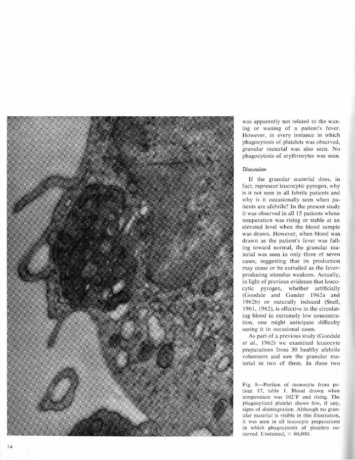
In Jeucocyte preparations from nine patients, phagocytosis of platelets by neutrophils was seen (fig. 8). The platelets were generally intact and could be easily recognized. Platelet phagocytes.is
7
8
TABLE 1
Data from 22 Patients with Fevers Due to a Variety of Diseases
Clinical Data Electron Microscope Observations
' Presence of granular material
Condition Phago-
Temperature• when afebrile Febrile specimen' cytosis No. Age Sex Race Disease (°F) at bleeding Treatmentb specimens Afebrile
of
were taken specimens plate-
Fresh Saline lets incubation
-- - - - -1 25 F w Hodgkin's disease, labial 104, stable Aspirin Improved I.C. Not done None No
abscess Demerol E .C. Penicillin
2 27 F N Lobar pneumonia 105. 6, stable Penicillin Improved LC. Not done None No Codeine
3 29 M w Von Recklinghausen 's 100.2 1' Aspirin Improved I.C. Not done None No disease Codeine
4 40 F w Malignant melanoma 102 1 Penicillin Deadd I.C. None Not done Yes Streptomycin
5 40 F N Cholangitis jaundice 102.7 1' Neomycin Deadd I.C. Not done Not done Yes E .C.
6 46 F w Fracture of humerus 103 1 Aspirin Improved None None None No Nembutal Codeine
7 47 F w Fracture of femur 101.5 1 Penicillin Improved None I.C. None Yes Streptomycin
8 46 M w Acute pyelonephritis 101 .3 1' Chloromycetin Improved I.C. LC. None Yes Cortisone
9 49 F w Hodgkin's disease 101 1' Chloromycetin Improved I.C . I.C. None Yes Aspirin E.C. E.C. Phenobarbital
10 50 M w Thrombophlebitis with 101 1' Penicillin Improved I.C. Not done I.C. Yes popliteal abscess E.C. E.C.
11 54 M w Thrombotic thrombo- 101.5 1 Cortisone Deadd None None Not done No cytopenic purpura Chloromycetin with cerebral hemor- Aspirin rhage
12 54 F w Carcinoma of breast 103 1 Penicillin D eadd E.C. None Not done No with metastases Aspirin
Chloromycetin 13 57 M w Carcinoma of tongue 104, stable Streptomycin Deadd I.C. Not done Not done No
Penicillin E.C. Codeine Leucovorin
14 60 M w Bilateral leg amputation 102 1 None D eadd None Not done Not done No for gangrene
Postoperative pneumo-nia
15 61 M w Subacute bacterial en- 100.5 1 Penicillin Improved None None None No docarditis Aspirin
16 62 F w Lymphosarcoma 100 .5, stable Aspirin Deadd I.C. l.C. Not done No Thorazine
I Codeine
0 Direction of arrow indicates whether the temperature was rising or falling at the time the blood was drawn; temperatures are oral unless otherwise indicated and taken within 15 minutes of the blood sample.
b Treatment listed is that which the patient was receiving when the blood was drawn and for at least the previous 24 hours. ' I.C. = intracellular ; E .C. = extracellular. d Patient was febrile until death.
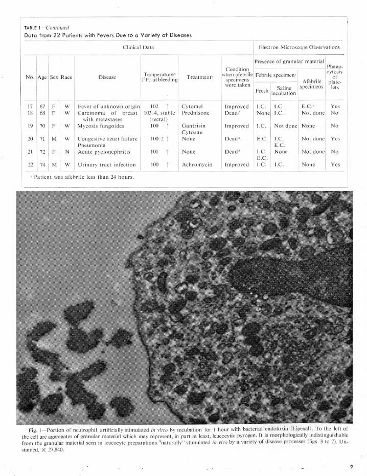
TABLE !-Continued
Data from 22 Patients with Fevers Due to a Variety of Diseases
Clinical Data Electron Microscope Observations
I Presence of granular material
Condition Phago-
No. Age Temperature" when afebrile Febrile specimen' cytosis
Sex Race Disease Treatment' of (°F) at bleeding specimens Afebrile plate-were taken Saline specimens lets
Fresh incubation -- -- - - --
17 67 F w Fever of unknown origin 102 r Cytomel Improved I.C. I.C. E.C.' Yes 18 68 F w Carcinoma of breast 103 .4, stable Prednisone Deadd None I.C. Not done No
with metastases (rectal) 19 70 F w Mycosis fungoides 100 r Gantrisin Improved I.C . Not done None No
Cytoxan 20 71 M w Congestive heart failure 100 .2 r None Deadd E .C. l.C. Not done Yes
Pneumonia E.C. 21 72 F N Acute pyelonephritis 101 r None Deadd I.C . None Not done No
E.C. 22 74 M w Urinary tract infection 100 r Achromycin Improved I.C . I.C . None Yes
' Patient was afebrile less than 24 hours.
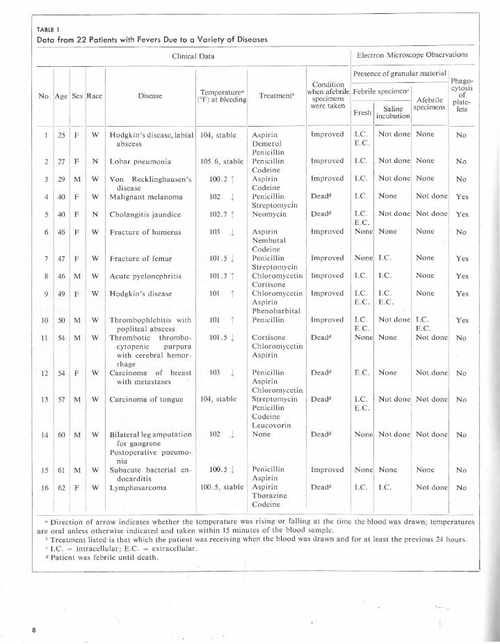
Fig. I- Portion of neutrophil , artificially stimulated in vitro by incubation for 1 hour with bacterial endotoxin (Lipexal) . To the left of the cell are aggregates of granular material which may represent, in part at least, leucocytic pyrogen. It is morphologically indistinguishable from the granular material seen in leucocyte preparations "naturally" stimulated in vivo by a variety of disease processes (figs . 3 to 7) . Un-. stained, X 27,840.
9
10
TABLE 2
Summary of Electron Microscopic Findings in Leucocyte Preparations
from 22 Febrile Patients. Blood samples for controls could
be obtained from only 12 patients because the remaining 10 patients died before becoming afebrile .
Presence of Granular
Temperature of Patient Material
Yes No
Rising or at peak .. 15* 0 Falling . . . . . . . . 3* 4 Afebrile controls . . . . . . 2 10
* In one of these cases granular material was not seen in the fresh~ specimen but was present within leucocytes in the saline-incubated specimen.
Fig. 2-To the left and below the visible portion of the lymphocyte, and between parts of two erythrocytes, is an aggregate of granular material about 1.0 µ, in diameter. In this ease 10 ml of whole blood from one of the authors was incubated for 1 hour in vitro with 0.5 µ, of bacterial endotoxin (Lipexal) and then returned intravenously to the donor. Twenty-five minutes later chills and rising fever began, at which time blood for this preparation was drawn. The granular material here is made up of fine particles averaging approximately 50 A diameter. Stained with uranyl acetate, X 48,000.
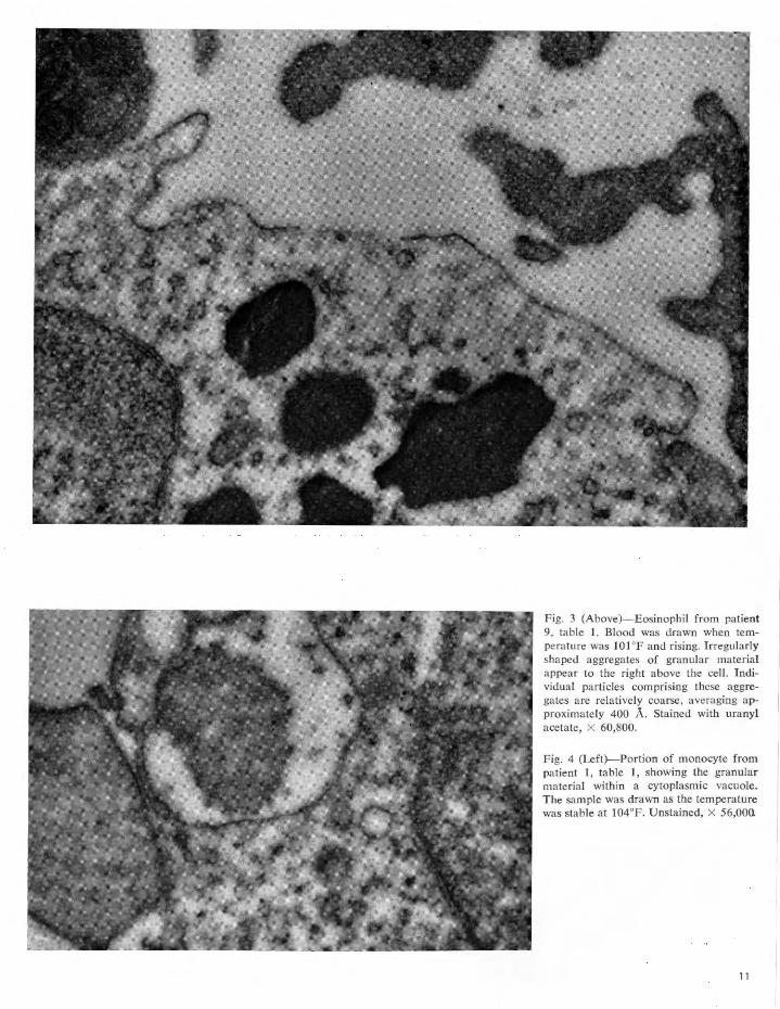
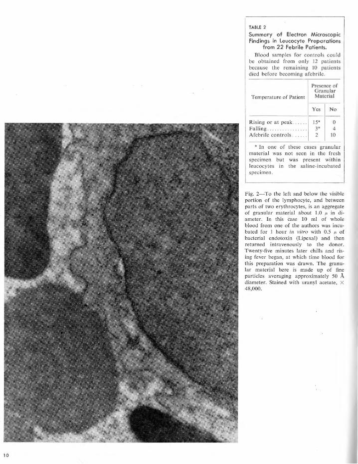
Fig. 3 (Above)-Eosinophil from patient 9, table 1. Blood was drawn when temperature was 101°F and rising. Irregularly shaped aggregates of granular material appear to the right above the cell. Individual particles comprising these aggregates are relatively coarse, averaging approximately 400 A. Stained with uranyl acetate, X 60,800.
Fig. 4 (Left)- Portion of monocyte from patient 1, table 1, showing the granular material within a cytoplasmic vacuole. The sample was drawn as the temperature was stable at 104°F. Unstained, X 56,000.
11
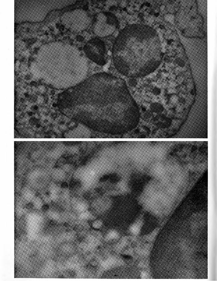
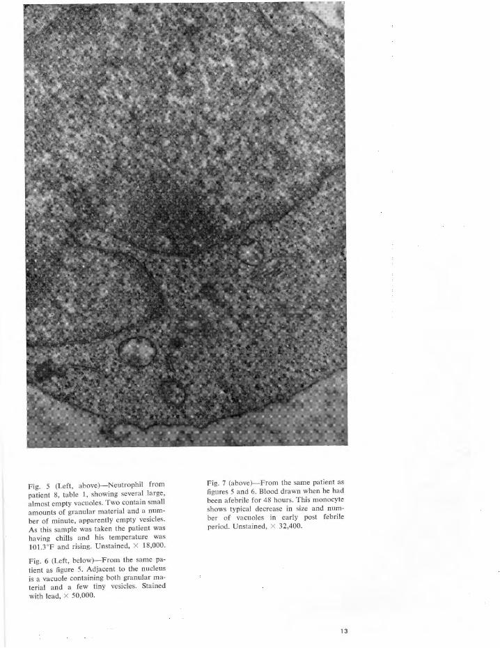
Fig. 5 (Left, above)- Neutrophil from patient 8, table 1, showing several large, almost empty vacuoles. Two contain small amounts of granular material and a number of minute, apparently empty vesicles. As this sample was taken the patient was having chills and his temperature was 101.3°F and rising. Unstained, x 18,000.
Fig. 6 (Left, below)- From the same pa
tient as figure 5. Adjacent to the nucleus is a vacuole containing both granular material and a few tiny vesicles. Stained
with lead, X 50,000.
Fig. 7 (above)-From the same patient as figures 5 and 6. Blood drawn when he had been afebrile for 48 hours. This monocyte shows typical decrease in size and number of vacuoles in early post febrile period. Unstained, X 32,400.
13
14
was apparently not related to the waxing or waning of a patient's fever. However, in every instance in which phagocytosis of platelets was observed, granular material was also seen. No phagocytosis of erythrocytes was seen.
Discussion
If the granular material does, in fact, represent leucocytic pyrogen, why is it not seen in all febrile patients and why is it occasionally seen when patients are afebrile? In the present study it was observed in all 15 patients whose temperature was rising or stable at an elevated level when the blood sample was drawn. However, when blood was drawn as the patient's fever was falling toward normal, the granular material was seen in only three of seven cases, suggesting that its production may cease or be curtailed as the feverproducing stimulus weakens. Actually, in light of previous evidence that leucocytic pyrogen, whether artificially (Goodale and Gander 1962a and 1962b) or naturally induced (Snell, 1961 , 1962), is effective in the circulating blood in extremely low concentration, one might anticipate difficulty seeing it in occasional cases.
As part of a previous study (Goodale et al., 1962) we examined leucocyte preparations from 30 healthy afebrile volunteers and saw the granular material in two of them. In these two
Fig. 8-Portion of monocyte from patient 17, table 1. Blood drawn when temperature was 102°F and rising. The phagocytized platelet shows few, if any, signs of disintegration. Although no granular material is visible in this illustration, it was seen in all leucocyte preparations in which phagocytosis of platelets occurred. Unstained, X 60,000.
cases we felt that the granular material might have been due to contamination of the leucocyte preparation by bacterial endotoxin prior to incubation. In the present study there were two patients whose leucocyte preparations contained the granular material, not only when they were febrile but also when they were afebrile. One of the patients C* 17, table 1) had been afebrile Jess than 24 hours and became febrile again the day after her control sample was drawn. The other patient C* 10), although his temperature was normal, had a large, open, and infected wound on one leg. Perhaps a threshold exists beneath which there is insufficient circulating pyrogen to elicit a measurable febrile response, but still enough to be sometimes visible by electron microscopy.
The only difference we have noted between leucocyte preparations which have been artificially stimulated in vitro to release their pyrogen, and those "naturalJy" stimulated, is that in the latter the granular material often appears in the cell cytoplasm within vacuoles. Whether it is being formed inside the celJ or whether it has been produced at the cell surface and then phagocytized, we do not know. Conditions are undoubtedly more favorable for phagocytosis in vivo than in vitro.
We are not able at present to assess accurately the amount of granular material present in a leucocyte preparation. Therefore, we can make no statements as to whether height or duration of fever, type of disease, or method of treatment had any detectable effect on the amount or location of granular material. Cortisone has been reported (Atkins et al, 1955) to have antipyretic properties. Three of the patients (*8, 11, and 18, table 1) in the present study were receiving either cortisone or
prednisone. In patients * 8 and 18, whose temperatures were rising or at peak, leucocytic pyrogen was observed. In patient *11, whose temperature was falling, leucocytic pyrogen was not seen. From these few observations we can say only that if the granular material is or contains Jeucocytic pyrogen, then cortisone does not apparently exert its antipyretic effect by preventing the formation or release of the pyrogen. However, the quantification of the granular material must await more precise biological or chemical methods than are now available.
When phagocytosis of platelets by leucocytes occurred, it always occurred in the presence of granular material. Sometimes platelets in advanced stages of disintegration within cytoplasmic vacuoles resembled the granular material. We have observed platelet phagocytosis in only one artificially stimulated leucocyte preparation. Its meaning in the present study is uncertain.
Summary
A granular material was visible by electron microscopy in leucocyte preparations from 1 7 of 22 febrile patients with a variety of diseases. It was seen in all 15 patients whose temperature was rising when the blood sample was drawn, in 3 of 7 patients whose temperature was falling, and in 2 of 12 afebrile patients.
The granular material is morphologically identical to that seen in leucocyte preparations artificially stimulated in vitro by bacterial endotoxins and known to contain leucocytic pyrogen.
The present study would tend to strengthen the association between the granular material and Jeucocytic pyrogen, but the exact relationship has yet to be proved.
References
ATKINS, E., F . ALLISON, R. M. SMITH, AND w. B. WOOD. Studies on the antipyretic action of cortisone on pyrogen-induced fever. J. Exptl. Med. 101: 353-366, 1955.
CRANSTON, W. I., F. GOODALE, E. S. SNELL, AND F. WENDT. The role of leucocytes in the initial action of bacterial pyrogens in man. Clin. Sci. 15: 219-226, 1956.
GANDER, G. W., AND F. GOODALE. Method for the purification of human leucocytic pyrogen. Proc. Intern. Soc. Hematol. 9th Congr. 1: 359-397, 1962a.
GANDER, G. W., AND F. GOODALE. Chemical properties of leucocytic pyrogen. I. Partial purification of rabbit leucocytic pyrogen. Exp. Mol. Pathol. 1: 417-426, 1962b.
GOODALE, F., R. FILLMORE, AND E. HILLMAN. Electron microscopic observations of human leucocytes. I. Response in vitro to bacterial endotoxin. Exp. Mol. Pathol. 1, 229-250, 1962.
KARNOVSKY, M. J. Simple methods of "staining with lead" at high pH in electron microscopy. J. Biophys. Biochem. Cytol. 11: 729-732, 1961.
SNELL, E. S., F. GOODALE, F. WENDT, AND w. I. CRANSTON. Properties of human endogenous pyrogen. Clin. Sci. 21: 115-124, 1961.
SNELL, E. S. An examination of the blood of febrile subjects for pyrogenic properties. Clin. Sci. 21, 115-124, 1961.
SNELL, E. S. Pyrogenic properties of human pathological fluids. Clin. Sci. 23: 141-150, 1962.
15
Oxygen Affinities
and Electrophoretic
Patterns of Hemoglobins
in Trout and Basses
from Virginia*
JACK D. BURKE
Department of Anatomy Medical College of Virginia, Richmond
Multiple hemoglobins in several different species of fishes were described by using electrophoresis in 1959 by Buhler and Shanks in the United States Chandrasekhar in India, and Hashi: moto and Matsuura in Japan. Consequently, using gene frequency data, variant hemoglobins have been studied in relation to such parameters as proportional changes of hemoglobins with growth in salmon (Hashimoto and Matsuura, 1960), and intraspecific variation in cod and whiting by Sick (1961). This paper is concerned with hemoglobin polymorphism as it is related to interspecific variation in Micropterus dolomieui, the smallmouth bass, and Micropterus salmoides, the largemouth bass, as well as Salmo gairdneri, the rainbow trout, and Salmo trutta, the brown trout. Since the brook trout, Salvelinus fontinalis, was available, it was also possible to compare the trout hemoglobins generically.
A striking example of distribution of fishes in freshwater is that of trout and catfishes. Trout are ordinarily found in cool, well aerated water having a high oxygen content, but catfishes can be found in shallow, warm water with a low oxygen concentration. A comparison of the results by Irving et al. (1941) on trout, with results reported by Haws and Goodnight (1962) for catfishes, shows that hemoglobin affinity for oxygen is greater in catfishes than in trout, when the measurements were made with similar temperatures and carbon dioxide tensions. With other conditions being favorable for reproduction and sustenance, it seems that the affinity of hemoglobin for oxygen is a limiting factor in the distribution of trout and catfishes. This may be a general ecophysiological relationship-even operating at the interspecific level-since it appears to be a factor in the distribution of fishes such as smallmouth and largemouth basses, as well as in trout. In this study is has been found that the affinity of hemoglobin for oxygen is different in basses and trout, and electropherograms on cellulose acetate membranes show hemoglobin polymorphism in the basses as well as the trout.
* Supported by U. S. Public Health Research Grants H-365(and H-8774.
16
Materials and Methods
The smallmouth bass were caught on artificial bait in or near the rapids at the Fall Line of the James River at Richmond, Virginia (mean oxygen content: greater than 7 mg per L). The largemouth bass were taken on both artificial bait and live minnows from different ponds and lakes in east-central counties of Virginia (mean oxygen content: less than 5 mg per L). All fishes were collected in the spring, summer, and fall months of the year. The fishes were weighed on a simple field balance described by Burke (1963), and the oxygen content of the water where the fishes were caught was determined by a modification of the Winkler method with a 10-ml syringe (Burke, 1962a). All of the trout were obtained from the State Trout Hatchery at Marion, Virginia.
Blood was removed from the fishes in the field, or in the laboratory if the transportation distances were short. The procedure of securing blood as described by Burke (1962b) was employed. Essentially, the technique used here is that the posterior part of the operculum was removed, a slit made through the posterior branchial chamber and the pericardium, the heart clipped, and the blood collected in a heparinized pipette as it "welled" into the pericardia! cavity.
Hemoglobin solutions for oxygen affinity studies were prepared as follows. The blood was centrifuged, the plasma decanted with a vacuum pipette, and the cells washed three times in 0.11 M NaCl solution with intermittent centrifugation. The cells then were hemolyzed by suspending them in an equal volume of distilled water overnight in a refrigerator. After gentle agitation on a rotator, the suspension was filtered, centrifuged at high speed, and filtered again. The hemoglobin was made up in phosphate buffers with an ionic strength of 0.3 and a pH of either 7.4 or 6.8; all spectrophotometric readings at 640 m,u were made at 25 ± 1 °C. Oxyhemoglobin affinity curves were determined by the spectrophotometric method of Burke and Powell (1962). After equilibration with air, percentages of oxyhemoglobin were determined for various oxygen tensions obtained manometrically using a tonometer, and the following equation: y I 100 = A, -
A ,/ A, - A , where y is percentage oxyhemoglobin, A is absorbance, and r, s, and o represent "reduced" hemoglobin, partially oxygenated hemoglobin, and fully oxygenated hemoglobin, respectively. This procedure was modified from the methods described by Hall (1935), Riggs (1951), Redmond (1955), Rossi-Fanelli and Antonini (1958), and personal communication with Dr. Clyde Manwell of the University of Illinois, Urbana. At least three oxyhemoglobin affinity curves were determined, both at pH 7.4 and 6.8 on pooled blood samples, from two to four fish of each species that were sexually mature. Therefore, the points for each curve shown in figs. 3 to 7 represent mean values.
Before dilution, the hemoglobin solutions prepared for the determination of the oxyhemoglobin affinity curves were also used in spotting for electropherograms. Electrophoresis was carried out in a Gelman electrocab containing a barbital buffer with a pH of 8.6, and an ionic strength of 0.05 µ,
at 250 V for 1 hour at room temperature. At least three electrophoretic patterns were run on each hemoglobin sample. The patterns were developed on cellulose acetate membranes by staining with bromphenol blue or amido Black lOB, clearing in dilute acetic acid solution, and drying in air.
Results
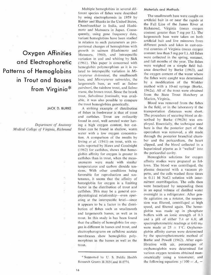
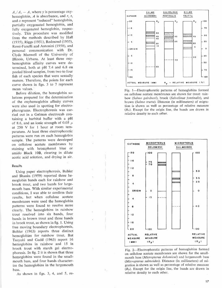
Using paper electrophoresis, Buhler and Shanks (1959) reported three hemoglobin bands each for rainbow and brook trout, and two bands for largemouth bass. With similar experimental conditions, I was able to confirm their results, but when cellulose acetate membranes were used the hemoglobin patterns were found to resolve more clearly. The hemoglobins in rainbow trout resolved into six bands, four bands in brown trout and three bands in brook trout, as shown in fig. 1. Using free moving boundary electrophoresis, Buhler (1963) reports three distinct hemoglobins for rainbow trout. But Tsuyuki and Gadd (1963) report 16 hemoglobins in rainbow and 15 in brook trout with starch gel electrophoresis. In fig. 2 it is shown that three hemoglobins were found in the smallmouth bass, and four bands characterize the hemoglobins in the largemouth bass.
As shown in figs. 3, 4, and 5, re-
SAL MO SALVELINUS SA LMO
CATH ODE GA IR ONER I FONTI NALi S TR U TTA
20 RF RF RF 100
15 70
55 10
45 45 45 50
40
25 25 20 20 10
ORIGI N 0
-- - - 30 25
- - -- 40
I 0 5 0
-- -- 65
15 70
20 100
ACTUAL MEAS URE 0011 RF REL ATI VE M EASURE ( .. , Fig. 1-Electrophoretic patterns of hemoglobins formed on cellulose acetate membranes are shown for trout: rainbow (Salmo gairdneri) , brook (Salvelinus fontinalis), and brown (Sa/mo trutta). Distance (in millimeters) of migration is shown as well as percentage of relative measure (RF). Except for the origin line, the bands are drawn in relative density to each other.
CATHOOE
20
16
I Z
8
4
ORIGIN
4
8
12
16
20
ACTUAL
ME ASURE
(MM)
MICROPTERUS
OO L OM IEUI
100
75 70
0
15
100
RELATIVE
ME ASURE
( RFI
MICROPTERUS
S AL MOIOES
100
50
35
Z 5
0
40
100
REL ATI VE
ME A SURE
< R F l
Fig. 2-Electrophoretic patterns of hemoglobins formed on cellulose acetate membranes are shown for the smallmouth bass (Micropterus dolomieui) and largemouth bass (Micropterus salmoides). Distance (in millimeters) of migration is shown as well as percentage of relative measure (RF). Except for the origin line, the bands are drawn in relative density to each other.
17
SA L M O G AI RDNER I
I ' 608 pH . ' 7 ,4 p H
100
• z 0
.... 8 0 .. a:: :::> .... 60 .. I/)
.... z
"' u
a::
"' Q. 20
0 10 20 30 40 70 80 90
O XY GEN TENS I ON
Fig. 3-0xybemoglobin affinity curves for rainbow trout (Salmo gairdneri) determined at 25°C.
100
z 0
80 ;:: .. a:: :::> .... 60 .. I/)
.... z 40 ... u
cc ... 20 Q.
0 10 'l 0 30 40 5 0
SA LVELINUS FONTI N A LI S
, , 5,9 p H
• ,7,4pH
60 7 0 80 90
O X YG E N TENS I ON
Fig. 4-0xyhemoglobin affinity curves for brook trout (Salvelinus fontinalis) determined at 25°C.
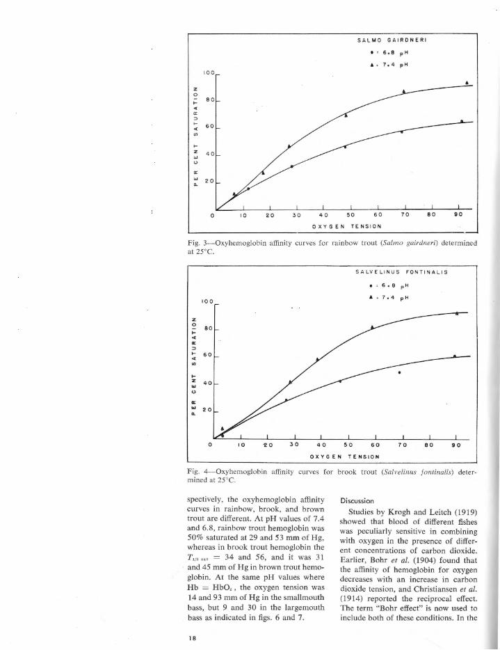
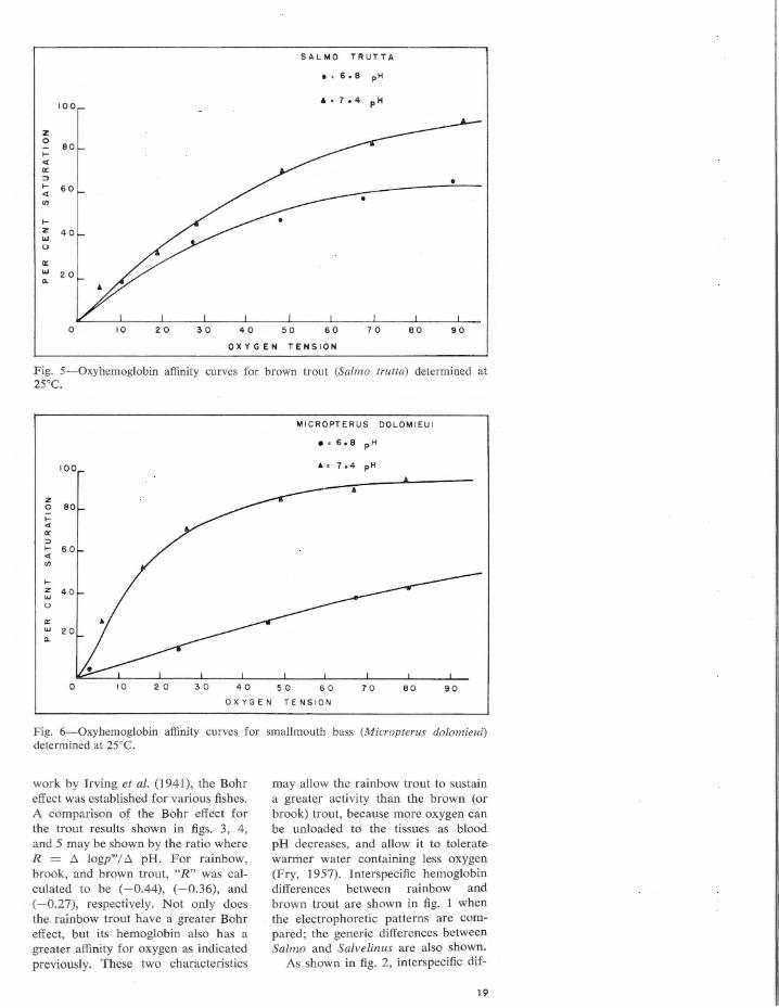
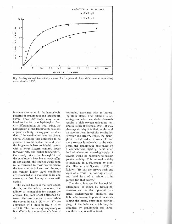
spectively, the oxyhemoglobin affinity curves in rainbow, brook, and brown trout are different. At pH values of 7.4 and 6.8, rainbow trout hemoglobin was 50% saturated at 29 and 53 mm of Hg, whereas in brook trout hemoglobin the T,1• " ' = 34 and 56, and it was 31 and 45 mm of Hg in brown trout hemoglobin. At the same pH values where Hb = HbO, , the oxygen tension was 14 and 93 mm of Hg in the smallmouth bass, but 9 and 30 in the largemouth bass as indicated in figs. 6 and 7.
18
Discussion
Studies by Krogh and Leitch (1919) showed that blood of different fishes was peculiarly sensitive in combining with oxygen in the presence of different concentrations of carbon dioxide. Earlier, Bohr et al. (1904) found that the affinity of hemoglobin for oxygen decreases with an increase in carbon dioxide tension, and Christiansen et al. (1914) reported the reciprocal effect. The term "Bohr effect" is now used to include both of these conditions. In the
SALMO TRUTTA
e , 6, 8 pH
100
z 0
80 f-<t a:: :> f- 60 <t (/)
f-z 40 w (,)
a:: w 20 Q.
0 10 20 30 40 50 60 70 eo 90
OXYGEN TENSION
Fig. 5-0xyhemoglobin affinity curves for brown trout (Sa/mo trutta) determined at 25°C.
100
z 0
>= <l a:: :> f-<l (/)
f-z w 40 (,)
a:: w 20 Q.
0 10 20 30 40 50
MICROPTERUS OOLOMIEUI
e • 6, 8 pH
• • 7, 4 pH
60 70 80 90
OXYGEN TENSION
Fig. 6-0xyhemoglobin affinity curves for smallmouth bass (Micropterus dolomieui) determined at 25°C.
work by Irving et al. (1941), the Bohr effect was established for various fishes. A comparison of the Bohr effect for the trout results shown in figs. 3, 4 , and 5 may be shown by the ratio where R = ~ logp'°/ ~ pH. For rainbow, brook, and brown trout, "R" was calculated to be (-0.44), (-0.36), and (-0.27), respectively. Not only does the rainbow trout have a greater Bohr effect, but its hemoglobin also has a greater affinity for oxygen as indicated previously. These two characteristics
may allow the rainbow trout to sustain a greater activity than the brown (or brook) trout, because more oxygen can be unloaded to the tissues as blood pH decreases, and allow it to tolerate warmer water containing less oxygen (Fry, 1957). lnterspecific hemoglobin differences between rainbow and brown trout are shown in fig. 1 when the electrophoretic patterns are compared; the generic differences between Salmo and Salvelinus are also shown.
As shown in fig. 2, interspecific dif-
19
MICROPTERUS SALMOIDES
•=6.8 PH
100 .t.=7.4 pH
z 80 0 • ;::: ~
a: ::> .... 60 ~
"' .... z 40 UJ u
a: 20 UJ Q.
0 10 20 30 40 50 60 70 80 90
OXYGEN TENSION
Fig. 7-0xyhemoglobin affinity curves for largemouth bass (Micropterus salmoides) determined at 25°C.
ferences also occur in the hemoglobin patterns of smallmouth and largemouth basses. These differences may be related to the two ecophysiological factors differentiating the trout. First, the hemoglobin of the largemouth bass has a greater affinity for oxygen than does that of the smallmouth bass, as shown above. Assuming this difference to be genetic, it would explain the ability of the largemouth bass to inhabit waters with a lower oxygen content, lower turnover rate, and higher temperature. Conversely, since the hemoglobin of the smallmouth bass has a lower affinity for oxygen, this species would tend to be restricted to those waters where the temperature is lower and the oxygen content higher. Such conditions are associated with mountain lakes and streams, or fast flowing streams with rapids.
The second factor is the Bohr effect; this is, as the acidity increases, the affinity of hemoglobin for oxygen decreases. The Bohr effect difference between the two species is seen when the curves in fig. 6 (R = -1. 3 7) are compared with those in fig. 7 (R = -0.73). The decreasing oxyhemoglobin affinity in the smallmouth bass is
20
noticeably associated with an increasing Bohr effect. This relation is advantageous when metabolic demands require a high oxygen unloading tension in tissues (Foreman, 1954). It may also explain why it is that, as the acid metabolites form in cellular respiration (Prosser and Brown, 1961), the hemoglobin is buffered at a lower pH and more oxygen is unloaded to the cells. Thus, the smallmouth bass takes on .a characteristic fighting habit when hooked, where an increasing supply of oxygen would be necessary to sustain greater activity. This unusual activity is indicated in a statement by Henshall (Harlan and Speaker, 1951) as follows: "He has the arrowy rush and vigor of a trout, the untiring strength and bold leap of a salmon . . . the gamest fish that swims."
Therefore, interspecific hemoglobin differences-as shown by certain parameters such as electrophoretic patterns, oxyhemoglobin affinities, and Bohr effects-are important in establishing the limits, sometimes overlapping, of the habitats which may be occupied by smallmouth and largemouth basses, as well as trout.
Summary
1. Hemoglobin solutions were prepared from pooled samples of blood taken from each of the following species; Sa/mo gairdneri, the rainbow trout; Salvelinus fontinalis, the brook trout; Salmo trutta, the brown trout; Micropterus dolomieui, the smallmouth bass; Micropterus salmoides, the largemouth bass.
2. Hemoglobin electrophoretic patterns for each species were developed on cellulose acetate membranes.
3. Oxyhemoglobin affinity curves were determined spectrophotometrically on different hemoglobin solutions from each species.
4. Interspecific differences con-cerned with hemoglobin electrophoretic patterns, oxyhemoglobin affinities, and the Bohr effect were shown for both trout and basses.
Acknowledgments
Travel expenses, in part, were provided by the Virginia Academy of Science. Technical assistance was given by Mr. Phillip Brandt, Mr. Joseph Lively, and Mr. James Powell. The Virginia Commission of Game and Inland Fisheries, through Mr. Robert Martin, contributed their facilities with kindness. Dr. Allan Powell, Department of Chemistry, University of Richmond, Va., offered helpful suggestions during the study.
References
BOHR, C., K. HASSELBALCH, AND A. KROGH. Ueber einen in biologischer Beziehung wichtigen Einfluss, den die Kohlensaurespannung des Blutes auf dessen Sauerstoff-bindung ubt. Skand. Arch. Physiol. 16: 402-412, 1904.
BUHLER, D. R. Studies on fish hemoglobins. J. Biol. Chem. 238: 1665-1674, 1963.
BUHLER, D. R., AND W. E. SHANKS. Multiple hemoglobins in fishes. Science. 129: 899-900, 1959.
BURKE, J. D . Determination of oxygen in water using a 10 ml. syringe. J. Elisha Mitchell Sci. Soc. 78: 145-147, 1962a.
BURKE, J. D. A simple technique for immobilizing fish to remove blood. Copeia 1962: No. 4, 852-854, 1962b.
BURKE, J. D . A simple balance for weighing small animals. Turtox News 41: 76-77, 1963.
BURKE, J. D ., AND W. A. POWELL. A spectrophotometric procedure for the determination of oxy-hemoglobin
affinity curves. Virginia J. Sci. 13: 243, 1962.
CHANDRASEKHAR, N. Multiple haemoglobins in fish. Nature 184: 1652-1653, 1959.
CHRISTIANSEN, J., C. G. DOUGLAS, AND J . S. HALDANE. The absorption and dissociation of carbon dioxide by human blood. J. Physiol. 48: 244-271, 1914.
FOREMAN, C. W. A comparative study of the oxygen dissociation of mammalian hemoglobin. J. Cellular Comp. Physiol. 44: 421-430, 1954.
FRY, F . E. J . The aquatic respiration of fish. In: The Physiology of Fishes. New York: Academic Press, 1957, pp. 1-63.
HASHIMOTO, K., AND F. MATSUURA. Multiple hemoglobins in fish. Bull. Japan. Soc. Sci. Fisheries 24: 719-723 , 1959.
HASHIMOTO, K., AND F . MATSUURA. Comparative studies on two hemoglobins of salmon. V. Change in proportion of two hemoglobins with growth. Bull. Japan . Soc. Sci. Fisheries 26: 931-937, 1960.
HALL, F. G. A spectro-comparator for the study of hemoglobin. J. Elisha Mitchell Sci. Soc. 51: 289-293, 1935.
HARLAN, J. R., AND E. B. SPEAKER. Iowa Fish and Fishing. Des Moines, State Conservation Commission, 1951.
HAWS, T. G., AND c. J. GOODNIGHT. Some aspects of the hematology of two species of catfish in relation to their habitats. Physiol. Zoo/. 35: 8-17, 1962.
IRVING, L., E. C. BLACK, AND V. SAFFORD. The influence of temperature upon the combination of oxygen with the blood of trout. Biol. Bull. 80: 1-17, 1941.
KROGH, A., AND I. LEITCH. The respiratory function of the blood in fishes. J. Physiol. (London) 52: 288-300, 1919.
PROSSER, C. L., AND F. A. BROWN. Comparative Animal Physiology. Philadelphia: Saunders, 1961.
REDMOND, J . R. The respiratory function of hemocyanin in crustacea. J. Cellular Comp. Physiol. 46: 209-247, 1955.
RIGGS, A . The metamorphosis of hemoglobin in the bullfrog. J. Gen. Physiol. 35: 23-40, 1951.
Rossi-FANELLI, A., AND E. ANTONINI. Studies on the oxygen and carbon monoxide equilibria of human myoglobin. Arch. Biochem . Biophys. 77: 478-492, 1958.
S1cK, K. Haemoglobin polymorphism in fishes . Nature 192: 894-896, 1961.
TSUYUKI, H ., AND R. E. A. GADD. The multiple hemoglobins of some members of the Salmonidae family. Biochim. et Biophys. Acta 71: 219-221, 1963.
"Some of my friends have even asserted that a Ph.D. thesis should be the greatest scientific work a man has ever done and perhaps ever will do, and should wait until he is thoroughly able to state his life work. I do not go along with this. I mean merely that if the thesis is not in fact such an overwhelming task, it should at least be in intention the gateway to vigorous creative work. Lord only knows that there are enough problems yet to be solved, books to be written, and music to be composed! Yet for all but a very few, the path to these lies through the performance of perfunctory tasks which in nine cases out of ten have no compelling reason to be performed. Heaven save us from the first novels which are written because a young man desires the prestige of being a novelist rather than because he has something to say! Heaven save us likewise from the mathematical papers which are correct and elegant but without body or spirit. Heaven save us above all from the snobbery which not only admits the possibility of this thin and perfunctory work, but which cries out in a spirit of shrinking arrogance against the competition of vigor and ideas, wherever these may be found!"
Norbert Weiner, The Human Use of Human Beings: Cybernetics and Society . Garden City, New York: Doubleday and Company, Inc., 1954, p. 133 .
21
On the Mathematical
Basis of
Medical Diagnosis*
MALCOLM E. TURNER
Department of Biometry Emory University School of Medicine Atlanta, Georgia
It used to be that a good physician could assimilate, retain, and recall most of the known facts about medicine. Beginning with the years prior to World War II, it became evident this was no longer possible. The systemizations to condense facts in other fields had not progressed as far as had the accumulation of facts in medicine. Thus, we have seen the emergence of the "medical specialist" and the "team approach" to disease. Even this multiple physician approach is beginning to fail before the exponentially increasing array of information about pathological processes.
I believe that there are just three things that can be done about this overwhelming wealth of information: (1) We can develop more specialists that are even more specialized, but already this approach is being hampered by problems of communication. (2) We can develop more encompassing theories of disease so as to reduce the large number of facts to a relatively few simple hypotheses. This is the goal of the model builders, perhaps the ideal approach, but we cannot afford to wait for this nirvana. And even if this were possible now we would only reach a temporary plateau upon which new mountains of data would pile. (3) The third approach is to utilize such mechanical and electronic slaves as are available to help us organize, retain, recall, and communicate those observations on disease worthy of record. I believe we
* Based on a lecture presented at Grand Rounds, Department of Medicine, Kansas University Medical Center, December 9, 1964, and supported in part by the Kaw Valley Heart Association.
22
are forced to develop this third approach while evolving the best compromise for the first approach and pursuing the second with all possible vigor.
In order to utilize the latest engineering achievements we must be very clear about what instructions we give. If we are not, we may find ourselves in the position of the "sorcerer's apprentice" who failed to learn how to turn off the water; only here we will have stacks and stacks of meaningless paper.
It is sensible to examine first how the human computer works when making medical diagnoses (or any kind of inductive or scientific inference) by discussing hypotheses which have attempted to describe this process. The machinery should be taught how to imitate the human diagnostician. Perhaps then we can find ways to improve processes when coupling the machine and human brain.
The concepts to be presented here have been discussed before in connection with medical diagnosis by a number of authors. I draw attention particularly to the papers by Neyman (1947, 1950), Yerushalmy (1947), Chiang (1951), Chiang, Hodges, and Yerushalmy (1955), Paycha (1958), Arnois, Silverman, and Turner (1959), Ledley and Lusted (1959, 1960), Shephard and Turner (1959), Tanimoto (1960), Cady et al. (1961), Van Woerkom and Brodman (1961), Ward and Hook (1962), Entwisle and Entwisle (1963), Collen et al. (1964), and Nissen-Meyer (1964).
The discussion is divided into two sections. The first section deals with the problem of construction of a scheme of classification of disease entities. This may be termed the problem of classification. The second section deals with the credibility of a diagnosis after a patient has been assigned to a disease entity. The degree of credence attached to possible assignments may be used as the basis of assignment, and hence the basis of medical diagnosis itself. I will term this the problem of credence.
The Problem of Classification
It is convenient to refer to the state of a patient at a particular instant of time. Let us suppose that at some such instant a patient (or normal individual) may be completely
characterized by the concomitant values of a sufficiently large number of variables. Some of these variables, such as sex, are constant throughout life; some, such as height and weight, change relatively slowly. Others, such as blood cholesterol, vary dramatically at different times. Some are nearly constant because of feedback control mechanisms. Some are periodic. Some strikingly reflect impacts from the environment. Some are random. Many are interrelated in complex fashions, and their nature is sought by the model builders. Whatever are the characteristics of the various variables, however, the set of values applying to a sufficiently large collection of variables uniquely characterizes the state of the individual at the particular point in time. It may be helpful to think in geometric terms. Suppose each variable is the axis of a geometrical space. If there are n variables there will be n axes for our space, and we will have an n-dimensional space. (It will not harm our concept to visualize a two-dimensional space with, for example, the first axis, X1 , equal to the weight of the individual, and the second axis, X2 , equal to the systolic blood pressure of the individual.) Now, at a particular instant of time, the set of values for the n variables will determine a single point in the state space. This single point will be called the state point of the individual at the particular instant. It is easy to visualize that in an instant the point can shift slightly from its original position. So, through life, from the moment of birth (or earlier) to the moment of death, the individual will be uniquely described by a succession of adjacent state points. Imagining these points strung together we have a line of state points twisting and bending through the state space from birth to death. This is indeed an abstract view of a patient, as a line in state space, a life line. One may immediately object to this "coldy mechanized" view. However, there is no reason in principle why some of the n variables cannot represent the emotional and affective states of the individual at each instant. Theoretically, every human experience and feeling can be represented as values on the axes, or on combinations of the axes, in the state space.
Suppose there is a line in state space for every person in the world, a
bundle of more than three billion lines! As the course of life is somewhat similar for all of us, the life lines will have parallel tendencies although no two lines will be identical (possible but improbable). Typical individuals will lie toward the center of the bundle, atypical ones toward the outside. Clearly, life lines will not exist in all parts of the state space. The dictates of life are such that the living mechanism will not function in all possible states. Thus, very extreme life lines will not exist. Possible but still extreme life lines will occur rarely, whereas mild, atypical lines will occur much more frequently in this conception. (This central tendency of the life lines is predicted by the "central limit theorems" of mathematical probability, and is confirmed by common experience; the mathematical function most used to describe the density of lines at various distances from the center of the bundle is termed the normal or Gaussian distribution.)
Some of the lines may represent lives which at times have more than negligible malfunction. Then the individual is diseased. Satellite bundles of diseased life lines occur with new centers of density. If it is clear (sufficiently low density of lines between regions of high density) that these satellite clusters are not fortuitous irregularities in the tail of the normal density, then the satellites themselves are recognized as distinct disease entities and are appropriately named. Sometimes partial tails of the normal density function are taken to be disease entities (although not distinct) when the respective states represent some degree of malfunction or pathology. It is useful to distinguish these two patterns of disease.
In the past, recognition of disease pattern has been largely heuristic. Now, powerful quantitative tools exist for aiding this process. Of particular interest is the generalized measure of distance (squared) between cluster centers, due to Mahalanobis (1930, 1936) and known as Mahalanobis' D2 • The central idea in the use of D2
is the measure of the distance (squared) between cluster centers, taking into account the functional dependencies between state variables. If the distance between centers is large, compared to the scatter about the centers, then distinct disease entities are recognized. Statistical tests of significance help to
23
distinguish real from accidental clustering. The method of "discriminant functions," due to Fisher (1938), and the "generalized T' test," due to Hotelling (1931), are mathematically equivalent procedures to D2 • These and some other procedures with similar objectives are frequently referred to as "cluster analysis." An over-all view of the rationale, mathematical derivation, and uses of these procedures is found in Rao (1952). The elementary discussions in Shephard and Turner (1959) and Hanna, Turner, and Hughes (1963) may be helpful.
Measurements of distances between cluster centers may be made for various fixed ages yielding a "distance function" of age. Alternatively, adjustments of the states for age may be made by replacing observed states with corresponding (sliding up or down the average life line) states at some age. The principles of "covariance analysis" are appropriate here.
There is one final consideration about choice of procedures for cluster analysis before we pass on to the problem of credence. The D2-T' discriminant function procedure is based upon one rather restrictive assumption about the equality of scatter, and interdependencies between variables, about two centers which we wish to measure the distance between. This assumption often is not even approximately true when comparing normal and diseased life line bundles. In this case, generalized procedures are available (Kendall, 1957), although they have not been used widely.
The Problem of Credence
Suppose we have divided the state space into a set of not necessarily mutually exclusive regions recognized as disease entities plus the "normal" region. It is immaterial whether informal or formal procedures were used in arriving at the regions. We will take the regions to be fixed for purposes of application of the ideas of this section. Let us realize, however, that these regions will be rearranged at times as information about the state space accumulates. Further suppose that a physician has observations corresponding to the values of some of the state variables. At this point he arrives at a provisional diagnosis (i.e., he assigns the "patient" to one of the regions in state space). But this diagnosis suffers from uncertainty due to
24
at least two causes: (1) his information is incomplete as he cannot measure all state variables, and (2) those measurements he has (signs, symptoms, tests, etc.) contain intrinsic errors of a random or systematic nature, due either to physiological variation or to measurement error. The physician now decides whether to take more measurements (new measurements or replications of old ones), to begin treatment based on his provisional diagnosis, or both. His behavior in these two important respects is predicated largely upon his belief in his own diagnosis. Thus there is the problem of how best to measure and reason about the subjective phenomenon, credibility.
We will relate credibility to probability by first examining some concepts of probability. The notion of mathematical probability first arose in the Italian Renaissance as a theory of repetitive happenings which was applied to games of chance and even to life insurance. The philosophical and mathematical bases of the theory of probability were subject to much dispute until the purely mathematical aspe.cts of the theory were abstracted (cf. Kolmogorov, 1956). In this modern guise the essential ideas of the theory of probability can be simply stated. We consider the set of possible results of an experiment. Call these results Ai, A., · · ·, Ak. Suppose B is another kind of result of the same experiment. We will let A ,B stand for the event, "both A , and B happen." We will let Ai U B stand for the event, "either A, or B, or both A, and B happen." We will let S stand for the event which must happen, and 0 stand for the event which cannot happen. Then if we write A,A. = 0 we imply that both Ai and A. cannot both happen. Or if we write Ai U B = S we imply that either A, or B must happen. Now the theory of probability concerns certain real numbers which are assigned to each possible experimental result and are called "probabilities." Thus; we wi\l write p(A,) and read, "the probability that Ai happens," or write p(A , B) and read "the probability that either A, or B happens," and so forth. It is important to realize that the theory of probability itself does rtot provide prescriptions for assigning the probabilities. These prescriptions must be obtained from other considerations. However, the probabilities must satisfy three restrictions
(called the axioms of probability): (1) p(A ,) ~ 0 where A , is any result, (2) p(S) = 1, and (3) if AiA2 = 0 then p(A, U A .) = p(A,) + p(A2). This is all we need to establish from the theorems of the theory of probability. For example, we can derive that p(O) = 0, that 0 ;:;:; p(A,) ;:;:; 1, that p(A , U B) = p(A,) + p(B) - p(A ,B), and many more. Before proceeding we will need to make one further definition. Let p(BI A,) = p(AiB)I p(A,) and read p(B I Ai) as "the probability that B will happen given that Ai has already happened," or "the probability of B given A , ," for short. Then we say that A , and B are independent if p(B I A,) = p(B). If B and A, are independent then we see that p(A ,B) = p(A,)p(B), the famous rule of multiplication for independent events.
It would be easy to demonstrate the truth of a very remarkable formula discovered by Thomas Bayes (1763) and now known as Bayes' Theorem. This formula can be written:
p(A , I B) p(A,)p(B I A,) / p(B).
where p(B) = p(A,)p(B I A ,) + p(A2) p(B I A ,) + · · · p(Ak)p(B I A k), and supposing that A,, A,, · · ·, Ak are mutually exclusive events.
Probabilities have to do with the frequency of occurrence of possible outcomes of an experiment. Let us put aside all thoughts about probabilities and think about a set of possible hypotheses, H i , H. , · · ·, H • , to explain some observed phenomenon. Suppose we would like to measure the credence we place in each hypothesis. What restrictions should we impose upon our measure? It has been suggested (cf. Polya, 1954) that rational humans behave as though their credences (write C(Hi), C(H2), etc., for real measures) obeyed the following three restrictions: (1) C(H,) ~ 0 where H , is any hypothesis, (2) C(S) = 1 where S = H, U H, U · · · U H., and (3) if H,H2 = 0 then C(H, U H2) = C(H,) + C(H2). Restriction (1) says that the measure of credibility which we will use is never negative. Restriction (2) says that the credence in at least one hypothesis is assigned the numerical quantity one. Finally, restriction (3) says that if two hypotheses cannot both be right then the degree of credence to be placed upon the compound hypothesis "either H, or H 2" is simply the sum of the respective individual credences.
The theory of credibility is identical in mathematical content to the theory of probability, although the purposes of the two theories are quite different. But since they are mathematically equivalent, any theorem of probability can be taken over for credence theory, and in fact, there is no logical reason why we cannot mix probabilities and credences in any valid formula derived from the axioms of probability. For example, the following mixed version of Bayes' Theorem is perfectly valid:
C (H, I B ) = C (H, )p (B I Hi)/p (B ).
This formula may be interpreted to say that if one wants to calculate the credence to be placed in hypothesis number 1, given the observations B, then we need to know two things: (1) the credence placed in hypothesis number 1 before B was observed, and (2) the probability that B would be observed if hypothesis number 1 were true. Having similar information for all alternative hypotheses will allow computation of the denominator. This is a remarkable result because it provides a complete solution to the problem of assigning credences to various hypotheses or diagnoses in light of any given observations.
The key to using the mixed version of Bayes' Theorem for measuring or comparing credence in alternative diagnoses is in the source of the prior credences, C(H,), C(H2), etc. We consider four different situations.
1. Prior credences estimated as relative frequencies of disease entity in a particular population. Sometimes it is possible to estimate how often each disease entity occurs in a population from which a current patient was drawn at random. Such relative frequencies then may be used as proper measures of the prior credence.
2. Prior credences locally uniform. The posterior credence, C(H, [ B) will not be much affected by C(H,), the prior credence, if there is sufficient information in the observations B. This situation can be ensured by increasing the quality and quantity of the observations (more examinations, tests, etc.).
3. Prior credences subjective. The physician may not have formal information of the type encountered in situation 1 but may have strong, intuitively developed measures of prior
25
credence based upon experience. Numerical evaluation of these credences can be evoked but can lead to dangerous conclusions. Polya (1954) and others warn against attempting it; however, there is no doubt that all practicing physicians act as though they were making such an evaluation.
4. Minimax prior credences. Consider just two competing diagnoses, H, and H2 . Suppose the physician would take a certain action if the patient had disease entity number 1 and another action if the patient had disease entity number 2. What would be the loss to the patient if the wrong action were taken? We can choose prior credences so that we minimize the maximum loss to the patient. This approach necessitates very strong observational information before the physician will depart from the "conservative action." The idea applies as well to more than two possible diagnoses.
Most of the current attempts to use Bayes' Theorem with electronic computers to aid in making medical diagnoses involve situation 1 or 2. Thus, by situation 1 we replace C(H,), C(H2), etc. by observed relative frequencies of the respective disease entities, or by situation 2 we set the prior credences equal to each other; that is, C(H,) = C(H,) = · · · = C(H") = 1/k. In either case we still need to know the second factors in the mixed Bayes' Theorem, namely p(B [ H ,), p(B [ H,), etc. We recall that these factors represent the probabilities of observing the set of signs, symptoms, and tests, given that a particular diagnosis is correct. In current applications these usually are empirically determined from the same population as are the prior credences of situation 1. That is, these probabilities are replaced by the relative frequencies of particular sign, symptom, and test configurations in the various diagnostic cluster groups.
Let us suppose we wish to compare two competing diagnoses. Let us form the ratio of p(B [ H,) to p(B [ H,). When B has been observed, this ratio is termed the likelihood ratio (LR). By rearranging Bayes' Theorem we have:
LR = p(B I Hi) /p(B I H,)
C(H, I B) / C (H,)
C(H2 [ B) / C(H 2)
We then see that the likelihood ratio amounts to a comparison of the pos-
26
terior-to-prior credence ratios for the two diagnoses. If this LR is large we might wish to favor H , , or if it is small we might wish to favor H , . The LR is the basis for the discriminant function techniques mentioned in the last section, and is the principal idea underlying the procedures adopted in medical diagnosis by Neyman (1947, 1950) and Collen et al. (1964).
Alternatively, one could employ directly the posterior credence ratio (CR) given by
CR = C(H, I B )/ C(H2 [ B )
That is, if CR is large we would favor H , , but if CR is small we would favor H2 . In order to compute CR we need to specify the prior credences as well as the likelihoods. This can be done by appeal to any one of the four situations enumerated. In particular, situation 1 has been considered. In the case of situation 2, the LR = CR and this has often been used to justify the LR method.
Conclusions
Armed with an overwhelming accumulation of data about disease, how can we ensure that they will all be employed effectively to make a correct diagnosis in a particular patient? The use of electronic computers can be of some help in the collation, correlation, storage, and communication of the accumulated information, but we must be careful in instructing the machinery so we will not one day find a monster whose behavior is unpredictable. A reasonable procedure would be to analyse our own thought processes carefully to ascertain how the human diagnostician arrives at his conclusions. The matter is certainly not settled but the concepts of state spaces and the theory of credences seem to form a plausible "first model" of the human inference maker at work making medical diagnoses. It is hoped that a wider appreciation of these ideas will lead to the construction of better models that could enable the great potential of the "computer age" to have its full impact upon medical care.
References
ARNOIS, D. C., F. N. SILVERMAN, AND M. E. TURNER. The radiographic evaluation of pulmonary vasculature in children with congenital cardio-
vascular disease. Radiology 72: 689-698, 1959.
BAYES, T. An essay towards solving a problem in the doctrine of chances. Phil. Trans. Roy. Soc. London 53: 370-418, 1763. (Reproduced in Biometrika 45: 296-315, 1958.)
CADY, L. D., M. M. GERTLER, L. G. GOTTSCH, AND M. A. WOODBURY. The factor structure of variables concerned with coronary artery disease. Behavioral Sciences 6: 37-41, 1961.
CHIANG, C. L. On the design of mass medical surveys. Human Biol. 23: 242-271 , 1951.
CHIANG, C. L. , J . L. HODGES, AND J. YERUSHALMY. In: Proceedings of Third Berkeley Symposium on Mathematics, Statistics and Probability. Berkeley: University of California Press, 1955, pp. 121-133 .
COLLEN, M. F ., L. RUBIN, J . NEYMAN, G. B. DANTZIG, R. M. BAER, AND M. S. SIEGELAUB. Automated multiphasic screening and diagnosis. Am. J. Public Health 54: 741-750, 1964.
ENTWISLE, G., AND D . ENTWISLE. The use of a digital computer as a teaching machine. J. Med. Educ. 38: 803-812, 1963.
FISHER, R. A. The statistical utilization of multiple measurements. Ann. Eugenics 8: 376-386, 1938.
HANNA, B. L., M. E. TURNER, AND R. D. HUGHES. Family studies of the facial complex. J. Dental Research 6: Suppl.42, 1322-1329, 1963.
HOTELLING, H . The generalization of Student's ratio. Ann. Math. Stat. 2: 360-378, 1931.
KENDALL, M. G. A Course in Multivariate Analysis. London: Charles Griffin and Company, 1957, pp. 157-158.
KoLMOGOROV, A. N. Foundations of the Theory of Probability. New York: Chelsea Publishing Company, 1956.
LEDLEY, R. S. The use of electronic computers in medical data processing. IRE Trans. on Med. Electronics ME-7: 31-47, 1960.
LEDLEY, R. s., AND L. B. LUSTED. Reasoning foundations of medical diagnosis. Science 130: 9-21, 1959.
LIPKIN, M., R. L. EAGLE, B. J. DAVIS, V. K. ZWORYKIN, R. EBALD, M. SENDROW, AND C. BERKELEY. Digital computer as an aid to differential diagnosis. Arch. Internal Med. 108: 56-72, 1961.
MAHALANOBIS, P. c. On tests and measures of group divergence, I. theoretical formulae. J. Proc. Asiatic Soc. Benga/26: 541-588, 1930.
MAHALANOBIS, P. C. On the generalized distance in statistics. Proc. Nat . Inst. Sci. India 2: 49-55, 1936.

NEYMAN, J. First Course in Probability and Statistics. New York: Henry Holt, 1950, pp. 250-343.
N EYMAN, J . Outline of statistical treatment of the problem of diagnosis. Public Health Repts. (U.S.) 62: 1449-1456, 1947.
NISSEN-MEYER, S. Evaluation of screening tests in medical diagnosis. Biometrics 20: 730- 755, 1964.
OVERALL, J. E., AND C. M. WILLIAMS. Models for medical diagnosis. Behavioral Sciences 6: 134-141, 1961.
OVERALL, J . £., AND C. M. WILLIAMS. Conditional probability program for diagnosis of thyroid function. J. Am. Med. Assoc. 183: 307-313, 1963 .
PAYCHA, F. Medical diagnosis and cybernetics. Mechanization of Thought Processes. London: National Physics Laboratory, vol. 2, syrup. 10, 1958.
POLYA, G. Patterns of Plausible Inference Mathematics and Plausible Reasoning. Princeton: Princeton University Press, vol. 2, 1954.
RAO, C. R. Advanced Statistical Methods in Biometric Research. New York: John Wiley and Sons, 1952.
SHEPHARD, R. J., AND M. E. TuRNER. On the probability of correct diagnosis by pulmonary function tests. Thorax 14: 300- 304, 1959.
SMITH, R. F. Quantitative interpretation of the exercise electrocardiogram: use of computer techniques in the cardiac evaluation of aviation personnel. Pensacola, Florida: United States Naval School of Aviation Medicine, 1964 (preprint).
TANIMOTO, T. IBM type 704 medical diagnosis program. IRE Trans. on Med. Electronics ME-7: 280-282, 1960.
THORNER, R. M., AND Q. R. REMEIN. Principles and procedures in the evaluation of screening for disease. Public Health Monograph No. 67 Washington, D.C.: Government Printing Office, 1961.
VAN WOERKOM, A. J., AND K. BRODMAN. Statistics for a diagnostic model. Biometrics 17: 299- 318, 1961.
WARD, J. H., AND M. E. HOOK. Use of regular analogue and electronic computers in the prediction of coronary artery disease. Behavioral Sciences 7: 120-126, 1962.
WARNER, H. R., A. F . TORONTO, L. G. VEASEY, AND R. A. STEPHENSON. A mathematical approach to medical diagnosis. J. Am. Med. Assoc. 177: 177-183, 1961.
YEROSHALMY, J. Statistical problems in assessing methods of medical diagnosis, with special reference to x-ray techniques. Public Health Repts. (U.S.) 62: 1432- 1449, 1947.
I
" ... What then is a good experiment? It is that which informs us of something besides an isolated fact; it is that which enables us to foresee , that is, that which enables us to generalize.
"For without generalization foreknowledge is impossible. The circumstances under which one has worked will never reproduce themselves all at once. The observed action then will never recur; the only thing that can be affirmed is that under analogous circumstances an analogous action will be produced. In order to foresee, then, it is necessary to invoke at least analogy, that is to say, already then to generalize ....
" ... Thus, thanks to generalization, each fact observed enables us to foresee a great many others; only we must not forget that the first alone is certain, that all others are merely probable. No matter how solidly founded a prediction may appear to us, we are never absolutely sure that experiment will not contradict it, if we undertake to verify it. The probability, however, is often so great that practically we may be content with it. It is far better to foresee even without certainty than not to foresee at all."
Henri Poincare, transl. by G. B. Halsted. The Foundations of Science Lancaster, Pennsylvania: The Science Press, 1946, pp. 128-129.
27
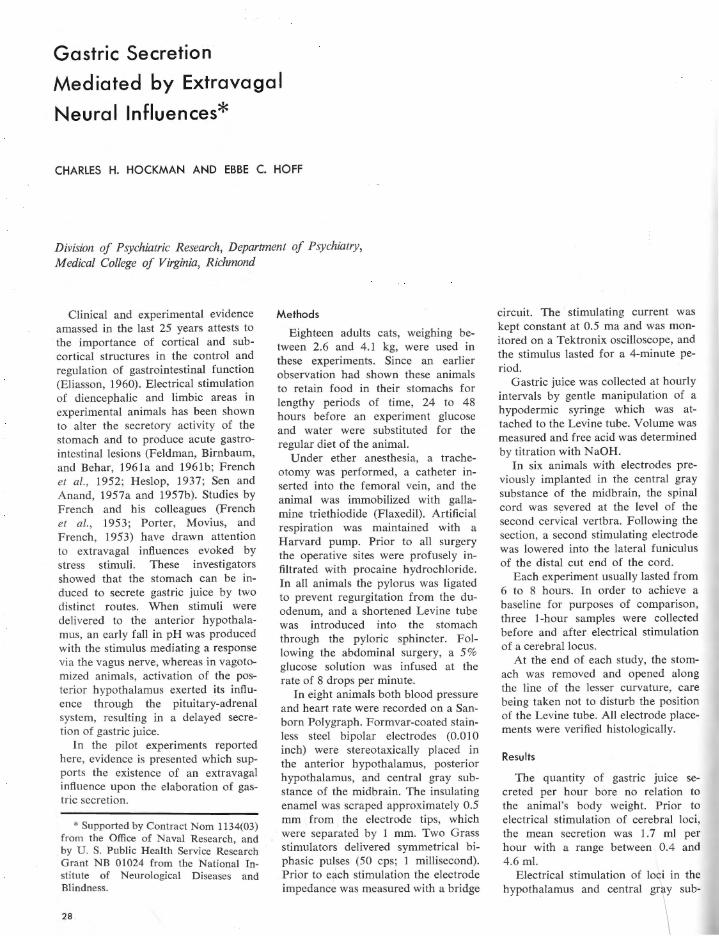
Gastric Secretion
Mediated by Extravagal
Neural Influences*
CHARLES H. HOCKMAN AND EBBE C. HOFF
Division of Psychiatric Research, Department of Psychiatry, Medical College of Virginia, Richmond
Clinical and experimental evidence amassed in the last 25 years attests to the importance of cortical and subcortical structures in the control and regulation of gastrointestinal. funct~on (Eliasson, 1960). Electrical. stJmulatJ~n of diencephalic and limbic areas m experimental animals has been shown to alter the secretory activity of the stomach and to produce acute gastrointestinal lesions (Feldman, Birnbaum, and Behar, 1961a and 1961b; French et al., 1952; Heslop, 1937; Sen and Anand, 1957a and 1957b). Studies by French and his colleagues (French et al., 1953; Porter, Movius, and French, 1953) have drawn attention to extravagal influences evoked by stress stimuli. These investigators showed that the stomach can be induced to secrete gastric juice by two distinct routes. When stimuli were delivered to the anterior hypothalamus, an early fall in pH was produced with the stimulus mediating a response via the vagus nerve, whereas in vagotomized animals, activation of the posterior hypothalamus exerted its influence through the pituitary-adrenal system, resulting in a delayed secretion of gastric juice.
In the pilot experiments reported here, evidence is presented which supports the existence of an extravagal influence upon the elaboration of gastric secretion.
*Supported by Contract Norn 1134(03) from the Office of Naval Research, and by U . S. Public Health Service Research Grant NB 01024 from the National Institute of Neurological Diseases and Blindness.
28
Methods
Eighteen adults cats, weighing between 2.6 and 4.1 kg, were used in these experiments. Since an earlier observation had shown these animals to retain food in their stomachs for lengthy periods of time, 24 to 48 hours before an experiment glucose and water were substituted for the regular diet of the animal.
Under ether anesthesia, a tracheotomy was performed, a catheter inserted into the femoral vein, and the animal was immobilized with gallamine triethiodide (Flaxedil). Artificial respiration was maintained with a Harvard pump. Prior to all surgery the operative sites were profusely infiltrated with procaine hydrochloride. In all animals the pylorus was ligated to prevent regurgitation from the duodenum, and a shortened Levine tube was introduced into the stomach through the pyloric sphincter. Following the abdominal surgery, a 5% glucose solution was infused at the rate of 8 drops per minute.
In eight animals both blood pressure and heart rate were recorded on a Sanborn Polygraph. Formvar-coated stainless steel bipolar electrodes (0.010 inch) were stereotaxically placed in the anterior hypothalamus, posterior hypothalamus, and central ~ray s~bstance of the midbrain. The msulatmg enamel was scraped approximately 0.5 mm from the electrode tips, which were separated by 1 mm. Two Grass stimulators delivered symmetrical biphasic pulses (50 cps; 1 millisecond). Prior to each stimulation the electrode impedance was measured with a bridge
circuit. The stimulating current was kept constant at 0.5 ma and was monitored on a Tektronix oscilloscope, and the stimulus lasted for a 4-minute period.
Gastric juice was collected at hourly intervals by gentle manipulation of a hypodermic syringe which was attached to the Levine tube. Volume was measured and free acid was determined by titration with NaOH.
In six animals with electrodes previously implanted in the central gray substance of the midbrain, the spinal cord was severed at the level of the second cervical vertbra. Following the section, a second stimulating electrode was lowered into the lateral funiculus of the distal cut end of the cord.
Each experiment usually lasted from 6 to 8 hours. In order to achieve a baseline for purposes of comparison, three 1-hour samples were collected before and after electrical stimulation of a cerebral locus.
At the end of each study, the stomach was removed and opened along the line of the lesser curvature, care being taken not to disturb the position of the Levine tube. All electrode placements were verified histologically.
Results
The quantity of gastric juice secreted per hour bore no relation to the animal's body weight. Prior to electrical stimulation of cerebral loci, the mean secretion was 1. 7 ml per hour with a range between 0.4 and 4.6 ml.
Electrical stimulation of loci in the hypothalamus and central gray sub-
\
stance produced both an increase in the volume of gastric juice secreted and in its free acid content. The increase in volume within the first hour following electrical stimulation varied from 21 to 200%, the largest increase in volume resulting from stimulation of the posterior hypothalamus. An increase in free acid occurred from 1 to 3 hours following electrical stimulation at all loci, ' the increase being from 50 to 250%.
In two animals with electrode placements in the posterior hypothalamus, and one with a placement in the central gray, there were no changes either in volume of juice secreted or acid content following electrical stimulation .
Sectioning of the spinal cord at C-2 usually elicited an immediate and copious secretion of gastric juice. Following cordotomy, electrical stimulation of the central gray substance had no effect whatsoever upon gastric secretion which was usually inhibited by the section; however, subsequent stimulation of the distal end of the severed cord, in the lateral funiculus, elicited a profuse secretion of gastric juice during the first hour following the onset of stimulation.
Control animals that did not receive stimulation typically showed a gradual reduction in hourly volume of gastric secretion with a concomitant decrease of acid content in the sample. In animals in which blood pressure and heart rate were recorded, electrical stimulation of the stated loci also elicited marked arterial blood pressure elevations, arrhythmic electrocardiographic complexes, pupillary dilatation, piloerection, and urination.
A visual examination of the stomach of all experimental animals revealed irregular hemorrhagic erosions from one to several millimeters in length. Although in some animals these lesions appeared to be super -ficial, in most they were both mucosa! and submucosal and were filled with extravasated blood. The severity of the lesions did not appear to bear any relation to the area stimulated.
It appeared rthat, in some animals, mechanical withdrawal of hourly samples might account for an irritation of the stomach mucosa, especially where the rugae came into contact with the openings in the Levine tube. A careful examination of the position
of the tube confirmed our suspicion in some animals.
Discussion
These pilot studies support the results of other investigators by showing that gastric secretion can be altered by electrical stimulation of cerebral loci, and present further evidence for extravagal control of gastric secretion.
In animals from which blood pressure was recorded, a significant pressor response was always observed during electrical stimulation of the stated cerebral loci. This was usually accompanied by other sympathetic phenomena, including pupillary dilatation, piloerection, and urination. It is noteworthy that this massive sympathetic activation accompanied the experimental production of the acute gastrointestinal lesions which were observed. Al ·· though some superficial erosions were undoubtedly the result of the mechanical procedure of sample withdrawal, we can confidently state that this was not the case for all lesions which were observed. We draw attention to this point since a similar method has been used by other investigators.
French et al. (1953) and Porter et al. (1953) have suggested that the extravaQal route for gastric secretion might be humorally mediated. Although this possibility is not disputed by the present study, the fact that stimulation of the central gray substance did not elicit secretion following cordotomy, and that subsequent stimulation of the distal cut end 1 of the cord caused an immediate secretion, would seem to argue for a more direct neural influence. Additional experimental work in this area is required in order to permit an assessment of the role played by the sympathetic division of the autonomic nervous system in the control of gastrointestinal function. However, these results suggest that the sympathetic division does indeed play a role in the elaboration of gastric secretion. This raises important and interesting questions with respect to stress-provoking stimuli and their relation to psychosomatic disturbances of the gastrointestinal tract.
Summary
In adult cats anesthetized with ether and immobolized with Flaxedil, the effects of electrical stimulation of cerebral loci on gastric secretion were studied. Stimulation of mesencephalic
and diencephalic areas increased both the volume and acidity of samples collected hourly. Section of the spinal cord at the level of the second cervical vertebra abolished this secretion; however, subsequent stimulation in the lateral funiculus of the distal end of the severed cord elicited the response. These pilot studies present further evidence for extravagal mediation of gastric secretion.
Acknowledgments
We are grateful to Dr. N . M. Papadopoulos for advice and for the use of his laboratory facilities for chemical analyses, and to Messrs. F. T. Grove and R. M. Butler for technical assistance.
References
EuAssoN, S. G. Central control of digestive function. In: Handbook of Physiology, edited by J. Field. Washington, D.C.: American Physiological Society, 1960 Sect. 1, vol. 2, pp. 1163-1171.
FELDMAN, S., D. BIRNBAUM, AND A. J. BEHAR. · Gastric secretion and acute gastroduodenal lesions following hypothalamic and preoptic stimulation. J. Neuro~urg. 18: 661-670, 196la.
FELDMAN, S., A. J. BEHAR, AND D. BIRNBAUM. Gastric lesions following hypothn Jamie stimulation. Arch. Neural. 4: 308-317, 196lb.
FRENCH, J. D., R . L. LONGMIRE, R. W. PORTER, AND H. J. MOVIUS. Extravagal influences on gastric hydrochloric acid secretion induced by stress stimuli. Surgery 34: 621-632, 1953.
FRENCH, J. D., R. w. PORTER, F. K. VON AMERONGEN, AND R. B. RANEY. Gastrointestinal hemorrhage and ulceration associated with intracranial lesions. A clinical and experimental study. Surgery 32: 395-407, 1952.
HESLOP, T. S. The nervous control of gastric secretion. An experimental study. Brit. J. Surg. 25: 884-899, 1937.
PORTER, R. W., H. J. Movms, AND J . D. FRENCH. Hypothalamic influences on hydrochloric acid secretion of the stomach. Surgery 33: 875-880, 1953.
SEN, R. N., AND B. K. ANAND. Effect of el~ctrical stimulation of the limbic system of the brain ("visceral brain") on gastric secretory activity and ulceration. Indian J. M ed. Research 45: 515-521, 1957a.
SEN, R. N., AND B. K. ANAND. Effect of electrical stimulation of the hypothalamus on gastric secretory activity and ulceration. Indian J. Med. Research 45: 507-513, 1957b.
29
The Effect of ldoxuridine (IDU) on
Corneal Stromal Cells in Tissue Culture*
WALTER J. GEERAETS, GUY WONG, AND DUPONT GUERRY, Ill
Department of Ophthalmology, Medical College of Virginia, Richmond
An alarming increase in the severity and frequency of the stromal forms of herpetic keratitis has been reported during the last decade (Thygeson and Kimura, 1957; Thygeson, Kimura, and Hogan, 1956; Kimura and Goodner, 1963; Bianchetti, 1963). This has led to an intensive search for therapeutic means for its management. The introduction in 1962 by Kaufman, Nesburn, and Maloney of idoxuridine (IDU) as an effective agent against herpes infection of the cornea has stimulated clinical and laboratory research to evaluate its full therapeutic potential.
Initially, IDU was reported as favorably affecting the stromal forms of herpetic keratitis. Kimura and Goodner (1963) and Maxwell (1963) found this to be true in 50 to 85% of the cases reported. Subsequently, Payrau and Dohlman (1964) found that IDU adversely affected the healing of stromal wounds as evidenced by tensile strength determinations.
But other investigators (Polack and Rose, 1964; Kaufman et al., 1964), basing their results upon the decreased tritiated thymidine uptake, reported that IDU only moderately inhibited the healing of stromal wounds. The degree to which the decreased tritiated thymidine uptake accurately reflected a decreased mitosis has been questioned by Pelc (1963). The same author, as well as other investigators, has expressed "reservations about assuming an invariable sequential relationship between DNA synthesis and mitotic division." Pelc found that in many organs a fewer number of cells divided than would be expected from the number of labeled cells found, thus indicating that DNA can be replaced in a
* Supported, in part, by the Virginia Lions Organization through the Old Dominion Eye Bank and Research, Inc., Richmond.
30
cell nucleus without cell division taking place.
Since the complete effect of IDU on corneal cells is still not known, this investigation was conducted. During the experimental phase of this study, a few studies in vivo appeared in the literature (Kaufman et al., 1962; Maxwell, 1963; Kaufman et al., 1964; Ey, Hughes, and Holmes, 1964), as well as tissue culture work with various cells. However, to our knowledge, no study in vitro with corneal stromal cells has been reported, although it has been pointed out (Kaufman, personal communication) that the effect of IDU may vary with the cell type used.
Materials and Methods
Cell Culture
Corneal tissue cultures were used. Corneal explants were obtained from chinchilla rabbit eyes and cultured under perforated cellophane in growth medium (minimum essential medium),' with L-glutamine (3 mg per ml), penicillin (100 µ, per ml), streptomycin (0.5 mg per ml), and 10% calf serum. The pH was 7.4 which equilibrated with 5% C02 at 37°C.
A cell strain of corneal stromal cells which had undergone at least eight passages was obtained and used in this experiment (fig. 1).
ldoxuridine Medium
Growth medium containing idoxuridine was prepared by dissolving powdered IDU2 in the medium. First, a 1,000 µ,g per ml solution was prepared. One-tenth dilutions ranging through 0.1 µ,g per ml were then prepared. This material was filtered through Seitz fil-
' Grand Island Biological Company, Grand Island, N.Y.
2 Nutritional Biochemicals Corpora-tion, Cleveland, Ohio.
\
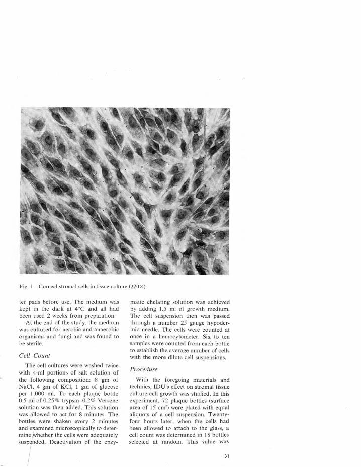
Fig. I-Corneal stromal cells in tissue culture (220 X).
ter pads before use. The medium was kept in the dark at 4 °C and all had been used 2 weeks from preparation.
At the end of the study, the medium was cultured for aerobic and anaerobic organisms and fungi and was found to be sterile.
Cell Count
The cell cultures were washed twice with 4-ml portions of salt solution of the following composition: 8 gm of NaCl, 4 gm of KC!, 1 gm of glucose per 1,000 ml. To each plaque bottle 0.5 ml of 0.25 % trypsin-0.2% Versene solution was then added. This solution was allowed to act for 8 minutes. The bottles were shaken every 2 minutes and examined microscopically to determine whether the cells were adequately '"'t dod. Deoctiv.tioo of tho my-
matic chelating solution was achieved by adding 1.5 ml of growth medium. The cell suspension then was passed through a number 25 gauge hypodermic needle. The cells were counted at once in a hemocytometer. Six to ten samples were counted from each bottle to establish the average number of cells with the more dilute cell suspensions.
Procedure
With the foregoing materials and technics, IDU's effect on stromal tissue culture cell growth was studied. In this experiment, 72 plaque bottles (surface area of 15 cm2 ) were plated with equal aliquots of a cell suspension. Twentyfour hours later, when the cells had been allowed to attach to the glass, a cell count was determined in 18 bottles selected at random. This value was
31
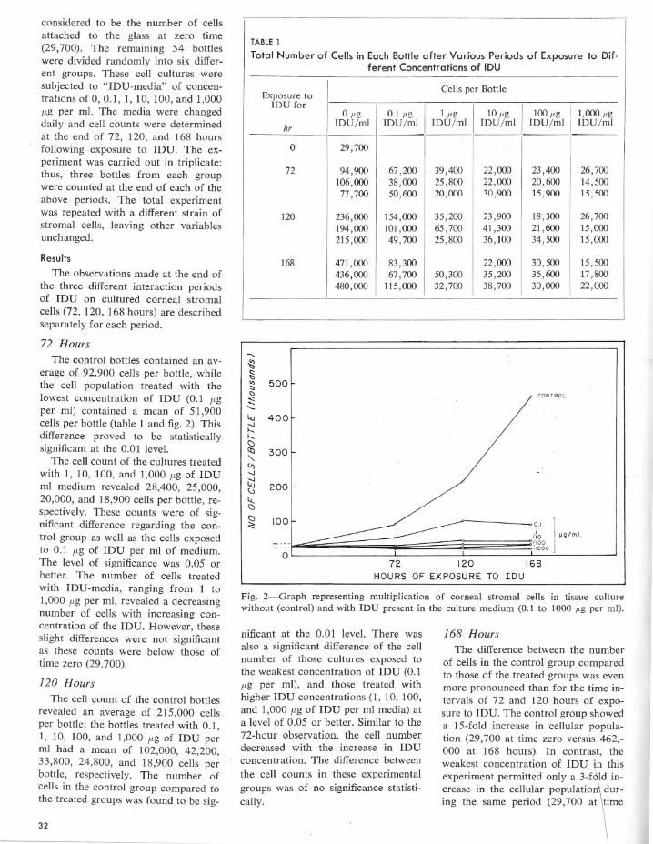
considered to be the number of cells attached to the glass at zero time (29,700). The remaining 54 bottles were divided randomly into six different groups. These cell cultures were subjected to "!DU-media" of concentrations of 0, 0.1, 1, 10, 100, and 1,000 µ.g per ml. The media were changed daily and cell counts were determined at the end of 72, 120, and 168 hours following exposure to IDU. The experiment was carried out in triplicate: thus, three bottles from each group were counted at the end of each of the above periods. The total experiment was repeated with a different strain of stromal cells, leaving other variables unchanged.
Results
The observations made at the end of the three different interaction periods of IDU on cultured corneal stromal cells (72, 120, 168 hours) are described separately for each period.
72 Hours The control bottles contained an av
erage of 92,900 cells per bottle, while the cell population treated with the lowest concentration of IDU (0.1 µ.g per ml) contained a mean of 51,900 cells per bottle (table 1 and fig. 2). This difference proved to be statistically significant at the 0.01 level.
The cell count of the cultures treated with 1, 10, 100, and 1,000 µ.g of IDU ml medium revealed 28,400, 25 ,000, 20,000, and 18,900 cells per bottle, respectively. These counts were of significant difference regarding the control group as well as the cells exposed to 0.1 µ.g of IDU per ml of medium. The level of significance was 0.05 or better. The number of cells treated with !DU-media, ranging from 1 to 1,000 µ.g per ml, revealed a decreasing number of cells with increasing concentration of the IDU. However, these slight differences were not significant as these counts were below those of time zero (29,700).
120 Hours
The cell count of the control bottles revealed an average of 215,000 cells per bottle; the bottles treated with 0.1, 1, 10, 100, and 1,000 µ.g of IDU per ml had a mean of 102,000, 42,200, 33,800, 24,800, and 18,900 cells per bottle, respectively. The number of cells in the control group compared to the treated groups was found to be sig-
32
TABLE 1
Total Number of Cells in Each Bottle after Various Periods of Exposure to Different Concentrations of IOU
Exposure to Cells per Bottle
IDU for 0 µg 0.1 µg 1 µg 10 µg 100 µg 1,000 µg
hr IDU/ ml IDU/ ml IDU/ ml IDU/ ml IDU/ ml IDU/ ml
0 29,700
72 94,900 67,200 39,400 22,000 23,400 26,700 106,000 38 ,000 25,800 22,000 20,600 14,500 77,700 50,600 20 ,000 30,900 15,900 15,500
120 236,000 154,000 35,200 23,900 18 ,300 26,700 194,000 101,000 65 ,700 41,300 21,600 15,000 215,000 49,700 25 ,800 36, 100 34,500 15,000
168 471,000 83,300 22,000 30,500 15,500 436,000 67,700 50 ,300 35,200 35,600 17,800 480,000 115,000 32,700 38,700 30,000 22,000
..... {l § "' 500 "' <:>
<:: CON TROL ..... ...... i...i 400 ~ h. C) Cl:l 300
" <J) -.J
~ 200 ~
li.. C)
C) 100 <:
0 72 120 168
HOURS OF EXPOSURE TO IOU
Fig. 2-Graph representing multiplication of corneal stromal cells in tissue culture without (control) and with IDU present in the culture medium (0.1 to 1000 µg per ml).
nificant at the 0.01 level. There was also a significant difference of the cell number of those cultures exposed to the weakest concentration of IDU (0.1 µ.g per ml), and those treated with higher IDU concentrations (1, 10, 100, and 1,000 µ.g of IDU per ml media) at a level of 0.05 or better. Similar to the 72-hour observation, the cell number decreased with the increase in IDU concentration. The difference between the cell counts in these experimental groups was of no significance statistically.
168 Hours The difference between the number
of cells in the control group compared to those of the treated groups was even more pronounced than for the time intervals of 72 and 120 hours of exposure to IDU. The control group showed a 15-fold increase in cellular population (29,700 at time zero versus 462,-000 at 168 hours). In contrast, the weakest concentration of IDU in this experiment permitted only a 3-fOld increase in the cellular population\~uring tho "m' period (29,700 .i \ m'
zero versus 88,900 at 168 hours). This 3-fold increase in the number of cells of the latter group again was significantly different from the populations treated with the greater concentrations of IDU. This was found at the 0.01 level.
As was noted in the group of shorter exposure to IDU, there was only an insignificant difference found between the number of cells in the 1, 10, 100, and 1,000 µg of IDU per ml of mediatreated populations, all which showed cell counts similar to those at time zero.
Throughout this experiment, there was no significant difference in cell counts within individual groups that could have been attributed to the different periods of IDU exposure. Thus, IDU interaction upon corneal stromal cells for 72, 120, and 168 hours in equal concentrations did not result in significantly different cell counts.
When the total experiment was repeated with a different strain of stromal cells, the results obtained were very similar to those above.
Discussion
The marked inhibition of cell population increase and its relation to the concentration of IDU is consistent with recent observations by others. The growth inhibition of mouse leukemic cells in culture by IDU was reported by Mathias, Fischer, and Prusoff (1959). These authors reported that IDU, at ordinarily accepted therapeutic concentrations and less, permitted only a single doubling of the population cells in culture, and thereafter "cell death invariably commenced."
Ey, Hughes, and Holmes (1964) found that, with concentrations of IDU at less than therapeutic strength, the multiplication of primary rabbit kidney cells in culture was entirely blocked. Cheong, Rich, and Eidenoff (1960) have reported similar results with cultures of human cervical carcinoma cells.
Studies in vivo by Polack and Rose (1 964) with therapeutic doses of IDU illustrated that there was a delay in cell repopulation after stromal injury in rabbits. Payrau and Dahlman (1964) also found that stromal wound healing was retf(rded in rabbits treated with IDU. As was theorized by Polack, "this inhibition should increase if IDU ooul/ ' m>de mifable to fill the p<e-
mitotic cells." The more marked quantitative differences in cell counts, after exposures to various concentrations of IDU in this study in vitro, can be based on this assumption of Polack.
The arrest of cellular multiplication after exposure to a critical concentration of IDU may be explained by lysis of the cells. Mathias and Fischer (1959) found that murine leukemia cells in 10- to 100-µg IDU solutions underwent lysis even after doubling in number.
The findings reported in this study suggest that the proliferation of corneal stromal cells is inhibited by IDU. These findings in vitro should be considered when IDU is clinically used in cases where there is severe stromal involvement.
Summary
Corneal stromal cells were cultured in vitro and exposed to various concentrations of idoxuridine (IDU), ranging from 0 to 1,000 µg of IDU per ml of medium. Inhibition of cell multiplication occurred with concentrations of 0.1 µg per ml. With concentrations of 1.0 µg per ml and greater, there was no increase in cell number from the time of exposure to IDU.
Acknowledgment
We are grateful for the valuable aid extended by Dr. Robert W. Tankersley, Jr., Medical College of Virginia, and Dr. Fred Stocker, Durham, N.C.
References
BIANCHETTI, M. Herpes simplex keratitis. Ophthalmologia 146: 395-410, 1963.
CHEONG, L., M. A. RICH, AND M. L. EIDINOFF. Introduction of the 5-halongenated uracil moiety into deoxyribonucleic acid of mammalian cells in culture. J. Biol. Chem. 235: 1441-1447, 1960.
EY, R. C., W. F . HUGHES, AND A. W. HOLMES. Clinical and laboratory evaluation of idoxuridine (IDU) therapy in herpes simplex keratitis. A . M.A. Arch. Ophthalmol. 71: 325-331, 1964.
KAUFMAN, H. E., A. B. NESBURN, AND E. D. MALONEY. IDU therapy of herpes simplex. A. M.A. Arch. Ophthalmol. 67: 583-591, 1962.
KAUFMAN, H. E., J. A. CAPELLA, E. D. MALONEY, J. E. ROBBINS, G. M. CooPER, AND M. H. UoTILA. Corneal toxicity of cytosine arabinoside. A. M. A. Arch. Ophthalmol. 72: 535-540, 1964.
KIMURA, S. J., AND E. K. GOODNER. Ocular Pharmacology and Therapeutics and the Problems of Medical Management. Philadelphia: F. A. Davis Co., 1963.
MATHIAS, A. P., G . A. FISCHER, AND w. H. PRUSOFF. Inhibition of the growth of mouse leukemia cells in culture by 5-iododeoxyuridine. Biochim. et Biophys. Acta 36: 560-561, 1959.
MATHIAS, A. P., AND G. A. FISCHER. Studies of the inhibition of thymidine-synthesis and -utilization by mouse leukemia cells in culture. Federation Proc. 18: 284, 1959.
MAXWELL, E. Treatment of herpes keratis with 5-iodo-2-deoxyuridine (IDU). Am. J. Ophthalmol. 56: 571-573, 1963 .
PAYRAU, P., AND c. H. DOHLMAN. IDU in corneal wound healing. Am. J. Ophthalmol. 57: 999-1002, 1964.
PELC, S. R. On the question of renewal of differentiated cells. Exp. Cell Research 29: 194-198, 1963 .
POLACK, F . M., AND J. ROSE. The effect of 5-iodo-2-deoxyuridine (IDU) in corneal healing. A. M. A . Arch . Ophthalmol. 71: 520-527, 1964.
THYGESON, P., S. J. KIMURA, AND M. J . HOGAN. Observations on herpetic keratitis and keratoconjunctivitis. A. M . A. Arch. Ophthalmol. 56: 375-388, 1956.
THYGESON, P., AND s. J. KIMURA. Deep forms of herpetic keratitis. Am. J. Ophthalmol. 43: 109-113, 1957.
33

The Challenge of Pulmonary Emphysema*
DAVID V. BATES
Department of Medicine, Royal Victoria Hospital McGill University, Montreal, Canada
I feel very privileged to be invited to be your Stoneburner Lecturer for this year. When I came to consider what I really wanted to say and what might interest people in widely different areas of medicine, there really was little choice. I perhaps can claim to be able to talk about emphysema from a rather broader standpoint than some other physicians. Not because I suffer from it, which is sometimes a good reason for talking about a disease, but because I have been trained both in England and in America, and the outlook on this disease has differed in Europe and in the States.
I started work on emphysema under the guidance of Dr. Christie in 1948, and in 1948 no one was much interested in the disease. I worked in Philadelphia in 1952, and there I gave a lecture on emphysema which was so controversial that it was disbelieved. The climate is different now, and I am talking about something of which most of you already know a fair amount. I have selected it as a topic because, whether you are a public health administrator, an internist, an allergist, an anesthetist, a surgeon, or even just a cigarette smoker, you should be interested in this condition.
One reason for contemporary concern about this disease is the considerable increase in the standardized mortality in the United States since 1945, for what physicians call on the death
* Presented as the first of the Seventeenth Annual Stoneburner Lectures at the Medical College of Virginia, March 11, 1964.
34
certificate "emphysema and fibrosis." The rate of increase of lung cancer has been similar, but since 1955 the diseases categorized as "emphysema" have been increasing more rapidly than has lung cancer. Over the same period, of course, mortality from tuberculosis has dipped. But the puzzle of nomenclature can be seen from the fact that bronchitis, as certified in the United States, has apparently not increased at all as a cause of mortality. If I were to show you a comparable graph from Europe, bronchitis would appear to have been the main cause of the increased mortality, and emphysema to a much smaller extent. I think we now realize that this is purely a semantic matter. Differences are not great between different industrialized communities in any of these diseases. It has been largely a matter of what the physician has called the condition he has looked at.
Certified causes of death at best are enigmatic and subject to shifting classification. The autopsy incidence of morphological emphysema as found in 138 random autopsies of inflated right lungs at the Massachusetts General Hospital in Boston has been reported by Dr. Thurlbeck. With reference to men only, and by dividing the autopsy population into 12 decades of age, the incidence of quite obvious morphological emphysema, excluding the little bits of emphysema at the apices and other minor forms, is striking. Once men are in the fifth and sixth decades, half of the random au
top•y populotion •how• oon•i\ ob1'
morphological emphysema. The same data in identical form are found in my own hospital in Montreal. So Montreal and Boston have a virtually identical autopsy incidence. Many pathologists have emphasized that the true incidence of emphysema can only be evaluated if the lungs are inflated. If they are fixed when collapsed, he will underestimate the incidence of this condition by at least half; and second, he will not be in a position to see, as I will show you in a few moments, its most damaging form. When the Massachusetts General Hospital group is broken down into categories, the malefem ale incidence is very different. In 59 females, 45 had no detectable morphological emphysema. In 79 males, only 25 had no emphysema. This reflects the 4: 1 prevalence of emphysema in men.
There is another reason why this group of diseases has become very important. You will recall the major episode of smog in London in 1952, but you may have forgotten that this episode killed 4,000 people in 6 days. The smog lasted from December 2 until 14, approximately. London is so large that it took the Registrar General's figures 3 weeks to catch up on the surplus mortality of 4,000 people. So this is another reason why this disease has become important.
The First Challenge
The first of emphysema's three challenges is to understand not what the acute episode can do, which we know very well in a population with some
I
lung disease, but to understand what lesser degrees of atmospheric pollution do, not over 6 days but over 20 years. I fancy it will be a long time before we understand the interrelationship between the acute sensational phenomenon and the chronic unsensational mortality.
I grew up in an era when chronic bronchitis did not have a respectable pathology. In Boyd's Pathology for 1947, chronic bronchitis was not a respectable disease. It was mainly an important complication of tuberculosis, or it was sometimes a nuisance in people with heart disease, but as a primary pathological entity it was little regarded. It is worth reminding you that now it has a highly respectable and extremely carefully quantified pathological existence, depending on hypertrophy of the bronchial mucous glands.
Now it is time we took up some practical examples of the kind of patient you deal with, and we deal with. I am not going out of my way to speak of the only case I have seen in 5 years that represented so and such. I am talking about things that I believe are extremely common. The first patient was a 66-year-old man who worked all his life with the Canadian Pacific Railway, largely an office job, and gave a rather clear history of some breath shortness for 2 years, some chronic cough for perhaps 15 years, not very much sputum, and an occasional episode of respiratory infection. He had dyspnea for 1 year. The function tests were done when he
had one such episode while in another hospital. He had left that hospital diagnosed as having arteriosclerotic heart disease. They found an abnormal EKG, swollen ankles, liver two fingersbreadth's enlarged, and a normal chest film. When he was studied in the function lab in November, 1958, the findings were: a vital capacity about half of what it should be, a lung volume much bigger than it ought to be-gross overinflation, and markedly uneven gas distribution. The F.E.V. (forced expiratory volume) should have been 70 L per minute, but was only 19 L per minute. His airflow rate should have been 3 L per second, but was only 0.2 L per second; therefore, he had terrible ventilatory obstruction. The transport of carbon monoxide, or the diffusing capacity, should have been about 14 and was 7 ml per minute per mm of Hg. This tells us either he had very uneven ventilation/ perfusion distribution in the lung, which is commonly found , or he had a reduced surface area for gas exchange, or a thickened alveolar membrane. His arterial C02 tension was 56 mm of Hg, and the pH 7.4. You know, therefore, this was a chronic situation because he had brought his bicarbonate up to adjust the pH. The oxygen saturation was a little down. About 9 months after these tests were done he came to the hospital with a very severe pneumonia, and he died as a tracheotomy was being done. The whole lung section of this man show,ed black areas which look at a distance like currants. These are holes which
35
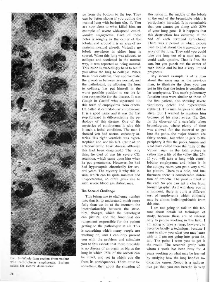
FIG. I-Whole lung section from patient with centrilobular emphysema. Barium added for clearer demarcation.
36
go from the bottom to the top. They can be better shown if you outline the normal lung with barium (fig. 1). You are now close to what killed him, an example of severe widespread centrilobular emphysema. Each of these holes is roughly in the center of the lobule, and around it is an area of remaining normal alveoli. Virtually no lobule anywhere in either lung is spared. When this lung was allowed to collapse and sectioned in the normal way, it was reported as being normal. This lesion is exceedingly hard to see if you allow the lung to collapse. When these holes collapse, they approximate; the alveoli in between are normal, and the pathologist, by allowing the lung to collapse, has put himself in the worst possible position to see the lesion responsible for the disease. It was Gough in Cardiff who separated out this form of emphysema from others. He called it centrilobular emphysema. It is a good name and it was the first step forward in differentiating the pathology of this disease. One of the mysteries of emphysema is why this is such a lethal condition. The man I showed you had normal coronary arteries. His right ventricle was hypertrophied and not his left. (He had no arteriosclerotic heart disease although this had been diagnosed.) The only thing he died of was his severe CO, retention, which came upon him when he got pneumonia. However, he had had hypercapnia chronically for several years. The mystery is why this lesion, which can be quite minimal and unspectacular, so often gives rise to such severe blood gas disturbance.
The Second Challenge
This brings me to challenge number two; that is, to understand much more fully than we do at the moment the interrelationship between the structural changes, which the pathologist can picture, and the functional derangement responsible for the patient getting to the pathologist at all. This is something which many people are working on, and I can only present you with the problem and stimulate you to this extent: that there probably is no disease of an organ as big as the lung in which 75 % of the alveoli can be intact, and yet in which you die from its consequences. There must be something then about the situation of
this lesion in the middle of the lobule at the end of the bronchiole which is particularly harmful. It is remarkable that you cannot get along with 25% of your lung gone, if it happens that this destruction has occurred at the end of each terminal bronchiole. There was a period in which people used to chat about the tremendous reserve of the lung. They said you could take one lung out of a man and he could walk upstairs. That is fine . He can, but you punch out the center of each lobule and he has a very limited prognosis.
My second example is of a man about the same age as the previous man, to illustrate the clues you can get in life that the lesion is centrilobular emphysema. This man's pulmonary function tests were similar to those of the first patient, also showing severe ventilatory defect and hypercapnia (table 1). This man happens to still be alive. But I wanted to mention him because of his chest x-rays (fig. 2a). In the close-up of a carefully taken bronchogram, where plenty of time was allowed for the material to get into the pools, the major bronchi are fairly normal, but when it gets to the periphery it fills the pools. Simon and Reid have called these the "Lily of the Valley" sign, as the total picture is rather like a lily of the valley (fig. 2b ). If you will take a lung with centrilobular emphysema and inject it in the autopsy room, you get a very similar picture. There is a hole, and furthermore there is considerable distortion of bronchi. The pool is filled at the end. So you can get a clue from bronchography. As I will show you in a moment, there is quite a different sort of emphysema which clinically may be almost indistinguishable from this one.
I am not going to talk in this lecture about details of technique of study, because these are of interest only to people working in this field. I am going to take a jump, however, to describe briefly a technique, because I want to show you what you may learn with it. I am not going into great detail. The point I want you to get is the result. The research group with whom I work has been busy for 4 years working on what may be learned by studying how the lung handles radioactive xenon. Xenon is a radioactive gas that you can breathe in very
\
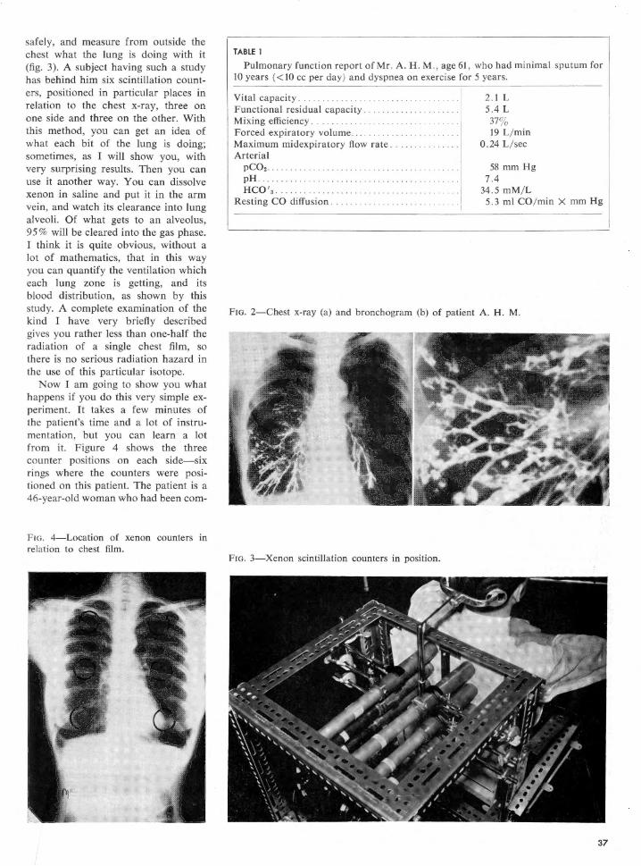
safely, and measure from outside the chest what the lung is doing with it (fig. 3). A subject having such a study has behind him six scintillation counters, positioned in particular places in relation to the chest x-ray, three on one side and three on the other. With this method, you can get an idea of what each bit of the lung is doing; sometimes, as I will show you, with very surprising results. Then you can use it another way. You can dissolve xenon in saline and put it in the arm vein, and watch its clearance into lung alveoli. Of what gets to an alveolus, 95% will be cleared into the gas phase. I think it is quite obvious, without a lot of mathematics, that in this way you can quantify the ventilation which each lung zone is getting, and its blood distribution, as shown by this study. A complete examination of the kind I have very briefly described gives you rather less than one-half the radiation of a single chest film, so there is no serious radiation hazard in the use of this particular isotope.
Now I am going to show you what happens if you do this very simple experiment. It takes a few minutes of the patient's time and a lot of instrumentation, but you can learn a lot from it. Figure 4 shows the three counter positions on each side-six rings where the counters were positioned on this patient. The patient is a 46-year-old woman who had been com-
FIG. 4-Location of xenon counters in relation to chest film.
TABLE 1
Pulmonary function report of Mr . A. H . M., age 61 , who had minimal sputum for IO years (<IO cc per day) and dyspnea on exercise for 5 years.
Vital capacity . . . . . . .. .. .. . . . Functional residual capacity ... . .. . . . .. . Mixing efficiency . . . ... . .. . . .. . . .. ...... . Forced expiratory volume... . ...... .. . . . . . .. . . . Maximum midexpiratory flow rate .. Arterial
pC02 •••• • • • • •..
pH . HCO'a.
Resting CO diffusion .
2 .1 L 5.4 L
37% 19 L/ min
0.24 L/ sec
58 mm Hg 7.4
34.5 mM/L 5.3 ml CO/ min X mm Hg
Fm. 2-Chest x-ray (a) and bronchogram (b) of patient A. H. M.
Fm. 3-Xenon scintillation counters in position.
37
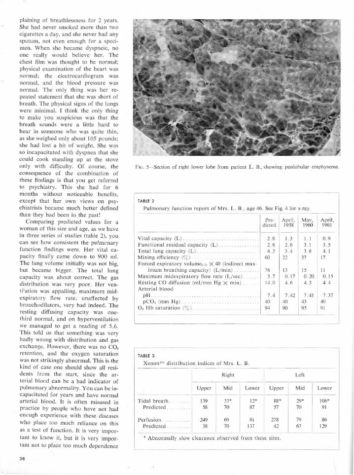
plaining of breathlessness for 2 years. She had never smoked more than two cigarettes a day, and she never had any sputum, not even enough for a specimen. When she became dyspneic, no one really would believe her. The chest film was thought to be normal; physical examination of the heart was normal; the electrocardiogram was normal, and the blood pressure was normal. The only thing was her repeated statement that she was short of breath. The physical signs of the lungs were minimal. I think the only thing to make you suspicious was that the breath sounds were a little hard to hear in someone who was quite thin, as she weighed only about 105 pounds ; she had lost a bit of weight. She was so incapacitated with dyspnea that she could cook standing up at the stove only with difficulty. Of course, the consequence of the combination of these findings is that you get referred to psychiatry. This she had for 6 months without noticeable benefits, except that her own views on psychiatrists became much better defined than they had been in the past!
Comparing predicted values for a woman of this size and age, as we have in three series of studies (table 2), you can see how consistent the pu!mcnary function findings were. Her vital capacity final!y came down to 900 ml. The lung volume initially was not big, but became bigger. The total lung capacity was about correct. The gas distribution was very poor. Her venti1ation was appalling, maximum midexpiratory flow rate, unaffected by bronchodilators, very bad indeed. The resting diffusing capacity was onethird normal, and on hyperventilation we managed to get a reading of 5.6. This told us that something was very badly wrong with distribution and gas exchange. However, there was no C02 retention , and the oxygen saturation was not strikingly abnormal. This is the kind of case one should show all residents from the start, since the arterial blood can be a bad indicator of pulmonary abnormality. You can be incapacitated for years and have normal arterial blood. It is often misused in practice by people who have not had enough experience with these diseases who place too much reliance on this as a test of function. It is very important to know it, but it is very important not to place too much dependence
38
FIG. 5- Section of right lower lobe from patient L. B., showing panlobular emphysema.
TABLE 2
Pulmonary function report of Mrs . L.B., age 46. See Fig. 4 for x-ray.
Pre- April , May, April, dieted 1958 1960 1961 --- - -- ------
Vital capacity (L) . . . . 2.8 1. 3 1. I 0 .9 Functional residual capacity (L). .. . 2.6 2.6 3 .1 3.5 Total lung capacity (L) . 4.2 3.4 3.8 4.1 Mixing efficiency (3) . 60 : 22 37 17 Forced expiratory volume0 75 X 40 (indirect max-
imum breathing capacity) (L/ min). 76 13 15 11 Maximum midexpiratory flow rate (L/ sec) . .. . .. 3.7 0.17 0.20 0. 15 Resting CO diffusion (ml/ mm Hg X min) . . . . . 14.0 4.6 4.3 4.4 Arterial blood
pH . . . . . . . . ' . . . . . . . . . . . . . . . . . . .. . . . 7.4 7.42 7.41 7 .37 pCO, (mm Hg) . . . . . . . . . . . . . . . . . . . .. . . .. . . 40 40 43 40
0 2 Hb saturation (3). . . . . . . . ... .. . .. . 94 90 95 91
TABLE 3
Xenon133 distribution indices of Mrs. L. B.
Right Left
Upper Mid Lower Upper Mid Lower
Tidal breath . ... . . . . 139 33* 12* 88* 29* 106* Predicted. .. . . ... . 58 70 87 57 70 91
Perfusion . .... . . .. 249 69 61 278 79 86 Predicted. 38 70 137 42 67 129
* Abnormally slow clearance observed from these sites.
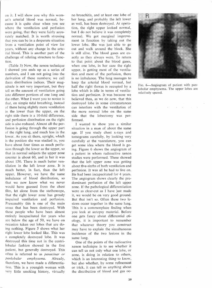
on it. I will show you why this woman's arterial blood was normal, because it is quite clear when you see where the ventilation and perfusion were going, that they were fairly accurately matched. It is worth stressing that you can be in a desperate situation from a ventilation point of view for years, without any change in the arterial blood. This is another part of the challenge of relating structure to function.
(Table 3) Now, the xenon technique I showed you ends up as a series of numbers, and I am not going into the derivation of these numbers; we call them distribution indices. Their magnitude is not very important, but they tell us the amount of ventilation going into different portions of one lung and the other. What I want you to notice is that, on simple tidal breathing, instead of there being slightly more ventilation in the lower than the upper, on the right side there is a 10-fold difference, and perfusion distribution on the right side is also reduced. Almost all the perfusion is going through the upper part of the right lung, and much less in the lower. As you sit there, upright, which is the position she was studied in, you have about four times as much perfusion through the lower as the upper, so that in normal subjects the upper zone counter is about 60, and in her it was about 150. There is much better ventilation in the left lower zone. It is much better, in fact, than the left upper. However, we have the same imbalance of blood distribution, so that we now know what we never would have guessed from the chest film, let alone from the stethoscope, that the right lower zone has grossly impaired ventilation and perfusion. Presumably this is one of the main areas that has been destroyed. With these people who have been almost entirely incapacitated for years who are below the age of 50, we have on occasion taken out lobes that are doing nothing. Figure 5 shows what her right lower lobe looked like. This was a completely destroyed lobe. It was destroyed this time not in the centrilobular fashion showed in the first patient, but generally destroyed. This often is referred to as panacinar or pan lobular emphysema. Already, therefore, we have made a differentiation. This is a youngish woman with very little smoking history, virtually
no bronchitis, and at least one lobe of her lung, and probably the left lower as well, has been destroyed. At operation, the right upper looked normal, but I do not believe it was completely normal. We got marginal improvement in function by taking out the lower lobe. She was just able to go out and walk around the block. She is still alive. The blood gases are exactly as they always were. To return to that point about the blood gases, when one lobe, in her case the right upper, is getting most of the ventilation and most of the perfusion, there is no imbalance. The lung manages to keep the arterial blood normal, but half the right thorax is occupied by a lobe which is idle in terms of ventilation and perfusion. It was because we believed then, as we do now, that this destroyed lobe in some circumstances can interfere with the ventilation of the more normal lobe on the same side that the lobectomy was performed.
I wanted to show you a similar situation in a man of about the same age. If you study chest x-rays and tomograms carefully, by looking very carefully at the vasculature, you can get some idea where the blood is going. Figure 6 shows the angiogram of a patient in whom radioactive xenon studies were performed. These showed that the left upper zone was getting about five-sixths of both ventilation and perfusion. It was all he had to live on. He had been incapacitated for 4 years. The angiogram shows clearly the predominant perfusion of the left upper zone. If the pathological differentiation were as clear-cut as I have just made it, we would be on very good ground. But that isn't so. Often these two lesions occur together in the same lung. This is a commonplace finding when you look at autopsy material. Before one gets fancy about differential etiology, it is important to remember that whatever theory you construct may have to explain the simultaneous incidence of the two lesions in the same lung.
One of the points of the radioactive xenon technique is to see whether it can tell us not only what one lobe, or zone, is doing in relation to others, which is an interesting thing to know, but also whether, by some refinement or tr ick, it can tell us anything about the distribution of blood and gas oc-
39
FIG. 6-Angiogram of patient with panlobular emphysema. The upper lobes are relatively spared.
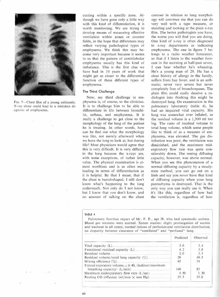
FIG. 7-Chest film of a young asthmatic. X-ray alone could lead to a mistaken diagnosis of emphysema.
curring within a specific zone. Although we have gone only a little way with this kind of differentiation, it is worth mentioning. We are trying to develop means of measuring effective ventilation within zones or counter fields, in the hope that differences may reflect varying pathological types of emphysema. We think this may become very important because it seems to us that the pattern of centrilobular emphysema usually has this kind of imbalance. This is the first clue we have had in 10 years of work that might get us closer to the differential function of these different types of emphysema.
The Third Challenge
Now, my third challenge in emphysema is, of course, to the clinician. It is to challenge him to be able to differentiate in life between bronchitis, asthma, and emphysema. It is really a challenge to get close to the morphology of the lung of the patient he is treating. In other words, how can he find out what the morphology was like, not merely afterward when we have the lung to look at, but during life? Most physicians would agree that this is very difficult. It is very difficult in the Jung because the x-rays are, with some exceptions, of rather little value. The physical examination is almost worthless and is as often misleading in terms of differentiation as it is helpful. By that I mean, that if the chest is barrel-shaped, I still don't know what's happening to the Jung underneath. Not only do I not know, but I know that you don't know, and no amount of talking on the chest
TABLE 4
contour in relation to lung morphology will convince me that you can do very well with a tape measure, or standing and looking at the plain x-ray film. The better pathologists you have, the worse you will find you are doing. One kind of x-ray is often diagnosed in x-ray departments as indicating emphysema. The one in figure 7 belongs to a radio weather forecaster, so that if I listen to the weather forecast in the morning at half-past seven, I can hear whether he's wheezing. He's a young man of 28. He has a clear history of allergy in the family, suffers from hay fever, and is an asthmatic; never very severe but never completely free of bronchospasm. The plain film could easily deceive a radiologist into thinking this might be destroyed lung. On examination in the pulmonary laboratory (table 4), he had an impaired vital capacity. His lung was somewhat over inflated, as the residual volume is a 1,300 ml too big. The ratio of residual volume to total lung volume, which some people like to think of as a measure of emphysema, was elevated. The gas distribution was poor, the ventilation was diminished, and the maximum midexpiratory flow rate was quite considerably down. The resting diffusing capacity, however, was above normal. When you see this phenomenon of a normal diffusing capacity by a steadystate method, you can go out on a limb and say you never have that kind of diffusing capacity when your lung parenchyma is destroyed. That is the only way you can really use it. When it's like this, regardless of how bad the ventilation is, regardless of how
Pulmonary function report of Mr. F. E., age 28, who had spasmodic asthma. Blood gas tensions were normal. Xenon studies: slight prolongation of wash in and washout in all zones; normal indices of perfusion and ventilation distribution; no disparity between clearance of " ventilated" and " perfused" lung.
Vital capacity (L) ... Functional residual capacity (L) . . .. .. . . . . . Residual volume ... . .... . Residual volume/ total lung capacity (% ) . . ... . . . .. . Mixing efficiency (%) .. . .. .. . . . ........... . Forced expiratory volumeo.75 X 40, (indirect maximum
breathing capacity) (L/ min) . . . Maximum rnidexpiratory flow rate (L/ sec) .... . . . . . . . . . Resting CO diffusion (ml/ min X mm Hg) .. .
40
Predicted Observed
5.6 3.4 4.2 3.8 2.0 3.3
26 49.5 65 34
140 83 4 .50 1.30
23. 5 33 .0
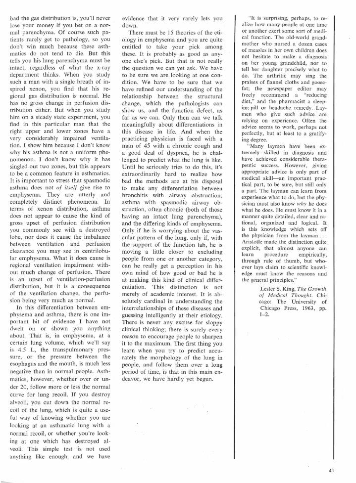
bad the gas distribution is, you'll never lose your money if you bet on a normal parenchyma. Of course such patients rarely get to pathology, so you don't win much because these asthmatics do not tend to die. But this tells you his lung parenchyma must be intact, regardless of what the x-ray department thinks. When you study such a man with a single breath of inspired xenon, you find that his regional gas distribution is normal. He has no gross change in perfusion distribution either. But when you study him on a steady state experiment, you find in this particular man that the right upper and lower zones have a very considerably impaired ventilation. I show him because I don't know why his asthma is not a uniform phenomenon. I don't know why it has singled out two zones, but this appears to be a common feature in asthmatics. It is important to stress that spasmodic asthma does not of itself give rise to emphysema. They are utterly and completely distinct phenomena. In terms of xenon distribution, asthma does not appear to cause the kind of gross upset of perfusion distribution you commonly see with a destroyed lobe, nor does it cause the imbalance between ventilation and perfusion clearance you may see in centrilobular emphysema. What it does cause is regional ventilation impairment without much change of perfusion. There is an upset of ventilation-perfusion distribution, but it is a consequence of the ventilation change, the perfusion being very much as normal.
In this differentiation between emphysema and asthma, there is one important bit of evidence I have not dwelt on or shown you anything about. That is, in emphysema, at a certain lung volume, which we'll say is 4.5 L, the transpulmonary pressure, or the pressure between the esophagus and the mouth, is much less negative than in normal people. Asthmatics, however, whether over or under 20, follow more or less the normal curve for lung recoil. If you destroy alveoli, you cut down the normal recoil of the lung, which is quite a useful way of knowing whether you are looking at an asthmatic lung with a normal recoil, or whether you're looking at one which has destroyed alveoli. This simple test is not used anything like enough, and we have
evidence that it very rarely lets you down.
There must be 15 theories of the etiology in emphysema and you are quite entitled to take your pick among these. It is probably as good as anyone else's pick. But that is not really the question we can yet ask. We have to be sure we are looking at one condition. We have to be sure that we have refined our understanding of the relationship between the structural change, which the pathologists can show us, and the function defect, as far as we can. Only then can we talk meaningfully about differentiations in this disease in life. And when the practicing physician is faced with a man of 45 with a chronic cough and a good deal of dyspnea, he is challenged to predict what the lung is like. Until he seriously tries to do this, it's extraordinarily hard to realize how bad the methods are at his disposal to make any differentiation between bronchitis with airway obstruction, asthma with spasmodic airway obstruction, often chronic (both of those having an intact lung parenchyma), and the differing kinds of emphysema. Only if he is worrying about the vascular pattern of the lung, only if, with the support of the function Jab, he is moving a little closer to excluding people from one or another category, can he really get a perception in his own mind of how good or bad he is at making this kind of clinical differentiation. This distinction is not merely of academic interest. It is absolutely cardinal in understanding the interrelationships of these diseases and guessing intelligently at their etiology. There is never any excuse for sloppy clinical thinking; there is surely every reason to encourage people to sharpen it to the maximum. The first thing you learn when you try to predict accurately the morphology of the lung in people, and follow them over a long period of time, is that in this main endeavor, we have hardly yet begun.
"It is surprising, perhaps, to realize how many people at one time or another exert some sort of medical function. The old-world grandmother who nursed a dozen cases of measles in her own children does not hesitate to make a diagnosis on her young grandchild, nor to tell her daughter precisely what to do. The arthritic may sing the praises of flannel cloths and goosefat; the newspaper editor may freely recommend a "reducing diet," and the pharmacist a sleeping-pill or headache remedy. Laymen who give such advice are relying on experience. Often the advice seems to work, perhaps not perfectly, but at least to a gratifying degree.
"Many laymen have been extremely skilled in diagnosis and have achieved considerable therapeutic success. However, giving appropriate advice is only part of medical skill-an important practical part, to be sure, but still only a part. The layman can learn from experience what to do, but the physician must also know why he does what be does. He must know it in a manner quite detailed, clear and rational, organized and logical. It is this knowledge which sets off the physician from the layman ... Aristotle made the distinction quite explicit, that almost anyone can learn procedure empirically, through rule of thumb, but whoever lays claim to scientific knowledge must know the reasons and the general principles."
Lester S. King, The Growth of M edical Thought. Chicago: The University of Chicago Press, 1963, pp. 1-2.
41
The Modern Treatment of
Respiratory Failure*
DAVID V. BATES
Department of Medicine, Royal Victoria Hospital McGill University, Montreal, Canada
I suppose it is appropriate that we come to respiratory failure at the end of a longish day. The definition of respiratory failure has an interesting history. Barcroft, 30 to 40 years ago, understood respiratory failure as a tissue phenomenon. He would have described cyanide poisoning to you as an example of respiratory failure. "Ventilatory failure" came into fashion but is not a very good term because total ventilation may be fine but gas exchange may be very poor. Europeans have invented various terms like "global insufficiency," which sounds very impressive in German, but always sounds to me more like a term from the Pentagon than a medical or physiological definition. I prefer to use the term "respiratory failure" to mean everything related to disordered gas tensions; but if you attempt a precise definition, you run into unexpected difficulties. If you define it in terms of blood gases only, you will call patients with an arteriovenous fistula in the lung sufferers from "respiratory failure," which is nonsense. You would also conclude that a man, walking up a mountain, whose arterial oxygen tension was lowered, had "respiratory failure," which isn't strictly true. So you can invent possible definitions only to discard them. I do not think there is much interest in following that line of thought.
I do think it is valuable to distinguish between acute and chronic respiratory failure. We can recognize that there is a state of chronic maladjustment or inadequacy, with com-
* Presented as the second of the Seventeenth Annual Stoneburner Lectures at the Medical College of Virginia, March 12, 1964.
42
pensatory adjustments of polycythemia and bicarbonate retention. We can recognize not only acute and chronic, but acute on chronic; this category includes many of the patients with respiratory failure that we commonly encounter. They have been precipitated into an acute dangerous situation, often having lived satisfactorily for several years with a chronic form of respiratory failure. In table 1 are set out some of the common forms of acute and chronic respiratory failure.
TABLE 1
Respiratory Failure
A. Chronic: 1. Chronic hypoventilation with
normal lungs : a. Neuromuscular disorders . b. Skeletal deformity. c. Primary alveolar hypoventi
lation syndrome. 2. Chronic hypercapnia (pC02i)
and hypoxia (p02ll as consequence of chronic lung disease (V /Q distribution abnormality). Arterial pH usually normal as a result of bicarbonate retention.
B. Acute: 1. Acute infection superimposed
on chronic respiratory fai lure. 2. Acute respiratory depression . 3. Acute status asthmaticus . 4. Chest injury. 5. Acute paralysis. 6. Following cardiac arrest or
pulmonary edema.
Insidious Nature of Respiratory Failure
The first point I wish to stress is that progressive hypoventilation is a dangerous condition and very difficult
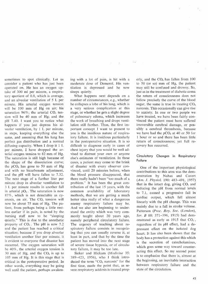
sometimes to spot clinically. Let us consider a patient who has just been operated on. He has an oxygen uptake of 300 ml per minute, a respiratory quotient of 0.8, which is average, and an alveolar ventilation of 5 L per minute. His arterial oxygen tension will be 100 mm of Hg on air; his saturation 96%; the arterial C02 tension will be 40 mm of Hg; and the pH 7.40. I want you to notice what happens if you just depress his alveolar ventilation, by 1 L per minute, in steps, keeping everything else the same, and assuming that his lung has perfect gas distribution and a normal diffusing capacity. When I drop it 1 L per minute, I have dropped the arterial oxygen tension to 82 mm of Hg. The saturation is still high because of the shape of the dissociation curve; the pC02 has gone to 50 mm of Hg and with no bicarbonate adjustment, and the pH will have fallen to 7.32. The next stage of a further liter per minute drop in alveolar ventilation to 3 L per minute results in another fall in arterial pO, . The saturation is now 87 % , which is not detectable as cyanosis, on air. The CO, tension will now be about 75 mm of Hg. The patient, from perhaps being a little restless earlier if in pain, is noted by the nursing staff now to be "sleeping quietly." This is due to the anesthetic property of C02 . The pH is now 7 .2 and the patient has reached a critical situation; because if you drop alveolar ventilation another liter per minute, it is evident to everyone that disaster has occurred. The oxygen saturation will be 40%, the arterial oxygen tension is 30 mm of Hg, and the C02 is up to 105 mm of Hg. It is this stage that is critical in the postoperative period. In other words, everything may be going well until the patient, perhaps awaken·
ing with a lot of pain, is hit with a moderate dose of Demerol. His ventilation is depressed and he now sleeps quietly.
What happens next depends on a number of circumstances, e.g., whether he collapses a lobe of his Jung, which is a very serious complication at this stage, or whether he gets a slight degree of pulmonary edema, which increases the work of breathing and drops ventilation still further. Thus, the first important concept I want to present to you is the insidious nature of respiratory failure. It is insidious particularly in the postoperative situation. It is so difficult to diagnose early in cases of chest injury that you would be well advised to distrust your own or anyone else's estimates of ventilation. In these cases, a patient may come to the brink of disaster with every observer convinced, until 20 minutes before, when the blood pressure disappeared, that there really hadn't been "too much of a problem." It has been the great contribution of the last 15 years, with the common availability of laboratory methods, that we are getting a much better idea really of what a dangerous enemy respiratory failure may be. And we also are beginning to understand the entity which was very commonly taught about 20 years ago, namely peripheral circulatory failure. The urgency of teaching about respiratory failure consists in recognizing that you can usually reverse it, at least in part, and that by the time the patient has moved into the next stage of severe tissue hypoxia, or of circulatory failure, it may be too late.
Sieker and Hickam (Medicine 35: 389-423 , 1956), who I think introduced the term "C02 narcosis" for the first time, made the point that, as severe respiratory acidosis is treated prop-
erly, and the C02 has fallen from 100 to 50 (or so) mm of Hg, the patient may still be confused and drowsy. So, just as in the treatment of diabetic coma the return of consciousness does not follow precisely the curve of the blood sugar, the same is true in treating C02 narcosis. This occasionally can give rise to anxiety. In one or two people we have treated, we have been fairly convinced the patient must have suffered irreversible cerebral damage, or possibly a cerebral thrombosis, because we have had the pC02 at 40 or 50 for 1 hour or so and there has been little return of consciousness; yet full recovery has occurred.
Circulatory Changes in Respiratory
Failure
One of the important physiological contributions to this area was the demonstration by Nahas and Cavert (Am. J. Physiol. 190: 483-491, 1957) that in the intact dog, giving C02 and reducing the pH from normal levels to 7 .1, caused a progressive fall in cardiac output, which fell almost linearly with the pH change. This was mainly due to a fall in stroke volume. Patterson (Proc. Roy. Soc. (London), Ser. B 88: 371-396, 1915) had demonstrated as early as 1915 that CO,, regardless of its pH effect, had a depressant effect on the isolated dog heart. It has also been shown that the body has a protection against this which is the secretion of catecholamines, which goes some way toward counteracting this effect. So my second point is to emphasize that there is, almost at the beginning, an inevitable interaction between respiratory failure and the state of the circulation.
43
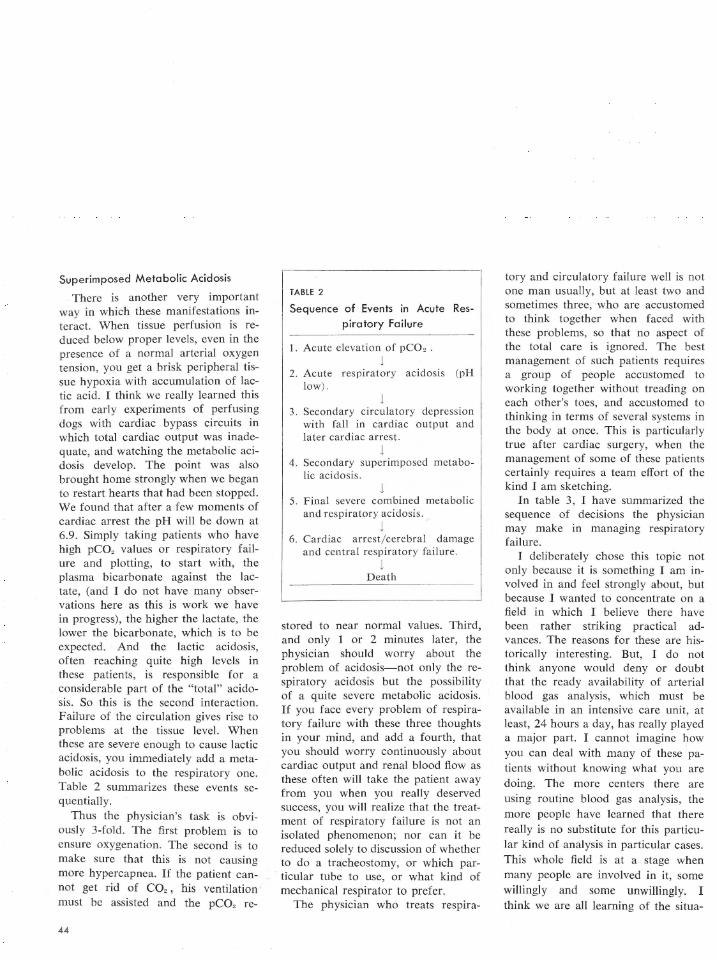
Superimposed Metabolic Acidosis
There is another very important way in which these manifestations interact. When tissue perfusion is reduced below proper levels, even in the presence of a normal arterial oxygen tension, you get a brisk peripheral tissue hypoxia with accumulation of lactic acid. I think we really learned this from early experiments of perfusing dogs with cardiac bypass circuits in which total cardiac output was inadequate, and watching the metabolic acidosis develop. The point was also brought home strongly when we began to restart hearts that had been stopped. We found that after a few moments of cardiac arrest the pH will be down at 6.9. Simply taking patients who have high pC02 values or respiratory failure and plotting, to start with, the plasma bicarbonate against the lactate, (and I do not have many observations here as this is work we have in progress), the higher the lactate, the lower the bicarbonate, which is to be expected. And the lactic acidosis, often reaching quite high levels in these patients, is responsible for a considerable part of the "total" acidosis. So this is the second interaction. Failure of the circulation gives rise to problems at the tissue level. When these are severe enough to cause lactic acidosis, you immediately add a metabolic acidosis to the respiratory one. Table 2 summarizes these events sequentially.
Thus the physician's task is obviously 3-fold. The first problem is to ensure oxygenation. The second is to make sure that this is not causing more hypercapnea. If the patient cannot get rid of CO, , his ventilation · must be assisted and the pC02 re-
44
TABLE 2
Sequence of Events in Acute Respiratory Failure
1. Acute elevation of pCO, . l
2. Acute respiratory acidosis (pH low).
l 3. Secondary circulatory depression
with fall in cardiac output and later cardiac arrest .
l 4. Secondary superimposed metabo
lic acidosis . l
5. Final severe combined metabolic and respiratory acidosis .
l 6. Cardiac arrest/ cerebral damage
and central respiratory failure. l
Death
stored to near normal values. Third, and only 1 or 2 minutes later, the physician should worry about the problem of acidosis-not only the respiratory acidosis but the possibility of a quite severe metabolic acidosis. If you face every problem of respiratory failure with these three thoughts in your mind, and add a fourth, that you should worry continuously about cardiac output and renal blood flow as these often will take the patient away from you when you really deserved success, you will realize that the treatment of respiratory failure is not an isolated phenomenon; nor can it be reduced solely to discussion of whether to do a tracheostomy, or which particular tube to use, or what kind of mechanical respirator to prefer.
The physician who treats respira-
tory and circulatory failure well is not one man usually, but at least two and sometimes three, who are accustomed to think together when faced with these problems, so that no aspect of the total care is ignored. The best management of such patients requires a group of people accustomed to working together without treading on each other's toes, and accustomed to thinking in terms of several systems in the body at once. This is particularly true after cardiac surgery, when the management of some of these patients certainly requires a team effort of the kind I am sketching.
In table 3, I have summarized the sequence of decisions the physician may make in managing respiratory failure.
I deliberately chose this topic not only because it is something I am involved in and feel strongly about, but because I wanted to concentrate on a field in which I believe there have been rather striking practical advances. The reasons for these are historically interesting. But, I do not think anyone would deny or doubt that the ready availability of arterial blood gas analysis, which must be available in an intensive care unit, at least, 24 hours a day, has really played a major part. I cannot imagine how you can deal with many of these patients without knowing what you are doing. The more centers there are using routine blood gas analysis, the more people have learned that there really is no substitute for this particular kind of analysis in particular cases. This whole field is at a stage when many people are involved in it, some willingly and some unwillingly. I think we are all learning of the situa-
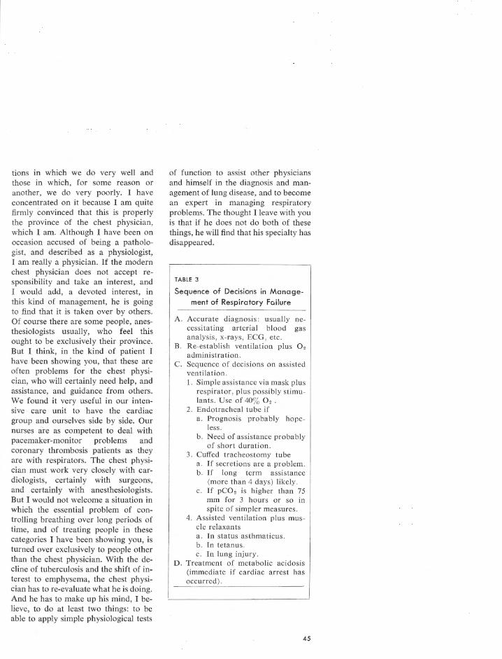
tions in which we do very well and those in which, for some reason or another, we do very poorly. I have concentrated on it because I am quite firmly convinced that this is properly the province of the chest physician, which I am. Although I have been on occasion accused of being a pathologist, and described as a physiologist, I am really a physician. If the modern chest physician does not accept responsibility and take an interest, and I would add, a devoted interest, in this kind of management, he is going to find that it is taken over by others. Of course there are some people, anesthesiologists usually, who feel this ought to be exclusively their province. But I think, in the kind of patient I have been showing you, that these are often problems for the chest physician, who will certainly need help, and assistance, and guidance from others. We found it very useful in our intensive care unit to have the cardiac group and ourselves side by side. Our nurses are as competent to deal with pacemaker-monitor problems and coronary thrombosis patients as they are with respirators. The chest physician must work very closely with cardiologists, certainly with surgeons, and certainly with anesthesiologists. But I would not welcome a situation in which the essential problem of controlling breathing over long periods of time, and of treating people in these categories I have been showing you, is turned over exclusively to people other than the chest physician. With the decline of tuberculosis and the shift of interest to emphysema, the chest physician has to re-evaluate what he is doing. And he has to make up his mind, I believe, to do at least two things: to be able to apply simple physiological tests
of function to assist other physicians and himself in the diagnosis and management of lung disease, and to become an expert in managing respiratory problems. The thought I leave with you is that if he does not do both of these things, he will find that his specialty has disappeared.
TABLE 3
Sequence of Decisions in Manage
ment of Respiratory Failure
A. Accurate diagnosis: usually necessitating arterial blood gas analysis, x-rays, ECG, etc.
B. Re-establish ventilation plus 0 2
administration. C. Sequence of decisions on assisted
ventilation. 1. Simple assistance via mask plus
respirator, plus possibly stimulants. Use of 403 02 .
2. Endotracheal tube if a. Prognosis probably hope
less . b. Need of assistance probably
of short duration. 3. Cuffed tracheostomy tube
a. If secretions are a problem. b. If long term assistance
(more than 4 days) likely. c. If pC02 is higher than 75
mm for 3 hours or so in spite of simpler measures .
4. Assisted ventilation plus muscle relaxants a. In status asthmaticus . b. In tetanus. c. In lung injury.
D . Treatment of metabolic acidosis (immediate if cardiac arrest has occurred).
45
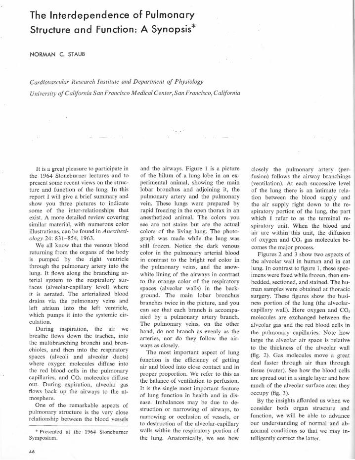
The Interdependence of Pulmonary
Structure and Function: A Synopsis*
NORMAN C. STAUB
Cardiovascular Research Institute and Department of Physiology
University of California San Francisco Medical Center, San Francisco, California
It is a great pleasure to participate in the 1964 Stoneburner lectures and to present some recent views on the structure and function of the lung. In this report I will give a brief summary and show you three pictures to indicate some of the inter-relationships that exist. A more detailed review covering similar material, with numerous color illustrations, can be found in Anesthesiology 24: 831-854, 1963.
We all know that the venous blood returning from the organs of the body is pumped by the right ventricle through the pulmonary artery into the lung. It flows along the branching arterial system to the respiratory surfaces (alveolar-capillary level) where it is aerated. The arterialized blood drains via the pulmonary veins and left atrium into the left ventricle, which pumps it into the systemic circulation.
During inspiration, the air we breathe flows down the trachea, into the multibranching bronchi and bronchioles, and then into the respiratory spaces (alveoli and alveolar ducts) where oxygen molecules diffuse into the red blood cells in the pulmonary capillaries, and C02 molecules diffuse out. During expiration, alveolar gas flows back up the airways to the atmosphere.
One of the remarkable aspects of pulmonary structure is the very close relationship between the blood vessels
* Presented at the 1964 Stoneburner Symposium.
46
and the airways. Figure 1 is a picture of the hilum of a lung lobe in an experimental animal, showing the main lobar bronchus and adjoining it, the pulmonary artery and the pulmonary vein. These lungs were prepared by rapid freezing in the open thorax in an anesthetized animal. The colors you see are not stains but are the actual colors of the living lung. The photograph was made while the lung was still frozen. Notice the dark venous color in the pulmonary arterial blood in contrast to the bright red color in the pulmonary veins, and the snowwhite lining of the airways in contrast to the orange color of the respiratory spaces (alveolar walls) in the background. The main lobar bronchus branches twice in the picture, and you can see that each branch is accompanied by a pulmonary artery branch. The pulmonary veins, on the other hand, do not branch as evenly as the arteries, nor do they follow the airways as closely.
The most important aspect of lung function is the efficiency of getting air and blood into close contact and in proper proportion. We refer to this as the balance of ventilation to perfusion. It is the single most important feature of lung function in health and in disease. Imbalances may be due to destruction or narrowing of airways, to narrowing or occlusion of vessels, or to destruction of the alveolar-capillary walls within the respiratory portion of the lung. Anatomically, we see how
closely the pulmonary artery (perfusion) follows the airway branchings (ventilation). At each successive level of the lung there is an intimate relation between the blood supply and the air supply right down to the respiratory portion of the lung, the part which I refer to as the terminal respiratory unit. When the blood and air are within this unit, the diffusion of oxygen and C02 gas molecules becomes the major process.
Figures 2 and 3 show two aspects of the alveolar wall in human and in cat lung. In contrast to figure 1, these specimens were fixed while frozen, then embedded, sectioned, and stained. The human samples were obtained at thoracic surgery. These figures show the business portion of the lung (the alveolarcapillary wall). Here oxygen and CO. molecules are exchanged between the alveolar gas and the red blood cells in the pulmonary capillaries. Note how large the alveolar air space is relative to the thickness of the alveolar wall (fig. 2). Gas molecules move a great deal faster through air than through tissue (water). See how the blood cells are spread out in a single layer and how much of the alveolar surface area they occupy (fig. 3).
By the insights afforded us when we consider both organ structure and function, we will be able to advance our understanding of normal and abnormal conditions so that we may intelligently correct the latter.
Fig. 1-Cat, X 2.4. Left lower lobe, main bronchus, and vessels at the hilum. This is a frozen, uninjected, unstained specimen. It demonstrates clearly the purplish color of partially desaturated (venous) blood in the pulmonary artery and the bright red color of well-oxygenated (arterial) blood in the pulmonary veins. The plane of section is just after the lobar bronchus enters the lung. The main bronchus is the large white opening. A small lateral branch to the apical segment of the lobe has been given off, and a large anterior branch is forming. The main bronchus continues nearly straight. Within the bronchus there is a faint circular ridging due to the cartilage in the walls. The pulmonary artery (PA) has divided, and a branch accompanies each airway. PA is one-third to one-half the diameter of the adjacent airway at all levels down to the terminal bronchiole. The pulmonary vein is larger than the artery. It is in the process of dividing. At the hilum the airway, pulmonary artery, and pulmonary vein are close together because of space limitations. This provides maximal mobility of the lung root. Note that even the largest airways and vessels are surrounded by alveolar tissue (orange background color).
Fig. 2-Human, X 100. Fixed, 10-µ section, stained. Shows red blood cells in alveolar capillaries confined to plane of wall, not bulging into airspaces as so often stated. About 50% of the alveolar septal volume is capillary lumen. Endothelium, interstitial connective tissue, and alveolar epithelium make up the remainder. Septae range from 5 to 10 µ in thickness in wellinflated lung. Human lungs were obtained at thoracic surgery. The anesthetist inflated the lung fully, then allowed it to deflate to a known airway pressure. The surgeon then clamped the edge of a lobe. At the time of clamping, pulmonary blood flow was intact. The specimens were frozen within 30 seconds of removal from the chest.
Fig. 3-Cat, X 200. Fixed, thick section, stained. Single alveolar wall in plane of focus. Individual red blood cells in alveolar capillaries are clearly seen. Normally the capillary net is not filled to capacity.
47
Energy Requirements
of Breathing*
W. T. THOMPSON, JR.
Department of Medicine
Medical College of Virginia, Richmond
The mass movement of gases into and out of the lungs is accomplished by muscular work. This requires energy expenditure, caloric consumption, oxygen utilization, and carbon dioxide production. Inspiration is always an active process, enlarging the volume of the thorax, thereby increasing the negative pressure so that air flows into the lungs. Expiration is ordinarily passive but may require muscular work.
The position of the chest wall at rest is a mechanically neutral point at which the tendency of the chest wall to expand is balanced by the tendency of the lungs to recoil. This is the end expiratory level or midposition, and the lung volume at this point is termed the functional residual capacity. An increase in chest size can be achieved only by exerting pressure greater than the elastic forces of the lungs. In addition, airway resistance to airflow and nonelastic tissue resistance must be overcome. In the normal individual, 60 to 70% of the total ventilatory work is required for overcoming elastic forces, the remainder for overcoming nonelastic resistance, largely due to airflow.
Expiration is passive in the normal individual during quiet breathing, the energy stored in the elastic lung tissue during inspiration being sufficient.
The diaphragm is the major muscle of inspiration and is probably always active even with so-called thoracic breathing. It arises from the xiphoid process, from the inner surface of the last six ribs and their cartliages, and from the lumbar vertebrae. By virtue of its position and attachments, it not only increases the vertical dimension of the chest by a downward movement, but also increases the transverse diameter by flaring the lower ribs.
The intercostal muscles are second in importance to the diaphragm. The external intercostal fibers run downward and forward and the internal intercostal fibers are directed upward and forward to the next rib, so that contraction elevates the rib with resulting increase in the anteroposterior diameter, transverse diameter, or both. Studies by Koepke et al. (1958) showed that the 1st intercostals are active in quiet breathing of most individuals. With increased ventilation, there is increased utilization of the intercostals, so that the majority of individuals use
* Presented at the 1964 Stoneburner Symposium.
48
all intercostal muscles when ventilation requirements approach 50% of the predicted vital capacity.
The diaphragm and intercostals are the only respiratory muscles used by the normal individual under ordinary circumstances. Other muscles are mobilized when ventilatory demands are increased, either because of increase in total ventilation in the normal individual or because of altered physiology of respiration in disease states. The socalled accessory muscles of respiration include particularly the scalenes, the sternomastoid, and the trapezius.
The scalene muscles arise from the transverse processes of the cervical vertebrae and insert in the 1st and 2nd ribs. They serve to stabilize and elevate the 1st and 2nd ribs. They are not ordinarily employed in quiet respiration.
Although a well trained subject was able to breathe 60 L per minute without electromyographic evidence of scalene activity, these muscles are ordinarily brought into play when an intrathoracic pressure of -5 to -6 cm of H20 is exerted and chest volume is increased by 800 to 900 ml of air. On rapid inspiration they are brought into action at the very beginning of inspiration (Thompson, Patterson, and Shapiro, 1964).
While many muscles of the thorax may be used in severe dyspnea, only the sternomastoid and the trapezius will be considered. The former arises from the manubrium and the sternal end of the clavicle and inserts at the mastoid process and lateral half of the nuchal line. It can assist in lifting the thorax. Campbell (1955) showed that there was never any detectable activity of this muscle in any subject during quiet respiration, but that increasing intensity of contraction was exhibited as intrathoracic pressures progressed from -10 to -50 cm of H,O.
The trapezius arises from the occiput, the ligamentum nuchae, and the spines of the last cervical and thoracic vertebrae. It inserts into the acromial 3rd of the clavicle, and the scapula. The upper fibers tend to lift the chest.
These muscles do not function with equal effectiveness as muscles of respiration, and they are described in order of decreasing efficiency. The diaphragm and the intercostals are peculiarly suited for their task of increasing the chest volume. The scalene muscles have a direct pull to lift the 1st rib. The sternomastoid and trape-
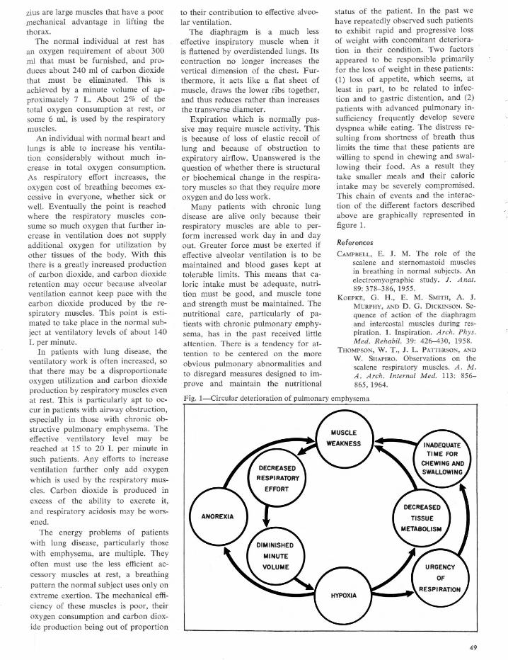
zius are large muscles that have a poor mechanical advantage in lifting the thorax.
The normal individual at rest has an oxygen requirement of about 300 ml that must be furnished, and produces about 240 ml of carbon dioxide that must be eliminated. This is achieved by a minute volume of approximately 7 L. About 2 % of the total oxygen consumption at rest, or some 6 ml, is used by the respiratory muscles.
An individual with normal heart and lungs is able to increase his ventilation considerably without much increase in total oxygen consumption. As respiratory effort increases, the oxygen cost of breathing becomes excessive in everyone, whether sick or well. Eventually the point is reached where the respiratory muscles consume so much oxygen that further increase in ventilation does not supply additional oxygen for utilization by other tissues of the body. With this there is a greatly increased production of carbon dioxide, and carbon dioxide retention may occur because alveolar ventilation cannot keep pace with the carbon dioxide produced by the respiratory muscles. This point is estimated to take place in the normal subject at ventilatory levels of about 140 L per minute.
In patients with lung disease, the ventilatory work is often increased, so that there may be a disproportionate oxygen utilization and carbon dioxide production by respiratory muscles even at rest. This is particularly apt to occur in patients with airway obstruction, especially in those with chronic obstructive pulmonary emphysema. The effective ventilatory level may be reached at 15 to 20 L per minute in such patients. Any efforts to increase ventilation further only add oxygen which is used by the respiratory muscles. Carbon dioxide is produced in excess of the ability to excrete it, and respiratory acidosis may be worsened.
The energy problems of patients with lung disease, particularly those with emphysema, are multiple. They often must use the less efficient accessory muscles at rest, a breathing pattern the normal subject uses only on extreme exertion. The mechanical efficiency of these muscles is poor, their oxygen consumption and carbon dioxide production being out of proportion
to their contribution to effective alveolar ventilation.
The diaphragm is a much less effective inspiratory muscle when it is flattened by overdistended lungs. Its contraction no longer increases the vertical dimension of the chest. Furthermore, it acts like a flat sheet of muscle, draws the lower ribs together, and thus reduces rather than increases the transverse diameter.
Expiration which is normally passive may require muscle activity. This is because of loss of elastic recoil of lung and because of obstruction to expiratory airflow. Unanswered is the question of whether there is structural or biochemical change in the respiratory muscles so that they require more oxygen and do less work.
Many patients with chronic lung disease are alive only because their respiratory muscles are able to perform increased work day in and day out. Greater force must be exerted if effective alveolar ventilation is to be maintained and blood gases kept at tolerable limits. This means that caloric intake must be adequate, nutrition must be good, and muscle tone and strength must be maintained. The nutritional care, particularly of patients with chronic pulmonary emphysema, has in the past received little attention. There is a tendency for attention to be centered on the more obvious pulmonary abnormalities and to disregard measures designed to improve and maintain the nutritional
status of the patient. In the past we have repeatedly observed such patients to exhibit rapid and progressive loss of weight with concomitant deterioration in their condition. Two factors appeared to be responsible primarily for the loss of weight in these patients: (1) loss of appetite, which seems, at least in part, to be related to infection and to gastric distention, and (2) patients with advanced pulmonary insufficiency frequently develop severe dyspnea while eating. The distress resulting from shortness of breath thus limits the time that these patients are willing to spend in chewing and swallowing their food. As a result they take smaller meals and their caloric intake may be severely compromised. This chain of events and the interaction of the different factors described above are graphically represented in figure l.
References
CAMPBELL, E. J. M. Tbe role of the scalene and sternomastoid muscles in breathing in normal subjects. An electromyographic study. J. Anat. 89: 378-386, 1955.
KOEPKE, G. H., E. M. SMITH, A. J . MURPHY, AND D. G. DICKINSON. Sequence of action of the diaphragm and intercostal muscles during respiration. 1. Inspiration. Arch. Phys. Med. Rehabil. 39: 426-430, 1958.
THOMPSON, W. T., J . L. PATTERSON, AND W. SHAPIRO. Observations on the scalene respiratory muscles. A. M . A . Arch. Internal Med. 113: 856-865, 1964.
Fig. 1-Circular deterioration of pulmonary emphysema
49
Relative Incidence of Pulmonary
Emphysema among Negroes
and Whites of Both Sexes*
EDWARD S. RAY
Department of Medicine, Medical College of Virginia, Richmond
A striking clinical feature of emphysema is its predominance among white males, and its relative infrequency among Negro females. This is true, at least in the state of Virginia, as shown by recent figures of death rates from the disease (table 1).
To further evaluate the incidence of this disease, the records of patients at the Medical College of Virginia were reviewed. Included in this study were hospital and clinic admissions for the years 1961 and 1962, and private outpatients seen during the years 1960, 1961, and 1962. The MCV hospitals admit approximately 35,000 patients yearly with a ratio of 51 % white and 49% Negro. The out-patient clinics admit about 90,000 patients annually with a ratio of 28% white and 72% Negro.
The criteria for making a diagnosis of emphysema were: shortness of breath as a major complaint, generalized suppression of breath sounds and expiratory wheezing, a I-second forced expiratory volume of less than 70% of the total vital capacity, and x-ray evi-
* Presented at the 1964 Stoneburner Symposium.
50
dence of limited motion and depression of the diaphragms. Only patients fulfilling these criteria, but showing no evidence of other lung diseases, were included in this study (370).
Smoking Habits
The great majority of the patients (315) gave a history of cigarette smoking (table 2). The sex and race distribution of these figures (table 3) almost parallels that of death rates from the disease. The data on smoking habits of these patients could not be evaluated accurately as the habit varied in individuals from year to year, and some patients had difficulty in recalling accurately their precise smoking habits over the years. Some individuals tended to underestimate the number of years they had been smoking, and some had reduced their smoking in recent years because of progressive respiratory symptoms. Of the 315 patients with emphysema, most had smoked for 30 years or longer (table 4). A great majority of these patients smoked a package or more of cigarettes daily (table 5). Thus there appears to be a correlation between the degree and duration of cigarette smoking and the incidence of significant pulmonary emphysema.
Cigarette Smoking among General Hospital and Clinic Patients
Because of the marked differences in the incidence of emphysema according to sex and race, differences in the smoking habits of these groups were examined. A survey was made of the smoking habits of patients on the general hospital wards and in the OutPatient Clinic. Only patients who were 40 years of age or older were included, as a pattern of smoking is not usually developed before this age. A total of 600 individuals were interviewed, 150 of each sex and race (table 6). There was a significant difference in the smoking habits of the two sexes and a smaller difference between the two races; white males being the heaviest smokers, followed by Negro males and white females (p <0.01 for each combination); Negro females smoked the least. These differences in smoking habits correspond to the difference in frequency of emphysema between the two sexes, but do not explain the greater difference in incidence between white and Negro males. Possibly the population selection was biased, as only clinic and hospital patients were interviewed. Possibly also other factors, such as occupation or host reaction, are important.
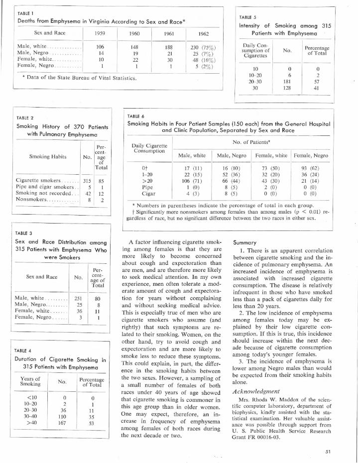
TABLE 1
Deaths from Emphysema in Virginia According to Sex and Race* TABLE 5
Intensity of Smoking among 315
Patients with Emphysema Sex and Race 1959 1960 1961 1962
Male, white . .. . ··· ·· ·· . . . 106 148 188 230 (753) Male, Negro . . .. ········ . 14 19 21 25 (73) Female, white. . . ... . .. . .. JO 22 30 48 (163) Female, Negro. . . .. .. . .. I I 1 5 (23)
Daily Con- Percentage sumption of No. of Total Cigarettes
10 0 0
* Data of the State Bureau of Vital Statistics. 10-20 6 2 20-30 181 57
TABLE 2
Smoking History of 370 Patients
with Pulmonary Emphysema
Smoking Habits
Cigarette smokers . . . Pipe and cigar smokers . . Smoking not recorded . . . Nonsmokers.
TABLE 3
Percent
No. age of
Total
315 85 5
42 12 8 2
Sex and Race Distribution among
315 Patients with Emphysema Who
were Smokers
Sex and Race No. Percent
age of Total
- ---- ---- ------
Male, white. . . . . . . . . 251 80 Male, Negro . .. . .. . . . 25 8 Female, white. . . . . . . 36 11 Female, Negro. . . . . 3 1
TABLE 4
Dura tion of Cigarette Smoking in
3 15 Patients with Emphysema
Years of Smoking
< 10 10-20 20-30 30-40
>40
No.
0 2
36 110 167
Percentage of Total
0 1
11 35 53
30 128 41
TABLE 6
Smoking Habits in Four Patient Samples (150 each) from the G eneral Hospital and Clinic Population, Separated by Sex and Race
Daily Cigarette No. of Patients*
Consumption Male, white Male, Negro Female, white Female, Negro
Ot 17 (11) 16 (JO ) 73 (50) 93 (62) 1-20 22 (15) 52 (36) 32 (20) 36 (24) > 20 106 (71 ) 66 (44) 43 (30) 21 (14) Pipe 1 (0) 8 (5) 2 (0 ) 0 (0) Cigar 4 (3 ) 8 (5) 0 (0 ) 0 (0)
* Numbers in parentheses indicate the percentage of total in each group. t Significantly more nonsmokers among females than among males (p < 0.01) re
gardless of race, but no significant difference between the two races in either sex.
A factor influencing cigarette smoking among females is that they are more likely to become concerned about cough and expectoration than are men, and are therefore more likely to seek medical attention. In my own experience, men often tolerate a moderate amount of cough and expectoration for years without complaining and without seeking medical advice. This is especially true of men who are cigarette smokers who assume (and rightly) that such symptoms are related to their smoking. Women, on the other hand, try to avoid cough and expectoration and are more likely to smoke less to reduce these symptoms. This could explain, in part, the difference in the smoking habits between the two sexes. However, a sampling of a small number of females of both races under 40 years of age showed that cigarette smoking is commoner in this age group than in older women. One may expect, therefore, an increase in frequency of emphysema among females of both races during the next decade or two.
Summary
1. There is an apparent correlation between cigarette smoking and the incidence of pulmonary emphysema. An increased incidence of emphysema is associated with increased cigarette consumption. The disease is relatively infrequent in those who have smoked less than a pack of cigarettes daily for less than 20 years.
2. The low incidence of emphysema among females today may be explained by their low cigarette consumption. If this is true, this incidence should increase within the next decade because of cigarette consumption among today's younger females.
3. The incidence of emphysema is lower among Negro males than would be expected from their smoking habits alone.
Acknowledgment Mrs. Rhoda W. Maddox of the scien
tific computer laboratory, department of biophysics, kindly assisted with the statistical examination. Her valuable assistance was possible through support from U. S. Public Health Service Research Grant FR 00016-03.
51
Pulmonary Surfactant
and its Relation to Disease*
SAMI I. SAIDt
Department of Medicine, Medical College of Virginia, Richmond
Perhaps the most important advance in respiratory physiology during the past decade is the discovery of the role of surface phenomena. This has been in large measure the result of pioneer work by Pattie, in England, and Clements, in the United States.
The following is a simplified and brief review of present knowledge of surface tension, as it relates to normal and abnormal pulmonary function. (For further reading on the subject pelase refer to these recent reviews: Clements, 1962a and 1962b, Pattie, 1958, 1961 , and 1965, and Mead, 1960.)
First, what is surface tension? When a liquid is in contact with the air, the molecules at the surface of the liquid will be under tension (fig. 1). This is because, while the molecules away from the surface are attracted equally by other molecules from all directions, those at the surface are influenced only by molecules below and to the side, but none above. The net effect is that the surface molecules tend to be pulled down, and therefore, the surface tends to get smaller.
And because surface tension is a force acting in the same plane as the surface, e.g., like the pull on the cord of a window curtain, it has units of force per length, e.g., gm per cm or dynes per cm, whereas pressure has the units of force per area, e.g. , gm per cm2 or pounds per square inch.
Now, what could this have to do with what goes on in the lungs? We will begin to appreciate the link when we consider the case of a gas-liquid interface that has a spherical shape, like that of an alveolus. A soap bubble is a good example. Here, the tendency of surface tension to shrink the surface will ultimately result in collapse of the bubble. And it can be shown that the total forces tending to empty the bubble will amount to twice the tension in the wall divided by the radius of curvature. This relationship is known as the law of Laplace, and it appears to hold for alveoli just as well as it holds for soap bubbles. It means simply that for a sphere, the surface forces will increase if the tension in the wall increases or if the radius of curvature decreases. It
* Presented at the 1964 Stoneburner Symposium.
t Recipient of U. S. Public Health Service Career Development A ward HEK3-18,432 from the National Heart Institute.
52
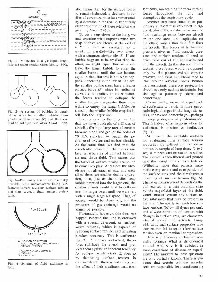
0 GAS
SURFACE - -..,. ----~ - - - - ....:F.'l~-= - -f~E~----~~--rs_-
~~_e==_4~~~~~~-~-;-;
f{fl!i{{{iil~~ ---- -- - --- - ------
Fig. I-Molecules at a gas-liquid interface are under tension (after Mead, 1960).
Fig. 2-A system of bubbles in parallel is unstable; smaller bubbles have greater surface forces (P) and therefore tend to collapse first (after Mead, 1960).
Fig. 3-Pulmonary alveoli are inherently unstable, but a surface-active lining (surfactant) lowers alveolar surface tension and thus protects them against atelectasis.
CAPILLARY
t HYDROSTATIC PRESSURE ALY. TISS. FLUID OSM. PRESSURE SURFACE TENSION
i PLASMA COLLOID OSMOTI C PRESSURE SURFACTANT
Fig. 4-Schema of fluid exchange in lung.
also means that, for the surface forces to remain balanced, a decrease in radius of curvature must be counteracted by a decrease in tension. A beautifully clear presentation of these relations was given by Mead (1960).
To get a step closer to the lung, we now examine what happens when two soap bubbles are blown at the end of a Y-tube and are arranged, so to speak, in parallel-like two alveoli with a common airway (fig. 2). If one bubble happens to be smaller than the other, we might expect that air would leave the larger bubble to enter the smaller bubble, until the two became equal in size. But this is not what happens. According to the law of Laplace, the smaller bubble must have a higher surface force (P), since its radius of curvature is smaller. In other words, the forces tending to collapse the smaller bubble are greater than those trying to empty the larger bubble. As a result, the smaller bubble empties itself into the larger one.
Turning now to the lung, we find that we have hundreds of millions of alveoli, offering a large area of contact between blood and gas (of the order of 70 M2 ) , sufficient to permit the exchange of oxygen and carbon dioxide. At the same time, we find that the alveoli also present, on their inner surface, a large area of contact between air and tissue fluid. This means that the forces of surface tension are bound to come into play. And since the alveoli are not all equal in size, and since all of them get smaller during expiration, then, just as the smaller soap bubble emptied into the larger one, the smaller alveoli would tend to collapse into the larger ones, until we were left with a single large air space. That, of course, would be disastrous, for the processes of gas exchange would no longer be possible.
Fortunately, however, this does not happen, because the lung is endowed with a special detergent, or surfaceactive material, which is capable of reducing surface tension and adjusting it when necessary. This is surfactant (fig. 3). Pulmonary surfactant, therefore, stabilizes the alveoli and protects them against an inherent tendency for collapse or atelectasis. It does so by decreasing surface tension in smaller alveoli, thereby balancing out the effect of their smallness and, con-
sequently, maintaining uniform surface forces throughout the lung and throughout the respiratory cycle.
Another important function of pulmonary surfactant is explained in figure 4. Normally, a delicate balance of fluid exchange exists between alveoli on the one hand, and capillaries on the other; only a thin fluid film lines the alveoli. The forces of hydrostatic pressure, alveolar fluid osmotic pressure, and surface tension, all tend to drive fluid out of the capillaries and into the alveoli. In the absence of surfactant, these forces would be opposed only by the plasma colloid osmotic pressure, and fluid and blood tend to leak into the alveolar spaces. Pulmonary surfactant, therefore, protects the alveoli not only against atelectasis, but also against pulmonary edema and hemorrhage.
Consequently, we would expect lack of surfactant to result in three major pathologic changes in the lung; atelectasis, edema and hemorrhage-perhaps in varying degrees of predominance. This is indeed what happens when the surfactant is missing or ineffective (fig. 5).
At present, the available methods for evaluating lung surface tension properties are indirect and not quantitative. A sample of lung tissue (1 to 3 gm) is minced and extracted in saline. The extract is then filtered and poured onto the trough of a surface balance (Wilhelmy type) that permits automatic compression and re-expansion of the surface area and the simultaneous recording of surface tension (fig. 6) . The latter measurement depends on the pull exerted on a thin platinum strip by the superficial layer of the fluid, which should contain any surface-active substances that may be present in the lung. The ability to reach low surface tensions (below 10 dynes per cm), and a wide variation of tension with changes in surface area, are characteristic of normal lung extracts. Lungs with abnormal surface properties give extracts that fail to reach a low surface tension even on maximal compression.
How is pulmonary surfactant normally formed? What is its chemical nature? And why is it deficient in some conditions of disease or experiment? The answers to these questions are only partially known. There is evidence that certain granular alveolar cells are responsible for manufacturing
53
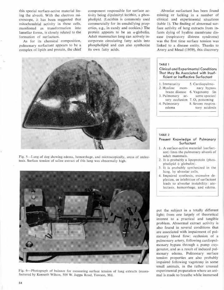
this special surface-active material lining the alveoli. With the electron microscope, it has been suggested that mitochondrial activity in these cells, manifested as transformation into lamellar forms, is closely related to the formation of surfactant.
As for its chemical composition, pulmonary surfactant appears to be a complex of lipids and protein, the chief
component responsible for surface activity being dipalmityl lecithin, a phospholipid. (Lecithin is commonly used commercially for its emulsifying properties, e.g., in candy and cookies.) The protein appears to be an a-globulin. Adult mammalian lung can actively incorporate circulating fatty acids into phospholipid and can also synthesize its own fatty acids.
Fig. 5-Lung of dog showing edema, hemorrhage, and microscopically, areas of atelectasis. Surface tension of saline extract of this lung was abnormally high.
Fig. 6-Photograph of balance for measuring surface tension of lung extracts (manufactured by Kenneth Wilson, 508 W. Joppa Road, Towson, Md.
54
Alveolar surfactant has been found missing or lacking in a number of clinical and experimental situations (table 1). The finding of abnormal surface activity of lung extracts from infants dying of hyaline membrane disease (respiratory distress syndrome) was the first time surface tension was linked to a disease entity. Thanks to Avery and Mead (1959), this discovery
TABLE 1
Clinical and Experimental Conditions That May Be Associated with Insuf-
ficient or Ineffective Surfactant
1. Immaturity 2. Hyaline mem
brane disease 3. Pulmonary ar
tery occlusion 4. Pulmonary
edema
TABLE 2
5. Cardiopulmonary bypass
6. Vagotomy (in some species)
7. O, poisoning 8. Severe respira
tory acidosis
Present Knowledge of Pulmonary Surfactant
1. A surface-active material (surfactant) lines the pulmonary alveoli of adult mammals.
2. It is probably a lipoprotein (phospholipid +globulin) .
3. It is probably synthesized in the lung , by alveolar cells.
4. Impaired synthesis , excessive depletion, or inhibition of surfactant leads to alveolar instability : atelectasis , hemorrhage, and edema.
put the subject in a totally different light; from one largely of theoretical interest to a practical and tangible problem. Abnormal extract activity is also found in several conditions that are associated with impairment of pulmonary blood flow: occlusion of a pulmonary artery, following cardiopulmonary bypass through a pump oxygenator, and as a result of induced pulmonary edema. Pulmonary surface tension properties are also probably impaired following vagotomy in some small animals, in the rather unusual experimental preparation where an animal is made to breathe while immersed
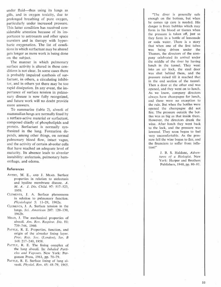
under fluid-thus using its lungs as gills, and in oxygen toxicity, due to prolonged breathing of pure oxygen, particularly under increased pressure. This latter condition has received considerable attention because of its importance to astronauts and other space travelers and in therapy with hyperbaric oxygenation. The list of conditions in which surfactant may be altered gets longer as more work is being done on the subject.
The manner in which pulmonary surface activity is altered in these conditions is not clear. In some cases there is probably impaired synthesis of surfactant, in others, a circulating inhibitor, and in others yet there may be too rapid dissipation. In any event, the importance of surface tension in pulmonary disease is now fully recognized, and future work will no doubt provide more answers.
To summarize (table 2), alveoli of mammalian lungs are normally lined by a surface-active material or surfactant, composed chiefly of phospholipids and protein. Surfactant is normally synthesized in the lung. Formation depends, among other things, on normal pulmonary blood flow, intact vagus, and the activity of certain alveolar cells that have reached an adequate level of maturity. Its absence leads to alveolar instability: atelectasis, pulmonary hemorrhage, and edema.
References
AVERY, M. E., AND J. MEAD. Surface properties in relation to atelectasis and hyaline membrane disease. A. M. A. J. Dis. Child. 97: 517-523, 1959.
CLEMENTS, J . A. Surface phenomena in relation to pulmonary function. Physiologist 5: 11-28, 1962a.
CLEMENTS, J . A. Surface tension in the lungs. Sci. American 207: 120-130, 1962b.
MEAD, J . The mechanical properties of alveoli. Am. Rev. Respirat. Dis. 81: 739-746, 1960.
PATTLE, R. E. Properties, function, and origin of the alveolar lining layer. Proc. Roy. Soc. (London), Ser. B 148: 217-240, 1958.
PATTLE, R. E. The lining complex of the lung alveoli . In: Inhaled Particles and Vapours. New York: Pergamon Press, 1961, pp. 70-79.
PATTLE, R. E. Surface lining of lung alveoli . Physiol. R ev. 45: 48-79, 1965.
"The diver is generally safe enough on the bottom, but when he comes up care is needed. His danger is from bubbles which may form in his blood or tissues when the pressure is taken off, just as they form in a bottle of lemonade or soda water. There is a story that when one of the first tubes was being driven under the Thames, the directors of the company celebrated its arrival under the middle of the river by having lunch in the tunnel. They went into an air lock, the steel door was shut behind them, and the pressure raised till it reached that in the end section of the tunnel. Then a door at the other end was opened, and they went on to lunch. As we know, company directors always have champagne for lunch, and these were no exception to the rule. But when the bottles were opened the champagne did not fizz. The pressure outside the bottles was as big as that inside them. However, the directors drank the wine. After lunch they went back to the lock, and the pressure was lowered. They soon began to feel very uncomfortable. As the pressure fell the wine began to fizz, and the financiers to suffer from inflation!"
J. B. S. Haldane, Adventures of a Biologist. New York: Harper and Brothers Publishers, 1940, pp. 80-81.
55
Books
Curiosities of Medicine. An Assembly of Medical Diversions. 1552-1962. Berton Roueche, ed. New York: Berkeley Publishing Corporation, 1964. 254 pp. $.60.
The welcome appearance in paperback form of the 1963 Little, Brown and Company edition should guarantee a wide audience for this collection of medical curiosa. The 21 selections, with introductory notes by the editor, range from a description of the sixteenth century's mysterious "Sweating Sickness" by John Caius to the 1962 JAMA paper on "Glue Sniffing in Children" by Glaser and Massengale. In between, the epidemiological classics of Panum (measles, 1847), Snow (cholera, 1854), Budd (typhoid, 1873), and Pickles (pleurodynia, 1939) vie with Matthew Carey's work on the 1793 Philadelphia yellow fever outbreak and William Beaumont's Experiments and Observations on the Gastric Juice and the Physiology of Digestion (1833).
Two papers from the 1961 Journal of the American Medical Association and the New England Journal of Medicine deal with "Stomach Cancer in Iceland" and "The Case of the Perilous Prune Pit," a refreshing autobiographical note on the small bowel meanderings of a fruit's inner core. Samuel Hopkins Adams, a layman who had the unique honor of becoming an associate member of the AMA, discusses the vagaries of Latrodectus mactans-the black widow spiderand Ashley Montagu, an anthropologist, reviews the peculiar mammalian habit of yawning. Cardiology and psychiatry each have two papers to their credit while an extract from Dubos' The White Plague (1952) shows how consumption was regarded during the Romantic Era of the 19th century. Readers interested in philosophy should like Dale's article presenting
56
the medical biography of Immanuel Kant.
Mr. Roueche, whose Eleven Blue M en1 has received popular acclaim, is author of the New Yorker's "Annals of Medicine." He received a 1961 Albert Lasker Award for his work-an indication of the clarity and accuracy of his writing. In his Curiosities of Medicine, Mr. Roueche has produced not only a most readable and enjoyable volume but also a work of scientific merit from both the historical and contemporary viewpoints.
FrederickJ.Spencer,M.B.,B.S.,M.P.H. Professor and Chairman Department of Preventive Medicine Medical College of Virginia
1 Roueche, Berton. Eleven Blue Men. New York: Berkeley Publishing Corporation, 1955. 189 pp. $.60.
Body Fluids and the Acid-Base Balance. Halvor N. Christensen. Philadelphia: W. B. Saunders, 1964. 506 pp. $6.50.
The rapid growth of scientific information has made it increasingly difficult for many teaching institutions to do justice to a subject matter in the lecture hall and the student laboratory. As a result, there has to be a certain amount of selection of topics, and at times, some degree of superficiality. More and more study "in depth" must be done by the student in the form of "homework." The conventional way of doing this is by making a thorough study of voluminous textbooks and monographs. Another method is by the use of a self study program. As the student proceeds in his studies, he is required to answer questions and solve problems. As a reward for having given the correct answers, he may continue with the program.
Body Fluids and the Acid-Base Bal-
ance, by Halvor N. Christensen, is such a program of self study written for students of the biological and medical sciences. As any preclinical and clinical lecturer can testify, students at all levels of training all too often encounter difficulties in comprehending biological acid-base problems. In the view of Dr. Christensen, this may result from the "presence of unexpected deficiencies, often minor, over which the student nevertheless cannot easily leap." This self study program, hopefully, may correct the situation. This volume of 506 pages with 1,100 "items," i.e., problems, simple and more difficult ones, covers all the material that any accredited medical school offers its students in this area: pH and Dissociation, Sodium and Chloride Distribution, Potassium and the Cellular Compartment, Calcium and Phosphorus, Gas Transport, Metabolic and Respiratory Aspects of Neutrality Regulation, and Renal Correction of the Neutrality. Written by a very competent biochemist (Christensen is professor and chairman of the department of biochemistry at the University of Michigan), the emphasis of the book is on understanding fluid and acid-base balance in the normal subject. No other subject matter cuts more deeply into all the biological and medical specialties than fluid balance and acid-base regulation. Since this self study program already has stood the test prior to its publication, it is hoped that preclinical and clinical teachers will familiarize themselves with this book and recommend it strongly to their students.
Ernst G. Huf, Ph.D., M.D. Professor of Physiology Medical College of Virginia
Respiratory Function in Disease. An Introduction to the Integrated Study of the Lung. David V. Bates and Ronald
V. Christie. Philadelphia: W. B. Saunders Company, 1964, 566 pp. $15.50.
Doctors Bates and Christie have written a much needed book. Their text, directed chiefly to the internist and the chest physician, is an up-to-date and comprehensive review of respiratory function alterations in disease, correlated with clinical , x-ray, and pathological findings.
The book opens with a brief analysis of methods for evaluation of different aspects of pulmonary function, and a summary of present knowledge of normal structure and function, including a discussion of variations due to posture, age, exercise, and altitude. Then follow several particularly strong chapters on airway diseases (spasmodic asthma, chronic bronchitis, and emphysema), and others on pulmonary cysts, bronchiectasis, atelectasis, diffuse interstitial fibrosis , sarcoidosis, acute infections, and tuberculosis. Separate treatment is also given to pulmonary function in diseases of the chest cage, in heart, collagen diseases, malignancies, and a few other less common entities. The last chapters are devoted to considerations of physiological principles in the management of respiratory failure, and in the evaluation of dyspnea and disability.
The book reads easily, is well indexed by author and subject, and has a wealth of references on practically every topic. My own feeling is that with a Comroe1 under one arm and a Bates and Christie under the other, teachers and practitioners of chest medicine should be extremely well prepared for their tasks.
Sarni I. Said, M.D .
'Comroe, J. H., Jr. , R. E. Forster, II, A. B. Dubois, W. A. Briscoe, and E. Carlsen. The Lung. Clinical Physiology and Pulmonary Function Tests (2nd ed.). Chicago: Year Book Medical Publishers, 1962.
57
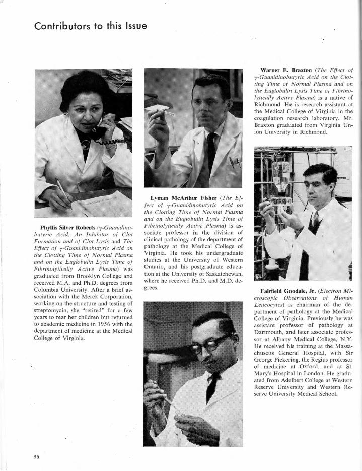
Contributors to this Issue
Phyllis Silver Roberts ( y-G uanidinobutyric Acid: An Inhibitor of Clot Formation and of Clot Lysis and The Effect of y-Guanidinobutyric Acid on the Clotting Time of Normal Plasma and on the Euglobulin Lysis Time of Fibrinolytically Active Plasma) was graduated from Brooklyn College and received M.A. and Ph.D. degrees from Columbia University. After a brief association with the Merck Corporation, working on the structure and testing of streptomycin, she "retired" for a few years to rear her children but returned to academic medicine in 1956 with the department of medicine at the Medical College of Virginia.
58
Lyman McArthur Fisher (The Effect of y-Guanidinobutyric Acid on the Clotting Time of Normal Plasma and on the Euglobulin Lysis Time of Fibrinolytically Active Plasma) is associate professor in the division of clinical pathology of the department of pathology at the Medical College of Virginia. He took his undergraduate studies at the University of Western Ontario, and his postgraduate education at the University of Saskatchewan, where he received Ph.D. and M.D. degrees.
Warner E. Braxton (The Effect of y-Guanidinobutyric Acid on the Clotting Time of Normal Plasma and on the Euglobulin Lysis Time of Fibrinolytically Active Plasma) is a native of Richmond. He is research assistant at the Medical College of Virginia in the coagulation research laboratory. Mr. Braxton graduated from Virginia Union University in Richmond.
Fairfield Goodale, Jr. (Electron Microscopic Observations of Human Leucocytes) is chairman of the department of pathology at the Medical College of Virginia. Previously he was assistant professor of pathology at Dartmouth, and later associate professor at Albany Medical College, N.Y. He received his training at the Massachusetts General Hospital, with Sir George Pickering, the Regius professor of medicine at Oxford, and at St. Mary's Hospital in London. He graduated from Adelbert College at Western Reserve University and Western Reserve University Medical School.
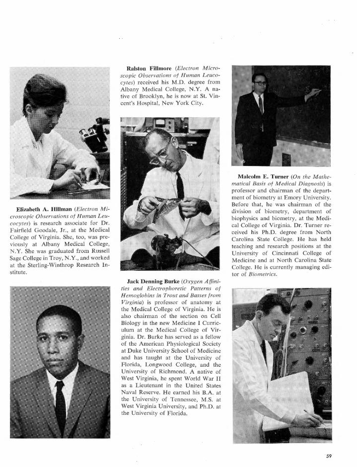
Elizabeth A. Hillman (Electron Microscopic Observations of Human Leucocytes) is research associate for Dr. Fairfield Goodale, Jr., at the Medical College of Virginia. She, too, was previously at Albany Medical College, N.Y. She was graduated from Russell Sage College in Troy, N.Y., and worked at the Sterling-Winthrop Research Institute.
Ralston Fillmore (Electron Microscopic Observations of Human L eucocytes) received his M.D. degree from Albany Medical College, N.Y. A native of Brooklyn, he is now at St. Vincent's Hospital, New York City.
Jack Denning Burke (Oxygen Affinities and Electrophoretic Patterns of Hemoglobins in Trout and Basses from Virginia) is professor of anatomy at the Medical College of Virginia. He is also chairman of the section on Cell Biology in the new Medicine I Curriculum at the Medical College of Virginia. Dr. Burke has served as a fellow of the American Physiological Society at Duke University School of Medicine and has taught at the University of Florida, Longwood College, and the University of Richmond. A native of West Virginia, he spent World War II as a Lieutenant in the United States Naval Reserve. He earned his B.A. at the University of Tennessee, M.S. at West Virginia University, and Ph.D. at the University of Florida.
Malcolm E. Turner (On the Mathematical Basis of Medical Diagnosis) is professor and chairman of the department of biometry at Emory University. Before that, he was chairman of the division of biometry, department of biophysics and biometry, at the Medical College of Virginia. Dr. Turner received his Ph.D. degree from North Carolina State College. He has held teaching and research positions at the University of Cincinnati College of Medicine and at North Carolina State College. He is currently managing editor of Biometrics.
·59
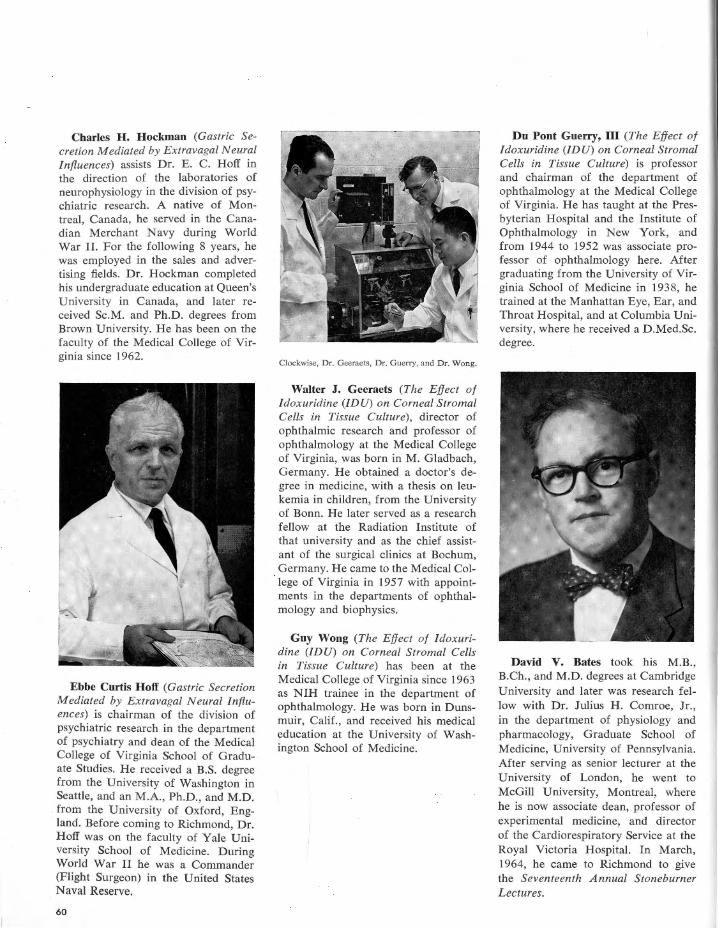
Charles H. Hockman (Gastric Secretion Mediated by Ex travagal Neural Infiuences) assists Dr. E. C. Hoff in the direction of the laboratories of neurophysiology in the division of psychiatric research. A native of Montreal , Canada, he served in the Canadian Merchant Navy during World War II. For the following 8 years, he was employed in the sales and advertising fields. Dr. Hockman completed his undergraduate education at Queen's University in Canada, and later received Sc.M. and Ph.D. degrees from Brown University. He has been on the faculty of the Medical College of Virginia since 1962.
Ebbe Curtis Hoff (Gastric Secretion Mediated by Extravagal Neural Infiuences) is chairman of the division of psychiatric research in the department of psychiatry and dean of the Medical College of Virginia School of Graduate Studies. He received a B.S. degree from the University of Washington in Seattle, and an M.A., Ph.D., and M.D. from the University of Oxford, England. Before coming to Richmond, Dr. Hoff was on the faculty of Yale University School of Medicine. During World War II he was a Commander (Flight Surgeon) in the United States Naval Reserve.
60
Clockwise, Dr. Geeraets, Dr. Guerry, and Dr. Wong.
Walter J. Geeraets (The Effect of ldoxuridine (!DU) on Corneal Stromal Cells in Tissue Culture), director of ophthalmic research and professor of ophthalmology at the Medical College of Virginia, was born in M. Gladbach, Germany. He obtained a doctor's degree in medicine, with a thesis on leukemia in children, from the University of Bonn. He later served as a research fellow at the Radiation Institute of that university and as the chief assistant of the surgical clinics at Bochum, Germany. He came to the Medical College of Virginia in 1957 with appointments in the departments of ophthalmology and biophysics.
Guy Wong (The Effect of ldoxuridine (!DU) on Corneal Stromal Cells in Tissue Culture) has been at the Medical College of Virginia since 1963 as NIH trainee in the department of ophthalmology. He was born in Dunsmuir, Calif., and received his medical education at the University of Washington School of Medicine.
Du Pont Guerry, ill (The Effect of ldoxuridine (/DU) on Corneal Stromal Cells in Tissue Culture) is professor and chairman of the department of ophthalmology at the Medical College of Virginia. He has taught at the Presbyterian Hospital and the Institute of Ophthalmology in New York, and from 1944 to 1952 was associate professor of ophthalmology here. After graduating from the University of Virginia School of Medicine in 1938, he trained at the Manhattan Eye, Ear, and Throat Hospital, and at Columbia University, where he received a D.Med.Sc. degree.
David V. Bates took his M.B., B.Ch., and M.D. degrees at Cambridge University and later was research fellow with Dr. Julius H. Comroe, Jr., in the department of physiology and pharmacology, Graduate School of Medicine, University of Pennsylvania. After serving as senior lecturer at the University of London, he went to McGill University, Montreal, where he is now associate dean, professor of experimental medicine, and director of the Cardiorespiratory Service at the Royal Victoria Hospital. In March, 1964, he came to Richmond to give the Seventeenth Annual Stoneburner Lectures.
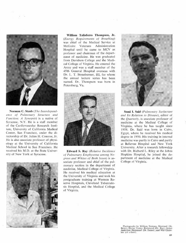
Norman C. Staub (The Interdependence of Pulmonary Structure and Function: A Synopsis) is a native of Syracuse, N.Y. He is a staff member of the Cardiovascular Research Institute, University of California Medical Center, San Francisco, under the directorship of Dr. Julius H . Comroe, Jr. He is also associate professor of physiology at the University of California Medical School in San Francisco. He received his M.D. at the State University of New York at Syracuse.
William Taliaferro Thompson, Jr. (Energy Requirements of Breathing) was chief of the Medical Service at McGuire Veterans Administration Hospital until he came to MCV as professor and chairman of the department of medicine. He was graduated from Davidson College and the Medical College of Virginia. He entered the Army and was a staff member of the 45th General Hospital overseas with Dr. L. T. Stoneburner, III, for whom the annual lecture series has been named. Dr. Thompson was born in Petersburg, Va.
Edward S. Ray (Relative Incidence of Pulmonary Emphysema among Negroes and Whites of Both Sexes} is associate professor and chief of the pulmonary section in the department of medicine, Medical College of Virginia. He received his medical education at the University of Virginia and took his postgraduate training at Western Reserve Hospitals, Cleveland Tuberculosis Hospital, and the Medical College of Virginia.
Sarni I. Said (Pulmonary Surfactant and Its Relation to Disease), editor of the Quarterly, is associate professor of medicine at the Medical College of Virginia, where he has taught since 1958. Dr. Said was born in Cairo, Egypt, where he received his medical degree in 1950. His training in internal medicine was partly in Cairo and partly at Bellevue Hospital and New York University. After a research fellowship with Dr. Richard L. Riley at the Johns Hopkins Hospital, he joined the department of medicine at the Medical College of Virginia.
Photographs by Willia m Notman , Montreal (Dr. Ba tes); Myron Tooley, Richmond (Dr. Ray); James Anderson, Richmond (Dr. Turner) ; and Wirt Christian (all others).
61

Calendar of Postgraduate Education MEDICAL COLLEGE OF vmGINIA/ SPRING 1965
APRIL 21-24, 1965
POSTGRADUATE COURSE IN PEDIATRIC RADIOLOGY WITH EMPHASIS ON TOPICS OF PRACTICAL IMPORTANCE
A three-and-one-half day course designed especially for the clinical radiologist, pediatric radiologist, and pediatrician, but open to all interested physicians. The course will be presented by members of the faculty of the Medical College of Virginia and guest faculty.
TUESDAY-APRIL 20
P.M.
4-8 Registration, Hotel John Marshall.
WEDNESDAY-APRIL 21
A.M.
8-8: 30
8 :30-8 :40
8 :40-8:45
Registration, Hotel John Marshall.
W elcome-Dr. R. Blackwell Smith, Presi-dent, MCV.
Introduction-Dr. Richard G. Lester, chairman, department of radiology, MCV.
8 :45-9 : 30 Radiation Hazards in Diagnostic Radiology -Dr. F. T. O'Foghludha, associate professor, division of radiation physics, department of radiology, MCV.
9 : 30-10 : 15 Pneumonias in the Pediatric A ge GroupDr. R. V. Platou, professor and chairman, department of pediatrics, Tulane University School of Medicine.
10 : 15-10: 45 Intermission
10 : 45-11 : 30 Pneumonias of Early Infancy with Emphasis on Roentgenologic Studies-Dr. E. B. Singleton, director of radiology, St. Luke's and Texas Children's Hospitals; associate professor of radiology, Baylor University College of Medicine.
11:30-12 : 15 Unusual Pneumonias-Dr. John P. Jimenez, instructor, division of radiodiagnosis, department of radiology, MCV.
P .M.
12 :30- 1:30 Lunch
1 :45-2 :30
2 :30-3 :15
3 : 15- 3:30
3:30--4 :15
4 : 15-5
62
Pathology of Congenital Heart Disease with Emphasis on Embryogenesis-Dr. J. B. Arey, pathologist, St. Christopher's Hospital for Children; professor of pathology, Temple University School of Medicine.
Plain Film Diagnosis 'in Congenital Heart Disease-Dr. Lester.
Intermission
Angiocardiographic Diagnosis of Congenital Heart Disease-Dr. Lester.
Vascular Rings-Dr. T. Keats, professor and chairman, department of radiology, University of Virginia Medical School.
THURSDAY-APRIL 22
A.M.
8: 30-9 : 15 Pediatric Urological Problems-Clinical A spects-Dr. Platou.
9 : 15- 10 Urologic Problems in Infants and Children -Roentgen Aspects-Dr. Singleton.
10-10 : 30 Intermission
10: 30-11 : 15 Retro peritoneal Masses in Infants and Children-Dr. J. A. Kirkpatrick, radiologist, St. Christopher's Hospital for Children and Children's Heart Hospital; associate professor of radiology, (pediatrics) Temple University School of Medicine.
11 : 15-11 : 45 Radiotherapy for Tumors of the Genitourinary Tract- Dr. E. R. King, professor and chairman, division of radiation therapy, department of radiology, MCV.
P.M.
11 :45-12:30 Voiding Cystourethrography-Dr. E. Van Epps, professor and chairman, department of radiology, State University of Iowa College of Medicine.
12:30-1:30 Lunch
1: 30-2: 15 Diagnostic Approach to Infants with a Large Head-Dr. Kirkpatrick.
2: 15-3 Neuroradiological A spects of Epilepsy-Dr. M. P. Neal, associate professor, division of radiodiagnosis, department of radiology, MCV.
3-3: 15 Intermission 3: 15-4 Cervical Spine in Infancy and Childhood-
Dr. Van Epps. 4-5 Film session 6:30-7:30 Social hour
7 :30 Dinner
FRIDAY-APRIL 23
A.M. 8 : 30-9: 15
9 :15-10
10-10:30
Gastrointestinal Disorders of the Newborn -Dr. Platou. Esophageal Atresia and Tracheoesophageal Fistula-Dr. Kirkpatrick.
Intermission
10: 30-11 : 15 Intestinal Obstruction in Infants and Children-Dr. Singleton.
11 : 15-12
P.M. 12-1
1-1 :45
Malrotation and Fixation of the Bowel: Radiologic Detection and Clinical Significance-Dr. M. H. Wittenborg, radiologist, Children's Hospital Medical Center of Boston; associate clinical professor of radiology, Harvard Medical School.
Lunch Pathologic Manifestation of Cystic Fibrosis of the Pancreas- Dr. Arey.
1 :45-2:30
3 : 15
Roentgen Aspects of Cystic Fibrosis of the Pancreas-Dr. Wittenborg.
Panel Discussion and Grand Rounds, MCV.
SATURDAY-APRIL 24
A.M.
8: 30-9: 15 Bony Manifestation of Sickle Cell Disease and Other Blood Dyscrasias-Dr. Neal.
9: 15-10 Skeletal Maturation, Its Assessment and Practical Application-Dr. Wittenborg.
10-10: 30 Intermission
10:30-11: 15 Bony Tumors and Tumor-Like Conditions in Childhood-Dr. Saul Kay, professor and chairman, division of surgical pathology, department of pathology, MCV.
11: 15-12 Radiographic Problems About the Knee and Elbow-Dr. Van Epps.
P.M.
12-12:45
TUITION
Problem Fractures in Childhood-Dr. M. J. Hoover, Jr., professor and chairman, division of orthopedic surgery, MCV.
$100 for the entire course, including registration fee, noon lunches, and one evening dinner. For physicians in residency training, the tuition is $50.
If an enrollment is cancelled in advance, a remittance of $90 ($40 for resident physicians) will be made. The registration is nontransferable and is made for the entire course.
Separate daily attendance fee is $30, plus $10 for registration.
Wives accompanying their husbands will be guests for dinner.
For further information, write or call: Dr. M. Pinson Neal, Jr., Director, Postgraduate Course, Department of Radiology, Medical College of Virginia, Richmond, Va. 23219. Phone: 703-644-9851.
APRIL 28-30, 1965
18TH ANNUAL STONEBURNER LECTURE SERIES AND SYMPOSIUM: REHABILITATION WITH SPECIAL EMPHASIS ON STROKE.
Stoneburner Lecturer Dr. Frank H. Krusen, professor of physical medicine and rehabilitation, Temple University School of Medicine.
WEDNESDAY-APRIL 28
P.M.
8: 30 Stoneburner Lecture I- R ehabilitation Adds Life to Years.
THURSDAY-APRIL 29
A.M.
9-10 Registration for Symposium, Egyptian Building, MCV.
10-10:30 Newer Concepts in Exercise Treatment-Dr.
10:30-11
Paul M. Elwood, Jr., medical director, Sister Kenny Foundation.
Ultra Sound-What is its Value?-Dr. Justus F. Lehmann, professor of physical medicine and rehabilitation, University of Washington School of Medicine.
11-11 :30 Massage, Manipulation, and Traction in Physical Medicine-Dr. James R. Oates, assistant professor of physical medicine, Baylor University College of Medicine.
11: 30-12 Thermotherapy-Dr. Edwin L. Lytle, assistant professor of physical medicine and rehabilitation, MCV.
P.M.
12-2 Lunch
2-3 Stoneburner Lecture II-New Developments in the Field of Biomedical Engineering.
3-3 :30
3: 30-4
4-4:30
Lower Extremity Prosthetics-Dr. Roy Hoover, medical director, Woodrow Wilson Rehabilitation Center.
Recent Advances in Upper Extremity Bracing -Dr. John M. Herring, assistant professor of physical medicine and rehabilitation, MCV.
Electromyography-Dr. John B. Redford, professor and chairman, department of physical medicine, MCV.
FRID_'\ Y-APRIL 30
A.M.
10-10:30 The Diagnosis of Cerebrovascular DiseaseDr. Laurie E. Rennie, assistant professor, department of neurology, MCV.
10: 30-11 Prognosis in Stroke-Dr. Elwood.
11-11: 30 Management of Acute Stage Stroke-Dr. Redford.
11 : 30-12 Management of Chronic Stage Stroke-Dr. Oates.
P.M.
12-2 Lunch
2-2: 30 Counseling of the Family of the Stroke Patient - Dr. Henry D. Lederer, chairman, department of psychiatry, MCV.
2: 30-3 Gait Training-Dr. Lehmann.
3-3: 30 The Communication Problem-Mrs. Martha L. Taylor, director, speech and hearing therapy, New York University Medical Center.
3: 30-4 Panel on Rehabilitation Problems.
TUITION
$10 per day for the Symposium. There is no charge for attending Dr. Krusen's lectures.
For further information, write: Dr. John B. Redford, Chairman, Department of Physical Medicine and Rehabilitation, Medical College of Virginia, Richmond Va. 23219.
63
The MEDICAL COLLEGE OF VIRGINIA QUARTERLY is designed
primarily for the postgraduate education of physicians. The QUARTERLY
will publish results of original research in basic and clinical sciences, and
report on seminars and symposiums held at the College. Contributions
from outside the MCV faculty are invited.
Manuscripts , submitted in duplicate, should be prepared according to
recommendations in the Style Manual for Biological Journals , 2nd ed.,
published in 1964 by the American Institute of Biological Sciences, 2000
P Street, N.W., Washington, D . C. 20036.
Correspondence : MEDICAL COLLEGE OF VIRGINIA QUARTERLY, Medical College of Virginia, Richmond, Va. 23219. Phone:
703-644-9851.
Subscription rates in the USA and Canada: 1 year, $4; 2 years, $7;
3 years, $9. All other countries: 1 year, $5; 2 years, $8; 3 years, $10
House staff, medical students, and fellows : 1 year, $2.
Editorial A<lvisory Board
William M. Feagans Fairfield Goodale, Jr. June L. Huntley Richard G . Lester
EDITOR
Sarni I. Said
Hunter H . McGuire, Jr. Kinloch Nelson Arnold M. Salzberg Frederick J. Spencer
Designer, Bruce W. Smith; Photographer, Wirt Christian; Editorial Assistant, Karen Kevorkian .
Printed by the Waverly Press, Baltimore, Md . Entered third class mail at Baltimore, Md. Copyright 1965 by the Medical College of Virginia.
The Medical Education Building, at the corner of 11th a11d Marshall Streets, opposite the Medical College Hospital, was designed by Merrill C. Lee, and completed in 1963. The 10-story structure houses offices for faculty and administration, student laboratories and study areas, research laboratories, and classrooms equipped with closed circuit television.
~2jA11'f/ZJ
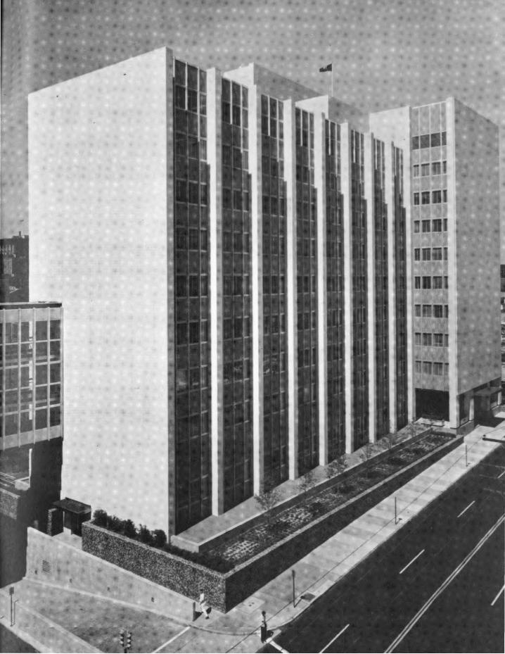


![Quarterly, Vol. 08, no. 1 (1962, March) [whole issue] - eGrove](https://img.pdfslide.net/doc/110x75/63339918a6138719eb0aafb0/quarterly-vol-08-no-1-1962-march-whole-issue-egrove.jpg)


