Embed Size (px)
Citation preview

Mesenchymal Stem Cells OverexpressingEphrin-B2 Rapidly Adopt an Early Endothelial
Phenotype with Simultaneous Reductionof Osteogenic Potential
Garry P. Duffy, Ph.D.,1,2 Sinead D’Arcy, B.Sc.,1 Tabassum Ahsan, Ph.D.,2 Robert M. Nerem, Ph.D.,2
Timothy O’Brien, M.D., Ph.D.,1 and Frank Barry, Ph.D.1
Restoration of the vascular supply to ischemic tissues is of high clinical relevance, and proangiogenic therapiesaim to reduce morbidity and mortality rates associated with the onset of cardiovascular disease. Stem celltherapy has been proposed as a potentially useful proangiogenic therapy. Mesenchymal stem cells (MSCs) havebeen shown to be proangiogenic and produce a number of cytokines involved in vessel development andmaturation. Preclinical studies have reported increased angiogenesis after MSC delivery to the heart, and similaroutcomes have been reported in recent clinical trials. Stem-cell-mediated neovascularization has been aug-mented by genetic modification with overexpression of angiogenic cytokines, including vascular endothelialgrowth factor (VEGF) and platelet-derived growth factor, showing promising results. In this study we aimed toenhance the proangiogenic capability of MSCs. MSCs were genetically modified to overexpress a versatilemolecule, Ephrin-B2, involved in tissue morphogenesis and vascular development to enhance inherent neo-vascularization potential. Using nucleofection, Ephrin-B2 was transiently overexpressed on the cell surface ofMSCs to recapitulate embryonic signaling and promote neovascularization. Ephrin-B2-expressing MSCs adop-ted an early endothelial phenotype under endothelial cell culture conditions increasing expression of vonWillebrand factor and VEGF-Receptor 2. The cells had an increased ability to form vessel-like structures, pro-duce VEGF, and incorporate into newly formed endothelial cell structures. These data indicate that MSCsexpressing Ephrin-B2 represent a novel proangiogenic cell source to promote neovascularization in ischemictissues.
Introduction
Restoration of a vascular supply to ischemic tissuesis of high clinical relevance, and proangiogenic thera-
pies aim to reduce morbidity and mortality rates associatedwith the onset of cardiovascular disease.1 Over the pastdecade there has been a huge leap in unraveling the mo-lecular mechanisms that govern new blood vessel forma-tion.2–4 A number of regulatory factors have been identifiedas key role players in the formation of blood vessels, in-cluding vascular endothelial growth factor (VEGF), platelet-derived growth factor, and fibroblast growth factor families.5
This has allowed the design and implementation of novelproangiogenic interventions and therapeutic approaches,including gene therapy,6 cellular therapy,7 and a combina-tion of both.8
Stem cell therapy has been proposed as a promisingproangiogenic therapy. Since initial reports demonstratedthe effectiveness of stem cell delivery to the postischemicmyocardium,9 mesenchymal stem cells (MSCs) have beenstudied extensively as a cardiac therapy10 and MSCs possessinherent proangiogenic capabilities producing a number ofcytokines involved in vessel development, including VEGFand platelet-derived growth factor.11–13 Preclinical studieshave reported increased angiogenesis after MSC delivery tothe heart,14 and similar outcomes have been reported inclinical trials.15 Shyu et al.16 reported that MSC delivery as astand-alone therapy surpassed angiogenic factor gene ther-apy in promoting myocardial regeneration in a mouse modelof acute myocardial infarction. Further, MSCs have beenshown to promote angiogenesis in peripheral vascular is-chemia as well as in wound healing.17,18
1Regenerative Medicine Institute (REMEDI), National University of Ireland Galway, Ireland.2Institute of Bioscience and Bioengineering, Georgia Institute of Technology, Atlanta, Georgia.
TISSUE ENGINEERING: Part AVolume 16, Number 9, 2010ª Mary Ann Liebert, Inc.DOI: 10.1089/ten.tea.2009.0623
2755

Stem-cell-mediated neovascularization has been aug-mented by the modification of these cells to overexpressangiogenic cytokines.19,20 As in proangiogenic gene ther-apy, initial studies focused on overexpression of a singleangiogenic factor. VEGF has proved popular, along withother factors such as Ang-1 and hepatocyte-like growthfactor (HGF), and initial preclinical reports point to im-provements in myocardial reperfusion over cell therapyalone.21–23 Further studies have utilized a multimodalapproach, where a number of factors are simultaneouslyoverexpressed in MSCs, resulting in further improvementin global heart function.8,24,25
These studies illustrate the importance of a multimodalapproach to vascular reperfusion in ischemic tissues. In thiswork we aimed to develop a novel combined stem cell–genetherapy approach for treating ischemic tissues with en-hanced proangiogenic capability compared to nonmodifiedcells. We genetically modified MSCs to overexpress a ver-satile molecule, Ephrin-B2, involved in tissue morphogenesisand vascular development to enhance inherent neovascu-larization potential.
The erythropoietin-producing hepatocellular carcinoma(Eph) receptors and their cell-surface-anchored ligands, theEphrins, comprise the largest of the receptor tyrosine kinasefamilies with 14 receptors and 8 ligands.26 The receptors aresubdivided into Eph-A and Eph-B categories based on se-quence homologies and binding properties to Ephrin ligandsand have known actions in the development of the vascularand nervous system. Eph receptor tyrosine kinases and theirEphrin ligands regulate a diverse array of cellular functions,including cell migration, repulsion and adhesion, and somiteformation.27 Eph-B4 and Ephrin-B2 are co-expressed in theyolk sac, the first site of hematopoiesis and vascular devel-opment during embryogenesis.28 Ephrin-B2 is specificallyexpressed in arterial angioblasts and endothelial and peri-vascular mesenchymal cells, whereas Eph-B4 is expressed onendothelial cells (ECs) belonging to the venous lineage.29 Arecent report has shown that vessel segregation into venousand arterial fates is also controlled by expression of Ephrin-B2 and Eph-B4 through directional control.30 Knockout micelacking either Eph-B4 or Ephrin-B2 resulted in early lethalityin the developing embryo as a result in the arrest of angio-genesis but not vasculogenesis,29,31 illustrating the impor-tance of both molecules in vascular development.32
Ephrin-B2 also plays a part in vessel maturation throughits involvement in smooth muscle recruitment and crosstalkbetween endothelial cells and pericytes.33 Ephrin-B2 hasalso been shown to be expressed during postnatal neo-vascularization and have positive effects on wound heal-ing.34,35 Further, a recent study has shown the delivery ofEphrin-B2 chimeric proteins to be efficacious in promotingan increase in capillary density in a mouse model of myo-cardial infarction.36 Another study has shown that ex vivomodification of skin autofibroblasts with Ad-Ephrin-B2 andsubsequent delivery into a rabbit model of hind-limb is-chemia resulted in increased arteriogenesis.37 Both studieshighlight the potential usage of Ephrin-B2 as a proangio-genic factor and confirm our approach to creating a moreefficacious stem cell therapy. Using nucleofection, Ephrin-B2 was transiently overexpressed on the cell surface ofMSCs to recapitulate embryonic signaling and promoteneovascularization.
Materials and Methods
MSC isolation and expansion
Bone marrow aspirates were obtained from the iliac crestof normal donors; all procedures were performed with in-formed consent and approved by the Clinical ResearchEthics Committee at University College Hospital, Galway.MSCs were isolated by direct plating of whole marrow andexpanded in culture as described previously.38 Briefly, as-pirates were washed with a medium (Dulbecco’s modifiedEagle’s medium–low glucose [DMEM-LG] containing 1%antibiotic) and centrifuged; the precipitated cells were sus-pended in the medium with 10% selected fetal bovine se-rum (FBS) and plated at a final density of *3.0�105 cells/cm2. Cells were seeded on T-175 flasks and maintained at378C with 95% humidity and 5% CO2 in the same medium.After 5 days red blood cells were washed off withphosphate-buffered saline (PBS) and a fresh medium wasadded. Colonies of adherent cells formed within 9 days. Atthe end of primary culture, adherent colonies were de-tached by treatment with 0.25% trypsin and 0.53 mMethylenediaminetetraacetic acid. Cells were plated in themedium [DMEM-LG; 10% FBS; 1% antibiotic (all fromSigma-Aldrich)] at 5.7�103 cells/cm2. Cultures were pas-saged at 4–6-day intervals and expanded to passage 5 forexperimentation.
Osteogenic differentiation assays
To assess the osteogenic potential of MSCs, cells wereplated at 30,000 cells per well in a six-well plate and culturedovernight. The following day cells were treated with theosteogenic medium [DMEM-LG; 100 nM dexamethasone;10 mM b-glycerophosphate; 50mM ascorbic acid 2-phosphate;10% fetal bovine serum (FB/FBS); 1% antibiotic (Sigma-Aldrich)]. The medium was replaced two times per week.Calcium deposition was measured on day 15. Wells werefixed with 10% neutral buffered formalin for 10 min, and1 mL of 3% silver nitrate solution was added to each well andincubated at room temperature in the dark for 10 min. Wellswere rinsed and exposed to bright light for 15 min and cal-cium deposits imaged. Quantification of mineral depositionwas performed as previously described.39
Adipogenic differentiation assay
To assess the adipogenic potential of MSCs, cells wereseeded at 2�105 per well in six-well plates and cultureduntil confluent. Cultures were then placed in an adipo-genic induction medium [DMEM high glucose 10% FBS;1 mm Dexamethasone; 200 mm Indomethacin; 10 mg/mLInsulin; and 0.5 mM Isobutylmethylxanthine; 1% antibiotic(all from Sigma-Aldrich)] for 3 days and subsequentlymoved into an adipogenic maintenance medium [DMEMhigh glucose; 10% FBS; and 10 mg/mL Insulin: 1% antibi-otic] for 1 day. Cells cultured in the normal medium wereused as controls. After three induction and maintenancecycles, cells were fixed with 10% neutral buffered formalin(Sigma-Aldrich) and stained using Oil Red O (Sigma-Aldrich). To quantify binding of Oil Red O to accumulatedlipid it was extracted with 100% isopropanol and absor-bance was read at 520 nm.
2756 DUFFY ET AL.

Cell surface phenotype analysis
MSCs at passage 5 were analyzed for purity and epitopeexpression using flow cytometry analysis. Cells were blockedin 5% goat serum/D-PBS for 30 min, and incubated with therespective phycoerythrin (PE)-conjugated antibodies (anti-CD45, anti-CD73, anti-CD105, and mouse IgG 1 isotypecontrol [BD Biosciences]) diluted in blocking buffer at 1:100.After incubation the cells were washed in PBS and analyzedusing a FACS LSR flow cytometer (Becton Dickenson). His-tograms of cell number versus fluorescence intensity wererecorded for 10,000 cells per sample and analyzed using FCSExpress 2 (DeNovo Software).
Eph-B4 Fc-chimera protein binding assay
To assess expression of Ephrin-B2 on the cell surface ofunmodified and transfected MSCs, the binding ability ofEph-B4 Fc-chimera, the receptor to Ephrin-B2, was mea-sured. Briefly, cells were detached from the flask using celldissociation agent (MP Bio) washed in D-PBS/5% FBS twiceand incubated with 2mg/mL Fc-chimera proteins (Ephrin-B2, Eph-B4) for 30 min on ice. After a further two washes,cells were incubated with FITC-conjugated goat anti-humanIgG1 antibody for 30 min at room temperature. After a fur-ther two washes, stained cells were analyzed using theGUAVA EasyCyte or the BD LSR. Histograms of cell numberversus fluorescence intensity were recorded for 10,000 cellsper sample and analyzed using FCS Express 2 (DeNovoSoftware).
Cloning of Ephrin B2 into a mammalianexpression vector
The full-length coding sequence of Ephrin B2 was ampli-fied from human coronary artery endothelial cell mRNAusing a two-step reverse transcriptase (RT)-polymerase chainreaction carried out with a proof-reading taq (PFX) that ad-ded a poly-A tail at both 50 and 30 ends.
Coding primer sequences:Forward, 50ATGGCTGTGAGAAGGGACTCCReverse, 50 TCAGACCTTGTAGTAAATGTTC
After amplification, the cloning sequence was initiallycloned into the TA- pTargeT Expression Vector (Promega) ina sterile 0.5 mL eppendorf tube. Inserts were verified withDNA sequencing (MWG Biotech—Genome SequencingServices). Subsequently, Ephrin B2 was subcloned from thepTargeT vector to the pIRES2-eGFP bi-cistronic mammalianexpression vector (Clontech). Insertion was verified by re-striction endonuclease mapping.
Nucleofection and transgene analysis
MSCs at passage 5 were washed with Hanks balance saltsolution (Sigma) and harvested by trypsinization. Cells werecounted and 5�105 cells were resuspended in 100 mL of pre-warmed Human MSC Nucleofection Solution (Amaxa).Two microgram of plasmid DNA in TRIS-EDTA (TE) buffer(pmaxGFP, pIRES2-eGFP, and pEphrin-B2/IRES2-eGFP)was added to the cell suspension. The sample was trans-ferred into an Amaxa cuvette and placed into the holder of
the Nucleofector Device II (Amaxa Biosystems) and sub-jected to the high transfection efficiency program (U-23). Thecuvette was removed immediately and 500 mL of the pre-warmed growth medium was added to the cell suspensionand transferred to a T25 cell culture flask. The cells wereincubated in a humidified 378C/5% CO2 incubator for 24 h.After 8 h of culture, viability was assessed by the proportionof cells attached to the culture surface using a coulter coun-ter. Cell viability was also measured by trypan blue exclu-sion. pmaxGFP acted as a positive nucleofection control andcells minus DNA acted as a negative control. GFP/eGFPtransgene expression was analyzed 48 h after nucleofectionby flow cytometry. The percent of GFP/eGFP-positive cellswere quantified versus negative control over 10 days usingthe BD LSR and analyzed using FCS Express 2 (DeNovoSoftware). Western blot analysis and Eph-B4/Fc bindingassays were carried out as described previously to verifyincreased expression of Ephrin-B2 in transfected MSCs.
Matrigel assay—vessel formation assay
Growth-factor-reduced Matrigel (120 mL; BD Biosciences)was placed in each well of an ice-cold 48-well plate. The platewas placed at 378C for 30 min to permit Matrigel to solidify.To assess the effect of overexpression of Ephrin-B2 on MSC-vessel-like structure formation, cells were detached 48 h afternucleofection and plated at 3�104 cells per well. Further toassess the effect of transfected MSCs on endothelial-cell-vessel-like structure formation ECs were detached, labeledwith 2 mM PK26 (Sigma-Aldrich), and plated at 3�104 cellsper well. MSCs were added to ECs at a ratio of 1:2 24 h afterEC seeding. MSC and EC/MSC tube formation was ob-served periodically over 72 h and at 5 days of coculture.Phase images were acquired using a Zeiss Axiovert 200 mi-croscope and Spot v4.6 digital imaging system. Magnifica-tion bars are added to one image to indicate themagnification of all images within the panel. Confocal im-ages and z-stacks were taking with a Zeiss LSM 510 andLSM Image Examiner. Image analysis was carried out usingOlympus Cell R imaging software. Lateral sprouts pervessel-like structure length, and cross-sectional diameterwere measured on 10�images (n¼ 5). For analysis of smoothmuscle actin (SMA) expression, ECs were labeled with 2 mMQ-tracker 645 qtracker dye to distinguish the cells fromGFP-labeled MSCs. PE-labeled anti-alpha SMA (a-SMA)antibodies (Abcam) were used, and increases in mean fluo-rescence intensity were measured as a surrogate measure ofSMA expression using flow cytometry. FITC and PE com-pensation was carried out using BD FACSDIVA software.
MSC/Ephrin-B2—endothelial cell differentiationand phenotype analysis
T25 cell culture flasks were coated with a thin layer ofgrowth-factor-reduced Matrigel (150 mg/mL) for 2 h. MSCs,MSCs transfected with pIRES2-eGFP (MSCs/eGFP), andMSCs transfected with pEphrin-B2/IRES2-eGFP (MSCs/Ephrin-B2) were seeded on the Matrigel-coated flasks andcultured in the endothelial growth medium-2 growth me-dium for 5 days. Phase images were acquired using a ZeissAxiovert 200 microscope and Spot v4.6 digital imaging sys-tem. Cells were harvested and fixed for 15 min in 4% para-formaldehyde. Cells were washed in PBS (�)/5% FBS and
MSCS/EPHRIN-B2 ADOPT AN ENDOTHELIAL PHENOTYPE 2757

permeabilized with 0.1% triton�100/PBS for 5 min. Cellswere incubated with one of the following antibodies for 1 hat room temperature: anti-a-SMA (goat anti-human; Sigma),anti–von Willebrand factor (vWF)–PE (Dako), anti-Flk-1(rabbit anti-human; Fitzgerald), and anti-CD73-PE (BD).Isotype controls acted as negative controls, and endothelialcells and smooth muscle cells were also used to ensurespecificity of the antibodies (data not shown). Secondary-only controls were used to ensure nonspecific binding whenusing polyclonal antibodies. After washing, cells stainedwith unconjugated primary antibodies were incubated withAlexa fluor 546–conjugated anti-goat/rabbit secondary an-tibodies (Molecular Probes) for 1 h at room temperature.Histograms of cell number versus fluorescence intensitywere recorded for 10,000 cells per sample at each time pointwith the BD LSR and analyzed using FCS Express 2 (DeNovoSoftware).
Assessment of VEGF production
After a 5-day exposure of MSCs/Ephrin-B2 to endothelialconditions, 5�105 MSCs were seeded in six-well plates in theserum-free medium. The cells were placed in a hypoxicchamber at 0.1% O2 for 4 h. The conditioned medium wascollected and the amount of VEGF produced was measuredusing a Quantikine human VEGF immunoassay kit (R&DSystems). MSCs transfected with pIRES2-eGFP acted ascontrols.
Statistical analysis
All values are presented as the mean� standard deviationof the mean. Datasets were tested for significance using theStudent’s t-test or a general linear model two-way analysis ofvariance in combination with a post hoc Tukey test to com-pare between groups. A level of p< 0.05 was consideredstatistically significant.
Results
MSCs were isolated from the bone marrowand expressed characteristic cell surface markers
Cell isolation, expansion, and differentiation of MSCs us-ing direct plating were established according to other re-ports.38 Isolated MSCs adhered to tissue culture plastic, hada fibroblastic morphology (Fig. 1A), and proliferated up topassage 5 with a doubling time of *2 days. MSCs also dif-ferentiated along the adipogenic and osteogenic pathways asevidenced by the accumulation of lipid droplets in the cy-toplasm and deposition of calcium in the wells (Fig. 1B, C).The isolated cells were negative for CD45, a known hema-topoietic marker, but positive for MSC characteristic markersCD73 and CD105 (Fig. 1D).
Nulceofection of MSCs with pIRES2-eGFP/Ephrin-B2
MSCs were transfected with the pEphrin-B2/eGFP plasmidusing the nucleofection technique previously described.40,41
Transfection efficiency was measured by analyzing expressionof eGFP in cells using both microscopy and FACS analysis. Atransfection efficiency of 45.6%� 1.48% was achieved with thepEphrin-B2/eGFP vector (Fig. 2A), whereas viability was re-duced to 66.3%� 1.48% after nucleofection. GFP-positive cells
were clearly seen in micrographs 48 h after transfection (Fig.2B). Cells remained viable in culture and expanded over 6days (data not shown). Expression of the transgene, Ephrin-B2, was examined 48 h after transfection by the binding of itscognate receptor, Eph-B4/Fc. Cells increased expression ofEphrin-B2 as there was an increase in the binding of Ephrin-B2 receptor, Eph B4/Fc, to the cell surface of transfected MSCsby 22%� 2.3% compared with normal cells (Fig. 2C). To as-sess the transient nature of transfection, we analyzed thepercentage of eGFP-positive cells every 2 days over 10 days inculture. There was a marked drop of expression from 46.8% to4.5% over the time period (Fig. 2D). The pmaxGFP vectoracted as a positive transfection control, and also showed adecrease in GFP expression from 73% to 18.2% over the same10-day period (Fig. 2D).
MSCs overexpressing Ephrin-B2 have a reducedosteogenic differentiation potential
To assess changes in the differentiation potential ofMSCs/Ephrin-B2, osteogenic induction was initiated 48 hafter nucleofection. It became apparent that the cells over-expressing Ephrin-B2 adopted a morphology similar to en-dothelial cells at days 2, 5 and 8 after osteogenic inductioncompared to cells alone and mature endothelial cells (Fig.3A, inset [i]). At day 15, however, MSCs/Ephrin-B2 ap-peared similar in morphology to osteogenic MSCs but with areduced cell number. Transgene expression was reverted tonormal levels at this time. There was a significant reductionin the amount of calcium deposited by MSCs/Ephrin-B2 incomparison to normal MSCs at day 15 of osteogenic induc-tion ( p< 0.01; Fig. 3B). This reduction in calcium depositionis also apparent in von Kossa–stained wells of transfectedcells versus normal cells (Fig. 3C). The data indicate thatthere was a reduction in the differentiation potential of MSCsoverexpressing Ephrin-B2 toward the osteogenic lineage.
Overexpression of Ephrin-B2 in MSCs increasesvessel-like structure formation
To further understand the changes in the cellular pheno-type seen after Ephrin-B2 overexpression, the vessel-likestructure formation potential of transfected MSCs was ana-lyzed. Two days after transfection, cells were plated ongrowth-factor-reduced Matrigel and vessel-like structure for-mation was imaged 24 h later. It was immediately apparentthat MSCs overexpressing Ephrin-B2 had an increased po-tential to form vessel-like structures on the Matrigel (Fig. 4B)compared with normal MSCs, which formed large cellularaggregates on the gel surface (Fig. 4A). This increase in vessel-like structure formation potential by MSCs/Ephrin-B2 wascomparable to endothelial cells (Fig. 4 inset [i]). Using confocalmicroscopy, we subsequently investigated the incorporationof eGFP-positive cells into vessel-like structures formed on theMatrigel as transfected cells express both eGFP and Ephrin-B2because of the bi-cistronic expression system (Fig. 4C, D). Alarge number of cells aggregated in the MSC-alone well as seenwith the large number of 406-diamidino-2-phenylindole(DAPI)-stained nuclei in cell bundles, whereas cells in theMSC/Ephrin-B2 wells formed vessel-like structures witheGFP-positive cells incorporated into them. These vessel-likestructures also remained present up to 5 days after cell seeding
2758 DUFFY ET AL.

(Fig. 4E) in comparison to cell bunches seen in untransfectedcells. (Fig. 4F).
MSCs/Ephrin-B2 adopt an early endothelial phenotypeunder endothelial culture conditions
To further characterize the cellular phenotype of theMSCs/Ephrin-B2, MSCs/eGFP, and MSCs after 5 days un-der endothelial cell culture conditions, cells were grown on a
thin layer of Matrigel in the endothelial medium for 5 days(Fig. 5A). Expression of early endothelial markers (vWF andFlk-1) was assessed by antibody staining and FACS analysis.MSCs/Ephrin B2 increased expression of both early endo-thelial markers, vWF and the Flk-1 (Fig. 5B), whereas therewas no appreciable change in expression in MSCs/eGFP orMSCs. Cells did not initially express a-SMA and there wereno changes in expression levels after culture. Interestingly,there was a decrease in the expression level of CD73 on
FIG. 1. MSC characterization anddifferentiation potential. (A) Phase-contrast micrograph showing thefibroblastic morphology of MSCs inculture at passage 5. (B) Quantifica-tion of Oil red O staining in MSCscultured under adipogenic condi-tions for 15 days. Stained positivelywith Oil red O for lipid droplets in thecytoplasm (n¼ 3). (C) Quantificationof calcium deposition in MSCscultured under osteogenic conditionsfor 15 days (n¼ 3). MSCs, mesen-chymal stem cells. (D) Tabledepicted the expression levels ofCD45, �CD73, and �CD105 onMSCs. Color images available onlineat www.liebertonline.com/ten.
MSCS/EPHRIN-B2 ADOPT AN ENDOTHELIAL PHENOTYPE 2759

FIG. 2. Nucleofection of MSCs with pIRES2-eGFP/Ephrin-B2. (A) A flow cytometry histogram showing the level of GFPexpression of MSCs transfected with pEphrin-B2/IRES2-eGFP. Cells were 66.3% viable after nucleofection. (B) Fluorescentmicrographs of eGFP-positive cells nucleofected with pEphrin-B2/IRES2-eGFP (arrows; 10�, 40�). (C) Flow cytometry his-togram showing the binding of preclustered Eph-B4/Fc protein to its ligand, Ephrin-B2, on transfected and normal MSCs.There is an increase in expression of Ephrin-B2 on the cell surface of nucleofected cells. (D) Flow cytometry scatter plots of thepercent GFP/eGFP-positive cells (Q2, upper right) over 10 days after nucleofection; the expression levels drop over the timecourse illustrating the transient nature of the transgene expression. Eph, erythropoietin-producing hepatocellular carcinoma.Color images available online at www.liebertonline.com/ten.
2760

FIG. 3. Osteogenic potential of Ephrin-B2 overexpressing MSCs. (A) Representative phase micrographs of normal andtransfected cells over 15 days of osteogenic differentiation (scale bar indicates 200 mm). MSCs overexpressing Ephrin-B2 showa morphology similar to an endothelial phenotype (inset: i). (B) Quantitative analysis of calcium deposition after osteogenicinduction. Data are presented as the mean� SD of calcium concentration (mg) per well, n¼ 3. *p< 0.05 versus Osteo alone.There is significantly less calcium deposition in osteogenic cultures of MSCs overexpressing Ephrin-B2 versus MSCs alone.(C) Light micrographs of von Kossa–stained osteogenic cultures; there is visibly less stained calcium deposits in transfectedcells (200�). Cells in the normal medium acted as controls. SD, standard deviation.
2761

FIG. 4. Effect of Ephrin-B2 overexpression on MSC-vessel-like structure formation. Phase micrographs of MSCs (A) andMSCs/Ephrin-B2 (B) cultured on Matrigel-coated wells for 24 h in the endothelial medium (scale bar indicates 200 mm).Fluorescent micrographs of MSCs (C) and MSCs/Ephrin-B2 (D) on Matrigel. Large cellular clusters form when MSCs arecultured on Matrigel, whereas MSCs/Ephrin-B2 form numerous vessel-like structures. Inset: ECs cultured on Matrigel (i).GFP-positive cells can be seen incorporating into vessel-like structures (nuclei, blue). (E) Representative phase images ofvessel-like structures still apparent 5 days after cultures in MSCs/Ephrin-B2. (F) MSCs alone acted as controls. Color imagesavailable online at www.liebertonline.com/ten.
2762

MSCs/Ephrin-B2 compared to MSCs/eGFP and MSCs afterEC culture. On further inspection this reduction corre-sponded to the level of CD73 normally expressed on aorticendothelial cells (Fig. 5B). These data suggest that MSCs/Ephrin-B2 adopt an early endothelial phenotype after expo-sure to EC cell culture conditions.
To further characterize the transfected cell population,MSCs/Ephrin-B2 and MSCs/eGFP were cultured underhypoxic conditions for 4 h and VEGF production was mea-sured. MSCs overexpressing Ephrin-B2 released significantly
higher levels of the proangiogenic factor VEGF than MSCs/eGFP (Fig. 5C: p< 0.01).
MSCs/Ephrin B2 incorporate with new vessel-likestructures formed by endothelial cells
To assess the ability of MSCs/Ephrin-B2 cells to incorpo-rate into newly formed EC-vessel-like structures (Fig. 6A–D),we cocultured endothelial cells with MSCs on growth-factor-reduced Matrigel. MSCs were added 24 h after EC seeding
FIG. 5. Cellular phenotype anal-ysis of MSCs/Ephrin-B2 under en-dothelial culture conditions. (A)Representative phase micrographsof cells grown under endothelialcell culture conditions for 5 days(50�). (B) Flow cytometry histo-grams showing the expression lev-els of early endothelial markersvWF and Flk-1 on MSCs, MSCstransfected with naked plasmid(pIRES-eGFP), and MSCs trans-fected with pIRES-eGFP/Ephrin-B2under EC culture conditions.Staining for alpha-smooth muscleactin acted as a negative control.Expression of CD73 was also ana-lyzed as an MSC marker control.MSCs/Ephrin-B2 showed an in-creased expression level of bothvWF and Flk-1, while also showinga reduction in CD73 expression.This reduction in CD73 expressionwas similar to the expression levelsseen on the cell surface of aorticendothelial cells. (C) Quantitativeanalysis of VEGF production after5 h of hypoxia (0.1% O2). Data arepresented as the mean� SD ofVEGF concentration (pg/mL),n¼ 6. *p< 0.01 versus MSCs/eGFP.VEGF, vascular endothelial growthfactor. Color images available on-line at www.liebertonline.com/ten.
MSCS/EPHRIN-B2 ADOPT AN ENDOTHELIAL PHENOTYPE 2763
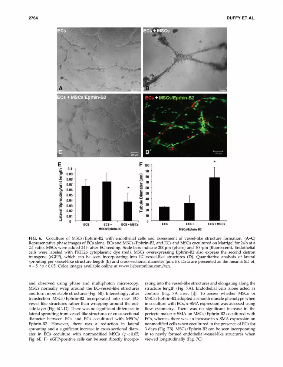
and observed using phase and multiphoton microscopy.MSCs normally wrap around the EC-vessel-like structuresand form more stable structures (Fig. 6B). Interestingly, aftertransfection MSCs/Ephrin-B2 incorporated into new EC-vessel-like structures rather than wrapping around the out-side layer (Fig. 6C, D). There was no significant difference inlateral sprouting from vessel-like structures or cross-sectionaldiameter between ECs and ECs cocultured with MSCs/Ephrin-B2. However, there was a reduction in lateralsprouting and a significant increase in cross-sectional diam-eter in ECs coculture with nonmodified MSCs ( p< 0.05;Fig. 6E, F). eGFP-positive cells can be seen directly incorpo-
rating into the vessel-like structures and elongating along thestructure length (Fig. 7A). Endothelial cells alone acted ascontrols (Fig. 7A inset [i]). To assess whether MSCs orMSCs/Ephrin-B2 adopted a smooth muscle phenotype whenin coculture with ECs, a-SMA expression was assessed usingflow cytometry. There was no significant increase in thepericyte maker a-SMA on MSCs/Ephrin-B2 cocultured withECs, whereas there was an increase in a-SMA expression onnonmodified cells when cocultured in the presence of ECs for3 days (Fig. 7B). MSCs/Ephrin-B2 can be seen incorporatingin to newly formed endothelial-vessel-like structures whenviewed longitudinally (Fig. 7C)
FIG. 6. Coculture of MSCs/Ephrin-B2 with endothelial cells and assessment of vessel-like structure formation. (A–C)Representative phase images of ECs alone, ECs and MSCs/Ephrin-B2, and ECs and MSCs cocultured on Matrigel for 24 h at a2:1 ratio. MSCs were added 24 h after EC seeding. Scale bars indicate 200mm (phase) and 100 mm (fluorescent). Endothelialcells were labeled with PKH26 cytoplasmic dye (red). MSCs overexpressing Ephrin-B2 also express the second cistrontransgene (eGFP), which can be seen incorporating into EC-vessel-like structures (D). Quantitative analysis of lateralsprouting per vessel-like structure length (E) and cross-sectional diameter (mm: F). Data are presented as the mean� SD of,n¼ 5. *p< 0.05. Color images available online at www.liebertonline.com/ten.
2764 DUFFY ET AL.

FIG. 7. Structural organization of EC-MSC/Ephrin-B2 cocultured vessel-like structures. (A) Multiphoton fluorescent mi-crograph of vessel-like structures formed during EC coculture with MSCs/Ephrin B2. eGFP-tagged MSCs are visible in-corporating into a vessel-like structure after 72 h of culture (arrows). Inset ECs alone. Scale bar indicates 20mm; ECs (red) andnuclei (blue). (B) Quantitative analysis of alpha smooth muscle expression on MSCs of MSCs/Ephrin-B2 cocultured with ECs.Data are presented as mean� SD of phycoerythrin (PE) mean fluorescent intensity, n¼ 3. *p< 0.05. (C) Longitudinal view(50�) of a confocal slice taken through a vessel-like structure formed in MSCs/Ephrin-B2 (eGFP/Green) and EC (Red)coculture. Color images available online at www.liebertonline.com/ten.
2765

Discussion
The need for an efficacious proangiogenic therapy withoutadverse side effects still remains unmet. Stem cell therapieshave been proposed as a potential solution to this problem,offering advantages in integration as well as the ability tosupply several trophic factors involved in neovasculariza-tion.12 Recent approaches have been undertaken to enhancethe inherent angiogenic capabilities of MSCs by geneticallymodifying the cells to express proangiogenic factors, in-cluding VEGF, Ang-1, and fibroblast growth factor,19,22 andall have been reported to increase vascular density in ische-mic tissues compared to unmodified cells. However, resultsfrom the clinical setting are still unpublished.8
In this study we aimed to assess a novel stem cell genetherapy combination using Ephrin-B2 as the therapeutic gene.Recent reports have demonstrated increased expression of theligand Ephrin-B2 in a number of tumor categories and to befunctionally involved in tumor angiogenesis.27,42 This factplaces important emphasis on considering the ‘‘The JanusPhenomenon’’ described by Epstein et al.,43 where an angio-genic factor aids targeted neovascularization but also pro-motes tumor progression by enhancing tumor angiogenesis indistal locations. It should be noted that Ephrin-B2 may beproangiogenic for tumors and could have an affect on tumorprogression while enhancing tissue revascularization. To re-duce this risk we chose to transiently deliver Ephrin-B2 toMSCs using a nonviral method of transfection, nucleofection.
Transfection rates of 80% have been reported in MSCs usingthe nucleofection technique.40,41 We achieved similar ratesusing a control plasmid pmaxGFP; however a lower rate oftransfection of 44% was achieved with our bi-cistronic vector(pIRES2-eGFP/Ephrin-B2). This may be due to the fact thatthe eGFP transgene is downstream of the IRES promoter andsome reports have noted significantly less expression of thesecond cistron transgene after transfection.44 There was asuccessful increase in the expression level of Ephrin-B2 inMSCs after nucleofection by Western blotting (data notshown) and the binding of its cognate receptor, Eph-B4/Fc, tothe cell surface of the transfected cells. Cell viability was re-duced to *65% using the Nucleofection technique, but issuessurrounding reduced cell viability were somewhat offset bythe high transfection efficiency and increased expression ofEphrin-B2 achieved in the MSC population. This high effi-ciency in combination with the alleviation of safety consider-ations associated with viral vectors points toward Nucleofectionbecoming a clinically applicable transfection technique in thefuture if viability can be improved.
Reports have demonstrated the involvement of Ephrin-B2/Eph-B4 signaling in the crosstalk between osteoclasts andosteoblasts during bone formation and homeostasis.45,46 Weexamined the osteogenesis potential to ensure that MSCsmodified with Ephrin-B2 did not increase bone matrix for-mation. After assessment MSCs/Ephrin-B2 had a reducedosteogenic differentiation potential compared with un-transfected cells after nucleofection. Interestingly, the trans-fected cells appeared to adopt an endothelial morphologyduring osteogenic induction, which led to the analysis of thevessel-like structure formation capability of MSCs/Ephrin-B2 on Matrigel. We found that cells overexpressing Ephrin-B2 formed vessel-like structures in a similar manner to ECson Matrigel, whereas untransfected MSCs formed charac-
teristic cell bunches on the gel surface. These vessel-likestructures remained present up to 5 days and cells at struc-ture junctions adopted a more cobblestone morphology. Tounderstand the changes in MSCs/Ephrin-B2 phenotype, ex-pression of early endothelial markers was examined after 5days under EC culture conditions. MSCs/Ephrin-B2 adoptedan early endothelial phenotype by increasing expression ofvWF and Flk-1.47 Further, it was found that MSCs/Ephrin-B2 cells incorporated into the newly formed endothelial-cell-vessel-like structures rather than associate with the ECs byforming an outer pericyte layer as seen with untransfectedMSCs in our previous study.48 This was further demon-strated in the quantitative difference in cross-sectional di-ameter between the EC and MSCs/Ephrin-B2 group and theEC/MSC group, where there was only a significant increasein vessel-like cross-sectional diameter of the EC/MSC group.These findings were in line with our previous experiments,48
where nonmodified MSCs resulted in greater vessel-likestructure formation, increased stability, and increase incomplexity of the structures formed. This was not the case inMSCs/Ephrin-B2 and EC cocultures where these cells coex-isted with the ECs forming normal lattice structures. Tounderstand the changes in trophic factor production betweenmodified and unmodified MSCs, VEGF production was an-alyzed after exposure to a hypoxic environment. MSCs/Ephrin-B2 produced a significantly higher amount of VEGF,a known proangiogenic factor. This result further demon-strates the ability of cell genetically modified with Ephrin-B2to assume an endothelial-like phenotype.
The differentiation of MSCs toward the endothelial lineagehas been previously reported,49 and a recent study alsoshowed that MSCs exposed to mixed esters of hyaluronanwith butyric and retinoic acid adopted an endothelial fate.50
In this study, we show that MSCs transiently transfected withEphrin-B2 rapidly adopt an endothelial phenotype and en-hance the production of VEGF. This novel stem cell and genetherapy approach may be of relevance in promoting neo-vascularization. Not only do the MSCs/Ephrin-B2 have thepotential to provide the endothelial hardware necessary tointegrate with newly formed vessels, but the cells also pro-duce other factors that enhance vessel formation. Futurestudies would have to be completed to assess the enhancedproangiogenic capability of MSCs/Ephrin-B2 in vivo onpathological ischemia and as an intervention after myocardialinfarction.
Further, the use of MSCs/Ephrin-B2 to promote angio-genesis has implications in the reperfusion of engineered tis-sues. As biomaterial and bioreactor technologies advance,there continues to be an increase in the complexity and size oftissues/organs generated in vitro.51 Maintaining the cells vi-able within these constructs will likely require vascularization.The novel cell source described here would be an ideal cellularcandidate to meet these advancing requirements.
In conclusion, these data demonstrate that we have suc-cessfully created a novel proangiogenic stem cell source thatmay have therapeutic potential in restoring perfusion to is-chemic tissues.
Author’s Contribution Summary
Garry Duffy: conception and design, collection and assemblyof data, data analysis and interpretation, and article writing.
2766 DUFFY ET AL.

Sinead D’Arcy: collection and assembly of data.Tabassum Ahsan: conception and design and final article
approval.Robert Nerem: conception and design, financial support,
and final article approval.Timothy O’Brien: financial support and final article ap-
proval.Frank Barry: conception and design, financial support,
and final article approval.
Disclosure Statement
Garry Duffy, Timothy O’Brien and Frank Barry have ap-plied for a patent.
References
1. Sellke, F.W., and Simons, M. Angiogenesis in cardiovasculardisease: current status and therapeutic potential. Drugs 58,
391, 1999.2. Jain, R.K. Molecular regulation of vessel maturation. Nat
Med 9, 685, 2003.3. Carmeliet, P. Angiogenesis in life, disease and medicine.
Nature 438, 932, 2005.4. Fraisl, P., Mazzone, M., Schmidt, T., and Carmeliet, P.
Regulation of angiogenesis by oxygen and metabolism. DevCell 16, 167, 2009.
5. Carmeliet, P. Angiogenesis in health and disease. Nat Med 9,
653, 2003.6. Losordo, D.W., and Dimmeler, S. Therapeutic angiogenesis
and vasculogenesis for ischemic disease. Part I: angiogeniccytokines. Circulation 109, 2487, 2004.
7. Losordo, D.W., and Dimmeler, S. Therapeutic angiogenesisand vasculogenesis for ischemic disease: part II: cell-basedtherapies. Circulation 109, 2692, 2004.
8. Yau, T.M., Kim, C., Li, G., Zhang, Y., Fazel, S., Spiegelstein,D., Weisel, R.D., and Li, R.K. Enhanced angiogenesis withmultimodal cell-based gene therapy. Ann Thorac Surg 83,
1110, 2007.9. Orlic, D., Kajstura, J., Chimenti, S., Jakoniuk, I., Anderson,
S.M., Li, B., Pickel, J., McKay, R., Nadal-Ginard, B., Bodine,D.M., Leri, A., and Anversa, P. Bone marrow cells regenerateinfarcted myocardium. Nature 410, 701, 2001.
10. Pittenger, M.F., and Martin, B.J. Mesenchymal stem cells andtheir potential as cardiac therapeutics. Circ Res 95, 9, 2004.
11. Kinnaird, T., Stabile, E., Burnett, M.S., Shou, M., Lee, C.W.,Barr, S., Fuchs, S., and Epstein, S.E. Local delivery ofmarrow-derived stromal cells augments collateral perfusionthrough paracrine mechanisms. Circulation 109, 1543, 2004.
12. Kinnaird, T., Stabile, E., Burnett, M.S., Lee, C.W., Barr, S.,Fuchs, S., and Epstein, S.E. Marrow-derived stromal cellsexpress genes encoding a broad spectrum of arteriogeniccytokines and promote in vitro and in vivo arteriogenesisthrough paracrine mechanisms. Circ Res 94, 678, 2004.
13. Ladage, D., Brixius, K., Steingen, C., Mehlhorn, U.,Schwinger, R.H., Bloch, W., and Schmidt, A. Mesenchymalstem cells induce endothelial activation via paracine mech-anisms. Endothelium 14, 53, 2007.
14. Nagaya, N., Fujii, T., Iwase, T., Ohgushi, H., Itoh, T., Ue-matsu, M., Yamagishi, M., Mori, H., Kangawa, K., and Ki-tamura, S. Intravenous administration of mesenchymal stemcells improves cardiac function in rats with acute myocardialinfarction through angiogenesis and myogenesis. Am JPhysiol Heart Circ Physiol 287, H2670, 2004.
15. Katritsis, D.G., Sotiropoulou, P.A., Karvouni, E., Karabinos,I., Korovesis, S., Perez, S.A., Voridis, E.M., and Papamichail,M. Transcoronary transplantation of autologous mesenchy-mal stem cells and endothelial progenitors into infarctedhuman myocardium. Catheter Cardiovasc Interv 65, 321,2005.
16. Shyu, K.G., Wang, B.W., Hung, H.F., Chang, C.C., and Shih,D.T. Mesenchymal stem cells are superior to angiogenicgrowth factor genes for improving myocardial performancein the mouse model of acute myocardial infarction. J BiomedSci 13, 47, 2006.
17. Wu, Y., Chen, L., Scott, P.G., and Tredget, E.E. Mesenchymalstem cells enhance wound healing through differentiationand angiogenesis. Stem Cells 25, 2648, 2007.
18. Au, P., Tam, J., Fukumura, D., and Jain, R.K. Bone marrow-derived mesenchymal stem cells facilitate engineering oflong-lasting functional vasculature. Blood 111, 4551, 2008.
19. Tirziu, D., and Simons, M. Angiogenesis in the human heart:gene and cell therapy. Angiogenesis 8, 241, 2005.
20. Dzau, V.J., Gnecchi, M., and Pachori, A.S. Enhancing stemcell therapy through genetic modification. J Am Coll Cardiol46, 1351, 2005.
21. Matsumoto, R., Omura, T., Yoshiyama, M., Hayashi, T., In-amoto, S., Koh, K.R., Ohta, K., Izumi, Y., Nakamura, Y.,Akioka, K., Kitaura, Y., Takeuchi, K., and Yoshikawa, J.Vascular endothelial growth factor-expressing mesenchymalstem cell transplantation for the treatment of acute myo-cardial infarction. Arterioscler Thromb Vasc Biol 25, 1168,2005.
22. Huang, S.D., Lu, F.L., Xu, X.Y., Liu, X.H., Zhao, X.X., Zhao,B.Z., Wang, L., Gong, D.J., Yuan, Y., and Xu, Z.Y. Trans-plantation of angiogenin-overexpressing mesenchymal stemcells synergistically augments cardiac function in a porcinemodel of chronic ischemia. J Thorac Cardiovasc Surg 132,
1329, 2006.23. Yang, J., Zhou, W., Zheng, W., Ma, Y., Lin, L., Tang, T., Liu,
J., Yu, J., Zhou, X., and Hu, J. Effects of myocardial trans-plantation of marrow mesenchymal stem cells transfectedwith vascular endothelial growth factor for the improve-ment of heart function and angiogenesis after myocardialinfarction. Cardiology 107, 17, 2007.
24. Yau, T.M., Kim, C., Li, G., Zhang, Y., Weisel, R.D., and Li,R.K. Maximizing ventricular function with multimodal cell-based gene therapy. Circulation 112, I123, 2005.
25. Spiegelstein, D., Kim, C., Zhang, Y., Li, G., Weisel, R.D., Li,R.K., and Yau, T.M. Combined transmyocardial revascular-ization and cell-based angiogenic gene therapy increasestransplanted cell survival. Am J Physiol Heart Circ Physiol293, 43311, 2007.
26. Pasquale, E. Eph-Ephrin bidirectional signalling in physi-ology and disease. Cell 133, 38, 2008.
27. Kertesz, N., Krasnoperov, V., Reddy, R., Leshanski, L., Ku-mar, S.R., Zozulya, S., and Gill, P.S. The soluble extracellulardomain of EphB4 (sEphB4) antagonizes EphB4-EphrinB2interaction, modulates angiogenesis, and inhibits tumorgrowth. Blood 107, 2330, 2006.
28. Adams, R.H., Wilkinson, G.A., Weiss, C., Diella, F., Gale,N.W., Deutsch, U., Risau, W., and Klein, R. Roles of ephrinBligands and EphB receptors in cardiovascular development:demarcation of arterial/venous domains, vascular morpho-genesis, and sprouting angiogenesis. Genes Dev 13, 295,1999.
29. Wang, H.U., Chen, Z.F., and Anderson, D.J. Molecular dis-tinction and angiogenic interaction between embryonic
MSCS/EPHRIN-B2 ADOPT AN ENDOTHELIAL PHENOTYPE 2767

arteries and veins revealed by ephrin-B2 and its receptorEph-B4. Cell 93, 741, 1998.
30. Herbert, S.P., Huisken, J., Kim, T.N., Feldman, M.E.,Houseman, B.T., Wang, R.A., Shokat, K.M., and Stainier,D.Y.R. Arterial-venous segregation by selective cell sprout-ing: an alternative mode of blood vessel formation. Science326, 294, 2009.
31. Gerety, S.S., Wang, H.U., Chen, Z.F., and Anderson, D.J.Symmetrical mutant phenotypes of the receptor EphB4 andits specific transmembrane ligand ephrin-B2 in cardiovas-cular development. Mol Cell 4, 403, 1999.
32. Adams, R.H., and Klein, R. Eph receptors and ephrin li-gands. Essential mediators of vascular development. TrendsCardiovasc Med 10, 183, 2000.
33. Foo, S.S., Turner, C.J., Adams, S., Compagni, A., Aubyn, D.,Kogata, N., Lindblom, P., Shani, M., Zicha, D., and Adams,R.H. Ephrin-B2 controls cell motility and adhesion duringblood-vessel-wall assembly. Cell 124, 161, 2006.
34. Hafner, C., Meyer, S., Hagen, I., Becker, B., Roesch, A.,Landthaler, M., and Vogt, T. Ephrin-B reverse signaling in-duces expression of wound healing associated genes in IEC-6intestinal epithelial cells. World J Gastroenterol 11, 4511, 2005.
35. Erber, R., Eichelsbacher, U., Powajbo, V., Korn, T., Djonov,V., Lin, J., Hammes, H.P., Grobholz, R., Ullrich, A., andVajkoczy, P. EphB4 controls blood vascular morphogenesisduring postnatal angiogenesis. EMBO J 25, 628, 2006.
36. Mansson-Broberg, A., Siddiqui, A., Genander, M., Grinne-ma, K., Hao, X., Andersson, A., Wardell, E., Sylven, C., andCorbascio, M. Modulation of EphrinB2 leads to increasedangiogenesis in ischemic myocardium and endothelial cellproliferation. Biochem Biophys Res Commun 373, 355, 2008.
37. Katsu, M., Koyama, H., Maekawa, H., Kurihara, H., Ucida,H., and Hamada, H. Ex vivo gene delivery of ephrin-B2 in-duces development of functional collateral vessels in a rabbitmodel of hind limb ischemia. J Vasc Surg 49, 192, 2009.
38. Murphy, J.M., Fink, D.J., Hunziker, E.B., and Barry, F.P.Stem cell therapy in a caprine model of osteoarthritis. Ar-thritis Rheum 48, 3464, 2003.
39. Jaiswal, N., Haynesworth, S.E., Caplan, A.I., and Bruder,S.P. Osteogenic differentiation of purified, culture-expandedhuman mesenchymal stem cells in vitro. J Cell Biochem 64,
295, 1997.40. Aslan, H., Zilberman, Y., Arbeli, V., Sheyn, D., Matan, Y.,
Liebergall, M., Li, J.Z., Helm, G.A., Gazit, D., and Gazit, Z.Nucleofection-based ex vivo nonviral gene delivery to hu-man stem cells as a platform for tissue regeneration. TissueEng 12, 877, 2006.
41. Haleem-Smith, H., Derfoul, A., Okafor, C., Tuli, R., Olsen,D., Hall, D.J., and Tuan, R.S. Optimization of high-efficiencytransfection of adult human mesenchymal stem cells in vitro.Mol Biotechnol 30, 9, 2005.
42. Zhang, S., Jiang, T., and Liang, M. Expression of Eph B4 andEphrin B2 in cervical cancer tissues and angiogenesis. Int JGynaecol Obstet 96, 46, 2007.
43. Epstein, S.E., Stabile, E., Kinnaird, T., Lee, C.W., Clavijo, L.,and Burnett, M.S. Janus phenomenon: the interrelated tra-deoffs inherent in therapies designed to enhance collateralformation and those designed to inhibit atherogenesis. Cir-culation 109, 2826, 2004.
44. Tahvanainen, J., Pykalainen, M., Kallonen, T., Lahteenmaki,H., Rasool, O., and Lahesmaa, R. Enrichment of nucleo-fected primary human CD4þ T cells: a novel and efficientmethod for studying gene function and role in humanprimary T helper cell differentiation. J Immunol Methods310, 30, 2006.
45. Zhao, C., Irie, N., Takada, Y., Shimoda, K., Miyamoto, T.,Nishiwaki, T., Suda, T., and Matsuo, K. BidirectionalephrinB2-EphB4 signaling controls bone homeostasis. CellMetab 4, 111, 2006.
46. Mundy, G.R., and Elefteriou, F. Boning up on ephrin sig-naling. Cell 126, 441, 2006.
47. Liu, J.W., Dunoyer-Geindre, S., Serre-Beinier, V., Mai, G.,Lambert, J.F., Fish, R.J., Pernod, G., Buehler, L., Bouna-meaux, H., and Kruithof, E.K. Characterization of endothe-lial-like cells derived from human mesenchymal stem cells.J Thromb Haemost 5, 826, 2007.
48. Duffy, G., Ahsan, T., O’Brien, T., Barry, F., and Nerem, N.Bone marrow-derived mesenchymal stem cells promoteangiogenic processes in a time- and dose-dependent mannerin vitro. Tissue Eng Part A 15, 2459, 2009.
49. Oswald, J., Boxberger, S., Jorgensen, B., Feldmann, S., Eh-ninger, G., Bornhauser, M., and Werner, C. Mesenchymalstem cells can be differentiated into endothelial cells in vitro.Stem cells (Dayton, Ohio) 22, 377, 2004.
50. Ventura, C., Cantoni, S., Bianchi, F., Lionetti, V., Cavallini,C., Scarlata, I., Foroni, L., Maioli, M., Bonsi, L., Alviano, F.,Fossati, V., Bagnara, G.P., Pasquinelli, G., Recchia, F.A.,and Perbellini, A. Hyaluronan mixed esters of butyric andretinoic Acid drive cardiac and endothelial fate in termplacenta human mesenchymal stem cells and enhancecardiac repair in infarcted rat hearts. J Biol Chem 282,
14243, 2007.51. Griffith, L.G., and Swartz, M.A. Capturing complex 3D tis-
sue physiology in vitro. Nat Rev Mol Cell Biol 7, 211, 2006.
Address correspondence to:Frank Barry, Ph.D.
Regenerative Medicine Institute (REMEDI)National University of Ireland
Orbsen BuildingGalwayIreland
E-mail: [email protected]
Received: September 16, 2009Accepted: April 5, 2010
Online Publication Date: May 17, 2010
2768 DUFFY ET AL.