Embed Size (px)
Citation preview
© 2013 Graves et al, publisher and licensee Dove Medical Press Ltd. This is an Open Access article which permits unrestricted noncommercial use, provided the original work is properly cited.
ImmunoTargets and Therapy 2013:2 73–90
ImmunoTargets and Therapy
Metastatic renal cell carcinoma: update on epidemiology, genetics, and therapeutic modalities
Angela Graves1
Hannah Hessamodini1
Germaine Wong2
Wai H Lim1,3
1Department of Renal Medicine, Sir Charles Gairdner Hospital, Perth, WA, Australia; 2Centre for Kidney Research, University of Sydney, Sydney, NSW, Australia; 3School of Medicine and Pharmacology, University of Western Australia, Perth, WA, Australia
Correspondence: Wai H Lim Department of Renal Medicine, Sir Charles Gairdner Hospital, Hospital Avenue, Nedlands, Perth, WA 6009, Australia Tel +61 8 9346 2799 Fax +61 8 9346 3942 Email [email protected]
Abstract: The treatment of advanced renal cell carcinoma (RCC) remains a major therapeutic
challenge for clinicians. Despite advances in the understanding of the immunobiology of RCC and
the availability of several novel targeted agents, there has been little improvement in the survival
of patients with metastatic RCC. This review will focus on the recent understanding of risk factors
and treatment options and outcomes of metastatic RCC, in particular, targeted therapeutic agents
that inhibit vascular endothelial growth factor and mammalian target of rapamycin pathways.
Prospective studies are required to determine whether sequential targeted therapy will further
improve progression-free survival in RCC. Ongoing research to develop novel agents with better
tolerability and enhanced efficacy in the treatment of metastatic RCC is required.
Keywords: metastatic renal cell carcinoma, targeted treatment, immunotherapy, cytokines
IntroductionRenal cell carcinoma (RCC) is usually a highly vascularized malignancy arising
from the lining of the proximal convoluted tubules within the kidney, and is the
most common form of kidney cancer in adults.1,2 Most RCCs are asymptomatic and
are detected incidentally on imaging. The classic triad of symptoms (macroscopic
hematuria, abdominal mass, and flank pain) occur in less than 20% of patients.3 Both
genetic and environmental risk factors for RCC have been identified, but the etiology
of a large proportion of RCCs remains unclear. Patients with metastatic RCC have a
poorer prognosis, as these cancers are relatively resistant to chemoradiotherapy. Since
the introduction of targeted therapy, overall progression-free survival has improved
to over 15 months from less than 5 months with nontargeted therapy, but the optimal
methods and frequency of delivery of these agents are largely unclear. This review will
focus on the treatment outcomes of metastatic RCC, including surgery, radiotherapy,
and targeted and nontargeted therapies. Nonparenchymal kidney cancers (eg, urothelial
tumors) and kidney cancers in children (eg, Wilms’s tumor) will not be discussed.
PrevalenceRCC is the 14th most common cause of cancer in the general population, account-
ing for 2%–3% of all new cancer cases detected per year worldwide. The estimated
worldwide incidence of RCC is 15 cases per 100,000 population, but there is a higher
incidence in males (annual incidence of 20.7 per 100,000 population) compared with
females (annual incidence of 10.5 per 100,000 population).4 The incidence of RCC
varies among countries, and is up to 15-fold higher in Europe, North America, and
Australia compared to Asia and Africa, suggesting the possibility of dissimilar patterns
Dovepress
submit your manuscript | www.dovepress.com
Dovepress 73
R E v I E W
open access to scientific and medical research
Open Access Full Text Article
http://dx.doi.org/10.2147/ITT.S31426

ImmunoTargets and Therapy 2013:2
of risk-factor exposure among various countries (Figure 1).5
The incidence of RCC peaked in the mid-1990s, possibly
reflecting the improvement in imaging modalities, but has
since declined.5,6 Better understanding of RCC risk factors
allowing target intervention to avoid or modify potential risk
factors may have contributed to the decreasing incidence
over the last decade.5
In kidney-transplant recipients, de novo RCC of the native
kidneys is the second most common cancer occurring post-
transplant, after nonmelanoma skin cancer.7 Although de novo
RCC can develop in the renal allograft, the incidence is much
lower (0.2%–0.5%) compared to de novo RCC of the native
kidneys (1%–5%).8,9 In kidney transplant recipients, the risks
of developing RCC from native kidneys are ten- to 100-fold
greater compared with end-stage kidney disease patients on
dialysis.10,11 Apart from the traditional risk factors associ-
ated with RCC identified in the general population, there
is a strong association between increasing dialysis duration
pretransplant and development of RCC in kidney-transplant
recipients.8,12 The median time to diagnosis of RCC in the
transplant recipients and general population is comparable,
at 132 months (range 1–244 months), but RCCs in kidney-
transplant recipients generally have a more favorable progno-
sis (except for stage IV RCC) compared with similar cancers
in the general population.7,13 In the general population and
kidney-transplant recipients, RCCs confined to the kidney
have a better prognosis and are potentially curable following
partial or total nephrectomy. Metastatic RCCs are poorly
responsive to treatment and have a poorer prognosis.14
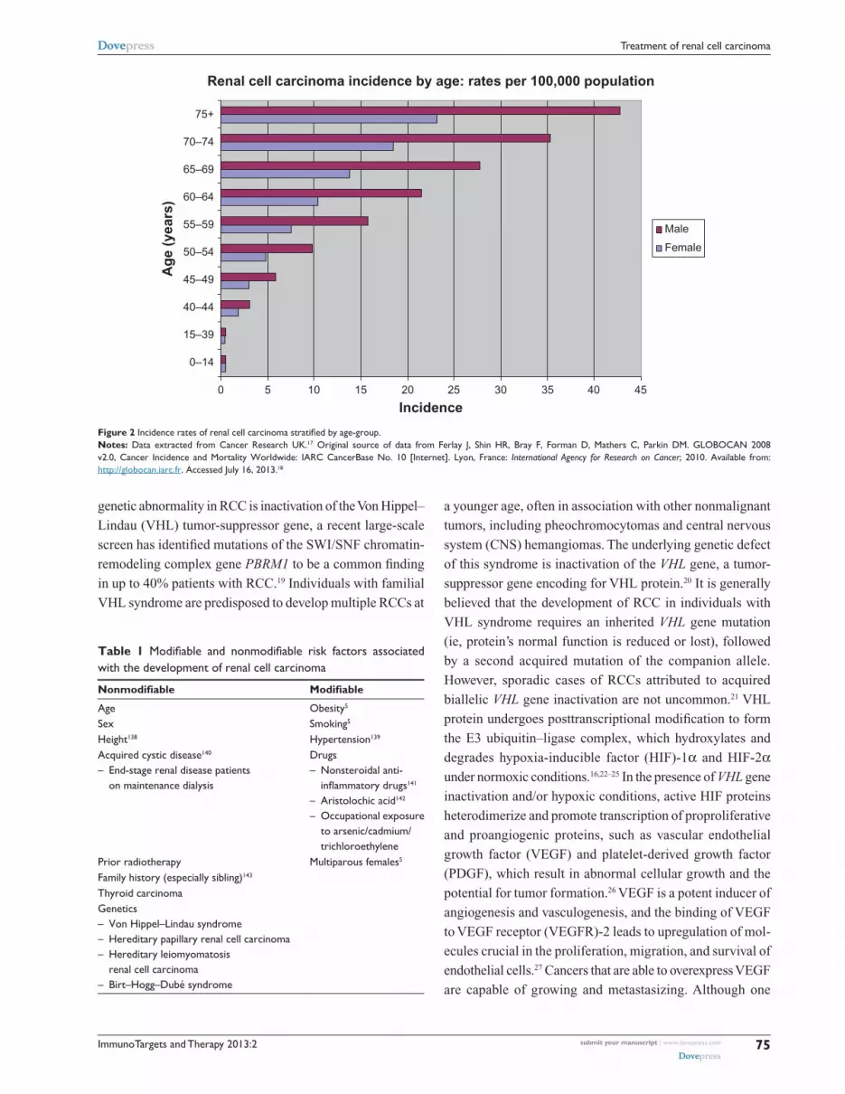
The mean age at diagnosis of RCC in the general popula-
tion is 64 years, and the incidence of RCC continues to rise
with increasing age (Figure 2).15 Although the majority of
RCCs are localized at the time of diagnosis, one in three cases
are at an advanced stage on initial presentation. The 5-year
survival rates of patients with and without metastatic disease
at presentation are 10% and 85%, respectively.16
Risk factorsRisk factors for RCC include genetic and environmental
factors, and these are shown in Table 1. There is a strong
association between increasing body mass index and the risk
of RCC, such that for every 5 kg/m2 increase in body mass
index, there is a 24% and 34% increased risk of RCC in
males and females, respectively.5 Similarly, tobacco exposure
is associated with a 50% and 20% greater risk of RCC in
males and females, respectively.5
Genetic factorsThe identification of several gene mutations has provided
new insights into the immunobiology of RCC, which is
crucial in prognosis and the future development of novel
treatment for this cancer. Although the most recognized
Age-standardized incidence rates of renal cell carcinoma per 100,000 population
Incidence0 2 4 6 8 10 12 14 16 18
North America
Central/Eastern Europe
Northern Europe
World
South America
West Asia
Carribean
Central Africa
East Africa
West Africa
Female
Male
Figure 1 Age-standardized incidence rates of renal cell carcinoma according to sex and country.Notes: Data extracted from Cancer Research UK.17 Original source of data from Ferlay J, Shin HR, Bray F, Forman D, Mathers C, Parkin DM. GLOBOCAN 2008 v2.0, Cancer Incidence and Mortality Worldwide: IARC CancerBase No. 10 [Internet]. Lyon, France: International Agency for Research on Cancer; 2010. Available from: http://globocan.iarc.fr. Accessed July 16, 2013.18
submit your manuscript | www.dovepress.com
Dovepress
Dovepress
74
Graves et al
ImmunoTargets and Therapy 2013:2
a younger age, often in association with other nonmalignant
tumors, including pheochromocytomas and central nervous
system (CNS) hemangiomas. The underlying genetic defect
of this syndrome is inactivation of the VHL gene, a tumor-
suppressor gene encoding for VHL protein.20 It is generally
believed that the development of RCC in individuals with
VHL syndrome requires an inherited VHL gene mutation
(ie, protein’s normal function is reduced or lost), followed
by a second acquired mutation of the companion allele.
However, sporadic cases of RCCs attributed to acquired
biallelic VHL gene inactivation are not uncommon.21 VHL
protein undergoes posttranscriptional modification to form
the E3 ubiquitin–ligase complex, which hydroxylates and
degrades hypoxia-inducible factor (HIF)-1α and HIF-2α
under normoxic conditions.16,22–25 In the presence of VHL gene
inactivation and/or hypoxic conditions, active HIF proteins
heterodimerize and promote transcription of proproliferative
and proangiogenic proteins, such as vascular endothelial
growth factor (VEGF) and platelet-derived growth factor
(PDGF), which result in abnormal cellular growth and the
potential for tumor formation.26 VEGF is a potent inducer of
angiogenesis and vasculogenesis, and the binding of VEGF
to VEGF receptor (VEGFR)-2 leads to upregulation of mol-
ecules crucial in the proliferation, migration, and survival of
endothelial cells.27 Cancers that are able to overexpress VEGF
are capable of growing and metastasizing. Although one
Renal cell carcinoma incidence by age: rates per 100,000 population
Incidence
Ag
e (y
ears
)
0 5 10 15 20 25 30 35 40 45
0–14
15–39
40–44
45–49
50–54
55–59
60–64
65–69
70–74
75+
Male
Female
Figure 2 Incidence rates of renal cell carcinoma stratified by age-group.Notes: Data extracted from Cancer Research UK.17 Original source of data from Ferlay J, Shin HR, Bray F, Forman D, Mathers C, Parkin DM. GLOBOCAN 2008 v2.0, Cancer Incidence and Mortality Worldwide: IARC CancerBase No. 10 [Internet]. Lyon, France: International Agency for Research on Cancer; 2010. Available from: http://globocan.iarc.fr. Accessed July 16, 2013.18
Table 1 Modifiable and nonmodifiable risk factors associated with the development of renal cell carcinoma
Nonmodifiable Modifiable
Age Obesity5
Sex Smoking5
Height138 Hypertension139
Acquired cystic disease140
– End-stage renal disease patients on maintenance dialysis
Drugs– Nonsteroidal anti-
inflammatory drugs141
– Aristolochic acid142
– Occupational exposure to arsenic/cadmium/trichloroethylene
Prior radiotherapy Multiparous females5
Family history (especially sibling)143
Thyroid carcinomaGenetics– von Hippel–Lindau syndrome– Hereditary papillary renal cell carcinoma– Hereditary leiomyomatosis
renal cell carcinoma– Birt–Hogg–Dubé syndrome
genetic abnormality in RCC is inactivation of the Von Hippel–
Lindau (VHL) tumor-suppressor gene, a recent large-scale
screen has identified mutations of the SWI/SNF chromatin-
remodeling complex gene PBRM1 to be a common finding
in up to 40% patients with RCC.19 Individuals with familial
VHL syndrome are predisposed to develop multiple RCCs at
submit your manuscript | www.dovepress.com
Dovepress
Dovepress
75
Treatment of renal cell carcinoma

ImmunoTargets and Therapy 2013:2
of the main actions of VEGF-targeted therapy is inhibiting
new blood-vessel growth, therefore starving the tumor cells
of the necessary oxygen and nutrients to sustain continued
growth, the full therapeutic potential of this agent is relatively
complex and likely to involve multiple mechanisms.28 The
mammalian target of rapamycin (mTOR) pathway is also
capable of regulating cellular growth in response to hypoxic
conditions. mTOR is a serine/threonine kinase activated in a
pathway involving VEGF along with other growth factors and
protein kinases.29 In RCC, mTOR expression is significantly
increased, and tumors with high levels of mTOR expression
have been shown to be more aggressive and associated with a
poorer prognosis. Interestingly, the use of chemotherapeutic
agents and ionizing radiation have been shown to enhance
mTOR expression by activating upstream regulators of
mTOR, which in part may contribute to the lack of efficacy
of these therapies in the treatment of RCC.30
Although multiple genetic mutations have been identi-
fied for all RCC subtypes, VHL gene inactivation appears
to be restricted to clear-cell RCC. Other reported genetic
mutations identified for clear-cell RCCs include deletions
of parts of chromosome 3p, mutation of gene PBRM1, gain
of chromosome 5q and loss of 8p, 9p, and 14q; trisomy of
chromosomes 7 and 17, loss of the Y chromosome, gain of
chromosomes 12, 16, and 20, mutation of the tricarboxylic
acid cycle enzyme fumarate-hydratase (a tumor-suppressive
gene), and rare mutations of the Met proto-oncogene reported
for papillary RCCs; and mutations of the tumor-suppressor
folliculin gene and loss of chromosomes 1, 2, 6, 10, 13, 17,
21, and Y reported for chromophobe RCCs.19,31–33
Types of renal cell carcinomaAlthough there are multiple histological subtypes of RCC,
clear-cell RCCs are the most common and account for up
to 80% of RCC in the general population. The characteris-
tic histological appearance of clear-cell RCCs is the clear
cytoplasm and well-defined cell membrane, with the trans-
parent cytoplasm attributed to accumulation of cholesterol
esters, glycogen, and phospholipids.20 In contrast to the
general population, papillary cell RCC is the predominant
cancer type in kidney-transplant recipients (15% versus
[vs] 44%) and is more likely to be bilateral and multifocal
at initial presentation.10 Chromophobe RCCs are relatively
uncommon, and account for up to 5% of RCC in the general
population. This tumor type rarely metastasizes and has the
best prognosis, with 5-year survival approaching 90%, com-
pared to 10% survival for patients with metastatic clear-cell
or papillary RCC.34
Prognostic factors for renal cell carcinomaThe Memorial Sloan-Kettering Cancer Center (MSKCC)
prognostic score is a useful tool for predicting survival
amongst those with advanced-stage disease treated with
immunotherapy or chemotherapy.35 The MSKCC score was
derived from a cohort of 400 patients who had received
interferon (IFN)-based therapy for metastatic RCC, and
comprises Karnofsky performance status, lactate dehydro-
genase (LDH) level, hemoglobin level, serum calcium level,
and prior nephrectomy. The median survival of patients with
an MSKCC score of 0 is 20 months (favorable prognosis),
reducing to 10 (intermediate prognosis) and 4 months (poor
prognosis) in those with scores of 1–2 and 3–5, respectively.
Another large multicenter study of 645 patients treated
with VEGF-targeted therapy demonstrated that prognostic
categories derived from performance status, calcium level,
hemoglobin level, neutrophil count, platelet count, and
time from diagnosis reliably predicted survival and may be
superior compared to the MSKCC score by more accurately
reclassifying patients into the correct prognostic categories
by almost 10% (survival in intermediate- and poor-prognosis
groups of 27 and 9 months respectively).36 Other adverse
prognostic factors identified in other studies include failed
treatment with radiotherapy, multiple metastatic sites, sar-
comatoid differentiation, elevated alkaline phosphatase,
neutrophilia, and thrombocytosis.37–39
Several inflammatory and tumor-specific biomarkers have
recently been identified as important prognostic markers of
survival in patients with metastatic RCC. There is an inverse
association between serum interleukin (IL)-6 level and progres-
sion-free and overall survivals. Serum IL-6 level above 35 pg/
mL is associated with a fourfold increased risk of cancer-related
mortality.40 The modified Glasgow Prognostic Score derived
from a cohort of 169 patients, and showed a strong association
between a score calculated from C-reactive protein and serum
albumin and cancer-specific survival (hazard ratio [HR] 5.13,
95% confidence interval [CI] 2.89–9.11; P , 0.01).41 Although
several tumor biomarkers, such as carbonic anhydrase IX,
HIF-1-α, p53, VEGFR-1, B7-HI, and survivin, appear promis-
ing in further improving the prognosis of cancer mortality, the
clinical utility of these markers has not been widely adopted
because of their availability and cost (Table 2).25,38,42
Treatment options for renal cell carcinomaThe finding that RCCs are relatively insensitive to standard
chemotherapeutic regimens has led to the development of
submit your manuscript | www.dovepress.com
Dovepress
Dovepress
76
Graves et al

ImmunoTargets and Therapy 2013:2
In patients with large tumors, the use of neoadjuvant
targeted therapies to reduce tumor bulk has allowed
successful CN to proceed.49 A study comparing 44 patients
with metastatic RCC who had received neoadjuvant targeted
therapies (bevacizumab or sorafenib) prior to CN to a
matched cohort of 58 patients who had undergone CN at
the outset showed a possible survival benefit with neoad-
juvant treatment (18% vs 31% mortality in neoadjuvant/
CN and CN groups, respectively).50 Another small study
of patients with metastatic RCC showed a lack of sur-
vival benefit in those who had received targeted therapy
(sorafenib/ sunitinib) following CN compared to targeted
therapy alone (median progression-free survival of 12 vs
9 months, respectively).46 A retrospective study of 188
patients with metastatic RCC demonstrated that patients
who had received targeted therapy without CN had a higher
median overall survival of 13 months, compared with a
median of 8 months in historical patients who had received
IFN-α without CN.51 Nevertheless, the role of adjuvant
targeted therapy prior to CN remains debatable and, future
studies addressing the role of CN with sequential targeted
therapy are required.
In kidney-transplant recipients with allograft RCC,
nephron-sparing surgery can be considered if the tumor is
superficial and its size is ,4 cm, but renal allograft nephre-
ctomy is often undertaken if the allograft has failed or if the
tumor is multifocal.10,52 Regular ultrasonography of the renal
allograft following nephron-sparing surgery is essential to
detect tumor recurrences.53
Stereotactic radiotherapyThe benefit of radiotherapy in the treatment of RCC remains
unclear and is not recommended.42 A recent meta-analysis
(n = 735 in a total of seven trials: five retrospective and two
prospective studies) demonstrated that postnephrectomy
radiotherapy significantly reduced the risk of local and/
or regional recurrences by 53% (pooled odds ratio 0.47,
99% CI 0.33–0.68), but this benefit did not translate to an
improvement in disease-free survival or overall survival.54
Although the incidence of adverse events was similar in
those receiving or not receiving radiotherapy, there were six
deaths from gastrointestinal and hepatic toxicities, which
were thought to be directly attributable to radiotherapy. The
suggestion that computer tomography-based planning prior
to radiotherapy might be associated with improved response
rate and reduced incidence of adverse events will need to
be carefully examined in future studies. Nevertheless, it is
unlikely that radiotherapy will be of benefit in many patients,
Table 2 Prognostic indicators of renal cell carcinoma
Patient factors Tumor factors Laboratory parameters
Performance statusSmoking
Tumor subtypeMetastasis ($2 sites)Time from diagnosis to treatmentRetroperitoneal nodal metastasisFailed treatment with radiotherapySarcomatoid differentiation
AlbuminC-reactive proteinHemoglobinLactate dehydrogenaseNeutrophil countPlatelet countLymphocyte countAlkaline phosphataseCalcium levelInterleukin-6
immunotherapeutic agents aimed at potentiating antitumor
immune surveillance. Both dendritic cells (DCs) and T cells
have been identified in tumor tissue, which indirectly suggests
the importance of these immune cells in the immunobiology
of RCC.43 Spontaneous tumor remission in the absence of
treatment occurs in less than 2% of cases, but cytoreduc-
tive nephrectomy (CN) has been associated with regression
of metastatic RCC, possibly by reducing tumor-derived
T-cell-inhibitory factors and reducing tumor-derived growth
factors.16,44
Cytoreductive nephrectomyAlthough the role of CN remains controversial in metastatic
RCC, it is generally accepted that surgery is often necessary
as an adjunctive treatment to immunotherapy (eg, IFN-α).45
Furthermore, CN may provide symptomatic relief, often
associated with a modest improvement in survival (median
3–6 months), especially in those with large tumor bulk or
paraneoplastic syndromes.46,47 Tumor size, performance status
presurgery, and recurrence/growth of tumor postsurgery are
well-recognized prognostic factors known to affect survival
following CN.14 In patients with limited metastatic disease,
metastasectomy may be considered in younger patients,
those with solitary non-CNS lesions, and those with disease
detected over 12 months following CN, with the expectation
of achieving a 5-year survival of 30%.20,47
In a large retrospective series of 566 patients with meta-
static RCC, the investigators identified hypoalbuminemia,
elevated LDH levels, tumor stage T3 or above, symptomatic
metastatic disease, presence of liver metastasis, and retro-
peritoneal or supradiaphragmatic lymph-node involvement
were factors associated with poorer survival.48 Furthermore,
patients with four or more of these risk factors did not benefit
from CN. In kidney-transplant recipients, symptomatic dis-
ease and tumor size of .40 mm were associated with poorer
survival following CN.10
submit your manuscript | www.dovepress.com
Dovepress
Dovepress
77
Treatment of renal cell carcinoma

ImmunoTargets and Therapy 2013:2
because up to 85% of patients who experience treatment
failure will develop metastatic disease.55
Stereotactic body radiotherapy (SBRT) is a novel technique
that utilizes short courses of intensive, highly focused radia-
tion delivered to metastatic lesions. This technique appears
promising, and is associated with excellent local control and
stabilization of tumor growth (defined as lack of tumor activity
on positron emission tomography scan or expansion of tumor
size of ,20%).56,57 Multiple case series have demonstrated
that SBRT was successful in both reducing local symptoms
from tumor bulk and stabilization of the growth of metastatic
lesions at both cranial and extracranial sites. Small tumor vol-
ume, greater numbers of fractions and dose per fraction, and
higher biological effective dose of SBRT have been shown to be
associated with improved symptom control from stabilization
of metastatic tumor growth in up to 90% of cases.58–60
Immune-based therapiesThere has been considerable focus on the effective-
ness of immunotherapy in patients with metastatic RCC.
Immune-based therapies can be broadly categorized into
non-tumor-targeted and tumor-targeted therapies. Nontargeted
therapies include subcutaneous IFN-α or intravenous or sub-
cutaneous IL-2, both of which can induce a nonspecific graft
versus tumor and inflammatory responses. Tumor-targeted
therapies include VEGF and mTOR inhibitors (Figure 3),
DC peptide-based vaccines (tumor antigens are presented
by patient’s antigen-presenting cells) and adoptive cell
transfer, the latter utilizing ex vivo expanded tumor antigen-
pulsed autologous lymphocytes that are reinfused back into
patients.43,61 Whilst targeted therapies may be associated with
superior progression-free survival compared with IFN-α/IL-2,
these nontargeted cytokine therapies have also been shown to
induce sustained, drug-free remission. It has also been shown
that VEGF-targeted therapy may be more effective at reducing
tumor bulk compared with mTOR inhibitors.62 In the absence
of definitive randomized studies in the treatment of non-clear-
cell RCC, it is generally recommended that this cancer type
should be treated with sunitinib, sorafenib, or temsirolimus,
but it remains unclear which agent is superior.63,64
Tumor cell
Endothelial cell
Inactive VHL
HIFα HIFα
Pro Pro OH
mTORC2
mTORC1
mTOR
PI3-K
S6K
S6FKBP
Geneactivation
Growthfactorie, IGF
MIP-3αIL-6IL-8VEGF
Maturationarrest
IFN-α
Processing
MHC class 2
Costimulatinginteraction
CD4+ T cellCD8+ T cell
Inhibited by myeloid derived suppressor cells
MHC class 1
Tumor antigen
Tumor antigen
Lysis
CytotoxicT cell
PDGF
VEGF
PI3-K
Trovaxanti-G250
X
TemsirolimusEverolimus
Dendritic cell
AKt
AKt
Geneactivation
VEGFR
XX
PDGFR
AfliberceptBevacizumab
SunitinibSorafenibAxitinibPazopanib
X
.
.
.
Figure 3 Site of actions of targeted therapy used in the treatment of metastatic renal cell carcinoma.Notes: The tumor cell possesses inactive von Hippel–Lindau (vHL), permitting the production of heterodimerized hypoxia-inducible factor (HIF) under normoxic conditions. Mammalian target of rapamycin (mTOR) activation further facilitates HIF production. HIF and S6 contribute to gene activation, leading to production of vascular endothelial growth factor (vEGF) and platelet-derived growth factor (PDGF), both of which act upon the endothelial cell to promote angiogenesis. Bevacizumab targets only vEGF, whereas sunitinib and sorafenib target vEGF receptors as well as PDGF and c-kit. Temsirolimus and everolimus inhibit the mTOR signaling pathway. Dendritic cells (DCs) are crucial in antitumor immunity, and mature DCs interact with both innate immune cells and antigen-specific T cells to elicit an immune response against tumor antigens. Tumor-derived factors such as vEGF, interleukin (IL)-6, and IL-8 may inhibit DC maturation, therefore escaping immune surveillance. Nontargeted therapies such as interferon (IFN)-α (by promoting maturation of DCs) and DC-based vaccination (by presenting tumor antigens to induce an antigen-specific cytotoxic T-cell response) are other effective treatment options for metastatic renal cell carcinoma.Abbreviations: MHC, major histocompatibility complex; PI3-K, phosphatidylinositide 3-kinase; MIP, macrophage inflammatory protein; X, site of action; VEGF, vascular endothelial growth factor; FKBP, FK-binding protein; IGF, insulin-like growth factor, PDGFR, platelet-derived growth factor receptor.
submit your manuscript | www.dovepress.com
Dovepress
Dovepress
78
Graves et al

ImmunoTargets and Therapy 2013:2
Nontargeted therapiesInterferon-α and interleukin-2IFN-α and IL-2 were the first two immunotherapeutic agents
approved for use in metastatic RCC. IFN-α is administered
subcutaneously, and IL-2 as an intravenous bolus or infusion.
Although the precise antitumor properties of IFN-α have
not been clearly defined, this cytokine has been shown to be
capable of inhibiting angiogenesis and cell cycling, as well
as enhancing the activity of several immune cells. IFN-α
is the predominant type I IFN produced by plasmacytoid
DCs.65 Type I IFNs are capable of coordinating the innate and
adaptive immune responses by directly affecting innate cells
(eg, natural killer [NK] cells) as well as antigen-specific T
cells and memory B cells.66 The ability of plasmacytoid DCs
to enhance cytotoxicity of NK and cluster of differentiation
(CD8)+ T cells as well as protecting DCs from NK cell-
mediated lysis of immature DCs is mediated by their ability
to produce type I IFN.67,68 Type I IFN may also promote the
cross-presentation and cross-priming of antigens by CD8+
T cells,69 as well as inducing T-cell activation (increased
expression of CD69) and survival,70 all of which may have
an important role in antitumor immunity. IL-2 is produced
by T cells in response to the interaction between antigen-
presenting cells and T cells (including T-cell recognition of
antigens presented by antigen-presenting cells), which leads
to the activation and proliferation of antigen-specific T cells.
Furthermore, IL-2 exerts an immunomodulatory effect by
promoting the apoptosis of activated T cells and the matura-
tion of regulatory T cells, the latter known to be capable of
suppressing immune reactivity of other immune cells.71
Although the therapeutic potential of IFN-α in the
treatment of metastatic RCC was identified in the 1980s,
randomized controlled prospective trials involving this agent
were not conducted until the 1990s. The first such study
compared 41 patients receiving IFN-α (subcutaneous dose
of 8 million units thrice weekly) and vinblastine (intravenous
dose of 0.1 mg/kg thrice weekly) with 35 patients receiving
hormonal therapy medroxyprogesterone (intramuscular dose
of 500 mg weekly). Survival was similar between the two
groups, but a greater proportion of the patients in the IFN-α
vinblastine group had achieved partial or complete remission
compared with patients in the medroxyprogesterone group.72
A subsequent study randomized 160 patients to receive either
vinblastine alone (dose of 0.1 mg/kg thrice weekly) or vin-
blastine in combination with IFN-α (dose of 3 million units
thrice weekly, increasing to 18 million units after the first
week) for 12 months or until disease progression.73 Median
survival (68 vs 38 weeks, P = 0.005) and progression-free
survival (13 vs 9 weeks, P , 0.001) in the vinblastine/IFN-α
group were significantly better compared with the vinblas-
tine group. A randomized study involving 335 patients with
metastatic RCC demonstrated that patients randomized to
IFN-α (two doses of 5 million units followed by 10 million
units in the first week, then 10 million units thrice weekly for
a further 11 weeks) had a 28% reduction in the risk of mor-
tality (HR 0.72, 95% CI 0.55–0.94) compared with patients
randomized to medroxyprogesterone acetate (300 mg daily
for 12 weeks). Similarly, patients randomized to the IFN-α
group had a survival advantage with median increase in
survival of 2.5 months (95% CI 0.5–5.0 months).74
There have been multiple studies comparing the effi-
cacy of IFN-α to IL-2, either alone or in combination.
In a large multicenter trial involving 425 patients with
metastatic RCC, patients randomized to IFN-α (18 × 106 IU
subcutaneously three times a week for 10 weeks, followed
by maintenance therapy for a further 12 weeks) achieved
similar overall survival compared to patients randomized to
IL-2 (four cycles of daily subcutaneous dose of 18 × 106 IU
per square meter of body surface area) and a combination
of both agents (IL-2 with the addition of IFN-α 6 × 10⁶ IU
thrice weekly).75 Patients receiving combination IFN-α/
IL-2 were significantly more likely to achieve a clinical
response (defined as 50% reduction in the size of all lesions
on serial computed tomography imaging) compared with
either therapy alone in intention-to-treat and on-treatment
analyses (P , 0.01). Patients receiving IL-2 therapy, alone or
in combination, experienced higher rates of adverse events,
including vasopressor-resistant hypotension and fevers.
In another study involving 492 treatment-naive patients
with metastatic RCC (any histological type, more than one
metastatic site with Karnofsky score of $80%, normal
liver function and hematological parameters, and baseline
creatinine of ,160 µmol/L), patients were randomized to
receive medroxyprogesterone (oral dose of 200 mg daily),
IFN-α (9 million IU thrice weekly), IL-2 (9 million IU daily
or alternate daily), or in combination (IL-2 with IFN-α at
6 million IU per dose) for 12 weeks, extending up to 24 weeks
in the absence of tumor progression. There was no significant
difference in progression-free survival or overall survival
between all four groups, suggesting no survival benefit with
the use of cytokine therapies either alone or in combination.76
Consistent with prior studies, IL-2 therapy, especially in com-
bination with IFN-α, was associated with a much higher risk
of adverse events compared with medroxyprogesterone (59%
versus 10%, P , 0.001) including performance impairment
(30% versus 2%), weight loss and fever (27% versus 0%),
submit your manuscript | www.dovepress.com
Dovepress
Dovepress
79
Treatment of renal cell carcinoma

ImmunoTargets and Therapy 2013:2
gastrointestinal disturbances (14% versus 1%), anemia (3%
versus 0%), leukopenia (3% versus 2%), and neutropenia
(4% versus 0%). Other randomized studies in previous
untreated patients with metastatic RCC also demonstrated
no survival benefit with the use of IL-2 or IFN-α, alone or in
combination with other chemotherapeutic agents.77,78 A recent
systematic review of 6880 patients with advanced renal cell
carcinoma who had received an immunotherapeutic agent in
at least one study arm and reported remission or survival by
allocation suggested that the use of IFN-α was associated
with an improvement in median survival by 2.8 months.44
The reported remission rates in patients participating in trials
were 1.8% in the group receiving control/placebo therapies,
7.6% with single-cytokine therapy, 12.9% with combined-
cytokine therapy, and 22.9% with high-dose IL-2 therapy.
Future studies evaluating the role of IFN-α and IL-2 with
targeted therapy as well as determining the optimal dose and
duration of nonspecific cytokine therapy are required.
Anti-programmed death ligand 1 therapyProgrammed death-receptor ligand 1 (PD-L1), also called
B7-H1, is a member of the B7 family, which on interaction
with PD-1 negatively regulate T-cell receptor signaling.79
It has been shown that aggressive forms of RCC express
PD-L1 and the interaction between tumor cells PD-L1 and
immune cells PD-1 contributes to immune dysregulation in
these patients and promotes cancer progression.80 In pre-
clinical models, blockade of interactions between PD-1 and
PD-L1 mediates antitumor activity, suggesting a potentially
novel form of antitumor immunotherapy.81 BMS-936559 is a
humanized anti-PD-L1 monoclonal antibody, which inhibits
PD-L1 binding to both PD1 and the T-cell ligand CD80.
In a multicenter phase I study involving 207 patients with
advanced solid organ cancers (including 17 patients with
metastatic RCC), patients received 6-week cycles of intrave-
nous anti-PD-L1 (doses of 0.3–10 mg/kg) up to a maximum
of 16 cycles. An objective response was observed in most
cancer types, including 12% of patients with RCC. Although
adverse events were common, the majority of symptoms
were mild, including infusion reactions, fatigue, diarrhea,
rash, and pruritus.82 BMS-936558, a humanized monoclonal
antibody that blocks PD-1, appears to be equally efficacious
in patients with advanced solid organ cancers and has been
shown to produce an objective response in up to 27% of
patients with RCC.83 Most adverse events were mild, includ-
ing fatigue, diarrhea, rash, anorexia, nausea, and pruritus,
but 11% of patients experienced more severe adverse events,
including pneumonitis and elevated transaminases. Phase II
and III studies are currently under way to define further the
role of these agents in the treatment of metastatic RCC.
Targeted therapiesvaccinesEarlier vaccines used in the treatment of metastatic RCC were
largely disappointing, but the newer peptide-based vaccines
appear more promising. Antigen selection for vaccine design
appears to be the key in the improved efficacy achieved with
peptide-based vaccines, but nevertheless peptide-based
vaccines do not provide a sustained antitumor response and
should be considered as adjunctive therapy to other immuno-
therapeutic or chemotherapeutic agents.84,85 In a randomized
controlled trial involving the use of TroVax, a recombinant
modified vaccinia virus Ankara vector encoding the oncofetal
target antigen 5T4 peptide-based vaccine (TroVax Renal
Immunotherapy Survival Trial), patients with metastatic RCC
were randomized to receive MVA-5T4 peptide-based vaccine
or placebo in combination with sunitinib, IL-2, or IFN-α.86,87
Although a survival advantage was not demonstrated with
the addition of this vaccine, a post hoc analysis restricted to
a cohort of patients with lower-grade MSKCC did show a
significant survival advantage if treated with TroVax vaccine
and IL-2, compared with placebo (mortality HR 0.54, 95%
CI 0.3–0.98; P = 0.046). It remains unclear whether the poor
response to vaccination is a reflection of inadequate vaccine
dose or that tumors were lacking 5T4 expression, the latter
being important for antitumor response. Another peptide-
based vaccination complex to tumor necrosis factor-α or
heat-shock proteins has been developed, and although this
vaccine appears efficacious in murine RCC models, it has
been disappointing in phase I human trials.85,88,89
DCs are a group of rare, heterogeneous, professional
antigen-presenting cells that can initiate primary immune
responses, and hence have the ability to regulate both innate
and adaptive immune responses (Figure 4).90,91 Precursor
DCs, arising from bone marrow progenitors, enter tissues
as immature DCs with superior phagocytic capabilities. DCs
then encounter foreign antigens, such as bacteria and tumor
antigens, resulting in the secretion of cytokines (eg, IFN)
and activation of NK cells, macrophages, and eosinophils.
Following antigen capture and processing, DCs undergo
maturation and migrate to secondary lymphoid tissues,
where they present processed antigen/peptide coupled to
major histocompatibility complexes to T cells, allowing for
selection and expansion of antigen-specific CD4+ T-helper
cells. These CD4+ T-helper cells subsequently amplify the
immune responses by regulating antigen-specific (eg, CD8+
submit your manuscript | www.dovepress.com
Dovepress
Dovepress
80
Graves et al

ImmunoTargets and Therapy 2013:2
cytotoxic T cells, B cells), and antigen nonspecific (eg, mac-
rophages, NK cells, and eosinophils) effector cells. As DCs
have a prominent role in the initiation of innate and adaptive
immune response against invading pathogens, they are likely
to have an equally important role in antitumor immunity. The
development of a tumor invariably involves the failure of the
immune system to recognize tumor antigens, leading to an
abnormal proliferation of tumor cells. Tumors may evade
immune recognition directly or indirectly (via the production
of suppressive cytokines and other mediators) by affecting
normal DC and T-cell functions.
Following initial success in eliciting immunogenicity
against antigens delivered by DCs in patients with cancer and
HIV infection, therapeutic DC-based vaccines such as the US
Food and Drug Administration (FDA)-approved DC-based
vaccine against metastatic castration-resistant prostate cancer,
have been developed and used in clinical studies to generate
protective immunity against certain types of tumors.92–94 These
clinical studies in humans were initiated following observa-
tions in mice that ex vivo generated DCs could induce both
humoral and tumor-specific immunity, and may be superior
to other forms of vaccines.92 Established protocols involving
mature DCs generated from CD34+ bone marrow-precursor
cells and monocytes have been successful in the induction of
antitumor immunity. DCs used in vaccination-based protocols
involving tumor antigen must be phenotypically mature to
ensure that DCs are capable of migrating to secondary lym-
phoid organs to initiate tumor antigen-specific T-cell immunity
when delivered into the host.95 Various maturation stimuli have
been trialed, including cytokines (eg, IL-1, IL-6, tumor necrosis
factor-α, prostaglandin E2), Toll-like receptor ligands, CD40 L,
major histocompatibility complex-binding antigens (including
peptides, protein, tumor lysates, apoptotic cells), and DNA
and RNA transfection of DCs. DC-based vaccination strategy
in human subjects has been shown to induce antigen-specific
T-cell response and may even generate tumor antigen-specific
cytotoxic T lymphocytes in tumor tissues.96,97
DC-tumor peptide-based vaccination could potentially
promote a vigorous tumor antigen-specific T-cell response
in patients with RCC.98 A systematic review of 29 random-
ized controlled trials of DC tumor antigen-based vaccination
comprising a total of 906 patients with either metastatic
Immatureconventional DC
Immatureplasmacytoid DC
Monocytes
Mature DC
NK cells
IFN-α/β, TNF-α IL-1, IL-6, IL-8, IL-12, IL-15, IL-18, IL-23
Positive feedback to promotefurther DC activation and
maturation
Stimulus (microbial products,TLR ligands etc)
Induce DCmaturation
NKG2D interactionCostimulatorymoleculesinteraction
Cell-cell contact
B cells
Plasmacells
Antibodies
Treg cellsTh1/2cells
Antigen-specific CTL
CD4+/CD8+
T cells IL-2
Lysis ofimmature
DCIFNγ andcytotoxicity
Figure 4 Overview of the relationship between dendritic cells and effector cells.Notes: Immature conventional or plasmacytoid DCs mature in response to appropriate stimuli (eg, microbial products, TLR ligands). Mature DCs secrete immunoregulatory cytokines (including IFN-α and IL-12), and through cell–cell interactions modulate effector cell response including NK cells and B and T cells, as well as providing positive feedback to DCs to initiate ongoing activation and maturation. Activated effector cells could in turn modulate DC activation, maturation, and survival, as well as enhancing other effector cell functions through the production of cytokines (IFN-γ) and/or via cell–cell contact.Abbreviations: DC, dendritic cell; IFN, interferon; IL, interleukin; NK, natural killer; CTL, cytotoxic T lymphocyte; Treg cells, regulatory T cells; Th, T helper; TLR, Toll-like receptor; TNF, tumor necrosis factor.
submit your manuscript | www.dovepress.com
Dovepress
Dovepress
81
Treatment of renal cell carcinoma

ImmunoTargets and Therapy 2013:2
RCC or patients with recurrent or metastatic prostate cancer
showed that the clinical benefit rate (a combined objective
response rate with stable disease rate) was 48% in patients
with metastatic RCC receiving DC-based vaccination.
Meta-analysis of individual patient data demonstrated that
cellular immune response and DC dose had a significant
influence on clinical benefit rate in patients with metastatic
RCC. Of patients with metastatic RCC, 92% had received
prior surgery or radiotherapy, 17% had received prior che-
motherapy, 36% had received prior immunotherapy, and 36%
had received concomitant IL-2 or combined IFN-α/IL-2 with
DC-based vaccine. There were a few mild adverse effects,
particularly local reactions at injection sites and nonspe-
cific constitutional symptoms, including fever and flu-like
symptoms. Potential mechanisms of immune surveillance
escape include the promotion of local lymphoid chemokine
expression, such as tumor-derived macrophage inflammatory
protein 3-α, which appears to promote recruitment of imma-
ture DCs into tumors, thereby inhibiting T-cell activation.99
Other soluble factors, including IL-8, IL-6, and VEGF, may
also inhibit DC maturation, and it is plausible that attempts
to mature these immature DCs could lead to the enhancement
of antitumor response.100
Novel targeted therapiesTargeted therapies directed against the VEGF and mTOR
pathways have become the treatment of choice for metastatic
RCC, with activity against both primary and metastatic
lesions (Table 3).36,42,101 Bevacizumab is a humanized mono-
clonal antibody directed against VEGF, whereas sunitinib,
sorafenib, and pazopanib are tyrosine kinase inhibitors that
target the downstream effects of VEGF activation. These
tyrosine kinase inhibitors have differing binding affinities
to molecular targets, and other than inhibition of VEGFR2
and VEGFR3, they may also inhibit PDGFR-β and/or
c-Kit. Temsirolimus and everolimus are specific mTOR
kinase inhibitors.102 Studies have identified several markers
that predict response to treatment and/or survival, which
include VEGF levels and gene single-nucleotide polymor-
phisms, particularly the Cytochrome P450 3A5*1 allele. A
tissue microarray-based immunohistochemical analysis of
upstream and downstream elements of the mTOR pathway
revealed pS6 as the strongest predictor of survival in both
localized and metastatic RCC.103,104 In murine models, loss
of phosphatase and tensin homologue, which reverses the
action of phosphatidylinositide 3-kinase PI3-K in activa-
tion of mTOR4, appeared to sensitize tumors to mTOR
inhibition.105
Tumors have developed several ways to escape immune
surveillance, and therefore become resistant to immuno-
therapeutic agents. RCCs have been shown to promote the
development of myeloid-derived suppressor cells, possibly
by production of granulocyte-macrophage colony stimulating
factor, resulting in T-cell hyporesponsiveness against tumor
cells.106,107 It has also been shown that certain VHL mutations
can lead to differences in VEGF activation, thereby result-
ing in the variable responses of RCC to VEGF inhibition.21
Agents directed against mTOR signaling are active against
the TOR1 complex subunit, which is important in the con-
trol of cell growth and proliferation, as well as stabilizing
HIF-1-α.108 In contrast, the TOR2 complex subunit is impor-
tant in cell morphology and adhesion, as well as promoting
Table 3 Summary of study outcomes using targeted therapy in the treatment of metastatic renal cell carcinomas
Treatment Study population Comparator Median progression-free survival (months)
Objective response
Bevacizumab + IFN-α114,116
Treatment-naive and prior nephrectomies IFN-αSunitinibBevacizumab + temsirolimus
10 vs 517 vs 817 vs 8
31% vs 13%39% vs 24%39% vs 27%
Sunitinib116,119 Treatment-naive ± previous nephrectomies IFN-αBevacizumab + temsirolimus
11 vs 58 vs 8
31% vs 6%24% vs 27%
Sorafenib126,127 Treatment-naive ± previous nephrectomies IFN-αSorafenib + IFN-αSorafenib + IL-2
6 vs 67 vs 89 vs 11
5% vs 9%30% vs 25%15% vs 27%
Pazopanib129 Treatment-naive or had previously failed cytokine-based therapy
Placebo 9 vs 4 –
Axitinib130 Failed prior treatment with sunitinib, bevacizumab/ IFN-alpha, temsirolimus, or cytokine therapy
Sorafenib 7 vs 5 –
Temsirolimus62 Treatment-naive ± previous nephrectomies IFN-α 6 vs 3 Overall survival 11 vs 7 months
Everolimus132 Failed previous cytokine or targeted therapy Placebo 4 vs 2 –
Abbreviations: IFN, interferon; IL, interleukin; vs, versus.
submit your manuscript | www.dovepress.com
Dovepress
Dovepress
82
Graves et al

ImmunoTargets and Therapy 2013:2
the expression of HIF-2-α expression (expression of HIF-1-α
can be promoted by either the TOR1 or TOR2 complex).109
Unlike the TOR1 complex, the TOR2 complex is resistant to
inhibition of mTOR signaling, thus providing another avenue
for mTOR inhibitor resistance in patients with metastatic
RCC.110,111 Increased insulin-like growth factor signaling as
a result of loss of mTOR/S6k inhibition may contribute to
mTOR resistance, but this remains debatable.109 Mutation
of the tricarboxylic acid-cycle enzyme fumarate-hydratase
(a tumor-suppressive gene) leads to upregulation and accu-
mulation of HIF-1-α, is more prevalent in papillary rather
than clear-cell RCC, and may explain why papillary RCC
may be more responsive to mTOR inhibition.109,112
BevacizumabBevacizumab is a recombinant humanized monoclonal
immunoglobulin G1 antibody produced in a mammalian
cell-culture system. Bevacizumab competitively binds to
and inhibits the activity of human VEGF both in vitro and
in vivo.113 In a large multicenter randomized controlled
study of 649 patients with metastatic RCC (phase III trial
of bevacizumab plus interferon alfa-2a in patients with
metastatic renal cell carcinoma), patients were randomized
to receive intravenous bevacizumab (at a dose of 10 mg/kg
every 2 weeks) together with subcutaneous IFN-α (dose of
9 million units thrice a week) or IFN-α with placebo.114 All
patients had prior nephrectomies and were treatment-naive.
Patients were block-randomized according to the country
of origin and prognostic grade determined by the MSKCC
score. The study was terminated prematurely following
interim analysis demonstrating that patients randomized
to bevacizumab and IFN-α treatment achieved better pro-
gression-free survival compared to the IFN-α-alone group
(median 10.2 versus 5.4 months, P , 0.001). There were no
significant differences between groups for the primary end
point in overall survival, likely explained by the decision
to unblind the study following interim analysis, with sub-
sequent crossover of the placebo group to the bevacizumab
and IFN-α treatment group (median overall survival in the
bevacizumab/IFN-α treatment vs IFN-α/placebo group
of 23.3 months vs 21.3 months, respectively; P = 0.34).
Fatigue, asthenia, proteinuria, and hypertension were the
most frequently reported adverse events, particularly in
the bevacizumab and IFN-α treatment group. Other less
common adverse events (#1%) attributed to the use of
bevacizumab included bleeding, myocardial ischemia and
infarction, left ventricular failure, gastrointestinal perfora-
tion, and thromboembolic events.115
The similarly designed CALGB 90206 and TORAVA
trials showed that the combination of bevacizumab/IFN-α
treatment was associated with higher median progression-free
survival compared with other treatments (CALGB 90206 –
bevacizumab/IFN-α 8.5 vs IFN-α 5.2 months; TORAVA –
bevacizumab/ IFN-α 16.8 vs sunitinib 8.2 vs bevacizumab/
temsirolimus 8.2 months).116,117 These studies do suggest that
the combination of bevacizumab and IFN-α may achieve
superior response in patients with favorable prognosis or
indolent disease. In contrast, the clinical benefit of combined
therapy with bevacizumab and IL-2 remains unclear. In a
phase II study, patients with untreated metastatic RCC who
had received bevacizumab and low-dose IL-2 demonstrated
an objective response rate of 15% with 38% of patients, with
reduction in tumor burden of ,30%.118
SunitinibSunitinib is an orally active multi-tyrosine kinase inhibitor,
which inhibits the actions of VEGF and angiogenesis, the
latter via inhibition of VEGFR1 and VEGFR2 and PDGFR-β.
In a large international, multicenter, randomized controlled
study of 750 patients with metastatic clear-cell RCC, patients
were randomized to receive oral sunitinib (dose of 50 mg
daily for 4 weeks) or IFN-α (sequentially escalating regi-
men to a maximum of 9 million units thrice weekly).119 All
patients were treatment-naive and were block-randomized
according to several prognostic factors, including LDH level,
Eastern Cooperative Oncology Group (ECOG) status, and
previous nephrectomy. Even though patients in the IFN-α
treatment group were allowed to cross over to the sunitinib
group following interim analysis, patients randomized to
the sunitinib group had significantly longer progression-free
survival compared to the IFN-α group at the end of the study
(11 months vs 5 months, respectively, P , 0.001). There
was no significant difference in overall survival between
groups, likely reflecting crossover of patients between treat-
ment groups. Common adverse events following sunitinib
use were gastrointestinal symptoms (diarrhea, stomatitis),
hepatotoxicity, constitutional symptoms (fatigue, reduced
appetite), cardiovascular abnormalities (hypertension, pro-
longed QT interval and left ventricular dysfunction of no
clinical significance), laboratory abnormalities (cytopenias,
elevated lipase and uric acid) and hand-foot syndrome.115,120
Fatigue was more common in patients randomized to IFN-α.
Other randomized phase II and III trials involving sunitinib
showed that the objective response rates were similar in those
receiving intermittent (4 weeks of 50 mg/day followed by
2 weeks off treatment) and continuous dosing (32% vs 28%);
submit your manuscript | www.dovepress.com
Dovepress
Dovepress
83
Treatment of renal cell carcinoma

ImmunoTargets and Therapy 2013:2
but there were lower objective response rates compared with
bevacizumab and IFN-α treatment (24% vs 39%).116,121 In
other studies involving the use of sunitinib, greater frequency
and severity of adverse events were observed in patients
of Korean ethnicity, possibly related to a difference in the
metabolism of this agent compared to other ethnic groups.122
Furthermore, the efficacy of sunitinib in non-clear-cell RCCs
has largely been disappointing.123
SorafenibSorafenib is an orally active multi-tyrosine kinase inhibitor,
with inhibitory actions against several protein kinases, includ-
ing VEGF, PDGFR, Raf-1, Flt-3, and c-Kit. Sorafenib is
approved for use in advanced RCC and advanced hepatocellu-
lar carcinoma. A large multicenter trial (TARGET; treatment
approaches in renal cancer global evaluation trial) random-
ized 903 patients with metastatic RCC to receive sorafenib
(dose of 400 mg twice daily) or matching placebo.124 Patients
were block-randomized according to country of enrollment
and prognostic score (low or intermediate risk on MSKCC
score), and all patients had received prior systemic therapy.
This study terminated prematurely when a planned interim
analysis demonstrated that patients randomized to sorafenib
had significantly lower risk of cancer progression compared
to placebo (HR 0.44, 95% CI 0.35–0.55, P , 0.01).
There was no significant difference in overall survival
between groups, likely reflecting crossover of patients
between treatment groups (ie, 48% of the placebo-assigned
group crossed over to the sorafenib group following interim
analysis). In a post hoc analysis censoring patients who
had crossed over from placebo to sorafenib, median overall
survival was significantly longer in the sorafenib group com-
pared to placebo (17.8 months vs 14.3 months, P = 0.03). In
addition, sorafenib appears to be well tolerated, has similar
efficacy in younger and elderly patients, and has been shown
to be associated with improved health-status questionnaire
scores across all age-groups.125 Although there was a higher
incidence of adverse events, especially in those aged , 70
years, including myocardial ischemia (,5% vs 0%), diar-
rhea (43% vs 13%), fatigue (36% vs 27%), hypertension
(18% vs 2%), hand-foot syndrome (31% vs 6%), and rash
(39% vs 15%) in patients who received sorafenib compared
to placebo, this drug may be better tolerated compared to
sunitinib. However, the cardiovascular-related adverse events
associated with these agents are unlikely to be of clinical
significance, and therefore sorafenib can be considered in
patients with cardiovascular disease. A number of phase II
trials comparing sorafenib with IFN-α or IL-2 alone or in
combination with sorafenib have failed to demonstrate any
differences in median progression-free survival between
treatment groups.126,127 It has been shown that patients treated
with either sorafenib or IFN-α with low serum levels of
IFN-α receptor 2 mRNA had poorer prognosis,128 and future
studies evaluating the response of cancer treatment according
to IFN-α receptor status are warranted.
PazopanibPazopanib is a potent, orally active multi-tyrosine kinase
inhibitor of VEGFRs 1–3, PDGFR, and c-Kit, all of which are
important in tumor growth and angiogenesis. It is approved
for use in advanced RCC and soft-tissue sarcomas, but also
has anti-tumor activities against ovarian cancers. A large
randomized double-blind placebo-controlled trial was con-
ducted involving 435 patients with locally advanced and/or
metastatic RCC.129 Recruited patients were either treatment-
naive or had had previously failed cytokine-based therapy.
Patients were block-randomized in a 2:1 ratio according to
ECOG status (0 versus 1), history of previous nephrectomy,
and prior systemic therapy to receive oral pazopanib (800 mg
daily) or matching placebo. Similar to other studies, patients
who had progressed on placebo were allowed to cross over
to the pazopanib group. Patients randomized to pazopanib
had significantly longer progression-free survival compared
to placebo (median 9.2 months vs 4.2 months, respectively;
P , 0.01), independent of previous treatment with cytokine
therapy. Treatment-related adverse events were more com-
mon in the pazopanib group, particularly hypertension and
hepatotoxicity with elevated transaminases and diarrhea.
Pazopanib-related mortality from cerebrovascular accident,
gastrointestinal perforation, and rectal hemorrhage occurred
in ,1% of patients. Discontinuation rates attributed to
adverse events were noted to be higher in those patients
who had received previous cytokine therapy. Results of a
recently completed large phase III noninferiority trial of
1110 patients with metastatic RCC randomized to pazopanib
or sunitinib showed that both agents were equally efficacious,
but pazopanib was better tolerated, with a significantly lower
incidence of hand-foot syndrome, mucositis, and stomatitis
(unpublished data).
AxitinibAxitinib is a potent, orally active inhibitor of VEGFRs 1–3
with minimal inhibitory effects of PDGFR and other recep-
tor kinases such as c-Kit. A large multicenter randomized
controlled study was conducted involving 723 patients with
progressive metastatic RCC and failed prior treatment with
submit your manuscript | www.dovepress.com
Dovepress
Dovepress
84
Graves et al

ImmunoTargets and Therapy 2013:2
sunitinib, bevacizumab/IFN-α, temsirolimus, or cytokine
therapy.130 Patients were block-randomized according to
ECOG status and previous systemic therapy to receive
axitinib (5 mg twice daily, and if tolerated, increasing to
a maximum of 10 mg twice daily) or sorafenib (400 mg
twice daily). Patients randomized to axitinib had longer
progression-free survival compared to patients receiving
sorafenib (median progression-free survival 6.7 months
vs 4.7 months, respectively), particularly those who had
received axitinib following cytokine treatment (median
progression-free survival 12.1 months vs 6.5 months). The
use of axitinib was associated with a significantly lower risk
of disease progression and/or mortality compared to sorafenib
(HR 0.67, 95% CI 0.54–0.67; P , 0.01). Treatment-related
adverse events were more common in the axitinib group,
particularly diarrhea, hypertension, fatigue, anorexia, nausea,
and dysphonia.
TemsirolimusTemsirolimus is a derivative of sirolimus, a commonly used
immunosuppressive agent in kidney and liver transplantation.
Temsirolimus was approved for use in advanced RCC in 1997.
This agent is a specific inhibitor of mTOR kinase, which
inhibits the synthesis of proteins that are crucial in regulating
tumor-cell proliferation, growth, and survival. By reduc-
ing VEGF, temsirolimus also inhibits tumor angiogenesis.
A phase III randomized controlled study of 626 patients
with metastatic RCC at high risk of progression (ie, patients
with elevated LDH level, low hemoglobin, elevated cor-
rected calcium, time from diagnosis to randomization of
less than 1 year, Karnofsky performance score of 60–70, and
multiple metastatic sites) were randomized to one of three
treatment groups: weekly dose of intravenous temsirolimus
(25 mg/week), thrice-weekly dose of subcutaneous IFN-α
(3–18 million units per dose as tolerated), or weekly dose of
oral temsirolimus (15 mg/week) in combination with thrice-
weekly dose of subcutaneous IFN-α (3 million units per dose
in the first week, increasing to 6 million units as tolerated).62
Patients were block-randomized according to country of
origin and previous nephrectomy. Almost 70% of patients
randomized to temsirolimus were considered as having a poor
prognosis by MSKCC score. Patients randomized to temsi-
rolimus had significantly lower risk of all-cause mortality
compared to the IFN-α group (HR 0.73, 95% CI 0.58–0.92;
P , 0.01). There was a nonsignificant trend towards longer
median overall survival in the temsirolimus group compared
with the combination temsirolimus/IFN-α and IFN-α groups
(10.9 months vs 8.4 months vs 7.3 months, respectively).
Adverse events were relatively common in all groups, but
particularly in patients receiving temsirolimus/IFN-α (87%),
followed by those receiving IFN-α (78%), and temsirolimus
(67%; P = 0.02). Reports of asthenia were more common in
the IFN-α group compared to the temsirolimus group (26%
vs 11%, respectively). Rash, peripheral edema, and stomatitis
were more common in temsirolimus- containing regimens,
affecting between 20% and 47% of patients. Features of
metabolic syndrome, including hypertension, dyslipidemia,
and hyperglycemia, were more frequent in the temsirolimus
groups. At the conclusion of this study, the authors suggested
that temsirolimus was moderately effective in patients with
metastatic RCC with poor prognostic indicators, but there
was no additional benefit if IFN-α was combined with
temsirolimus.
EverolimusEverolimus is a derivative of sirolimus, with a similar mode
of action. Like sirolimus, everolimus has been approved
in kidney transplantation to prevent the risk of rejection.
Since 2009, it has also been approved for use in advanced
RCC, although the dose used in RCC is much greater than
the immunosuppressive dose in kidney transplantation.131
A multicenter randomized controlled study of 410 patients
with metastatic clear-cell RCC who had failed previous
cytokine or targeted therapies were randomized in a 2:1
ratio to everolimus (10 mg daily) or matching placebo.132
Patients were block-randomized according to MSKCC score
and previous exposure to VEGF inhibitors. Patients random-
ized to everolimus had significantly longer median survival
compared to placebo (4 months vs 1.9 months, respectively;
P , 0.01). Similar to other studies, there was no difference
in overall survival between the two groups, likely reflecting
the decision to allow patients with progressive disease to
cross over from placebo to the everolimus group. Adverse
events were relatively common in the everolimus group,
particularly gastrointestinal complications (stomatitis and
diarrhea), cytopenias, hyperglycemia, dyslipidemia, rash,
and fatigue. Drug-related pneumonitis occurred in 8% of
patients, with most responding to discontinuation of treat-
ment. Drug discontinuation as a result of adverse events
was more common in the everolimus group compared to
placebo (10% versus 4%), but the tolerability of the drug was
generally manageable with conservative management and/
or reduction in dose, with most adverse events being grade I
or II severity. At the conclusion of this study, the authors
suggested that everolimus should be considered the agent of
choice in patients who have failed VEGF therapy.
submit your manuscript | www.dovepress.com
Dovepress
Dovepress
85
Treatment of renal cell carcinoma
ImmunoTargets and Therapy 2013:2
Other novel, combination, and sequential therapiesThere have been a few studies that have evaluated
simultaneous use of multiple novel agents in the treatment
of metastatic RCC. In a phase I study of three patients
with metastatic RCC, a simultaneous use of intravenous
temsirolimus 15 mg weekly in combination with 4 weeks
of oral sunitinib 25 mg daily was associated with an
unacceptably high risk of toxicity and treatment discon-
tinuation.133,134 A trial investigating the maximum toler-
able doses of temsirolimus and sunitinib (Clinical Trials
identifier NCT01122615) and two other trials to evaluate
the efficacy of the combination sunitinib and bevacizumab
(Clinical Trials identifier NCT01243359) or sorafenib and
bortezomib (Clinical Trials identifier NCT01100242) in
metastatic RCC are currently under way. Other novel agents
of interest include an adenosine triphosphate- competitive
mTOR inhibitor, A2D8055, which in combination with
alphaCD40 antibody appears promising in a murine
model of RCC.135 A recently completed phase II study in
treatment-naive patients with metastatic RCC has demon-
strated that treatment with cediranib, a potent angiogenesis
inhibitor, was associated with an 85% clinical response
rate (38% partial response) and a median overall survival
of 29 months.136
Although the sequential inhibition of several pathways
essential for tumor growth appears logical, the benefit of
this approach in the treatment of metastatic RCC remains
unclear, but should be considered in those with rapid disease
progression and/or development of new tumor sites, or those
with unacceptable drug-related toxicities.27 Several small stud-
ies have shown that sequential treatment of patients with the
tyrosine kinase inhibitor axitinib following first-line sorafenib
or sunitinib was associated with median progression-free
survival of 7.4 and 4.8 months, respectively.130,137 A prospec-
tive randomized trial evaluating the efficacy of sorafenib
followed by sunitinib versus sunitinib followed by sorafenib
is currently under way and will provide further insight into
the value of sequential treatment (http://clinicaltrials.gov/
ct2/show/NCT00732914).
ConclusionDespite the increased availability of several therapeutic
options for metastatic RCC, the prognosis of this disease
remains relatively poor. The optimal treatment of metastatic
RCC has yet to be elucidated, although targeted therapy is
now considered the treatment of choice. Nevertheless, it
is often likely that a combination of surgery, radiotherapy,
and nontargeted and/or targeted agents is required for
disease control. Clinicians must be cognizant of the need to
balance the risk and benefit of treatment and to tailor treat-
ment according to the individual.
DisclosureThe authors report no conflicts of interest in this work.
References 1. Gu FL, Cai SL, Cai BJ, Wu CP. Cellular origin of renal cell carcinoma –
an immunohistological study on monoclonal antibodies. Scand J Urol Nephrol Suppl. 1991;138:203–206.
2. Barjorin D. Tumors of the Kidney, Bladder, Ureters, and Renal Pelvis. Philadelphia: Saunders Elsevier; 2011.
3. Cohen HT, McGovern FJ. Renal-cell carcinoma. N Engl J Med. 2005; 353(23):2477–2490.
4. Howlader N, Noone A, Krapcho M, et al. SEER Cancer Statistics Review 1975–2009 (Vintage 2009 Populations). 2012. Available from: http://seer.cancer.gov/csr/1975_2009_pops09/index.html. Accessed April 17, 2013.
5. Chow WH, Dong LM, Devesa SS. Epidemiology and risk factors for kidney cancer. Nat Rev Urol. 2010;7(5):245–257.
6. Mathew A, Devesa SS, Fraumeni JF Jr, Chow WH. Global increases in kidney cancer incidence, 1973–1992. Eur J Cancer Prev. 2002;11(2): 171–178.
7. Végso G, Hajdu M, Sebestyén A. Lymphoproliferative disorders after solid organ transplantation-classification, incidence, risk factors, early detection and treatment options. Pathol Oncol Res. 2011;17(3): 443–454.
8. Tillou X, Doerfler A, Collon S, et al. De novo kidney graft tumors: results from a multicentric retrospective national study. Am J Transplant. 2012;12(12):3308–3315.
9. Barama A, St-Louis G, Nicolet V, Hadjeres R, Daloze P. Renal cell carcinoma in kidney allografts: a case series from a single center. Am J Transplant. 2005;5(12):3015–3018.
10. Karczewski M, Rzymski P, Karczewski J. De novo renal cell carcinoma of native kidneys in renal transplant recipients: a single-center experience. Exp Clin Transplant. 2012;10(4):310–313.
11. Gigante M, Neuzillet Y, Patard JJ, et al. Renal cell carcinoma (RCC) arising in native kidneys of dialyzed and transplant patients: are they different entities? BJU Int. 2012;110(11 Pt B):E570–E573.
12. Wong G, Turner RM, Chapman JR, et al. Time on dialysis and cancer risk after kidney transplantation. Transplantation. 2013;95(1):114–121.
13. Miao Y, Everly JJ, Gross TG, et al. De novo cancers arising in organ transplant recipients are associated with adverse outcomes compared with the general population. Transplantation. 2009;87(9):1347–1359.
14. Lara PN Jr, Tangen CM, Conlon SJ, Flanigan RC, Crawford ED; Southwest Oncology Group Trial S8949. Predictors of survival of advanced renal cell carcinoma: long-term results from Southwest Oncology Group Trial S8949. J Urol. 2009;181(2):512–516; discussion 516–517.
15. Ferlay J, Shin H, Bray F, et al. Incidence/mortality data. Available from: http://globocan.iarc.fr. Accessed April 17, 2013.
16. Hu B, Lara PN Jr, Evans CP. Defining an individualized treatment strategy for metastatic renal cancer. Urol Clin North Am. 2012;39(2): 233–249, vii.
17. Cancer Research UK [Internet]. CancerStats: Cancer Statistics for the UK. Available from: http://info.cancerresearchuk.org/cancerstats. Accessed July 16, 2013.
18. Ferlay J, Shin HR, Bray F, Forman D, Mathers C, Parkin DM. GLOBO-CAN 2008 v2.0, Cancer Incidence and Mortality Worldwide: IARC CancerBase No. 10 [Internet]. Lyon, France: International Agency for Research on Cancer; 2010. Available from: http://globocan.iarc.fr. Accessed July 16, 2013
submit your manuscript | www.dovepress.com
Dovepress
Dovepress
86
Graves et al

ImmunoTargets and Therapy 2013:2
19. Varela I, Tarpey P, Raine K, et al. Exome sequencing identifies frequent mutation of the SWI/SNF complex gene PBRM1 in renal carcinoma. Nature. 2011;469(7331):539–542.
20. Rini BI, Campbell SC, Escudier B. Renal cell carcinoma. Lancet. 2009;373(9669):1119–1132.
21. Rini BI, Jaeger E, Weinberg V, et al. Clinical response to therapy targeted at vascular endothelial growth factor in metastatic renal cell carcinoma: impact of patient characteristics and Von Hippel-Lindau gene status. BJU Int. 2006;98(4):756–762.
22. Kaelin WG Jr. The von Hippel-Lindau gene, kidney cancer, and oxygen sensing. J Am Soc Nephrol. 2003;14(11):2703–2711.
23. Heng DY, Kollmannsberger C, Chi KN. Targeted therapy for metastatic renal cell carcinoma: current treatment and future directions. Ther Adv Med Oncol. 2010;2(1):39–49.
24. Rini BI. Vascular endothelial growth factor-targeted therapy in metastatic renal cell carcinoma. Cancer. 2009;115(Suppl 10): 2306–2312.
25. Wykoff CC, Beasley NJ, Watson PH, et al. Hypoxia-inducible expression of tumor-associated carbonic anhydrases. Cancer Res. 2000;60(24):7075–7083.
26. Courtney KD, Choueiri TK. Updates on novel therapies for metastatic renal cell carcinoma. Ther Adv Medical Oncol . 2010;2(3):209–219.
27. Escudier B, Szczylik C, Porta C, Gore M. Treatment selection in metastatic renal cell carcinoma: expert consensus. Nat Rev Clin Oncol. 2012;9(6):327–337.
28. Ellis LM, Hicklin DJ. VEGF-targeted therapy: mechanisms of anti-tumour activity. Nat Rev Cancer. 2008;8(8):579–591.
29. Hutson TE. Targeted therapies for the treatment of metastatic renal cell carcinoma: clinical evidence. Oncologist. 2011;16 Suppl 2:14–22.
30. McCubrey JA, Steelman LS, Kempf CR, et al. Therapeutic resistance resulting from mutations in Raf/MEK/ERK and PI3K/PTEN/Akt/mTOR signaling pathways. J Cell Physiol. 2011;226(11):2762–2781.
31. Jonasch E, Futreal PA, Davis IJ, et al. State of the science: an update on renal cell carcinoma. Mol Cancer Res. 2012;10(7):859–880.
32. Nickerson ML, Jaeger E, Shi Y, et al. Improved identification of von Hippel-Lindau gene alterations in clear cell renal tumors. Clin Cancer Res. 2008;14(15):4726–4734.
33. Klomp JA, Petillo D, Niemi NM, et al. Birt-Hogg-Dubé renal tumors are genetically distinct from other renal neoplasias and are associated with up-regulation of mitochondrial gene expression. BMC Med Genomics. 2010;3:59.
34. Patard JJ, Leray E, Rioux-Leclercq N, et al. Prognostic value of histo-logic subtypes in renal cell carcinoma: a multicenter experience. J Clin Oncol. 2005;23(12):2763–2771.
35. Motzer RJ, Bacik J, Mazumdar M. Prognostic factors for survival of patients with stage IV renal cell carcinoma: Memorial Sloan-Kettering Cancer Center experience. Clin Cancer Res. 2004;10(18 Pt 2): 6302S–6303S.
36. Heng DY, Xie W, Regan MM, et al. Prognostic factors for overall survival in patients with metastatic renal cell carcinoma treated with vascular endothelial growth factor-targeted agents: results from a large, multicenter study. J Clin Oncol. 2009;27(34):5794–5799.
37. Cho KS, Choi YD, Kim SJ, et al. A comprehensive prognostic stratifica-tion for patients with metastatic renal clear cell carcinoma. Yonsei Med J. 2008;49(3):451–458.
38. Muriel López C, Esteban E, Berros JP, et al. Prognostic factors in patients with advanced renal cell carcinoma. Clin Genitourin Cancer. 2012;10(4):262–270.
39. Manola J, Royston P, Elson P, et al. Prognostic model for survival in patients with metastatic renal cell carcinoma: results from the interna-tional kidney cancer working group. Clin Cancer Res. 2011;17(16): 5443–5450.
40. Negrier S, Perol D, Menetrier-Caux C, et al. Interleukin-6, interleukin-10, and vascular endothelial growth factor in metastatic renal cell carci-noma: prognostic value of interleukin-6 – from the Groupe Francais d’Immunotherapie. J Clin Oncol. 2004;22(12):2371–2378.
41. Lamb GW, Aitchison M, Ramsey S, Housley SL, McMillan DC. Clinical utility of the Glasgow Prognostic Score in patients undergoing curative nephrectomy for renal clear cell cancer: basis of new prognostic scoring systems. Br J Cancer. 2012;106(2):279–283.
42. Kenney PA, Wood CG. Integration of surgery and systemic therapy for renal cell carcinoma. Urol Clin North Am. 2012;39(2):211–231, vii.
43. Itsumi M, Tatsugami K. Immunotherapy for renal cell carcinoma. Clin Dev Immunol. 2010;2010:284581.
44. Coppin C, Le L, Porzsolt F, Wilt T. Targeted therapy for advanced renal cell carcinoma. Cochrane Database Syst Rev. 2008;2:CD006017.
45. Flanigan RC, Salmon SE, Blumenstein BA, et al. Nephrectomy followed by interferon alfa-2b compared with interferon alfa-2b alone for metastatic renal-cell cancer. N Engl J Med. 2001;345(23): 1655–1659.
46. Crispen PL, Blute ML. Role of cytoreductive nephrectomy in the era of targeted therapy for renal cell carcinoma. Curr Urol Rep. 2012;13(1): 38–46.
47. Russo P. Multi-modal treatment for metastatic renal cancer: the role of surgery. World J Urol. 2010;28(3):295–301.
48. Culp SH, Tannir NM, Abel EJ, et al. Can we better select patients with metastatic renal cell carcinoma for cytoreductive nephrectomy? Cancer. 2010;116(14):3378–3388.
49. Cowey CL, Amin C, Pruthi RS, et al. Neoadjuvant clinical trial with sorafenib for patients with stage II or higher renal cell carcinoma. J Clin Oncol. 2010;28(9):1502–1507.
50. Margulis V, Matin SF, Tannir N, et al. Surgical morbidity associated with administration of targeted molecular therapies before cytoreduc-tive nephrectomy or resection of locally recurrent renal cell carcinoma. J Urol. 2008;180(1):94–98.
51. Richey SL, Culp SH, Jonasch E, et al. Outcome of patients with metastatic renal cell carcinoma treated with targeted therapy without cytoreductive nephrectomy. Ann Oncol. 2011;22(5):1048–1053.
52. Végső G, Toronyi É, Deák PÁ, Doros A, Langer RM. Detection and management of renal cell carcinoma in the renal allograft. Int Urol Nephrol. 2013;45(1):93–98.
53. Ploussard G, Chambade D, Meria P, et al. Biopsy-confirmed de novo renal cell carcinoma (RCC) in renal grafts: a single-centre manage-ment experience in a 2396 recipient cohort. BJU Int. 2012;109(2): 195–199.
54. Tunio MA, Hashmi A, Rafi M. Need for a new trial to evaluate postopera-tive radiotherapy in renal cell carcinoma: a meta-analysis of randomized controlled trials. Ann Oncol. 2010;21(9):1839–1845.
55. Aref I, Bociek RG, Salhani D. Is post-operative radiation for renal cell carcinoma justified? Radiother Oncol. 1997;43(2):155–157.
56. Stinauer MA, Kavanagh BD, Schefter TE, et al. Stereotactic body radiation therapy for melanoma and renal cell carcinoma: impact of single fraction equivalent dose on local control. Radiat Oncol. 2011;6:34.
57. Masucci GV, Wersäll P, Kiessling R, Lundqvist A, Lewensohn R. Ste-reotactic ablative radio therapy (SABR) followed by immunotherapy a challenge for individualized treatment of metastatic solid tumours. J Transl Med. 2012;10:104.
58. Ranck MC, Golden DW, Corbin KS, et al. Stereotactic body radiotherapy for the treatment of oligometastatic renal cell carcinoma. Am J Clin Oncol. Epub August 2, 2012.
59. Teh B, Bloch C, Galli-Guevara M, et al. The treatment of primary and metastatic renal cell carcinoma (RCC) with image-guided stereotactic body radiation therapy (SBRT). Biomed Imaging Interv J. 2007;3(1):e6.
60. Lwu S, Goetz P, Monsalves E, et al. Stereotactic radiosurgery for the treatment of melanoma and renal cell carcinoma brain metastases. Oncol Rep. 2013;29(2):407–412.
61. Galluzzi L, Vacchelli E, Eggermont A, et al. Trial Watch: Adoptive cell transfer immunotherapy. Oncoimmunology. 2012;1(3):306–315.
62. Hudes G, Carducci M, Tomczak P, et al. Temsirolimus, interferon alfa, or both for advanced renal-cell carcinoma. N Engl J Med. 2007;356(22): 2271–2281.
submit your manuscript | www.dovepress.com
Dovepress
Dovepress
87
Treatment of renal cell carcinoma

ImmunoTargets and Therapy 2013:2
63. Dutcher JP, de Souza P, McDermott D, et al. Effect of temsirolimus versus interferon-alpha on outcome of patients with advanced renal cell carcinoma of different tumor histologies. Med Oncol. 2009;26(2): 202–209.
64. Escudier B, Eisen T, Porta C, et al. Renal cell carcinoma: ESMO Clinical Practice Guidelines for diagnosis, treatment and follow-up. Ann Oncol. 2012;23 Suppl 7:vii65–vii71.
65. Coccia E, Severa M, Giacomini E, et al. Viral infection and Toll-like receptor agonists induce a differential expression of type I and lambda interferons in human plasmacytoid and monocyte-derived dendritic cells. Eur J Immunol. 2004;34(3):796–805.
66. Basler C, Garcia-Sastre A. Viruses and the type I interferon antivi-ral system: induction and evasion. Int Rev Immunol. 2002;21(4–5): 305–337.
67. Dalod M, Hamilton T, Salomon R, et al. Dendritic cell responses to early murine cytomegalovirus infection: subset functional specializa-tion and differential regulation by interferon alpha/beta. J Exp Med. 2003;197(7):885–898.
68. Krug A, French A, Barchet W, et al. TLR9-dependent recognition of MCMV by IPC and DC generates coordinated cytokine responses that activate antiviral NK cell function. Immunity. 2004;21(1):107–119.
69. Le Bon A, Etchart N, Rossmann C, et al. Cross-priming of CD8+ T cells stimulated by virus-induced type I interferon. Nat Immunol. 2003;4(10):1009–1015.
70. Agnello D, Lankford C, Bream J, et al. Cytokines and transcription factors that regulate T helper cell differentiation: new players and new insights. J Clin Immunol. 2003;23(3):147–161.
71. Malek TR. The biology of interleukin-2. Ann Rev Immunol. 2008;26: 453–479.
72. Kriegmair M, Oberneder R, Hofstetter A. Interferon alfa and vinblastine versus medroxyprogesterone acetate in the treatment of metastatic renal cell carcinoma. Urology. 1995;45(5):758–762.
73. Pyrhönen S, Salminen E, Ruutu M, et al. Prospective randomized trial of interferon alfa-2a plus vinblastine versus vinblastine alone in patients with advanced renal cell cancer. J Clinical Oncol. 1999;17(9): 2859–2867.
74. [No authors listed]. Interferon-alpha and survival in metastatic renal carcinoma: early results of a randomised controlled trial. Medical Research Council Renal Cancer Collaborators. Lancet. 1999;353(9146): 14–17.
75. Negrier S, Escudier B, Lasset C, et al. Recombinant human interleukin-2, recombinant human interferon alfa-2a, or both in metastatic renal-cell carcinoma. Groupe Français d’Immunothérapie. N Engl J Med. 1998;338(18):1272–1278.
76. Negrier S, Perol D, Ravaud A, et al. Medroxyprogesterone, interferon alfa-2a, interleukin 2, or combination of both cytokines in patients with metastatic renal carcinoma of intermediate prognosis: results of a randomized controlled trial. Cancer. 2007;110(11):2468–2477.
77. Gore ME, Griffin CL, Hancock B, et al. Interferon alfa-2a versus combination therapy with interferon alfa-2a, interleukin-2, and fluorou-racil in patients with untreated metastatic renal cell carcinoma (MRC RE04/EORTC GU 30012): an open-label randomised trial. Lancet. 2010;375(9715):641–648.
78. Akaza H, Tsukamoto T, Fujioka T, et al. Combined immunotherapy with low-dose IL-2 plus IFN-alpha for metastatic renal cell carcinoma: survival benefit for selected patients with lung metastasis and serum sodium level. Jpn J Clin Oncol. 2011;41(8):1023–1030.
79. Blank C, Gajewski TF, Mackensen A. Interaction of PD-L1 on tumor cells with PD-1 on tumor-specific T cells as a mechanism of immune evasion: implications for tumor immunotherapy. Cancer Immunol Immunother. 2005;54(4):307–314.
80. Thompson RH, Dong H, Lohse CM, et al. PD-1 is expressed by tumor-infiltrating immune cells and is associated with poor outcome for patients with renal cell carcinoma. Clin Cancer Res. 2007;13(6):1757–1761.
81. Dong H, Strome SE, Salomao DR, et al. Tumor-associated B7-H1 promotes T-cell apoptosis: a potential mechanism of immune evasion. Nat Med. 2002;8(8):793–800.
82. Brahmer JR, Tykodi SS, Chow LQ, et al. Safety and activity of anti-PD-L1 antibody in patients with advanced cancer. N Engl J Med. 2012;366(26):2455–2465.
83. Topalian SL, Hodi FS, Brahmer JR, et al. Safety, activity, and immune correlates of anti-PD-1 antibody in cancer. N Engl J Med. 2012;366(26): 2443–2454.
84. Kim DW, Krishnamurthy V, Bines SD, Kaufman HL. TroVax, a recom-binant modified vaccinia Ankara virus encoding 5T4: lessons learned and future development. Hum Vaccin. 2010;6(10):784–791.
85. Kaufman HL. Vaccines for melanoma and renal cell carcinoma. Semin Oncol. 2012;39(3):263–275.
86. Elkord E, Shablak A, Stern PL, Hawkins RE. 5T4 as a target for immunotherapy in renal cell carcinoma. Expert Rev Anticancer Ther. 2009;9(12):1705–1709.
87. Amato RJ, Hawkins RE, Kaufman HL, et al. Vaccination of metastatic renal cancer patients with MVA-5T4: a randomized, double-blind, placebo-controlled phase III study. Clin Cancer Res. 2010;16(22): 5539–5547.
88. Schwaab T, Ernstoff MS. Therapeutic vaccines in renal cell carcinoma. Therapy. 2011;4(8):369–377.
89. Wang Y, Wang XY, Subjeck JR, Shrikant PA, Kim HL. Temsirolimus, an mTOR inhibitor, enhances anti-tumour effects of heat shock protein cancer vaccines. Br J Cancer. 2011;104(4):643–652.
90. Banchereau J, Steinman RM. Dendritic cells and the control of immunity. Nature. 1998;392(6673):245–252.
91. Palucka AK, Ueno H, Fay J, Banchereau J. Dendritic cells: a critical player in cancer therapy? J Immunother. 2008;31(9):793–805.
92. Banchereau J, Palucka A. Dendritic cells as therapeutic vaccines against cancer. Nat Rev Immunol. 2005;5(4):296–306.
93. Davis ID, Jefford M, Parente P, Cebon J. Rational approaches to human cancer immunotherapy. J Leukoc Biol. 2003;73(1):3–29.
94. Lu W, Arraes LC, Ferreira WT, Andrieu JM. Therapeutic dendritic-cell vaccine for chronic HIV-1 infection. Nat Med. 2004;10(12): 1359–1365.
95. Schultze J, Grabbe S, von Bergwelt-Baildon M. DCs and CD40-activated B cells: current and future avenues to cellular cancer immu-notherapy. Trends Immunol. 2004;25(12):659–664.
96. Dhodapkar MV, Krasovsky J, Steinman RM, Bhardwaj N. Mature den-dritic cells boost functionally superior CD8+ T cell in humans without foreign helper epitopes. J Clin Invest. 2000;105(6):R9–R14.
97. Gilliet M, Kleinhans M, Lantelme E, Schadendorf D, Burg G, Nestle FO. Intranodal injection of semimature monocyte-derived dendritic cells induces T helper type I responses to protein neoantigen. Blood. 2003;102(1):36–42.
98. Draube A, Klein-González N, Mattheus S, et al. Dendritic cell based tumor vaccination in prostate and renal cell cancer: a systematic review and meta-analysis. PloS One. 2011;6(4):e18801.
99. Middel P, Brauneck S, Meyer W, Radzun HJ. Chemokine-mediated distribution of dendritic cell subsets in renal cell carcinoma. BMC Cancer. 2010;10:578.
100. Figel AM, Brech D, Prinz PU, et al. Human renal cell carcinoma induces a dendritic cell subset that uses T-cell crosstalk for tumor-permissive milieu alterations. Am J Pathol. 2011;179(1): 436–451.
101. Atkins MB, Bukowski RM, Escudier BJ, et al. Innovations and chal-lenges in renal cancer: summary statement from the Third Cambridge Conference. Cancer. 2009;115(Suppl 10):2247–2251.
102. Porta C, Tortora G, Linassier C, et al. Maximising the duration of disease control in metastatic renal cell carcinoma with targeted agents: an expert agreement. Med Oncol. 2012;29(3):1896–1907.
103. Hager M, Haufe H, Alinger B, Kolbitsch C. pS6 expression in normal renal parenchyma, primary renal cell carcinomas and their metastases. Pathol Oncol Res. 2012;18(2):277–283.
104. Pantuck AJ, Seligson DB, Klatte T, et al. Prognostic relevance of the mTOR pathway in renal cell carcinoma: implications for molecu-lar patient selection for targeted therapy. Cancer. 2007;109(11): 2257–2267.
submit your manuscript | www.dovepress.com
Dovepress
Dovepress
88
Graves et al

ImmunoTargets and Therapy 2013:2
105. Neshat MS, Mellinghoff IK, Tran C, et al. Enhanced sensitivity of PTEN-deficient tumors to inhibition of FRAP/mTOR. Proc Natl Acad Sci U S A. 2001;98(18):10314–10319.
106. Ko JS, Zea AH, Rini BI, et al. Sunitinib mediates reversal of myeloid-derived suppressor cell accumulation in renal cell carcinoma patients. Clin Cancer Res. 2009;15(6):2148–2157.
107. Finke J, Ko J, Rini B, Rayman P, Ireland J, Cohen P. MDSC as a mechanism of tumor escape from sunitinib mediated anti-angiogenic therapy. Int Immunopharmacol. 2011;11(7):856–861.
108. Agarwala SS, Case S. Everolimus (RAD001) in the treatment of advanced renal cell carcinoma: a review. Oncologist. 2010;15(3): 236–245.
109. Anandappa G, Hollingdale A, Eisen T. Everolimus – a new approach in the treatment of renal cell carcinoma. Cancer Manag Res. 2010;2: 61–70.
110. Hudes GR. Targeting mTOR in renal cell carcinoma. Cancer. 2009;115(Suppl 10):2313–2320.
111. Shen C, Beroukhim R, Schumacher SE, et al. Genetic and functional studies implicate HIF1alpha as a 14q kidney cancer suppressor gene. Cancer Discov. 2011;1(3):222–235.
112. Manuelli M, De Luca L, Iaria G, et al. Conversion to rapamycin immu-nosuppression for malignancy after kidney transplantation. Transplant Proc. 2010;42(4):1314–1316.
113. Los M, Roodhart JM, Voest EE. Target practice: lessons from phase III trials with bevacizumab and vatalanib in the treatment of advanced colorectal cancer. Oncologist. 2007;12(4):443–450.
114. Escudier B, Bellmunt J, Négrier S, et al. Phase III trial of bevaci-zumab plus interferon alfa-2a in patients with metastatic renal cell carcinoma (AVOREN): final analysis of overall survival. J Clin Oncol. 2010;28(13):2144–2150.
115. Ravaud A. Editorial comment to therapy management of cardiovascular adverse events in the context of targeted therapy for metastatic renal cell carcinoma. Int J Urol. 2012;19(9):805.
116. Négrier S, Gravis G, Pérol D, et al. Temsirolimus and bevacizumab, or sunitinib, or interferon alfa and bevacizumab for patients with advanced renal cell carcinoma (TORAVA): a randomised phase 2 trial. Lancet Oncol. 2011;12(7):673–680.
117. Rini BI, Halabi S, Rosenberg JE, et al. Phase III trial of bevacizumab plus interferon alfa versus interferon alfa monotherapy in patients with metastatic renal cell carcinoma: final results of CALGB 90206. J Clin Oncol. 2010;28(13):2137–2143.
118. Garcia JA, Mekhail T, Elson P, et al. Clinical and immunomodulatory effects of bevacizumab and low-dose interleukin-2 in patients with metastatic renal cell carcinoma: results from a phase II trial. BJU Int. 2011;107(4):562–570.
119. Motzer RJ, Hutson TE, Tomczak P, et al. Sunitinib versus interferon alfa in metastatic renal-cell carcinoma. N Engl J Med. 2007;356(2): 115–124.
120. Ravaud A. Treatment-associated adverse event management in the advanced renal cell carcinoma patient treated with targeted therapies. Oncologist. 2011;16 Suppl 2:32–44.
121. Motzer RJ, Hutson TE, Olsen MR, et al. Randomized phase II trial of sunitinib on an intermittent versus continuous dosing schedule as first-line therapy for advanced renal cell carcinoma. J Clin Oncol. 2012;30(12):1371–1377.
122. Yoo C, Kim JE, Lee JL, et al. The efficacy and safety of sunitinib in korean patients with advanced renal cell carcinoma: high incidence of toxicity leads to frequent dose reduction. Jpn J Clin Oncol. 2010; 40(10):980–985.
123. Choueiri TK, Plantade A, Elson P, et al. Efficacy of sunitinib and sorafenib in metastatic papillary and chromophobe renal cell carcinoma. J Clin Oncol. 2008;26(1):127–131.
124. Escudier B, Eisen T, Stadler WM, et al. Sorafenib for treatment of renal cell carcinoma: Final efficacy and safety results of the phase III treatment approaches in renal cancer global evaluation trial. J Clin Oncol. 2009;27(20):3312–3318.
125. Eisen T, Oudard S, Szczylik C, et al. Sorafenib for older patients with renal cell carcinoma: subset analysis from a randomized trial. J Natl Cancer Inst. 2008;100(20):1454–1463.
126. Procopio G, Verzoni E, Bracarda S, et al. Sorafenib with interleukin-2 vs sorafenib alone in metastatic renal cell carcinoma: the ROSORC trial. Br J Cancer. 2011;104(8):1256–1261.
127. Jonasch E, Corn P, Pagliaro LC, et al. Upfront, randomized, phase 2 trial of sorafenib versus sorafenib and low-dose interferon alfa in patients with advanced renal cell carcinoma: clinical and biomarker analysis. Cancer. 2010;116(1):57–65.
128. Furuya N, Kamai T, Shirataki H, et al. Serum interferon alpha receptor 2 mRNA may predict efficacy of interferon alpha with/without low-dose sorafenib for metastatic clear cell renal cell carcinoma. Cancer Immunol Immunother. 2011;60(6):793–808.
129. Sternberg CN, Davis ID, Mardiak J, et al. Pazopanib in locally advanced or metastatic renal cell carcinoma: results of a randomized phase III trial. J Clin Oncol. 2010;28(6):1061–1068.
130. Rini BI, Escudier B, Tomczak P, et al. Comparative effectiveness of axitinib versus sorafenib in advanced renal cell carcinoma (AXIS): a randomised phase 3 trial. Lancet. 2011;378(9807):1931–1939.
131. Coppin C. Everolimus: the first approved product for patients with advanced renal cell cancer after sunitinib and/or sorafenib. Biologics. 2010;4:91–101.
132. Motzer R, Escudier B, Oudard S, et al. Efficacy of everolimus in advanced renal cell carcinoma: a double-blind, randomised, placebo-controlled phase III trial. Lancet. 2008;372(9637):449–456.
133. Miller RE, Larkin JM. Combination systemic therapy for advanced renal cell carcinoma. Oncologist. 2009;14(12):1218–1224.
134. Patel PH, Senico PL, Curiel RE, Motzer RJ. Phase I study combining treatment with temsirolimus and sunitinib malate in patients with advanced renal cell carcinoma. Clin Genitourin Cancer. 2009;7(1): 24–27.
135. Jiang Q, Weiss JM, Back T, et al. mTOR kinase inhibitor AZD8055 enhances the immunotherapeutic activity of an agonist CD40 antibody in cancer treatment. Cancer Res. 2011;71(12):4074–4084.
136. Sridhar SS, Mackenzie MJ, Hotte SJ, et al. A phase II study of cediranib (AZD 2171) in treatment naive patients with progressive unresectable recurrent or metastatic renal cell carcinoma. A trial of the PMH phase 2 consortium. Invest New Drugs. Epub January 26, 2013.
137. Rini BI, Wilding G, Hudes G, et al. Phase II study of axitinib in sorafenib-refractory metastatic renal cell carcinoma. J Clin Oncol. 2009;27(27):4462–4468.
138. Green J, Cairns BJ, Casabonne D, Wright FL, Reeves G, Beral V. Height and cancer incidence in the Million Women Study: prospective cohort, and meta-analysis of prospective studies of height and total cancer risk. Lancet Oncol. 2011;12(8):785–794.
139. Corrao G, Scotti L, Bagnardi V, Sega R. Hypertension, antihypertensive therapy and renal-cell cancer: a meta-analysis. Curr Drug Saf. 2007;2(2):125–133.
140. Marple JT, MacDougall M, Chonko AM. Renal cancer complicat-ing acquired cystic kidney disease. J Am Soc Nephrol. 1994;4(12): 1951–1956.
141. Cho E, Curhan G, Hankinson SE, et al. Prospective evaluation of anal-gesic use and risk of renal cell cancer. Arch Intern Med. 2011;171(16): 1487–1493.
142. Cosyns JP. Aristolochic acid and ‘Chinese herbs nephropathy’: a review of the evidence to date. Drug Saf. 2003;26(1):33–48.
143. Clague J, Lin J, Cassidy A, et al. Family history and risk of renal cell carcinoma: results from a case-control study and systematic meta-analysis. Cancer Epidemiol Biomarkers Prev. 2009;18(3):801–807.
submit your manuscript | www.dovepress.com
Dovepress
Dovepress
89
Treatment of renal cell carcinoma
ImmunoTargets and Therapy
Publish your work in this journal
Submit your manuscript here: http://www.dovepress.com/immunotargets-and-therapy-journal
ImmunoTargets and Therapy is an international, peer-reviewed open access journal focusing on the immunological basis of diseases, potential targets for immune based therapy and treatment protocols employed to improve patient management. Basic immunology and physiology of the immune system in health, and disease will be also covered. In addition, the journal will focus on the impact of manage-
ment programs and new therapeutic agents and protocols on patient perspectives such as quality of life, adherence and satisfaction. The manuscript management system is completely online and includes a very quick and fair peer-review system, which is all easy to use. Visit http://www.dovepress.com/testimonials.php to read real quotes from published authors.
ImmunoTargets and Therapy 2013:2submit your manuscript | www.dovepress.com
Dovepress
Dovepress
Dovepress
90
Graves et al


