Embed Size (px)
Citation preview

Review
MicroRNAs as modulators and biomarkers of inflammatory andneuropathic pain conditions
Hjalte H. Andersen a,b, Meg Duroux b, Parisa Gazerani a,b,⁎a Center for Sensory-Motor Interaction, Department of Health Science and Technology, Faculty of Medicine, Aalborg University, Denmarkb Laboratory of Cancer Biology, Biomedicine, Department of Health Science and Technology, Faculty of Medicine, Aalborg University, Denmark
a b s t r a c ta r t i c l e i n f o
Article history:Received 7 May 2014Revised 26 July 2014Accepted 2 August 2014Available online 10 August 2014
Keywords:NociceptionNeuropathyNeuropathic painInflammationSurrogate pain modelChronic painMicroRNAChronic constriction injury
Thepost-transcriptional regulatormolecules,microRNAs, have emerged as important biomarkers andmodulators ofnumerous pathophysiological processes including oncogenesis and cardiovascular diseases. Recently, a significantnumber of dysregulations in microRNAs have been reported in patients suffering from painful disorders such ascomplex regional pain syndrome, cystitis-induced chronic pain and irritable bowel disorder, in both affected tissuesand the circulation. Moreover, microRNAs are known to be involved in pain processing based on several recentfindings in animal models of inflammatory and neuropathic pain.Thebasis of this reviewwas to cover and summarize available articles in English encompassing “microRNA and pain”.In animal pain models widespread microRNA modulation is present and manifests on multiple levels i.e.: thedorsal root ganglia, the spinal dorsal horn and the brain. Numerous functional in vivo studies have found that dys-regulatedmicroRNAs are involved in the post-transcriptionalmodulation of genes implicated in pain generationand maintenance. Lastly, a few animal studies have delivered promising results as to the possibility of applyingmicroRNAs as therapeutics to alleviate established pain and several clinical studies have highlighted the poten-tial in applying microRNAs as biomarkers in painful conditions such as complex regional pain syndrome and fi-bromyalgia. This review briefly introduces the basics of microRNAs, their biogenesis and function, and mainlyfocuses on the recent advances made in understanding the role of microRNAs in relation to pain processingand painful conditions. It also provides an overview of widely diverse methodological approaches and resultswith a potential for future implications of microRNAs in the diagnosis and treatment of pain.
© 2014 The Authors. Published by Elsevier Inc. This is an open access article under the CC BY-NC-ND license(http://creativecommons.org/licenses/by-nc-nd/3.0/).
Contents
Introduction . . . . . . . . . . . . . . . . . . . . . . . . . . . . . . . . . . . . . . . . . . . . . . . . . . . . . . . . . . . . . . . . 160Search methodology and structure . . . . . . . . . . . . . . . . . . . . . . . . . . . . . . . . . . . . . . . . . . . . . . . . . . . 160MicroRNA structure and function . . . . . . . . . . . . . . . . . . . . . . . . . . . . . . . . . . . . . . . . . . . . . . . . . . . . 161
Molecular mechanisms of pain: a role for microRNAs? . . . . . . . . . . . . . . . . . . . . . . . . . . . . . . . . . . . . . . . . . . . . . 161Central and peripheral sensitization . . . . . . . . . . . . . . . . . . . . . . . . . . . . . . . . . . . . . . . . . . . . . . . . . . . 161MicroRNA facilitated regulation of pain genes . . . . . . . . . . . . . . . . . . . . . . . . . . . . . . . . . . . . . . . . . . . . . . 161
MicroRNAs in pain and nociception . . . . . . . . . . . . . . . . . . . . . . . . . . . . . . . . . . . . . . . . . . . . . . . . . . . . . 162MicroRNAs involved in acute and prolonged inflammatory pain . . . . . . . . . . . . . . . . . . . . . . . . . . . . . . . . . . . . . . 162MicroRNAs in animal models of osteoarthritis . . . . . . . . . . . . . . . . . . . . . . . . . . . . . . . . . . . . . . . . . . . . . . 162MicroRNAs as potential therapeutic targets for inflammation . . . . . . . . . . . . . . . . . . . . . . . . . . . . . . . . . . . . . . . 162MicroRNAs involved in neuropathic pain . . . . . . . . . . . . . . . . . . . . . . . . . . . . . . . . . . . . . . . . . . . . . . . . 163MicroRNAs as potential therapeutic targets for neuropathic pain . . . . . . . . . . . . . . . . . . . . . . . . . . . . . . . . . . . . . . 164
MicroRNAs in clinical pain conditions . . . . . . . . . . . . . . . . . . . . . . . . . . . . . . . . . . . . . . . . . . . . . . . . . . . . 164MicroRNAs in visceral pain conditions . . . . . . . . . . . . . . . . . . . . . . . . . . . . . . . . . . . . . . . . . . . . . . . . . . 164MicroRNAs as biomarkers in pain disorders . . . . . . . . . . . . . . . . . . . . . . . . . . . . . . . . . . . . . . . . . . . . . . . 164
Conclusion and future directions . . . . . . . . . . . . . . . . . . . . . . . . . . . . . . . . . . . . . . . . . . . . . . . . . . . . . . 165
Neurobiology of Disease 71 (2014) 159–168
⁎ Corresponding author at: Department of Health Science and Technology, Aalborg University, Fredrik Bajers Vej 7D3, Aalborg Ø, DK-9220, Denmark.E-mail address: [email protected] (P. Gazerani).Available online on ScienceDirect (www.sciencedirect.com).
http://dx.doi.org/10.1016/j.nbd.2014.08.0030969-9961/© 2014 The Authors. Published by Elsevier Inc. This is an open access article under the CC BY-NC-ND license (http://creativecommons.org/licenses/by-nc-nd/3.0/).
Contents lists available at ScienceDirect
Neurobiology of Disease
j ourna l homepage: www.e lsev ie r .com/ locate /ynbd i

Conflict of interest . . . . . . . . . . . . . . . . . . . . . . . . . . . . . . . . . . . . . . . . . . . . . . . . . . . . . . . . . . . . . 166Author contributions . . . . . . . . . . . . . . . . . . . . . . . . . . . . . . . . . . . . . . . . . . . . . . . . . . . . . . . . . . . . 166Acknowledgments . . . . . . . . . . . . . . . . . . . . . . . . . . . . . . . . . . . . . . . . . . . . . . . . . . . . . . . . . . . . . 166References . . . . . . . . . . . . . . . . . . . . . . . . . . . . . . . . . . . . . . . . . . . . . . . . . . . . . . . . . . . . . . . . . 166
Introduction
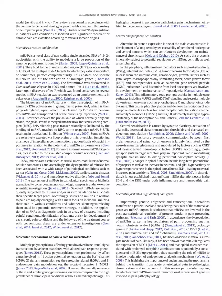
Human disorders associated with significant pathological pain suchas osteoarthritis (OA), post-herpetic neuralgia, diabetic polyneuropathyandmigraine are all highly prevalent, reduce life quality for affected pa-tients and thus represent a large socioeconomic burden (Berger et al.,2004; Manack et al., 2011; Pérez et al., 2012; Phillips, 2003). Moreover,these conditions share a high incidence of co-morbidities and largelysuboptimal treatment possibilities (Finch, 2013; Pérez et al., 2012).Significant progress has been made in terms of identifying matrices in-volved in normal and pathological pain processing as well as under-standing the molecular mechanisms underlying nociception and pain(Pace et al., 2006). Similarly, advances have been made in understand-ing the causes underlying pain chronification (Seifert and Maihöfner,2011). Within the last six years, accumulating evidence has suggestedthat a class of small non-coding inhibitory RNAs known as microRNAs(miRNAs) play an important role in regulating pain-processing within awide range of experimental models and clinical pain disorders (seeFig. 1) (Kynast et al., 2013a; Lutz et al., 2014; Niederberger et al., 2011).This review aims at presenting the advances made in understandingthe role of miRNAs as micro-modulators, biomarkers and potential ther-apeutic entities of painful conditions.
Search methodology and structure
Based on the PubMed entry: “(Pain OR nociception) AND (miRNA ORmicroRNA)”, with no time limit on the 24th of April 2014, 120 indexedarticleswere identified, demonstrating that research onmicroRNAs in re-lation to pain is still very limited. All publications were curated based ontitle and abstract; from those, 49 were found relevant and included,whereas 69 were excluded. It is important to note that this particularsearch strategy is not intended to cover all miRNA alterations in diseasesthat can give rise to pain per se. For example, various cancers, such asglioblastoma, involve extensive miRNA dysregulation and can result inexcruciating pain (Edvardsson andPersson, 2012), however, this exampleand similar areas are not dealt with in this review since the primary focusof the literature in such fields are inherently related to the miRNA-involvement in oncogenesis and not pain processing (Edvardsson andPersson, 2012; Møller et al., 2013). The studies included herein can bebroadly subdivided into the following 3 categories of which several over-lap: 1) explorative studies ofmiRNA-alterations induced bymodeled painconditions in vivo, i.e. both pain related to inflammation and neuropathy,2) studies investigating miRNA alterations in patients suffering from var-ious conditions related to pain, and 3) studies that explore the functionalproperties of one or more specific miRNA in relation to a particular pain
Fig. 1. Clinical pain condition and in vivo painmodels accompanied by significant miRNAmodulation and the tissue(s) where themiRNA changes manifest. Typical pathological locationsare represented. Blue = animal surrogate model of pain, red = clinical pain conditions, orange = animal surrogate model of pain and clinical pain conditions.
160 H.H. Andersen et al. / Neurobiology of Disease 71 (2014) 159–168
model (in vitro and in vivo). The review is sectioned in accordance withthe commonly perceived etiology of pain models as either inflammatoryor neuropathic pain (Pace et al., 2006). Studies of miRNA dysregulationin patients with conditions associated with significant recurrent orchronic pain are classified according to various somatic origins.
MicroRNA structure and function
miRNA is a novel class of non-coding single-stranded RNA of 19–24nucleotides with the ability to modulate a large proportion of thegenome post-transcriptionally (Bartel, 2009; Lagos-Quintana et al.,2001). They bind to the 3′ untranslated region (UTR), or occasionally5′ UTRs, of the multiple mRNA targets to which they exhibit imperfect,or sometimes, perfect complementarity. This enables one specificmiRNA to inhibit the translation of multiple genes (Thomsonet al., 2011; Ørom et al., 2008). The first miRNA was discovered inCaenorhabditis elegans in 1993 and named: lin-4 (Lee et al., 1993).Later, upon discovery of let-7, which was found conserved in severalspecies, miRNA-regulation was recognized as an omnipresent entity ineukaryotes (Pasquinelli et al., 2000; Reinhart et al., 2000).
The biogenesis of miRNA starts with the transcription of miRNA-genes by RNA polymerase II, giving rise to pri-miRNA, which is thenpoly-adenylated, upon which it is cleaved by Drosha to becomepre-miRNA and transported out of the nucleus by Exportin-5 (Yi et al.,2003). Dicer then cleaves the pre-miRNA of which normally only onestrand, the guide-strand, ismerged into the RNA-induced silencing com-plex (RISC). RNA-silencing post-transcriptionally is facilitated by thebinding of miRNA attached to RISC, to the respective mRNA 3′ UTR,resulting in translational inhibition (Winter et al., 2009). SomemiRNAsare selectively excreted via lipoproteins or micro-vesicles, functioningas a mode of intercellular communication, a notion that is of crucial im-portance in relation to the potential of miRNA as biomarkers (Chenet al., 2012; Stoorvogel, 2012). For more information onmiRNA biogen-esis, please refer to the available comprehensive reviews (Tran andHutvagner, 2013; Winter et al., 2009).
Today,miRNAs are established, as crucialmicro-modulators of normalcellular homeostasis and accordingly, the dysregulation of miRNAs hasbeen associated with a wide range of pathological conditions such ascancer (Calin and Croce, 2006; McManus, 2003), cardiovascular diseases(Vickers et al., 2014), and neurodegenerative disorders (Abe and Bonini,2013). The expression of miRNAs in pathological specimens or biofluidsnormalized to corresponding non-pathologic samples is under extensivescientific investigation (Jia et al., 2014). Selected miRNAs are subse-quently subjected to in silico and/or in vitro validation to elucidatetheir specific target genes. Accordingly, studies on miRNAs in relationto pain are rapidly emerging with a main focus on individual miRNAs,their role in various conditions and whether silencing/mimickingthem could be a potential treatment strategy. In addition, the applica-tion of miRNAs as diagnostic tools in an array of diseases, includingpainful conditions, identification of patients at risk for development ofe.g. chronic pain conditions and the follow-up of the treatment coursewith conventional drugs are under extensive investigation (Chenet al., 2014; Im et al., 2012; Willemen et al., 2012).
Molecular mechanisms of pain: a role for microRNAs?
Multiple polymorphisms, affecting genes involved in neuronal signaltransduction, have been associated with altered pain response pheno-types (James, 2013). These polymorphisms most predominantly, affectgenes involved in: 1) action potential generation e.g. the Na+-channelSCN9A, 2) signal transmission e.g. the serotonin related SLC6A4, and 3)endogenous pain modulation e.g. the μ-opioid receptor 1 (MOR1)(James, 2013; Reyes-Gibby et al., 2007). However, the overall prevalenceof these and similar genotypes remains low when compared to the highprevalence of chronic pain conditions without a hereditary etiology. This
highlights the great importance in pathological pain mechanisms not re-lated to the genomic layout (Breivik et al., 2006; Houlden et al., 2006).
Central and peripheral sensitization
Alteration in protein expression is one of the main characteristics indevelopment of a long-term hyper-excitability of peripheral nociceptiveand central neurons, which can contribute to development or mainte-nance of chronic pain (Gold and Gebhart, 2010). As such, the process isinherently subject to potential regulation by miRNAs, centrally as wellas peripherally.
In the periphery, inflammatory mediators such as prostaglandin E2(PGE2), interleukin-1 beta (IL-1β), tumor necrosis factor alpha (TNFα)release from the immune cells, keratinocytes, growth factors such asgranulocyte–macrophage colony-stimulating factor, nerve growth factor(NGF) and neuropeptides such as calcitonin gene-related peptide(CGRP), substance P and histamine from local nociceptors, are involvedin development or maintenance of hyperalgesia (Gangadharan andKuner, 2013). This inflammatory soup prompts activity in multiple genepathways includingMAPK3 and JAK-STAT-signaling and recruitsmultipledownstream enzymes such as phospholipase C and phosphoinositide3-kinase. This causes phosphorylation and de novo transcription of no-ciceptive molecules such as transient receptor potential cation channelsubfamily V member 1 (TRPV1) and Nav1.8, ultimately leading to hyper-excitability of the nociceptive Aδ- and C-fibers (Gold and Gebhart, 2010;Julius and Basbaum, 2001).
Central sensitization is caused by synaptic plasticity, activation ofglial cells, decreased signal transmission thresholds and decreased en-dogenous modulation (Sandkühler, 2009; Scholz and Woolf, 2007;Woolf, 2011). Excitatory synaptic communication between firstorder neurons and spinal cord neurons is largely facilitated by theneurotransmitter glutamate and modulated by factors such as CGRPand brain-derived neurotrophic factor (BDNF). Accordingly, post-synaptic glutamatergic receptors play an important role in modulatingsynaptic transmission following persistent nociceptive activity (Jiet al., 2003). Changes in spinal function include long-term potentiationof synapses as well as an increase in neuronal and glial hyper activity orhyper responsiveness in the spinal dorsal horn (SDH), leading to overallincreased pain sensitivity (Ji et al., 2003; Sandkühler, 2009). In this rela-tion, it is now established that significantmiRNAalterations occur in theSDH and the DRG under both inflammatory and neuropathic painconditions.
MicroRNA facilitated regulation of pain genes
Importantly, genetic, epigenetic and transcriptional alterationsmanifest on a protein-level and considering that N60% of themammaliangenes are miRNA-targets, it is inherent that miRNAs are involved in thepost-transcriptional regulation of proteins crucial in pain processingpathways (Friedman and Farh, 2009). In accordance, the dysregulationof miRNAs targeting key regulators of pain processing such asγ-aminobutyric acid-α1 (GABAα1) (Sengupta et al., 2013), cyclooxy-genase 2 (Akhtar and Haqqi, 2012; Park et al., 2013), TRPV1 (Li et al.,2011) and multiple Na+ and Ca2+-channels (Favereaux et al., 2011; Liet al., 2011; von Schack et al., 2011) has been observed in various surro-gate models of pain. Similarly, it has been shown that miR-23b regulatesthe expression of MOR1 (Ni et al., 2013) and that opioid-tolerance asso-ciated with prolonged morphine-administration is potentially a conse-quence of miR-23b upregulation, thus extending the role of miRNA toinvolve modulation of endogenous analgesic mechanisms (Wu et al.,2008). This highlights the importance of understanding themechanismsbehind the transition from acute physiological pain to pathologicalchronification, and in the context of this review particularly mappingto which extend miRNA-induced transcriptional repression of genes isinvolved in pain pathophysiology.
161H.H. Andersen et al. / Neurobiology of Disease 71 (2014) 159–168

MicroRNAs in pain and nociception
MicroRNAs involved in acute and prolonged inflammatory pain
A number of different in vivo models of inflammation have beenused to investigate the role of miRNA in inflammatory pain processing.Generally, widespread miRNA alterations are observed upon pain induc-tion (see Table 1). In a study by Zhao et al. (2010) itwas shown that Dicerknockout mice had an attenuated pain response to inflammationassessed by carrageenan injection into the hind paw, highlighting thatmiRNAmodulation may predominantly constitute a detrimental mecha-nism in the context of inflammation induced pain andhyperalgesia (Zhaoet al., 2010). This section highlights findings pertaining to the role ofmiRNA modulation upon induced inflammation.
To date, four studies using Freund's complete adjuvant-induced(CFA) inflammatory hyperalgesia have been published. In apioneering study in 2007, it was shown that inflammation inducedby injection of CFA into the masseter muscle of rats causedsignificant downregulation of 7 different miRNAs (see Table 1) inthe mandibular branch of the trigeminal nerve, amongst them wasmiR-124 widely described as a key miRNA in pain-processing (Baiet al., 2007). Also using CFA, Kusuda et al. (2011) applied a different ap-proach only investigating four preselected brain-specific miRNAs ofwhich miR-1 and miR-16 expression was markedly downregulated for7 days, but surprisingly upregulated in the SDH along with miR-206,24 h after induction. The same year, miR-143was shown to be downreg-ulated in the DRG upon CFA-induction but notably not in a neuropathicpain model applying sciatic nerve transection (Kusuda et al., 2011). Theauthors suggested Vcan1/2, known tomodulate neurogenesis, as a puta-tive target based on a corresponding ipsilateral increase in mRNA, but aluciferase-based target validation was not performed (Kusuda et al.,2011). Most recently, it was confirmed thatmiR-134, previously demon-strated to be decreased under neuropathic conditions (Bai et al., 2007),was downregulated in the DRG upon inflammatory induction with CFAand that it was inversely correlated with MOR1-expression. The studysubsequently applied a luciferase-assay to validate MOR1 as a target ofmiR-134 elucidating a potential endogenous pain modulation mecha-nism and highlighting a novel therapeutic opportunity (Ni et al., 2013).
MicroRNAs in animal models of osteoarthritis
Several studies have used animal models of OA to study the miRNAchanges in associationwith the pathogenesis of this prevalent andpainfulcondition. Firstly, a prominent increase inmiR-155 in synovial fibroblastsand monocytes was revealed in patients with rheumatoid arthritis (RA)(Stanczyk et al., 2008). It was later discovered that miR-146a was signif-icantly increased in peripheral blood mononuclear cells in RA (Pauley
et al., 2008). Elaborating on these results, Li et al. (2011) used the MIAOA-model analyzing miRNA-expression both in the DRG and the SDHand analyzed miR-146a modulation in human synoviocytes (Li et al.,2011). Surprisingly, in rodent models of OA a steep decrease in miR-146a expression in both the DRG and SDH was seen (as opposed to theknown upregulation in peripherally affected cells). Expanding on thatfinding, the influence of miR-146a was investigated applying transienttransfection ofmiR-146a in astrocytes andmicroglia, resulting in a signif-icant decrease in pro-inflammatory transcripts such as: TNFα, COX2,nitric oxide synthase (iNOS) and interleukin 6 (Li et al., 2011). In astudy comparing cultured chondrocytes from healthy and OA donorsmiR-558 was found to have significantly decreased in the latter. Thestudy further showed that miR-558 deficiency is inducible by IL-1β andconfirmed COX2 as a direct target mRNA, highlighting its potential im-portance in joint inflammation (Park et al., 2013). The same group alsoused a surgical animal model of OA analyzing miRNA-expression bothin the DRG and the SDH, here, a significant reduction in the expressionof miR-146a andmiR-183was found (X. Li et al., 2013). This finding con-curred with increased mRNA levels of a number of pro-inflammatorymediators such as NFκΒ, TNFα and IL-1β, but luciferase validation wasnot conducted on these potential targets. Interestingly, the study con-ducted a series of loss/gain of function studies on microglia (BV2 cellline) and astrocytes supporting the notion of miR-146a as being a keyregulator of inflammation, as previously reported by the same group (Liet al., 2011).
MicroRNAs as potential therapeutic targets for inflammation
miR-124 has been found centrally downregulated in two studies ap-plying different models of peripheral inflammatory hyperalgesia;intraplantar IL-1β-injection and formalin application and is validatedto target MeCP2, previously implicated in nociception (Kynast et al.,2013b;Willemen et al., 2012). Interestingly, miR-124 appears tomodu-late the reactivity of microglia promotingM1 toM2 switch; thus poten-tially decreasing central sensitization (Willemen et al., 2012). Whenapplied intravenously miR-124 and anti-miR-124 caused decreasedand increased pain behavior, respectively, albeit only significantly in asubset of the assessment timeframe (Kynast et al., 2013b). Otherknown centrally acting miRNA alterations include upregulation ofmiR-155 and miR-233 in the prefrontal cortices of rats uponcarrageenan-facilitated facial inflammation (Poh et al., 2011). Withinvisceral inflammation, a very recent study investigated modulation ofmiRNA in the developing spinal cord following neonatal cystitis in arodent-model applying zymosan. A steep increase of miR-181awas revealed concomitantly with an apparent repression of theGABAAα1-subunit. Subsequent administration of the GABAA-receptoragonist, muscimol, did not attenuate the viscero-motor response (VMR)
Table 1Overview of results from studies onmiRNAmodulation in animal surrogate models of inflammatory pain. Superscript numerals annotate a miRNA and its target gene(s). Only luciferase-validatedmRNA targets are included. The abbreviation behind individual highlightedmiRNAs denotes the origin of the sample: HC = hippocampus, SC = spinal cord, SDH = spinal dorsalhorn, DRG = dorsal root ganglion, and ACC = anterior cingulate cortex.
Study Pain model Highlighted miRNAs(sample origin)
Central/peripheral AberrantmiRNAs
Luciferase-validatedtargets
Bai et al. (2007) CFA-induced trigeminal pain miR-10a, miR-29a, miR-98, miR-99a,miR-124, miR-134, miR-183↓ (TG)
Both 7
Li et al. (2011) Inflammatory joint pain miR-146a↓ (DRG, SDH)/miR-146a↑ (JC) Both 1Tam Tam et al. (2011) CFA-induced pain miR-143↓ (DRG) Peripheral 1Kusuda et al. (2011) CFA-induced pain miR-1↑, miR-16↑, miR-206↑ (DRG)
miR-1↓, miR-16↓, miR-206↓ (SDH)Both 3
Poh et al. (2011) Facial inflammation miR-155↑, miR-233↑ (PC) Central 1Willemen et al. (2012) IL-1β-induced inflammatory hyperalgesia miR-124↓ (SCM) Central 1Xu et al. (2012) IL-1β-induced inflammation in chondrocytes miR-194↑ (JC) Peripheral 1 SOX5X. Li et al. (2013) Surgically induced joint pain miR-146a, miR-183↓ (DRG) Peripheral 9Kynast et al. (2013b) Formalin-induced inflammatory pain miR-124a↓ (SDH) Central 1 MECP2Ni et al. (2013) CFA-induced pain miR-134↓ (DRG) Peripheral 1 MOR1Park et al. (2013) Inflammatory joint pain miR-558↓ (JC) Peripheral 1 COX2Pan et al. (2014) CFA-induced pain miR-219↓ (SC) Central 1 CAMK2G
162 H.H. Andersen et al. / Neurobiology of Disease 71 (2014) 159–168

to colon distension in rats with neonatal cystitis, whereas in cystitis-induced adult rats the drug produced significant decrease in VMR(Sengupta et al., 2013). The authors concluded that it is likely thatmiR-181a is directly involved in toning spinal cord inhibition,unmasking excitatory pathways to facilitate prolonged or even chronicpain and hyperalgesia (Sengupta et al., 2013). Very recently, Park et al.(2014) reported that extracellular miR-let-7b induces inward currentsin rat DRG through coupling between Toll-like receptor-7 and the noci-ceptive ion-channel transient receptor potential cation channel sub-family A. Furthermore, the study elucidated a direct nociceptive effectof miR-let-7b in vivo, documented that miR-let-7b is released fromnociception neurons upon activation, and showed a reduction informalin-induced pain by concomitant administration of miR-let-7b-inhibitor (Park et al., 2014). This report indicates that circulatorymiRNAs could be as physiologically relevant as intracellular miRNAsand highlights their biomarker potential.
MicroRNAs involved in neuropathic pain
Lesions to the somatosensory nervous system frequently give rise toneuropathic pain, generally classified as either central or peripheral. It isestimated that≈8% of theWestern population suffers fromoneormoresubcategories of painful neuropathywith diabetic small-fiber neuropathybeing themost common subetiology (Torrance et al., 2006). Several stud-ies investigating changes inmiRNA expression upon induced neuropathyin rodent models have demonstrated a significant dysregulation innumerous miRNAs (see Table 2). A study by Genda et al. (2013) foundthat 111 miRNAs were significantly modulated in the SDH after chronicconstriction injury (CCI) in mice, but no further functional characteriza-tionwas performed (Genda et al., 2013). Illustrating the lack of consolida-tion in this research field, a recent study, also using CCI was unable toshow any significant difference between CCI- and sham-operated mice(Brandenburger et al., 2012). An early study utilizing a sciatic nerve injurymodel in mice investigated both the differential expression of miRNA indorsal root ganglion (DRG) neurons compared to scam injured mice,and also the effect of nerve injury on the proteins involved inmiRNA bio-synthesis. While subunits of the RISC were highly upregulated, levels ofvarious p-body machinery proteins differed significantly betweengroups, indicating that not only miRNA-deregulation but also regulatoryproteins involved in the miRNA-processing pathway could be of
importance in the peripheral response to injury (Wu et al., 2011). In aL5 spinal nerve ligation model, 63 miRNAs were found dysregulated, 59of which were concomitantly dysregulated in the ipsilateral L4 DRG sug-gesting that miRNA changes in adjacent uninjured afferents may alsocontribute to the development and continuation of neuropathic pain.The study did not elaborate further on these projectedmiRNA alterationsand their underlying cause (von Schack et al., 2011). Another recentstudy using a spinal nerve ligation model utilized genome-wide miRNAsequencing in two genetically similar rat strains displaying opposite re-sponses to neuropathic injuries, i.e. pain-prone and pain-resistant. Thestudy discovered a 3-miRNAs signature being highly downregulated inthe pain-prone rat strain and used in silico target prediction to associatethemiRNAs rno-miR-30d-5p and rno-miR-125b-5pwith the neuropathicpain related genes, TNF and BDNF (Bali et al., 2014).
Several studies have investigated neuropathy-inducedmiRNA alter-ations at the central level. Imai et al. (2011) found that sciatic nervelesion in mice induced significant downregulation of miR-200b and-429 in post-synaptic neurons of nucleus accumbens and that thisdownregulation corresponded with increased levels of DNA (cyto-sine-5)-methyltransferase 3A (DNMT3a). The authors proposedthat the DNMT3a increase associated with decreased levels of miR-200b and -429 is involved in the generation of dysfunction of themesolimbic motivation/valuation circuitry linking prolonged nocicep-tive stimuli with co-morbid conditions such as anxiety and sleep distur-bances (Imai et al., 2011). In a similar study using CCI in mice, thehippocampal formationwas analyzed using TaqMan low-density arraysand 51 miRNAs were found to be significantly altered compared to con-tralateral sham surgery.With complementary qRT-PCR the study investi-gated the downregulation of miRNAs miR-125b and -132 at days 7 and15 post injury but conducted no miRNA target-validation (Arai et al.,2013). In a recent study using bilateral CCI in rats and performingmiRNAmicroarray analysis on DRG, SDH, hippocampus and anterior cin-gulate cortex, a significant upregulation of miR-341 was observed inDRG-neurons and a downregulation of miR-181a-3p, -203 and -541-3pwas observed in the SDH, demonstrating miRNA alterations in boththe CNS and PNS in response to chronic sciatic constriction. None ofthese miRNAs was further functionally analyzed. In contrast to thestudy by Arai et al. (2013), no significant miRNAmodulations were ob-served in the hippocampus, representing a substantial discrepancy inthe literature (H. Li et al., 2013).
Table 2Overview of results from studies on miRNA modulation in animal surrogate models of neuropathic pain. Only luciferase-validated mRNA targets are included. The abbreviation behindindividual highlightedmiRNAs denotes the origin of the sample: TG = trigeminal ganglion, SDH = spinal dorsal horn, DRG = dorsal root ganglion, SCM = spinal cordmicroglia, JC = jointchondrocytes, and PC = prefrontal cortex.
Study Pain model Highlighted miRNAs/(sample origin) AberrantmiRNAs
Central/peripheral
Luciferase-validated targets
Aldrich et al. (2009) Spinal nerve ligation miR-96↑, miR-182↑, miR-183↑ (all DRG) 3 BothLiu et al. (2009) Contusive spinal cord injury miR-137, miR-181a, miR-219-2-3p, miR-7a↓,
miR-21↑ (all SDH)269 Central
Nakanishi et al.(2010)
Spinal cord injury miR-223↑, miR-124a↓ (SC) 2 Central
Imai et al. (2011) Sciatic nerve lesion miR-34c↓, miR-200b↓, miR-429↓ (all ACC) 17 CentralFavereaux et al.(2011)
Spinal nerve ligation miR-103↓ (DRG) 1 Peripheral CACNA1C, CACNA2D1,CACNB1
Kusuda et al. (2011) Spinal nerve ligation andaxotomy
Axotomy: miR-1 (↑SDH/↓DRG), miR-16↑ (SDH),miR-206↑ (SDH)Spinal nerve ligation: miR-1↓ (DRG), miR-206↓(DRG)
3 Both
Wu et al. (2011) Sciatic nerve crush miR-124↓, miR-221↑, miR-142-5p↑,miR-21↑ (All DRG)
4 Peripheral
Im et al. (2012) Spinal cord injury miR-23b↓ (SDH) Unspecified Central NOX4Genda et al. (2013) CCI miR-21, miR-221, miR-500 (all DRG) 111 PeripheralArai et al. (2013) CCI miR-125b↓, miR-132↓ (both HC) 51 CentralH. Li et al. (2013) CCI miR-341↑ (DRG), miR-203↓ (SDH),
miR-541-5p↓ (SDH),miR-181a-1-3p↓ (SDH)
1 Central
Chen et al. (2014) CCI miR-96↓ (DRG) 1 Peripheral(Li et al., 2014) Bilateral constriction injury miR-203↓ (SDH) Unspecified Central RAP1A
163H.H. Andersen et al. / Neurobiology of Disease 71 (2014) 159–168

MicroRNAs as potential therapeutic targets for neuropathic pain
In the light of four recent studies, the outlook of applying anti-miRNAs/miRNA-mimics to treat neuropathic pain and hyperalgesiaseems bright. In streptozotocin-induced diabetic neuropathy in rats, amiRNA-construct targeting Navα-subunits was delivered by a herpessimplex virus-based vector successfully reversing cold allodynia andthermal and mechanical hyperalgesia (Chattopadhyay et al., 2012). Inanother comprehensive study, intrathecal administration of miR-23b-mimics significantly alleviated mechanical and thermal hyperalgesia ina mice model of spinal cord injury. Furthermore, miR-23b was shownto target NADPH oxidase 4 (NOX4), an inflammation-promoting enzyme,also known to decrease production of the inhibitory neurotransmitterGABA through inhibition of glutamic acid decarboxylase (Im et al.,2012). Similarly, miR-124 (which is highly expressed in quiescentmicroglia) was shown to shift the M1/M2 balance towards the anti-inflammatory M2 phenotype and to decrease mechanical hyperalgesiaand pain behavior in a spared nerve model in mice (Willemen et al.,2012). Most recently, Chen et al. (2014) showed that CCI causes asteep decrease of miR-96 in DRG neurons, coinciding with an increasein Nav1.3, an ion-channel implicated in neuropathic hyper-excitabilityand a predicted target of miR-96. Subsequently, the study proved intra-thecal injections to be efficient in alleviating both mechanical and ther-mal hyperalgesia associated with the CCI procedure (Chen et al., 2014).It is evident that miRNAs have a potential as specific biomarkers andpossibly therapeutic entities within neuropathic conditions and newstudies on functional miRNA characterization and exploration of deliv-ery methodology could hold promise in future targeted therapy of neu-ropathic pain.
MicroRNAs in clinical pain conditions
As described, it is evident that animal models of neuropathic andinflammatory pain demonstrate widespread miRNA alterations andfurthermore that many of these alterations are detrimental to thegiven condition (Chen et al., 2014; Genda et al., 2013). This section high-lights novel findings on the implication of miRNAs from clinical painfulconditions with diverse characteristics and origin. In the context of in-vestigating the link between miRNAs and pain processing, it is impor-tant to note that particularly when it comes to clinical pain conditionsit is unclear to which extent miRNAs merely aggravate the underlyingpathogenesis subsequently leading to pain perception and not pain pro-cessing itself.
The most comprehensive body of literature is available on the sub-ject of knee arthritis, first and foremost OA and RA. Human specimensfrom patients clearly indicate that miRNAs are entwined in the patho-genesis of RA and OA, potentially contributing to cartilage degenerationand possibly even pain chronification (Furer et al., 2010; Stanczyk et al.,2008; Yu et al., 2011). It is beyond the scope of this review to summarizethe findings of miRNAs implicationwithin OA, RA and autoimmune dis-eases in general (refer to Furer et al., 2010 for more on this particulartopic).
MicroRNAs in visceral pain conditions
In several visceral painful conditions such as irritable bowl syndrome(IBS), bladder pain syndrome (BPS), endometriosis and cystitis-inducedchronic pain (CICP) miRNA dysregulation has been reported. In IBS, themiRNA profile appears to facilitate an increased permeability of thegastrointestinal tract (Zhou et al., 2010). A noteworthy study by Verneand colleagues (2010) demonstrated that miR-29a is markedly in-creased in patients with IBS in intestinal biopsies and in a subset ofdiarrhea-predominant IBS patients' miR-29a was found significantlyupregulated in circulating miRNA-containing exosomes (see Table 3).The study used a luciferase-reporter to validate that miR-29a targetsthe glutamine synthetase gene GLUL, thus decreasing enterocytic
glutamine levels and aggravating the increased intestinal permeability(Zhou and Verne, 2011; Zhou et al., 2010). In a study of BPS, 31miRNAswere differentially expressed and a direct correlation between theincreased expression of miR-449b and miR-500 concomitantly withdownregulation of the substance P receptor NK1-receptor was found(Sanchez Freire et al., 2010). The authors suggested this to constitute amiRNA-dependent adaptive mechanism to the prolonged NK1R overex-pression which was previously described in BPS (Marchand et al., 1998;Sanchez Freire et al., 2010). In endometriosis, characterized bymenstrualcycle-dependent pelvic pain, a great deal of recent research has revealedthe influence of miRNAs. The first study reporting widespread miRNAdysregulation in endometriosis was published in 2007 (Pan et al., 2007)and 45 studies have been published since then on that subject.1 Notably,Hawkins et al. (2011) performed a full microRNAome analysis ofendometriosis patients and found 22 miRNAs to be dysregulated. Thestudy further investigated the highly upregulated miR-29c and eluci-dated its ability to significantly inhibit several important genesinvolved in extracellular matrix function such as collagen type VII A1(COL7A1). Lastly, the same study also validly pointed out the extensivediscrepancy present in terms of the specific miRNAs found across simi-lar studies but ascribed it to methodological differences e.g. samplingtime in relation to menstrual cycle (Hawkins et al., 2011). In the highlyprevalent pain disorder, lower back pain, Zhao et al. (2014) investigatedone of the frequent underlying causes being intervertebral disc degen-eration (IDD). Here, miRNA expression in the nucleus pulposus wascompared between IDD patients and acute intervertebral injury pa-tients giving rise to a large miRNA signature, but no further analysiswas performed (Zhao et al., 2014).
MicroRNAs as biomarkers in pain disorders
Investigating miRNAs as diagnostic and prognostic biofluid-derivedbiomarkers is well progressed in various fields, particularly oncologyand cardiology, but is novel in relation to pain conditions (Ahmadet al., 2013; Kinet et al., 2013). Research in miRNAs are of great interestin terms of biomarker potential since they are present in virtually allbiofluids, exhibit more superior stability than mRNA and appear to behighly sensitive to slight changes in various physiological processes(Jung et al., 2010; Kemppainen et al., n.d.; Tomaselli et al., 2012).
In an innovative study by Orlova et al. (2011), whole blood sampleswere taken from 41 patients with complex regional pain syndrome(CRPS) and 20 controls upon which analysis of miRNA expression, cyto-kines and numerous clinical parameters were conducted (see Table 3).Unsurprisingly, cytokines such as CCL2 and VEGF were notably elevatedin the CRPS group compared to control and a CRPSmiRNA-signaturewasevident. Interestingly, extensive correlation analyses revealed that4 miRNAs (miR-296-5p, -532-3p, -361-3p and -30d) were positivelycorrelated with CRPS-associated pain level, miR-150 was correlatedwith the occurrence of migraine within the CRPS patient cohort andan extensive array of miRNAs was found to correlate with the levels ofcirculating cytokines (Orlova et al., 2011). A recent study with a similardesign investigated the miRNA profile of the cerebrospinal fluid infibromyalgia patients and identified 10miRNAs differentially expressedbetween affected patients and healthy controls. Most notably, the studyfound that decreased levels of miR-145-5p in the CSF were associatedwith reported symptomatology i.e. pain intensity and fatigue (Bjersinget al., 2013).
The prospect of applying miRNAs as biofluid-derived biomarkers inpain conditions is enticing due to their high specificity, e.g. circulatingmiRNA-signatures have been ascertained to discriminate betweeneccentric and concentric exercise (Banzet et al., 2013). However,numerous obstacles are present when using circulating miRNAs: 1) itremains unclear why and to which extent miRNAs are selectively
1 NB. Studies that include "endometriosis" and "microRNA".
164 H.H. Andersen et al. / Neurobiology of Disease 71 (2014) 159–168

exported (Boon and Vickers, 2013), 2) there are numerous qualityissues associated with sampling, particularly hemolysis and low isola-tion yields, and 3) plasma miRNA expression is highly affected by vari-ous hormonal and metabolic factors (Tomaselli et al., 2012).
Conclusion and future directions
Despite being a subfield in its infancy, it is clear thatmiRNAalterationsare essential in a variety of pain models and in clinical conditions charac-terized by severe pain (Kynast et al., 2013a). Until now studies have beenconducted either in animal painmodels or in fewer caseswithin a patientcohort posing inherent limitations as to the translatability between thesetwo types of studies. The use of human surrogate models have beenbeneficial in improving understanding of pain processing by temporarilyrecreating concise pain symptoms such as spontaneous burning pain,heat hyperalgesia or cold allodynia (Andersen et al., 2014; Arendt-Nielsen and Yarnitsky, 2009). Studies mapping the local and systemicmiRNA alterations likely associated with induced local inflammation inwidely used models such as the heat/capsaicin, ultraviolet B and NGFmodels (Arendt-Nielsen and Yarnitsky, 2009) are warranted.
Within animal models of neuropathic and inflammatory pain suchas rodent spinal nerve ligation and CFA-induced inflammation, wide-spread miRNA modulation is observed (Bai et al., 2007; Genda et al.,2013). It appears that miRNA alterations occur more rapidly in responseto inflammatory pain, and some suggest it to be an adaptive mechanismaimed at pain reduction, as opposed to miRNA alterations in response toneuropathic pain suggested mainly to be a detrimental process (Bali andKuner, 2014; Gheinani et al., 2013). Despite the fact that numerousmiRNAs are frequently reported aberrantly expressed, onlymodest over-lap between dysregulated miRNA such as miR-103 in neuropathy andmiR-146a in inflammation is present in the literature (Favereaux et al.,2011; Li et al., 2011; Pauley et al., 2008; Yamasaki et al., 2009). Further re-search is warranted to adequately elucidate whether this is a conse-quence of limited published data or high specificity between miRNAalterations between various pain conditions. Interestingly, a fewmiRNAs,such as miR-183, have been found consistently downregulated in bothneuropathic and inflammatory pain conditions while others, such asmiR-1, have opposite responses between neuropathic and inflammatorymodels and even within different neuropathic pain models (Bali andKuner, 2014; Kusuda et al., 2011).
Although administration of anti-miRs and miR-mimics has alreadybeen successfully applied in animal models of neuropathic and inflam-matory pain, it is necessary to obtain consistent knowledge about therole of miRNAs in pain processing during various normal and pathologi-cal pain conditions before any diagnostic or therapeutic utilization can beaccomplished (Chattopadhyay et al., 2012; Chen et al., 2014; Im et al.,2012; Willemen et al., 2012). Frequently, a large number of miRNAsare found dysregulated in a given condition (Genda et al., 2013). Thisis, at the same time, an obstacle and also an opportunity. The obviouschallenge lies in increased complexity and choosing the most promising
miRNA(s) for further investigation. To overcome this challenge severalstrategies can be utilized: 1) the median absolute deviation can beapplied to assess the most differentially expressed miRNAs across sam-ples in combination with statistical testing, 2) in silico target predictionalgorithms can be used to predict feasible targets relevant for pain pro-cessing, and 3) specific promising miRNAs can be tested individually inmiRNA transfection studies. As for the opportunity related to the factthat manymiRNAs are often found dysregulated in a given condition, in-creased specificity is the primary gain. As such, miRNAs as biomarkersare promising not only in terms of diagnosis, but also in relation to pa-tient stratification. A seemingly high level of specificity could hold prom-ise in stratification of heterogeneous patient populations, currentlydifficult to subcategorize. The literature is growing dramatically interms of reporting aberrantly expressed miRNAs in multiple pain condi-tions; however, functionallywell-characterizedmiRNAs is highly lackingand needs particular attention (Arai et al., 2013; Genda et al., 2013).
Another pronounced bottleneck is represented by the problem ofdistributing miRNA-mimic or anti-miRNA to the target tissue, particu-larly when it comes to deliveringmiRNAs across the blood brain barrier,whichwould be relevant to formulate a centrally acting analgesic. Novelresults indicate a great potential of exosomes, already known to carrymicroRNAs in cell-to-cell communication and to deliver siRNA to targetcells (Chen et al., 2012; Johnsen et al., 2014). This approachwas utilizedby Alvarez-Erviti et al. (2011), who applied exosomes expressingneuron-specific targeting surface peptides by systemic injection. Thisenabled cell specific delivery of the siRNA-cargo across the blood brainbarrier (Alvarez-Erviti et al., 2011). Although promising, some concernshave beenmade as towhether exosomal cell-to-cell interaction is phys-iologically relevant and whether exosome-carried therapeutics willprove clinically feasible (Sverdlov, 2012).
Intricately, pain gives rise to miRNA alterations on multiple levels(see Fig. 2). In circulation, miRNAs-signatures have been discovered ine.g. IBS and CRPS-patients (Orlova et al., 2011; Zhou et al., 2010). Inlocal tissues miRNA dysregulation in response to pain involve residingprimary afferents, fibroblasts and keratinocytes e.g. in post-operationalskin inflammation (Sun et al., 2012). Knowledge is scarce regarding thepurpose of circulatingmiRNAs and local cell-to-cell interactionsmediatedby miRNAs, but these fields are of great interest in regards to biomarkersand potential formulation of topical analgesics, respectively. In the DRGand the SDH widespread miRNA dysregulation has been documented inresponse to both neuropathic and inflammatory pain and it is evidentthat miRNA expression is selectively modulated between the SDH andDRG neurons in response to the exact same injury. For instance, miR-16expression was significantly reduced in the DRG, but upregulated in theSDH after CFA-induced inflammation in mice, i.e. exhibiting opposite ex-pressional response between peripheral and central compartments(Kusuda et al., 2011). Many of these studied miRNA-driven alterationsprompt the initiation or maintenance of a more excitable phenotype,thus increasing the conductance of nociceptive stimuli (Chen et al.,2014; Zhao et al., 2010). It is known that both microglia and astrocytes
Table 3Examples of painful disorders where serum microRNA aberrations are associated with symptomatology. *Notice that these are proposed endogenous control miRNAs, but are foundsignificantly dysregulated. ↑/↓ = increase/decrease.
Reference Condition MicroRNA(s) Sample / sample size
Orlova et al.(2011)
Complex regional painsyndrome
(↓) miR-939, -25, -7c, -7a, -7b, -320b, -126, -629, -664, -320, -1285, -625,-532-3p, -181a(↑) RNU48*, RNU44*, miR-720, -1201
Whole blood/41 patients, 20 controls
Bjersing et al.(2013)
Fibromyalgia (↓) miR-21-5p, 145-5p, 29a-3p, 99b-5p, 125b-5p, 23a-3p, 23b-3p, 195-5p,miR-223-3p
Cerebrospinal fluid/10 patients, 20 controls
Zhou et al.(2010)
Irritable bowel syndrome (↑) miR-29a Serum micro-vesicles/19 patients, 10 controls
Wang et al.(2013)
Endometriosis (↑) miR-199a miR-122(↓) miR-145a-3p, -141-5p, -542-3p, -9-3p
Serum/10 patients, 10 controls (screening) | 60patients,25 controls (validation)
Beyer et al.(2014)
Osteoarthritis (↓) let-7e, miR-454, -885-5p
165H.H. Andersen et al. / Neurobiology of Disease 71 (2014) 159–168

are involved in central sensitization and that miRNA dysregulation inthese cells is correspondingly associated with CNS hypertrophy and in-creased reactivity (Bhalala et al., 2012; Ponomarev et al., 2011). Circula-torymiRNAs are also present in the cerebrospinalfluid and here specificmiRNAs have been linked to pain in fibromyalgia patients (Bjersinget al., 2013). Lastly, miRNA alterations have been mapped in severalbrain matrices associatedwith higher pain processing in rodent modelsof pain (Poh et al., 2011).
In summary, miRNAs are rapidly emerging as pivotal players in-volved in pain from the first single study in the field in 2007 (Baiet al., 2007) to 37 publications in 2013 alone. These small non-codingRNA molecules represent promising potential biomarkers and thera-peutic targets in a variety of human diseases, including pain conditions.However, pronounced bench-to-bedside hindrances warrants furtherstudies before miRNAs can be applied clinically in pain patients forprognosis, diagnosis, follow-up of the treatment course, and potentiallynovel treatment strategies.
Conflict of interest
No conflict of interest to declare.
Author contributions
All authors contributed in the conception, writing and discussion ofthis manuscript.
Acknowledgments
Grosserer L. F. Foghts Fond grant number: 20734 provided financialsupport to the research project subsequently inciting this review paper.The Novo Nordisk Scholarship Grant of 2013 supported H. H. Andersen.P. Gazerani’s research is supported by a 2010 FSS grant from the DanishResearch Council, grant number: 831301.
References
Abe, M.,Bonini, N.M., 2013. MicroRNAs and neurodegeneration: role and impact. TrendsCell Biol. 23, 30–36. http://dx.doi.org/10.1016/j.tcb.2012.08.013.
Ahmad, J.,Hasnain, S.E., Siddiqui, M.A.,Ahamed, M.,Musarrat, J., Al-Khedhairy, A.A., 2013.MicroRNA in carcinogenesis & cancer diagnostics: a new paradigm. Indian J. Med.Res. 137, 680–694.
Akhtar, N.,Haqqi, T.M., 2012. MicroRNA-199a* regulates the expression of cyclooxygenase-2in human chondrocytes. Ann. Rheum. Dis. 71, 1073–1080. http://dx.doi.org/10.1136/annrheumdis-2011-200519.
Fig. 2. Locations ofmiRNA alterations in response to induced pain and painful disorders. The tables at each level list examples of well-investigatedmiRNAs and their correspondingmRNAtargets. MiRNA modulation is reflected in: the circulation, DRG neurons, SDH, glial cells and in several brain areas. DRG = dorsal root ganglion and SDH = spinal dorsal horn. *Notvalidated with a luciferase-reporter assay.Red arrows denote miRNA up- and downregulation and the specified locations.
166 H.H. Andersen et al. / Neurobiology of Disease 71 (2014) 159–168

Aldrich, B.T.,Frakes, E.P.,Kasuya, J.,Hammond, D.L.,Kitamoto, T., 2009. Changes in expres-sion of sensory organ-specific microRNAs in rat dorsal root ganglia in associationwith mechanical hypersensitivity induced by spinal nerve ligation. Neuroscience164, 711–723. http://dx.doi.org/10.1016/j.neuroscience.2009.08.033.
Alvarez-Erviti, L.,Seow, Y.,Yin, H.,Betts, C.,Lakhal, S.,Wood, M.J. a, 2011. Delivery of siRNAto the mouse brain by systemic injection of targeted exosomes. Nat. Biotechnol. 29,341–345. http://dx.doi.org/10.1038/nbt.1807.
Andersen, H.H.,Olsen, R.V.,Møller, H.G., Eskelund, P.W.,Gazerani, P., Arendt-Nielsen, L.,2014. A review of topical high-concentration L-menthol as a translational model ofcold allodynia and hyperalgesia. Eur. J. pain 18, 315–325. http://dx.doi.org/10.1002/j.1532-2149.2013.00380.x.
Arai, M.,Genda, Y., Ishikawa, M., 2013. The miRNA and mRNA changes in Rat hippocampiafter chronic constriction injury. Pain Med. 14, 720–729.
Arendt-Nielsen, L.,Yarnitsky, D., 2009. Experimental and clinical applications of quantitativesensory testing applied to skin, muscles and viscera. J. Pain 10, 556–572. http://dx.doi.org/10.1016/j.jpain.2009.02.002.
Bai, G.,Ambalavanar, R.,Wei, D.,Dessem, D., 2007. Downregulation of selective microRNAsin trigeminal ganglion neurons following inflammatory muscle pain. Mol. Pain 3, 15.http://dx.doi.org/10.1186/1744-8069-3-15.
Bali, K.K.,Kuner, R., 2014. Noncoding RNAs: key molecules in understanding and treatingpain. Trends Mol. Med. 1–12 http://dx.doi.org/10.1016/j.molmed.2014.05.006.
Bali, K.K., Hackenberg, M., Lubin, A., Kuner, R., Devor, M., 2014. Sources of individualvariability: miRNAs that predispose to neuropathic pain identified using genome-wide sequencing. Mol. Pain 10, 22. http://dx.doi.org/10.1186/1744-8069-10-22.
Banzet, S.,Chennaoui, M.,Girard, O.,Racinais, S.,Drogou, C.,Chalabi, H.,Koulmann, N., 2013.Changes in circulating microRNAs levels with exercise modality. J. Appl. Physiol.http://dx.doi.org/10.1152/japplphysiol.00075.2013.
Bartel, D.P., 2009. MicroRNAs: target recognition and regulatory functions. Cell 136,215–233. http://dx.doi.org/10.1016/j.cell.2009.01.002.
Berger, A., Dukes, E.M., Oster, G., 2004. Clinical characteristics and economic costs ofpatients with painful neuropathic disorders. J. Pain 5, 143–149. http://dx.doi.org/10.1016/j.jpain.2003.12.004.
Beyer, C.,Zampetaki, A.,Lin, N.-Y.,Kleyer, A.,Perricone, C.,Iagnocco, A.,Distler, A.,Langley, S.R., Gelse, K., Sesselmann, S., Lorenzini, R., Niemeier, A., Swoboda, B., Distler, J.H.W.,Santer, P.,Egger, G.,Willeit, J.,Mayr, M.,Schett, G.,Kiechl, S., 2014. Signature of circulat-ing microRNAs in osteoarthritis. Ann. Rheum. Dis. http://dx.doi.org/10.1136/annrheumdis-2013-204698.
Bhalala, O.G.,Pan, L.,Sahni, V.,McGuire, T.L.,Gruner, K.,Tourtellotte,W.G.,Kessler, J.A., 2012.MicroRNA-21 regulates astrocytic response following spinal cord injury. J. Neurosci.32, 17935–17947. http://dx.doi.org/10.1523/JNEUROSCI.3860-12.2012.
Bjersing, J.L., Lundborg, C., Bokarewa, M.I.,Mannerkorpi, K., 2013. Profile of cerebrospinalmicroRNAs in fibromyalgia. PLoS One 8, e78762. http://dx.doi.org/10.1371/journal.pone.0078762.
Boon, R.A.,Vickers, K.C., 2013. Intercellular transport of microRNAs. Arterioscler. Thromb.Vasc. Biol. 33, 186–192. http://dx.doi.org/10.1161/ATVBAHA.112.300139.
Brandenburger, T., Castoldi, M., Brendel, M.,Grievink, H., Schlösser, L.,Werdehausen, R.,Bauer, I.,Hermanns, H., 2012. Expression of spinal cord microRNAs in a rat model ofchronic neuropathic pain. Neurosci. Lett. 506, 281–286. http://dx.doi.org/10.1016/j.neulet.2011.11.023.
Breivik, H.,Collett, B.,Ventafridda, V.,Cohen, R.,Gallacher, D., 2006. Survey of chronic painin Europe: prevalence, impact on daily life, and treatment. Eur. J. Pain 10, 287–333.http://dx.doi.org/10.1016/j.ejpain.2005.06.009.
Calin, G.A., Croce, C.M., 2006. MicroRNA-cancer connection: the beginning of a new tale.Cancer Res. 66, 7390–7394. http://dx.doi.org/10.1158/0008-5472.CAN-06-0800.
Chattopadhyay, M., Zhou, Z.,Hao, S.,Mata, M., Fink, D.J., 2012. Reduction of voltage gatedsodium channel protein in DRG by vector mediated miRNA reduces pain in rats withpainful diabetic neuropathy. Mol. Pain 8, 17. http://dx.doi.org/10.1186/1744-8069-8-17.
Chen, X.,Liang, H.,Zhang, J.,Zen, K.,Zhang, C.-Y., 2012. Secreted microRNAs: a new form ofintercellular communication. Trends Cell Biol. 22, 125–132. http://dx.doi.org/10.1016/j.tcb.2011.12.001.
Chen, H.-P.,Zhou, W.,Kang, L.-M.,Yan, H.,Zhang, L.,Xu, B.-H.,Cai, W.-H., 2014. IntrathecalmiR-96 inhibits Nav1.3 expression and alleviates neuropathic pain in rat followingchronic construction injury. Neurochem. Res. 76–83 http://dx.doi.org/10.1007/s11064-013-1192-z.
Edvardsson, B.,Persson, S., 2012. Cluster headache and parietal glioblastoma multiforme.Neurologist 18, 206–207. http://dx.doi.org/10.1097/NRL.0b013e31825cf181.
Favereaux, A., Thoumine, O., Bouali-Benazzouz, R., Roques, V., Papon, M.-A., Salam, S.A.,Drutel, G.,Léger, C.,Calas, A.,Nagy, F.,Landry, M., 2011. Bidirectional integrative regu-lation of Cav1.2 calcium channel by microRNA miR-103: role in pain. EMBO J. 30,3830–3841. http://dx.doi.org/10.1038/emboj.2011.249.
Finch, J.W., 2013. Challenges of chronic pain management: public health consequencesand considered responses. N. C. Med. J. 74 (243–5), 247–248.
Friedman, R., Farh, K., 2009. Most mammalian mRNAs are conserved targets ofmicroRNAs. Genome Res. 19, 92–105. http://dx.doi.org/10.1101/gr.082701.108.
Furer, V., Greenberg, J.D., Attur, M., Abramson, S.B., Pillinger, M.H., 2010. The role ofmicroRNA in rheumatoid arthritis and other autoimmune diseases. Clin. Immunol.136, 1–15. http://dx.doi.org/10.1016/j.clim.2010.02.005.
Gangadharan, V., Kuner, R., 2013. Pain hypersensitivity mechanisms at a glance. Dis.Model. Mech. 6, 889–895. http://dx.doi.org/10.1242/dmm.011502.
Genda, Y.,Arai, M.,Ishikawa, M.,Tanaka, S.,Okabe, T.,Sakamoto, A., 2013. MicroRNA changesin the dorsal horn of the spinal cord of rats with chronic constriction injury: aTaqMan® Low Density Array study. Int. J. Mol. Med. 31, 129–137. http://dx.doi.org/10.3892/ijmm.2012.1163.
Gheinani, A.H., Burkhard, F.C.,Monastyrskaya, K., 2013. Deciphering microRNA code inpain and inflammation: lessons from bladder pain syndrome. Cell. Mol. Life Sci.http://dx.doi.org/10.1007/s00018-013-1275-7.
Gold, M.S.,Gebhart, G.F., 2010. Nociceptor sensitization in pain pathogenesis. Nat. Med.16, 1248–1257. http://dx.doi.org/10.1038/nm.2235.
Hawkins, S.M.,Creighton, C.J.,Han, D.Y.,Zariff, A.,Anderson, M.L.,Gunaratne, P.H.,Matzuk,M.M., 2011. Functional microRNA involved in endometriosis. Mol. Endocrinol. 25,821–832. http://dx.doi.org/10.1210/me.2010-0371.
Houlden, H., King, R., Blake, J., Groves, M., Love, S.,Woodward, C.,Hammans, S.,Nicoll, J.,Lennox, G.,O'Donovan, D.G.,Gabriel, C.,Thomas, P.K.,Reilly, M.M., 2006. Clinical, patho-logical and genetic characterization of hereditary sensory and autonomic neuropathytype 1 (HSAN I). Brain 129, 411–425. http://dx.doi.org/10.1093/brain/awh712.
Im, Y.B., Jee, M.K., Choi, J.I., Cho, H.T.,Kwon, O.H.,Kang, S.K., 2012. Molecular targeting ofNOX4 for neuropathic pain after traumatic injury of the spinal cord. Cell Death Dis.3, e426. http://dx.doi.org/10.1038/cddis.2012.168.
Imai, S., Saeki, M., Yanase, M., Horiuchi, H., Abe, M., Narita, M., Kuzumaki, N., Suzuki, T.,Narita, M., 2011. Change in microRNAs associated with neuronal adaptive responsesin the nucleus accumbens under neuropathic pain. J. Neurosci. 31, 15294–15299.http://dx.doi.org/10.1523/JNEUROSCI.0921-11.2011.
James, S., 2013. Human pain and genetics: some basics. Br. J. Pain 7, 171–178. http://dx.doi.org/10.1177/2049463713506408.
Ji, R.-R.,Kohno, T.,Moore, K.a.,Woolf, C.J., 2003. Central sensitization and LTP: do pain andmemory share similar mechanisms? Trends Neurosci. 26, 696–705. http://dx.doi.org/10.1016/j.tins.2003.09.017.
Jia, S.,Zocco, D.,Samuels, M.L.,Chou, M.F.,Chammas, R.,Skog, J.,Zarovni, N.,Momen-Heravi,F.,Kuo, W.P., 2014. Emerging technologies in extracellular vesicle-based moleculardiagnostics. Expert. Rev. Mol. Diagn. 14, 307–321. http://dx.doi.org/10.1586/14737159.2014.893828.
Johnsen, K.B.,Gudbergsson, J.M.,Skov, M.N.,Pilgaard, L.,Moos, T.,Duroux, M., 2014. A com-prehensive overview of exosomes as drug delivery vehicles — endogenousnanocarriers for targeted cancer therapy. Biochim. Biophys. Acta http://dx.doi.org/10.1016/j.bbcan.2014.04.005.
Julius, D., Basbaum, a.I., 2001. Molecular mechanisms of nociception. Nature 413,203–210. http://dx.doi.org/10.1038/35093019.
Jung, M.,Schaefer, a.,Steiner, I.,Kempkensteffen, C.,Stephan, C.,Erbersdobler, a., Jung, K.,2010. Robust microRNA stability in degraded RNA preparations from human tissueand cell samples. Clin. Chem. 56, 998–1006. http://dx.doi.org/10.1373/clinchem.2009.141580.
Kemppainen, J., Shelton, J., Kelnar, K., Volz, S., Peltier, H., Szafranska, A., Ovcharenko, D.,Illmer, T., Labourier, M., Latham, G., Brown, D., 2010. MicroRNAs as biomarkers inblood and other biofluids — commercial publication. AsuraGen.
Kinet, V.,Halkein, J.,Dirkx, E.,Windt, L.J. De, 2013. Cardiovascular extracellular microRNAs:emerging diagnostic markers and mechanisms of cell-to-cell RNA communication.Front. Genet. 4, 214. http://dx.doi.org/10.3389/fgene.2013.00214.
Kusuda, R., Cadetti, F., Ravanelli, M.I., Sousa, T.a., Zanon, S.,De Lucca, F.L., Lucas, G., 2011.Differential expression of microRNAs in mouse pain models. Mol. Pain 7, 17.
Kynast, K.L.,Russe, O.Q.,Geisslinger, G.,Niederberger, E., 2013a. Novel findings in pain pro-cessing pathways: implications for miRNAs as future therapeutic targets. Expert Rev.Neurother. 13, 515–525. http://dx.doi.org/10.1586/ern.13.34.
Kynast, K.L.,Russe, O.Q.,Möser, C.V.,Geisslinger, G.,Niederberger, E., 2013b. Modulation ofcentral nervous system-specificmicroRNA-124a alters the inflammatory response in theformalin test in mice. Pain 154, 368–376. http://dx.doi.org/10.1016/j.pain.2012.11.010.
Lagos-Quintana, M., Rauhut, R., Lendeckel, W., Tuschl, T., 2001. Identification of novelgenes coding for small expressed RNAs. Science 294, 853–858. http://dx.doi.org/10.1126/science.1064921.
Lee, R.C.,Feinbaum, R.L.,Ambros, V., 1993. The C. elegans heterochronic gene lin-4 encodessmall RNAs with antisense complementarity to lin-14. Cell 75, 843–854.
Li, X.,Gibson, G.,Kim, J.-S.,Kroin, J.,Xu, S.,vanWijnen, A.J.,Im, H.-J., 2011. MicroRNA-146a islinked to pain-related pathophysiology of osteoarthritis. Gene 480, 34–41. http://dx.doi.org/10.1016/j.gene.2011.03.003.
Li, H., Shen, L.,Ma, C.,Huang, Y., 2013a. Differential expression of miRNAs in the nervoussystem of a rat model of bilateral sciatic nerve chronic constriction injury. Int. J. Mol.Med. 32, 219–226. http://dx.doi.org/10.3892/ijmm.2013.1381.
Li, X.,Kroin, J.S.,Kc, R.,Gibson, G.,Chen, D.,Corbett, G.T.,Pahan, K., Fayyaz, S.,Kim, J.-S.,vanWijnen, A.J.,Suh, J.,Kim, S.-G.,Im, H.-J., 2013b. Altered spinal microRNA-146a and themicroRNA-183 cluster contribute to osteoarthritic pain in knee joints. J. Bone Miner.Res. http://dx.doi.org/10.1002/jbmr.2002.
Liu, N.-K.,Wang, X.-F., Lu, Q.-B.,Xu, X.-M., 2009. Altered microRNA expression followingtraumatic spinal cord injury. Exp. Neurol. 219, 424–429. http://dx.doi.org/10.1016/j.expneurol.2009.06.015.
Lutz, B.M., Bekker, A., Tao, Y.-X., 2014. Noncoding RNAs: new players in chronic pain.Anesthesiology http://dx.doi.org/10.1097/ALN.0000000000000265 (Epub ahead ofprint).
Manack, A.N.,Buse, D.C.,Lipton, R.B., 2011. Chronic migraine: epidemiology and disease bur-den. Curr. Pain Headache Rep. 15, 70–78. http://dx.doi.org/10.1007/s11916-010-0157-z.
Marchand, J.E.,Sant, G.R.,Kream, R.M., 1998. Increased expression of substance P receptor-encoding mRNA in bladder biopsies from patients with interstitial cystitis. Br. J. Urol.81, 224–228.
McManus, M.T., 2003. MicroRNAs and cancer. Semin. Cancer Biol. 13, 253–258.Møller, H.G.,Rasmussen, A.P.,Andersen, H.H., Johnsen, K.B.,Henriksen, M.,Duroux, M., 2013.
A systematic review of microRNA in glioblastomamultiforme: micro-modulators in themesenchymal mode of migration and invasion. Mol. Neurobiol. 47, 131–144. http://dx.doi.org/10.1007/s12035-012-8349-7.
Nakanishi, K.,Nakasa, T.,Tanaka, N.,Ishikawa, M.,Yamada, K.,Yamasaki, K.,Kamei, N.,Izumi,B.,Adachi, N.,Miyaki, S.,Asahara, H.,Ochi, M., 2010. Responses of microRNAs 124a and223 following spinal cord injury in mice. Spinal Cord 48, 192–196. http://dx.doi.org/10.1038/sc.2009.89.
Ni, J.,Gao, Y.,Gong, S.,Guo, S.,Hisamitsu, T., Jiang, X., 2013. Regulation of μ-opioid type 1receptors by microRNA134 in dorsal root ganglion neurons following peripheral
167H.H. Andersen et al. / Neurobiology of Disease 71 (2014) 159–168

inflammation. Eur. J. Pain 17, 313–323. http://dx.doi.org/10.1002/j.1532-2149.2012.00197.x.
Niederberger, E.,Kynast, K., Lötsch, J.,Geisslinger, G., 2011. MicroRNAs as new players inthe pain game. Pain 152, 1455–1458. http://dx.doi.org/10.1016/j.pain.2011.01.042.
Orlova, I.a.,Alexander, G.M.,Qureshi, R.a.,Sacan, A.,Graziano, A.,Barrett, J.E.,Schwartzman, R.J.,Ajit, S.K., 2011. MicroRNA modulation in complex regional pain syndrome. J. Transl.Med. 9, 195. http://dx.doi.org/10.1186/1479-5876-9-195.
Ørom, U.A.,Nielsen, F.C., Lund, A.H., 2008. MicroRNA-10a binds the 5′ UTR of ribosomalprotein mRNAs and enhances their translation. Mol. Cell 30, 460–471. http://dx.doi.org/10.1016/j.molcel.2008.05.001.
Pace, M.C.,Mazzariello, L.,Passavanti, M.B.,Sansone, P.,Barbarisi, M.,Aurilio, C., 2006. Neu-robiology of pain. J. Cell. Physiol. 209, 8–12. http://dx.doi.org/10.1002/jcp.20693.
Pan, Q.,Luo, X.,Toloubeydokhti, T.,Chegini, N., 2007. The expression profile of micro-RNAin endometrium and endometriosis and the influence of ovarian steroids on their ex-pression. Mol. Hum. Reprod. 13, 797–806. http://dx.doi.org/10.1093/molehr/gam063.
Pan, Z.,Zhu, L.-J., Li, Y.-Q.,Hao, L.-Y.,Yin, C., Yang, J.-X.,Guo, Y., Zhang, S.,Hua, L.,Xue, Z.-Y.,Zhang, H.,Cao, J.-L., 2014. Epigenetic Modification of Spinal miR-219 Expression Reg-ulates Chronic Inflammation Pain by Targeting CaMKII. J. Neurosci. 34, 9476–9483.http://dx.doi.org/10.1523/JNEUROSCI.5346-13.2014.
Park, S.J., Cheon, E.J., Kim, H.A., 2013. MicroRNA-558 regulates the expression ofcyclooxygenase-2 and IL-1β-induced catabolic effects in human articular chondrocytes.Osteoarthritis Cartilage 21, 981–989. http://dx.doi.org/10.1016/j.joca.2013.04.012.
Park, C.-K., Xu, Z.-Z., Berta, T., Han, Q., Chen, G., Liu, X.-J., Ji, R.-R., 2014. ExtracellularmicroRNAs activate nociceptor neurons to elicit pain via TLR7 and TRPA1. Neuron82, 47–54. http://dx.doi.org/10.1016/j.neuron.2014.02.011.
Pasquinelli, A.E.,Reinhart, B.J., Slack, F.,Martindale, M.Q.,Kuroda, M.I.,Maller, B.,Hayward,D.C., Ball, E.E.,Degnan, B.,Müller, P., Spring, J., Srinivasan, A., Fishman, M., Finnerty, J.,Corbo, J., Levine, M., Leahy, P.,Davidson, E.,Ruvkun, G., 2000. Conservation of the se-quence and temporal expression of let-7 heterochronic regulatory RNA. Nature 408,86–89. http://dx.doi.org/10.1038/35040556.
Pauley, K.M.,Satoh, M.,Chan, A.L.,Bubb, M.R.,Reeves, W.H.,Chan, E.K., 2008. UpregulatedmiR-146a expression in peripheral blood mononuclear cells from rheumatoid arthri-tis patients. Arthritis Res. Ther. 10, R101. http://dx.doi.org/10.1186/ar2493.
Pérez, C.,Ribera, M.V.,Gálvez, R.,Micó, J.a.,Barutell, C.,Failde, I.,Sánchez-Magro, I.,Stern, a.,2012. Highprevalence of confirmed, but also of potential and believed, neuropathic painin pain clinics. Eur. J. Pain 1–10 http://dx.doi.org/10.1002/j.1532-2149.2012.00204.x.
Phillips, C.J., 2003. Pain management: health economics and quality of life considerations.Drugs 63, 47–50.
Poh, K.-W.,Yeo, J.-F.,Ong, W.-Y., 2011. MicroRNA changes in the mouse prefrontal cortexafter inflammatory pain. Eur. J. Pain 15. http://dx.doi.org/10.1016/j.ejpain.2011.02.002 (801.e1–12).
Ponomarev, E.D., Veremeyko, T., Barteneva, N., Krichevsky, A.M., Weiner, H.L., 2011.MicroRNA-124 promotes microglia quiescence and suppresses EAE by deactivatingmacrophages via the C/EBP-α-PU.1 pathway. Nat. Med. 17, 64–70. http://dx.doi.org/10.1038/nm.2266.
Reinhart, B.J., Slack, F.J., Basson, M., Pasquinelli, A.E., Bettinger, J.C., Rougvie, A.E.,Horvitz, H.R.,Ruvkun, G., 2000. The 21-nucleotide let-7 RNA regulates developmentaltiming in Caenorhabditis elegans. Nature 403, 901–906. http://dx.doi.org/10.1038/35002607.
Reyes-Gibby, C.,Shete, S.,Rakvåg, T., 2007. Exploring joint effects of genes and the clinicalefficacy of morphine for cancer pain: OPRM1 and COMT gene. Pain 130, 25–30.
Sanchez Freire, V., Burkhard, F.C., Kessler, T.M.,Kuhn, A.,Draeger, A.,Monastyrskaya, K.,2010. MicroRNAs may mediate the down-regulation of neurokinin-1 receptor inchronic bladder pain syndrome. Am. J. Pathol. 176, 288–303. http://dx.doi.org/10.2353/ajpath.2010.090552.
Sandkühler, J., 2009. Models and mechanisms of hyperalgesia and allodynia. Physiol. Rev.707–758 http://dx.doi.org/10.1152/physrev.00025.2008.
Scholz, J.,Woolf, C.J., 2007. The neuropathic pain triad: neurons, immune cells and glia.Nat. Neurosci. 10, 1361–1368. http://dx.doi.org/10.1038/nn1992.
Seifert, F., Maihöfner, C., 2011. Functional and structural imaging of pain-inducedneuroplasticity. Curr. Opin. Anaesthesiol. 24, 515–523. http://dx.doi.org/10.1097/ACO.0b013e32834a1079.
Sengupta, J.N.,Pochiraju, S., Pochiraju, S.,Kannampalli, P.,Bruckert, M.,Addya, S.,Yadav, P.,Miranda, A., Shaker, R.,Banerjee, B., 2013. MicroRNA-mediated GABA Aα-1 receptorsubunit down-regulation in adult spinal cord following neonatal cystitis-inducedchronic visceral pain in rats. Pain 154, 59–70. http://dx.doi.org/10.1016/j.pain.2012.09.002.
Stanczyk, J.,Pedrioli, D.M.L.,Brentano, F.,Sanchez-Pernaute, O.,Kolling, C.,Gay, R.E.,Detmar, M.,Gay, S.,Kyburz, D., 2008. Altered expression of microRNA in synovial fibroblasts and sy-novial tissue in rheumatoid arthritis. Arthritis Rheum. 58, 1001–1009. http://dx.doi.org/10.1002/art.23386.
Stoorvogel, W., 2012. Functional transfer of microRNA by exosomes. Blood 119, 646–648.http://dx.doi.org/10.1182/blood-2011-11-389478.
Sun, Y.,Li, X.-Q.,Sahbaie, P.,Shi, X.-Y.,Li, W.-W.,Liang, D.-Y.,Clark, J.D., 2012. miR-203 reg-ulates nociceptive sensitization after incision by controlling phospholipase A2 acti-vating protein expression. Anesthesiology 117, 626–638. http://dx.doi.org/10.1097/ALN.0b013e31826571aa.
Sverdlov, E.D., 2012. Amedeo Avogadro's cry: what is 1 μg of exosomes? Bioessays 34,873–875. http://dx.doi.org/10.1002/bies.201200045.
Tam Tam, S.,Bastian, I.,Zhou, X.F.,Vander Hoek, M.,Michael, M.Z.,Gibbins, I.L.,Haberberger,R.V., 2011. MicroRNA-143 expression in dorsal root ganglion neurons. Cell Tissue Res.346, 163–173. http://dx.doi.org/10.1007/s00441-011-1263-x.
Thomson, D.W., Bracken, C.P.,Goodall, G.J., 2011. Experimental strategies for microRNAtarget identification. Nucleic Acids Res. 39, 6845–6853. http://dx.doi.org/10.1093/nar/gkr330.
Tomaselli, S.,Panera, N.,Gallo, A.,Alisi, A., 2012. CirculatingmiRNA profiling to identify bio-markers of dysmetabolism. Biomark. Med 6, 729–742. http://dx.doi.org/10.2217/bmm.12.91.
Torrance, N.,Smith, B.H.,Bennett, M.I.,Lee, A.J., 2006. The epidemiology of chronic pain ofpredominantly neuropathic origin. Results from a general population survey. J. Pain 7,281–289. http://dx.doi.org/10.1016/j.jpain.2005.11.008.
Tran, N.,Hutvagner, G., 2013. Biogenesis and the regulation of the maturation of miRNAs.Essays Biochem. 54, 17–28. http://dx.doi.org/10.1042/bse0540017.
Vickers, K.C., Rye, K.-A., Tabet, F., 2014. microRNAs in the onset and development ofcardiovascular disease. Clin. Sci. (Lond.) 126, 183–194. http://dx.doi.org/10.1042/CS20130203.
Von Schack, D.,Agostino, M.J.,Murray, B.S., Li, Y.,Reddy, P.S.,Chen, J.,Choe, S.E.,Strassle, B.W., Li, C., Bates, B., Zhang, L., Hu, H., Kotnis, S., Bingham, B., Liu, W.,Whiteside, G.T.,Samad, T.a.,Kennedy, J.D.,Ajit, S.K., 2011. Dynamic changes in the microRNA expres-sion profile reveal multiple regulatory mechanisms in the spinal nerve ligationmodelof neuropathic pain. PLoS One 6, e17670. http://dx.doi.org/10.1371/journal.pone.0017670.
Wang, W.,Zhao, Y.,Han, B.,Hong, S.,Chen, Y., 2013. Circulating microRNAs identified in agenome-wide serum microRNA expression analysis as noninvasive biomarkers forendometriosis. J. Clin. Endocrinol. Metab. 98, 281–289. http://dx.doi.org/10.1210/jc.2012-2415.
Willemen, H.L.D.M.,Huo, X.-J.,Mao-Ying, Q.-L.,Zijlstra, J.,Heijnen, C.J., Kavelaars, A., 2012.MicroRNA-124 as a novel treatment for persistent hyperalgesia. J. Neuroinflamma-tion 9, 143. http://dx.doi.org/10.1186/1742-2094-9-143.
Winter, J., Jung, S., Keller, S., Gregory, R.I.,Diederichs, S., 2009. Many roads to maturity:microRNA biogenesis pathways and their regulation. Nat. Cell Biol. 11, 228–234.http://dx.doi.org/10.1038/ncb0309-228.
Woolf, C.J., 2011. Central sensitization: implications for the diagnosis and treatment ofpain. Pain 152, S2–S15. http://dx.doi.org/10.1016/j.pain.2010.09.030.
Wu, Q.,Law, P.-Y.,Wei, L.-N.,Loh, H.H., 2008. Post-transcriptional regulation of mouse muopioid receptor (MOR1) via its 3′ untranslated region: a role for microRNA23b. FASEBJ. 22, 4085–4095. http://dx.doi.org/10.1096/fj.08-108175.
Wu, D.,Raafat, M., Pak, E.,Hammond, S.,Murashov, A.K., 2011. MicroRNA machinery re-sponds to peripheral nerve lesion in an injury-regulated pattern. Neuroscience 190,386–397. http://dx.doi.org/10.1016/j.neuroscience.2011.06.017.
Xu, J., Kang, Y., Liao, W.,Yu, L., 2012. MiR-194 regulates chondrogenic differentiation ofhuman adipose-derived stem cells by targeting Sox5. PLoS One 7, e31861. http://dx.doi.org/10.1371/journal.pone.0031861.
Yamasaki, K.,Nakasa, T.,Miyaki, S., Ishikawa, M.,Deie, M.,Adachi, N.,Yasunaga, Y.,Asahara,H.,Ochi, M., 2009. Expression of microRNA-146a in osteoarthritis cartilage. ArthritisRheum. 60, 1035–1041. http://dx.doi.org/10.1002/art.24404.
Yi, R.,Qin, Y.,Macara, I.G.,Cullen, B.R., 2003. Exportin-5mediates the nuclear export of pre-microRNAs and short hairpin RNAs. Genes Dev. 17, 3011–3016. http://dx.doi.org/10.1101/gad.1158803.
Yu, C.,Chen, W.-P.,Wang, X.-H., 2011. MicroRNA in osteoarthritis. J. Int. Med. Res. 39, 1–9.http://dx.doi.org/10.1177/147323001103900101.
Zhao, J., Lee, M.-C.,Momin, A., Cendan, C.-M., Shepherd, S.T.,Baker, M.D.,Asante, C., Bee, L.,Bethry, A., Perkins, J.R., Nassar, M.a., Abrahamsen, B., Dickenson, A., Cobb, B.S.,Merkenschlager, M.,Wood, J.N., 2010. Small RNAs control sodium channel expression,nociceptor excitability, and pain thresholds. J. Neurosci. 30, 10860–10871. http://dx.doi.org/10.1523/JNEUROSCI.1980-10.2010.
Zhao, B.,Yu, Q., Li, H.,Guo, X.,He, X., 2014. Characterization of microRNA expression pro-files in patients with intervertebral disc degeneration. Int. J. Mol. Med. 33, 43–50.http://dx.doi.org/10.3892/ijmm.2013.1543.
Zhou, Q.,Verne, G.N., 2011. New insights into visceral hypersensitivity—clinical implica-tions in IBS. Nat. Rev. Gastroenterol. Hepatol. 8, 349–355. http://dx.doi.org/10.1038/nrgastro.2011.83.
Zhou, Q., Souba, W.W.,Croce, C.M.,Verne, G.N., 2010. MicroRNA-29a regulates intestinalmembrane permeability in patients with irritable bowel syndrome. Gut 59,775–784. http://dx.doi.org/10.1136/gut.2009.181834.
168 H.H. Andersen et al. / Neurobiology of Disease 71 (2014) 159–168