Embed Size (px)
Citation preview

MIDBRAIN DOPAMINERGIC NEURONS IN THE MOUSE:CO-LOCALIZATION WITH CALBINDIN-D28K AND
CALRETININ
C.-L. LIANG, C. M. SINTON and D. C. GERMAN*Department of Psychiatry, University of Texas, Southwestern Medical Center, Dallas, TX 75235-9070,
U.S.A.
Abstract––The calcium-binding proteins Calbindin-D28k and calretinin are co-localized with dopamine insome of the midbrain dopaminergic neurons in the rat and monkey; the present study sought to examinethe pattern of co-localization in the mouse. Double immunofluorescence staining procedures were used fortyrosine hydroxylase (a dopaminergic cell marker) and Calbindin-D28k or calretinin. Midbrain dopamin-ergic neurons were examined at four rostrocaudal levels, and the percentage of cells that contained bothtyrosine hydroxylase and either of the two calcium-binding proteins was determined in nucleus A8(retrorubral field), nucleus A9 (substantia nigra pars compacta, pars reticulata and pars lateralis) andnucleus A10 (nucleus paranigralis, ventral tegmental area, interfascicular nucleus, central linear nucleus).The two calcium-binding proteins were distributed similarly in midbrain dopaminergic neurons in theseveral nuclear groups that comprise nuclei A8, A9 and A10. The calcium-binding proteins were found inthe majority (50–100%) of nucleus A10 neurons, whereas in nuclei A8 and A9 (except for the substantianigra pars lateralis) less than 40% of the cells contained either calcium-binding protein. The pattern ofco-localization in the mouse is similar to that reported for the rat and monkey.The calcium-binding proteins mark the population of midbrain dopaminergic neurons that are
less vulnerable to degeneration in the 1-methyl-4-phenyl-1,2,3,6-tetrahydropyridine mouse model ofParkinson’s disease. Copyright ? 1996 IBRO. Published by Elsevier Science Ltd.
Key words: calcium-binding proteins, immunocytochemistry, tyrosine hydroxylase.
Calbindin-D28k and calretinin are calcium-bindingproteins that are widely distributed in the CNS ofmammals, and in the rat brain they are localizedprimarily in non-overlapping neuronal popula-tions.1,2,6,13,20,24,30,37,38,39,40,41,43,44,48 Calbindin-D28k
and calretinin are EF-hand homolog proteins, in thesame family as calmodulin, parvalbumin and theS100s, which bind Ca2+ with dissociation constantsin the micromolar range, and they are modulated bystimulus-induced increases in cytosolic free Ca2+.19,36
The dopaminergic (DA) neurons in the mid-brain have been found to contain Calbindin-D28k
and/or calretinin. In the human, monkey and rat,Calbindin-D28k is localized in many midbrain DAneurons.7,13,15 In the rat, some midbrain DA neuronscontain both Calbindin-D28k and calretinin.
43
In Parkinson’s disease, and in animals treatedwith the neurotoxin 1-methyl-4-phenyl-1,2,3,6-
tetrahydropyridine (MPTP; which produces altera-tions in intracellular Ca2+ as one component of itstoxicity), there is a preferential loss of those midbrainDA neurons that lack Calbindin-D28k
15,23,25,54 orcalretinin.31 These observations suggest thatCalbindin-D28k and/or calretinin may play a neuro-protectant role in these DA neurons. Recentimmunohistochemical investigations have demon-strated that Calbindin-D28k gene expression may be acritical factor which determines neuronal survival inthe substantia nigra during Parkinson’s disease.15,25
It is possible that reduced Calbindin-D28k geneexpression in key brain areas leads to excito-toxic vulnerability and Ca2+-mediated neuronaldegeneration.23
Although there have been no studies of the distri-bution of the two calcium-binding proteins in themouse brain, the mouse is an important species inwhich to examine this issue because it representsan animal model of Parkinson’s disease.18,26,51
The C57BL/6 mouse strain is very susceptibleto MPTP-induced degeneration of midbrain DAneurons.5,17,18,23 In the present study, we useddouble-labeling immunofluorescence methods tovisualize simultaneously tyrosine hydroxylase (TH;the rate-limiting enzyme in the synthesis of dopaminein the midbrain neurons) and Calbindin-D28k orcalretinin in the midbrain of the C57BL/6 mouse
*To whom correspondence should be addressed.Abbreviations: CLi, central linear nucleus; DA, dopaminer-gic; IF, interfascicular nucleus; MPTP, 1-methyl-4-phenyl-1,2,3,6-tetrahydropyridine; PBP, nucleus para-brachialis; PBS, phosphate-buffered saline; PBST, PBScontaining 0.3% Triton X-100; PN, nucleus paranigralis;RRF, retrorubral field; SNC, substantia nigra parscompacta; SNL, substantia nigra pars lateralis; SNR,substantia nigra pars reticulata; TH, tyrosine hydroxy-lase; VTA, ventral tegmental area.
Pergamon
Neuroscience Vol. 75, No. 2, pp. 523–533, 1996Copyright ? 1996 IBRO. Published by Elsevier Science Ltd
Printed in Great Britain0306–4522/96 $15.00+0.00PII: S0306-4522(96)00228-X
523

strain. We also examined the FVB/N mouse strain,because this strain is ideally suited for transgenicstudies.53
EXPERIMENTAL PROCEDURES
Animals
Three adult male C57BL/6 mice (Taconic Farms,Germantown, NY, U.S.A.) and three adult male FVB mice(Harlan Sprague Dawley, Indianapolis, IN, U.S.A.) wereused for immunocytochemical staining (20–25 g). The micewere deeply anesthetized with Nembutal (120 mg/kg, i.p.),and perfused through the ascending aorta with 0.1 Mphosphate-buffered saline (PBS; pH 7.4, 2–3 min) and 10%neutral buffered formalin (10 min). The brains were blockedin a coronal plane and postfixed in formalin for 5–10 h.After immersion in a 20% sucrose solution for 12 hor longer, 20-µm-thick sections were cut on a freezingmicrotome.
Immunocytochemistry
In order to choose representative areas for doubleimmunostaining, tissue sections were first stained with apolyclonal antibody against TH (Eugene Tech Inter-national, Allendale, NJ, U.S.A.), using the peroxidase–antiperoxidase method. Details of the staining procedurehave been published previously.10 The immunostained sec-tions were used to map the locations of TH-containing cellson a computer-imaging system. Details of the computer-imaging procedure have been published previously.10,14 Thesections adjacent to those that were stained for TH wereprocessed for simultaneous two-color fluorescence immuno-cytochemistry. The sections were rinsed with 0.1 M PBScontaining 0.3% Triton X-100 (PBST) for 30 min, and thenincubated overnight with a mixture of two primary anti-bodies that were raised from different species. The anti-bodies include rabbit Calbindin-D28k antiserum (1 : 1000,from Dr A. M. Iacopino), mouse monoclonal Calbindin-D28k antibody (1 : 150, SWant, Bellinzona, Switzerland),sheep anti-TH antibody (1 : 150, Pel-Freez) and rabbitcalretinin antiserum (1 : 1000, SWant). The TH antibodyprovided very distinct immunostaining of the midbrain DAneurons without background staining. The Calbindin-D28k
antibody has been shown not to cross-react with calretinin,and has been used previously in our laboratory, amongothers.13,23 The calretinin antibody has also been shown notto cross-react with Calbindin-D28k.
47
After incubation in the primary antibodies, sectionswere washed three times in PBS (10 min per wash), andthen incubated with a mixture of fluorescein-conjugateddonkey anti-mouse IgG (1 : 50, Jackson, West Grove) orfluorescein-conjugated donkey anti-sheep IgG (1 : 50,Jackson) and Texas Red-conjugated donkey anti-rabbit IgG(1 : 50, Jackson) for 3 h. The sections were washed twice inPBST and twice in PBS, and then mounted on gelatin-coated slides and air-dried. The sections were coverslippedusing a phenylenediamine/glycerin/PBS mounting medium.Immunocytochemically stained sections were viewed with aLeitz fluorescence microscope equipped with appropriatefilter systems, which included: N2.1, a 515–560-nm band-pass filter used for exciting fluorescein, and a 580-nmlongpass barrier filter to limit emission to red; L3, a450–490-nm bandpass filter used for exciting Texas Red,and a 515–560-nm bandpass barrier filter to limit emissionto green; and G/R, a filter system for green and red, abandpass 490/20-nm and 675/30-nm filter for exciting bothfluorescein and Texas Red, and a bandpass 525/20-nm and635/30-nm barrier filter to limit emission to both green andred.For all six brains, the extent of co-localization of TH with
Calbindin-D28k or calretinin was determined. All brains
were examined by two people, and a semi-quantitativemethod was used to determine the percentage of cells thatcontained both TH and a calcium-binding protein. In onebrain from a C57BL/6 mouse, computer-imaging proce-dures10,14 were used to quantify the number of cells thatcontained TH alone, TH and Calbindin-D28k, and TH andcalretinin from rostral to caudal in four representativemidbrain regions.
RESULTS
Sections for analysis
Sections from four rostrocaudal levels were chosenfor immunocytochemical staining (Fig. 1). Level 1 isat the middle portion of the mammillary nucleus,which contains the DA neurons in the substantianigra pars compacta (SNC), substantia nigra parsreticulata (SNR) and rostral ventral tegmental area(VTA) (Fig. 1A). Level 2 is at the caudal portion ofthe mammillary nucleus, which contains the sameregions as in level 1, and also the interfascicularnucleus (IF; Fig. 1B) just medial to the fasciculusretroflexus. Level 3 is at the rostral interpeduncularnucleus (Fig. 1C), which contains the substantianigra pars lateralis (SNL) and nucleus paranigralis(PN). Level 4 is at the caudal interpeduncularnucleus, and contains the caudal extent of nucleusA10, the central linear nucleus (CLi) and theretrorubral field (RRF) (Fig. 1D).Calbindin-D28k and calretinin immunoreactivity
are present throughout the midbrain DA neuronareas. The cell distributions were very similar in bothC57BL/6 and FVB/N mouse strains. The followingdata will therefore be described only in the C57BL/6strain, as an example of the results applying to bothstrains.There are substantial differences in the intensity of
Calbindin-D28k and calretinin immunoreactivityin different DA cells. Using the filter system thatallowed simultaneous visualization of red and greenfluorescence (G/R filter), the TH-containing cellswere green (using fluorescein isothiocyanate), thecalcium-binding protein-containing cells were red(using Texas Red) and the double-labeled cells wereshades of green–yellow (Fig. 2). If the cells containrelatively high concentrations of TH and calcium-binding protein, they appear yellow. If the cellscontain relatively high concentrations of TH andonly faint amounts of calcium-binding protein, theyappear apple green. For the latter cells, very faint redimmunostaining is observed using the L3 filter sys-tem. These observations indicate that DA neuronscan contain different quantities of Calbindin-D28k orcalretinin.
Co-localization of Calbindin-D28k and tyrosinehydroxylase
The magnitude of co-localization of TH with thetwo calcium-binding proteins is based upon semi-quantitative analysis in six mice, and quantitative
524 C.-L. Liang et al.

measurements in one of these mice (Fig. 1 andTable 1). The two methods gave converging data.In nucleus A8, in the RRF, about 30% of the DA
neurons contain Calbindin-D28k. As can be observedin Fig. 1, the double-labeled cells are confined to thedorsal portion of the RRF, whereas the ventral
portion of the RRF contains DA neurons thatmainly lack Calbindin-D28k.Depending upon the specific portion of nucleus
A9, from 0% to 100% of the DA neurons containCalbindin-D28k. In the SNC, from rostral to caudal(Fig. 1A–C), about 20% of the DA cells contain
Fig. 1. Computer-generated maps of midbrain cell locations in representative sections from rostral (A) tocaudal (D) in the C57BL/6 mouse. Open circles represent TH-immunoreactive cells, closed circlesrepresent TH-immunoreactive cells that also contain a calcium-binding protein, and open trianglesrepresent cells that only contain a calcium-binding protein. The tissue sections are 20 µm thick. Grid lines
are separated by 0.2 mm, and the abbreviations are defined in the text.
Calcium-binding proteins in midbrain DA neurons 525

Fig. 2. Double immunofluorescent labeling of TH and Calbindin-D28k. (A) In the SNC severalTH-immunoreactive neurons (green) are located above the SNR. In the dorsal portion of the SNC, cellsoften contain both TH and Calbindin-D28k (yellow). The arrow illustrates a cell that only containsCalbindin-D28k (red). (B) In the VTA there are numerous cells that contain both TH and Calbindin-D28k
(yellow), but some of the larger multipolar neurons contain only TH. Scale bar = 12 µm.
526 C.-L. Liang et al.
Calbindin-D28k. The cells that contain Calbindin-D28k are mainly medium- or small-sized (18#12 µm)multipolar and fusiform neurons (Fig. 3B), and arelocated in the medial and dorsal portions of the SNC.The TH-immunoreactive neurons that lackCalbindin-D28k have large multipolar somata(25#20 µm–20#15 µm), are round to oval in shape,and have a nucleus that is unstained for TH(Fig. 3A). In the SNL, more than 90% of theTH-positive cells contain Calbindin-D28k immuno-reactivity. Both the TH and Calbindin-D28k
immunoreactivities are very weak. Throughout therostrocaudal extent of the SNR, less than 5% of theTH-containing cells are Calbindin-D28k positive.In nucleus A10, which includes the VTA (defined
here to include cells in the nucleus parabrachialispigmentosus (PBP)), CLi, PN, IF and A10c, fromless than 10% to 100% of the DA cells containCalbindin-D28k. Numerous double-immunostainedneurons can be seen in the VTA region (Fig. 3C, D).In the VTA, about 50% of the DA cells containCalbindin-D28k. In the IF, PN (situated immediatelylateral to the interpeduncular nucleus) and CLi,nearly all of the DA cells (80–100%) containCalbindin-D28k immunoreactivity. These DA cellshave smaller somata than DA cells in the SNCand PBP. There are many cells that are only THimmunoreactive on the dorsal edge of the VTA. Allof them have large somata and clear unstained nuclei,like the SNC cells that only contain TH. About 50%of the TH + Calbindin-D28k neurons in nucleus A10exhibit intense Calbindin-D28k immunofluorescence,and they are located in all subnuclei.
Co-localization of calretinin and tyrosine hydroxylase
The distribution of TH + calretinin neurons in themidbrain is similar to that of TH + Calbindin-D28k
neurons (Figs 1, 4). It is important to note that thereare many calretinin-immunoreactive cells around themidbrain DA complex that do not contain TH. Thereare many calretinin-immunoreactive cells dorsal tothe VTA, in the medial portion of the SNC, andalong the midline above the IF.In nucleus A8, the distribution of TH + calretinin
neurons is similar to that of the TH + Calbindin-D28k neurons. Overall in the RRF, about 40% of the
DA neurons contain calretinin. In the dorsal portionof the RRF, however, many of the DA neuronscontain calretinin but few double-labeled neurons arelocated in the ventral portion of the nucleus (Figs 1D,4E, F).In nucleus A9, most TH + calretinin neurons are
located in the medial SNC, and the cells are small-to medium-sized multipolar neurons (Fig. 1A).Some TH + calretinin neurons were located in thedorsal–lateral portion of the SNC; these were typi-cally small, fusiform-shaped neurons (Fig. 4A, B).Nearly all SNL DA neurons contain calretininimmunostaining (Fig. 1C), but both the TH andcalretinin stainings are of weak intensity. In theSNR there are no DA neurons that containcalretinin (Fig. 1A–C).In nucleus A10 regions, many of the subnuclei
contain high proportions of calretinin-containingneurons. In the VTA, about 40% of the DA neuronscontain calretinin (Figs 1C, 4C, D). In CLi, PN andIF, 65–95% of the DA cells contain calretinin. How-ever, in nucleus A10c none of the DA cells containscalretinin. Comparing the intensity of calretininimmunoreactivity, DA neurons with strong calretininimmunostaining only appear on the dorsal portion ofthe VTA.
Co-localization of Calbindin-D28k and calretinin
In nucleus A10 regions (including the IF, PNand CLi), and in the SNL, most neurons areCalbindin-D28k + calretinin double-immunostained.We observed different intensities of Calbindin-D28k
and calretinin immunoreactivity in different neurons.The medial SNC appears to form an extension ofthe VTA because most of these cells have double-staining characteristics similar to those in theVTA. In the SNC, a few fusiform-shapedcalretinin-immunoreactive neurons do not containCalbindin-D28k, and some small Calbindin-D28k-immunoreactive neurons do not contain calretinin.In the dorsal half of the RRF, about 50–60% of theimmunoreactive neurons are Calbindin-D28k +calretinin double-labeled, about 20% of the immuno-reactive neurons are only Calbindin-D28k positive,and 25% are only calretinin positive.
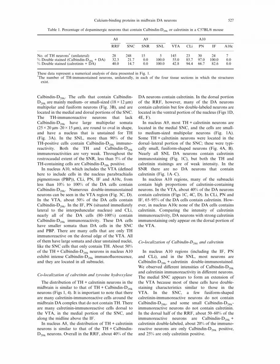
Table 1. Percentage of dopaminergic neurons that contain Calbindin-D28k or calretinin in a C57BL/6 mouse
A8 A9 A10
RRF SNC SNR SNL VTA CLi PN IF A10c
No. of TH neurons* (unilateral) 28 248 15 5 145 23 30 24 7% Double stained (Calbindin-D28k + DA) 32.3 21.7 0.0 100.0 55.0 85.7 97.0 100.0 0.0% Double stained (calretinin + DA) 40.0 14.7 0.0 100.0 42.8 94.4 66.7 82.6 0.0
These data represent a numerical analysis of data presented in Fig. 1.*The number of TH-immunostained neurons, unilaterally, in each of the four tissue sections in which the structuresexist.
Calcium-binding proteins in midbrain DA neurons 527

Fig. 3. Double-immunostained sections for TH and Calbindin-D28k. (A, C, E) Sections stained for TH; (B,D, F) the same sections stained for Calbindin-D28k. (A, B) SNC cells, above the SNR: most of the cellsonly stain for TH, but some of the dorsal SNC cells stain for both TH and Calbindin-D28k. (C, D) Stainingin the VTA: many VTA cells stain for both TH and Calbindin-D28k. (E, F) Staining in the RRF: only afew cells stain for both TH and Calbindin-D28k. Arrows point to cells stained in corresponding right andleft panels, and provide a reference point to compare the two photomicrographs. Scale bar = 22 µm.

Fig. 4. Double-immunostained sections for TH and calretinin. (A, C, E) Sections stained for TH; (B, D,F) the same sections stained for calretinin. (A, B) SNC cells: most of the cells only stain for TH, but someof the dorsal SNC cells stain both for TH and calretinin. (C, D) Staining in the VTA. (E, F) Staining inthe RRF. Note that a small portion of the nucleus A8 DA neurons contains calretinin. Arrows point tocells stained in corresponding right and left panels, and provide reference points to compare the two
photomicrographs. Scale bar = 22 µm.
Calcium-binding proteins in midbrain DA neurons 529

DISCUSSION
Characteristics of dopaminergic neurons that containCalbindin-D28k or calretinin
Most of the TH + Calbindin-D28k and TH +calretinin double-labeled neurons are medium- tosmall-sized, multipolar and fusiform-shaped neurons.In contrast, the midbrain DA neurons that lackthe calcium-binding proteins Calbindin-D28k andcalretinin, especially in the SNC and VTA, are large-sized multipolar neurons, often greater than 20 µm inthe long axis. These neurons are located mainly in theSNC and the dorsal portion of the VTA (within thePBP), and some at the lateral extent of the CLi.Gerfen et al.8,9 used anterograde axonal tracing withPhaseolus vulgaris-leucoagglutinin, combined withimmunocytochemical staining for TH and Calbindin-D28k, and autoradiographic localization of µ-opiatereceptors to study the mesostriatal DA projections inthe rat. They found that the DA neurons that containCalbindin-D28k in the VTA, dorsal tier of the SNCand RRF correspond to the DA neurons that projectto the striatal matrix compartment; these fibers havethin diameters and fewer axonal varicosities. The DAneurons that do not express Calbindin-D28k (those inthe ventral tier of the SNC and SNR) are distributedin a pattern that matches the origin of the DAprojection to the striatal patches; these fibers haveslightly thicker axonal diameters, with more frequentvaricosities. The large DA neurons which do notcontain Calbindin-D28k are likely to be the neuronsinnervating striatal patches, and the medium-sizedDA neurons which contain Calbindin-D28k and/orcalretinin are neurons which innervate the matrix ofthe ventromedial striatum, including the nucleusaccumbens.The present study provides data in the mouse
concerning the proportions of midbrain DA neuronsthat contain Calbindin-D28k and/or calretinin. Up to95% of the DA neurons in midline regions of nucleusA10 (such as PN, CLi, IF) contain Calbindin-D28k
and calretinin. In A10 subnuclei further from themidline, 40–80% of the DA neurons containCalbindin-D28k and calretinin; these include the VTAand PBP. These data from the mouse are similar todata from the rat.24,42 However, the greatest discrep-ancy involves the co-localization of calretinin withTH in the SNC and SNR; mouse nigral DA neuronsinfrequently contain Calbindin-D28k or calretinin(not counting the medial SNC cells), but approxi-mately 70% of rat SNC and SNR DA neuronscontain calretinin.42 The calretinin data of Isaacs andJacobowitz,24 in the rat, are more in line with thepresent data in the mouse. Why the two studies in therat are different is unclear.The distribution of cells labeled for both TH and
calcium-binding proteins is similar to that of themidbrain DA neurons that contain the neuro-active peptides cholecystokinin and neuro-tensin.21,22,49,50 The midbrain DA neurons that
contain Calbindin-D28k and calretinin are most fre-quently found in nucleus A10 regions and in the SNLportion of nucleus A9. In the rat, it has been demon-strated that Calbindin-D28k is localized within thesame population of midbrain DA neurons thatcontains cholecystokinin and neurotensin.11 Thecalcium-binding proteins may play a role in theregulation of intracellular calcium levels to modulatethe co-release of peptides with dopamine.
Nuclear organization of calcium-binding proteins
Since the identification of DA neurons in midbrainnuclei A8, A9 and A10 by Dahlstrom and Fuxe,3
the establishment of boundaries between the threenuclear groups has been problematic.4,52 The bordersbetween nuclei A9 and A10 have most often beenestablished using the rat brain atlas of Paxinos andWatson;35 this was done for both the rat12 andmouse32 midbrain DA neurons. In the present study,many of the DA neurons in the medial SNC containcalcium-binding proteins in similar proportions tothe DA neurons in the VTA region. Therefore, themedial SNC neurons may represent part of nucleusA10, and not part of nucleus A9. A similar conclu-sion has been drawn in the human brain regardingthe localization of Calbindin-D28k in the medialSNC.28,29 These neurochemical data would supportthe inclusion of cells currently positioned in themedial SNC (nucleus A9) with cells in the lateralVTA (nucleus A10). Consistent with this proposal,the medial SNC DA cells in the rat have been shownto project to similar limbic regions as other nucleusA10 cell groups.52
The organization of midbrain DA neurons inthe mouse is similar to that in humans. In themouse substantia nigra there are three tiers of cells(see Fig. 1), beginning dorsally with the SNC DAneurons that contain the calcium-binding proteins,then the SNC DA neurons that lack the twocalcium-binding proteins, and finally the SNR DAneurons that lack the calcium-binding proteins. Thisis similar to the ô, â and ó tiers of the humansubstantia nigra as described by Olszewski andBaxter.34 The Calbindin-D28k- and calretinin-containing DA neurons begin rostrally in thedorsal, medial and lateral portions of the SNC andVTA regions, whereas the ventral SNC and SNRDA neurons do not contain the calcium-bindingproteins. This pattern continues to the caudal-mostportion of the midbrain DA cell complex, where innucleus A8 the double-labeled cells reside in thedorsal portion of the RRF, and the ventral portionof the RRF and nucleus A10c do not containdouble-labeled cells. The DA neurons that containthe calcium-binding proteins may represent a separ-ate functional system from the DA neurons thatlack the calcium-binding proteins.
530 C.-L. Liang et al.
Calcium-binding proteins and neurotoxicity
The midbrain DA neurons that contain thecalcium-binding proteins are less vulnerable toMPTP toxicity.25,26 Three and six hours after treat-ment with MPTP (20 mg/kg), in the C57BL/6 mouse,there were 42% and 128% increases in Calbindin-D28k protein concentrations in the VTA, respectively,as determined by western blot analysis.33 The eleva-tion in Calbindin-D28k, in response to MPTP, mayprotect the VTA DA neurons from neurotoxicity.The SNC neurons, most of which lack Calbindin-D28k and calretinin, are most vulnerable to degenera-tion in Parkinson’s disease, and to MPTPtoxicity.14,15,16,25,26 However, MPTP causes degen-eration of some of the DA neurons that containCalbindin-D28k, at a time when DA neurons thatlack Calbindin-D28k are still preserved.23,26 Thus,Calbindin-D28k is not the only factor that determineswhich cells survive MPTP toxicity. The activity of thedopamine transporter, which transports the MPTPtoxin into the DA neurons, also appears to play arole in determining which DA neurons are vulnerableto MPTP toxicity. The mRNA for the dopaminetransporter is low in midbrain DA cell regions thatare spared MPTP-induced degeneration in themouse,45 and there is a positive correlation betweenthe location of the vulnerable cells and the location ofcells that contain high levels of dopamine transportermRNA.46 Cells that contain low dopamine trans-porter activity and calcium-binding proteins should
be least vulnerable to degeneration in Parkinson’sdisease and following MPTP treatment.
CONCLUSIONS
The calcium-binding proteins Calbindin-D28k andcalretinin are localized within a subpopulation ofmidbrain DA neurons in the mouse. Midline nuclei,within nucleus A10, and the SNL contain the highestproportion of double-labeled neurons (labeled bothfor TH and one or both of the calcium-bindingproteins). The midline nuclei include the IF, PN andCLi. Within nucleus A8, less than half of the DAneurons contain Calbindin-D28k and/or calretinin,and within nucleus A9 (SNC and SNR), less than20% of the cells contain either calcium-binding pro-tein. This pattern of co-localization in the mouse issimilar to that found previously in the rat. Thebuffering of intracellular calcium by Calbindin-D28k
and/or calretinin may serve several functions for themidbrain DA neurons, such as regulating theco-release of neuroactive peptides (cholecystokinin,neurotensin) with dopamine, and protecting theneurons from MPTP neurotoxicity.
Acknowledgements—This research was supported by grantsfrom the NIH (NS-30406), American Parkinson DiseaseAssociation and the James Webb Fund of the DallasFoundation. The authors thank Ms Judy Burdette forsecretarial assistance.
REFERENCES
1. Arai R., Winsky L., Arai M. and Jacobowitz D. M. (1991) Immunohistochemical localization of calretinin in the rathindbrain. J. comp. Neurol. 310, 21–44.
2. Celio M. R. (1990) Calbindin D28k and parvalbumin in the rat nervous system. Neuroscience 35, 375–475.3. Dahlstrom A. and Fuxe K. (1962) Evidence for the existence of monoamine-containing neurons in the central nervous
system. I. Demonstration of monoamines in the cell bodies of brain stem neurons. Acta physiol. scand. Suppl. 232,1–31.
4. Fallon J. and Moore R. Y. (1978) Catecholamine innervation of the basal forebrain: IV. Topography of the dopamineprojection to the basal forebrain and neostriatum. J. comp. Neurol. 180, 545–580.
5. Fuller R. W. and Steranka L. R. (1985) Central and peripheral catecholamine depletion by 1-methyl-4-phenyl-1,2,3,6-tetrahydropyridine (MPTP) in rodents. Life Sci. 36, 243–247.
6. Garcia-Segura L. M., Baetens D., Roth J., Norman A. W. and Orci L. (1984) Immunohistochemical mapping ofcalcium-binding protein immunoreactivity in the rat central nervous system. Brain Res. 296, 75–86.
7. Gerfen C. R., Baimbridge K. G. and Thibault J. (1985) The neostriatal mosaic: compartmental distribution of CaBPand parvalbumin in the basal ganglia of the rat and monkey. Proc. natn. Acad. Sci. U.S.A. 82, 8780–8784.
8. Gerfen C. R., Herkenham M. and Thibault J. (1987) The neostriatal mosaic: II. Patch- and matrix-directedmesostriatal dopaminergic and non-dopaminergic systems. J. Neurosci. 7, 3915–3934.
9. Gerfen C. R., Herkenham M. and Thibault J. (1987) The neostriatal mosaic: III. Biochemical and developmentaldissociation of patch–matrix mesostriatal systems. J. Neurosci. 7, 3935–3944.
10. German D. C., Dubach M., Askari S., Speciale S. G. and Bowden D. M. (1988) 1-Methyl-4-phenyl-1,2,3,6-tetrahydropyridine-induced parkinsonian syndrome inMacaca fascicularis: which midbrain dopaminergic neurons arelost? Neuroscience 24, 161–174.
11. German D. C. and Liang C.-L. (1993) Neuroactive peptides exist in the midbrain dopaminergic neurons that containCalbindin-D28k. NeuroReport 4, 491–494.
12. German D. C. and Manaye K. F. (1993) Midbrain dopaminergic neurons (nuclei A8, A9 and A10): three-dimensionalreconstruction in the rat. J. comp. Neurol. 331, 297–309.
13. German D. C., Manaye K. F., Brown J. and Gerfen C. R. (1990) Calcium-binding protein immunoreactivity in thehuman midbrain: relationship to dopaminergic neurons. Soc. Neurosci. Abstr. 16, 696.
14. German D. C., Manaye K. F., Smith W. K., Woodward D. J. and Saper C. B. (1989) Midbrain dopaminergic cell lossin Parkinson’s disease: computer visualization. Ann. Neurol. 26, 507–514.
15. German D. C., Manaye K. F., Sonsalla P. K. and Brooks B. A. (1992) Midbrain dopaminergic cell loss in Parkinson’sdisease and MPTP-induced parkinsonism: sparing of Calbindin-D28k-containing cells. Ann. N.Y. Acad. Sci. 648,42–62.
Calcium-binding proteins in midbrain DA neurons 531

16. German D. C., Nelson E. L., Liang C.-L., Speciale S. G., Sinton C. R. and Sonsalla P. K. (1996) The neuro-toxin MPTP causes degeneration of specific nucleus A8, A9 and A10 dopaminergic neurons in the mouse.Neurodegeneration, in press.
17. Hallman H., Large J., Olson L., Stromberg I. and Jonsson G. (1985) Neurochemical and histochemical characteriz-ation of neurotoxic effects of MPTP on brain catecholamine neurons in the mouse. J. Neurochem. 44, 117–127.
18. Heikkila R. E., Hess A. and Duvoisin R. C. (1984) Dopaminergic neurotoxicity of 1-methyl-4-phenyl-1,2,5,6-tetrahydropyridine (MPTP) in mice. Science 224, 1451–1453.
19. Heizmann C. W. and Braun K. (1992) Changes in Ca2+-binding proteins in human neurodegenerative disorders.Trends Neurosci. 15, 259–264.
20. Heppelmann B., Senaris R. and Emson P. C. (1994) Combination of alkaline phosphatase in situ hybridization withimmunohistochemistry: colocalization of calretinin-mRNA with calbindin and tyrosine hydroxylase immunoreactivityin rat substantia nigra neurons. Brain Res. 635, 293–299.
21. Hokfelt T., Everitt B. J., Theodorsson-Norheim E. and Goldstein M. (1984) Occurrence of neurotensin-likeimmunoreactivity in subpopulations of hypothalamic, mesencephalic, and medullary catecholamine neurons. J. comp.Neurol. 222, 543–559.
22. Hokfelt T., Skirboll L., Rehfeld J. F., Goldstein K., Markey K. and Dann O. (1980) A subpopulation ofmesencephalic dopamine neurons projecting to limbic areas contains a cholecystokinin-like peptide: evidence fromimmunohistochemistry combined with retrograde tracing. Neuroscience 5, 2093–2124.
23. Iacopino A., Christakos S., German D. C., Sonsalla P. K. and Altar C. A. (1992) Calbindin-D28k-containing neuronsin animal models of neurodegeneration: possible protection from excitotoxicity. Molec. Brain Res. 13, 251–261.
24. Isaacs K. R. and Jacobowitz D. M. (1994) Mapping of the colocalization of calretinin and tyrosine hydroxylase in therat substantia nigra and ventral tegmental area. Expl Brain Res. 99, 34–42.
25. Lavoie B. and Parent A. (1991) Dopaminergic neurons expressing calbindin in normal and parkinsonian monkeys.NeuroReport 2, 601–604.
26. Liang C.-L., Sinton C. M., Sonsalla P. K. and German D. C. (1996) Midbrain dopaminergic neurons in the mouse thatcontain Calbindin-D28k exhibit reduced vulnerability to MPTP-induced neurodegeneration. Neurodegeneration,submitted.
27. Manaye K. F., Sonsalla P. K., Barnett G., Heikkila R. E., Woodward D. J., Smith W. K. and German D. C. (1989)1-Methyl-4-(2*-methylphenyl)-1,2,3,6-tetrahydropyridine (2*CH3-MPTP) induced degeneration of mesostriataldopaminergic neurons in the mouse: biochemical and neuroanatomical studies. Brain Res. 491, 307–315.
28. McRitchie D. A. and Halliday G. M. (1995) Calbindin D28k-containing neurons are restricted to the medial substantianigra in humans. Neuroscience 65, 87–91.
29. McRitchie D. A., Hardman C. D. and Halliday G. M. (1996) Cytoarchitectural distribution of calcium bindingproteins in midbrain dopaminergic regions of rats and humans. J. comp. Neurol. 364, 121–150.
30. Miettinen R., Gulyas A. I., Baimbridge K. G., Jacobowitz D. M. and Freund T. F. (1992) Calretinin is present innon-pyramidal cells of the rat hippocampus. II. Co-existence with other calcium binding proteins and GABA.Neuroscience 48, 29–43.
31. Mouatt-Prigent A., Agid Y. and Hirsch E. C. (1994) Does the calcium-binding protein calretinin protect dopaminergicneurons against degeneration in Parkinson’s disease? Brain Res. 668, 62–70.
32. Nelson E. L., Liang C.-L., Sinton C. M. and German D. C. (1996) Midbrain dopaminergic neurons in the mouse:computer-assisted mapping. J. comp. Neurol. 369, 361–371.
33. Ng M. C., Iacopino A. M., Quintero E. M., Marches F., Sonsalla P. K., Liang C.-L., Speciale S. G. and German D.C. (1996) The neurotoxin MPTP increases calbin din-D28k levels in mouse midbrain dopaminergic neurons. Molec.Brain Res. 36, 329–336.
34. Olszewski J. and Baxter D. (1954) Cytoarchitecture of the Human Brain Stem. Karger, Basel.35. Paxinos G. and Watson C. (1986) The Rat Brain in Stereotaxic Coordinates. Academic Press, New York.36. Persechini A., Moncrief N. D. and Kretsinger R. H. (1989) The EF-hand family of calcium-modulated proteins.
Trends Neurosci. 12, 462–467.37. Resibois A. and Rogers J. H. (1992) Calretinin in rat brain: an immunohistochemical study. Neuroscience 46,
101–134.38. Rogers J. H. (1989) Two calcium-binding proteins mark many chick sensory neurons. Neuroscience 31, 697–709.39. Rogers J. H. (1989) Immunoreactivity for calretinin and other calcium-binding proteins in cerebellum. Neuroscience
31, 711–721.40. Rogers J. H. (1989) Calretinin: a gene for a novel calcium-binding protein expressed principally in neurons. J. Cell
Biol. 105, 1343–1353.41. Rogers J. H. (1992) Immunohistochemical markers in rat cortex: co-localization of calretinin and Calbindin-D28k with
neuropeptides and GABA. Brain Res. 587, 147–157.42. Rogers J. H. (1992) Immunohistochemical markers in rat brain: colocalization of calretinin and Calbindin-D28k with
tyrosine hydroxylase. Brain Res. 587, 203–210.43. Rogers J. H. and Resibois A. (1992) Calretinin and Calbindin-D28k in rat brain: patterns of partial co-localization.
Neuroscience 51, 843–865.44. Sanchez F., Alonso J. R., Arevalo R., Carretero J., Vazquez R. and Aijon J. (1991) Calbindin D28k- and
parvalbumin-reacting neurons in the hypothalamic magnocellular neurosecretory nuclei of the rat. Brain Res. Bull. 28,39–46.
45. Sanghera M. K., Manaye K. F., Liang C.-L., Iacopino A. M., Bannon M. J. and German D. C. (1994) Low dopaminetransporter mRNA levels in midbrain regions containing calbindin. NeuroReport 5, 1641–1644.
46. Sanghera M. K., Simmerly R. B. and German D. C. (1995) Dopamine transporter activity in midbrain neuronsis directly correlated with vulnerability to MPTP toxicity: relationship to Calbindin-D28k. Soc. Neurosci. Abstr. 21,1380.
47. Schwaller B., Buchwald P., Blumcke I., Celio M. R. and Hunziker W. (1993) Characterization of a polyclonalantiserum against the purified human recombinant calcium binding protein calretinin. Cell Calcium 14, 639–648.
532 C.-L. Liang et al.

48. Seress L., Nitsch R. and Leranth C. (1993) Calretinin immunoreactivity in the monkey hippocampal formation. I.Light and electron microscopic characteristics and co-localization with other calcium-binding proteins. Neuroscience55, 775–796.
50. Seroogy K. B., Mehta A. and Fallon J. H. (1987) Neurotensin and cholecystokinin coexistence within neurons of theventral mesencephalon: projections to forebrain. Expl Brain Res. 68, 277–289.
49. Seroogy K., Ceccatelli S., Schalling M., Hokfelt T., Frey P., Walsh J., Dockray G., Brown J., Buchan A. andGoldstein M. (1989) A subpopulation of dopaminergic neurons in the rat ventral mesencephalon contains bothneurotensin and cholecystokinin. Brain Res. 455, 88–98.
51. Sonsalla P. K., Youngster S. K., Kindt M. V. and Heikkila R. E. (1987) N-Methyl-4-(2*-methylphenyl)-1,2,3,6-tetrahydropyridine-induced neurotoxicity. J. Pharmac. exp. Ther. 242, 850–855.
52. Swanson L. W. (1982) The projections of the ventral tegmental area and adjacent regions: a combined fluorescentretrograde tracer and immunofluorescence study in the rat. Brain Res. Bull. 9, 321–353.
53. Taketo M., Schroeder A. C., Mobraaten L. E., Gunning K. B., Hanten G., Fox R. R., Roderick T. H., Stewart C. L.,Lilly F., Hansen C. T. and Overbeek P. A. (1991) FVB/N: an inbred mouse strain preferable for transgenic analyses.Proc. natn. Acad. Sci. U.S.A. 88, 2065–2069.
54. Yamada T., McGeer P. L., Baimbridge K. K. and McGeer E. G. (1990) Relative sparing in Parkinson’s disease ofsubstantia nigra dopamine neurons containing Calbindin-D28k. Brain Res. 526, 303–307.
(Accepted 26 March 1996)
Calcium-binding proteins in midbrain DA neurons 533


