Embed Size (px)
Citation preview
Journal of Bioenergetics and Biomembranes, Vol. 37, No. 3, June 2005 ( C© 2005)DOI: 10.1007/s10863-005-6567-7
Mitochondrial Ceramide and the Induction of Apoptosis
Leah J. Siskind1
In most cell types, a key event in apoptosis is the release of proapoptotic intermembrane space proteinsfrom mitochondria to the cytoplasm. In general, it is the release of these intermembrane space proteinsthat is responsible for the activation of caspases and DNases that are responsible for the execution ofapoptosis. The mechanism for the increased permeability of the mitochondrial outer membrane duringthe induction phase of apoptosis is currently unknown and highly debated. This review will focuson one such proposed mechanism, namely, the formation of ceramide channels in the mitochondrialouter membrane. Ceramides are known to play a major regulatory role in apoptosis by inducingthe release of proapoptotic proteins from the mitochondria. As mitochondria are known to containthe enzymes responsible for the synthesis and hydrolysis of ceramide, there exists a mechanism forregulating the level of ceramide in mitochondria. In addition, mitochondrial ceramide levels havebeen shown to be elevated prior to the induction phase of apoptosis. Ceramide has been shown toform large protein permeable channels in planar phospholipid and mitochondrial outer membranes.Thus, ceramide channels are good candidates for the pathway with which proapoptotic proteins arereleased from mitochondria during the induction phase of apoptosis.
KEY WORDS: Ceramide; mitochondria; ceramide synthase; Bcl-2, apoptosis; sphingosine; cytochrome c;channel.
INTRODUCTION
Early in apoptosis, there is often an increase in thepermeability of the mitochondrial outer membrane thatleads to the release of intermembrane space proteins, in-cluding cytochrome c, procaspases, apoptosis-inducingfactor (AIF), heat shock proteins, Smac/Diablo, and en-donuclease G (for review see Saelens et al., 2004). Therelease of intermembrane space proteins into the cyto-plasm is crucial for the activation of specific caspases andDNases that are responsible for the execution of apoptosis.The mechanism for the increased permeability of the mi-tochondrial outer membrane during the induction phaseof apoptosis is currently unknown and highly debated.It is thought to occur by either the permeability transi-tion or the creation of a pore in the mitochondrial outermembrane (for review see Henry-Mowatt et al., 2004).Several pathways have been proposed for this pore. This
1 Department of Biology, University of Maryland College Park, CollegePark, Maryland 20742; e-mail: [email protected].
review will focus on one such proposed pathway, namely,ceramide channels.
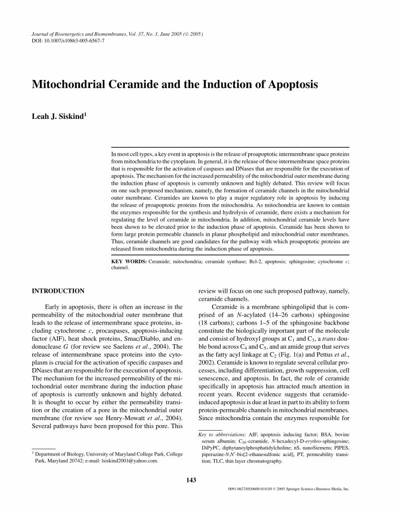
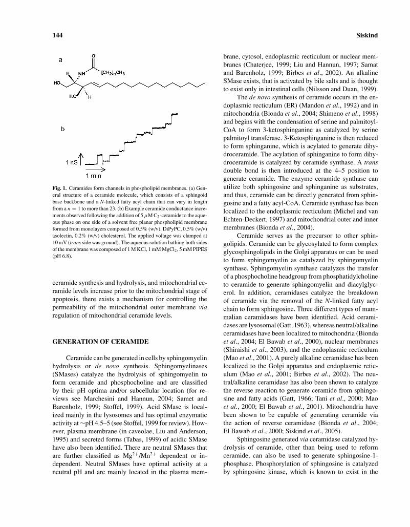
Ceramide is a membrane sphingolipid that is com-prised of an N-acylated (14–26 carbons) sphingosine(18 carbons); carbons 1–5 of the sphingosine backboneconstitute the biologically important part of the moleculeand consist of hydroxyl groups at C1 and C3, a trans dou-ble bond across C4 and C5, and an amide group that servesas the fatty acyl linkage at C2 (Fig. 1(a) and Pettus et al.,2002). Ceramide is known to regulate several cellular pro-cesses, including differentiation, growth suppression, cellsenescence, and apoptosis. In fact, the role of ceramidespecifically in apoptosis has attracted much attention inrecent years. Recent evidence suggests that ceramide-induced apoptosis is due at least in part to its ability to formprotein-permeable channels in mitochondrial membranes.Since mitochondria contain the enzymes responsible for
Key to abbreviations: AIF, apoptosis inducing factor; BSA, bovineserum albumin; C16-ceramide, N-hexadecyl-D-erythro-sphingosine;DiPyPC, diphytanoylphosphatidylcholine; nS, nanoSiemens; PIPES,piperazine-N,N′-bis[2-ethanesulfonic acid], PT, permeability transi-tion; TLC, thin layer chromatography.
1430091-0627/05/0600-0143/0 C© 2005 Springer Science+Business Media, Inc.
144 Siskind
Fig. 1. Ceramides form channels in phospholipid membranes. (a) Gen-eral structure of a ceramide molecule, which consists of a sphingoidbase backbone and a N-linked fatty acyl chain that can vary in lengthfrom a n = 1 to more than 23. (b) Example ceramide conductance incre-ments observed following the addition of 5 µM C2-ceramide to the aque-ous phase on one side of a solvent free planar phospholipid membraneformed from monolayers composed of 0.5% (w/v). DiPyPC, 0.5% (w/v)asolectin, 0.2% (w/v) cholesterol. The applied voltage was clamped at10 mV (trans side was ground). The aqueous solution bathing both sidesof the membrane was composed of 1 M KCl, 1 mM MgCl2, 5 mM PIPES(pH 6.8).
ceramide synthesis and hydrolysis, and mitochondrial ce-ramide levels increase prior to the mitochondrial stage ofapoptosis, there exists a mechanism for controlling thepermeability of the mitochondrial outer membrane viaregulation of mitochondrial ceramide levels.
GENERATION OF CERAMIDE
Ceramide can be generated in cells by sphingomyelinhydrolysis or de novo synthesis. Sphingomyelinases(SMases) catalyze the hydrolysis of sphingomyelin toform ceramide and phosphocholine and are classifiedby their pH optima and/or subcellular location (for re-views see Marchesini and Hannun, 2004; Samet andBarenholz, 1999; Stoffel, 1999). Acid SMase is local-ized mainly in the lysosomes and has optimal enzymaticactivity at ∼pH 4.5–5 (see Stoffel, 1999 for review). How-ever, plasma membrane (in caveolae, Liu and Anderson,1995) and secreted forms (Tabas, 1999) of acidic SMasehave also been identified. There are neutral SMases thatare further classified as Mg2+/Mn2+ dependent or in-dependent. Neutral SMases have optimal activity at aneutral pH and are mainly located in the plasma mem-
brane, cytosol, endoplasmic recticulum or nuclear mem-branes (Chaterjee, 1999; Liu and Hannun, 1997; Samatand Barenholz, 1999; Birbes et al., 2002). An alkalineSMase exists, that is activated by bile salts and is thoughtto exist only in intestinal cells (Nilsson and Duan, 1999).
The de novo synthesis of ceramide occurs in the en-doplasmic recticulum (ER) (Mandon et al., 1992) and inmitochondria (Bionda et al., 2004; Shimeno et al., 1998)and begins with the condensation of serine and palmitoyl-CoA to form 3-ketosphinganine as catalyzed by serinepalmitoyl transferase. 3-Ketosphinganine is then reducedto form sphinganine, which is acylated to generate dihy-droceramide. The acylation of sphinganine to form dihy-droceramide is catalyzed by ceramide synthase. A transdouble bond is then introduced at the 4–5 position togenerate ceramide. The enzyme ceramide synthase canutilize both sphingosine and sphinganine as substrates,and thus, ceramide can be directly generated from sphin-gosine and a fatty acyl-CoA. Ceramide synthase has beenlocalized to the endoplasmic recticulum (Michel and vanEchten-Deckert, 1997) and mitochondrial outer and innermembranes (Bionda et al., 2004).
Ceramide serves as the precursor to other sphin-golipids. Ceramide can be glycosylated to form complexglycosphingolipids in the Golgi apparatus or can be usedto form sphingomyelin as catalyzed by sphingomyelinsynthase. Sphingomyelin synthase catalyzes the transferof a phosphocholine headgroup from phosphatidylcholineto ceramide to generate sphingomyelin and diacylglyc-erol. In addition, ceramidases catalyze the breakdownof ceramide via the removal of the N-linked fatty acylchain to form sphingosine. Three different types of mam-malian ceramidases have been identified. Acid cerami-dases are lysosomal (Gatt, 1963), whereas neutral/alkalineceramidases have been localized to mitochondria (Biondaet al., 2004; El Bawab et al., 2000), nuclear membranes(Shiraishi et al., 2003), and the endoplasmic recticulum(Mao et al., 2001). A purely alkaline ceramidase has beenlocalized to the Golgi apparatus and endoplasmic retic-ulum (Mao et al., 2001; Birbes et al., 2002). The neu-tral/alkaline ceramidase has also been shown to catalyzethe reverse reaction to generate ceramide from sphingo-sine and fatty acids (Gatt, 1966; Tani et al., 2000; Maoet al., 2000; El Bawab et al., 2001). Mitochondria havebeen shown to be capable of generating ceramide viathe action of reverse ceramidase (Bionda et al., 2004;El Bawab et al., 2000; Siskind et al., 2005).
Sphingosine generated via ceramidase catalyzed hy-drolysis of ceramide, other than being used to reformceramide, can also be used to generate sphingosine-1-phosphase. Phosphorylation of sphingosine is catalyzedby sphingosine kinase, which is known to exist in the
Mitochondrial Ceramide 145
cytosol and endoplasmic recticulum (Ghosh et al., 1994;Olivera et al., 1999). Sphingosine-1-phosphate has beenshown to be protective/anti-apoptotic. In fact, it has beenproposed that it is the ratio of sphingosine-1-phosphateto ceramide/sphingosine that determines the fate of a cell(Cuvillier et al., 1996; Olivera and Spiegel, 1993).
A CONSERVED ROLE FOR CERAMIDEIN APOPTOSIS
There are a number of observations that support aproapoptotic role for ceramide in apoptosis. First, ce-ramide generation is a common cellular response of a vari-ety of cell types following exposure to apoptosis-inducingagents. These include TNFα (Obeid et al., 1993; Moduret al., 1996; Garcıa-Ruiz et al., 1997; Geilen et al., 1997),interleukin-1 (Masamune et al., 1996), CD95/Fas/APO-1(Cifone et al., 1994; Cremesti et al., 2001; Brenner et al.,1998; Gulbins et al., 1995; Paris et al., 2001; Tepper et al.,1995), γ -interferon (Birbes et al., 2002), NO (Takedaet al., 1999), ionizing radiation (Vit and Rosselli, 2003;Alphonse et al., 2002), serum withdrawal (Caricchio et al.,2002), heat (Jenkins, 2003), etoposide (Sawada et al.,2000a,b), staurosporine (Wiesner and Dawson, 1996),daunorubicin (Come et al., 1999), and the corticosteroiddexamethasone (Cifone et al., 1999). Second, the effec-tive doses of these agents required to induce ceramidegeneration closely matches the dose required to induceapoptosis (Kolesnick and Kronke, 1998). Third, elevationsin cellular ceramide in response to these agents occursprior to the execution phase of apoptosis (for example,Birbes et al., 2002; Hannun, 1996; Dbaibo et al., 1997).Fourth, exogenous addition of cell-permeable ceramideanalogues induces apoptosis in a variety of cell lines (forexample, Obeid et al., 1993; Jarvis et al., 1994; Quintanset al., 1994; Cifone et al., 1994). Fifth, ceramide-inducedapoptosis is very specific, as the naturally occurring ce-ramide precursor dihydroceramide (lacks the 4–5 transdouble bond present in ceramide) does not induce apop-tosis (Obeid et al., 1993; Bielawska et al., 1993). Sixth,apoptosis can be inhibited upon blockage of ceramide gen-eration and cells that are incapable of generating ceramideare often incapable of undergoing apoptosis (Selzner et al.,2001; Chmura et al., 1997a,b; Bruno et al., 1998; Sautinet al., 2000; Alphonse et al., 2002; Riboni et al., 2002).
Apoptosis can often be induced in cancer cells byelevation of endogenous ceramide levels via the addi-tion of inhibitors of ceramide metabolism (for example,Chmura et al., 1997a; Raisova et al., 2002; Rodriguez-Lafrasse et al., 2002; Alphonse et al., 2004; Bielawskaet al., 1996; Selzner et al., 2001) or by exogenously added
cell-permeable ceramide analogues (for example, Sautinet al., 2000; Lozano et al., 2001; Selzner et al., 2001;Kimura et al., 2003). Ceramidase inhibitors have beenshown to elevate ceramide levels and induce apoptosis inHL-60 leukemia cells (Bielawska et al., 1996) humancolon cancer cells (Selzner et al., 2001), HaCaT ker-atinocytes, and human melanoma cells (Raisova et al.,2002). Defective ceramide generation has been correlatedwith resistance to radiation-induced apoptosis (Chmuraet al., 1997a,b; Bruno et al., 1998; Sautin et al., 2000;Alphonse et al., 2002). In fact, Alphonse et al., 2002demonstrated that SQ20B adenocarcinoma cells are re-sistant to both anti-Fas and γ -irradiation because of aninability to generate ceramide. In a further study, theyshowed that the apoptotic death pathway is present, butnot functional, in the absence of a certain threshold ofceramide generation (Alphonse et al., 2004). Thus, ce-ramide clearly plays an important role in apoptosis in-duction. However, the mechanism by which ceramide in-duces apoptosis is still highly debated. It is thought thatceramide-induced apoptosis involves a direct action ofceramide on mitochondria.
MITOCHONDRIAL CERAMIDEGENERATION AND APOPTOSIS
Mitochondria have been shown to contain ceramidewith a three-fold higher concentration in the outer mem-branes than the inner membranes (Ardail et al., 2001).In fact, mitochondria isolated from healthy rat livers havehigher levels of dihydroceramide (lacks the 4-5 trans dou-ble bond present in ceramide, not apoptotic) than ceramide(Ardail et al., 2001). This is not surprising given the factthat higher levels of ceramide in mitochondria would notbe expected in healthy cells. Thus, conversion of dihy-droceramide to ceramide would be one way to rapidlygenerate ceramide in mitochondria and induce apoptosis.
Increases in cellular ceramide levels during apopto-sis have been shown to occur prior to the mitochondrialphase of apoptosis (Witty et al., 1996; Raisova et al., 2000;Bose et al., 1995; Charles et al., 2001; Rodriguez-Lafrasseet al., 2001; Kroesen et al., 2001; Thomas et al., 1999).As stated above, mitochondria contain the enzymes re-sponsible for ceramide synthesis and hydrolysis, namely,ceramide synthase and ceramidase (El Bawab et al., 2000;Shimeno et al., 1998; Bionda et al., 2004). In fact, Biondaet al., 2004 reconfirmed the presence of ceramide syn-thase and ceramidase in ultrapure mitochondria free fromcontaminating endoplasmic recticulum membranes (mi-tochondrial associated membranes; MAM), and micro-somes. They were able to show that both mitochondrial
146 Siskind
outer and inner membranes are capable of generating ce-ramide. CD95-, TNFα-radiation, and UV-induced apop-tosis have all been shown to occur via an increase in mi-tochondrial ceramide levels (Birbes et al., 2005; Vance,1990; Garcia-Ruiz et al., 1997; Matsko et al., 2001; Daiet al., 2004). Garcia-Ruiz et al., 1997 showed that mito-chondria isolated from TNF treated cells had a two- tothree-fold increase in ceramide levels than mitochondriafrom untreated cells. A similar increase in mitochondrialceramide levels was also shown by Birbes et al., 2005in TNF treated MCF7 breast cancer cells. Dai et al.,2004 showed that UV irradiation of HeLa cells resultsin increased mitochondrial ceramide levels that precededcytochrome c release and apoptosis. Inhibitors of sph-ingolipid metabolism that prevented ceramide synthesisafter UV irradiation also prevented apoptosis (Dai et al.,2004). Birbes et al., 2001, targeted the bacterial sphin-gomyelinase protein (bSMase) to different intracellularlocations in MCF7 breast cancer cells in order to generateceramide solely in these locations. Only when the bSMasewas targeted to mitochondria and ceramide generated inmitochondria was there cytochrome c release and apop-tosis. A mutant inactive bSMase targeted to mitochondriaresulted in no ceramide generation and no apoptosis. Tar-geting the bSMase to the plasma membrane, cytoplasm,endoplasmic reticulum, the Gogli, and the nucleus did notresult in apoptosis despite the generation of ceramide inthese compartments. Thus, ceramide-induced apoptosisoccurs at the level of mitochondria.
Ceramides have been reported to have numerous af-fects on mitochondria, including enhanced generation ofreactive oxygen species (Zamzami et al., 1995; Di Paolaet al., 2000; Quillet-Mary et al., 1997; France-Lanordet al., 1997; Garcia-Ruiz et al., 1997), alteration of cal-cium homeostasis of mitochondria and endoplasmic retic-ulum (Zamzami et al., 1995; Quillet-Mary et al., 1997;Garcia-Ruiz et al., 1997; Pinton et al., 2001; Muriel et al.,2000), ATP depletion (Arora et al., 1997), collapse inthe inner mitochondrial membrane potential (Zamzamiet al., 1995; Arora et al., 1997; Di Paola et al., 2000;Ghafourifar et al., 1999), inhibition and/or activation ofthe activities of various components of the mitochondrialelectron transport chain (Di Paola et al., 2000; Gudzet al., 1997), and release of intermembrane space pro-teins (Ghafourifar et al., 1999; Di Paola et al., 2000,2004; Siskind et al., 2002). Short-chain cell perme-able ceramide analogues, such as N-acetyl-D-erythro-sphingosine (C2-ceramide) and N-hexanoyl-D-erythro-sphingosine (C6-ceramide) have been shown to inducecytochrome c release when added to whole cell cultures(for example, Zamzami et al., 1995; Castedo et al., 1996;Susin et al., 1997a,b; DeMaria et al., 1997; Zhang et al.,
1997; Amarante-Mendes et al., 1998) and isolated mito-chondria (Arora et al., 1997; Di Paola et al., 2000, 2004;Ghafourifar et al., 1999); this cytochrome c release waspreventable by preincubation with or overexpression ofthe anti-death protein Bcl-2 (Ghafourifar et al., 1999;Zhang et al., 1996; Geley et al., 1997), or transfectionof cells with Bcl-xL (Gottschalk et al., 1994; Wiesneret al., 1997). Long chain naturally occurring ceramideshave also been shown to induce the release of both cy-tochrome c (Di Paola et al., 2000, 2004) from isolatedmitochondria. In addition to cytochrome c, long- andshort-chain ceramides have been shown to induce the re-lease of apoptosis-inducing factor (AIF) (Di Paola et al.,2004), AK-2 (Di Paola et al., 2004), and adenylate ki-nase (Siskind et al., 2002) from isolated mitochondria.Until recently, it was not clear how ceramide increasedthe permeability of the mitochondrial outer membraneto intermembrane space proteins. Recent evidence indi-cates that it is due to a direct action of ceramide on themitochondrial outer membrane. In fact, ceramides havebeen shown to form large protein permeable channels inplanar phospholipid and mitochondrial outer membranes(Siskind and Colombini, 2000; Siskind et al., 2002, 2003).
CERAMIDE CHANNEL FORMATION
Both short- and long-chain naturally occurring ce-ramides form large channels in planar phospholipid mem-branes. The addition of ceramide to the aqueous phase oneither one or both sides of a planar phospholipid mem-brane results in pore formation as indicated by discretestepwise current increases (Siskind and Colombini, 2000;Siskind et al., 2002, 2003, 2005; Fig. 1(b)). These dis-crete increments in conductance are, by definition, chan-nels, reflecting the formation of continuous water-filledpathways through the membrane. Ceramide channel for-mation requires the presence of the 4-5 trans double bondas dihydroceramide does not form channels even at con-centrations up to 25 times higher than that required for ce-ramide channel formation (Siskind and Colombini, 2000).Recall that dihydroceramide is the metabolic precursorto ceramide. It does not induce cytochrome c release orapoptosis. Thus, the channel forming ability of ceramidescorrelates with its apoptotic activity.
Similar ceramide channels form in the mitochondrialouter membrane. Ceramide does not induce a cytochromec secretion or release mechanism, but simply raises thepermeability of the mitochondrial outer membrane, viaceramide channel formation, to include all small proteins.Evidence for ceramide channel formation in mitochon-drial outer membranes comes from work with isolated
Mitochondrial Ceramide 147
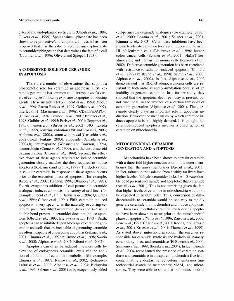
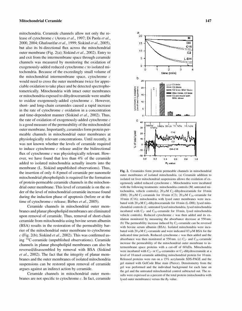
mitochondria. Ceramide channels allow not only the re-lease of cytochrome c (Arora et al., 1997; Di Paola et al.,2000, 2004; Ghafourifar et al., 1999; Siskind et al., 2005),but also its bi-directional flux across the mitochondrialouter membrane (Fig. 2(a); Siskind et al., 2002). Entry toand exit from the intermembrane space through ceramidechannels was measured by monitoring the oxidation ofexogenously-added reduced cytochrome c to isolated mi-tochondria. Because of the exceedingly small volume ofthe mitochondrial intermembrane space, cytochrome cwould need to cross the outer membrane twice for appre-ciable oxidation to take place and be detected spectropho-tometrically. Mitochondria with intact outer membranesor mitochondria exposed to dihydroceramide were unableto oxidize exogenously-added cytochrome c. However,short- and long-chain ceramides caused a rapid increasein the rate of cytochrome c oxidation in a concentrationand time-dependent manner (Siskind et al., 2002). Thus,the rate of oxidation of exogenously-added cytochrome cis a good measure of the permeability of the mitochondrialouter membrane. Importantly, ceramides form protein per-meable channels in mitochondrial outer membranes atphysiologically relevant concentrations. Until recently, itwas not known whether the levels of ceramide requiredto induce cytochrome c release and/or the bidirectionalflux of cytochrome c was physiologically relevant. How-ever, we have found that less than 4% of the ceramideadded to isolated mitochondria actually inserts into themembrane (L. Siskind unpublished observations). Thus,the insertion of only 4–8 pmol of ceramide per nanomolemitochondrial phospholipids is required for the formationof protein-permeable ceramide channels in the mitochon-drial outer membrane. This level of ceramide is on the or-der of the level of mitochondrial ceramide increase foundduring the induction phase of apoptosis (before or at thetime of cytochrome c release; Birbes et al., 2005).
Ceramide channels in mitochondrial outer mem-branes and planar phospholipid membranes are eliminatedupon removal of ceramide. Thus, removal of short-chainceramide from mitochondria using bovine serum albumin(BSA) results in the restoration of the permeability bar-rier of the mitochondrial outer membrane to cytochromec (Fig. 2(b); Siskind et al., 2002). This was confirmed us-ing 14C-ceramide (unpublished observations). Ceramidechannels in planar phospholipid membranes can also bereversed/disassembled by removal with BSA (Siskindet al., 2002). The fact that the integrity of planar mem-branes and the outer membranes of isolated mitochondriasuspensions can be restored upon removal of ceramideargues against an indirect action by ceramide.
Ceramide channels in mitochondrial outer mem-branes are not specific to cytochrome c. In fact, ceramide
Fig. 2. Ceramides form protein permeable channels in mitochondrialouter membranes of isolated mitochondria. (a) Ceramide addition toisolated rat liver mitochondrial suspensions allows the oxidation of ex-ogenously added reduced cytochrome c. Mitochondria were incubatedwith the following treatments: mitochondria controls (M; untreated mi-tochondria, vehicle controls); 20 µM C2-dihydroceramide for 10 min(DH); 20 µM C2-ceramide for 10 min (C2); 20 µM C16-ceramide for10 min (C16); mitochondria with lysed outer membranes were incu-bated with 20 µM C2-dihydroceramide for 10 min (L-DH); lysed mito-chondrial controls (L; untreated lysed mitochondria, lysed mitochondriaincubated with C2- and C16-ceramide for 10 min, lysed mitochondriavehicle controls). Reduced cytochrome c was then added and its ox-idation monitored by measuring the absorbance decrease at 550 nm.(b) The permeability increase induced by C2-ceramide can be reversedwith bovine serum albumin (BSA). Isolated mitochondria were incu-bated with 20 µM C2-ceramide and were indicated 83 µM BSA for theindicated time periods. Reduced cytochrome c was then added and theabsorbance was then monitored at 550 nm. (c) C2- and C16-ceramideincrease the permeability of the mitochondrial outer membrane to in-termembrane space proteins with a cut-off of 60 kDa. Mitochondriawere incubated with C2- or C16-ceramides or C2-dihydroceramide at alevel of 18 nmol ceramide added/mg mitochondrial protein for 10 min.Released proteins were run on a 15% acrylamide SDS-PAGE and thegel stained with GelCode Blue stain (Pierce). Densitometry from thegel was performed and the individual background for each lane onthe gel and the untreated mitochondrial control subtracted out. The re-sults were expressed as a percent of the total protein (mitochondria withlysed outer membranes) versus the RF value.
148 Siskind
channels allow the release of low molecular weight pro-teins from mitochondria, but not high molecular weightones. There is a sharp molecular weight cut-off of 60,000for the size of intermembrane space proteins that are re-leased through the ceramide channels (Fig. 2(c); Siskindet al., 2002). Even though this cut-off was measured un-der denaturing conditions and thus is most likely an un-derestimate, it is still in line with the size of proapoptoticproteins released from mitochondria during apoptosis (cy-tochrome c 12 kDa, (Dickerson et al., 1971); endonucleaseG 28 kDa, (Schafer et al., 2004); AIF 57 kDa, (Mateet al., 2002); Smac/DIABLO 42 kDa, (Chai et al.,2001)).
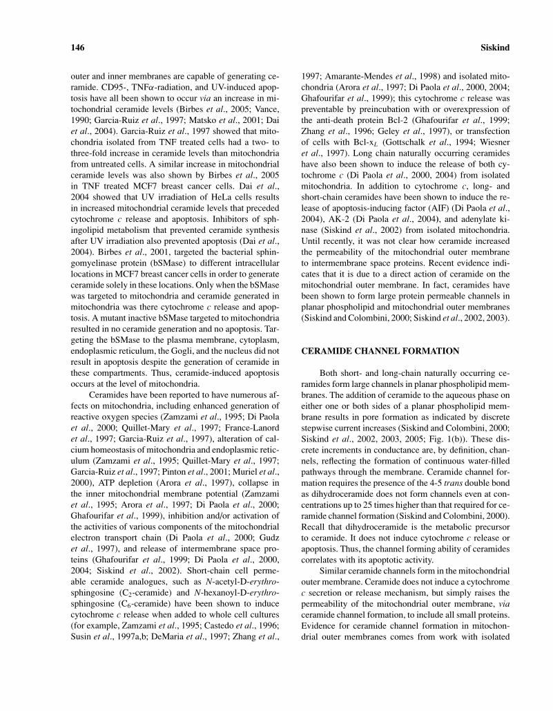
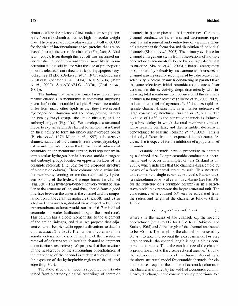
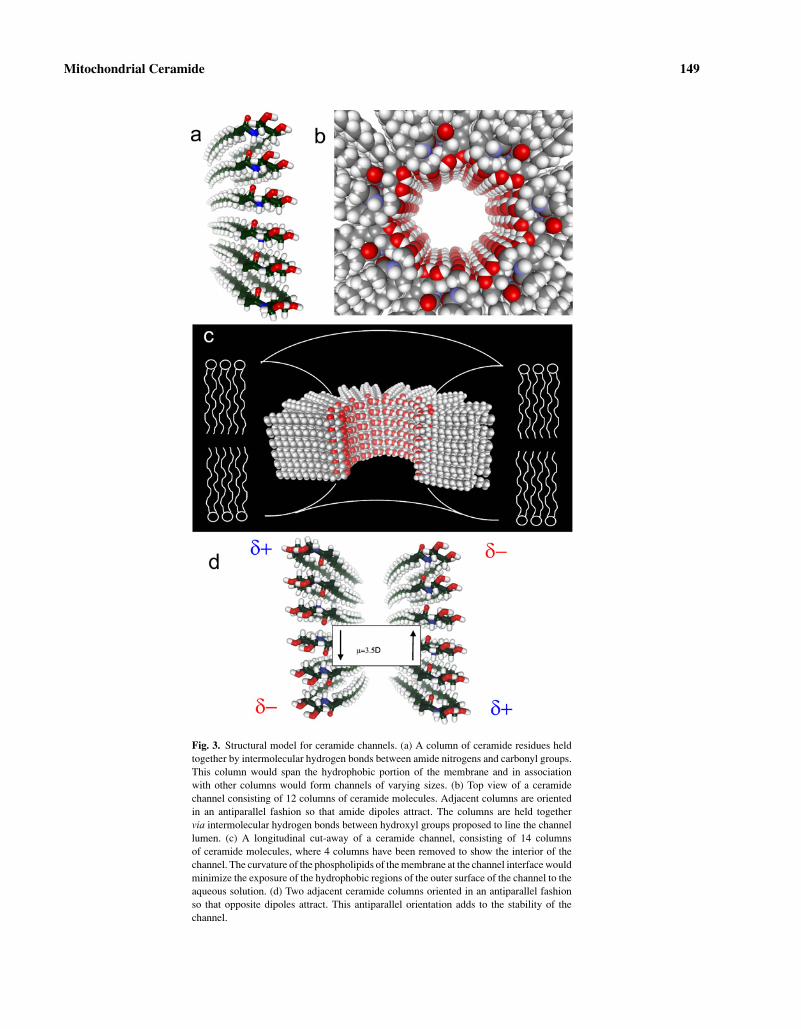
The finding that ceramide forms large protein per-meable channels in membranes is somewhat surprisinggiven the fact that ceramide is a lipid. However, ceramidesdiffer from many other lipids in that they have severalhydrogen-bond donating and accepting groups, namelythe two hydroxyl groups, the amide nitrogen, and thecarbonyl oxygen (Fig. 1(a)). We developed a structuralmodel to explain ceramide channel formation that is basedon their ability to form intermolecular hydrogen bonds(Pascher et al., 1976; Moore et al., 1997) and extensivecharacterization of the channels from electrophysiologi-cal recordings. We propose the formation of columns ofceramides on the membrane surface, held together by in-termolecular hydrogen bonds between amide nitrogensand carbonyl groups located on opposite surfaces of theceramide molecule (Fig. 3(a) for the proposed structureof a ceramide column). These columns could swing intothe membrane, forming an annulus stabilized by hydro-gen bonding of the hydroxyl groups lining the channel(Fig. 3(b)). This hydrogen-bonded network would be sim-ilar to the structure of ice, and thus, should form a goodinterface between the water in the channel and the nonpo-lar portion of the ceramide molecule (Figs. 3(b) and (c) fora top and cut-away longitudinal view, respectively). Eachtransmembrane column would consist of 6–7 individualceramide molecules (sufficient to span the membrane).This column has a dipole moment due to the alignmentof the amide linkages, and thus, we propose that adja-cent columns be oriented in opposite directions so that thedipoles attract (Fig. 3(d)). The number of columns in theannulus determines the size of the channel; the insertion orremoval of columns would result in channel enlargementor contracture, respectively. We propose that the curvatureof the headgroups of the surrounding phospholipids atthe outer edge of the channel is such that they minimizethe exposure of the hydrophobic regions of the channeledge (Fig. 3(c)).
The above structural model is supported by data ob-tained from electrophyiological recordings of ceramide
channels in planar phospholipid membranes. Ceramidechannel conductance increments and decrements repre-sent the enlargement and contracture of ceramide chan-nels rather than the formation and dissolution of individualchannels (Siskind et al., 2003). The primary evidence forchannel enlargement stems from observations of multipleconductance increments followed by one large decrementto baseline (Siskind et al., 2003). Channel enlargementis supported by selectivity measurements; increases inchannel size are usually accompanied by a decrease in ionselectivity, whereas channels conducting in parallel havethe same selectivity. Initial ceramide conductances favorcations, but this selectivity drops dramatically with in-creasing total membrane conductance until the ceramidechannel is no longer selective (Siskind et al., 2003, 2005),indicating channel enlargement. La+3 induces rapid ce-ramide channel disassembly in a manner indicative oflarge conducting structures (Siskind et al., 2003). Theaddition of La+3 to the ceramide channels is followedby a brief delay, in which the total membrane conduc-tance remains constant, and then a sudden decrease inconductance to baseline (Siskind et al., 2003). This isdistinctly different from the exponential conductance de-crease that is expected for the inhibition of a population ofchannels.
Ceramide channels have a propensity to contractby a defined size. Larger ceramide conductance decre-ments tend to occur as multiples of 4 nS (Siskind et al.,2003), which indicates that the channels disassemble bymeans of a fundamental structural unit. This structuralunit cannot be a single ceramide molecule. Rather, a ce-ramide column or pairs of ceramide columns (see Fig. 3(b)for the structure of a ceramide column) as in a barrel-stave model may represent the larger structural unit. Theconductance of a channel (G) can be calculated fromthe radius and length of the channel as follows (Hille,1992):
G = (κsp πr2)/(L + 0.5 πr ) (1)
where r is the radius of the channel, κsp the specificconductance (equal to 112 for 1.0 M KCl; Robinson andStokes, 1965) and L the length of the channel (estimatedto be ∼5 nm). The length of the channel is increased by0.5(πr) to take into account the axis resistance. For verylarge channels, the channel length is negligible as com-pared to its radius. Thus, the conductance of the channelis proportional not to the cross-sectional area (πr2), but tothe radius or circumference of the channel. According tothe above structural model for ceramide channels, the cir-cumference is equal to the number of ceramide columns inthe channel multiplied by the width of a ceramide column.Hence, the change in the conductance is proportional to a
Mitochondrial Ceramide 149
Fig. 3. Structural model for ceramide channels. (a) A column of ceramide residues heldtogether by intermolecular hydrogen bonds between amide nitrogens and carbonyl groups.This column would span the hydrophobic portion of the membrane and in associationwith other columns would form channels of varying sizes. (b) Top view of a ceramidechannel consisting of 12 columns of ceramide molecules. Adjacent columns are orientedin an antiparallel fashion so that amide dipoles attract. The columns are held togethervia intermolecular hydrogen bonds between hydroxyl groups proposed to line the channellumen. (c) A longitudinal cut-away of a ceramide channel, consisting of 14 columnsof ceramide molecules, where 4 columns have been removed to show the interior of thechannel. The curvature of the phospholipids of the membrane at the channel interface wouldminimize the exposure of the hydrophobic regions of the outer surface of the channel to theaqueous solution. (d) Two adjacent ceramide columns oriented in an antiparallel fashionso that opposite dipoles attract. This antiparallel orientation adds to the stability of thechannel.
150 Siskind
change in the number of columns comprising the ceramidechannel and equation (1) becomes:
�G = 3.56 × column width
× number of columns lost from the channel
(2)
at 1 M KCl and G in nS (see Siskind et al., 2003 for an in-depth description and underlying mathematical details).Because the number of columns lost from the channelmust always be an integer, the change in conductancewould equal to 4 nS if the column width were 1.12 nm.This is compatible with the dimension of two ceramidecolumns, indicating that the fundamental unit of disas-sembly is a two-column unit. Thus, two columns may lineup in opposite directions resulting in a favorable dipole–dipole interaction (Fig. 3(d)).
The larger conductance decrements occurring asmultiples of 4 nS is only consistent with ceramide chan-nels that are cylindrical structures with preferred diame-ters rather than a continuum of sizes or shapes (Siskindet al., 2003). The electrophysiological data provides ev-idence that ceramide channels are not flaccid structures,but rather rigid cylinders whose structure is determinedby the hydrogen-bonded network forming the inner liningof the channel. Other models that propose that ceramidesact by simply disrupting the membrane or forming raftsthat result in defects between these and the surroundingphospholipids are all in sharp disagreement with the ob-served behavior of ceramide channels. Taken together, thedata indicates that ceramides form barrel-stave channelswhose size can change by the loss or insertion of multiplesof columns.
FUTURE DIRECTIONS
Future studies need to be aimed at determining theexact submitochondrial location of ceramide generationduring the induction phase of apoptosis. It is known thatmitochondrial ceramide levels increase prior to the re-lease of propapoptotic intermembrane space proteins andthat mitochondrial outer and inner membranes are capa-ble of generating ceramide. However, it is not known inwhich mitochondrial membrane the ceramide is generatedduring the induction phase of apoptosis. Generation of ce-ramide in the mitochondrial outer membrane would leadto the formation of pathways that would allow the releaseof intermembrane space contents. Alternatively, ceramidegeneration in the mitochondrial inner membrane wouldlead to the collapse in the mitochondrial inner membranepotential and swelling similar to a mitochondrial perme-ability transition. So the submitochondrial location of the
ceramide channels will determine whether mitochondrial-mediated apoptosis occurs via the formation of a pore inthe mitochondrial outer membrane and release of inter-membrane space proteins without a collapse in the mito-chondrial inner membrane potential or alternatively, thepermeability transition.
In addition, the role that the Bcl-2 family membersplay in ceramide-induced apoptosis, needs to be estab-lished. As stated above, ceramide-induced cytochrome crelease is inhibited by preincubation with or overexpres-sion of the anti-death protein Bcl-2 (Ghafourifar et al.,1999; Zhang et al., 1996; Geley et al., 1997), or transfec-tion of cells with Bcl-xL (Gottschalk et al., 1994; Wiesneret al., 1997). However, it is still debated whether thisis through a direct interaction with ceramide (for exam-ple, inhibition of ceramide channels) or an indirect action(for example, altering the activity of one or more en-zymes responsible for ceramide synthesis). Overexpres-sion of Bcl-xL in the B-lymphocyte cell line WEHI 231protected against ceramide-induced apoptosis, but not ce-ramide formation (Wiesner et al., 1997). There have alsobeen several reports that overexpression of Bcl-2 blockedceramide-induced apoptosis without inhibiting ceramidegeneration (Zhang et al., 1996; Allouche et al., 1997;Birbes et al., 2001), indicating that ceramide acts up-stream of the anti-apoptotic Bcl-2 proteins. For example,Ghafourifar et al., 1999 showed that ceramide additionto isolated mitochondria induces cytochrome c releaseand this cytochrome c release was completely preventedby preincubation with Bcl-2. Alternatively, other studieshave shown that Bcl-2 inhibits ceramide-induced apop-tosis by preventing ceramide accumulation (Yoshimuraet al., 1998; Tepper et al., 1999; Sawada et al., 2000b;Kawatani et al., 2003). The conflicting reports on the modeof Bcl-2 and/or Bcl-xL inhibition of ceramide-inducedapoptosis may be due to different pathways for the gen-eration of ceramide. Some inducers of apoptosis that actvia ceramide generation, do so via activation of the denovo synthesis pathway, whereas others do so via ac-tivation of sphingomyelin hydrolysis. Until recently, itwas thought that mitochondrial ceramide generation onlyoccurred via the de novo synthesis pathway. However,recent studies indicate that a mitochondrial pool of sphin-gomyelin and ceramide exist and are involved in apopto-sis (Birbes et al., 2001, 2005). Thus, the manner in whichBcl-2 and/or Bcl-xL inhibit ceramide-induced apoptosis,whether down- or up-stream of ceramide generation, maydepend on whether mitochondrial ceramide generationoccurs via de novo synthesis or sphingomyelin hydrol-ysis. Future work needs to determine the mechanism ofBcl-2 and/or Bcl-xL protection from ceramide-inducedapoptosis.
Mitochondrial Ceramide 151
Evidence indicates that pro-apoptotic Bcl-2 familymembers act in concert with ceramide. For example, Pas-torino et al., 1999 showed that exogenously-added ce-ramide potentiates the ability of Bax to initiate apoptosis.It has been reported that for some cell types and tissues,Bax translocation from the cytosol to mitochondria oc-curs following ceramide production (Kim et al., 2001).Birbes et al., 2005 showed that treatment of MCF7 cellswith TNF resulted in increased mitochondrial ceramidelevels that were associated with Bax translocation to mi-tochondria. Kashkar et al., 2005 reported that additionof C16-ceramide to isolated mitochondria stimulated Baxtranslocation to mitochondria and subsequent cytochromec/Smac release. They found that the ceramide-inducedBax conformational change occurred in isolated mito-chondria fractions and not in mitochondrial protein lysatesor cytosolic fractions (Kashkar et al., 2005). While ce-ramide can clearly induce cytochrome c release and apop-tosis under Bax deficient conditions, the presence of Baxoften enhances ceramide-induced apoptosis (von Haefenet al., 2002). Clearly Bax is not required for ceramidechannel formation (Siskind and Colombini, 2000; Siskindet al., 2002, 2003) as ceramide channels are observedin planar phospholipid membranes free of proteins andceramide channels allow the release of intermembranespace proteins less than 60 kDa in size from isolated ratliver mitochondria (reported to lack Bax, Polster et al.,2001). However, it is possible that Bax and ceramide mayform hybrid channels. Alternatively, Bax could promotethe enlargement or stabilization of ceramide channels (orvice versa) or ceramide could promote the insertion ofBax into mitochondrial membranes. Further investigationis required in order to better understand how ceramidechannels are regulated in vivo.
CONCLUSIONS
Ceramide forms large and stable barrel-stave chan-nels in membranes. These channels are large enough toallow mitochondrial proapoptotic intermembrane spaceproteins to cross the outer membrane and thus are goodcandidates for the pathway by which proapoptotic pro-teins are released from mitochondria during the inductionphase of apoptosis. Mitochondrial ceramide levels havebeen shown to be elevated during apoptosis, specificallyprior to the mitochondrial phase of apoptosis (Witty et al.,1996; Raisova et al., 2000; Bose et al., 1995; Charles et al.,2001; Rodriguez-Lafrasse et al., 2001; Kroesen et al.,2001; Thomas et al., 1999). Recent studies indicate thatonly ceramide generation in mitochondria, as opposed toother organelles, leads to the release of cytochrome c and
apoptosis (Birbes et al., 2001). Mitochondrial membranescontain the enzymes responsible for ceramide synthesisand hydrolysis, namely, ceramide synthase and cerami-dase. In fact, Bionda et al., 2004 recently showed thatmitochondrial outer membranes contain ceramide syn-thase activity. Thus, the permeability of the mitochondrialouter membrane can be directly regulated by controllingthe level of ceramide in the membrane and thus the sizeof the ceramide channels.
Interestingly, it was recently found that the ceramidemetabolite sphingosine forms channels that are too smallto allow proteins to permeate, but large enough to allowmetabolites to pass (Siskind et al., 2005). Sphingosinechannels have actually been proposed to exert a protectiverole if sphingosine is generated in the mitochondrial outermembrane as the channels would allow for the exchangeof metabolites across the mitochondrial outer membrane(in the case when VDAC is closed), but would not al-low the release of intermembrane space proteins (Siskindet al., 2005). Conversion to ceramide would lead to the for-mation of much larger channels that would then allow therelease of intermembrane space proteins. Thus, the con-version between ceramide and sphingosine could regulatethe permeability of the mitochondrial outer membrane andthus the life and death of the cell. Alternatively, as statedpreviously, dihydroceramide does not form channels inmembranes. As healthy mitochondria are known to con-tain a higher percentage of dihydroceramide in their outermembranes than ceramide (Ardail et al., 2001), intercon-version between dihydroceramide and ceramide wouldalso determine the fate of the cell.
ACKNOWLEDGMENT
This work was supported by National Institutes ofHealth grant NS42025.
REFERENCES
Allouche, M., Bettaieb, A., Vindis, C., Rousse, A., Grignon, C., andLaurent, G. (1997). Oncogene 14, 1837–1845.
Alphonse, G., Aloy, M. T., Broquet, P., Gerard, J. P., Louisot, P., Rousson,R., and Rodriguez-Lafrasse, C. (2002). Int. J. Radiat. Biol. 78, 821–835.
Alphonse, G., Bionda, C., Aloy, M.-T., Ardail, D., Rousson, R., andRodriguez-Lafrasse, C. (2004). Oncogene 23, 2703–2715.
Amarante-Mendes, G. P., Naekyung Kim, C., Liu, L., Huang, Y., Perkins,C. L., Green, D. R., and Bhalla, K. (1998). Blood 91, 1700–1705.
Ardail, D., Popa, I., Alcantara, K., Pons, A., Zanetta, J. P., Louisot, P.,Thomas, L., and Portoukalian, J. (2001). FEBS Lett. 488, 160–164.
Arora, A. S., Jones, B. J., Patel, T. C., Bronk, S. F., and Gores, G. J.(1997). Hepatology 25, 958–963.
Bielawska, A., Crane, H. M., Liotta, D., Obeid, L. M., and Hannun, Y. A.(1993). J. Biol. Chem. 268, 26226–26232.
152 Siskind
Bielawska, A., Greenberg, M. S., Perry, D., Jayadev, S., Shayman, J. A.,McKay, C., and Hannun, Y. A. (1996). J. Biol. Chem. 271, 12646–12654.
Bionda, C., Portoukalian, J., Schmitt, D., Rodriguez-Lafrasse, C., andArdail, D. (2004). Biochem. J. 382, 527–533.
Birbes, H., El Bawab, S., Hannun, Y. A., and Obeid, L. M. (2001).FASEB J. 15, 2669–2679.
Birbes, H., El Bawab, S., Obeid, L. M., and Hannun, Y. A. (2002). Adv.Enzyme Regul. 42, 113–129.
Birbes, H., Luberto, C., Hsu, Y. T., El Bawab, S., Hannun, Y. A., andObeid, L. M. (2005). Biochem. J. 386, 445–451.
Bose, R., Verheij, M., Haimovitz-Friedman, A., Scotto, K., Fuks, Z., andKolesnick, R. (1995). Cell 82, 405–414.
Brenner, B., Ferlinz, K., Grassme, H., Weller, M., Koppenhoefer, U.,Dichgans, J., Sandhoff, K., Lang, F., and Gulbins, E. (1998). CellDeath Differ. 5, 29–37.
Bruno, A. P., Laurent, G., Averbeck, D., Demur, C., Bonnet, J., Bettaieb,A., Levade, T., and Jaffrezou, J. P. (1998). Cell Death Differ. 5,172–182.
Caricchio, R., D’Adamio, L., and Cohen, P. L. (2002). Cell Death Differ.9, 574–580.
Castedo, M., Hirsch, T., Susin, S. A., Zamzami, N., Marchetti, P., Macho,A., and Kroemer, G. (1996). J. Immunol. 157, 512–521.
Charles, A. G., Han, T. Y., Liu, Y. Y., Hansen, N., Giuliano, A. E., andCabot, M. C. (2001). Cancer Chemother. Pharmacol. 47, 444–450.
Cifone, M. G., De Maria, R., Roncaioli, P., Rippo, M. R., Azuma, M.,Lanier, L. L., Santoni, A., and Testi, R. (1994). J. Exp. Med. 180,1547–1552.
Cifone, M. G., Migliorati, G., Parroni, R., Marchetti, C., Millimaggi, D.,Santoni, A., and Riccardi, C. (1999). Blood 93, 2282–2296.
Chai, J., Shiozaki, E., Srinivasula, S. M., Wu, Q., Datta, P., Alnemri,E. S., Shi, Y., and Dataa, P. (2001). Cell 104, 769–780.
Chaterjee, S. (1999). Chem. Phys. Lipids 102, 79–96.Chmura, S. J., Mauceri, H. J., Advani, S., Heimann, R., Beckett, M. A.,
Nodzenski, E., Quintans, J., Kufe, D. W., and Weichselbaum, R. R.(1997a). Cancer Res. 57, 4340–4347.
Chmura, S. J., Nodzenski, E., Beckett, M. A., Kufe, D. W., Quintans, J.,and Weichselbaum, R. R. (1997b). Cancer Res. 57, 1270–1275.
Come, M. G., Bettaieb, A., Skladanowski, A., Larsen, A. K., and Laurent,G. (1999). Int. J. Cancer. 81, 580–587.
Cremesti, A., Paris, F., Grassme, H., Holler, N., Tschopp, J., Fuks, Z.,Gulbins, E., and Kolesnick, R. (2001). J. Biol. Chem. 276, 23954–23961.
Cuvillier, O., Pirianov, G., Kleuser, B., Vanek, P. G., Coso, O. A.,Gutkind, S., and Spiegel, S. (1996). Nature 381, 800–803.
Dai, Q., Liu, J., Chen, J., Durrant, D., McIntyre, T. M., and Lee, R. M.(2004). Oncogene 23, 3650–3658.
Dbaibo, G. S., Perry, D. K., Gamard, C. J., Platt, R., Poirier, G. G., Obeid,L. M., and Hannun, Y. A. (1997). J. Exp. Med. 185, 481–490.
DeMaria, R., Lenti, L., Malisan, F., d’Agostino, F., Tomasini, B., Zeuner,A., Rippo, M. R., and Testi, R. (1997). Science 277, 1652–1655.
Dickerson, R. E., Takano, T., Eisenberg, D., Kallai, O. B., Samson, L.,Cooper, A., and Margoliash, E. (1971). J. Biol. Chem. 246, 1511.
Di Paola, M., Cocco, T., and Lorusso, M. (2000). Biochemistry 39,6620–6628.
Di Paola, M., Zaccagnino, P., Montedoro, G., Cocco, T., and Lorusso,M. (2004). J. Bioenerg. Biomembr. 36, 165–170.
El Bawab, S., Roddy, P., Qian, T., Bielawska, A., Lemasters, J. J., andHannun, Y. A. (2000). J. Biol. Chem. 275, 21508–21513.
El Bawab, S., Birbes, H., Roddy, P., Szulc, Z. M., Bielawska, A., andHannun, Y. A. (2001). J. Biol. Chem. 276, 16758–16776.
France-Lanord, V., Brugg, B., Michel, P. P., Agid, Y., and Ruberg, M.(1997). J. Neurochem. 69, 1612–1621.
Garcia-Ruiz, C., Colell, A., Mari, M., Morales, A., and Fernandez-Checa, J. C. (1997). J. Biol. Chem. 272, 11369–11377.
Gatt, S. (1963). J. Biol. Chem. 238, 3131–3133.Gatt, S. (1966). J. Biol. Chem. 241, 3724–3730.Geilen, C. C., Bektas, M., Wieder, T., Kodelja, V., Goerdt, S., and
Orfanos, C. E. (1997). J. Biol. Chem. 272, 8997–9001.
Geley, S., Harmann, B. L., and Kofler, R. (1997). FEBS Lett. 400, 15–18.
Ghafourifar, P., Klein, S. D., Schucht, O., Schenk, U., Pruschy, M.,Rocha, S., and Richter, C. (1999). J. Biol. Chem. 274, 6080–6084.
Ghosh, T. K., Bian, J., and Gill, D. L. (1994). J. Biol. Chem. 269, 22628–22635.
Gottschalk, A., Boise, L., Thompson, C., and Quintans, J. (1994). Proc.Natl. Acad. Sci. USA 91, 7350–7354.
Gulbins, E., Bissonnette, R., Mahboubi, A., Martin, S., Nishioka, W.,Brunner, T., Baier, G., Baier-Bitterlich, G., Byrd, C., Lang, F.,Kolesnick, R., Altman, A., and Green, D. (1995). Immunity 2, 341–351.
Gudz, T. I., Tserng, K.-Y., and Hoppel, C. L. (1997). J. Biol. Chem. 272,24154–24158.
Hannun, Y. A. (1996). Science 274, 1855–1859.Henry-Mowatt, J., Dive, C., Martinou, J.-C., and James, D. (2004).
Oncogene 23, 2850–2860.Hille, B. (1992). Ionic Channels of Excitable Membranes, 2nd edn.
Sinauer Association, Sunderland, MA.Jarvis, W. D., Kolesnick, R. N., Fornari, F. A., Traylor, R. S., Gewirtz,
D. A., and Grant, S. (1994). Proc. Natl. Acad. Sci USA 91, 73–77.
Jenkins, G. M. (2003). Cell Mol. Life Sci. 60, 701–710.Kashkar, H., Wiegmann, K., Yazdanpanah, B., Haubert, D., and Kronke,
M. (2005). J. Biol. Chem. 280, 20804–20813.Kawatani, M., Uchi, M., Simizu, S., Osada, H., and Imoto, M. (2003).
Exp. Cell Res. 286, 57–66.Kim, H. J., Mun, J. Y., Chun, Y. J., Choi, K. H., and Kim, M. Y. (2001).
FEBS Lett. 505, 264–268.Kimura, K., Markowski, M., Edsall, L. C., Spiegel, S., and Gelmann,
E. P. (2003). Cell Death Differ. 10, 240–248.Kolesnick, R. N., and Kronke, M. (1998). Annu. Rev. Physiol. 60, 643–
665.Kroesen, B.-J., Pettus, B., Luberto, C., Busman, M., Sietsma, H., Leij,
L. D., and Hannun, Y. A. (2001). J. Biol. Chem. 276, 13606–13614.
Liu, B., and Hannun, Y. A. (1997). J. Biol. Chem. 272, 16281–16287.Liu, P., and Anderson, R. G. (1995). J. Biol. Chem. 270, 27179–27185.Lozano, J., Menendez, S., Morales, A., Ehleiter, D., Liao, W. C.,
Wagman, R., Haimovitz-Friedman, A., Fuks, Z., and Kolesnick,R. (2001). J. Biol. Chem. 276, 442–448.
Mandon, E. C., Ehses, I., Rother, J., van Echten, G., and Sandhoff, K.(1992). J. Biol. Chem. 267, 11144–11148.
Mao, C., Xu, R., Szulc, Z. M., Bielawska, A., Galadari, S. M., and Obeid,L. M. (2001). J. Biol. Chem. 276, 26577–26588.
Mao, C., Xu, R., Bielawska, A., and Obeid, L. M. (2002). J. Biol. Chem.275, 6876–6884.
Marchesini, N., and Hannun, Y. A. (2004). Biochem. Cell Biol. 82, 27–44.
Masamune, A., Igarashi, Y., and Hakomori, S. (1996). J. Biol. Chem.271, 9368–9375.
Mate, M. J., Ortiz-Lombardia, M., Boitel, B., Haouz, A., Tello, D., Susin,S. A., Penninger, J., Kroemer, G., and Alzari, P. M. (2002). Nat.Struct. Biol. 9, 442–446.
Matsko, C. M., Hunter, O. C., Rabinowich, H., Lotze, M. T., andAmoscato, A. A. (2001). Biochem. Biophys. Res. Commun. 287,1112–1120.
Michel, C., and van Echten-Deckert, G. (1997). FEBS Lett. 416, 153–155.
Modur, V., Zimmermann, G. A., Prescott, S. M., and McIntyre, T. M.(1996). J. Biol. Chem. 271, 13094–13102.
Muriel, M.-P., Lamberg, N., Darios, F., Michel, P. P., Hirsch, E. C., Agid,Y., and Ruberg, M. (2000). J. Comp. Neurol. 426, 297–315.
Nilsson, A., and Duan, R. D. (1999). Chem. Phys. Lipids 102, 97–105.Obeid, L. M., Linardic, C. M., Karolak, L. A., and Hannun, Y. A. (1993).
Science 259, 1769–1771.Olivera, A., and Spiegel, S. (1993). Nature 356, 557–560.Olivera, A., Kohama, T., Edsall, L., Nava, V., Cuvillier, O., Poulton, S.,
and Spiegel, S. (1999). J. Cell. Biol. 147, 545–558.
Mitochondrial Ceramide 153
Paris, F., Grassme, H., Cremesti, A., Zager, J., Fong, Y., Haimovitz-Friedman, A., Fuks, Z., Gulbins, E., and Kolesnick, R. (2001).J. Biol. Chem. 276, 8297–8305.
Pastorino, J. G., Tafani, M., Rothman, R. J., Marcineviciute, A., Hoek,J. B., and Farber, J. L. (1999). J. Biol. Chem. 274, 31734–31739.
Pettus, B. J., Chalfant, C. E., and Hannun, Y. A. (2002). Biochim. Bio-phys. Acta 1585, 114–125.
Pinton, P., Ferrari, D., Rapizzi, E., Di Virgilio, F., Pozzan, T., andRizzuto, R. (2001). EMBO J. 20, 2690–2701.
Polster, B. M., Kinnally, K. W., and Fiskum, G. (2001). J. Biol. Chem.276, 37887–37894.
Quillet-Mary, A., Jaffrezou, J., Mansat, V., Bordier, C., Naval, J., andLaurent, G. (1997). J. Biol. Chem. 272, 21388–21395.
Quintans, J., Kilkus, J., McShan, C. L., Gottschalk, A. R., and Dawson,G. (1994). Biochem. Biophys. Res. Commun. 202, 710–714.
Raisova, M., Bektas, M., Wieder, T., Daniel, P., Eberle, P., Orfanos,C. E., and Geilen, C. C. (2000). FEBS Lett. 473, 27–32.
Raisova, M., Goltz, G., Bektas, M., Bielawska, A., Riebeling, C.,Hossini, A. M., Eberle, J., Hannun, Y. A., Orfanos, C. E., andGeilen, C. C. (2002). FEBS Lett. 516, 47–52.
Riboni, L., Campanella, R., Bassi, R., Villani, R., Gaini, S. M.,Martinelli-Boneschi, F., Viani, P., and Tettamanti, G. (2002). Glia39, 105–113.
Rodriguez-Lafrasse, C., Alphonse, G., Broquet, P., Aloy, M., Louisot,P., and Rousson, R. (2001). Biochem. J. 357, 407–416.
Rodriguez-Lafrasse, C., Alphonse, G., Aloy, M.-T., Ardail, D., Gerard,J.-P., Louisot, P., and Rousson, R. (2002). Int. J. Cancer 101, 589–98.
Saelens, X., Festjens, N., Walle, L. V., van Gurp, M., van Loo, G., andVandenabeele, P. (2004). Oncogene 23, 2861–2874.
Samet, D., and Barenholz, Y. (1999). Chem. Phys. Lipids 102, 65–77.Sawada, M., Nakashima, S., Banno, Y., Yamakawa, H., Hayashi, K.,
Takenaka, K., Nishimura, Y., Sakai, N., and Nozawa, Y. (2000a).Cell Death Differ. 7, 761–772.
Sawada, M., Nakashima, S., Banno, Y., Yamakawa, H., Takenaka, K.,Shinoda, J., Nishimura, Y., Sakai, N., and Nozawa, Y. (2000b).Oncogene 19, 3508–3520.
Sautin, Y., Takamura, N., Shklyaev, S., Nagayama, Y., Ohtsusu, A.,Namba, H., and Yamashita, S. (2000). Thyroid 10, 733–740.
Schafer, P., Scholz, S. R., Gimadutdinow, O., Cymerman, I. A., Bujnicki,J. M., Ruiz-Carrillo, A., Pingoud, A., and Meiss, G. (2004). J. Mol.Biol. 338, 217–228.
Selzner, M., Bielawska, A., Morse, M. A., Rudiger, H. A., Sindram,D., Hannun, Y. A., and Clavien, P. A. (2001). Cancer Res. 61,1233–1240.
Shimeno, H., Soeda, S., Sakamoto, M., Kouchi, T., Kowakame, T., andKihara, T. (1998). Lipids 33, 601–605.
Shiraishi, T., Imai, S., and Uda, Y. (2003). Biol. Pharm. Bull. 26, 775–779.
Siskind, L. J., and Colombini, M. (2000). J. Biol. Chem. 275, 38640–38644.
Siskind, L. J., Kolesnick, R. N., and Colombini, M. (2002). J. Biol.Chem. 277, 26796–26803.
Siskind, L. J., Davoody, A., Lewin, N., Marshall, S., and Colombini, M.(2003). Biophys. J. 85, 1560–1575.
Siskind, L. J., Fluss, S., Bui, M., and Colombini, M. (2005). J. Bioener.Biomembr. in press.
Stoffel, W. (1999). Chem. Phys. Lipids 102, 107–121.Susin, S. A., Zamzami, N., Castedo, M., Daugas, E., Wang, E. G., Geley,
S., Fassy, F., Reed, J. C., and Kroemer, G. (1997a). J. Exp. Med.186, 25–37.
Susin, S. A., Zamzami, N., Larochette, N., Dallaporta, B., Marzo, I.,Brenner, C., Hirsch, T., Petit, P. X., Geuskens, M., and Kroemer,G. (1997b). Exp. Cell Res. 236, 397–403.
Tabas, I. (1999). Chem. Phys. Lipids 102, 123–130.Takeda, Y., Tashima, M., Takahashi, A., Uchiyama, T., and Okazaki, T.
(1999). J. Biol. Chem. 274, 10654–10660.Tani, M., Okino, N., Mori, K., Tanigawa, T., Izu, H., and Ito, M. (2000).
J. Biol. Chem. 275, 11229–11234.Tepper, C. G., Jayadev, S., Liu, B., Bielawska, A., Wolff, R., Yonehara,
S., Hannun, Y. A., and Seldin, M. F. (1995). Proc. Natl. Acad. Sci.USA 92, 8443–8447.
Tepper, A. D., de Vries, E., van Bitterswijk, W. J., and Borst, J. (1999).J. Clin. Invest. 103, 971–978.
Thomas, R. L., Jr., Matsko, C. M., Lotze, M. T., and Amoscato, A. A.(1999). J. Biol. Chem. 274, 30580–30588.
Vance, J. E. (1990). J. Biol. Chem. 265, 7248–7256.Vit, J. P., and Rosselli, F. (2003). Oncogene 22, 8645–8652.Von Haefen, C., Wieder, T., Gillissen, B., Starck, L., Graupner, V.,
Dorken, B., and Daniel, P. T. (2002). Oncogene 21, 4009–4019.Wiesner, D. A., and Dawson, G. (1996). J Neurochem. 66, 1418–1425.Wiesner, D. A., Kilkus, J. P., Gottschalk, A. R., Quintans, J., and Dawson,
G. (1997). J. Biol. Chem. 272, 9868–9876.Witty, J. P., Bridgham, J. T., and Johnson, A. L. (1996). Endocrinology
137, 5269–5277.Yoshimura, S., Banno, Y., Nakashima, S., Takenaka, K., Sakai, H.,
Nishimura, Y., Sakai, N., Shimizu, S., Eguchi, Y., Tsujimoto, Y.,and Nozawa, Y. (1998). J. Biol. Chem. 273, 6921–6927.
Zamzami, N., Marchette, P., Castedo, M., Decaudin, D., Macho, A.,Hirsch, T., Susin, S. A., Petit, P. X., Mignotte, B., and Kroemer, G.(1995). J. Exp. Med. 182, 367–377.
Zhang, P., Liu, B., Kang, S. W., Soe, M. S., Rhee, S. G., and Obeid,L. M. (1997). J. Biol. Chem. 272, 30615–30618.
Zhang, J., Alter, N., Reed, J. C., Borner, C., Obeid, L. M., and Hannun,Y. A. (1996). Proc. Natl. Acad. Sci. USA 93, 5325–5328.


