Embed Size (px)
Citation preview
ORIGINAL RESEARCHpublished: 16 December 2021
doi: 10.3389/fvets.2021.794228
Frontiers in Veterinary Science | www.frontiersin.org 1 December 2021 | Volume 8 | Article 794228
Edited by:
Iryna Goraichuk,
Institute of Experimental and Clinical
Veterinary Medicine, Ukraine
Reviewed by:
Tereza Cristina Cardoso,
Universidade Estadual de São
Paulo, Brazil
Saima Ashraf,
University of Veterinary and Animal
Sciences, Pakistan
Riaz Hussain,
Pir Mehr Ali Shah Arid Agriculture
University, Pakistan
*Correspondence:
Guangxing Li
Specialty section:
This article was submitted to
Veterinary Epidemiology and
Economics,
a section of the journal
Frontiers in Veterinary Science
Received: 13 October 2021
Accepted: 25 November 2021
Published: 16 December 2021
Citation:
Abbas G, Zhang Y, Sun X, Chen H,
Ren Y, Wang X, Ahmad MZ, Huang X
and Li G (2021) Molecular
Characterization of Infectious
Bronchitis Virus Strain HH06 Isolated
in a Poultry Farm in Northeastern
China. Front. Vet. Sci. 8:794228.
doi: 10.3389/fvets.2021.794228
Molecular Characterization ofInfectious Bronchitis Virus StrainHH06 Isolated in a Poultry Farm inNortheastern ChinaGhulam Abbas 1, Yue Zhang 1, Xiaowei Sun 1, Huijie Chen 2, Yudong Ren 3, Xiurong Wang 4,
Muhammad Zulfiqar Ahmad 5, Xiaodan Huang 1 and Guangxing Li 1*
1Heilongjiang Key Laboratory for Animal and Comparative Medicine, College of Veterinary Medicine, Northeast Agricultural
University, Harbin, China, 2College of Pharmaceutical Engineering, Jilin Agriculture Science and Technology University, Jilin,
China, 3Department of Computer Science and Technology, College of Electrical and Information Technology, Northeast
Agricultural University, Harbin, China, 4 State Key Laboratory of Veterinary Biotechnology, Harbin Veterinary Research
Institute, Chinese Academy of Agricultural Science, Harbin, China, 5Department of Plant Breeding and Genetics, Faculty of
Agriculture, Gomal University, Dera Ismail Khan, Pakistan
Spike (S) glycoprotein is an important virulent factor for coronaviruses (CoVs), and
variants of CoVs have been characterized based on S gene analysis. We present
phylogenetic relationship of an isolated infectious bronchitis virus (IBV) strain with
reference to the available genome and protein sequences based on network, multiple
sequence, selection pressure, and evolutionary fingerprinting analysis in People’s
Republic of China. One hundred and elven strains of CoVs i.e., Alphacoronaviruses
(Alpha-CoVs; n = 12), Betacoronaviruses (Beta-CoVs; n = 37), Gammacoronaviruses
(Gamma-CoVs; n = 46), and Deltacoronaviruses (Delta-CoVs; n = 16) were selected
for this purpose. Phylogenetically, SARS-CoV-2 and SARS-CoVs clustered together
with Bat-CoVs and MERS-CoV of Beta-CoVs (C). The IBV HH06 of Avian-CoVs was
closely related to Duck-CoV and partridge S14, LDT3 (teal and chicken host). Beluga
whale-CoV (SW1) and Bottlenose dolphin-CoVs of mammalian origin branched distantly
from other animal origin viruses, however, making group with Avian-CoVs altogether into
Gamma-CoVs. The motif analysis indicated well-conserved domains on S protein, which
were similar within the same phylogenetic class and but variable at different domains of
different origins. Recombination network tree indicated SARS-CoV-2, SARS-CoV, and
Bat-CoVs, although branched differently, shared common clades. The MERS-CoVs of
camel and human origin spread branched into a different clade, however, was closely
associated closely with SARS-CoV-2, SARS-CoV, and Bat-CoVs. Whereas, HCoV-OC43
has human origin and branched together with bovine CoVs with but significant distant
from other CoVs like SARSCoV-2 and SARS-CoV of human origin. These findings explain
that CoVs’ constant genetic recombination and evolutionary process that might maintain
them as a potential veterinary and human epidemic threat.
Keywords: molecular epidemiology, zoonosis, coronaviruses, infectious bronchitis virus, evolution
Abbas et al. Molecular Characteristics of IBV
INTRODUCTION
Coronaviruses (CoVs) are a group of RNA viruses that mainlyinfect respiratory systems of domestic and wild birds as wellas mammals including humans. These viruses belong to thesubfamily Orthocoronavirinae of the family Coronaviridae (1,2), further classified into Alphacoronavirus, Betacoronavirus,Gammacoronavirus, and Deltacoronavirus genera (3). The CoVsare enveloped viruses with a helical-symmetry nucleocapsidthat projects club-shaped spikes. The genome is a positive-sense single-stranded RNA of 26-32 kilobase pairs that encodesmain structural proteins i.e., spike (S) glycoprotein comprising2 subunits (S1 and S2), envelop (E) protein, membrane (M)protein, and nucleocapsid (N) protein (4–6). The S glycoproteinis an important virulent factor i.e., plays role in viral adsorptionand invasion into the host cells (7, 8). The evolution of S proteinis more active and it often undergoes mutation. The changesof certain amino acids influence the conformation of antigenicdeterminants, resulting in the generation of new strains (9).Usually, difference of amino acids in S1 by 20∼50% is consideredfor different serotypes, however in some instances, only 2%or 10∼15 amino acids variation may lead to the emerging ofdifferent serotypes of infectious bronchitis virus (IBV) (10, 11).Hence, evolution process is considered important factor whichplays major role in many emerging serotypes. Indicating thepositions of amino acids evolutionary conservation is importantfor maintaining the protein structure and function (12, 13).Therefore, detection of selected sites may enlighten the selectionforces and detects the functionally significant sites for CoVs Sprotein interaction.
IBV was the first coronavirus described, and was found bySchalk and Hawn (1931) in North Dakota of the United Statesof America (USA) (14). After that, the related CoVs havebeen isolated from other birds, mammals, and rodents (15);however, the first CoV in a human was identified in the 1960sand was associated with the common cold (16, 17). In thelast couple of decades, disease pandemic viruses Severe AcuteRespiratory Syndrome Coronavirus (SARS-CoV) and MiddleEast Respiratory Syndrome (MERS) have caused a larger numberof mortalities (18, 19). The last few days of the year 2019 baredthe advent of a pathogenic disease caused by a novel epidemicof CoV (Severe Acute Respiratory Syndrome 2; SARS-CoV-2)(20). During the manifestation of disease, the CoVs has broadertissue tropism, mainly toward respiratory system, however canpotentially infect other organ systems e.g., gastrointestinal andreproductive tract (21, 22). Avian coronavirus (Avian-CoV) fromthe genus Gammacoronavirus causes avian infectious bronchitiswhich is highly infectious, and affects respiratory, renal, andreproductive system. It causes significant drop in weight gain(in broilers) and egg production (in layers) (10, 23). Though,chickens (Gallus gallus) are considered natural hosts of IBV,these viruses have been reported to cause enteric diseases inturkeys (Turkey-CoV) (24), renal and respiratory disease inpheasants (Pheasant-CoV) (25). Duck coronavirus (Duck-CoV)(26), peafowl coronavirus (PeF-CoV) (27), pigeon coronavirus(Pi-CoV) (28), Canada goose coronavirus (GCoV) (29) seemto be less pathogenic. However, the host range might be even
broader e.g., swans, mallards, geese, and gulls also exhibited IBV-like symptoms and yielded viruses that had gene fragments fromM41, 793B, and QX lineages (25).
During replication, Avian-CoVs have high geneticallyrecombination potential (30). Genetic techniques have playedan important role in understanding the genetic relatedness ofdifferent microorganisms, pathogens, and the diseases caused bythe pathogens as well as their evolutionary mechanism (31, 32).Recent pandemic of COVID-19 has drawn attention to thepotential zoonotic threats of the CoVs (33, 34). In order tocontrol and prevent the occurrence of such pandemics, it isimportant to understand the virus origin, genetic mechanics andits mode of transmission between intra-host species. In our study,we presented phylogenetic relationship of an isolated IBV strainwith reference to the available genome and protein sequencesbased on phylogenetic, recombinant network, multiple sequence,selection pressure, and evolutionary fingerprinting analysis.
MATERIALS AND METHODS
Chicken EmbryosSpecific-pathogen-free (SPF) chicken embryos were used forvirus isolation and titration. 9–11-day-old SPF chicken embryoswere purchased from the Experimental Animal Center of theHarbin Veterinary Research Institute (HVRI), Chinese Academyof Agricultural Sciences, People’s Republic of China (PRC).
IBV HH06 Isolation and IdentificationThe IBV isolate HH06 (GenBank accession numberMH181793.1) was isolated from Hy-Line chicken suspected ofhaving infectious bronchitis infection in a farm at NortheasternChina and kept in the Veterinary Pathology Laboratory ofthe College of Veterinary Medicine, Northeast AgriculturalUniversity as earlier described by Ren et al. (35). Briefly, thepurification and propagation of the isolate was done by threetimes passaging in allantoic cavity of 9-day-old SPF embryonatedchicken eggs (ECE) and distinct IBV characteristics e.g., embryodwarfing, hemorrhages, curling or stunting of embryos wereobserved (36). The 50% embryo infectious dose (EID50) wasmeasured by inoculating 10-fold dilutions in groups of 9-day-oldECE as described previously (37).
Viral RNA Extraction and ReverseTranscription Polymerase Chain ReactionRNA was extracted from the allantoic fluid using TRIzolreagent (TaKaRa, Dalian, China), according to the protocolof manufacturer. Reverse transcriptase reaction was performedaccording to procedures provided by Qiagen RT-PCR kit. Briefly,a total of 20 µl mix was prepared as follows; 8 µL DEPC, 4µl 5×RT-buffer, 1 µl dNTP, 1 µl Oligo (dT), 5 µl RNA, 0.5µl m-MLV, and 0.5 µl RNase. After preparation of cDNA, IBV-N primers (189 bp) [Sense: CAAGCTAGGTTTAAGCCAGGT;Antisense: TCTGAAAACCGTAGCGGATAT] (38) were usedfor RT-PCR IBV detection. PCR reactions included initialdenaturation for 95◦C for 5min, followed by 40 cycles ofdenaturation for 30 sec at 94◦C, annealing for 30 sec at 55.7◦C,and extension for 2min, at 72◦C and a final extension cycle at
Frontiers in Veterinary Science | www.frontiersin.org 2 December 2021 | Volume 8 | Article 794228
Abbas et al. Molecular Characteristics of IBV
72◦C for 10min with holding temperature of 4◦C. PCR productswere run on 1% agarose gel electrophoresis for confirmation andvisualized by subsequent UV trans-illumination (Bio-Best 140E,SIM, USA) (39).
Cloning and Sequencing of Target GeneGel Extraction Mini Kit (Omega, USA) was used for DNApurification and recovery of the PCR products. Purified PCRproducts ligated with a TA cloning vector pMD18-T (TaKaRa,Japan) were transformed into competent E. coli cells strainJM109 (Beijing TransGen Biotech, PRC). Confirmation of clonescontaining recombinant plasmid was achieved by PCR andrestriction enzyme digestion. The PCR conditions were thesame as that for the above-mentioned PCR amplification.Three positive clones were randomly selected and cultured.Recombinant plasmids were sequenced at Shanghai Sang-gong Biological Engineering Technology & Services Co., Ltd(Shanghai, China).
Genetic, Phylogenetic, Motif Analysis, andComparative Sequence AlignmentA total of 111 corona viruses from the Coronaviridae familywere selected to analyze phylogeny and genetic relatedness. Thesequence of IBV strain HH06 (GenBank number MH181793.1)isolated in this study along with 110 viruses were aligned usingClustalW multiple alignment algorithm. The phylogenetic treewas constructed based on a maximum-likelihood method (JTTmodel) using the MEGA 7.0 version with bootstrap replicates(1,000) (https://www.megasoftware.net) (40). The sequencesfrom GenBank (https://www.ncbi.nlm.nih.gov/genbank), whichrepresent the well-established four genera of Coronaviridaeare enlisted in Supplementary Table S1. The motif analysiswas performed using the protein sequence of the S genesthrough the online database the MEME (https://meme-suite.org/meme/tools/meme). The ClustalW in MEGA 7.0 was usedto align the amino acid sequence of S proteins of 31 CoVsrepresenting different hosts, origins, and genotypes amongall selected 111 CoVs of four genera of the Coronaviridaefamily (Supplementary Table S1). Aforementioned sequenceswere subjected to the GeneDoc program to shade the conservedamino acids in alignment (40).
Recombinant Network TreeThe spike protein sequence was analyzed to evaluate the degreeof possible recombination. A network tree was assembledfrom protein sequences alignment of IBV strain HH06 and110 reference CoV strains from different genera by usingthe SplitTree 4.13.1 (http://www.bio-soft.net/tree/SplitsTree.htm) (41).
Selection Pressure AnalysisOnline database SELECTION (https://selecton.tau.ac.il/) wasused to ratify codon sites under selection pressure. Aligned codonsequence of CoVs proteins was tested in the SELECTION thatallows shifting the ω ratio between different codons within thealigned sequence and this was measured by maximum-likelihoodtest through Bayesian inference method (42). Moreover, the
selection results are shown with color scales demonstratingvarious types of selection. The identification and accessionnumbers of protein coding sequence of gene (CDS) are presentedin Supplementary Table S1.
Evolutionary FingerprintingWe used the EFP model to represent evolutionary fingerprintsas probability distributions and presented a methodology forcomparing these distributions in a way that is robust againstvariations in data set size and divergence. The EFP was doneby using an online Data Monkey classical tool (https://www.datamonkey.org/) (43) on the aligned CDS sequences of selectedCoVs including SARS-CoV-2, SARS-CoV, MERS –CoV, and IBV.
Evolutionary Analysis of DiversifyingSelectionNeutrality analysis was done based on maximum likelihoodcomputation of dN-dS using the HyPhy software programimplemented in MEGA 7.0, using the Nei-Gojobori method(44). All position gaps and missing data were eliminated.The evolutionary history was inferred using the maximumlikelihood method based on the Kimura 2-parameter model andthe phylogenetic tree of CoVs S gene was constructed usingMEGA 7.0 software package based on maximum likelihood(45). ClustalW software was used for genetic sequence and thesimilarity analysis of S genes. For each codon, estimates ofthe numbers of inferred synonymous (s) and non-synonymous(n) substitutions are presented along with the numbers of sitesthat are estimated to be synonymous (S) and non-synonymous(N). These estimates were produced using the joint MaximumLikelihood reconstructions of ancestral states under a Muse-Gaut model of codon substitution and Tamura-Nei model ofnucleotide substitution (43). For estimating ML values, a treetopology was automatically computed. The test statistic dN–dS isused for detecting codons that have undergone positive selection,where dS is the number of synonymous substitutions per site(s/S) and dN is the number of non-synonymous substitutionsper site (n/N). A positive value for the test statistic indicatesan overabundance of non-synonymous substitutions. In thiscase, the probability of rejecting the null hypothesis of neutralevolution (p-value) was calculated (46). Values of p < 0.05are considered significant at a 5% level and are highlighted.Normalized dN–dS for the test statistic is obtained using thetotal number of substitutions in the tree (measured in expectedsubstitutions per site). It is useful for making comparisons acrossdata sets. Maximum Likelihood computations of dN and dSwere conducted using HyPhy software package. The analysisinvolved 110 nucleotide sequences. Codon positions includedwere 1st+2nd+3rd+Noncoding. All positions containing gapsand missing data were eliminated. There were a total of2,625 positions in the final dataset. Evolutionary analyses wereconducted in MEGA7 (40). Evolutionary analysis of diversifyingselection was performed by various approaches to detect theepisodic diversifying detection affecting individual codon sites.Mixed-effects model evolution (MEME) combines the fixedeffects to identify instances of both episodic diversifying selectionand pervasive positive selection at the individual branch site
Frontiers in Veterinary Science | www.frontiersin.org 3 December 2021 | Volume 8 | Article 794228
Abbas et al. Molecular Characteristics of IBV
level using Markov Chain Monte Carlo (MCMC) routine, whichensures the robustness against model misspecification overpredefined sites through approximate Bayesian method (47). Thefitting of MEME to alignment, MG94xREV codon model, wasapplied using parameter estimatesω= β/α fitted to the data usingthe GTR nucleotide model as initial values. The selective pressurewas measured with two parameters β: β- < α and β+ and thealternative model include four parameters for each site: β-, β+,and α estimating site to site substitution variability rates 42]. Thevalues of p < 0.05 were considered as significant from the LRTbased on χ2 asymptotic distribution (44).
RESULTS
Isolation, Identification, and Confirmationof IBV HH06IBV strain HH06 was isolated using 9–11-day-old SPFECE. The morphology and gross changes were observed,dwarfism, hemorrhage, and congestion were found(Supplementary Figure S1). The allantoic fluid washarvested. The presence of IBV HH06 was confirmed byRT-PCR using IBV-N specific product (189 bp) primers(Supplementary Figure S2).
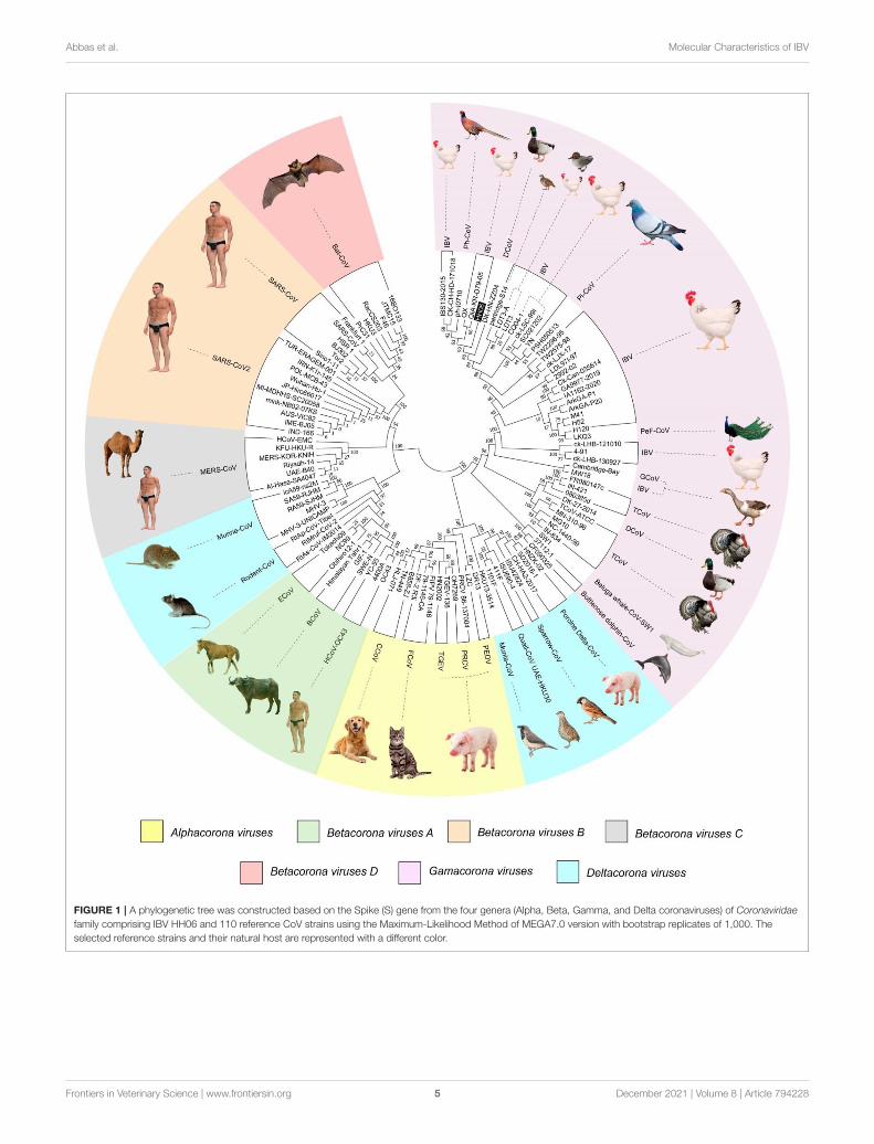
Genetic, Phylogenetic, and MOTIF Analysisof the S ProteinA phylogenetic tree (Figure 1) was constructed based on aminoacid CDS of S glycoprotein to assess the genetic relevancy anddiscrimination among the four main genera (Alphacoronavirus,Betacoronavirus, Gammacoronavirus, and Deltacoronaviruse ofthe Coronaviridae family) comprising different mammalian andavian coronaviruses. Among selected viruses were SARS-CoV-2, SARS-CoV, and MERS-CoV of mammalian origin along withIBV strain HH06 of avian origin isolated in this study. TheIBV strain HH06 clustered into the GI-19 genotype (QX-type)of Avian-CoVs that belongs to Gammacoronavirus and wasclosely related to Duck-CoV DK/CH/HN/ZZ2004 (GenBankaccession number AEO86768.1) and partridge S14 (GenBankaccession number AAT70772.1), LDT3 (GenBank accessionnumber AAU14248.1) (teal and chicken host) of GI-18 (LDT3-A). Ph-CoV strains ph/China/I0710 (GenBank accession numberQDA76255.1) and PSH050513 (GenBank accession numberAAZ85066.1) of Avian-CoVs also clustered closely in the samegroup. These indicate the intra-host evolution of Avian-CoVsfrom one genotype to another and from one host to anotherhost. The S gene glycoprotein sequence of Beluga whale-CoV SW1 (GenBank accession number ABW87820.1) andBottlenose dolphin-CoVs 37112-1 (GenBank accession numbersQII89019.1) and HKU22 (GenBank accession numbers 211AHB63481.1) clustered at distant; however, making group alongwith Avian-CoVs altogether into Gammacoronaviruses. Anothersequences set of delta-CoVs that comprises sparrow CoV, muniaCoV, and Quail-CoV of Avian-CoVs along with porcine delta-CoV clustered together with Feline CoVs, Canine CoVs. PRCV,TGEV, and PEDV belonging to alpha CoVs, hence makinga discrete cluster covering the coronaviruses from avian and
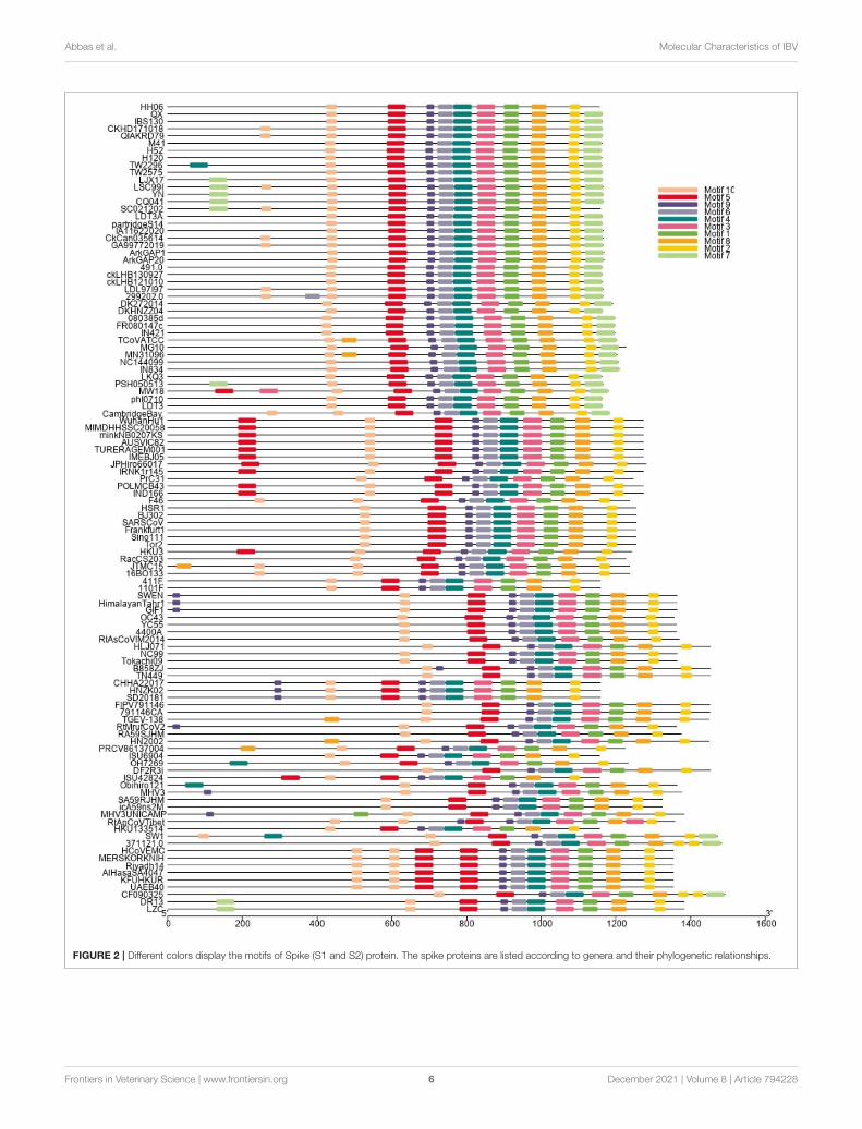
mammalian species. A cluster of exactingly CoVs, branch off intothe human CoVs (HCV-OC43), and a cluster containing murineCoVs, equine CoVs, and rodent CoVs. TheMERS viruses of Beta-CoVs (C) were closely associated with SARS-CoVs-2 and SARS-CoVs. In the same manner, Bat-CoVs show close associationwith SARS-CoVs. Currently, the S gene mainly determinesthe serotype and tissue tropism of different virus strains. Theconserved domains of spike proteins are determined by theMEME (Figure 2). In total, we found ten motifs in the S gene,and their annotations confirmed through the Pfam databases. AllS protein motifs are generally well-conserved and similar withinthe same phylogenetic class; however, variation was also observed(Supplementary Table S2).
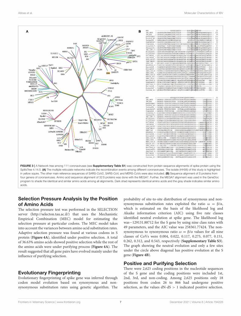
Recombinant Network AnalysisFurther, a recombinant network tree was generated (Figure 3A)of 111 selected strains from four genera (Alpha, Beta, Gamma,and Delta) of CoVs comprising human, animals, and avianorigin. The CoVs of human origin distributed in separateclades. The Betacoronaviruses B (SARS-CoV-2 and SARS-CoV)and Betacoronaviruses D (Bat-CoVs) shared common clades,although branched differently, however possible to be originatedfrom bats. The MERS-CoVs of camel and human origin fromBetacoronaviruses C distributed in individual clade, however, wasclosely associated with Betacoronaviruses B and D. Althoughhuman coronavirus (HCoV-OC43) has a human origin, itbranched together with Bovine-CoVs and showed a significantdistance from other human CoVs like SARS-CoV-2 and SARS-CoV. Feline- and Canine-CoVs showed common origin thusbranched together, however, were significantly distant from otherSARS-related viruses. The Gammacoronaviruses of avian originindicated association at a distant place from Betacoronaviruses(SARS-CoV2, SARS-CoV, Bat-CoV and MERS-CoVs), however,Beluga whale-CoV strain SW1 and Bottlenose dolphin-CoVsstrains 37112-1 and CF090325 of animal origin differentiatedinto separate clade from other CoVs of animal origin andclustered at distant however making group along with Avian-CoVs altogether into Gammacoronaviruses. Isolate HH06 fromthe current study was clustered into Avian-CoVs group,mainly ofchicken origin, however, Duck-CoV and Pheasant-CoV showed aclose relationship with the HH06 isolate from chicken. Similarly,Turkey-CoVs separated clade from other avian mainly chickenhowever 3 Turkey-CoVs differentiated into a separate clade.
Comparative Sequence AlignmentAlignment of interrelated amino acids sequences was performedin comparison to 4 genera of coronaviruses and these isolatesexpressed distinctive amino acids mutations in the HVRscompared to different species. The result of the alignment of the Sgene of IBV isolate HH06 from this study shown the occurrenceof numerous mutational sites (Figure 3B). The majority ofmutation sites were located in HVRI in spike protein structurethat was comparable to sequence alignment of amino acids ofselected 31 CoVs of 4 genera of Coronaviridae family.
Frontiers in Veterinary Science | www.frontiersin.org 4 December 2021 | Volume 8 | Article 794228
Abbas et al. Molecular Characteristics of IBV
FIGURE 1 | A phylogenetic tree was constructed based on the Spike (S) gene from the four genera (Alpha, Beta, Gamma, and Delta coronaviruses) of Coronaviridae
family comprising IBV HH06 and 110 reference CoV strains using the Maximum-Likelihood Method of MEGA7.0 version with bootstrap replicates of 1,000. The
selected reference strains and their natural host are represented with a different color.
Frontiers in Veterinary Science | www.frontiersin.org 5 December 2021 | Volume 8 | Article 794228
Abbas et al. Molecular Characteristics of IBV
FIGURE 2 | Different colors display the motifs of Spike (S1 and S2) protein. The spike proteins are listed according to genera and their phylogenetic relationships.
Frontiers in Veterinary Science | www.frontiersin.org 6 December 2021 | Volume 8 | Article 794228
Abbas et al. Molecular Characteristics of IBV
FIGURE 3 | A Network tree among 111 coronaviruses (see Supplementary Table S1) was constructed from protein sequence alignments of spike protein using the
SplitsTree 4.14.5. (A) The multiple reticulate networks indicate the recombination events among different coronaviruses. The isolate (HH06) of this study is highlighted
in yellow squire. The other main reference sequences of SARS-CoV2, SARS-CoV, and MERS-CoVs were also included. (B) Sequence alignment of S proteins from
four genera of coronaviruses. Amino acid sequence alignment of 32S proteins was done with the MEGA7. Further, the MEGA7 alignment was used in the GeneDoc
program to shade the identical and similar amino acids among all alignments. Dark shad represents identical amino acids and the gray shade indicates similar amino
acids.
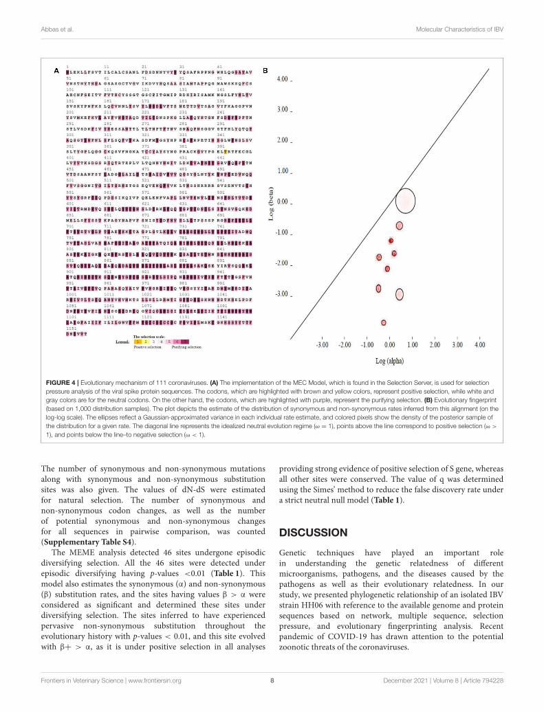
Selection Pressure Analysis by the Positionof Amino AcidsThe selection pressure test was performed in the SELECTIONserver (http://selecton.tau.ac.il/) that uses the MechanisticEmpirical Combination (MEC) model for estimating theselection pressure at particular codons. The MEC model takesinto account the variances between amino acid substitution rates.Adaptive selection pressure was found at various codons in Sprotein (Figure 4A), identified under positive selection. A totalof 36.63% amino acids showed positive selection while the rest ofthe amino acids were under purifying process (Figure 4A). Theresult suggested that all gene pairs have evolved mainly under theinfluence of purifying selection.
Evolutionary FingerprintingEvolutionary fingerprinting of spike gene was inferred throughcodon model evolution based on synonymous and non-synonymous substitution rates using genetic algorithm. The
probability of site-to-site distribution of synonymous and non-synonymous substitution rates exploited the ratio ω = β/α,which is estimated on the basis of the likelihood log andAkaike information criterion (AIC) using five rate classesidentified neutral evolution at spike gene. The likelihood logwas−129131.88712 for the S gene by using nine class rates with49 parameters, and the AIC value was 258361.77424. The non-synonymous to synonymous ratio ω = β/α values for all nineclasses of CoVs were 0.004, 0.022, 0.117, 0.275, 0.077, 0.151,0.262, 0.312, and 0.543, respectively (Supplementary Table S3).The graph showing the neutral evolution and only a few sitesunder the circle above diagonal has positive evolution at the Sgene (Figure 4B).
Positive and Purifying SelectionThere were 2,625 coding positions in the nucleotide sequencesof the S gene and the coding positions were included 1st,2nd, 3rd, and non-coding. Among 2,625 positions only 18positions from codon 26 to 866 had undergone positiveselection, as the values dN-dS > 1 indicated positive selection.
Frontiers in Veterinary Science | www.frontiersin.org 7 December 2021 | Volume 8 | Article 794228
Abbas et al. Molecular Characteristics of IBV
FIGURE 4 | Evolutionary mechanism of 111 coronaviruses. (A) The implementation of the MEC Model, which is found in the Selection Server, is used for selection
pressure analysis of the viral spike protein sequences. The codons, which are highlighted with brown and yellow colors, represent positive selection, while white and
gray colors are for the neutral codons. On the other hand, the codons, which are highlighted with purple, represent the purifying selection. (B) Evolutionary fingerprint
(based on 1,000 distribution samples). The plot depicts the estimate of the distribution of synonymous and non-synonymous rates inferred from this alignment (on the
log-log scale). The ellipses reflect a Gaussian-approximated variance in each individual rate estimate, and colored pixels show the density of the posterior sample of
the distribution for a given rate. The diagonal line represents the idealized neutral evolution regime (ω = 1), points above the line correspond to positive selection (ω >
1), and points below the line–to negative selection (ω < 1).
The number of synonymous and non-synonymous mutationsalong with synonymous and non-synonymous substitutionsites was also given. The values of dN-dS were estimatedfor natural selection. The number of synonymous andnon-synonymous codon changes, as well as the numberof potential synonymous and non-synonymous changesfor all sequences in pairwise comparison, was counted(Supplementary Table S4).
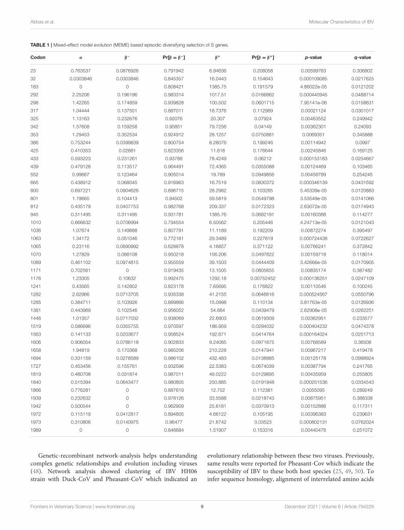
The MEME analysis detected 46 sites undergone episodicdiversifying selection. All the 46 sites were detected underepisodic diversifying having p-values <0.01 (Table 1). Thismodel also estimates the synonymous (α) and non-synonymous(β) substitution rates, and the sites having values β > α wereconsidered as significant and determined these sites underdiversifying selection. The sites inferred to have experiencedpervasive non-synonymous substitution throughout theevolutionary history with p-values < 0.01, and this site evolvedwith β+ > α, as it is under positive selection in all analyses
providing strong evidence of positive selection of S gene, whereasall other sites were conserved. The value of q was determinedusing the Simes’ method to reduce the false discovery rate undera strict neutral null model (Table 1).
DISCUSSION
Genetic techniques have played an important rolein understanding the genetic relatedness of differentmicroorganisms, pathogens, and the diseases caused by thepathogens as well as their evolutionary relatedness. In ourstudy, we presented phylogenetic relationship of an isolated IBVstrain HH06 with reference to the available genome and proteinsequences based on network, multiple sequence, selectionpressure, and evolutionary fingerprinting analysis. Recentpandemic of COVID-19 has drawn attention to the potentialzoonotic threats of the coronaviruses.
Frontiers in Veterinary Science | www.frontiersin.org 8 December 2021 | Volume 8 | Article 794228
Abbas et al. Molecular Characteristics of IBV
TABLE 1 | Mixed-effect model evolution (MEME) based episodic diversifying selection of S genes.
Codon α β− Pr[β = β−] β+ Pr[β = β+] p-value q-value
23 0.763537 0.0876926 0.791942 6.94636 0.208058 0.00599763 0.306802
32 0.0303846 0.0303846 0.845357 16.0443 0.154643 0.000109085 0.0217625
183 0 0 0.808421 1385.75 0.191579 4.86022e-05 0.0121202
292 2.25206 0.196196 0.983314 1017.51 0.0166862 0.000440945 0.0488714
298 1.42265 0.174859 0.939828 100.502 0.0601715 7.95141e-06 0.0158631
317 1.04444 0.137501 0.887011 18.7376 0.112989 0.00021124 0.0301017
325 1.13163 0.232676 0.92076 20.307 0.07924 0.00463552 0.249942
342 1.57608 0.159258 0.95851 79.7256 0.04149 0.00362301 0.24093
353 1.29453 0.352534 0.924912 28.1257 0.0750881 0.0069351 0.345888
386 0.753244 0.0399839 0.800754 8.28076 0.199246 0.00114942 0.0997
425 0.410353 0.02881 0.823356 11.616 0.176644 0.00245846 0.169125
433 0.593223 0.231261 0.93788 78.4249 0.06212 0.000153183 0.0254667
439 0.479128 0.113517 0.964491 72.4365 0.0355088 0.00124469 0.103465
552 0.99667 0.123464 0.905014 19.789 0.0949856 0.00458789 0.254245
665 0.438912 0.068045 0.916963 16.7519 0.0830372 0.000346139 0.0431592
800 0.697221 0.0904626 0.896715 28.2982 0.103285 5.45339e-05 0.0120883
801 1.19665 0.104413 0.94502 59.5819 0.0549798 3.53549e-05 0.0141066
812 0.435179 0.0407753 0.982768 209.337 0.0172323 2.63072e-05 0.0174943
945 0.311495 0.311495 0.931781 1385.76 0.0682191 0.00160388 0.114277
1010 0.666632 0.0706994 0.794554 6.50562 0.205446 4.24713e-05 0.0121043
1036 1.07674 0.149688 0.807791 11.1189 0.192209 0.00872274 0.395497
1063 1.34172 0.051046 0.772181 29.3489 0.227819 0.000724438 0.0722627
1065 0.23116 0.0690992 0.628878 4.16857 0.371122 0.00766241 0.372842
1070 1.27829 0.088108 0.950218 156.206 0.0497822 0.00159718 0.118014
1089 0.461102 0.0974815 0.955559 39.1503 0.0444409 3.42666e-05 0.0170905
1171 0.702561 0 0.919435 13.1505 0.0805655 0.00835174 0.387482
1176 1.23305 0.10632 0.992475 1292.18 0.00752452 0.000136251 0.0247109
1241 0.43565 0.142802 0.823178 7.65695 0.176822 0.00110546 0.100245
1282 2.62966 0.0713705 0.935338 41.2155 0.0646616 0.000524567 0.0550796
1285 0.384711 0.103926 0.889866 15.0998 0.110134 3.81763e-05 0.0126936
1381 0.443969 0.102546 0.956052 54.664 0.0439479 2.62908e-05 0.0262251
1446 1.01357 0.0717032 0.938069 22.6903 0.0619309 0.00362951 0.233577
1519 0.586696 0.0355755 0.970597 186.959 0.0294032 0.000404232 0.0474378
1563 0.141133 0.0203677 0.958524 192.671 0.0414764 0.000164024 0.0251713
1606 0.906054 0.0786118 0.902833 9.24085 0.0971675 0.00768589 0.36508
1658 1.94819 0.170368 0.985206 210.228 0.0147941 0.00967217 0.419478
1694 0.331159 0.0278589 0.986102 432.483 0.0138985 0.00125178 0.0998924
1727 0.453456 0.155761 0.932596 22.5383 0.0674039 0.00387794 0.241765
1819 0.480708 0.031874 0.987011 49.0222 0.0129895 0.00435959 0.255805
1840 0.515394 0.0643477 0.980805 250.885 0.0191948 0.000251536 0.0334543
1866 0.776281 0 0.887619 12.702 0.112381 0.0055095 0.289249
1939 0.232632 0 0.978126 33.5588 0.0218743 0.00875951 0.388338
1942 0.500544 0 0.962909 25.6181 0.0370913 0.00152886 0.117311
1972 0.115119 0.0412817 0.894805 4.66122 0.105195 0.00396383 0.239631
1973 0.310806 0.0140975 0.96477 21.6742 0.03523 0.000802131 0.0762024
1989 0 0 0.846684 1.51907 0.153316 0.00440476 0.251072
Genetic-recombinant network-analysis helps understandingcomplex genetic relationships and evolution including viruses(48). Network analysis showed clustering of IBV HH06strain with Duck-CoV and Pheasant-CoV which indicated an
evolutionary relationship between these two viruses. Previously,same results were reported for Pheasant-Cov which indicate thesusceptibility of IBV to these both host species (25, 49, 50). Toinfer sequence homology, alignment of interrelated amino acids
Frontiers in Veterinary Science | www.frontiersin.org 9 December 2021 | Volume 8 | Article 794228
Abbas et al. Molecular Characteristics of IBV
sequences was done which indicated the majority of mutationsites in HVRI of the spike protein structure. Currently, CoVs areclassified based on sequence comparison of structural protein,mainly spike protein gene (51). Spike protein facilitates the entryof virus to receptors present on the surface of host cells (52).These proteins have two cleaved forms called S1 and S2. TheS1 plays a crucial role in the viral adsorption to the cellularglycoprotein receptor during the process of virus invasion intothe host cell. The S2 play important role in fusion and forms stalkappearance to spike molecule. There is a very high conservedregion in the C-terminal S2 mutation rate of S2 as it containsan extra cleavage site known as the furin S2’ site, which directlyinfluences the invasion mechanism of coronaviruses by host cells(53). The S protein is consideredmain determining factor in virusadaptation and tropism in the organs of host. Mutations thatoccurred in the S protein may influence the targeted host organ(54). We found 10 motifs in the S gene, and their annotationsconfirmed through the Pfam databases and assume their functionin attachment and invasion/infection of the virus to the host cells.
A total of 36.63% positive selection on S protein aminoacids indicated an influence of negative selection on all genepairs. The selective removal of deleterious alleles that arisethrough random mutations can result in stabilizing selection.We selected Gaussian-approximate variance to evaluate theevolutionary fitness of our IBV strains based on S gene. It showeda majority of neutral evolution as compared to positive evolutionon the S gene. To understand the dynamics of molecularsequence evolution, we estimated synonymous (α) and non-synonymous (β) substitution rates by MEME analysis, whichindicated 46 episodic diversifying selection sites. In the presentstudy, the phylogenic analysis indicated a broad mechanism, inwhich the MERS-CoVs of Beta-CoVs (C) were closely associatedwith SARS-CoVs-2 and SARS-CoVs of Beta-CoVs (B). In thesame manner, Bat-CoVs show a close association with SARS-CoVs. Thus, similarly, in SARS-CoV-2 zoonotic transmission tohumans, the viruses are considered to have originated in eitherbats or pangolins (31). Hence, the adaptation and recombinationof SARS-CoV-2 have happened in another intermediate orreservoir host with the possibility of contact with pangolins orbats. Mutation and adaptation have determined the co-evolutionof CoVs and their hosts (9). Gene recombination and mutationare both important means in producing multiple strains of CoVs,with multiple research reports on CoVs-recombination (55).There is a possibility of recombination among various strains,the recombination area and the antigenic profile of recombinantvirus had important guiding significance for predicting theevolution and prevention of CoVs infection afterward (56,57). Evolutionary conservation is critical for detecting aminoacid positions and for sustaining the structural protein role.Consequently, during the selection pressure analysis, the sitesthat were detected as purifying and positive selection mayinstruct and clarify the spike protein gene function and evolution.
In the present analysis, purifying selection was observedduring the selection pressure analysis. Li et al. (58) havereported purifying selection in different host species, alongwith frequent recombination among coronaviruses, proposesa common evolutionary mechanism that could lead to new
emerging human coronaviruses. Similarly in our recombinantnetwork tree analysis, the Betacoronaviruses B (SARS-CoV2and SARS-CoV) and Betacoronaviruses D (Bat-CoVs) sharedcommon clades, although branched differently, however possibleto be originated from bats. Incidence of such intra-speciestransmission happenings in birds and mammals might bemanifested by the predominant occurrence of CoVs at alarge scale (59). In terms of Gamma-CoVs, thought provokingtrends were observed during phylogenetic analysis. The Belugawhale- and Bottlenose dolphin-CoVs of mammals were closelyassociated with the branches of Avian-CoVs. Similarly, threeavian-CoVs (sparrow, munia, and qual) were branched togetherwith Porcine-CoV and this was suggestive of transmission amongdifferent species. Moreover, outcomes of the current analysisadvocate that variation at the genomic and molecular level ofthe S gene sequence made viable CoVs adaptation to differenthost species. Additionally, the analysis of isolate from the presentstudy IBV HH06 indicated that it belongs to QX-type G-19 ofIBV, however, it was also closely associated with Duck-CoV andPheasant-CoV. Abro et al. (60) carried bioinformatics of CoVsand found inter-species transmission of avian and mammalianCoVs that suggested inter-species transmission and were inagreement with our findings.
CONCLUSION
Based on the S glycoprotein, SARS-CoV-2, SARS-CoV, Bats-CoV, and MERS-CoV have close genetic relationship. IBV canpotentially infect wider range of bird species beyond theirnatural hosts chicken e.g., ducks, teal, partridge, turkeys, andpheasants. Duck-CoV, and Pheasant-CoV grouped together withIBV strain HH06 and other QX-type viruses, hence there is animperative need to work further on the transmission mechanismof these CoVs. Purifying selection contributed predominately tothe evolutionary process in selection pressure analysis. Sufficientchances of recombination and evolution of CoVs result in therise of novel CoVs, which have great ability of intra-speciestransmission and zoonosis. Thus, their continuous emergingworldwide needs to be controlled more efficiently.
DATA AVAILABILITY STATEMENT
The datasets presented in this study can be found in onlinerepositories. The names of the repository/repositoriesand accession number(s) can be found in thearticle/Supplementary Material.
ETHICS STATEMENT
Ethical review and approval was not required for the studyon human participants in accordance with the local legislationand institutional requirements. Written informed consent forparticipation was not required for this study in accordance withthe national legislation and the institutional requirements.
Frontiers in Veterinary Science | www.frontiersin.org 10 December 2021 | Volume 8 | Article 794228
Abbas et al. Molecular Characteristics of IBV
AUTHOR CONTRIBUTIONS
GA and YZ: conceptualization. GA and GL: data curation.GA and XS: formal analysis. GL: funding acquisition, projectadministration, resources, and supervision. HC and XH:investigation. GA: methodology and writing–original draft. GAand MA: software. YR and XW: validation. GA, HC, andGL: writing–review and editing. All authors agreed to thefinal version.
FUNDING
The National Natural Science Foundation of China under Grant(31172295 and 31272569) supported this research.
ACKNOWLEDGMENTS
China Scholarship Council was highly thanked for funding thestay of GA for conduction of his doctoral research.
SUPPLEMENTARY MATERIAL
The Supplementary Material for this article can be foundonline at: https://www.frontiersin.org/articles/10.3389/fvets.2021.794228/full#supplementary-material
Supplementary Table S1 | The information of Coronaviruses protein and gene
sequences used in bioinformatics.
Supplementary Table S2 | Analysis and description of motif present in spike
glycoprotein sequences of coronaviruses.
Supplementary Table S3 | Evolutionary fingerprinting analysis of Coronaviruses
proteins.
Supplementary Table S4 | Maximum likelihood analysis of S protein for
codon-by-codon positive selection.
Supplementary Figure S1 | Dwarfism and hemorrhage of embryos 72 h
post-infection with IBV HH06 at the left, the control embryo at the right.
Supplementary Figure S2 | Confirmation of IBV Strain HH06 using IBV-N
specific primers.
REFERENCES
1. Zhu N, Zhang D, Wang W, Li X, Yang B, Song J, et al. China Novel
Coronavirus, Research T. A Novel Coronavirus from Patients with Pneumonia
in China. N Engl J Med. (2020) 382:727–33. doi: 10.1056/NEJMoa200
1017
2. Weiss SR, Leibowitz JL. Coronavirus pathogenesis. Adv Virus Res. (2011)
81:85–164. doi: 10.1016/B978-0-12-385885-6.00009-2
3. ICTV. Coronaviridae. Available online at: https://talk.ictvonline.org/ictv-
reports/ictv_9th_report/positive-senserna-viruses-2011/w/posrna_viruses/
222/coronaviridae (accessed September 25, 2021).
4. Fehr AR, Perlman S. Coronaviruses: an overview of their
replication and pathogenesis. Methods Mol Biol. (2015) 1282:1–
23. doi: 10.1007/978-1-4939-2438-7_1
5. Masters PS. The molecular biology of coronaviruses. Adv Virus Res. (2006)
66:193–292. doi: 10.1016/S0065-3527(06)66005-3
6. Locker JK, Rose JK, Horzinek MC, Rottier PJ. Membrane assembly of
the triple-spanning coronavirus M protein. Individual transmembrane
domains show preferred orientation. J Biol Chem. (1992) 267:21911–
8. doi: 10.1016/S0021-9258(19)36699-2
7. Lau SK, Lee P, Tsang AK, Yip CC, TseH, Lee RA, et al. Molecular epidemiology
of human coronavirus OC43 reveals evolution of different genotypes over
time and recent emergence of a novel genotype due to natural recombination.
J Virol. (2011) 85:11325–37. doi: 10.1128/JVI.05512-11
8. Hoffmann M, Kleine-Weber H, Schroeder S, Krüger N, Herrler T, Erichsen
S, et al. SARS-CoV-2 cell entry depends on ACE2 and TMPRSS2 and is
blocked by a clinically proven protease inhibitor. Cell. (2020) 181:271–
80.e8. doi: 10.1016/j.cell.2020.02.052
9. I.Wickramasinghe NA, de Vries RP, Gröne A C.de Haan AM,
Verheije MH. Binding of avian coronavirus spike proteins to host
factors reflects virus tropism and pathogenicity. J Virol. (2011)
85:8903–12. doi: 10.1128/JVI.05112-11
10. Cavanagh D. Coronavirus avian infectious bronchitis virus. Vet Res. (2007)
38:281–97. doi: 10.1051/vetres:2006055
11. Hodgson T, Casais R, Dove B, Britton P, Cavanagh D. Recombinant
infectious bronchitis coronavirus Beaudette with the spike protein gene of the
pathogenic M41 strain remains attenuated but induces protective immunity. J
Virol. (2004) 78:13804–11. doi: 10.1128/JVI.78.24.13804-13811
12. A Rao S, Shetty NP. Evolutionary selectivity of amino acid is inspired from
the enhanced structural stability and flexibility of the folded protein. Life Sci.
(2021) 281:119774. doi: 10.1016/j.lfs.2021.119774
13. Toro H, van Santen VL, Jackwood MW. Genetic diversity and selection
regulates evolution of infectious bronchitis virus.Avian Dis. (2012) 56:449–55.
doi: 10.1637/10072-020212-Review.1
14. Schalk AF, Hawn MC. An apparently new respiratory disease in baby chicks. J
Am Vet Med Assoc. (1931) 78:413–22.
15. Beaudette F. Cultivation of the virus of infectious bronchitis. J Am Vet Med
Assoc. (1937) 90:51–60.
16. Kendall E, BynoeM, Tyrrell D. Virus isolations from common colds occurring
in a residential school. Br Med J. (1962) 2:82. doi: 10.1136/bmj.2.5297.82
17. Bradburne AF, BynoeML, Tyrrell DA. New respiratory virus. BrMed J. (1967)
3:752. doi: 10.1136/bmj.3.5568.752
18. Varia M, Wilson S, Sarwal S. Hospital Outbreak Investigation Team.
Investigation of a nosocomial outbreak of severe acute respiratory syndrome
(SARS) in Toronto, Canada. CMAJ. (2003) 169:285–92.
19. De Groot RJ, Baker SC, Baric RS, Brown CS, Drosten C, Enjuanes L, et
al. Commentary: Middle east respiratory syndrome coronavirus (mers-cov):
announcement of the coronavirus study group. J Virol. (2013) 87:7790–
2. doi: 10.1128/JVI.01244-13
20. Oude Munnink BB, Worp N, Nieuwenhuijse DF, Sikkema RS,
Haagmans B, Fouchier RA, et al. The next phase of SARS-CoV-
2 surveillance: real-time molecular epidemiology. Nat Med. (2021)
27:1–7. doi: 10.1038/s41591-021-01472-w
21. Hu B, Guo H, Zhou P, Shi ZL. Characteristics of SARS-CoV-2 and COVID-19.
Nat Rev Microbiol. (2021) 19:141–54. doi: 10.1038/s41579-020-00459-7
22. Banerjee A. Virus hunters: discovering the evolutionary origins of SARS-CoV-
2. Cell Host Microbe. (2021) 29:1031–3. doi: 10.1016/j.chom.2021.06.012
23. Cavanagh D, Naqi SA. Infectious bronchitis. In: Calnek BW, Barnes HJ, Beard
CW, McDougald LR, Saif YM, editors, Disease of Poultry. Hoboken, NJ: Iowa
State University Press (1997). p. 511–26
24. De Wit J. Detection of infectious bronchitis virus. Avian Pathol. (2000)
29:71–93. doi: 10.1080/03079450094108
25. Cavanagh D, Mawditt K D.d.Welchman B, Britton P, Gough R. Coronaviruses
from pheasants (Phasianus colchicus) are genetically closely related to
coronaviruses of domestic fowl (infectious bronchitis virus) and turkeys.
Avian Pathol. (2002) 31:81–93. doi: 10.1080/03079450120106651
26. Wu X, Pan S, ZhouW,Wu Y, Huang Y,Wu B. The isolation and identification
of infectious bronchitis virus PTFY strain inmuscovy ducks. Bing DuXue Bao.
(2016) 32:203–9. doi: 10.13242/j.cnki.bingduxuebao.002908
27. Liu S, Chen J, Chen J, Kong X, Shao Y, Han Z, et al. Isolation of avian
infectious bronchitis coronavirus from domestic peafowl (Pavo cristatus) and
teal (Anas). J Gen Virol. (2005) 86:719–25. doi: 10.1099/vir.0.80546-0
Frontiers in Veterinary Science | www.frontiersin.org 11 December 2021 | Volume 8 | Article 794228
Abbas et al. Molecular Characteristics of IBV
28. Qian DH, Zhu GJ, Wu LZ, Hua GIsolation X, and characterization of a
coronavirus from pigeons with pancreatitis. Am J Vet Res. (2006) 67:1575–
9. doi: 10.2460/ajvr.67.9.1575
29. Papineau A, Berhane Y, Wylie TN, Wylie KM, Sharpe S, et al.
Genome organization of canada goose coronavirus, a novel species
identified in a mass die-off of Canada Geese. Sci Rep. (2019)
9:5954. doi: 10.1038/s41598-019-42355-y
30. Cook JK, Jackwood M, Jones RC. The long view: 40 years
of infectious bronchitis research. Avian Pathol. (2012) 41:239–
50. doi: 10.1080/03079457.2012.680432
31. Goraichuk IV, Arefiev V, Stegniy BT, Gerilovych AZoonotic P, and
Reverse Zoonotic Transmissibility of SARS-CoV-2. Virus Res. (2021)
302:198473. doi: 10.1016/j.virusres.2021.198473
32. Alluwaimi AM, Alshubaith IH, Al-Ali AM, Abohelaika S. The coronaviruses
of animals and birds: their zoonosis, vaccines, and models for SARS-CoV and
SARS-CoV2. Front Vet Sci. (2020) 7:655. doi: 10.3389/fvets.2020.582287
33. Zhang G, Li B, Yoo D, Qin T, Zhang X, Jia Y, et al. Animal
coronaviruses and SARS-CoV-2. Transbound Emerg Dis. (2021) 68:10971110.
doi: 10.1111/tbed.13791
34. Rabaan AA, Al-Ahmed SH, Sah R, Alqumber MA, Haque S,
Patel SK, et al. MERS-CoV: epidemiology, molecular dynamics,
therapeutics, future challenges. Ann Clin Microbiol Antimicrob. (2021)
20:1–14. doi: 10.1186/s12941-020-00414-7
35. Ren X, Yin J, Ma D, Li Characterization G, and membrane gene-based
phylogenetic analysis of avian infectious bronchitis virus Chinese strain
HH06. Virus Genes. (2009) 38:39–45. doi: 10.1007/s11262-008-0280-7
36. Hewson K, Noormohammadi AH, Devlin JM, Mardani K, Ignjatovic J. Rapid
detection and non-subjective characterisation of infectious bronchitis virus
isolates using high-resolution melt curve analysis and a mathematical model.
Arch. Virol. (2009) 154:649–60. doi: 10.1007/s00705-009-0357-1
37. Reed LJ, MuenchHA. simple method of estimating fifty per cent endpoints12.
Am J Epidemiol. (1938) 27:493–7. doi: 10.1093/oxfordjournals.aje.a118408
38. Sun X, Li L, Pan L, Wang Z, Chen H, Shao C, et al. Infectious bronchitis virus:
identification of Gallus gallus APN high-affinity ligands with antiviral effects.
Antiviral Res. (2021) 186:104998. doi: 10.1016/j.antiviral.2020.104998
39. Chen H, Muhammad I, Zhang Y, Ren Y, Zhang R, Huang X, et al.
Antiviral Activity Against Infectious Bronchitis Virus and Bioactive
Components of Hypericum perforatum L. Front Pharmacol. (2019)
10:1272. doi: 10.3389/fphar.2019.01272
40. Kumar S, Stecher G, Tamura K. MEGA7: molecular evolutionary genetics
analysis version 7.0 for bigger datasets. Mol Biol Evol. (2016) 33:1870–
4. doi: 10.1093/molbev/msw054
41. Huson DH, Bryant D. Application of phylogenetic networks in evolutionary
studies.Mol Biol Evol. (2006) 23:254–67. doi: 10.1093/molbev/msj030
42. J-Yang R, Liao BY, Zhuang SM, Zhang J. Protein misinteraction avoidance
causes highly expressed proteins to evolve slowly. Proc Natl Acad Sci U S A.
(2012) 109:E831–40. doi: 10.1073/pnas.1117408109
43. Kosakovsky Pond SL, Scheffler K, Gravenor MB, Poon AF, Frost SD.
Evolutionary fingerprinting of genes. Mol Biol Evol. (2010) 27:520–
36. doi: 10.1093/molbev/msp260
44. Kosakovsky Pond SL, Frost SD. Not so different after all: a comparison of
methods for detecting amino acid sites under selection. Mol Biol Evol. (2005)
22:1208–22. doi: 10.1093/molbev/msi105
45. Kimura M. A simple method for estimating evolutionary rates of base
substitutions through comparative studies of nucleotide sequences. J Mol Evol.
(1980) 16:111–20. doi: 10.1007/BF01731581
46. Murrell B, Moola S, Mabona A, Weighill T, Sheward D, Kosakovsky Pond
SL, et al. FUBAR: a fast, unconstrained bayesian approximation for inferring
selection.Mol Biol Evol. (2013) 30:1196–205. doi: 10.1093/molbev/mst030
47. Newman M. Networks. Oxford: Oxford University Press (2018).
48. Wang Y, Zeng J, Zhang C, Chen C, Qiu Z, Pang J, et al. New
framework for recombination and adaptive evolution analysis with
application to the novel coronavirus SARS-CoV-2. Brief Bioinform. (2021)
22:bbab107. doi: 10.1093/bib/bbab107
49. Torres CA, Listorti V, Lupini C, Franzo G, Drigo M, Catelli E, et al. Gamma
and Deltacoronaviruses in quail and pheasants from Northern Italy. Poult Sci.
(2017) 96:717–22. doi: 10.3382/ps/pew332
50. Chen GQ, Zhuang QY, Wang KC, Liu S, Shao JZ, Jiang WM, et al.
Identification and survey of a novel avian coronavirus in ducks. PLoS ONE.
(2013) 8:e72918. doi: 10.1371/journal.pone.0072918
51. Gonzalez JM, Gomez-Puertas P, Cavanagh D, Gorbalenya AE,
Enjuanes L. A comparative sequence analysis to revise the
current taxonomy of the family Coronaviridae. Arch Virol. (2003)
148:2207–35. doi: 10.1007/s00705-003-0162-1
52. Zhang X, Tan Y, Ling Y, Lu G, Liu F, Yi Z, et al. Viral and host factors
related to the clinical outcome of COVID-19. Nature. (2020) 583:437–
40. doi: 10.1038/s41586-020-2355-0
53. Andersen KG, Rambaut A, Lipkin WI, Holmes EC, Garry
RF. The proximal origin of SARS-CoV-2. Nat Med. (2020)
26:450–2. doi: 10.1038/s41591-020-0820-9
54. Li W. Delving deep into the structural aspects of a furin cleavage site
inserted into the spike protein of SARS-CoV-2: a structural biophysical
perspective. Biophys Chem. (2020) 264:106420. doi: 10.1016/j.bpc.2020.106
420
55. Ye ZW, Yuan S, Yuen KS, Fung SY, Chan CP, Jin DY. Zoonotic origins of
human coronaviruses. Int J Biol Sci. (2020) 16:1686. doi: 10.7150/ijbs.45472
56. Graham RL, Baric RS. Recombination, reservoirs, and the modular spike:
mechanisms of coronavirus cross-species transmission. J Virol. (2010)
84:3134–46. doi: 10.1128/JVI.01394-09
57. Shafique L, Ihsan A, Liu Q. Evolutionary trajectory for the
emergence of novel coronavirus SARS-CoV-2. Pathogens. (2020)
9:240. doi: 10.3390/pathogens9030240
58. Li X, Giorgi EE, Marichannegowda MH, Foley B, Xiao C, Kong XP, et al.
Emergence of SARS-CoV-2 through recombination and strong purifying
selection. Sci Adv. (2020) 6:eabb9153. doi: 10.1126/sciadv.abb9153
59. Hulswit RJ, de Haan CA, Bosch BJ. Coronavirus spike protein and tropism
changes. Adv Virus Res. (2016) 96:29–57. doi: 10.1016/bs.aivir.2016.08.004
60. Abro SH, Ullman K, Belak S, Baule C. Bioinformatics and
evolutionary insight on the spike glycoprotein gene of QX-like and
Massachusetts strains of infectious bronchitis virus. Virol J. (2012)
9:211. doi: 10.1186/1743-422X-9-211
Conflict of Interest: The authors declare that the research was conducted in the
absence of any commercial or financial relationships that could be construed as a
potential conflict of interest.
Publisher’s Note: All claims expressed in this article are solely those of the authors
and do not necessarily represent those of their affiliated organizations, or those of
the publisher, the editors and the reviewers. Any product that may be evaluated in
this article, or claim that may be made by its manufacturer, is not guaranteed or
endorsed by the publisher.
Copyright © 2021 Abbas, Zhang, Sun, Chen, Ren, Wang, Ahmad, Huang and Li.
This is an open-access article distributed under the terms of the Creative Commons
Attribution License (CC BY). The use, distribution or reproduction in other forums
is permitted, provided the original author(s) and the copyright owner(s) are credited
and that the original publication in this journal is cited, in accordance with accepted
academic practice. No use, distribution or reproduction is permitted which does not
comply with these terms.
Frontiers in Veterinary Science | www.frontiersin.org 12 December 2021 | Volume 8 | Article 794228


