Embed Size (px)
Citation preview

712
© Urban & Vogel 2002
Herz 27 · 2002 · Nr. 8 © Urban & Vogel
Herz
Molecular Mechanisms of Inherited VentricularArrhythmiasThomas Wichter1, 2, Eric Schulze-Bahr1, 2, Lars Eckardt1, Matthias Paul1, Bodo Levkau1, 2, Matthias Meyborg1, Michael Schäfers3, Wilhelm Haverkamp1, Günter Breithardt1, 2
Background: Inherited ventricular arrhythmias such as thelong QT syndrome (LQTS), Brugada syndrome, catecholamin-ergic polymorphic ventricular tachycardia (CPVT), idiopathicventricular fibrillation (VF), and arrhythmogenic right ventric-ular cardiomyopathy (ARVC) account for a relevant proportionof sudden cardiac death cases in young patient cohorts. Thedetailed pathogenetic mechanisms of inherited ventricular ar-rhythmias are still poorly understood because systematic in-vestigations are difficult to perform due to low patient num-bers and the lack of appropriate experimental models. Howev-er, recent advances in research and science have identified agenetic background for many of these diseases.Present Knowledge: In LQTS, various mutations in differentgenes encoding for cardiac potassium and sodium channelproteins have been identified (“channelopathy”), and initialprogress in genotype-phenotype correlation is made. Muta-tions in the cardiac sodium channel gene have also been iden-tified in a subset of patients with Brugada syndrome, whereasa genetic background has not yet been demonstrated in idio-pathic VF and right ventricular outflow-tract tachycardia
Molekulare Mechanismen angeborener ventrikulärer Arrhythmien
Hintergrund: Angeborene ventrikuläre Arrhythmien wie QT-Syndrom (LQTS), Brugada-Syndrom, katecholaminerge poly-morphe ventrikuläre Tachykardie (CPVT), idiopathisches Kam-merflimmern sowie arrhythmogene rechtsventrikuläre Kar-diomyopathie (ARVC) sind wesentliche Ursachen plötzlicherHerztodesfälle bei jungen Patienten. Die detaillierten patho-genetischen Mechanismen sind bislang nur in Anfängen auf-geklärt, da bei geringen Patientenzahlen und fehlenden Tier-modellen systematische Untersuchungen erschwert sind.Dennoch konnten jüngste Fortschritte in klinischer und expe-rimenteller Forschung neue Erkenntnisse zum genetischen
Hintergrund angeborener ventrikulärer Arrhythmien beitra-gen.Aktuelle Erkenntnisse: Bei LQTS wurden in den letzten Jahrenzahlreiche Mutationen in verschiedenen Genen nachgewie-sen, die für Proteine kardialer Kalium- und Natriumkanäle ko-dieren („Ionenkanalerkrankung“). Auch wurden erste Ergeb-nisse bei der Genotyp-Phänotyp-Korralation erzielt. Muta-tionen im kardialen Natriumkanalgen wurden auch beimBrugada-Syndrom nachgewiesen, während bei Patienten mitidiopathischem Kammerflimmern und rechtsventrikulärerAusflusstrakttachykardie bislang keine genetischen Verände-
Key Words: Ion channel · Gene mutation · Cardiomyopathy · Long QT syndrome · Brugada syndrome · Suddendeath · Tachyarrhythmias · Ventricular tachycardia
Herz 2002;27:712–39
DOI 10.1007/s00059-002-2436-x
1 Department of Cardiology and Angiology,2 Institute for Arteriosclerosis Research, and3 Department of Nuclear Medicine, University of Münster, Germany.
(RVO-VT). Very recently, mutations in the cardiac ryanodine re-ceptor gene have been identified in CPVT and in a subgroup ofpatients with ARVC. Although several chromosomal loci weresuggested, no other responsible genes or mutations havebeen found in autosomal dominant forms of ARVC. However,in Naxos disease, a recessive form of ARVC with coexpressionof palmoplantar keratoderma and woolly hair, a mutation inthe plakoglobin gene has recently been discovered, thus un-derscoring the potential role of genetic alterations in cy-toskeletal proteins in ARVC.Future Perspectives: In the next years, significant progress in thegenetic diagnosis, pathophysiologic understanding of diseasemechanisms, genotype-phenotype correlation, and the devel-opment of gene- or target-directed treatment strategies can beexpected in the field of inherited ventricular arrhythmias.Conclusion: This review summarizes the current knowledge ofthe molecular mechanisms, including aspects of pathoanato-my, autonomic innervation, genetics, and genotype-pheno-type correlations with their potential implications for diagno-sis and treatment of inherited ventricular arrhythmias.
Review Article

Wichter T, et al. Molecular Mechanisms of Inherited Ventricular Arrhythmias
713Herz 27 · 2002 · Nr. 8 © Urban & Vogel
Inherited Ventricular ArrhythmiasVentricular tachyarrhythmias and sudden cardiac arrestare uncommon in children and the young adult popula-tion. However, whenever such an event occurs, it has se-vere consequences for the patient and his relatives aswell as for the physicians involved in patient manage-ment and medical care.
Rare inherited ventricular arrhythmias such as thelong QT syndrome (LQTS), Brugada syndrome, cate-cholaminergic polymorphic ventricular tachycardia(CPVT), idiopathic ventricular fibrillation (VF), and ar-rhythmogenic right ventricular cardiomyopathy (ARVC)account for the majority of sudden cardiac death cases inyoung patient cohorts. However, the life-time prevalenceof inherited ventricular arrhythmias is less than 5/10,000and less or equal than 1/10,000 for the individual condi-tions.
Inherited ventricular arrhythmic disorders can bedivided into two subgroups:1. “primary electrical disorders” in which an organic
heart disease is not detectable:a) long QT syndrome,b) Brugada syndrome,c) idiopathic ventricular fibrillation,d) catecholaminergic polymorphic ventricular tachy
cardia,e) idiopathic right ventricular outflow-tract tachycar-
dia (RVO-VT).2. “arrhythmogenic cardiomyopathies”, in which an in-
herited myocardial disease may primarily manifestwith ventricular tachyarrhythmias:a) arrhythmogenic right ventricular cardiomyopathy,
b) dilated cardiomyopathy (DCM),c) hypertrophic cardiomyopathy (HCM).
This review summarizes the current knowledge ofthe molecular mechanisms of inherited ventricular ar-rhythmias, including aspects of pathoanatomy, auto-nomic innervation, molecular genetics, and genotype-phenotype correlations and the potential implicationsfor diagnosis and treatment.
The molecular and genetic background and de-tailed pathogenetic mechanisms of inherited ventricu-lar arrhythmias have only recently begun to be uncov-ered (for reviews see [115–117]) because systematic in-vestigations were difficult or impossible to perform dueto low patient numbers and the lack of appropriate ex-perimental models. The late development in compari-son to other inherited cardiac disorders was due to therarity of the diseases and to the high arrhythmic mor-tality and early disease onset resulting in mostly smallnucleus families. Therefore, traditional genetic linkagestudies that are based on the genetic information ob-tained from large multigeneration families, were madedifficult.
Despite these limitations, recent research has iden-tified a genetic background for many of these diseases.To date, all primary electrical diseases in which thecausative genes have been identified turned out to be“channelopathies”. The causative genes encode forchannel subunits that regulate important ion currentsthat tune the cardiac action potential. In contrast, thegene mutations identified in arrhythmogenic cardiomy-opathies mainly focus on sarcolemmal and cytoskeletalproteins.
rungen gefunden wurden. Kürzlich konnten Mutationen imGen des kardialen Ryanodinrezeptors bei Patienten mit CPVTund einer Untergruppe der ARVC nachgewiesen werden. Da-gegen konnte bei anderen Formen autosomal-dominant ver-erbter ARVC trotz Lokalisation mehrerer chromosomaler Locibislang kein Gendefekt identifiziert werden. Bei der rezessivvererbten Naxos-Erkrankung, einer Sonderform der ARVC mitKoexpression von palmoplantarer Keratose, wurde unlängsteine Mutation im Plakoglobingen nachgewiesen, wodurch diepotentielle Rolle genetischer Veränderungen in zytoskelet-tären Proteinen bei ARVC unterstrichen wird.Perspektiven: Auch in den kommenden Jahren sind entschei-
dende Fortschritte in der genetischen Diagnostik, dem Ver-ständnis pathogenetischer Mechanismen, der Genotyp-Phäno-typ-Korrelation und der Entwicklung von genotyporientiertenTherapiestrategien bei angeborenen ventrikulären Arrhythmi-en zu erwarten.Schlussfolgerung: Diese Übersicht fasst den aktuellen Wis-sensstand der molekularen Mechanismen zusammen und dis-kutiert dabei Aspekte von Pathoanatomie, autonomer Inner-vation, Genetik, und Genotyp-Phänotyp-Korrelation mit ihrenpotentiellen Implikationen für die Diagnostik und Therapieangeborener ventrikulärer Arrhythmien.
Schlüsselwörter: Ionenkanal · Genmutation · Kardiomyopathie · QT-Syndrom · Brugada-Syndrom · PlötzlicherHerztod · Tachyarrhythmie · Ventrikuläre Tachykardie

Wichter T, et al. Molecular Mechanisms of Inherited Ventricular Arrhythmias
714 Herz 27 · 2002 · Nr. 8 © Urban & Vogel
Primary electrical cardiac disorders show a high ge-netic heterogeneity. Within all disease genes, the muta-tions are spread all over the entire gene (allelic hetero-geneity). In addition, more than one disease mutationmay be present. This complexity requires complete mu-tation analysis of all disease-causing genes before med-ical advice should be given.
The discovery of the genetic bases of the LQTS be-came a new methodologic paradigm, because with theuse of “classic” genetic linkage strategies (positional can-didate strategies) not only the causative genes werefound but, moreover, functional components with a fun-damental role for normal repolarization were discovered.Disease mutations turned out to be not only a family-spe-cific event with a distinct phenotype and the potential ofan additional diagnostic tool, but also, when expressed inheterologous expression systems, characterize the defec-tive ion channel in a topological way and lead to a morespecific understanding of ion channel function.
Meanwhile, genotype-phenotype correlations inlarge families are on the way to evaluate intergene, interfamilial and intrafamilial differences in the clini-cal phenotype reflecting gene-specific, gene-site-specific and individualconsequences of a giv-en mutation. A wide-spread phenotypicheterogeneity evenwithin mutation carri-ers in the same familyraises the importanceof modifying factorsand genes that aremostly unknown todate. The reducedpenetrance and vari-able expressivity asso-ciated with the LQTmutations remain tobe explained. First in-sights into the com-plex actions of muta-tions are coming fromexpression data. Thesepreliminary resultsmay lead to potentialimplications for a spe-cific (gene-site-direct-ed) therapy [59].
Pathophysiology of Cardiac Ion Channels (“Channelopathies”)
The electrical potential of myocardial cells is based on aseries of ionic events that are either responsible for de-polarization (based on the rapid sodium inward current[INa]) or a balance between various inward and outwardcurrents during repolarization which mainly carrypotassium ions (transient outward potassium current[Ito], slowly activating delayed rectifier [IKs], rapidly ac-tivating delayed rectifier [IKr]; Figure 1). Whereas depo-larization is a quite straight-forward process, repolariza-tion is more complicated depending on the equilibriumof various currents. Disturbances of repolarization mayoccur either due to a reduction in potassium outwardcurrents or to a persistence of inward sodium or calciumcurrents or both, which all may lead to a prolongation ofrepolarization.
The prolongation of the ventricular action potentialand the QT interval may be due to 1. a reduction in out-ward currents, notably the potassium currents IKr or IKs,or 2. due to an enhancement of inward currents, partic-ularly the sodium current (INa) and the L-type calciumcurrent [ICa] [1]. A prolongation of the action potential
Figure 1. Ionic currents contributing to the regulation of the cardiac action potential. The time-dependency ofactivation and the flow direction of the currents are shown in relation to the phases of the action potential (in-ward orientation in blue above, outward orientation in red below the line). (Reprinted from [116] with permis-sion of publisher.)Abbildung 1. Ionenströme, die zur Regulation des kardialen Aktionspotentials beitragen. Die Zeitfolge der Ak-tivierung und die Flussrichtung der Ionenströme sind in Abhängigkeit von der Phase des Aktionspotentialsdargestellt (Einwärtsorientierung in Blau oberhalb, Auswärtsorientierung in Rot unterhalb der Linie). (Abge-druckt aus [116] mit Erlaubnis des Verlags.)

Wichter T, et al. Molecular Mechanisms of Inherited Ventricular Arrhythmias
715Herz 27 · 2002 · Nr. 8 © Urban & Vogel
duration, especially at slow heart rates or followingpauses, is likely to favor early afterdepolarizations,which in conjunction with either a transmural or a re-gional heterogeneity in repolarization (“dispersed re-polarization”), may trigger torsade-de-pointes tachy-cardias [167], a characteristic feature of the LQTS.
Very recently, the functional characterization ofmutant ion channels by in vitro models facilitated firstinsights into pathogenic mechanisms. Mutations in ionchannels can be characterized by the mutation type (nu-cleic acid exchange) and the genetic status (homozy-gous vs heterozygous). Mutations can lead to a loss ofion channel function (haploinsufficiency or dominantnegative effect) or change of function.
Haploinsufficiency results mostly from nonsense,but also rearrangement (not in-frame) mutations. Thealtered protein does not interact with its wild-type coun-terpart, and the resulting ion channel is composed of on-ly 50% wild-type protein, thus reducing number of func-tional ion channels to half. Recent studies showed thatalso missense mutations that affect glycosylation, intra-cellular transport and channel subunit co-assembly (pri-or to membrane integration) can be associated withhaploinsufficiency [6, 32, 50, 86, 134, 152, 196]. Thus, theinteractions of a given mutation in the ion channel for-mation may be complex with significant impact on po-tential therapeutic strategies in mutation carriers.
Mutations with a dominant negative effect (most ofthe missense mutations) result in mutant proteins thathave a steric interaction with the wild-type protein, thusreducing wild-type function. Therefore, the number offunctional channels is less than 50%, and the affectedmutation carriers may exhibit a more severe phenotype.
The complete genetic, biochemical and electrophys-iologic understanding of a given mutation is importantfor future treatment strategies that specifically attemptto recompensate the clinical phenotype.
Long QT Syndrome (LQTS)Congenital LQTS is characterized by a prolonged QTinterval in the surface electrocardiogram (ECG), recur-rent syncope or sudden death resulting from ventriculartachyarrhythmias, especially of the torsade-de-pointestype. The incidence of congenital LQTS has been esti-mated as 1 : 7,000 to 1 : 10,000 live births.
In 1993, Schwartz et al [148] from the InternationalLQTS Registry proposed a diagnostic score that weighsdifferent clinical parameters (Table 1). A sum of ≥ 4points is associated with a high probability, 1.5–3.5
points with an intermediate, and ≤ 1 point with a lowprobability for LQTS. Since the causal genes were notidentified at that time, the impact of gene mutationswere not introduced into the scoring system.
Moss et al [102] prospectively investigated the clin-ical characteristics and long-term course of 3,343 indi-viduals from 328 families in which one or more mem-bers were identified as affected with LQTS. The groupof probands was younger at first contact (age 21 ± 15years) and had a higher incidence of female gender(69%), congenital deafness (7%), resting heart rate < 60/min (31%), a history of ventricular tachyarrhyth-mia (47%), and pre-enrollment syncope or cardiac ar-rest with resuscitation (80%) than other affected andunaffected family members. Syncope often occurred inassociation with acute physical, emotional, or auditoryarousal and was frequently misinterpreted as a seizuredisorder. By the age of 12 years, 50% of the probandshad died or experienced at least one syncopal episode.
Pathogenesis of LQTSTwo pathogenetic hypotheses for LQTS had been pro-posed to cause abnormal cardiac repolarization andventricular arrhythmias. One suggested a predomi-
Score
ElectrocardiogramHeart-rate corrected QT interval• ≥ 0.48 s 3• 0.46–0.47 s 2• ≥ 0.45 s (males) 1Torsade de pointes 2T-wave alternans 1Notched T-wave (in 3 leads) 1Low heart rate (for age) 0.5
Clinical historySyncope• With stress 2• Without stress 1Congenital deafness 0.5
Family historyFamily members with definite LQTS 1Unexplained SCD below age 30 among immediate family members 0.5
Table 1. Long QT syndromes (LQTS): diagnostic score (from [148]).A sum of ≥ 4 points is proposed to be associated with a high probabil-ity, 1.5–3.5 points with an intermediate, and ≤ 1 point with a low prob-ability for LQTS.Tabelle 1. Diagnostischer Score für QT-Syndrom (LQTS) (aus [148]). Beieinem Score von ≥4 besteht eine hohe Wahrscheinlichkeit, bei 1,5–3,5Punkten eine mittlere und bei ≤1 Punkt eine geringe Wahrschein-lichkeit für das Vorliegen eines LQTS.

Wichter T, et al. Molecular Mechanisms of Inherited Ventricular Arrhythmias
716 Herz 27 · 2002 · Nr. 8 © Urban & Vogel
nance of left autonomic innervation, the other an inher-ited ion channel defect (“channelopathy”).
The hypothesis of abnormal cardiac sympathetic in-nervation was supported by the clinical observation ofeffective treatment of patients with LQTS by �-adren-ergic blockers and/or left stellate ganglionectomy. In ad-dition, animal studies in dogs demonstrated the induc-tion of arrhythmias by removal of the right stellate gan-glion. Gohl et al [64] hypothesized a sympatheticimbalance and investigated five symptomatic patientswith LQTS using the radionuclide agent 123I-meta-iodobenzylguanidine (= MIBG; marker of presynapticnorepinephrine reuptake) and single-photon emissioncomputed tomography (SPECT). The results demon-strated a reduced tracer uptake in the inferior and infer-oseptal left ventricle which was interpreted as a “con-genital myocardial sympathetic dysinnervation”. How-ever, subsequent studies on myocardial innervation inLQTS using different tracers (123I-MIBG, 11C-hydroxy-ephedrine) and imaging techniques (SPECT, positronemission tomography [PET]) gave conflicting results[139], so that the pathogenetic role of sympatheticdysinnervation in LQTS remained controversial [29].Meanwhile, modulation of the autonomic tone is con-sidered an important modifier triggering polymorphicventricular tachycardia (VT) and torsade de pointes inthe setting of an inherited ion channel defect [8].
The second pathogenetic hypothesis for LQTS sug-gested that mutations in cardiac ion channel genes (ormodulator genes) cause delayed myocellular repolar-ization which could promote reactivation of L-type cal-cium channels and thereby secondary depolarizationswhich would induce torsade-de-pointes arrhythmias.This hypothesis was supported by the observation thatpharmacologic block of potassium channels induce QTprolongation and arrhythmias related to abnormal re-polarization in animal models and humans. The discov-ery of the first mutation in a cardiac potassium channelgene finally confirmed the myocellular hypothesis andinitiated intensive research in this field.
LQTS Genes and Genetic HeterogeneityThe Romano-Ward syndrome is the autosomal domi-nant and more frequent form of inherited LQTS. TheJervell and Lange-Nielsen (JLN) syndrome is a congen-ital form of LQTS with autosomal recessive trait. Thesechildren inherit two abnormal IKs alleles and presentwith severe QT prolongation and arrhythmias. Since IKsalso plays a role in ion homeostasis in the inner ear, the
JLN form of LQTS is associated with sensoneuronaldeafness or deaf-mutism. In some families, both syn-dromes exist together [95, 133, 161]. Autosomal domi-nant (= Romano-Ward syndrome) and recessive traitsare known for the LQT-1, -2, -3, and -5 genes; the occur-rence of two mutations in trans in the LQT-1 or LQT-5gene are predominantly associated with inner ear deaf-ness (= JLN syndrome).
Using genetic linkage analyses, LQTS is quite het-erogeneous from a pathophysiologic and genetic back-ground. More than six chromosomal loci, five genes(LQT-1, -2, -3, -5, and -6), and > 150 independent genemutations have been identified so far [160]. The candi-date gene approach had been used to identify genes re-sponsible for LQT-2 (gene encoding the IKr potassiumchannel protein [KCNH1 = HERG]) on chromosome 7,LQT-3 (gene encoding the cardiac sodium channel[SCN5A]) on chromosome 3, and LQT-5 (gene encodingminK, a �-subunit for the IKs channel complex [KCNE1;minK]) and LQT-6 (gene encoding MiRP1, a subunit forthe IKs channel complex [KCNE2, MiRP1]) on chromo-some 21. Positional cloning led to the detection of theLQT-1 gene (gene encoding the a-subunit of the IKspotassium channel protein [KCNQ1= KVLQT1]) onchromosome 11. The gene causing Andersen’s syndrome(LQT-7; IKir) has been proposed as another gene forLQTS. It is expected that the LQT-1 and LQT-2 repre-sent the major forms of inherited LQTS (covering ~70%of current gene mutations). In 30% of patients, the genedefect remains unidentified after analysis of the current-ly known LQTS genes. The various forms of LQTS arelisted in more detail in Table 2, and their mechanisms ofaction potential prolongation are illustrated in Figure 2.
At the LQT-1 locus on chromosome 11, the KCNQ1gene (formerly KVLQT1) produces the pore-forming α-subunit that interacts together with the �-subunit minK(minimal potassium channel) to form the slowly activat-ing component of the delayed rectifier (IKs) potassiumchannel [10, 32, 131, 135, 183]. The IKs channel is com-posed of three different proteins in a specific ratio (KCNQ1 isoforms 1 and 2, and minK) [46]. The loss ofchannel function decreases the IKs current, resulting inprolongation of action potential duration and ventricularrepolarization. The effects of a mutation in the KCNQ1gene depend on its exonic position in the gene. Differ-ences in the functional effects of a mutation within theisoform interplay and the specific expression pattern ofisoforms may explain, at least in part, the widespreadphenotypic variation found in LQT-1 patients.

Wichter T, et al. Molecular Mechanisms of Inherited Ventricular Arrhythmias
717Herz 27 · 2002 · Nr. 8 © Urban & Vogel
The LQT-2 locus is associated with mutations in thehuman eag-related gene (HERG, recently assigned asKCNH1) [43, 146]. The HERG polypeptide strongly re-sembles the properties of the rapidly activating compo-nent of delayed rectifier (IKr) channel in the humanheart [134, 136, 158, 159]. Blockade of the IKr channelduring phase 3 of the action potential is one of the im-portant mechanisms of action of class III antiarrhyth-mics and other drugs [72, 73, 174] associated with ac-quired LQTS. This suggests a functional link betweenboth forms of the LQTS [136]. Mutations in HERG re-duce the IKr current and, thus, prolong action potentialduration and ventricular repolarization.
At the LQT-3 locus on chromosome 3p21-p24, thegene for the α-subunit of the cardiac sodium channelhas been localized. Mutations in SCN5A were localizedin functionally important domains of channel inactiva-tion or voltage sensing [184, 185]. In contradistinction tothe other forms of LQTS in which a reduction of repo-larizing currents causes action potential prolongation,LQT-3 syndrome is due to a delayed activation or in-complete inactivation (gain of function) of the sodiumchannel that is responsible for the initial, rapid depolar-
ization phase of the action potential (phase 0) leading toa persistence of sodium influx beyond phase 0 of the ac-tion potential [17, 181].
For the LQT-4 locus, the causative gene has notbeen identified so far.
In autosomal LQT-5, mutations in the KCNE1 genethat encodes minK have recently been shown to reducethe IKs current with subsequent QT prolongation [162].This gene is also expressed in the cochlea and labyrinth of
Chromosome Inheritance Gene Ion current Frequency Arrhythmia ECG QT response Drug therapychange trigger to exercise
LQT-1 11p15.5 Aut.-dom. KCNQ1 �IKs 40% Exercise Broad T-wave Failure to shorten �-blockers(KVLQT1) (swimming), (+++)
emotionLQT-2 7q35-q36 Aut.-dom. KCNH1 �IKr 40% Auditory Notched Normal �-blockers
(HERG) (and others) T-wave, (+)low amplitude
LQT-3 3p21-p24 Aut.-dom. SCN5A �INa 10% Rest, sleep Long isoelectric Supranormal MexiletineST, peaked, shorteningnarrow T
LQT-4 4q25-q27 Aut.-dom. ? ? Rare LQT-5 21q22.1 Aut.-dom. KCNE1 �IKs Rare �-blockers
(minK) (++)LQT-6 21q22.1 Aut.-dom. KCNE2 �IKr Rare
(MIRP1)LQT-7 ? Aut.-dom. KCNE1 �IKir RareLQT-n ? ? ? ? RareJLN-1 11p15.5 Aut.-rec. KCNQ1 ��IKs Rare �-blockers
(KVLQT1) (+++)JLN-2 21q22.1 Aut.-rec. KCNE1 ��IKs Rare �-blockers
(++)
Table 2. Long QT (LQT) syndromes: genes and genotype-specific features. Aut.-dom.: autosomal dominant trait; Aut.-rec.: autosomal recessivetrait; JLN: Jervell and Lange-Nielsen. See text for details.Tabelle 2. Gene und Genotyp-spezifische Befunde bei QT-Syndromen (LQTS). Aut.-dom.: autosomal-dominante Vererbung; Aut.-rec.: autosomal-recessive Vererbung; JLN: Jervell-Lange-Nielsen-Syndrom. Zu Details siehe Text.
Figure 2. Mechanisms of action potential prolongation in LQTS sub-types by genetic alterations of ion currents.Abbildung 2. Mechanismen der Aktionspotentialverlängerung inLQTS-Subtypen durch genetische Veränderungen von Ionenströmen.

Wichter T, et al. Molecular Mechanisms of Inherited Ventricular Arrhythmias
718 Herz 27 · 2002 · Nr. 8 © Urban & Vogel
the inner ear. Therefore, KCNE1 appeared to be a goodcandidate gene for the autosomal recessive JLN syn-drome. Recently, we and others identified KCNE1 muta-tions in JLN without linkage to the LQT-1–4 loci (JLN-2)[49, 145, 147, 175]. The physiologic cooperation of theKCNE1 gene (minK) and the KCNQ1 gene products inthe formation of the IKs channel moreover suggested apossible role of the KCNQ1 gene in non-LQT-5-linkedJLN syndrome [168]. Independently, two groups demon-strated linkage to the LQT-1 locus and, subsequently, de-tected mutations in this gene (JLN-1) [109, 161]. Het-erozygous parents of patients with JLN syndrome with asingle mutant allele have a normal hearing and mostlynormal surface ECG. Affected individuals with two mu-tant alleles have a more severe QT prolongation andmore adverse clinical course with higher incidence ofsudden cardiac death (gene dosage effect).
Genotype-Phenotype CorrelationsSystematic mutation screening has now been started toidentify mutations in large LQT families and to corre-late the genetic, gene-specific findings with the clinicalfeatures of the affected family members.
Moss et al [103] identified a gene-specific phenotypein the repolarization pattern of the surface ECG. Patientswith LQT-1 show a broad and prolonged T-wave, where-as LQT-2 patients have a notched, low-amplitude T-wave. Patients with LQT-3 demonstrate a long isoelectricST segment with a delayed, peaked narrow T-wave [118].These results are consistent with experimental data [63,154, 155]. Thus, the functional behavior of LQT-3 pa-tients with a defective sodium ion channel appears to bedifferent from that of patients with defective potassiumion channels. LQT-3 patients shortened their QTc inter-val with increases in heart rate more than other LQT pa-tients, and they appear to respond specifically to sodiumchannel blockade [118, 154]. This also relates to clinicalarrhythmia triggers and the efficacy of �-blockers. InLQT-3, cardiac events occur more frequently at rest orduring sleep, whereas they are typically related to emo-tion or exercise (in particular swimming) in LQT-1 andauditory stimuli in LQT-2. The recurrence rate of ar-rhythmic events on �-blocker therapy was significantlylower in LQT-1 (19%) when compared to LQT-2 (41%)and LQT-3 (50%) patients [150]. These genotype-specif-ic features hold promise for a future potential of gene-di-rected pharmacologic therapy [118, 154, 156].
Recently, Locati et al [84] assessed the risk of car-diac events among genotyped LQT patients considering
gender- and age-related effects. This risk was higher inmales until puberty and higher in females during adult-hood. The same pattern was evident among LQT-1 genecarriers suggesting a preliminary evidence of gene-spe-cific differences in age-gender modulation.
In large international collaborative LQTS studies[103, 150, 198], it was demonstrated that the genotype ofthe LQTS influences the clinical course. The incidenceof cardiac events (syncope, aborted cardiac arrest, orsudden death) from birth to the age of 40 years was sig-nificantly higher among subjects with mutations at theLQT-1 (63%) or LQT-2 locus (46%) than among thosewith mutations at the LQT-3 locus (18%). Genotypeand the corrected QT interval were found significant in-dependent predictors of a first cardiac event. The cumu-lative mortality was independent of the LQT locus.However, the likelihood of dying during a cardiac eventwas significantly higher among LQT-3 mutation carriersbecause although cardiac events were less frequent,they were more likely to be lethal (20%) when com-pared to LQT-1 and LQT-2 mutations (4% each).
The phenotypic appearance of a specific mutationwithin an LQT gene depends on the functional impor-tance of the mutated domain (e.g., transmembraneousand pore domains are major functional domains) [48].Moss et al [104] investigated the clinical features andprognostic implications of mutations involving the poreand non-pore regions of the HERG channel in LQT-2and found that pore mutations had a markedly in-creased risk for arrhythmia-associated cardiac events(syncope, cardiac arrest, or sudden death) comparedwith those with non-pore mutations.
Since family members (carrying the identical muta-tion) as well as families with the same mutation show in-complete penetrance and variable clinical expressivity,the impact of modifying disease factors influencing theclinical course and the propensity to ventricular ar-rhythmia are existing. These factors are incompletelyunderstood to date.
However, besides a genetic background, there areother causes of QTc prolongation in the surface ECG.The incidence of torsade-de-pointes tachyarrhythmiasdue to an “acquired LQTS”, in which the clinical signsof LQTS appear provocable by environmental factorsand disease triggers, is higher than the familial (inherit-ed) LQTS. This is probably caused by the fact that manyof these factors and triggers may cause QTc prolonga-tion by affecting the same ion channels that are alteredin congenital LQTS. According to current understand-

Wichter T, et al. Molecular Mechanisms of Inherited Ventricular Arrhythmias
719Herz 27 · 2002 · Nr. 8 © Urban & Vogel
ing, the acquired LQTS is partly due to an unmaskingand enhancement of mostly subclinical inherited ionchannel defects by certain drugs. This results in markedprolongation and alteration of presumably normal car-diac repolarization, thus mimicking the congenitalLQTS. Complete listings of drugs that prolong the QTinterval and have been linked to torsade-de-pointes ar-rhythmias (Figure 3) are available at websites such aswww.longqt.org or www.qtdrugs.org.
Gene-Directed Treatment in LQTSAfter the genetic characterization of the LQTS as a“channelopathy”, attempts have been made to recom-pensate the various LQT phenotypes in vitro. The ex-pression data from in vitro analyses of mutant LQTchannels indicate that the molecular mechanisms evenwithin a single LQT gene may require specified med-ical approaches. LQT-1 patients with a dominant nega-tive effect caused by the mutation may benefit frompotassium channel openers, whereas LQT-1 patientswith a haploinsufficient effect due to the mutation maynot.
There is good evidence that �-blockade is highly ef-fective in patients with IKs lesions (LQT-1, LQT-5), butmay be less effective in IKr lesions (LQT-2, LQT-6). Pa-tients with LQT-2 may be particularly susceptible toevents triggered by adrenergic stimulation and hy-pokalemia. They poorly respond to cardiac pacing butmay benefit from �-blocker therapy [150]. The uniquefeature of the IKr channel to paradoxically increase thepotassium outward current concurrent with an increaseof extracellular potassium, was used to improve channeldysfunction in LQT-2 patients. As a result, a 24% re-duction in the QTc and normalization of T-wave abnor-malities were observed by acute, short-term elevation ofthe potassium serum level [35]. Potassium channelopeners (i.e., pinacidil, nicorandil) may also become atherapeutic option for selected LQT-1, LQT-2 andLQT-5 patients [137, 156].
In LQT-3, mexiletine in vitro selectively suppressesthe mutant channel phenotype by inhibition of lateopenings [182], and lidocaine showed comparable ef-fects [5]. Recently, Priori et al [118] were able to show asignificant reduction of QTc prolongation in LQT-3 pa-tients carrying mutant sodium channels that wereknown to be influenced by mexiletine in vitro.
These preliminary but exciting gene-directed thera-peutic approaches have to await further confirmationand detailed, prospective investigations. A clear elec-trophysiologic characterization of a given mutation willbe essential before starting gene-directed therapies anddeveloping therapeutic rationales. However, they sug-gest a new paradigm for the management of genotypedpatients with LQTS and may serve as an example forother types inherited ventricular arrhythmias.
Brugada SyndromeIn 1989, Martini et al [94] described six patients with apparently idiopathic VF, three of whom had a distinc-tive ECG pattern characterized by an upsloping ST seg-ment in the right precordial leads (“early repolariza-tion”) in association with right bundle branch block andT-wave inversion. In these patients, they documented“subtle” structural abnormalities of the right ventricleafter detailed clinical investigation. In 1992, Brugada &Brugada [25] described eight patients with the sameECG changes who had experienced cardiac arrest dueto VF. They introduced the term “right bundle branchblock, ST segment elevation, and sudden cardiac deathsyndrome” to characterize and describe the new clinicalentity as a life-threatening primary electrical disease.Today, this syndrome is most commonly referred to as“Brugada syndrome”.
EpidemiologyIn 5–10% of survivors of cardiac arrest due to ventricu-lar arrhythmia, no structural abnormality of the heart asthe underlying cause is found. Several authors [4, 129]suggested that Brugada syndrome accounts for approx-imately up to 20–40% of such cases of VF previouslyclassified as “idiopathic”. In 1999, Alings & Wilde [4]stated that only about 200 cases of Brugada syndromehad been reported. Meanwhile, over 500 cases havebeen reported worldwide, and the numbers are con-stantly increasing.
Brugada syndrome is especially prevalent in males(ratio 8 : 1) and appears to be most prevalent in South-east Asia and Japan [105, 197], where this disorder may
Figure 3. Polymorphic ventricular tachycardia of torsade-de-pointestype in a patient with long QT syndrome and recurrent syncope.Abbildung 3. Polymorphe ventrikuläre Tachykardie vom Typ Torsade-de-Pointes bei einem Patienten mit QT-Syndrom und rezidivierendenSynkopen.

Wichter T, et al. Molecular Mechanisms of Inherited Ventricular Arrhythmias
720 Herz 27 · 2002 · Nr. 8 © Urban & Vogel
be a leading cause of natural death among young men.The annual mortality rate was estimated to be as high as26–38/100,000 [105]. Since symptoms occur mostly atnight, this syndrome is also assigned as “sudden unex-pected nocturnal death syndrome”. The folklore ofmany of these countries is replete with stories of youngmen with “Lai Tai” (Thailand), “Bangungut” (Philip-pines), or “Pokkuri” (Japan), thrashing, screaming, andthen dying suddenly in their beds during the night.
Patients manifest with episodes of polymorphic VT,syncope, and cardiac arrest at a mean age of 40 years,however, with a large age range. Familial Brugada syn-drome displays an autosomal dominant mode of inheri-tance with incomplete penetrance. Sporadic disease ac-counts for 70–80% of patients. The diagnostic and prog-nostic impact of an incidental finding of Brugada-typeECG signs in asymptomatic individuals without familyhistory (prevalence of 0.14–0.43%; predominantly sad-dle-back type) [9, 60, 101] represents a controversialand currently unresolved, yet growing problem in clini-cal decision-making.
Although there is still a debate whether or not Bru-gada syndrome is associated with structural abnormali-ties of the right ventricle resembling those found inARVC [38, 94], no association to any chromosomal locithus far described for ARVC wasfound, and widespread agreementexists that the two diseases representseparate entities.
Diagnostic FeaturesThere are no stringent diagnostic cri-teria for Brugada syndrome [65–67].It is mandatory to exclude myocar-dial ischemia and organic heart dis-ease, particularly affecting the rightventricle (i.e., injury, compression,ARVC) as well as extracardiac andelectrolyte abnormalities beforemaking the diagnosis of Brugada syn-drome that is currently considered a“channelopathy” which belongs tothe group of “primary electrical dis-eases” of the heart. Exclusion ofstructural heart disease requires de-tailed cardiac investigation.
The electrocardiogram usuallysuggests the diagnosis in a pathogno-monic manner. The pattern of the
right precordial leads resembles those of atypical rightbundle branch block (with absence of broad S-waves inV5–6) together with variable (downsloping) ST segmentelevation (≥ 2 mm) and a coved or saddle-type appear-ance (Figure 4). Three ECG components can be typical-ly found: 1. elevated terminal portion of the QRS com-plex, 2. non-injury-related elevated descending ST seg-ment, and 3. negative T-wave in the same right-sidedprecordial leads. The QTc interval is usually normalwhereas the PR and HV interval may be prolonged[157]. Intermittent normalization of the ECG signatureof this syndrome may lead to an underestimation of thetrue prevalence of the disease, leaving asymptomaticpatients unrecognized or those after survived cardiacarrest with a diagnosis of “idiopathic VF”. The ECGchanges may not be apparent (concealed disease) unlesstheir appearance is provoked by interventions or agentsthat inhibit the sodium ion channel or modulate the au-tonomic tone. Intravenous administration of sodiumchannel blockers (i.e., ajmaline, flecainide, pro-cainamide) may therefore be used as a diagnostic test inpatients presenting with unexplained syncope or car-diac arrest whose resting ECG is normal or in whomdoubt about the diagnosis remains. This test may also bevaluable in the screening of family members [27].
Figure 4. ECG features of Brugada syndrome. The 12-lead surface ECG demonstrates a typi-cal coved-type ST segment elevation with atypical right bundle branch block in the rightprecordial leads. Less pronounced ECG manifestations of abnormal repolarization may in-clude the early repolarization syndrome and the saddle-back type. The onset of polymor-phic ventricular tachycardia is shown in precordial ECG leads (bottom right).Abbildung 4. EKG-Charakteristika des Brugada-Syndroms. Das 12-Kanal-EKG zeigt die typi-sche bogenförmige ST-Elevation bei atypischem Rechtsschenkelblock in den rechtspräkor-dialen Ableitungen. Weniger ausgeprägte EKG-Manifestationen abnormer Repolarisationbei Brugada-Syndrom beinhalten das Syndrom der frühen Repolarisation und die sattelför-mige ST-Elevation. Der Beginn einer polymorphen ventrikulären Tachykardie ist in den prä-kordialen Ableitungen dargestellt (unten rechts).
Saddle-back Coved

Wichter T, et al. Molecular Mechanisms of Inherited Ventricular Arrhythmias
721Herz 27 · 2002 · Nr. 8 © Urban & Vogel
Molecular Genetics and Genotype-Phenotype Correlations
As pointed out by Rook et al [132], pharmacologic sodium channel blockade elicits or worsens the electro-cardiographic features associated with Brugada syn-drome, thus making the cardiac sodium channel geneSCN5A a plausible candidate gene. Using a candidategene approach in patients with Brugada syndrome andno structural heart disease, Chen et al [31] studied sixsmall families and two sporadic patients and first identi-fied distinct mutations in the SCN5A gene which en-codes a pore-forming α-subunit.
Several inherited arrhythmogenic disorders are as-sociated with mutant alleles of the SCN5A gene, name-ly the LQTS (LQT-3), progressive cardiac conductiondisease [143], sudden infant death syndrome [173, 186]and Brugada syndrome. Therefore, overlapping syn-dromes have been described that are characterized byindividuals with at least two features of the SCN5A-linked syndromes in the same family. Bezzina et al [18]presented a large, eight-generation Dutch family with aspecific SCN5A mutation (1795insD) and a history ofsudden death, most of which had occurred at night.Some living members of this family demonstrated ECGfeatures compatible with both, Brugada syndrome andQT prolongation characteristic of LQT-3 syndrome [18,33]. SNC5A mutations have also been found in theSoutheast Asian sudden unexplained nocturnal deathsyndrome, thus confirming the hypothesis that this syn-drome is in close relationship (or possibly identical)with Brugada syndrome.
In Brugada syndrome, less than 20 gene mutationsin the SCN5A gene are known to date. The functionalsignificance of these mutations assessed by heterolo-gous expression of the mutant sodium channel proteinsin Xenopus oocytes revealed that the majority of muta-tions are loss-of-function mutations. In consequence,they diminish the available sodium channels and reducecardiac INa density. Mutations include frameshift anddeletion mutations that cause failure of the channel toexpress, and missense mutations that shift the voltageand time dependence of INa activation, inactivation andreactivation [7]. Interestingly, this change in the sodiumchannel function is temperature-dependent and ob-served at physiologic, but not at room temperature.Since the premature inactivation of the sodium channelis accelerated even further at higher temperature, clini-cal manifestations with severe arrhythmias may be un-covered or fascilitated during a febrile state [7].
In contrast to familial Brugada syndrome in whichSCN5A mutations can be found (20–30%), the diseaseis mostly sporadic (70–80%) without evidence for other affected family members. Sporadic cases wererarely found to harbor SCN5A mutations [144] which indicates the genetic (and probably patho-physiologic) heterogeneity of Brugada syndrome. Therole of genetic factors in sporadic disease has to be further investigated and structural heart disease (i.e., mild variants of ARVC) cannot be excluded de-finitively.
Cellular and Ionic MechanismsConcerning the cellular and ionic basis for the Bruga-da syndrome, a disturbance in the balance between in-ward directed currents (reduced INa or ICa) and out-ward directed currents (augmented IKr, IKs or Ito) of thecardiac action potential has been discussed [7, 63, 66](Figures 5a to 5f). It is assumed that transmural voltagegradients occur primarily due to an early repolariza-tion (and thereby action potential shortening) in theepicardial layer of the right ventricle (Figure 5b). Anoutward shift of net transmembrane current active atthe end of phase 1 of the right ventricular epicardialaction potential (where Ito is most prominent) can ac-centuate the action potential notch and cause loss ofthe epicardial action potential dome and marked ab-breviation of the action potential. Accelerated inacti-vation of INa or reduction of INa by other mechanismsmay leave Ito unopposed during phase 1 of the actionpotential, leading to a predominance of outward repo-larizing current at the end of phase 1 [7, 63, 74]. Thetransmural voltage gradient causes exaggeration of theJ-wave (ST segment elevation) and results in the sad-dle-back configuration on the surface ECG (Figure5c). Further accentuation of the action potential notchwith prolongation of the epicardial action potentialmay reverse transmural voltage gradients and lead tothe coved-type of ST segment elevation (Figure 5d),which is the typical ECG manifestation in patientswith Brugada syndrome. A delay in epicardial impulsepropagation (activation) may also contribute to inver-sion of the T-wave. A loss of action potential dome inthe epicardium results in a marked transmural as wellas epicardial dispersion of repolarization and refrac-toriness (Figure 5e) which forms the substrate forphase 2 reentrant mechanisms (Figure 5f). This maytrigger a closely coupled extrasystole which may in-duce polymorphic VT or VF [7].

Autonomic ModulationThere is increasing evidence that autonomic modula-tion may change not only the ECG appearance in Bru-gada syndrome but may also represent an importanttrigger mechanism for the occurrence of life-threaten-ing ventricular tachyarrhythmias. Typical ECG featuresof Brugada syndrome are variable over time and can bemodulated by exercise or pharmacologic interventionsthat interact with the cardiac autonomic innervation.For example, ECG signs of Brugada syndrome dimin-ished or disappeared after intravenous isoprenaline orexercise whereas they were unmasked or exaggeratedafter �-blockers, α-adrenergic receptor stimulation(norepinephrine), parasympathomimetic drugs (edro-phonium), and sodium channel blockers (i.e., ajmaline,flecainide, procainamide) [27, 100].
Recently, 123I-MIBG-SPECT imaging demonstrat-ed regional sympathetic dysinnervation of basal infer-oseptal left ventricular regions in 47% of patients withBrugada syndrome [194], thus further underscoring therole of autonomic imbalance in the pathophysiologyand arrhythmogenenis of Brugada syndrome. A re-duced adrenergic activity with subsequent dominanceof the parasympathetic tone may be discussed as amechanism of autonomic imbalance. A lack of sympa-thetic drive and acetylcholine stimulation reduce cAMPproduction, with a potential impact on protein phos-phorylation and spatial heterogeneity of calcium tran-sients, which may be arrhythmogenic. This imbalancemay be even more intense at times of physiologic down-regulation of adrenergic activity, which partly explainsthe propensity for ventricular tachyarrhythmias and
Wichter T, et al. Molecular Mechanisms of Inherited Ventricular Arrhythmias
722 Herz 27 · 2002 · Nr. 8 © Urban & Vogel
Figures 5a to 5f. Schematic representation of right ventricular action potential changes proposed to underlie the ECG manifestation of Brugadasyndrome. See text for details. (Reprinted from [7] with permission of publisher.)Abbildungen 5a bis 5f. Schematische Darstellung der Änderungen des rechtsventrikulären Aktionspotentials, die den EKG-Manifestationen desBrugada-Syndroms unterliegen. Zu Einzelheiten siehe Text. (Abgedruckt aus [7] mit Erlaubnis des Verlags.)
Wichter T, et al. Molecular Mechanisms of Inherited Ventricular Arrhythmias
723Herz 27 · 2002 · Nr. 8 © Urban & Vogel
sudden death at rest or during sleep in Brugada syn-drome [194]. An autonomic imbalance in favor ofparasympathetic branch is further supported by a recentcase report [71] and our own repeated observation ofmarked exaggeration coved-type ECG signs of Brugadasyndrome during vasovagal reactions.
Gene-Related Diagnosis, Risk Stratification, and Treatment
The prognosis of Brugada syndrome is poor with a mor-tality rate of approximately 10% per year. Since VF isthe most important and frequently first manifestation ofBrugada syndrome, appropriate diagnosing and earlyrisk stratification using noninvasive and invasive proce-dures are vital for patient management and preventionof sudden cardiac death. However, clinical and geneticassessment are still insufficient for several reasons.
First, genetic screening is a time- and cost-consum-ing procedure, and it is limited in availability only tohighly specialized genotyping centers. Once genotypinghas been initiated, the expected results are influencedby allelic heterogeneity at the SCN5A locus and, more-over, by widespread genetic heterogeneity. Only recent-ly, a second genetic locus for Brugada syndrome hasbeen identified on chromosome 3p22-25 [187], but thecorresponding gene and mutation are unknown to date.
Second, the clinical assessment of patients with Bru-gada syndrome has been improved, but the diagnosiscan still be established with only a degree of certainty.Several clinical noninvasive and invasive procedureshave been applied for diagnostic and risk stratificationpurposes. Signal-averaged ECG has been shown to de-tect slow conduction and fragmentation of electrical im-pulse propagation. Recent studies indicated that late po-tentials were present in 73% of patients and correlatedwith the development of life-threatening arrhythmias (p= 0.006) [68] as well as with the inducibility of VT [51] inBrugada syndrome. By contrast, T-wave alternans anddispersion of the (corrected) QT interval were not feasi-ble as risk stratifiers. Body surface potential mappinghas recently been shown to contribute to improvementin electrocardiographic diagnosis and characterizationof Brugada syndrome [28, 51], however, no impact onrisk assessment has been reported so far.
Programmed electrical stimulation has a positivepredictive value of 50% to identify patients at risk and anegative predictive value of 46%, whereas the positivepredictive value of pharmacologic challenge with sodi-um channel blockers is 35% [118]. Using a population
with different baseline characteristics, Brugada et al [26]reported inducibility of VT in 51% of patients. Sympto-matic patients with Brugada syndrome were more fre-quently inducible (73%) than asymptomatic individuals(33%; p = 0.0001), and inducibility was found a power-ful predictor for cardiac events in both groups. Our owngroup [52] and others [119, 120] reported similar rates ofVT inducibility in symptomatic and asymptomatic pa-tients. It was concluded that the stimulation protocolmarkedly influenced the extent of inducibility of VT inBrugada syndrome [52].
Because the available data on risk approximation forsymptomatic and asymptomatic patients in Brugada syn-drome are inconclusive, therapeutic strategies and patientmanagement are currently controversial and under de-bate. In patients considered at high risk for sudden death,implantable cardioverter defibrillator (ICD) therapy isthe treatment of choice in secondary as well as primaryprevention [23]. In other patient subgroups, however, avariety of treatment strategies and recommendations forpatient management are presently being discussed.
Future development of gene-directed therapeuticapproaches may help to overcome this clinical dilemma.Because a prominent Ito is required for phase 2 reentry,agents that inhibit Ito (i.e., 4-aminopyridine, quinidine,tedisamil) may restore the action potential dome andelectrical homogeneity and stability. The only clinicallyavailable agent capable of blocking the Ito is quinidine.Earlier reports from Belhassen et al [16] on the favor-able long-term outcome of patients with idiopathic VFwith quinidine treatment and recent case reports ofBrugada syndrome [61, 164] indicate a potential effica-cy of quinidine in Brugada syndrome. Cardioselectiveand ion channel-specific Ito blockers are not available todate but their development would be promising and ofsubstantial clinical and scientific interest. �-adrenergic(i.e., isoproterenol) or anticholinergic agents may behelpful in restoring the balance of currents during phase1, whereas �-blockers and amiodarone have demon-strated no clinical efficacy [24].
Idiopathic Ventricular FibrillationVF with unexplained cardiac arrest is rare in the ab-sence of demonstrable structural heart disease, myocar-dial ischemia, drug effects, electrolyte or metabolic ab-normalities, and toxicity (for reviews see [15, 36, 88, 179,191]). However, it appears to be more frequent thanpreviously thought and accounts for approximately6–12% of all sudden deaths (life-time prevalence

< 0.5/10,000) with a higher percentage in the young pop-ulation below the age of 40 years.
The Brugada syndrome (see previous section) hasbeen considered a distinct subgroup of idiopathic VFpresenting with a characteristic electrocardiographicpattern of right bundle branch block and elevation of STsegment in leads V1–3. VF associated with this ECG pat-tern may account for 20–40% of all patients with idio-pathic VF [67, 129].
Viskin & Belhassen [178] reviewed the literaturedealing with sudden death and identified 19 articles inwhich ostensibly healthy patients with documented VFunrelated to any known cardiac or noncardiac etiologywere reported. 54 patients fulfilled the criteria for idio-pathic VF, and over 90% of them had required resusci-tation. The mean age of patients in studies that reportedage data was 36 years, with a male-to-female ratio of 2.5 : 1. Available electrophysiologic data revealed a 69%inducibility of sustained VT or VF using no aggressiveprotocols of ventricular stimulation in most cases. In-duced tachyarrhythmias were poorly tolerated, andwere mostly of polymorphic configuration. Class Ia an-tiarrhythmic agents were highly effective in preventingreinduction of these arrhythmias. Available data sug-gested an 11% rate of sudden death within 1 year of di-agnosis.
The pathogenetic mechanisms of idiopathic VF arestill barely understood [98, 121]. Interestingly, a singlereport [3] recently described a novel SCN5A mutationassociated with idiopathic VF but without the typicalECG features of Brugada syndrome.
VF in patients with apparently normal hearts mayrepresent a true “primary electrical disease” of theheart but it may also be the first manifestation of a car-diomyopathy. Furthermore, the defect may also be aprimary disorder of cellular, metabolic or biochemicalfunction providing the substrate for electrical instabili-ty. Subtle abnormalities of the heart may either charac-terize diseases with minimal anatomic abnormalities orearly stages or minor manifestations of entities knownto be associated with ventricular tachyarrhythmias andsudden cardiac death (i.e., myocarditis, sarcoidosis oramyloidosis and DCM, HCM or ARVC). The incidenceof undetermined structural heart disease in cases of so-called “idiopathic VF” is not known. However, recentfindings suggest that morphologic alterations are morefrequently present than realized [121, 191]. The diagno-sis of idiopathic VF must therefore be made by exclu-sion, implying that adequate and extensive diagnostic
evaluation is necessary in order to rule out subclinicalstructural heart disease.
Viskin et al [180] reviewed the ECGs of patientswith idiopathic VF to identify features characteristic ofidiopathic VF. Documentation of the onset of sponta-neous arrhythmias was available for 22 VF episodes innine patients (six men and three women, aged 41 ± 16years) with idiopathic VF. In all instances, spontaneousVF followed a rapid polymorphic VT, which was initiat-ed by premature ventricular complexes with very shortcoupling intervals. The extrasystole initiating VF had acoupling interval of 302 ± 52 ms and a prematurity indexof 0.4 ± 0.07. These premature beats occurred within 40ms of the peak of the preceding T-wave. Pause-depen-dent arrhythmias were never observed. The authorshave concluded that cardiac arrest among patients withidiopathic VF has a distinctive mode of onset and thatdocumentation of a polymorphic VT that is not pause-dependent is of diagnostic value.
Similar to the Brugada syndrome, idiopathic VF isassociated with a high mortality rate, resulting from re-currence rates of up to 30% 5 years after an initialepisode of survived cardiac arrest [36, 192]. Therefore,effective treatment is mandatory to improve the long-term prognosis. Because other treatment options haveproven either inappropriate or ineffective, implantationof a cardioverter defibrillator (ICD) is considered thetreatment of choice in patients with idiopathic VF.
Future efforts should be directed toward the im-provement of risk stratification after an episode of sur-vived cardiac arrest (secondary prevention) and theidentification of asymptomatic patients in the generalpopulation (primary prevention) in order to reducemortality by selective ICD treatment or potentiallycause-specific therapy. This, however, requires furtherprogress in the elucidation of the underlying pathome-chanisms of idiopathic VF.
Catecholaminergic Polymorphic Ventricular Tachycardia (CPVT)
Catecholaminergic VT occurring in the structurally in-tact heart was described by Coumel et al [42] and byLeenhardt et al [78] as a distinct clinical entity with manifestations in childhood and adolescence. The termsCPVT and familial polymorphic VT both apply to thesame clinical entity, which has recently been character-ized in more detail [13, 77, 92, 93, 122, 165, 179]. Affect-ed individuals present with a distinctive pattern of high-ly reproducible, stress-related, bidirectional and poly-
Wichter T, et al. Molecular Mechanisms of Inherited Ventricular Arrhythmias
724 Herz 27 · 2002 · Nr. 8 © Urban & Vogel

Wichter T, et al. Molecular Mechanisms of Inherited Ventricular Arrhythmias
725Herz 27 · 2002 · Nr. 8 © Urban & Vogel
morphic VT (Figure 6), syncope and sudden death inthe absence of both structural heart disease and a pro-longed QT interval. A family history of juvenile suddendeath and stress-induced syncope is present in approxi-mately one-third of cases. Mortality is high and reachesup to 30–50% by the age of 30 years [53].
Molecular Genetics and Genotype-Phenotype Correlations
In 1999, Swan et al [166] described two unrelatedFinnish families with an autosomal dominant cardiacsyndrome causing stress-induced polymorphic VT andsyncope in the absence of structural abnormalities ofthe heart. Administration of flecainide did not induceECG abnormalities of the type seen in Brugada syn-drome. Of 24 affected individuals, ten had succumbed,including six cases of sudden death; the 14 survivorsshowed clinical evidence of the disease. Exercise stresstesting induced ventricular bigeminy or polymorphicVT in affected individuals. Three children initially ex-amined before 10 years of age developed arrhythmiasduring a 4-year period of follow-up. The cumulative car-diac mortality by the age of 30 years was 31%.
Linkage analysis assigned the disease locus to chro-mosome 1q42-q43 with a maximum LOD score of 4.74in the two families combined [166]. The same locus hadbeen described earlier in families with exercise-inducedpolymorphic VT and minor right ventricular abnormal-ities, thus considered as a subgroup of ARVC (ARVC-2) with unusual clinical manifestations [127].
On the basis of the typical ECG pattern and the hy-pothesis that delayed afterdepolarizations underlie thearrhythmia in this disorder, Priori et al [123] investigat-ed in a candidate gene approach the cardiac ryanodinereceptor (RyR2) gene, which maps to 1q42-q43. Theyincluded twelve probands without structural heart dis-ease but with documented bidirectional VT that was re-producibly induced by exercise and/or isoproterenol in-fusion. A family history of syncope and sudden deathwas present in five of the twelve patients. All patientshad a normal ECG at enrollment, normal atrioventricu-lar conduction, and a normal QT interval. Four mis-sense mutations in the RyR2 gene were reported thatcosegregated with the clinical phenotype. In 2002, thesame group [122] reported their extended clinical andgenetic experience in patients with CPVT. The clinicalphenotype was evaluated and mutation screening onthe RyR2 gene was performed in 30 probands and 118family members. RyR2 mutations were identified in 14
of 30 probands and nine of 118 family members (foursilent gene carriers). Genetype-phenotype analysisshowed that men were higher risk of cardiac events (i.e.,syncope) and that genotyped patients became sympto-matic at a younger age [122].
Three isoforms of the ryanodine receptor (RyR)have been defined so far: a “skeletal” RyR1, a “cardiac”RyR2 and a “brain” RyR3. The RyR2 is the major calci-um release channel on the sarcoplasmatic reticulum incardiomyocytes and is supposed to consist of two majorcomponents: a larger domain assembly resembling asquare prism and a smaller “baseplate” [163]. The RyR2protein is activated or inhibited by cytosolic calciumconcentration [91, 92] in myocardial cells, induces or in-hibits calcium release from the sarcoplasmatic reticu-lum into the cytosol and thus represents the main deter-minant for excitation-contraction coupling in the heart.It is hypothesized that RyR2 missense mutations are re-sponsible for an impairment of intracellular calcium re-lease resulting in a myocellur overload and thus repre-sent a trigger of potential electrical instability, clinicallymanifesting in CPVT [13, 172].
A total of eleven RyR2 missense mutations havebeen linked to CPUT or ARVC-2 which all cluster to re-gions of RyR2 that are homologous to the three regionson skeletal muscle RyR1 channel that are associatedwith mutations found in malignant hyperthermia andcentral core disease [22, 93].
Adrenergic stimulation leads to phosphorylation ofprotein kinase A which activates the channel. It is hy-pothesized that RyR2 missense mutations may alter theregulation of the channel (“gain of function”) resultingin increased sarcoplasmatic calcium leaks during sym-pathetic stimulation. Sarcoplasmatic calcium leakage
Figure 6. Exercise test in a patient with catecholaminergic polymor-phic ventricular tachycardia (CPVT): exercise-induced bidirectional VT(140 beats/min) terminates to sinus rhythm.Abbildung 6. Belastungs-EKG bei einem Patienten mit katechola-minerger polymorpher ventrikulärer Tachykardie (CPVT): belastungs-induzierte bidirektionale Kammertachykardie (Frequenz 140/min),die in Sinusrhythmus übergeht.

during diastole can generate delayed afterdepolariza-tions that can trigger the ventricular tachyarrhythmias(triggered activity) [93].
The finding of RyR2 mutations in both catecholamin-ergic VT without structural heart disease [77, 122, 123]and ARVC-2 [12, 172] raises the issue of possible allelismof these diseases or puts into question the initial diagnosisof atypical ARVC with polymorphic exercise-inducedVT and minor right ventricular abnormalities.
Recently, an autosomal recessive form of CPVT hasbeen described by Lahat et al [75] who located the dis-ease gene on chromosome 1p13-21 and identified a mis-sense mutation (D307H) of the calsequestrin 2 gene(CASQ2) in seven Bedouin families living in Israel [76].Calsequestrin serves as the major calcium reservoir inthe sarcoplasmatic reticulum of cardiac myocytes and ispart of a protein complex that contains the RyR. Themutation is likely to disrupt calcium binding by eitherdirectly increasing the calcium content in the sarcoplas-matic reticulum, alter the function of the RyR, or impairthe calcium release process. The resulting intracellularcalcium overload can be further enhanced by cate-cholamines and may trigger both early and delayed af-terdepolarizations as underlying arrhythmogenic mech-anisms in CPVT. In the homozygous state, the CASQ2mutation results in a severe clinical phenotype of the af-fected individuals [76] (gene dosing effect in autosomalrecessive disorders).
Current treatment of CPVT consists of �-adrener-gic blockers, antiarrhythmic drugs and/or ICD treat-ment, mainly based on empiric grounds or the results ofserial exercise-pharmacologic testing [53]. However,with increasing knowledge on the molecular pathogen-esis from future studies of biophysical properties of mu-tant channels and animal models, it may be possible todesign and develop novel and gene- or target-specifictherapeutic approaches that may improve patient man-agement and prognosis in CPVT.
Idiopathic Right Ventricular Outflow-Tract Tachycardia (RVO-VT)
Idiopathic VT in the absence of structural heart diseasemay arise from the right or left ventricle and has charac-teristic electrocardiographic features (for review see[15, 21, 191]). Although the pathophysiologic mecha-nisms are barely understood and the majority of casesappear to occur sporadic rather than familial, the condi-tions are generally considered inherited, “primary elec-trical diseases”.
Idiopathic RVO-VT represents the most frequentform of idiopathic VT and is important in the differen-tial diagnosis of various entities, in particular mild orsubclinical forms of ARVC. Due to major differences intreatment and prognosis, a clear-cut diagnostic separa-tion from structural heart disease is important but chal-lenging and requires complete noninvasive and invasivediagnostic work-up to exclude structural cardiac abnor-malities. In contrast to VT in structural heart disease, id-iopathic RVO-VT has a favorable long-term prognosiswith only single cases of unexplained sudden death re-ported in the literature, and may be cured by radiofre-quency catheter ablation of the arrhythmogenic focus.Due to the preserved global ventricular function,episodes of VT are usually well tolerated. However,syncope, seriously symptomatic or life-threatening tachy-arrhythmias and sudden cardiac death have been re-ported in single cases, putting into question the generalbenignity of these arrhythmias.
The pathogenesis of idiopathic RVO-VT is almostentirely unknown but likely to be heterogeneous, sincedifferent types of VT mechanisms and characteristicshave been described. Familial forms of idiopathic RVO-VT are rare but suggest a potential underlying role ofgenetic factors. RVO-VT is focal in origin, frequentlyprovocable by exercise, stress or catecholamine expo-sure, and terminates in response to �-blockers, vera-pamil, vagal maneuvers, or adenosine. In many of thesecases, the mechanism of arrhythmia is thought to resultfrom cAMP-mediated triggered activity.
The clinical characteristics and the role of adrener-gic stimulation for the onset, provocation and termina-tion of arrhythmia indicate the potential role of the au-tonomic nervous system in the arrhythmogenesis and,possibly, pathogenesis of RVO-VT. Numerous studiesreported on the exercise- or catecholamine-provocablenature of RVO-VT [21, 191]. Radionuclide studies fromothers as well as from our own group showed regionallyimpaired presynaptic innervation as demonstrated byreduced uptake of the radioionated norepinephrineanalog 123I-MIBG and SPECT imaging [62, 140]. How-ever, these findings were controversial until quantita-tive PET imaging showed significant abnormalities ofthe presynaptic as well as postsynaptic sympathetic in-nervation in patients with RVO-VT [138]. Presynaptictracer reuptake (11C-hydroxyephedrine) as well as post-synaptic �-adrenergic receptor density (11C-CGP12177)were significantly reduced, thus indicating increasedlevels of synaptic norepinephrine in the heart which re-
Wichter T, et al. Molecular Mechanisms of Inherited Ventricular Arrhythmias
726 Herz 27 · 2002 · Nr. 8 © Urban & Vogel
Wichter T, et al. Molecular Mechanisms of Inherited Ventricular Arrhythmias
727Herz 27 · 2002 · Nr. 8 © Urban & Vogel
sults in downregulation of myocyte b-receptors (see Fig-ure 8). These abnormalities may have a profound andpotentially arrhythmogenic impact on intracellular sig-nal transduction via G-protein and cAMP pathwayswhich regulate intracellular calcium homeostasis. Ab-normal adrenergic signal transduction may induce dis-persion of refractoriness and delayed afterdepolariza-tion which are prerequisites for reentrant mechanismsand triggered activity as dominant arrhythmia mecha-nism in RVO-VT.
In this context, Lerman et al [79] reported an inter-esting finding of a focal somatic myocardial cell muta-tion in G-protein subunit Gαi2 in a patient with adrener-gically mediated idiopathic RVO-VT unresponsive toadenosine and vagal maneuvers. A point mutation(F200L) in the GTP-binding domain of the inhibitoryG-protein Gαi2 gene was identied in a myocardial biop-sy sample from the arrhythmogenic focus in a patientwith RVO-VT. The mutation was found to increasestimulated levels of cAMP and to prevent adenosinesuppression of cAMP and thereby was consistent withthe patient’s VT characteristics. The authors concludedthat somatic cell mutations in the cAMP-dependent sig-nal transduction pathway occurring during myocardialdevelopment may be responsible for some forms of id-iopathic VT [79].
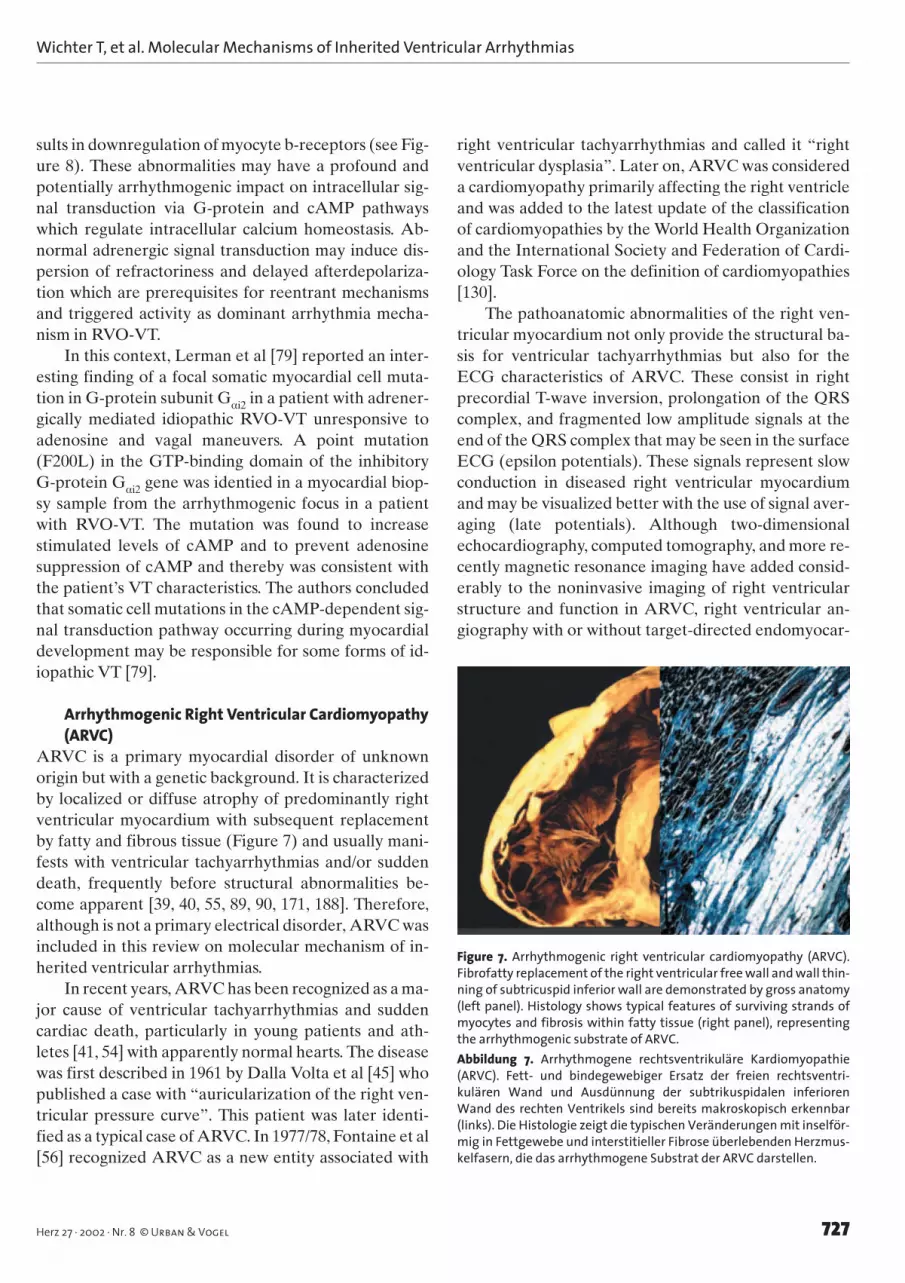
Arrhythmogenic Right Ventricular Cardiomyopathy(ARVC)
ARVC is a primary myocardial disorder of unknownorigin but with a genetic background. It is characterizedby localized or diffuse atrophy of predominantly rightventricular myocardium with subsequent replacementby fatty and fibrous tissue (Figure 7) and usually mani-fests with ventricular tachyarrhythmias and/or suddendeath, frequently before structural abnormalities be-come apparent [39, 40, 55, 89, 90, 171, 188]. Therefore,although is not a primary electrical disorder, ARVC wasincluded in this review on molecular mechanism of in-herited ventricular arrhythmias.
In recent years, ARVC has been recognized as a ma-jor cause of ventricular tachyarrhythmias and suddencardiac death, particularly in young patients and ath-letes [41, 54] with apparently normal hearts. The diseasewas first described in 1961 by Dalla Volta et al [45] whopublished a case with “auricularization of the right ven-tricular pressure curve”. This patient was later identi-fied as a typical case of ARVC. In 1977/78, Fontaine et al[56] recognized ARVC as a new entity associated with
right ventricular tachyarrhythmias and called it “rightventricular dysplasia”. Later on, ARVC was considereda cardiomyopathy primarily affecting the right ventricleand was added to the latest update of the classificationof cardiomyopathies by the World Health Organizationand the International Society and Federation of Cardi-ology Task Force on the definition of cardiomyopathies[130].
The pathoanatomic abnormalities of the right ven-tricular myocardium not only provide the structural ba-sis for ventricular tachyarrhythmias but also for theECG characteristics of ARVC. These consist in rightprecordial T-wave inversion, prolongation of the QRScomplex, and fragmented low amplitude signals at theend of the QRS complex that may be seen in the surfaceECG (epsilon potentials). These signals represent slowconduction in diseased right ventricular myocardiumand may be visualized better with the use of signal aver-aging (late potentials). Although two-dimensionalechocardiography, computed tomography, and more re-cently magnetic resonance imaging have added consid-erably to the noninvasive imaging of right ventricularstructure and function in ARVC, right ventricular an-giography with or without target-directed endomyocar-
Figure 7. Arrhythmogenic right ventricular cardiomyopathy (ARVC).Fibrofatty replacement of the right ventricular free wall and wall thin-ning of subtricuspid inferior wall are demonstrated by gross anatomy(left panel). Histology shows typical features of surviving strands ofmyocytes and fibrosis within fatty tissue (right panel), representingthe arrhythmogenic substrate of ARVC.Abbildung 7. Arrhythmogene rechtsventrikuläre Kardiomyopathie(ARVC). Fett- und bindegewebiger Ersatz der freien rechtsventri-kulären Wand und Ausdünnung der subtrikuspidalen inferiorenWand des rechten Ventrikels sind bereits makroskopisch erkennbar(links). Die Histologie zeigt die typischen Veränderungen mit inselför-mig in Fettgewebe und interstitieller Fibrose überlebenden Herzmus-kelfasern, die das arrhythmogene Substrat der ARVC darstellen.

dial biopsy has remained the gold standard of imagingin ARVC. The structural, imaging and electrocardio-graphic features along with the typical left bundlebranch block characteristics of ventricular tachy-arrhythmias are included in the diagnostic criteria ofARVC proposed by an International Task Force forARVC [96]. The catalog of criteria includes major andminor criteria in different categories and facilitates thediagnosis of ARVC with the use of a scoring system(Table 3).
EpidemiologyLittle is known about the prevalence and incidence ofARVC [40]. Epidemiologic data indicate regional varia-tions with higher in northern Italy, France and Ger-many. A prospective investigation of sudden cardiacdeath in young people recognized ARVC in approxi-mately 20% of cases as the underlying cause of death[171]. Similarly, a population-based study demonstratedthat ARVC accounts for 17% of sudden cardiac deathsin young people below 40 years of age [153]. Regionaldifferences in the prevalence of the disease may be dueto true differences which may be based on the genetic
background, or on differences in the awareness of thedisease (medical education) and the diagnostic accuracyin its detection.
The lack of qualified autopsies and the dissense inthe interpretation of diagnostic procedures contributeto the fact that ARVC is both clinically and pathologi-cally underdiagnosed. Because of the variety of symp-toms and the complexity of diagnostic criteria, a com-plete and detailed diagnostic work-up is necessary tomake the correct diagnosis. Otherwise, patients withARVC may be both pathologically and clinically misdi-agnosed as suffering from, i.e., idiopathic ventriculartachyarrhythmias (no structural heart disease) or DCM[106, 108, 169, 188].
Pathoanatomic FeaturesPathologically, ARVC is characterized by localized ordiffuse atrophy of predominantly right ventricular my-ocardium with subsequent replacement by fatty and fi-brous tissue and surviving islands of hypertrophied my-ocyte fibers within fat and fibrosis [39, 55, 171] (Figure7). Predilection areas include the right ventricular out-flow tract, the apex, and the inflow tract (subtricuspid
Wichter T, et al. Molecular Mechanisms of Inherited Ventricular Arrhythmias
728 Herz 27 · 2002 · Nr. 8 © Urban & Vogel
Table 3. Diagnostic criteria of arrhythmogenic right ventricular cardiomyopathy (ARVC): the diagnosis is made in the presence of at least two ma-jor criteria or one major and two minor criteria, or four minor criteria from different diagnostic groups (I-VI). ECG: electrocardiogram; EF: ejectionfraction; LBBB: left bundle branch block; LV: left ventricle; RBBB: right bundle branch block; RV: right ventricle; VT: ventricular tachycardia.Tabelle 3. Diagnostische Kriterien der arrhythmogenen rechtsventrikulären Kardiomyopathie (ARVC): Die Diagnose wird gestellt bei Vorliegenvon mindestens zwei Hauptkriterien oder einem Hauptkriterium und zwei Nebenkriterien oder bei vier Nebenkriterien aus verschiedenen Diag-nosekategorien (I-VI). ECG: Elektrokardiogramm; EF: Ejektionsfraktion; LBBB: Linksschenkelblock; LV: linker Ventrikel; RBBB: Rechtsschenkelblock;RV: rechter Ventrikel; VT: ventrikuläre Tachykardie.
I. Global and/or regional dysfunction and structural alterationsa
• Major– Severe dilatation and reductions of RVEF with no
(or only mild) LV impairment– Localized RV aneurysms (akinetic or dyskinetic areas with diastolic
bulging)– Severe segmental dilatation of RV
• Minor– Mild global RV dilatation and/or EF reduction with
normal LV– Mild segmental dilatation of the RV– Regional RV hypokinesia
II. Tissue characterization of RV wall• Major
– Fibrofatty replacement of myocardium on endomyocardial biopsy
III. Repolarization abnormalities• Minor
– Inverted T-waves in right precordial leads (V2 and V3; age > 12 years,absence of RBBB)
adetected by echocardiography, angiography, magnetic resonance imaging, or radionuclide scintigraphy
IV. Depolarization or conduction abnormalities• Major
– Epsilon waves or localized prolongation (> 110 ms) of QRS complex inright precordial leads (V1–3)
• Minor– Late potentials (signal-averaged ECG)
V. Arrhythmias• Minor
– LBBB type (non)sustained VT (ECG, Holter, exercise test)– Frequent ventricular extrasystoles (> 1,000/24 h; Holter)
VI. Family history• Major
– Familial disease confirmed at necropsy or surgery• Minor
– Family history of premature sudden death (< 35 years) due to sus-pected ARVC
– Familial history (clinical diagnosis based on present criteria)

Wichter T, et al. Molecular Mechanisms of Inherited Ventricular Arrhythmias
729Herz 27 · 2002 · Nr. 8 © Urban & Vogel
area) of the right ventricular free wall, also called the“triangle of dysplasia” [90], whereas the ventricular sep-tum is usually spared. The pathogenetic basis of thesestructural myocardial abnormalities is widely unknown.
A congenital absence of myocardium, as presentin Uhl’s anomaly with parchment-like thinning theright ventricular wall [176], can be excluded in ARVC,because it is usually not present at birth but developsduring adolescence or early adulthood. Myocarditishas been discussed as a primary pathogenetic factor[11, 55, 170] as well as an inflammatory complicationsuperimposed on preexisting, genetically predeter-mined abnormalities of the right ventricular my-ocardium [55]. Recently, a study involving myocardialsamples from twelve patients with ARVC identifiedenteroviral sequences in seven and adenovirus type 5in another two patients, and the authors suggested a
potential link between ARVC and the presence of vi-ral genome in the myocardium [20]. By contrast, otherstudies had not shown a high incidence of virus-posi-tive findings in ARVC. Despite an ongoing scientificdebate, the available data are inconclusive, and therole of myocarditis in the pathogenesis of ARVC re-mains controversial.
Myocardial apoptosis [85] has been discussed as an-other potential pathogenetic mechanism of predomi-nantly right-sided myocardial atrophy and subsequentreplacement by fatty and fibrous tissue in ARVC. Fol-lowing initial findings by James [69], evidence of apop-tosis was found in tissue samples of ARVC patients ob-tained during surgery, transplantation or endomyocar-dial biopsies [87, 110, 177]. Significant apoptosis wasdetected particularly in those patients, in whom a recentonset or acute bursts of arrhythmias were present [177].Because apoptosis may be triggered by various mecha-nisms discussed in ARVC (including disruption of celladhesion, myocardial stretch, catecholamine stress,etc.), it may rather be a secondary phenomenon than aprimary disease mechanism.
Autonomic Modulation and DysfunctionIn ARVC, ventricular arrhythmias often occur duringphysical exercise or mental stress and may be provokedby intravenous catecholamine infusion during electro-physiologic study (catecholamine sensitivity). By con-trast, ventricular tachyarrhythmias are frequently sup-pressed by an antiarrhythmic drug regimen with antia-drenergic properties [190]. These clinical observationssuggested an involvement of the cardiac sympatheticnervous system. 123I-MIBG scintigraphy with the use ofSPECT demonstrated regional abnormalities of thepresynaptic myocardial sympathetic function in ARVC[193]. Subsequent studies using quantitative PET inves-tigated the presynaptic (11C-hydroxyephedrine) andpostsynaptic (11C-CGP12177) sympathetic innervationand demonstrated a significant reduction of myocardial�-adrenergic receptor density in patients with ARVC[195] (Figure 8). These findings may result from a sec-ondary downregulation of �-receptors after increasedlocal synaptic norepinephrine levels caused by in-creased firing rates of the efferent neurons or as the re-sult of impaired presynaptic catecholamine reuptakeconfined to the heart, since plasma catecholamines andpulmonary �-receptor densities were normal [141].
Recent in vitro studies in adult rat cardiac myocytesreported norepinephrine-stimulated apoptosis mediat-
Figure 8. Potential mechanisms of autonomic dysfunction in arrhyth-mogenic right ventricular cardiomyopathy (ARVC) [141, 193, 195] andidiopathic right ventricular outflow-tract tachycardia (RVO-VT) [138,140]. From quantitative radionuclide studies using SPECT and PETimaging, reuptake of presynaptic tracers (123I-MIBG, 11C-HED) was re-duced [138, 140, 193], which may be explained by reduced uptake-1 (UP)or by competitive inhibition due to increased norepinephrine release(RE). Both mechanisms would lead to an increased concentration ofnorepinephrine in the synaptic cleft with subsequent downregulationof postsynaptic �-adrenergic receptors (�), as demonstrated by 11C-CGP12177 and quantitative [138, 141, 195]Abbildung 8. Potentielle Mechansimen der autonomen Dysfunktionbei arrhythmogener rechtsventrikulärer Kardiomyopathie (ARVC)[141, 193, 195] und idiopathischer rechtsventrikulärer Ausflusstrakt-Tachykardie (RVO-VT) [138, 140]. Quantitative szintigraphische Unter-suchungen mittels SPECT und PET zeigten eine Reduktion der prä-synaptischen Wiederaufnahme der Tracer (123I-MIBG, 11C-HED) [138,140, 193], die durch gestörten Uptake-1-Mechanismus (UP) oder durchkompetitive Hemmung durch gesteigerte Noradrenalinausschüttung(RE) bedingt sein kann. Beide Mechanismen würden eine gesteigerteNoradrenalinkonzentration im synaptischen Spalt mit konsekutiverHerrunterregulierung der postsynaptischen �-Rezeptor-Dichte (�) be-wirken, wie sie mittels 11C-CGP12177 und quantitativer PET nachgewie-sen wurde [138, 141, 195].

ed by �-adrenergic stimulation of intracellular cAMPand subsequent activation of protein kinase A, whichincreased calcium influx, thus inducing apoptotic celldeath [34]. In patients with ARVC, increased synapticconcentrations of norepinephrine therefore may notonly increase the propensity to ventricular tachy-arrhythmias but may contribute to the progression ofmyocardial atrophy mediated by apoptotic cell death,which recently has been discussed as a pathogeneticmechanism in ARVC.
Molecular Genetics of Autosomal Dominant ARVCFrom family pedigree analyses, it was deduced thatmore than 30% of the cases are not only familial but ob-viously follow a Mendelian autosomal dominant pat-tern of inheritance with variable penetrance and poly-morphic phenotype [107]. This still holds true for the sixreported chromosomal loci in ARVC [114] (Table 4).
In 1994, Rampazzo et al [126] were the first to re-port on a chromosomal abnormality. Via linkage analy-sis in two families over four generations (82 subjects, 19affected patients), they detected the chromosomal locuson 14q23-q24 (ARVC-1).
1 year later, the same group [127] detected a geneti-cally novel variant of ARVC assigned to chromosome1q42-q43 (ARVC-2). Clinically, this subgroup present-ed with an atypical form of ARVC characterized byCPVT and only minimal structural abnormalities of theright ventricle [12]. Since both 14q23-q24 and 1q42-q43chromosomal loci map in proximity of α-actinin-2 and -3 genes (ACTN2+3) [14], Rampazzo et al [127] postu-lated a possible pathogenetic implication of the cardiac-
specific protein α-actinin, which anchors myofibrillaractin filaments to the Z-disks. However, Tiso et al [172]recently identified mutations in the RyR2 gene in fami-lies affected with ARVC-2 and excluded actinin-2 as acandidate gene. Therefore, it is difficult to determinewhether ARVC-2 patients truly represent an atypicalsubgroup of ARVC or rather correspond to the lateridentified entity of idiopathic catecholaminergic VT(CPVT) as described earlier in the review.
In 1996, a second locus on the long arm of chromo-some 14 was identified in a study of three small familiesof various Italian, Belgian and Slovenian descent(ARVC-3) [151]. The mode of inheritance was de-scribed as autosomal dominant but LOD scores wererather low. In 1997, Rampazzo et al [128] performedlinkage analysis in three independent families and de-tected a new locus on chromosome 2q32.1-q32.3(ARVC-4). Interestingly, a localized involvement of theleft ventricle together with a left bundle branch block inthe 12-lead surface electrocardiogram was found insome affected family members.
In 1998, Ahmad et al [2] investigated a large New-foundland family consisting of more than 200 membersspanning seven generations, and linkage analysis re-vealed a new locus on chromosome 3p23 (ARVC-5).The average overall though incomplete penetrance wasestimated at 67%. The clinical presentation was hetero-geneous, partly consisting of right ventricular dilatationand ventricular arrhythmias with a left bundle branchblock pattern, partly only showing nonspecific ECG ab-normalities. In four of 17 individuals who died suddenly(mean age: 34 ± 7 years), ARVC was confirmed at au-
Wichter T, et al. Molecular Mechanisms of Inherited Ventricular Arrhythmias
730 Herz 27 · 2002 · Nr. 8 © Urban & Vogel
Author First Chromosome LOD Mode of Penetrance Genedescription score inheritance
ARVC-1 Rampazzo et al [126] 1994 14q23-q24 5.4 Autosomal dominant High ?ARVC-2 Rampazzo et al [127] 1995 1q42-q43 4.0 Autosomal dominant High hRyR2ARVC-3 Severini et al [151] 1995 14q12-q22 4.7 Autosomal dominant ? ?ARVC-4 Rampazzo et al [128] 1997 2q32.1-q32.3 3.5 Autosomal dominant ? ?ARVC-5 Ahmad et al [2] 1998 3p23 6.9 Autosomal dominant ? ?ARVC-6 Li et al [81] 2000 10p14-p12 3.9 Autosomal dominant ? ?ARVC-7 Melberg et al [99] 1999 10q22.3 3.1 Autosomal dominant ? ?Naxos Coonar et al [37] 1998 17q21 3.6 Autosomal recessive 100% PlakoglobinARVC/APC Frances et al [57] 1997 14q24–q term Autosomal recessive ? ?
Table 4. Genetics of arrhythmogenic right ventricular cardiomyopathy (ARVC) – chromosomal loci and potential genes. LOD: logarithm of theodds.Tabelle 4. Genetik der arrhythmogenen rechtsventrikulären Kardiomyopathie (ARVC): chromosomale Loci und potentielle Gendefekte. LOD: lo-garithm of the odds.

Wichter T, et al. Molecular Mechanisms of Inherited Ventricular Arrhythmias
731Herz 27 · 2002 · Nr. 8 © Urban & Vogel
topsy. The clinical presentation of affected patients inthis family shows features of both ARVC and DCM. Inthat light, it is of particular interest that a locus for fa-milial DCM had previously been mapped to the samechromosomal region (3q22-25) [112, 113].
In 2000, Li et al [81, 83] investigated a North Amer-ican family consisting of 47 members over five genera-tions and identified another locus for ARVC mappingto chromosome 10p12-p14 (ARVC-6). This form ap-pears to be more malignant with an early onset of typi-cal alterations, as demonstrated by endomyocardialbiopsy in patients as early as 2 years of age. In the searchfor candidate genes mapping to that locus, the cy-toskeletal protein vimentin, which among others seemsto account for DCM [47], is supposed to play a role inadipogenesis [58]. D’Amati et al [44] described a fe-male patient with familial DCM who underwent hearttransplantation where histological, immunohistochemi-cal and ultrastructural analysis of the heart was sugges-tive of a transdifferentiation from myocytes to matureadipocytes which had previously been discussed as a po-tential mechanism of fatty replacement in ARVC [55].Recently, a gene encoding for the neuroblastoma apop-tosis-related RNA-binding protein, which is located onchromosome 10 (10p13-p14), could be excluded as agene causing ARVC [82]. This again elucidates theproblem of correct phenotyping in a variable, some-times subtle and subclinical disease with limited accura-cy of clinical diagnostic criteria.
Melberg et al [99] recently located another locuswith autosomal dominant inheritance on chromosome10q22.3. This locus was identified by linkage analysis(multipoint peak LOD score of 3.06) in a Swedish fami-ly affected by ARVC and myofibrillar myopathy.
Molecular Genetics of Autosomal Recessive ARVC(“Naxos Disease”)
In 1986, Protonotarios et al. [125] first reported on a pe-culiar “coincidence” of diffuse nonepidermolytic palmo-plantar keratoderma, woolly hair and typical clinicalsigns of ARVC including right VT. Because all families aswell as the authors originated from the Aegean island ofNaxos, this disease was named “Naxos disease”. Naxosdisease shows autosomal recessive inheritance with highpenetrance and early onset of cardiac manifestations.
In a larger study of the Naxos population, Coonar etal [37] identified 22 affected subjects from nine familiesof 150 individuals. All patients with a homozygosity forthe mutation presented with hair and skin abnormalities
in early infancy which were reliable predictors for subse-quent manifestation of ARVC, which became overt withpalpitations or syncope approximately at 15 years of age.The disease is 100% penetrant by adolescence, and thecumulative survival rate at the age of 35 years was 74%with an annual disease-related mortality of 3% [124].
Linkage analysis mapped the locus of Naxos diseaseto chromosome 17q21. Initially, the type-1 keratingroup (KRT9) was considered a target gene because it isinvolved in cell stability. However, meanwhile a ho-mozygous two-base pair deletion (TG) in the plakoglo-bin gene (Pk2157del2) was identified as the diseasecausing mutation, resulting in a frameshift and prema-ture termination of the protein [97]. Plakoglobin is a cy-toskeletal protein essential for cell-cell contact both incardiac and dermal-epidermal junctions. Together withα-catenin and �-catenin, plakoglobin is suggested toform the core structure in a protein network regulatingcell-cell adhesion and intracellular signaling [19, 80,199] (Figure 9). Based on results of earlier in vitro stud-ies [142], McKoy et al [97] discussed whether a desmo-some dysfunction due to mutant plakoglobin might beresponsible for progressive myocyte death in conditionsof mechanical stress in Naxos disease.
In Ecuador, a comparable disease was recentlyidentified. Similar to Naxos disease, affected patientspresent with keratoderma and woolly hair at infancyand later develop cardiac manifestations with left ven-tricular DCM, often resulting in cardiac enlargementand heart failure during adolescence [111]. Clinicalsigns of right ventricular cardiomyopathy were not re-ported. The disease also follows an autosomal recessivetrait and was mapped to chromosome 6p23-24. In a
Figure 9. Interaction of cardiac cytoskeletal proteins in cell-to-cell con-tacts and signaling.Abbildung 9. Interaktion kardialer zytoskelettärer Proteine bei Zell-Zell-Kontakten und Signaltransduktion.

study involving three families from Ecuador, a mutation(7901delG) was found in the gene encoding desmo-plakin, a cytoskeletal protein with important functionsin the attachment of intermediate filaments to thedesmosome. Truncation of this protein leads to disrup-tion of the desmoplakin-intermediate filament interac-tion and thereby causes the clinical manifestations ofDCM, woolly hair and keratoderma [111].
A second autosomal recessive form of ARVC withdifferent comanifestation was reported by Frances et al[57]. Both, brother and sister of healthy parents pre-sented with palpitations. ARVC was diagnosed on thebasis of typical cardiac and ECG alterations as the un-derlying cause of the documented ventricular tachy-arrhythmias. Physical examination disclosed an asymp-tomatic linear-shaped opacification of the crystallinelens in the anterior and posterior subcapsular zones. Al-though no generally accepted pathogenesis of cataractformation has been established, overexpression of thecytoskeletal protein vimentin resulted in the develop-ment of cataracts in a mouse animal model [30], whichrenders additional insights into a putative relationshipof cytoskeletal proteins and ARVC.
Pathophysiology of Cytoskeletal Proteins (Cell-to-Cell Contacts)
The discovery of a mutation in the plakoglobin gene inNaxos disease has refocused the scientific interest on cy-toskeletal proteins and cell-to-cell contacts in the patho-physiology of ARVC. However, although in a candidategene approach several cytoskeletal proteins were inves-tigated (vimentin, α-actinin, type-1 keratin) for differ-ent chromosomal loci, no mutations were identified inthe relevant genes. Also, mutations in the plakoglobingene have not been confirmed in patients with autoso-mal dominant forms of “classical” ARVC. However,our group has characterized a differential regulation ofadherens junction proteins with reduction of plakoglo-bin in right ventricular myocardium of patients withARVC (manuscript submitted).
However, the concept that cytoskeletal proteins andcell-to-cell signaling may play an important role for thepathogenesis and pathophysiology of ARVC remainsappealing. Mutations in genes encoding for such pro-teins may interfere with intercellular signaling, cell-to-cell contact and adhesion, as well as cell stability and tis-sue integrity [80, 199]. An impairment of intercellularcontacts may cause intercellular disruption, particularlyin relation to mechanical stress or stretch, thus provid-
ing a potential explanation for the high prevalence ofARVC in athletes and the frequent occurrence of ven-tricular tachyarrhythmias and sudden death during ex-ercise. However, although attractive and plausible,these pathophysiologic concepts remain widely hypo-thetical at the present time since no proof could be pro-vided yet.
More recently, also abnormalities in myocardial gapjunctions and connexin patterns with reduction of con-nexins 43 and 45 were observed in preliminary reportson Naxos disease [70] and our own studies in ARVC(unpublished observations).
Genetic Testing and ScreeningFurther to the variable phenotypic presentation and dis-ease progression, genetic heterogeneity contributes tothe difficulties of diagnosis and risk stratification inARVC. In the past decade, many efforts have been un-dertaken to enlarge the knowledge on clinical and mol-ecular genetics, pathophysiology and disease mecha-nisms of ARVC. Six chromosomal loci for autosomaldominant forms of ARVC and two loci for autosomalrecessive inheritance (one of which is Naxos disease)have been identified. However, specific mutations haveonly been found for Naxos disease (plakoglobin) andthe ARVC-2 locus (RyR2).
Therefore, routine genetic testing of patients or rel-atives with a suspicious diagnosis of ARVC is presentlynot available, and the pathogenetic implications arisingfrom the available data require further investigations.However, with the rapid improvements in molecular ge-netics, genetic testing for disease-causing mutations inindex patients as well as family members may becomethe future gold standard for diagnosis and possibly alsorisk stratification of ARVC with potential implicationsfor the expansion of knowledge in pathogenesis andmanagement of the disease.
Summary and Future DirectionsInherited ventricular arrhythmias have reached the eraof molecular genetics and an increasing understandingof the underlying molecular mechanisms and arrhyth-mogenesis. However, the genetic and phenotypic het-erogeneity of the various disease entities remains com-plex at the current stage. Mutations in the same genemay not only be related to differences in the severity ofthe clinical course but may also be associated with mul-tiple diseases. In other entities (i.e., ARVC), the identi-fication of gene mutations is only at the beginning, and
Wichter T, et al. Molecular Mechanisms of Inherited Ventricular Arrhythmias
732 Herz 27 · 2002 · Nr. 8 © Urban & Vogel

Wichter T, et al. Molecular Mechanisms of Inherited Ventricular Arrhythmias
733Herz 27 · 2002 · Nr. 8 © Urban & Vogel
the interaction of the genetic background with molecu-lar mechanisms and structural abnormalities is stillwidely unclear.
During the past decade, evidence has constantlygrown that establishing accurate genetic diagnoses anddissection of molecular disease mechanisms can have animpact on prognosis and help direct therapy in a rangeof cardiovascular diseases. Further achievements in theareas of clinical and molecular research, improvementof medical education and expansion of genotyping facil-ities will open a new era not only for the correct identi-fication of affected patients or family members with lowor borderline clinical disease expressivity, but also forthe improvement of risk stratification and possiblygene-directed therapeutic approaches to recompensatethe clinical phenotype. These important steps, however,will require an integrative approach from the identifica-tion of genetic mutations to functional analysis of ionchannels and electrical/mechanical properties of my-ocytes to whole organ electrophysiology and finallyclinical patient management.
AcknowledgmentsThis work was supported in part by1. Deutsche Forschungsgemeinschaft (DFG), Bonn, Germany, Son-derforschungsbereich 556 (Projects A1, C1, C4),2. European Commission, Brussels, Belgium, within the 5th Frame-work Program 1998–2002 (QLG1-CT-2000-01091),3. Fondation Leducq, Paris, France,4. Alfried Krupp von Bohlen und Halbach Stiftung, Essen, Germany.
GlossaryARVC Arrhythmogenic right ventricular cardiomyopathyCASQ2 Calsequestrin 2 geneCPVT Catecholaminergic polymorphic ventricular tachycardiaDCM Dilated cardiomyopathyECG ElectrocardiogramHCM Hypertrophic cardiomyopathyHERG Human eag-related gene (see KCNH1)ICD Implantable cardioverter defibrillatorICa L-type calcium currentIKr Rapidly activating delayed rectifierIKs Slowly activating delayed rectifierINa Fast sodium currentIto Transient outward potassium currentJLN Jervell and Lange-NielsenKCNE1 Gene encoding minK, a �-subunit for the IKs channel com-
plexKCNE2 Gene encoding MiRP1, a subunit for the IKr channel com-
plexKCNH1 (= HERG) Gene encoding the IKr potassium channel pro-
teinKCNQ1 (= KVLQT1) Gene encoding the α-subunit of the IKs
potassium channel proteinKVLQT1 see KCNQ1
LQTS Long QT syndromeMIBG Meta-iodobenzylguanidinePET Positron emission tomographyRVO-VT Right ventricular outflow-tract tachycardiaRyR Ryanodine receptorRyR2 Cardiac ryanodine receptorSCN5A Gene encoding the cardiac sodium channelSPECT Single-photon emission computed tomographyVF Ventricular fibrillationVT Ventricular tachycardia
References1. Ackerman MJ. The long QT syndrome: ion channel diseases of the
heart. Mayo Clin Proc 1998;73:250–9.2. Ahmad F, Li D, Karibe A, Gonzalez O, Tapscott T, Hill R, Weil-
baecher D, Blackie P, Furey M, Gardner M, Bachinski LL, Roberts R.Localization of a gene responsible for arrhythmogenic right ven-tricular dysplasia to chromosome 3p23. Circulation 1998;98:2791–5.
3. Akai J, Makita N, Sakurada H, Shirai N, Ueda K, Kitabatake A,Nakazawa K, Kimura A, Hiraoka M. A novel SCN5A mutation as-sociated with idiopathic ventricular fibrillation without typicalECG findings of Brugada syndrome. FEBS Lett 2000;479:29–34.
4. Alings M, Wilde A. Brugada syndrome: clinical data and suggest-ed pathophysiological mechanism. Circulation 1999;99:666–73.
5. An RH, Bangalore R, Rosero SZ, Kass RS. Lidocaine block of LQT-3mutant human Na+ channels. Circ Res 1996;79:103–8.
6. An RH, Wang XL, Kerem B, Benhorin J, Medina A, Goldmit M, KassRS. Novel LQT-3 mutation affects Na+ channel activity throughinteractions between alpha- and beta1-subunits. Circ Res1998;83:141–6.
7. Antzelevitch C. The Brugada syndrome: ionic and arrhythmiamechanisms. J Cardiovasc Electrophysiol 2001;12:268–72.
8. Antzelevitch C. Sympathetic modulation of the long QT syn-drome. Eur Heart J 2002;23:1246–52.
9. Atarashi H, Ogawa S, Harumi K, Sugimoto T, Inoue H, MurayamaM, Toyama J, Hayakawa H, for the Idiopathic Ventricular Fibrilla-tion Investigators. Three-year follow-up of patients with rightbundle branch block and ST segment elevation in the right pre-cordial leads. J Am Coll Cardiol 2001;37:1916–20.
10. Barhanin J, Lesage F, Guillemare E, Fink M, Lazdunski M, Romey G.K(V)LQT1 and lsK (minK) proteins associate to form the I(Ks) car-diac potassium current. Nature 1996;384:78–80.
11. Basso C, Thiene G, Corrado D, Angelini A, Nava A, Valente M. Ar-rhythmogenic right ventricular cardiomyopathy: dysplasia, dys-trophy, or myocarditis? Circulation 1996;94:983–91.
12. Bauce B, Nava A, Ramazzo A, Daliento L, Muriamo M, Basso C,Thiene G, Danieli GA. Familial effort polymorphic ventricular ar-rhythmias in arrhythmogenic right ventricular cardiomyopathymap to chromosome 1q42-43. Am J Cardiol 2000;85:573–9.
13. Bauce B, Rampazzo A, Basso C, Bagattin A, Daliento L, Tiso N, Tur-rini P, Thiene G, Danieli GA, Nava A. Screening for ryanodine re-ceptor type 2 mutations in families with effort-induced polymor-phic ventricular arrhythmias and sudden death. J Am Coll Cardiol2002;40:341–9.
14. Beggs AH, Byers TJ, Knoll JH, Boyce FM, Bruns GAP, Kunkel LM.Cloning and characterization of two human skeletal muscle al-pha-actinin genes located on chromosome 1 and 11. J Biol Chem1992;267:9281–8.
15. Belhassen B, Viskin S. Idiopathic ventricular tachycardia and fib-rillation. J Cardiovasc Electrophysiol 1993;4:356–68.

Wichter T, et al. Molecular Mechanisms of Inherited Ventricular Arrhythmias
734 Herz 27 · 2002 · Nr. 8 © Urban & Vogel
16. Belhassen B, Viskin S, Fish R, Glick A, Setbon I, Eldar M. Effects ofelectrophysiologic-guided therapy with class Ia antiarrhythmicdrugs on the long-term outcome of patients with idiopathic ven-tricular fibrillation with or without Brugada syndrome. J Cardio-vasc Electrophysiol 1999;10:1301–12.
17. Bennett PB, Yazawa K, Makita N, George ALJ. Molecular mecha-nism for an inherited cardiac arrhythmia. Nature 1995;376:683–5.
18. Bezzina C, Veldkamp MW, van den Berg MP, Postma AV, RookMB, Viersma JW, van Langen IM, Tan-Sindhunata G, Bink-Boelkens MTE, van der Hout AH, Mannens MMAM, Wilde AAM. Asingle Na+ channel mutation causing both long-QT and Brugadasyndromes. Circ Res 1999;85:1206–13.
19. Bornslaeger ES, Godsel LM, Corcoram CM, Park JK, Hatzfeld M,Kowalczyk AP, Green KJ. Plakophilin 1 interferes with plakoglobinbinding to desmoplakin, yet together with plakoglobin promotesclustering of desmosomal plaque complexes at cell-cell borders.J Cell Sci 2001;114:727–38.
20. Bowles NE, Ni J, Marcus F, Towbin JA. The detection of car-diotropic viruses in the myocardium of patients with arrhythmo-genic right ventricular cardiomyopathy. J Am Coll Cardiol 2002;39:892–5.
21. Brooks R, Burgess JH. Idiopathic ventricular tachycardia: a review.Medicine (Baltimore) 1988;67:271–94.
22. Brown RL, Pollock N, Couchman KG, Hodges M, Hutchinson DO,Waaka R, Lynch P, McCarthy TV, Stowell KM. A novel ryanodinereceptor mutation and genotype-phenotype correlation in alarge malignant hyperthermia New Zealand Maori pedigree.Hum Mol Genet 2000;9:1515–24.
23. Brugada J, Brugada R, Antzelevitch C, Towbin J, Nademanee K,Brugada P. Long-term follow-up of individuals with the electro-cardiographic pattern of right bundle branch block and ST-seg-ment elevation in precordial leads V1 to V3. Circulation 2002;105:73–8.
24. Brugada J, Brugada R, Brugada P. Right bundle branch block andST-segment elevation in leads V1 through V3. A marker for suddendeath in patients without demonstrable structural heart disease.Circulation 1998;97:457–60.
25. Brugada P, Brugada J. Right bundle branch block, persistent STsegment elevation and sudden cardiac death: a distinct clinicaland electrocardiographic syndrome: a multicenter report. J AmColl Cardiol 1992;20:1391–6.
26. Brugada P, Geelen P, Brugada R, Mont L, Brugada J. Prognosticvalue of electrophysiologic investigations in Brugada syndrome.J Cardiovasc Electrophysiol 2001;12:1004–7.
27. Brugada R, Brugada J, Antzelevitch C, Kirsch GE, Potenza D, Tow-bin JA, Brugada P. Sodium channel blockers identify risk for sud-den death in patients with ST-segment elevation and right bun-dle branch block but structurally normal hearts. Circulation2000;101:510–5.
28. Bruns HJ, Eckardt L, Vahlhaus C, Schulze-Bahr E, Haverkamp W,Borggrefe M, Breithardt G, Wichter T. Body surface potentialmapping in patients with Brugada syndrome: right precordial STsegment variations and reverse changes in left precordial leads.Cardiovasc Res 2002;54:58–66.
29. Calkins H, Lehmann MH, Allman K, Wieland D, Schwaiger M.Scintigraphic pattern of regional cardiac sympathetic innerva-tion in patients with familial long QT syndrome using positronemission tomography. Circulation 1993;87:1616–21.
30. Capetanaki Y, Smith S, Heath JP. Overexpression of the vimentingene in transgenic mice inhibits normal lens cell differentiation.J Cell Biol 1989;109:1653–64.
31. Chen Q, Kirsch GE, Zhang D, Brugada R, Brugada J, Brugada P,Potenza D, Moya A, Borggrefe M, Breithardt G, Ortiz-Lopez R,Wang Z, Antzelevitch C, O’Brien RE, Schulze-Bahr E, Keating MT,
Towbin JA, Wang Q. Genetic basis and molecular mechanism foridiopathic ventricular fibrillation. Nature 1998;392:293–5.
32. Chouabe C, Neyroud N, Guicheney P, Lazdunski M, Romey G,Barhanin J. Properties of KvLQT1 K+ channel mutations in Ro-mano-Ward and Jervell and Lange-Nielsen inherited cardiac ar-rhythmias. EMBO J 1997;16:5472–9.
33. Clancy CE, Rudy Y. Na+ channel mutation that causes both Bru-gada and long QT syndrome phenotypes. A simulation study ofmechanism. Circulation 2002;105:1208–13.
34. Communal C, Singh K, Pimentel DR, Colucci WS. Norepinephrinestimulates apoptosis in adult rat ventricular myocytes by activa-tion of the �-adrenergic pathway. Circulation 1998;98:1329–34.
35. Compton SJ, Lux RL, Ramsey MR, Strelich KR, Sanguinetti MC,Green LS, Keating MT, Mason JW. Genetically defined therapy ofinherited long-QT syndrome. Correction of abnormal repolariza-tion by potassium. Circulation 1996;94:1018–22.
36. Consensus Statement of the Joint Steering Committees of theUnexplained Cardiac Arrest Registry of Europe and of the Idio-pathic Ventricular Fibrillation Registry of the United States. Sur-vivors of out-of-hospital cardiac arrest with apparently normalheart: need for definition and standardized clinical evaluation.Circulation 1997;95:265–72.
37. Coonar AS, Protonotarios N, Tsatsopoulou A, Needham EWA, Houl-ston RS, Cliff S, Otter MI, Murday VA, Mattu RK, McKenna WJ. Genefor arrhythmogenic right ventricular cardiomyopathy with diffusenonepidermolytic palmoplantar keratoderma and woolly hair(Naxos disease) maps to 17q21. Circulation 1998;97:2049–58.
38. Corrado D, Basso C, Buja G, Nava A, Rossi L, Thiene G. Right bun-dle branch block, right precordial ST-segment elevation and sud-den death in young people. Circulation 2001;103:710–7.
39. Corrado D, Basso C, Thiene G, McKenna WJ, Davies MJ, FontaliranF, Nava A, Silvestri F, Blomström-Lundqvist C, Wlodarska EK,Fontaine G, Camerini F. Spectrum of clinicopathologic manifes-tations of arrhythmogenic right ventricular cardiomyopathy/dysplasia: a multicenter study. J Am Coll Cardiol 1997;30:1512–20.
40. Corrado D, Fontaine G, Marcus FI, McKenna WJ, Nava A, Thiene G,Wichter T. Arrhythmogenic right ventricular dysplasia/car-diomyopathy: need for an international registry. Circulation2000;101:e101–6.
41. Corrado D, Thiene G, Nava A, Rossi L, Pennelli N. Sudden death inyoung competitive athletes: clinicopathologic correlation in 22cases. Am J Med 1990;89:588–96.
42. Coumel P, Fidelle J, Lucet V, Attuel P, Bouvrain Y. Catecholaminer-gic-induced severe ventricular arrhythmias with Adams-Stokessyndrome in children: report of four cases. Br Heart J 1978;40:Suppl:28–37.
43. Curran ME, Splawski I, Timothy KW, Vincent GM, Green ED, Keat-ing MT. A molecular basis for cardiac arrhythmia: HERG muta-tions cause long QT syndrome. Cell 1995;80:795–803.
44. D’Amati G, Di Gioia CR, Giordano C, Gallo P. Myocyte transdiffer-entiation: a possible pathogenetic mechanism for arrhythmo-genic right ventricular cardiomyopathy. Arch Pathol Lab Med2000;124:287–90.
45. Dalla Volta S, Battagiia G, Zerbini E. “Auricularization” of theright ventricular pressure curve. Am Heart J 1961;61:25–33.
46. Demolombe S, Baro I, Pereon Y, Bliek J, Mohammad-Panah R, Pol-lard H, Morid S, Mannens M, Wilde A, Barhanin J, Charpentier F,Escande D. A dominant negative isoform of the long QT syn-drome 1 gene product. J Biol Chem 1998;273:6837–43.
47. Di Somma S, Marotta M, Salvatore G, Cudemo G, Cuda G, De VivoF, Di Benedetto MP, Ciaramella F, Caputo G, de Divitiis O. Changesin myocardial cytoskeletal intermediate filaments and myocytecontractile dysfunction in dilated cardiomyopathy: an in vivostudy in humans. Heart 2000;84:659–67.

Wichter T, et al. Molecular Mechanisms of Inherited Ventricular Arrhythmias
735Herz 27 · 2002 · Nr. 8 © Urban & Vogel
48. Donger C, Denjoy I, Berthet M, Neyroud N, Cruaud C, BennaceurM, Chivoret G, Schwartz K, Coumel P, Guicheney P. KVLQT1 C-ter-minal missense mutation causes a forme fruste long-QT syn-drome. Circulation 1997;96:2778–81
49. Duggal P, Vesely M R, Wattanasirichaigoon D, Villafane J,Kaushik V, Beggs AH. Mutation of the gene for IsK associatedwith both Jervell and Lange-Nielsen and Romano-Ward forms oflong-QT syndrome. Circulation 1998;97:142–6.
50. Dumaine R, Wang Q, Keating MT, Hartmann HA, Schwartz PJ,Brown AM, Kirsch GE. Multiple mechanisms of Na+ channel-linked long-QT syndrome. Circ Res 1996;78:916–24.
51. Eckardt L, Bruns HJ, Paul M, Kirchhof P, Schulze-Bahr E, Wichter T,Breithardt G, Borggrefe M, Haverkamp W. Body surface area ofST elevation and the presence of late potentials correlate to theinducibility of ventricular tachyarrhythmias in Brugada syn-drome. J Cardiovasc Electrophysiol 2002;13:742–9.
52. Eckardt L, Kirchhof P, Schulze-Bahr E, Rolf S, Ribbing M, Loh P,Bruns HJ, Witte A, Milberg P, Borggrefe M, Breithardt G, WichterT, Haverkamp W. Electrophysiologic investigation in Brugadasyndrome: yield of programmed ventricular stimulation at twoventricular sites with three premature beats. Eur Heart J 2002:23:1394–1401.
53. Fisher JD, Krikler D, Hallidie-Smith KA. Familial polymorphic ven-tricular arrhythmias: a quarter century of successful medicaltreatment based on serial exercise-pharmacologic testing. J AmColl Cardiol 1999;34:2015–22.
54. Fontaine G, Fontaliran F, Frank R, Chomette G, Grosgogeat I.Causes of sudden death in athletes. Arch Mal Cœur 1989;82:107–11.
55. Fontaine G, Fontaliran F, Frank R. Arrhythmogenic right ventricu-lar cardiomyopathies. Clinical forms and main differential diag-noses. Circulation 1998;97:1532–5.
56. Fontaine G, Guiraudon G, Frank R, Vedel J, Grosgogeat Y, Cabrol C,Facquet J. Stimulation studies and epicardial mapping in ventric-ular tachycardia: study of mechanism and selection for surgery.In: Kulbertus HE, ed. Reentrant arrhythmias. Lancaster: MTP,1977:334–50.
57. Frances R, Rodriguez Benitez AM, Cohen DR. Arrhythmogenicright ventricular dysplasia and anterior polar cataract. Am J MedGenet 1997;73:125–6.
58. Franke WW, Gergt M, Grund C. Rearrangement of the vimentincytoskeleton during adipose conversion: formation of an inter-mediate filament cage around lipid globules. Cell 1987;49:131–41.
59. Franz WM, Müller OJ, Katus HA. Cardiomyopathies: from genet-ics to the prospect of treatment. Lancet 2001;358:1627–37.
60. Furuhashi M, Tsuchihashi K, Nagahara D, Hyakukoku M, OhtomoT, Satoh S, Nishimiya T, Shimamoto K. Prevalence of asympto-matic ST segment elevation in right precordial leads with rightbundle branch block (Brugada-type ST shift) among the generalJapanese population. Heart 2001;86:161–6.
61. Garg A, Finneran W, Feld GK. Familial sudden cardiac death asso-ciated with a terminal QRS abnormality on the surface 12-leadelectrocardiogram in the index case. J Cardiovasc Electrophysiol1998;9:642–7.
62. Gill JS, Hunter GJ, Gane J, Ward DE, Camm AJ. Asymmetry of car-diac 123I-meta-iodobenzylguanidine scans in patients with ven-tricular tachycardia and a “clinically normal” heart. Br Heart J1993;69:6–13.
63. Gima K, Rudy Y. Ionic current basis of electrocardiographic wave-forms: a model study. Circ Res 2002;90:889–96.
64. Gohl K, Feistel H, Weikl A, Bachmann K, Wolf F. Congenital my-ocardial sympathetic dysinnervation (CMSD) – a structural de-fect of idiopathic long QT syndrome. PACE 1991;14:1544–53.
65. Grace AA. Brugada syndrome. Lancet 1999;354:445–6.
66. Gussak I, Antzelevitch C, Bjerregaard P, Towbin JA, Chaitman B.The Brugada syndrome: clinical, electrophysiologic and geneticaspects. J Am Coll Cardiol 1999;33:5–15.
67. Gussak I, Bjerregaard P, Hammill SC. Clinical diagnosis and riskstratification in patients with Brugada syndrome. J Am Coll Car-diol 2001;37:1635–8.
68. Ikeda T, Sakurada H, Sakabe K, Sakata T, Takami M, Tezuka N,Nakae T, Noro M, Enjoji Y, Tejima T, Sugi K, Yamaguchi T. Assess-ment of noninvasive markers in identifying patients at risk in theBrugada syndrome: insight into risk stratification. J Am Coll Car-diol 2001;37:1628–34.
69. James TN. Normal and abnormal consequences of apoptosis inthe human heart: from postnatal morphogenesis to paroxysmalarrhythmias. Circulation 1994;90:556–73.
70. Kaplan SR, Protonotarios N, Tsatsopoulou A, Fontaine G, SaffitzJE. Diminished myocardial connexin 43 expression and gap junc-tion remodeling in Naxos disease. PACE 2002;24(Part II):525.
71. Kasanuki H, Ohnishi S, Ohtuka M, MatsudaN, Nirei T, Isogai R,Shoda M, Toyoshima Y, Hosoda S. Idiopathic ventricular fibrilla-tion induced with vagal activity in patients without obviousheart disease. Circulation 1997;95:2277–85.
72. Kiehn J, Lacerda AE, Wible B, Brown AM. Molecular physiologyand pharmacology of HERG. Single-channel currents and blockby dofetilide. Circulation 1991;94:2572–9.
73. Kiehn J, Wible B, Ficker E, Taglialatela M, Brown AM. Cloned hu-man inward rectifier K+ channel as a target for class III methane-sulfonanilides. Circ Res 1995;77:1151–5.
74. Kurita T, Shimizi W, Inagaki M, Sayama K, Taguchi A, Satomi K, Ai-hara N, Kamakura S, Kobayashi J, Kosakai Y. The electrophysio-logic mechanism of ST-segment elevation in Brugada syndrome.J Am Coll Cardiol 2002;40:330–4.
75. Lahat H, Eldar M, Levy-Nissenbaum E, Bahan T, Friedman E,Khouri A, Lorber A, Kastner DL, Goldman B, Pras E. Autosomal-re-cessive catecholamine- or exercise-induced polymorphic ventric-ular tachycardia: clinical features and assignment of the diseasegene to chromosome 1p13-21. Circulation 2001;103:2822–7.
76. Lahat H, Pras E, Olender T, Avidan N, Ben-Asher E, Man O, Levy-Nissenbaum E, Khoury A, Lorber A, Goldman B, Lancet D, Eldar M.A missense mutation in a highly conserved region of CASQ2 is as-sociated with autosomal recessive catecholamine-induced poly-morphic ventricular tachycardia in Bedouin families from Israel.Am J Hum Genet 2001;37:378–84.
77. Laitinen PJ, Brown KM, Piippo K, Swan H, Devaney JM, Brahm-blatt B, Donarum EA, Marino M, Tiso N, Viitasalo M, Toivonen L,Stephan DA, Kontula K. Mutations of the cardiac ryanodine re-ceptor (RyR2) gene in familial polymorphic ventricular tachycar-dia. Circulation 2001;103:485–90.
78. Leenhardt A, Lucet V, Denjoy I, Grau F, Ngoc DD, Coumel P. Cate-cholaminergic polymorphic ventricular tachycardia in children: a7-year follow-up of 21 patients. Circulation 1995;91:1512–9.
79. Lerman BB, Dong B, Stein KM, Markowitz SM, Linden J, CatanzaroDF. Right ventruicular outflow tract tachycardia due to a somaticcell mutation in G protein subunit �i2. J Clin Invest 1998;101:2862–8.
80. Leung CL, Green KJ, Liem RKH. Plakins: a family of versatile cy-tolinker proteins. Trends Cell Biol 2002;12:37–45.
81. Li D, Ahmad F, Gardner MJ, Weilbaecher D, Hill R, Karibe A, Gon-zalez O, Tapscott T, Sharratt G, Bachinski LL, Roberts R. The locusof a novel gene responsible for arrhythmogenic right ventriculardysplasia characterized by early onset and high penetrance mapsto chromosome 10p12-p14. Am J Hum Genet 2000;66:148–56.
82. Li D, Bachinski LL, Roberts R. Genomic organization and isoform-specific tissue expression of human NAPOR (CUGBP2) as a candi-date gene for familial arrhythmogenic right ventricular dyspla-sia. Genomics 2001;74:396–401.

Wichter T, et al. Molecular Mechanisms of Inherited Ventricular Arrhythmias
736 Herz 27 · 2002 · Nr. 8 © Urban & Vogel
83. Li D, Gonzalez O, Bachinski LL, Roberts R. Human protein thyro-sine phosphatase-like gene: expression profile, genomic struc-ture, and mutation analysis in patients with ARVD. Gene2000;256:237–43.
84. Locati EH, Zareba W, Moss AJ, Schwartz PJ, Vincent GM, LehmannMH, Towbin JA, Priori SG, Napolitano C, Robinson JL, Andrews M,Timothy K, Hall WJ. Age- and sex-related differences in clinicalmanifestations in patients with congenital long-QT syndrome:findings from the International LQTS Registry. Circulation1998;97:2237–44.
85. Maisch B. How cardiac cells die – necrosis, oncosis and apoptosis.Herz 1999;24:181–8.
86. Makita N, Shirai N, Nagashima M, Matsuoka R, Yamada Y, TohseN, Kitabatake A. A de novo missense mutation of human cardiacNa+ channel exhibiting novel molecular mechanisms of long QTsyndrome. FEBS Lett 1998;423:5–9.
87. Mallat Z, Tedgui A, Fontaliran F, Frank R, Durigon M, Fontaine G.Evidence of apoptosis in arrhythmogenic right ventricular dys-plasia. N Engl J Med 1996;335:1190–6.
88. Marcus FI. Idiopathic ventricular fibrillation. J Cardiovasc Electro-physiol 1997;8:1075–83.
89. Marcus FI, Fontaine G. Arrhythmogenic right ventricular dyspla-sia/cardiomyopathy: a review. Pacing Clin Electrophysiol 1995;18:1298–314.
90. Marcus FI, Fontaine G, Guiraudon G, Frank R, Laurenceau JL, Ma-lergue C, Grosgogeat Y. Right ventricular dysplasia: a report of 24cases. Circulation 1982;65:384–99.
91. Marks AR. Cardiac intracellular calcium release channels. Circ Res2000;87:8–11.
92. Marks AR. Clinical implications of cardiac ryanodine receptor/cal-cium release channel mutations linked to sudden cardiac death.Circulation 2002;106:8–10.
93. Marks AR, Priori SG, Memmi M, Kontula K, Laitinen PJ. Involve-ment of the cardiac ryanodine receptor/calcium release channelin catecholaminergic polymorphic ventricular tachycardia. J CellPhysiol 2002;190:1–6.
94. Martini B, Nava A, Thiene G, Buja GF, Canciani B, Scognamiglio R,Daliento L, Dalla Volta S. Ventricular fibrillation without appar-ent heart disease: description of six cases. Am Heart J1989;118:1203–9.
95. Mathews ECJ, Blount AW, Blount AW Jr, Townsend JI. QT prolon-gation and ventricular arrhythmias, with and without deafness,in the same family. Am J Cardiol 1972;29:702–11.
96. McKenna WJ, Thiene G, Nava A, Fontaliran F, Blomström-Lundqvist C, Fontaine G, Camerini F. Diagnosis of arrhythmo-genic right ventricular dysplasia/cardiomyopathy. Task Force ofthe Working Group Myocardial and Pericardial Disease of the Eu-ropean Society of Cardiology and of the Scientific Council on Car-diomyopathies of the International Society and Federation ofCardiology. Br Heart J 1994;71:215–8.
97. McKoy G, Protonotarios N, Crosby A, Tsatsopoulou A, AnastasakisA, Coonar A, Norman M, Baboonian C, Jeffrey S, McKenna WJ.Identification of a deletion in plakoglobin in arrhythmogenicright ventricular cardiomyopathy with palmoplantar keratoder-ma and woolly hair (Naxos disease). Lancet 2000;355:2119–24.
98. McRae JR, Wagner GS, Rogers MC, Canent RV. Paroxysmal famil-ial ventricular fibrillation. J Pediatr 1974;84:515–8.
99. Melberg A, Oldfors A, Blomström-Lundqvist C, Stalberg E, Carls-son B, Larsson E, Lidell C, Eeg-Olofsson KE, Wikstrom G, Henriks-son G, Dahl N. Autosomal dominant myofibrillar myopathy witharrhythmogenic right ventricular cardiomyopathy linked to chro-mosome 10q. Ann Neurol 1999;46:684–92.
100. Miyazaki T, Mitamura H, Miyoshi S, Soejima K, Aizawa Y, OgawaS. Autonomic and antiarrhythmic drug modulation of ST seg-
ment elevation in patients with Brugada syndrome. J Am CollCardiol 1996;27:1061–70.
101. Monroe MH, Littmann L. Two-year case collection of the Brugadasyndrome electrocardiogram pattern at a large teaching hospi-tal. Clin Cardiol 2000;23:849–51.
102. Moss AJ, Schwartz PJ, Crampton RS, Tzivoni D, Locati EH, Mac-Cluer J, Hall WJ, Weitkamp L, Vincent GM, Garson A Jr, RobinsonJL, Benhorin J, Choi S. The long QT syndrome: prospective longi-tudinal study of 328 families. Circulation 1991;84:1136–44.
103. Moss AJ, Zareba W, Benhorin J, Locati EH, Hall WJ, Robinson JL,Schwartz PJ, Towbin JA, Vincent GM, Lehmann MH. ECG T-wavepatterns in genetically distinct forms of the hereditary long QTsyndrome. Circulation 1995;92:2929–34.
104. Moss AJ, Zareba W, Kaufman ES, Gartman E, Peterson DR, BenhorinJ, Towbin JA, Keating MT, Priori SG, Schwartz PJ, Vincent GM, Robin-son JL, Andrews ML, Feng C, Hall WJ, Medina A, Zhang L, Wang Z. In-creased risk of arrhythmic events in long-QT syndrome with muta-tions in the pore region of the human ether-a-go-go-related genepotassium channel. Circulation 2002;105:794–9.
105. Nademanee K, Veerakul G, Nimmannit S, Chaowakul V, Bhuripa-nyo K, Likittanasombat K, Tunsanga K, Kuasirikul S, Malasit P,Tansupasawadikul S, Tatsanavivat P. Arrhythmogenic marker forthe sudden unexplained death syndrome in Thai men. Circula-tion 1997;96:2595–600.
106. Nava A, Thiene G, Canciani B, Martini B, Daliento L, Buja GF, FasoliG. Clinical profile of concealed form of arrhythmogenic right ven-tricular cardiomyopathy presenting with apparently idiopathicventricular arrhythmias. Int J Cardiol 1992;35:195–206.
107. Nava A, Thiene G, Canciani B, Scognamiglio R, Daliento L, Buja GF,Martini B, Stritoni P, Fasoli G. Familial occurrence of right ventric-ular dysplasia: a study involving nine families. J Am Coll Cardiol1988;12:1222–8.
108. Nemec J, Edwars BS, Osborn MJ, Edwards WD. Arrhythmogenicright ventricular dysplasia masquerading as a dilated cardiomy-opathy. Am J Cardiol 1999;84:237–9.
109. Neyroud N, Tesson F, Denjoy I, Leibovici M, Donger C, Barhanin J,Faure S, Gary F, Coumel P, Petit C, Schwartz K, Guicheney P. A nov-el mutation in the potassium channel gene KVLQT1 causes theJervell and Lange-Nielsen cardioauditory syndrome. Nat Genet1997;15:186–9.
110. Nishikawa T, Ishiyama S, Nagata M, Sakomura Y, Nakazawa M,Momma K, Hiroe M, Kasajima T. Programmed cell death in themyocardium of arrhythmogenic right ventricular cardiomyopa-thy in children and adults. Cardiovasc Pathol 1999;8:185–9.
111. Norgett EE, Hatsell SJ, Carvajal-Huerta L, Ruiz Cabezas JC, Com-mon J, Purkis PE, Whittock N, Leigh IM, Stevens HP, Kelsell DP. Re-cessive mutation in desmoplakin disrupts desmoplakin-interme-diate filament interactions and causes dilated cardiomyopathy,woolly hair and keratoderma. Hum Mol Genet 2000;9:2761–6.
112. Olson TM, Keating MT. Mapping a cardiomyopathy locus to chro-mosome 3q22-25. J Clin Invest 1996;97:528–32.
113. Olson TM, Michels VV, Thibodeau SN, Tai YS, Keating MT. Actinmutations in dilated cardiomyopathy, a heritable form of heartfailure. Science 1998;280:750–2.
114. Paul M, Wichter T, Schulze-Bahr E, Breithardt G. Genetics of ar-rhythmogenic right ventricular cardiomyopathy. In: Schalij MJ,Janse MJ, van Oosterom A, van der Wall EE, Wellens HJJ, eds. 100years of electrocardiography. Leiden, The Netherlands. TheEinthoven Foundation: Einthoven, 2002:265–72.
115. Priori SG, Barhanin J, Hauer RN, Haverkamp W, Jongsma HJ, Kle-ber AG, McKenna WJ, Roden DM, Rudy Y, Schwartz K, Schwartz PJ,Towbin JA, Wilde AM. Genetic and molecular basis of cardiac ar-rhythmias: impact on clinical management (parts 1 and 2). Circu-lation 1999;99:518–28.

Wichter T, et al. Molecular Mechanisms of Inherited Ventricular Arrhythmias
737Herz 27 · 2002 · Nr. 8 © Urban & Vogel
116. Priori SG, Barhanin J, Hauer RN, Haverkamp W, Jongsma HJ, Kle-ber AG, McKenna WJ, Roden DM, Rudy Y, Schwartz K, Schwartz PJ,Towbin JA, Wilde AM. Genetic and molecular basis of cardiac ar-rhythmias: impact on clinical management (part 3). Circulation1999;99:674–81.
117. Priori SG, Barhanin J, Hauer RNW, Haverkamp W, Jongsma HJ,Kleber AG, McKenna WJ, Roden DM, Rudy Y, Schwartz K,Schwartz PJ, Towbin JA, Wilde A. Genetic and molecular basis ofcardiac arrhythmias. Eur Heart J 1999;20:174–95.
118. Priori SG, Napolitano C, Cantu F, Brown AM, Schwartz PJ. Differ-ential response to Na+ channel blockade, beta-adrenergic stimu-lation, and rapid pacing in a cellular model mimicking the SCN5Aand HERG defects present in the long-QT syndrome. Circ Res1996;78:1009–15.
119. Priori SG, Napolitano C, Gasparini M, Pappone C, Della Bella P,Brignole M, Giordano U, Giovannini U, Menozzi C, Bloise R, CrottiL, Terreni L, Schwartz PJ. Clinical and genetic heterogeneity ofright bundle branch block and ST-segment elevation syndrome:a prospective evaluation of 52 families. Circulation 2000;102:2509–15.
120. Priori SG, Napolitano C, Gasparini M, Pappone C, Della Bella P,Giordano U, Bloise R, Giustetto C, De Nardis R, Grillo M, Ronchet-ti E, Faggiano G, Naftoli J. Natural history of Brugada syndrome.Insights for risk stratification and management. Circulation2002;105:1342–7.
121. Priori SG, Napolitano C, Grillo M. Concealed arrhythmogenic syn-dromes: the hidden substrate of idiopathic ventricular fibrilla-tion? Cardiovasc Res 2001;50:218–23.
122. Priori SG, Napolitano C, Memmi M, Colombi B, Drago F, GaspariniM, DeSimone L, Coltorti F, Bloise R, Keegan R, Cruz Filho FES, Vig-nati G, Benatar A, DeLogu A. Clinical and molecular characteriza-tion of patients with catecholaminergic polymorphic ventriculartachycardia. Circulation 2002;106:69–74.
123. Priori SG, Napolitano C, Tiso N, Memmi M, Vignati G, Bloise R, Sor-rentino V, Danieli GA. Mutations in the cardiac ryanodine recep-tor gene (hRyR2) underlie catecholaminergic polymorphic ven-tricular tachycardia. Circulation 2001;103:196–200.
124. Protonotarios N, Tsatsopoulou A, Anastasakis A, Sevdalis E, McKoy G, Stratos K, Gatzoulis K, Tentolouris K, Spiliopoulou C, Pana-giotakos D, McKenna W, Toutouzas P. Genetype-phenotype as-sessment in autosomal recessive arrhythmogenic right ventricu-lar cardiomyopathy (Naxos disease) caused by a deletion inplakoglobin. J Am Coll Cardiol 2001;38:1477–87.
125. Protonotarios N, Tsatsopoulou A, Patsourakos P, Alexopoulos D,Gezerlis P, Simitsis S, Scampardonis G. Cardiac abnormalities infamilial palmoplantar keratosis. Br Heart J 1986;56:321–6.
126. Rampazzo A, Nava A, Danieli GA, Buja G, Daliento L, Fasoli G,Scognamiglio R, Corrado D, Thiene G. The gene for arrhythmo-genic right ventricular cardiomyopathy maps to chromosome14q23-q24. Hum Mol Genet 1994;3:959–62.
127. Rampazzo A, Nava A, Erne P, Eberhard M, Vian E, Slomp P,Tiso N,Thiene G, Danieli GA. A new locus for arrhythmogenic right ven-tricular cardiomyopathy (ARVD 2) maps to chromosome 1q42-q43. Hum Mol Genet 1995;4:2151–4.
128. Rampazzo A, Nava A, Miorin M, Fonderico P, Pope B, Tiso N, Livol-si B, Zimbello R, Thiene G, Danieli GA. ARVD4, a new locus for ar-rhythmogenic right ventricular cardiomyopathy, maps to chro-mosome 2 long arm. Genomics 1997;45:259–63.
129. Remme CA, Wever EFD, Wilde AAM, Derksen R, Hauer RNW. Di-agnosis and long-term folllow-up of the Brugada syndrome inpatients with idiopathic ventricular fibrillation. Eur Heart J2001;22:400–9.
130. Richardson P, McKenna WJ, Bristow M, Maisch B, Mautner B,O’Connell J, Olsen E, Thiene G, Goodwin J, Gyarfas I, Martin I,
Nordet P. Report of the 1995 World Health Organization/Interna-tional Society and Federation of Cardiology Task Force on the de-finition and classification of cardiomyopathies. Circulation1996;93:841–2.
131. Romey G, Attali B, Chouabe C, Abitbol I, Guillemare E, Barhanin J,Lazdunski M. Molecular mechanism and functional significanceof the MinK control of the KvLQT1 channel activity. J Biol Chem1997;272:16713–6.
132. Rook MB, Alshinawi CB, Groenewegen WA, van Gelder IC, vanGinneken ACG, Jongsma HJ, Mannens MMAM, Wilde AAM. Hu-man SCN5A gene mutations alter cardiac sodium channel kinet-ics and are associated with the Brugada syndrome. CardiovascRes 1999;44:507–17.
133. Sanchez Cascos A, Sanchez Pernaute R, Cifuentes S. A child af-fected by the Romano-Ward syndrome born of a mother with theJervell and Lange-Nielsen syndrome. Rev Esp Cardiol 1990;43:406–7.
134. Sanguinetti MC, Curran ME, Spector PS, Keating MT. Spectrum ofHERG K+-channel dysfunction in an inherited cardiac arrhyth-mia. Proc Natl Acad Sci USA 1996;93:2208–12.
135. Sanguinetti MC, Curran ME, Zou A, Shen J, Spector PS, AtkinsonDL, Keating MT. Coassembly of K(v)LQT1 and minK (IsK) proteinsto form cardiac I(Ks) potassium channel. Nature 1996;384:80–3.
136. Sanguinetti MC, Jiang C, Curran ME, Keating MT. A mechanisticlink between an inherited and an acquired cardiac arrhythmia:HERG encodes the IKr potassium channel. Cell 1995;81:299–307.
137. Sato T, Hata Y, Yamamoto M, Morita H, Mizuo K, Yamanari H,Saito D, Ohe T. Early afterdepolarization abolished by potassiumchannel opener in a patient with idiopathic long QT syndrome. JCardiovasc Electrophysiol 1995;6:279–82.
138. Schäfers M, Lerch H, Wichter T, Rhodes CG, Lammertsma AA,Borggrefe M, Hermansen F, Schober O, Breithardt G, Camici PG.Cardiac sympathetic innervation in patients with idiopathic rightventricular outflow tract tachycardia. J Am Coll Cardiol 1998;32:181–6.
139. Schäfers M, Schober O, Lerch H. Cardiac sympathetic neurotrans-mission scintigraphy. Eur J Nucl Med 1998;25:435–41.
140. Schäfers M, Wichter T, Lerch H, Kuwert T, Schäfers K, BorggrefeM, Breithardt G, Schober O. Cardiac iodine-123-metaiodobenzyl-guanidine uptake in idiopathic ventricular tachycardia and fibril-lation. J Nucl Med 1999;40:1–5.
141. Schäfers MA, Wichter T, Schäfers KP, Rahman S, Rhodes CG, Lam-mertsma AA, Lerch H, Knickmeier M, Hermansen F, Schober O,Camici PG, Breithardt G. Pulmonary b-adrenoceptor density in ar-rhythmogenic right ventricular cardiomyopathy and idiopathicventricular tachycardia. Basic Res Cardiol 2001:96:91–7.
142. Schnittler HJ, Puschel B, Drenckhahn D. Role of cadherins and plakoglobin in interendothelial adhesion under restingconditions and under shear stress. Am J Physiol 1997;335:H2396–405.
143. Schott JJ, Alshinawi C, Kyndt F, Probst V, Hoorntje TM, HulsbeckM, Wilde AA, Escande D, Mannes MM, Le Marec H. Cardiac con-duction defects associate with mutation in SCN5A. Nat Genet1999;23:20–1.
144. Schulze-Bahr E, Eckardt L, Seidl KH, Seiler P, Wichter T, BreithardtG, Borggrefe M, Haverkamp W. Mutation analysis of the sodiumchannel gene SCN5A in 42 families with Brugada syndrome – ge-netic and diagnostic implications. ((¢7)) 2002:submitted.
145. Schulze-Bahr E, Haverkamp W, Wedekind H, Rubie C, Hordt M,Borggrefe M, Assmann G, Breithardt G, Funke H. Autosomal re-cessive long-QT syndrome (Jervell Lange-Nielsen syndrome) isgenetically heterogeneous. Hum Genet 1997;100:573–6.
146. Schulze-Bahr E, Haverkamp W, Wiebusch H, Schulte H, Hördt M,Borggrefe M, Breithardt G, Assmann G, Funke H. Molecular

Wichter T, et al. Molecular Mechanisms of Inherited Ventricular Arrhythmias
738 Herz 27 · 2002 · Nr. 8 © Urban & Vogel
analysis at the Harvey-Ras 1 gene in patients with long-QT syn-drome. J Mol Med 1995;73:565–9.
147. Schulze-Bahr E, Wang Q, Wedekind H, Haverkamp W, Chen Q,Sun Y, Rubie C, Hördt M, Towbin JA, Borggrefe M, Assmann G, QuX, Somberg JC, Breithardt G, Oberti C, Funke H. KCNE1 mutationscause Jervell and Lange-Nielsen syndrome. Nat Genet 1997;17:267–8.
148. Schwartz P, Moss AJ, Vincent GM, Crampton RS. Diagnostic crite-ria for the long QT syndrome: an update. Circulation 1993;88:782–4.
149. Schwartz PJ, Priori SG, Locati EH, Napolitano C, Cantu F, TowbinJA, Keating MT, Hammoude H, Brown AM, Chen LS. Long QT syn-drome patients with mutations of the SCN5A and HERG geneshave differential responses to Na+ channel blockade and to in-creases in heart rate. Implications for gene-specific therapy. Cir-culation 1995;92:3381–6.
150. Schwartz PJ, Priori SG, Spazzolini C, Moss AJ, Vincent GM, Napoli-tano C, Denjoy I, Guicheney P, Breithardt G, Keating MT, TowbinJA, Beggs AH, Brink P, Wilde AA, Toivonen L, Zareba W, RobinsonJL, Timothy KW, Corfield V, Wattanasirichaigoon D, Corbett C,Haverkamp W, Schulze-Bahr E, Lehmann MH, Schwartz K,Coumel P, Bloise R. Genotype-phenotype correlation in the long-QT syndrome: gene-specific triggers for life-threatening arrhyth-mias. Circulation 2001;103:89–95.
151. Severini GM, Krajinovic M, Pinamonti B, Sinagra G, Fioretti P,Brunazzi MC, Falaschi A, Camerini F, Giacca M, Mestroni L, andthe Heart Muscle Disease Study Group. A new locus for arrhyth-mogenic right ventricular dysplasia on the long arm of chromo-some 14. Genomics 1996;31:193–200.
152. Shalaby FY, Levesque PC, Yang WP, Little WA, Conder ML, Jenkins-West T, Blanar MA. Dominant-negative KvLQT1 mutations underliethe LQT1 form of long QT syndrome. Circulation 1997;96:1733–6.
153. Shen WK, Edwards WD, Hammill SC, Bailey KR, Ballard DJ, GershBJ. Sudden unexpected nontraumatic sudden death in 54 youngadults: a 30-year population-based study. Am J Cardiol 1995;76:148–52.
154. Shimizu W, Antzelevitch C. Sodium channel block with mexile-tine is effective in reducing dispersion of repolarization and pre-venting torsade de pointes in LQT2 and LQT3 models of the long-QT syndrome. Circulation 1997;96:2038–47.
155. Shimizu W, Antzelevitch C. Cellular basis for the ECG features ofthe LQT1 form of the long-QT syndrome: effects of �-adrenergicagonists and antagonists and sodium channel blockers on trans-mural dispersion of repolarization and torsade de pointes. Circu-lation 1998;98:2314–22.
156. Shimizu W, Kurita T, Matsuo K, Suyama K, Aihara N, Kamakura S,Towbin JA, Shimomura K. Improvement of repolarization abnor-malities by a K+ channel opener in the LQT1 form of congenitallong-QT syndrome. Circulation 1998;97:1581–8.
157. Smits JPP, Eckardt L, Probst V, Bezzina CR, Schott JJ, Remme CA,Haverkamp W, Breithardt G, Escande D, Schulze-Bahr E, LeMarecH, Wilde AAM. Genotype-phenotype relationship in Brugada syn-drome: electrocardiographic features differentiate SCN5A-relat-ed patients from non-SCN5A-related patients. J Am Coll Cardiol2002;40:350–6.
158. Spector PS, Curran ME, Keating MT, Sanguinetti MC. Class III an-tiarrhythmic drugs block HERG, a human cardiac delayed rectifi-er K+ channel. Open-channel block by methanesulfonanilides.Circ Res 1996;78:499–503.
159. Spector PS, Curran ME, Zou AR, Sanguinetti MC. Fast inactivationcauses rectification of the IKr channel. J Gen Physiol 1996;107:611–9.
160. Splawski I, Shen J, Timothy KW, Lehmann MH, Priori S, RobinsonJL, Moss AJ, Schwartz PJ, Towbin JA, Vincent GM, Keating MT.
Spectrum of mutations in long-QT syndrome genes. KVLQT1,HERG, SCN5A, KCNE1, and KCNE2. Circulation 2000;102:1178–85.
161. Splawski I, Timothy KW, Vincent GM, Atkinson DL, Keating MT.Molecular basis of the long-QT syndrome associated with deaf-ness. N Engl J Med 1997;336:1562–7.
162. Splawski I, Tristani-Firouzi M, Lehmann MH, Sanguinetti MC,Keating MT. Mutations in the hminK gene cause long QT syn-drome and suppress IKs function. Nat Genet 1997;17:338–40.
163. Stokes DL, Wagenknecht T. Calcium transport across the sar-coplasmatic reticulum. Eur J Biochem 2000;267:5274–9.
164. Suzuki H, Torigoe K, Numata O, Yazaki S. Infant case with a ma-lignant form of Brugada syndrome. J Cardiovasc Electrophysiol2000;11:1277–80.
165. Swan H, Laitinen PJ. Familial polymorphic ventricular tachycardia– intracellular calcium channel disorder. Card Electrophysiol Rev2002;6:81–7.
166. Swan H, Piippo K, Viitasalo M, Heikkila P, Paavonen T, Kainu-lainen K, Kere J, Keto P, Kontula K, Toivonen L. Arrhythmic disor-der mapped to chromosome 1q42-q43 causes malignant poly-morphic ventricular tachycardia in structurally normal hearts. JAm Coll Cardiol 1999;34:2035–42.
167. Tan HL, Hou CJ, Lauer MR, Sung RJ. Electrophysiologic mecha-nisms of the long QT interval syndromes and torsade de pointes.Ann Intern Med 1995;122:701–14.
168. Tesson F, Donger C, Denjoy I, Berthet M, Bennaceur M, Petit C,Coumel P, Schwartz K, Guicheney P. Exclusion of KCNE1 (IsK) as acandidate gene for Jervell and Lange-Nielsen syndrome. J MolCell Cardiol 1996;28:2051–5.
169. Thiene G, Basso C, Danieli GA, Ramazzo A, Corrado D, Nava A. Ar-rhythmogenic right ventricular cardiomyopathy: a still underrec-ognized clinical entity. Trends Cardiovasc Med 1997;7:84–90.
170. Thiene G, Corrado D, Nava A, Rossi L, Poletti A, Boffa GM, Dalien-to L, Pennelli N. Right ventricular cardiomyopathy: is there evi-dence of an inflammatory aetiology? Eur Heart J 1991;12:SupplD:22–5.
171. Thiene G, Nava A, Corrado D, Rossi L, Pennelli N. Right ventricularcardiomyopathy and sudden death in young people. N Engl J Med1988;318:129–33.
172. Tiso N, Stephan, DA, Bagattin A, Devaney JM, Stanchi F, LarderetG, Brahmbhatt B, Brown K, Bauce B, Muriamo M, Basso C, ThieneG, Danieli GA, Ramazzo A. Identification of mutations in the car-diac ryanodine receptor gene in families affected with arrhyth-mogenic right ventricular cardiomyopathy type 2 (ARVD2). HumMol Genet 2001;10:189–94.
173. Towbin JA, Ackermann MJ. Cardiac sodium channel gene muta-tions and sudden infant death syndrome. Confirmation of proofof concept? Circulation 2001;104:1092–3.
174. Trudeau MC, Warmke JW, Ganetzky B, Robertson GA. HERG, a hu-man inward rectifier in the voltage-gated potassium channelfamily. Science 1996;269:92–5.
175. Tyson J, Tranebjaerg L, Bellman S, Wren C, Taylor JF, Bathen J,Aslaksen B, Sorland SJ, Lund O, Malcolm S, Pembrey M, Bhat-tacharya S, Bitner-Glindzicz M. IsK and KvLQT1: mutation in eitherof the two subunits of the slow component of the delayed recti-fier potassium channel can cause Jervell and Lange-Nielsen syn-drome. Hum Mol Genet 1997;6:2179–85.
176. Uhl HS. A previously undescribed congenital malformation of theheart: almost total absence of the myocardium of the right ven-tricle. Bull Johns Hopkins Hosp 1972;91:197–204.
177. Valente M, Calabrese F, Thiene G, Angelini A, Basso C, Nava A,Rossi L. In vivo evidence of apoptosis in arrhythmogenic rightventricular cardiomyopathy. Am J Pathol 1998;152:479–84.
178. Viskin S, Belhassen B. Idiopathic ventricular fibrillation. Am HeartJ 1990;120:661–71.

Wichter T, et al. Molecular Mechanisms of Inherited Ventricular Arrhythmias
739Herz 27 · 2002 · Nr. 8 © Urban & Vogel
179. Viskin S, Belhassen B. Polymorphic ventricular tachyarrhythmiasin the absence of organic heart disease: classification, differen-tial diagnosis, and implications for therapy. Prog Cardiovasc Dis1998;41:17–34.
180. Viskin S, Lesh MD, Eldar M, Fish R, Setbon I, Laniado S, BelhassenB. Mode of onset of malignant ventricular arrhythmias in idio-pathic ventricular fibrillation. J Cardiovasc Electrophysiol 1997;8:1115–20.
181. Wang DW, Yazawa K, George ALJ, Bennett PB. Characterization ofhuman cardiac Na+ channel mutations in the congenital long QTsyndrome. Proc Natl Acad Sci U S A 1996;93:13200–5.
182. Wang DW, Yazawa K, Makita N, George ALJ, Bennett PB. Pharma-cological targeting of long QT mutant sodium channels. J Clin In-vest 1997;99:1714–20.
183. Wang Q, Curren ME, Splawski I, Burn TC, Millholland JM, VanRaayTJ, Shen J, Timothy KW, Vincent GM, de Jager T, Schwartz PJ, Tow-bin JA, Moss AJ, Atkinson DL, Landes GM, Connors TD, KeatingMT. Positional cloning of a novel potassium channel gene:KVLQT1 mutations cause cardiac arrhythmias. Nat Genet 1996;12:17–23.
184. Wang Q, Shen J, Li Z, Timothy K, Vincent GM, Priori SG, SchwartzPJ, Keating MT. Cardiac sodium channel mutations in patientswith long QT syndrome an inherited cardiac arrhythmia. HumMol Genet 1995;4:1603–7.
185. Wang Q, Shen J, Splawski I, Atkinson D, Li Z, Robinson JL, Moss AJ,Towbin JA, Keating MT. SCN5A mutations associated with an inher-ited cardiac arrhythmia long QT syndrome. Cell 1995;80:805–11.
186. Wedekind H, Smits JP, Schulze-Bahr E, Arnold R, Veldkamp MW,Bajanowski T, Borggrefe M, Brinkmann B, Warnecke I, Funke H,Bhuiyan ZA, Wilde AA, Breithardt G, Haverkamp W. A de-novomutation in the SCN5A gene associated with early onset of sud-den infant death. Circulation 2001;104:1158–64.
187. Weiss R, Barmada M, Nguyen T, Seibel JS, Cavlovich D, KornblitCA, Angelilli A, Villanueva F, McNamara DM, London B. Clinicaland molecular heterogeneity in the Brugada syndrome. A novelgene locus on chromosome 3. Circulation 2002;105:707–13.
188. Wichter T, Borggrefe M, Breithardt G. Arrhythmogene rechtsven-trikuläre Kardiomyopathie: Ätiologie, Diagnostik und Therapie.Med Klin 1998;93:268–77.
189. Wichter T, Borggrefe M, Breithardt G. How to diagnose and man-age right ventricular cardiomyopathy today. In: Raviele A, ed.Cardiac arrhythmias 1997. Milano: Springer, 1998:313–22.
190. Wichter T, Borggrefe M, Haverkamp W, Chen X, Breithardt G. Ef-ficacy of antiarrhythmic drugs in patients with arrhythmogenicright ventricular disease. Results in patients with inducible andnoninducible ventricular tachycardia. Circulation 1992;86:29–37.
191. Wichter T, Breithardt G, Borggrefe M. Ventricular tachycardia inthe normal heart. In: Podrid PJ, Kowey PR, eds.. Cardiac arrhyth-mias: mechanisms, diagnosis and management. Baltimore:Williams & Wilkins, 1995:1219–38.
192. Wichter T, Gunia S, Brunn J, Hammel D, Borggrefe M, Böcker D. Id-iopathic ventricular fibrillation: is ICD-implantation the treat-ment of choice? Circulation 2000;102:Suppl II:II-528.
193. Wichter T, Hindricks G, Lerch H, Bartenstein P, Borggrefe M,Schober O, Breithardt G. Regional myocardial sympathetic dysin-nervation in arrhythmogenic right ventricular cardiomyopathy:an analysis using 123I-meta-iodobenzylguanidine scintigraphy.Circulation 1994;89:667–83.
194. Wichter T, Matheja P, Borggrefe M, Schäfers K, Haverkamp W,Eckardt L, Schulze-Bahr E, Schober O, Breithardt G. Schäfers M.Cardiac autonomic dysfunction in Brugada syndrome. Circulati-on 2002;105:702–6.
195. Wichter T, Schäfers M, Borggrefe M, Rhodes CG, Lerch H, Lam-mertsma AA, Hermansen F, Schober O, Breithardt G, Camici PG.Abnormalities of cardiac sympathetic innervation in arrhythmo-genic right ventricular cardiomyopathy: quantitative asses-sment of presynaptic norepinephrine re-uptake and postsynap-tic �-adrenoceptor density using positron emission tomography.Circulation 2000;101:1552–8.
196. Wollnik B, Schroeder BC, Kubisch C, Esperer HD, Wieacker P,Jentsch TJ. Pathophysiological mechanisms of dominant and re-cessive KVLQT1 K+ channel mutations found in inherited cardiacarrhythmias. Hum Mol Genet 1997;6:1943–9.
197. Wong ML, Ong CN, Tan TC, Phua KH, Goh LG, Koh K, Lee HP,Chawalit S, Orapun M. Sudden unexplained death syndrome: areview and update. Trop Geogr Med 1992;44:S1–19.
198. Zareba W, Moss AJ, Schwartz PJ, Vincent GM, Robinson JL, PrioriSG, Benhorin J, Locati EH, Towbin JA, Keating MT, Lehmann MH,Hall WJ, International Long-QT Syndrome Registry ResearchGroup. Influence of the genotype on the clinical course of thelong-QT syndrome. N Engl J Med 1998;339:960–5.
199. Zhurinsky J, Shtutman M, Ben-Ze’ev A. Plakoglobin and �-catenin: protein interactions, regulation and biological roles. JCell Sci 2000;113:3127–39.
Correspondence AddressPriv.-Doz. Dr. Thomas WichterMedizinische Klinik und Poliklinik CKardiologie und AngiologieUniversitätsklinikum MünsterAlbert-Schweitzer-Straße 3348149 MünsterGermanyPhone (+49/251) 834-7585, Fax -7864e-mail: [email protected]


