Embed Size (px)
Citation preview

RESEARCH ARTICLE
Morphoregulation of Avian Beaks:Comparative Mapping of Growth ZoneActivities and Morphological EvolutionPing Wu,1 Ting-Xin Jiang,1 Jen-Yee Shen,2 Randall Bruce Widelitz,1 and Cheng-Ming Chuong1*
Avian beak diversity is a classic example of morphological evolution. Recently, we showed that localizedcell proliferation mediated by bone morphogenetic protein 4 (BMP4) can explain the different shapes ofchicken and duck beaks (Wu et al. [2004] Science 305:1465). Here, we compare further growth activitiesamong chicken (conical and slightly curved), duck (straight and long), and cockatiel (highly curved)developing beak primordia. We found differential growth activities among different facial prominences andwithin one prominence. The duck has a wider frontal nasal mass (FNM), and more sustained fibroblastgrowth factor 8 activity. The cockatiel has a thicker FNM that grows more vertically and a relativelyreduced mandibular prominence. In each prominence the number, size, and position of localized growthzones can vary: it is positioned more rostrally in the duck and more posteriorly in the cockatiel FNM,correlating with beak curvature. BMP4 is enriched in these localized growth zones. When BMP activity isexperimentally altered in all prominences, beak size was enlarged or reduced proportionally. When onlyspecific prominences were altered, the prototypic conical shaped chicken beaks were converted into anarray of beak shapes mimicking those in nature. These results suggest that the size of beaks can bemodulated by the overall activity of the BMP pathway, which mediates the growth. The shape of the beakscan be fine-tuned by localized BMP activity, which mediates the range, level, and duration of locallyenhanced growth. Implications of topobiology vs. molecular blueprint concepts in the Evo–Devo of avianbeak forms are discussed. Developmental Dynamics 235:1400–1412, 2006. © 2006 Wiley-Liss, Inc.
Key words: Evo–Devo; craniofacial development; Darwin’s finches; Mesozoic birds; BMP4; FGF; Shh
Accepted 14 February 2006
INTRODUCTION
During the morphological transforma-tion from reptiles to birds, new evolu-tionary challenges were imposed onthe early avian-like species (Chiappe,1995; Feduccia, 1999; Chuong et al.,2003). Accompanying the evolution offlight were novel eco-morphologicalopportunities that drove the evolutionof diverse beak shapes (Zweers et al.,1997; Hou et al., 2003, 2004). The re-
cruitment of forelimbs as wings al-lowed a newly found mobility result-ing from flight and opened vast eco-morphological possibilities. However,this change came at a cost, becauseanimals now needed to develop a newfeeding mechanism without the use offorearms. This development exertedselection pressures on the morphologyof the face; a strong, lightweight, andeffective feeding apparatus had to
evolve, leading to an amazing trans-formation of the snout into a largerange of beak shapes adapted to dif-ferent ecological niches. At the macro-scale, it involved a transformation ofthe reptile snout into a bird beak (Fe-duccia, 1999). At the micro-scale, itinvolved the fine tuning of the shapesof beaks in the Galapagos Islandfinches that inspired Darwin’s theoryof evolution (Darwin, 1859; Grant,
1Department of Pathology, Keck School of Medicine, University of Southern California, Los Angeles, California2Department of Dermatology, Keck School of Medicine, University of Southern California, Los Angeles, CaliforniaGrant sponsor: NIH; Grant number: AR42177; Grant number: AR47364; Grant sponsor: NCI; Grant number: CA83716.*Correspondence to: Cheng-Ming Chuong, M.D., Ph.D., Department of Pathology, University of Southern California, 2011Zonal Avenue, HMR 313B, Los Angeles, CA 90033. E-mail: [email protected]
DOI 10.1002/dvdy.20825Published online 29 March 2006 in Wiley InterScience (www.interscience.wiley.com).
DEVELOPMENTAL DYNAMICS 235:1400–1412, 2006
© 2006 Wiley-Liss, Inc.

1986). Although this work explainedthe basis of adaptive radiation ofbeaks, the mechanism that produceddifferent shaped beaks at the develop-mental level is mostly unknown.
The beak is made of multiple facialprominences (Francis-West et al.,1998; Helms and Schneider, 2003;Richman and Lee, 2003). These prom-inences consist of a neural crest-derived and mesodermally derivedmesenchymal core covered by anepithelial layer of ectoderm andendoderm. The frontal nasal mass(FNM), lateral nasal prominences(LNP), and maxillary prominences(MXP) comprise the upper beak. Themandibular prominence (MDP)forms the lower beak. Each promi-nence has a distinct shape, and allare coordinated with proportionalsizes to compose a unique beakshape in different species (Fig. 1A).The identities of facial prominencesare specified early in the neural creststage (Noden, 1988; LaBonne andBronner-Fraser, 1999; Le Douarinand Kalcheim, 1999) and may in-volve homeobox genes such as Hox(Couly et al., 1998), Dlx (Depew etal., 2002; Robledo et al., 2002), andso on. The identity of the MXP can berespecified to the FNM by a combi-nation of noggin and retinoic acid(Lee et al., 2001). Chimerae betweenduck and quail cephalic crest cellssuggested that the neural crest de-termines beak morphology (Schnei-der and Helms, 2003). Transplanta-tion studies showed that afrontonasal ectoderm, marked by fi-broblast growth factor 8 (FGF8) andSonic hedgehog (Shh) expression,could pattern the mesenchyme (Huet al., 2003). Further studies showedthat Shh and FGF8 act synergisti-cally to drive cartilage growth(Abzhanov and Tabin, 2004).
Some early events have been stud-ied (Francis-West et al., 1998; Helmsand Schneider, 2003; Richman andLee, 2003): from stage 20 to 29 theseprominences grow, interact, fuse, andmorph to form a beak with a species-specific shape. The morphogeneticprinciples operating in late stagesthough have not been studied thor-oughly. Recently, we found that, start-ing at stage 26, there are localizedmesenchymal zones with higher pro-liferative activity in the FNM. We de-
fine these zones as localized growthzones (LoGZ), meaning specific localregions where new cells are added at asignificantly higher rate than in adja-cent regions at a specific time. In amore broad sense, the growth could bemapped by subtracting the contour ofone developing stage from the other.Mechanistically, differential growthcan be caused by increasing cell pro-liferation, decreasing apoptosis, in-creasing cell immigration, or decreas-ing cell emigration (Wang et al.,1999a). Practically, we did not observechanges of apoptosis at this scale, anddetailed data on cell flux/influx inthese primordia are not yet available.We did observe multiple and dynami-cally changing localized niches of in-creased cell proliferation in develop-ing facial primordia (Wu et al., 2004).Therefore, in this study, we used1.5-hr 5-bromodeoxyuridine (BrdU)labeling to map the LoGZ.
Both chicken and duck embryoshave two LoGZ in the lateral FNM atstage 26 (chicken Hamburger andHamilton stage [H&H] or duck equiv-alent; Wu et al., 2004). The two LoGZconverged into one centrally localizedLoGZ at stage 28 in the chicken butremained separated as two lateralLoGZ in the duck. We showed thatthese LoGZ regions are enriched withbone morphogenetic protein 4 (BMP4)and further showed that BMP4 is in-volved in mediating LoGZ activity(Wu et al., 2004). Dr. Tabin’s groupinvestigated the molecular underpin-nings of Galapagos Island finch beakevolution directly. They compared ex-pression patterns of various growthfactors and found that mesenchymalexpression of Bmp4 in the upper beakstrongly correlates with a robust beakmorphology. They further went on touse chicken embryos as an experimen-tal model to show that BMP4 is func-tionally involved (Abzhanov et al.,2004). Thus, the concept has emergedthat a novelty for morphological evo-lution may not have to be based on thepresence or absence of a signallingpathway but can derive from alteringlevels of signalling molecule activitiesor changing configurations of signal-ling molecule expressing cell clusters.
In this work, we try to evaluate thisconcept further by additional compar-isons among three types of beaks: theconical and slightly curved chicken
beaks, the straight and long duckbeaks, and the highly curved cockatielbeaks. We ask how their developmen-tal processes differ from each other.When do the developing beaks start toshow different morphologies? Do allfacial prominences maintain similarratios in their growth activities? Canwe change these ratios and obtain dif-ferent beak morphologies? Is the cur-vature of beaks regulated by the posi-tion of the LoGZ? Can we make a beakcurve by producing an ectopic LoGZ?Using the chicken beak as a model, weare able to learn several principlesthat address the above questions. Wego on to show a working model ex-plaining how multiple prominencescan be integrated to form one func-tional unit with a large spectrum ofpossible variations in morphology.
RESULTS
Developing Morphology ofChicken, Duck, andCockatiel Beaks
We first compared the distinct mor-phology of three avian beaks (Fig. 1A).Chicken beaks are conical in shapeand moderately curved. Duck beaksare straight and wide. Cockatiel beaksare bigger, with more depth, and arehighly curved (Fig. 1A). We collectedseveral developmental stages for char-acterization. Comparisons of hema-toxylin and eosin (H&E) staining fromequivalent H&H (Hamburger andHamilton, 1951) stage chicken, duck,and cockatiel embryos were used (seethe Experimental Procedures sectionfor further discussion on staging).Chickens have a narrow FNM, whereasducks have a much wider and largerFNM at stage 29 (Fig. 2A). Cockatielshave a thicker FNM (dorsal–ventraldimension, brown line in Fig. 4B) thanchickens and ducks. Cartilage stain-ing (Fig. 1C) shows a similar patternas H&E staining at stage 35 (Fig. 1B).Skeleton staining shows that the ar-chitecture is established in chicken,duck, and cockatiel embryos by em-bryonic day (E) 14 (Fig. 1D) and al-ready resembles mature beaks.Among these three species, the majordifferences appear to be in the upperbeak. Therefore, we will focus more onthe FNM in the following experi-ments.
MORPHOREGULATION OF AVIAN BEAKS 1401

LoGZ and MolecularExpression
We mapped differences in cell prolifer-ation and molecular expression duringthe development of these differentbeaks by comparing equivalent staged(stage 20–29) chicken, ducks, andcockatiels. Scanning electron micros-copy (SEM) showed that, at stage 29,chickens and ducks already have dis-tinctly different beak shapes (Fig. 2A).Light microscopy showed that cocka-tiels have a narrow FNM at this stage(Fig. 2A). An initial study comparingthe LoGZ of chicken and duck FNM wasreported (Wu et al., 2004). Short (1.5-
hr) BrdU pulse labeling of stage 26 tostage 28 chickens showed that cell pro-liferation in both FNM lateral edgesshifted toward the rostral margin andgradually converged into one centrallylocalized zone. In ducks, the two bilat-erally positioned growth zones per-sisted in the lateral edges. These find-ings were demonstrated in the three-dimensional reconstruction of the LoGZ(Wu et al., 2004). Here, we further com-pare the whole-mount BrdU stainingbetween chickens and ducks at stage20. We found that both chickens andducks have an epidermal proliferationzone in the FNM (Fig. 2B). They are
distributed below the middle horizontalline of the two nasal pits at this stage.The upper border of this epidermis pro-liferation zone may correlate with theposition of the frontonasal ectodermalzone (FEZ) restricted by FGF8 and Shh(Hu et al., 2003). In chickens, this epi-dermis proliferation zone is diffuselydistributed (Fig. 2B); however, this zoneis restricted to the two lateral sides inthe duck (Fig. 2B). The persistence ofthis restricted epidermis proliferationzone above the two bilaterally posi-tioned mesenchymal growth zones (Wuet al., 2004) may contribute to the ex-panded width of the duck beak.
Fig. 1. Diversity of developing chicken, duck, and cockatiel beaks. A: Comparison of beak from newborn or adults (inset). B,B�: Stage 35, hematoxylinand eosin (H&E) staining. Boxed region in B is shown enlarged in B�. Arrows indicate the angle of the upper beak curvature. C–E: Cartilage (blue) andbone (red) staining of stage 35, embryonic day (E) 14 and E18 beaks, respectively. At stage 35, cartilage staining prevails. At E14, the beak showsgenerous bone growth and beak shapes are similar to adult birds. At E18, most of the staining is bone. Scale bar � 1 mm.Fig. 2. Comparative molecular expression in facial prominences during early chicken, duck, and cockatiel beak development. A: Stage 29 chicken,duck (scanning electron microscope), and cockatiel (light microscope) beak prominence configurations. B: Whole-mount BrdU staining of theepidermis growth zone in the FNM of stage 20 chickens and ducks. Arrows show regions of positive staining in the ectoderm. C–F: Whole-mount andsection FGF8 in situ hybridization of chicken and duck FNM ectoderm at stage 20 (C,D) and 23 (E,F). D and F are mid-sagittal sections from thecorresponding whole-mount samples depicted in C and E. BrdU, bromodeoxyuridine; E, eye; FNM, frontal nasal mass; NS, nasal slit; OC, oral cavity;SEM, scanning electron microscope. Scale bar � 0.5 mm.
Fig. 1. Fig. 2.
1402 WU ET AL.

When we compared FGF8 expres-sion in the FNM between chickensand ducks, we found that both chick-ens and ducks express FGF8 in theupper portion of the FNM ectoderm atstage 20 (Fig. 2C,D). However, whereas
ducks still express FGF8 in the FNMectoderm at stage 23, chickens do not(arrow, Fig. 2E,F). We propose thatthe persistent expression of FGF8may contribute to the distinct mor-phogenesis of the duck beak, causing
it to grow straight, longer, and to pro-duce more cartilage.
We then compared the expression ofsignalling molecules and LoGZ amongchicken, duck, and cockatiel beaks atstage 27 to stage 29 from mid-sagittal
Fig. 3. Comparative molecular expression and positioning of LoGZ during the development of chicken, duck, and cockatiel beaks. A–C: Frontal viewof beak prominences of chickens, ducks, and cockatiels at stage 27–29. D–F�: Mid-sagittal sections showing expression of Shh in chicken, duck, andcockatiel beaks at stage 27. The regions boxed in D, E, and F are shown enlarged in D�, E�, F�. Red arrows indicate the anterior end of the Shhexpression domain. Green arrows indicate a groove in the FNM as a reference point. G–I�; Mid-sagittal sections showing expression of BMP4 inchicken, duck, and cockatiel beaks at stage 27. Black arrows indicate the most-intense BMP4 expression. The regions boxed in G, H, and I are shownenlarged in G�, H�, I�. J–L�: Mid-sagittal sections showing bromodeoxyuridine (BrdU) staining patterns in stage 27 chickens, ducks, and cockatielbeaks. A grid with boxes 1–6, used to determine the data in Table 1, is indicated in J. Black arrows indicate the location of the most-intenseproliferation staining. The regions boxed in J, K, and L are shown enlarged in J�, K�, L�. The panels in the upper right corner, J�, K� and L� are fromthe lateral sagittal sections. The blue arrows in J�, K�, and L� indicate the location of the most-intense proliferation staining in the lateral sagittalsections. M–N�: Mid-sagittal sections showing expression of BMP4 in chicken and duck beaks at stage 28. Black arrows indicate the most intenseBMP4 expression. The regions boxed in M and N are shown enlarged in M� and N�. O–P�: Mid-sagittal sections showing BrdU staining patterns in stage28 chicken and duck beaks. Black arrows indicate the regions of most-intense proliferation staining. The regions boxed in O and P are shown enlargedin O� and P�. O� and P� are from the lateral sagittal sections. The blue arrows in O� and P� indicate the location of the most-intense proliferation stainingin the lateral sagittal sections. Q–S�: Mid-sagittal sections showing BrdU staining patterns in stage 29 chicken, duck, and cockatiel beaks. The blackarrow indicates the proliferation center. The regions boxed in Q, R, and S are shown enlarged in Q�, R�, S�. Q�, R�, and S� are from the lateral sagittalsections. The blue arrows in Q�, R�, and S� indicate the location of the most-intense proliferation staining in the lateral sagittal sections. l, lateral sagittalsection; m, mid-sagittal section.
MORPHOREGULATION OF AVIAN BEAKS 1403
sections (Fig. 3). At stage 27, afterFGF8 expression was diminished (datanot shown), Shh expression was main-tained. The Shh expression domainextended further anteriorly in ducks(Fig. 3E,E�) than in chickens (Fig.3D,D�); whereas the Shh expressiondomain extended less anteriorly incockatiels than in chickens (Fig.3F,F�). BMP4 expression was en-riched in the FNM mesenchyme inboth chickens and ducks at this stage;however, the BMP4 expression do-main was higher (more anterior) inducks (Fig. 3H,H�) than in chickens(Fig. 3G,G�). Cockatiels have a lowerBMP4 expression in the FNM at thisstage (Fig. 3I,I�).
We further examined the LoGZ ofthese three species at this stage (Fig. 3).We found that the distribution of con-centrated BrdU-positive cells showeddifferent patterns in these three spe-cies. The distributions followed a simi-lar trend as that of the BMP4 expres-sion pattern. The chicken LoGZ waspositioned distally and ventrally in theFNM (Fig. 3J,J�), the duck LoGZ posi-tion was slightly higher (Fig. 3K,K�),whereas the cockatiel LoGZ was lower(posterior; Fig. 3L,L�, see arrows). To
further analyze cell proliferation in theFNM, we studied the distribution ofproliferating cells in sagittal sections ofchicken, duck, and cockatiel beaks fromstage 27–29 (Fig. 3J–L�,O–P�,Q–S�).For each animal, a lower power view onthe left is used to show the overall dis-tribution of growth zone activities and ahigh power view on the right is used toshow proliferating cells. Both are sagit-tal sections along the middle plane. Theright upper panel is from the lateralsagittal sections (see Fig. 3A–C, for thesites of these sections).
When we compared the LoGZ be-tween chicken and duck beaks in mid-sagittal sections at stage 28, we foundthat the LoGZ in the duck is posi-tioned higher than in the chicken andextends further rostrally (Fig. 3O–P�).The higher position of the LoGZ is alsoseen in the lateral section of the duckbeak (Fig. 3P�) compared with thechicken beak at this stage (Fig. 3O�,also compare with Fig. 1C of Wu et al.,2004). BMP4 expression is enriched inthe LoGZ (Fig. 3M–N�). Due to differ-ences in the local growth activities ofstage 29 FNM, chicken, duck, andcockatiel beaks start to show distinctshapes (Fig. 3Q–S�). The duck FNM
shows a protruding process, whichcontains proliferating cells, before fur-ther elongation (Fig. 3R�). The cocka-tiel shows a more rectangular shapedFNM with its growth zone localized tothe posterior side of the mid-sagittalsection (Fig. 3S,S�). The lateral sagit-tal section shows a similar pattern asthe mid-sagittal section, and the LoGZis positioned further posteriorly (lin-gually; Fig. 3S�).
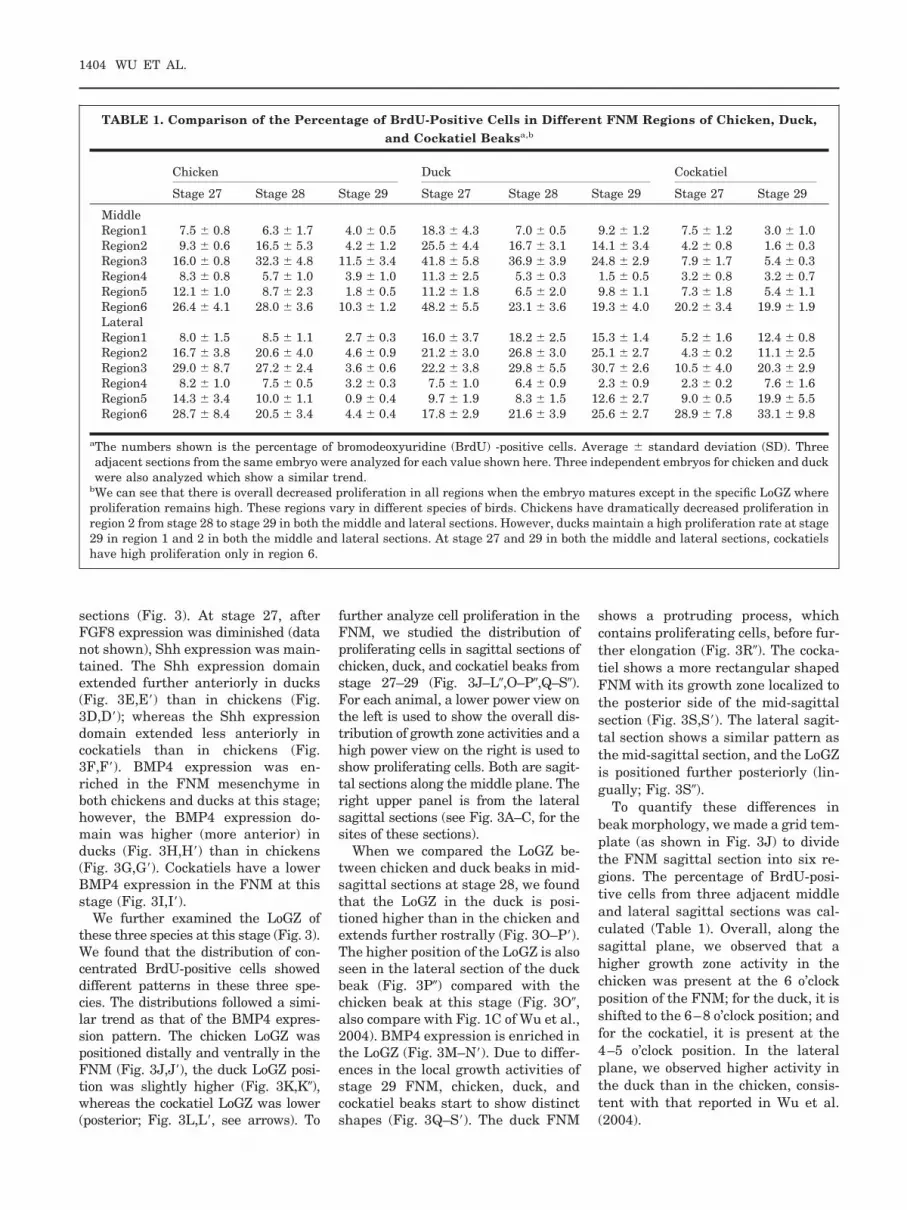
To quantify these differences inbeak morphology, we made a grid tem-plate (as shown in Fig. 3J) to dividethe FNM sagittal section into six re-gions. The percentage of BrdU-posi-tive cells from three adjacent middleand lateral sagittal sections was cal-culated (Table 1). Overall, along thesagittal plane, we observed that ahigher growth zone activity in thechicken was present at the 6 o’clockposition of the FNM; for the duck, it isshifted to the 6–8 o’clock position; andfor the cockatiel, it is present at the4–5 o’clock position. In the lateralplane, we observed higher activity inthe duck than in the chicken, consis-tent with that reported in Wu et al.(2004).
TABLE 1. Comparison of the Percentage of BrdU-Positive Cells in Different FNM Regions of Chicken, Duck,and Cockatiel Beaksa,b
Chicken Duck Cockatiel
Stage 27 Stage 28 Stage 29 Stage 27 Stage 28 Stage 29 Stage 27 Stage 29
MiddleRegion1 7.5 � 0.8 6.3 � 1.7 4.0 � 0.5 18.3 � 4.3 7.0 � 0.5 9.2 � 1.2 7.5 � 1.2 3.0 � 1.0Region2 9.3 � 0.6 16.5 � 5.3 4.2 � 1.2 25.5 � 4.4 16.7 � 3.1 14.1 � 3.4 4.2 � 0.8 1.6 � 0.3Region3 16.0 � 0.8 32.3 � 4.8 11.5 � 3.4 41.8 � 5.8 36.9 � 3.9 24.8 � 2.9 7.9 � 1.7 5.4 � 0.3Region4 8.3 � 0.8 5.7 � 1.0 3.9 � 1.0 11.3 � 2.5 5.3 � 0.3 1.5 � 0.5 3.2 � 0.8 3.2 � 0.7Region5 12.1 � 1.0 8.7 � 2.3 1.8 � 0.5 11.2 � 1.8 6.5 � 2.0 9.8 � 1.1 7.3 � 1.8 5.4 � 1.1Region6 26.4 � 4.1 28.0 � 3.6 10.3 � 1.2 48.2 � 5.5 23.1 � 3.6 19.3 � 4.0 20.2 � 3.4 19.9 � 1.9LateralRegion1 8.0 � 1.5 8.5 � 1.1 2.7 � 0.3 16.0 � 3.7 18.2 � 2.5 15.3 � 1.4 5.2 � 1.6 12.4 � 0.8Region2 16.7 � 3.8 20.6 � 4.0 4.6 � 0.9 21.2 � 3.0 26.8 � 3.0 25.1 � 2.7 4.3 � 0.2 11.1 � 2.5Region3 29.0 � 8.7 27.2 � 2.4 3.6 � 0.6 22.2 � 3.8 29.8 � 5.5 30.7 � 2.6 10.5 � 4.0 20.3 � 2.9Region4 8.2 � 1.0 7.5 � 0.5 3.2 � 0.3 7.5 � 1.0 6.4 � 0.9 2.3 � 0.9 2.3 � 0.2 7.6 � 1.6Region5 14.3 � 3.4 10.0 � 1.1 0.9 � 0.4 9.7 � 1.9 8.3 � 1.5 12.6 � 2.7 9.0 � 0.5 19.9 � 5.5Region6 28.7 � 8.4 20.5 � 3.4 4.4 � 0.4 17.8 � 2.9 21.6 � 3.9 25.6 � 2.7 28.9 � 7.8 33.1 � 9.8
aThe numbers shown is the percentage of bromodeoxyuridine (BrdU) -positive cells. Average � standard deviation (SD). Threeadjacent sections from the same embryo were analyzed for each value shown here. Three independent embryos for chicken and duckwere also analyzed which show a similar trend.
bWe can see that there is overall decreased proliferation in all regions when the embryo matures except in the specific LoGZ whereproliferation remains high. These regions vary in different species of birds. Chickens have dramatically decreased proliferation inregion 2 from stage 28 to stage 29 in both the middle and lateral sections. However, ducks maintain a high proliferation rate at stage29 in region 1 and 2 in both the middle and lateral sections. At stage 27 and 29 in both the middle and lateral sections, cockatielshave high proliferation only in region 6.
1404 WU ET AL.

Modulation of the OverallGrowth of MultipleProminences Leads toChanges of Beak Size
The avian beak is made of multipleprominences (Fig. 4A). We are inter-ested to know how these prominencescoordinate to form a distinct beakshape (Fig. 4B). We designed experi-ments to overexpress BMP4 in indi-vidual beak prominences, or overallthroughout the beak, to test for mor-phogenetic changes. First, we testedthe effect of the replication-competentavian sarcoma (RCAS) viral vector onbeak growth. When RCAS-LacZ wasinjected in all of the beak promi-nences, there were no observable ef-fects on beak growth (data not shown).Then, we tested the exogeneous geneexpression in a single prominence.When we overexpressed certain genesin a single prominence, we hoped thatthe overexpressed exogenous genewould remain restricted to that prom-inence and not infect other promi-nence(s). To test this theory, we in-jected RCAS LacZ in one prominence(right MXP) at stage 23. After 5 daysincubation, X-gal staining showedthat the �-galactosidase–positive cellswere restricted to the right edge of theupper mouth (Fig. 4C), which is de-rived from the right MXP. We theninjected RCAS noggin only in the rightLNP and right MXP. After 48-hr incu-bation, we examined noggin RNA dis-tribution by whole-mount in situ hy-bridization. The result showed thatexogenous noggin transcripts wereonly found in the right LNP and rightMXP (Fig. 4D). When RCAS nogginwas injected to the MDP, ectopicnoggin expression was restricted tothe lower beak (Fig. 4E). Comparedwith the endogenous noggin tran-scripts in the MDP, exogenous nogginis expressed to much higher levelsin the mesenchyme (compare Fig. 4E,E�). Furthermore, AMV-3C2 stain-ing showed that the distribution ofRCAS had a similar pattern as theexogenous noggin expression (com-pare Fig. 4E,E�). These results sug-gest that RCAS, being replicationcompetent, spreads the exogenousgene within but not beyond the in-jected prominence.
It was suggested that beak sizecould be determined by the level of
BMP pathway activity (Wu et al.,2004). Overexpression of BMP4 causedovergrowth of the beak when the viruswas injected in all of the beak promi-nences (Wu et al., 2004). Enlargedpremaxilla cartilage in the maxillaand Meckel’s cartilage in the mandi-ble were observed after BMP4 overex-pression. When a BMP antagonist,noggin, was used to neutralize endog-enous BMP activity, “mini beaks” withreduced cartilage formation and aminiaturized skeleton formed (Wu etal., 2004). Analyses of tissue sectionsfrom these specimens show BMP4-en-hanced cell proliferation and skeletaldifferentiation, whereas noggin re-duced cell proliferation and reducedskeletal differentiation (supplementof Wu et al., 2004). Increases in beakwidth and length are mainly due toincreases in skeletal mass as shownby beak cross-sections. Alcian blue andAlzarin red staining show that the skel-etal elements increase or decrease pro-portionally. These results suggest thatglobal misexpression of BMP4 or nogginin all beak prominences causes an over-all increase or reduction of the beaksize, without changing skeletal pat-terns. However, local delivery in selec-tive prominences or in part of one prom-inence will lead to changes in beakshape, as will be shown below.
Modulation of SelectiveProminences Leads toDifferential Growth ThatAlters Beak Shapes
Beak shapes vary among species (e.g.,some birds have relatively large upperor lower beaks). As the beak is madefrom multiple prominences, the differ-ential growth of different prominencesmay contribute to the distinct mor-phologies. To identify the role of eachcomponent in beak formation, we mi-croinjected RCAS virus to individualbeak prominences. Misexpression ofBMP4 in the FNM alone (Fig. 4F, n �4) caused an elongated upper beak(15.5 � 0.4 mm, average � SD) com-pared with the control (14.5 � 0.3 mm;P � 0.01). The lower beaks are of nor-mal size. Injection to both MXPsformed a wider upper beak (Fig. 4F,n � 4, 5.7 � 0.4 mm; control, 3.9 � 0.3mm; P � 0.001). If BMP was misex-pressed only in the MDP, a largerlower beak formed (Fig. 4F, n � 8, P �
0.01, 13.7 � 0.5 mm; control, 12.1 �0.4 mm), but the upper beak formednormally. Injection to all upper beakprominences (FNM, LNPs, and MXPs)produced a larger upper beak (Fig. 4F,n � 9, 16.0 � 0.4 mm vs. control,14.5 � 0.3 mm; P � 0.001) and a nor-mal lower beak.
In contrast, injection of Noggin tothe FNM alone produced a shorter up-per beak (Fig. 4F, n � 5, 10.9 � 1.0mm vs. control, 13.1 � 0.4 mm; P �0.001), but a normal lower beak. In-troduction of RCAS noggin to bothMXPs formed a narrower upper beak(Fig. 4F, n � 3, 3.4 � 0.2 mm vs.control, 3.9 � 0.3 mm; P � 0.01). Se-lective expression in the MDP sup-pressed lower beak formation (Fig. 4F,n � 4, 8.1 � 1.2 mm vs. control, 12.1 �0.4 mm; P � 0.001). Expression in theFNM, LNP, and MXP almost com-pletely inhibited the upper beak butleft the lower beak intact (Fig. 4F, n �10, P � 0.001). Therefore, differentialgrowth in different prominences canalter beak shapes by changing differ-ent dimensions.
Position of LoGZ and BeakCurvature
In nature, some birds exhibit upper orlower beaks with different degrees ofcurvature in the dorsoventral or evenleft–right plane. We surmise thatasymmetric growth can contribute tothis shaping mechanism. To test therole of growth zone activity in the for-mation of beak curvature, we microin-jected BMP/noggin to the lower FNMregion. However, the resolution of thistechnique is limited, as RCAS spreadswithin prominences but not betweenprominences in the time frame of ourexperiments. Therefore, we used theupper beak composed of multipleprominences to test the role of theBMP pathway in beak curvature. Weinjected RCAS-BMP4 into the rightupper beak (including the right side ofthe FNM, right LNP, and right MXP).The results showed that growth on theright side of the upper beak predomi-nated, causing the beak to curve to-ward the left (Fig. 4G). Injection withRCAS-noggin had the opposite effectand the beak curved toward the right(Fig. 4G). When RCAS-BMP4 viralmedia was diluted (1, 1:5, 1:25),growth on the right side of the upper
MORPHOREGULATION OF AVIAN BEAKS 1405
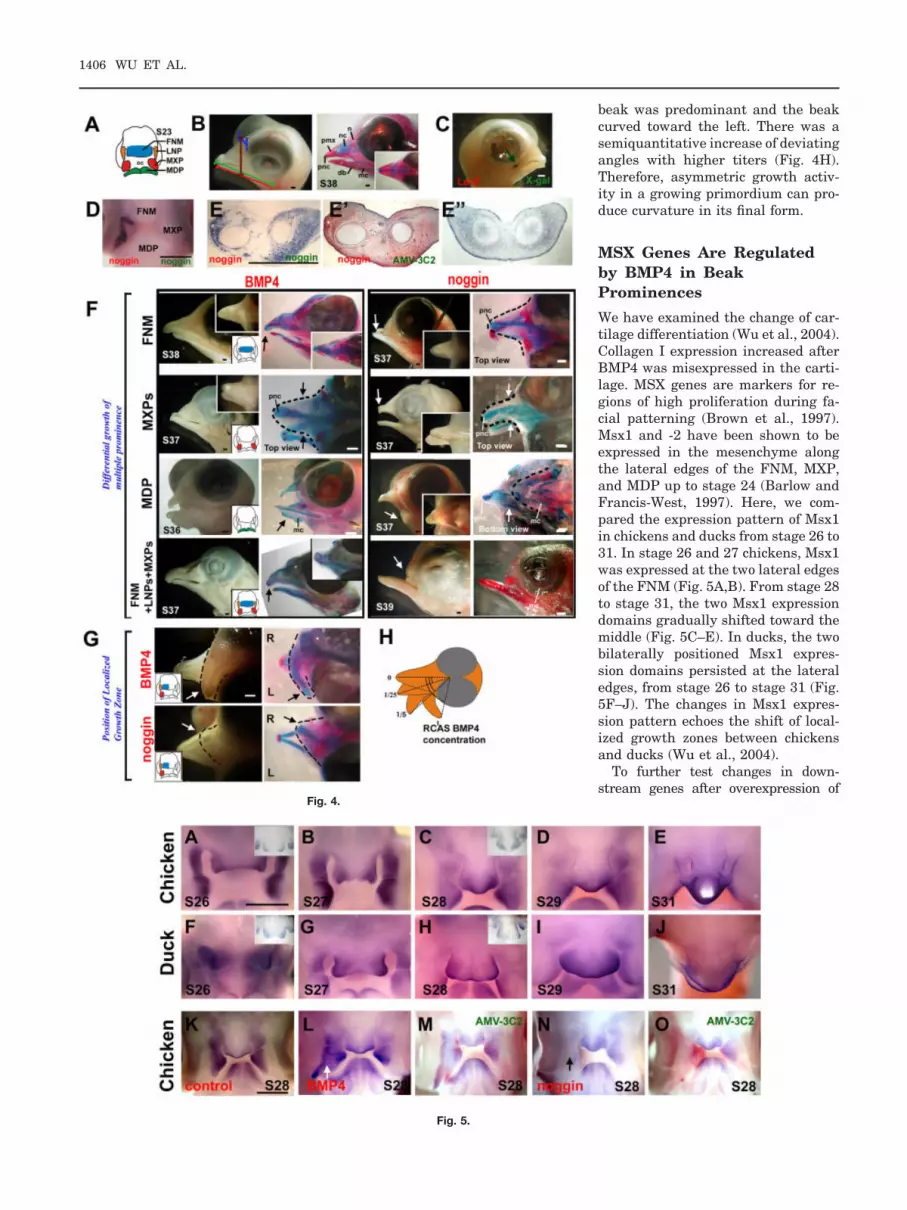
beak was predominant and the beakcurved toward the left. There was asemiquantitative increase of deviatingangles with higher titers (Fig. 4H).Therefore, asymmetric growth activ-ity in a growing primordium can pro-duce curvature in its final form.
MSX Genes Are Regulatedby BMP4 in BeakProminences
We have examined the change of car-tilage differentiation (Wu et al., 2004).Collagen I expression increased afterBMP4 was misexpressed in the carti-lage. MSX genes are markers for re-gions of high proliferation during fa-cial patterning (Brown et al., 1997).Msx1 and -2 have been shown to beexpressed in the mesenchyme alongthe lateral edges of the FNM, MXP,and MDP up to stage 24 (Barlow andFrancis-West, 1997). Here, we com-pared the expression pattern of Msx1in chickens and ducks from stage 26 to31. In stage 26 and 27 chickens, Msx1was expressed at the two lateral edgesof the FNM (Fig. 5A,B). From stage 28to stage 31, the two Msx1 expressiondomains gradually shifted toward themiddle (Fig. 5C–E). In ducks, the twobilaterally positioned Msx1 expres-sion domains persisted at the lateraledges, from stage 26 to stage 31 (Fig.5F–J). The changes in Msx1 expres-sion pattern echoes the shift of local-ized growth zones between chickensand ducks (Wu et al., 2004).
To further test changes in down-stream genes after overexpression of
Fig. 4.
Fig. 5.
1406 WU ET AL.

BMP4 or noggin, we injected the virusinto the right LNP and right MXP atstage 23. After 48-hr incubation, whole-mount Msx1 in situ hybridization wasperformed. When BMP4 was overex-pressed, we found an increased Msx1expression domain (Fig. 5L). When weoverexpressed noggin, we found a de-creased Msx1 expression domain (Fig.5N). Section in situ hybridization ofMsx1 in the mandible indicated thatBMP4 increased Msx1 expression,whereas noggin decreased it (data notshown). This result suggested thatMsx1’s activity is regulated by the BMPpathway. The colocalization of the BMPdownstream gene Msx1 and the prolif-eration activity in the FNM are consis-tent with the notion that BMP signal-ling is critical for beak morphogenesis.Our data are consistent with the previ-ous studies that demonstrated that ec-topic application of BMP protein-coatedbeads can activate MSXs gene expres-sion in the developing facial primordia(Barlow and Francis-West, 1997; Wanget al., 1999b; Ashique et al., 2002a;Mina et al., 2002).
DISCUSSION
Multicomponent, Multi-LoGZMorphogenesis of the Beakand the Generation ofDiverse Beak Forms
All bird beaks are made of the samedifferentiation materials (e.g., bone,
horny sheath, soft tissues), but theyform diverse shapes in different spe-cies. The different shapes are based ondifferent topobiologically arranged cel-lular activities. By varying the propor-tion of the width, depth, and length,the architecture of the beak is estab-lished. Here, we used a biological ap-proach to examine how the varied dis-tribution of cell proliferation activityduring beak morphogenesis of differ-ent bird species contributes to differ-ent beak morphologies.
The beak is made from the coordi-nated growth of multiple facial prom-inences. We suggest that the followingtwo categories of growth activitiescombine to alter beak shape: (1) Con-certed overall growth activities thatoccur in all prominences are responsi-ble for the global expansion and,therefore, the overall size of the beak;(2) Differential growth activities insome prominences or in part of a sin-gle prominence can contribute to al-tering the relative dimensions and,therefore, the shape of the beak. Asthese represent localized growth, wecalled these LoGZ (please see the in-troduction for further discussion onthe definition of LoGZ). This conceptcan be applied to different levels ofgrowth. One is at the interprominencelevel, i.e., the varied growth rates ofmultiple facial primordia in differentspecies. For example, at stage 27, theratio of the MXP/FNM is larger in the
duck than in the chicken (Wu et al.,2004). At stage 29, the relative size ofthe FNM to MXP and MDP is larger inthe duck. At E14, the ratio of the MDPto the maxilla is much smaller in thecockatiel than in the chicken and theduck. The other level is intrapromi-nence. Strategically positioned LoGZfine-tune the specific shapes of eachprominence. In the working model(Fig. 6), each facial prominence canhave its distinct distribution of LoGZ,and the number, size, and activitylevel of LoGZ within the same promi-nence in different birds can also bedifferent. Thus, the diverse beak mor-phology of each bird is derived from aunique combination of the above threecategories of growth activities actingat different hierarchical levels duringprominence morphogenesis (Fig. 6A–C).
To think about how specific organforms, such as diverse beak shapes,are “designed” from the genome, oneusually thinks about specific molecu-lar coding. This process seems to beunrealistic when we face endlessforms in nature. New concepts areneeded. Weiss (2005) suggests thatdevelopment uses a “phenogenetic”logic, using a few simple relationalprinciples to build complex traits. Therole of genes is in the process ratherthan the specifics of the trait, much asa variety of complex music can bemade by playing different tones on the
Fig. 4. Modulation of beak shapes by perturbing the BMP pathway activity. Replication-competent avian sarcoma (RCAS) -BMP4 or noggin viruseswere microinjected to either all or selective prominences. A: Schematic drawing of a stage 23 chicken beak. B: Normal embryonic day (E) 12 chickenhead. Left panel, measurement of upper beak length (green), lower beak length (red), depth (brown), and width (blue) performed according to Wu etal. (2004). Right panel, Alcian blue (cartilage) and Alzarin red (bone) staining to show different skeletal elements. C: Limited viral spread when virus ismicroinjected into a single prominence as determined by X-gal staining of RCAS-LacZ injected beak prominences. D: Whole-mount in situhybridization for noggin in RCAS-noggin injected LNP and MXP. E: Gene misexpression is verified by noggin expression on the coronal sections ofthe forming mandible. E�, distribution of virus detected by AMV-3C2 immunohistochemical staining. E�, endogenous noggin expression in a controlchicken mandible. F,G: RCAS-mediated gene misexpression. Chicken embryos injected at stage 23 were harvested at the indicated stages. Theinjected prominences are colored in the diagram within the inset. Side (F) and top (G) views are presented. The left column is a whole-mount view, andthe right column shows Alcian blue/Alzarin red staining. Resulting enlargements (black arrows) or reductions (white arrows) of beak sizes are indicated.F: Microinjection of BMP4 and noggin to selected maxillary prominences (FNM, MXP, and so on). G: Injection of RCAS BMP4 or RCAS noggin to theright maxilla. Arrows, curved beaks. H: Semiquantitative correlation of the degree of curvature and the titer of RCAS-BMP4 virus. BMP4, bonemorphogenetic protein 4; db, dentary bone; FNM, frontal nasal mass; L, left; LNP, lateral nasal prominence; LoGZ, localized growth zones; mc,Meckel’s cartilage; MDP, Mandibular prominence; MXP, maxillary prominences; n, nasal bone; nc, nasal chonchae; pnc, prenasal cartilage; pmx,premaxilla bone. R, right. RCAS, replication-competent avian sarcoma. Scale bars � 0.5 mm.
Fig. 5. Expression of Msx1 in chicken and duck beaks. Whole-mount in situ hybridization. A–E: Msx1 expression in normal stage 26–31 chicken beaks.Inset showing the section in situ hybridization. F�–J: Msx1 expression in normal duck beaks from equivalent stages. Inset shows a section in situhybridization. Note the Msx1-positive zones in chicken merge from two at stage 27 to one at stage 28; whereas that of duck remains as two at stage28. The larger FNM in the duck is obvious at stage 29 and 30. K–O: Expression of Msx1 after Replication competent avian sarcoma (RCAS) -bonemorphogenetic protein 4 (BMP4) and RCAS-noggin injection into the right MXP of chicken embryos. Stage 28 control (K), RCAS-BMP4 (L),RCAS-BMP4/virus staining (M), RCAS-noggin (N) and RCAS-noggin/virus staining (O). In situ hybridization is shown in blue. The presence of virus isrevealed by antibody AMV-3C2 and shown in red. Scale bar � 1 mm.
MORPHOREGULATION OF AVIAN BEAKS 1407

same instrument. Our recent work onthe mapping of feather stem cellsshows that the different relative posi-tioning of stem cells and transient am-plifying cells (TA cells in ectodermalorganogenesis terminology; they arealso defined by a short BrdU labelingand are equivalent to the LoGZ here)can lead to different feather morphol-ogies. Specifically, stem cells are con-figured as a ring, positioned horizon-tally in the radially symmetric downyfeathers but tilted anteriorly in bilat-erally symmetric feathers (Yue et al.,2005). An anterior–posterior Wnt 3agradient is critical for forming the bi-lateral but not radial symmetry offeathers (Yue et al., 2006). Thus, wefurther developed the original conceptof topobiology (Edelman, 1988) to sug-gest that topologically positioned mo-lecular activities and cell behavior canbe used to generate diverse organforms (Chuong et al., 2006). In thisway, subtle differences in the stem cellring configuration (e.g., by simple tilt-ing) can lead to a continuum of radialto bilateral symmetry in feathers.Similarly, the timing and positioningof multiple localized growth activitiesat different hierarchical levels in fa-cial primordia morphogenesis canhave profound consequences in themaking and fine-tuning of facial mor-phology. Thus, instead of searchingfor a molecular blueprint, the moreflexible and pleiomorphic concept ofphenogenetics and topobiology shouldbe developed further between ge-nomes and organ forms.
Because there are five beak promi-nences comprising the upper beak, thenumber of possible ways that beakshapes can be modulated are enor-mous. However, this complex morpho-genesis process is also prone to errorsas seen in the high incidence of cleftpalate/lips due to a lack of coordina-tion of cellular events (MacDonald etal., 2004). When all beak prominencesincrease their growth activities glo-bally, a large beak formed: the beaksize increased proportionally main-taining the overall shape. When onlycertain prominences enlarge, the beakassumes a new shape with altered rel-ative dimensions (Fig. 6, top row, B).We also found that the curvature ofupper beaks correlates with the posi-tion of the LoGZ within the FNM. Dif-ferent degrees of curvature formed
due to asymmetric growth that devi-ates from the original proximal–distalaxis (Fig. 6C).
Molecular Basis of GrowthActivities That PatternDifferent Beak Shapes
What mediates the growth activitiesin beak morphogenesis? BMPs wereshown to be expressed during aviancraniofacial morphogenesis (Francis-West et al., 1998). At stage 20, BMP4is expressed in the epithelium, but theexpression is gradually switched tothe mesenchyme from stage 24 to 28.BMP2 is more diffusely expressed inthe epithelium from stage 20 to 28
(Francis-West et al., 1994). Ectopic de-livery of BMP2 and BMP4 protein-coated beads cause a thickened or bi-furcated cartilage to form, dependingon the position of the beads (Barlowand Francis-West, 1997). The impor-tance of the BMP pathway in cranio-facial skeleton morphogenesis is fur-ther demonstrated by RCAS deliveryof constitutive and dominant-negativeBMP receptors, which lead to over-growth or suppression of cartilagegrowth, respectively (Ashique et al.,2002b). BMP- and Noggin-coated beadstudies show the homeostasis of BMPactivity is important in epithelial–mesenchymal interactions in the FNMand can regulate mesenchymal growth
Fig. 6. Working model for the shaping of the beak. Schematic chicken, duck, and cockatiel beaksare shown in the top row. A: Multiple prominences (coronal plane) have to be integrated to form onebeak. The basic configurations of chicken, duck and cockatiel beaks are different. Ducks havebigger FNM and MXP. Cockatiels have a smaller MDP. B: Frontal views show different numbers andpositions of LoGZ in chickens (one in the midline) and ducks (two bilateral LoGZ and a central LoGZform a diffuse growth zone) in the FNM. The cockatiel growth zone is in the bottom. Thesedifferences lead to the wider duck beaks. C: Sagittal views show different dorso-ventral positionsof LoGZ in the FNM. This could lead to vectorial differences in growth activity resulting in curvedoutgrowth. This asymmetric growth mechanism may be used to generate curved beaks as seen inthe dorsal–ventral curved upper beaks (no curvature in the duck, less curvature in the chicken,more curvature in either the cockatiel or eagle), or left–right curved beaks. D, dorsal; L, left; R, right;V, ventral. D: Growth activity can be regulated by BMP and/or other pathways. Here we showBMP4 pathway activity enhances beak growth by increasing cell proliferation and differentiation,whereas noggin suppression of BMP activity decreased proliferation and differentiation.
1408 WU ET AL.

and epithelial survival (Ashique et al.,2002a). These experimental resultsshow that the BMP pathway plays apivotal role in early stages of craniofa-cial morphogenesis.
Recently, it was found that the BMPpathway also plays an important rolein shaping the beaks in late stagemorphogenesis (Abzhanov et al., 2004;Wu et al., 2004). Mesenchymal BMP4transcript expression is found to par-allel that of the LoGZ (Wu et al.,2004). RCAS-mediated misexpressionof BMP4 in the beak prominence mes-enchyme causes proportional enlarge-ment, whereas RCAS noggin causesproportional reduction of beak size(Abzhanov et al., 2004; Wu et al.,2004). Specific application of BMP4protein-coated beads to the position ofthe LoGZ could increase beak width,therefore, changing the shape of thebeak (Wu et al., 2004). That we ob-tained different altered skeletal phe-notypes from those reported before(Barlow and Francis-West, 1997), byleaving BMP4 beads in different posi-tions, further highlights the wholeconcept of LoGZ and topobiology(Chuong et al., 2006). Together these,results suggest that, in the late stagesof craniofacial morphogenesis whenthe morphology of the beak is deter-mined, the size of the beak can bemodulated by the overall activity ofthe BMP pathway, which mediates itsgrowth. Beak shape can be fine-tunedby localized BMP activity, which is in-volved in mediating the range, level,and duration of the LoGZ activity.
The key question to the generationof beak diversity is how LoGZ areinduced and positioned. Whereaselegant transplantation experimentsshowed that the shapes of duck or quailbeaks are determined by the cephaliccrest (Schneider and Helms, 2003), theinformation residing in the crest mesen-chyme still has to be executed. At stage20, the interface of the FGF8 and Shhexpression domains was shown to bethe growth point of the FNM and wastermed the frontonasal ectodermal zone(FEZ; Hu et al., 2003). Misexpression ofShh around stage 20 can duplicate theFNM and form duplicated beaks (Huand Helms, 1999). This experimentmay be explained by duplication ofLoGZ, but more experiments will be re-quired to test this hypothesis. Retinoicacid-coated and noggin-coated beads
were shown to convert the MXP to theFNM (Lee et al., 2001). This findingmay occur because the experimentalconditions create differential growth ac-tivity and form an ectopic LoGZ in theMXP. It is possible that the early epi-thelial FEZ may directly or indirectlylead to the induction of the mesenchy-mal LoGZ in later stages (at and afterstage 26). This finding needs to be stud-ied further. The mechanism of howLoGZ are positioned is critical as it willlink our understanding of earlier mor-phogenesis events to the late beak mor-phogenesis events.
In different birds, the ectodermalexpression of FGF8 and Shh in theFNM show differences in their timingand locations. In the chicken, FGF8was expressed at the upper part of theectodermal frontonasal zone and mayfunction to induce cartilage formation(Hu et al., 2003; Abzhanov and Tabin,2004). Its expression disappeared inthis region after stage 20. On theother hand, ducks still express FGF8in the FNM at stage 23 (Fig. 2D). Wealso found that both chickens andducks have an epidermis proliferationzone distributed below the middle hor-izontal line of the two nasal pits atstage 20 (Fig. 2B). This epidermis pro-liferation zone is distributed diffuselyin chickens but restrictively to the twolateral sides in ducks (Fig. 2B). Thus,we suggest that the persistent expres-sion of FGF8 may contribute to thedistinct morphogenesis of the duckbeak, causing it to grow longer, wider,and produce more cartilage.
We found that the anterior border ofthe Shh expression domain correlateswith the position of the LoGZ. If wecompare the position of the anteriorpoint of Shh expression among ducks,chickens, and cockatiels at stage 27(Fig. 3D–F�), we find the pattern isduck � (more anterior than) chicken �cockatiel. In comparing different birds,we noticed the position of the LoGZ inthe mesenchyme is located beneath theanterior point of the Shh expression do-main in the epidermis. These findingsare consistent with the assumption thatthe position of the anterior border of theShh expression domain may determinethe position of the LoGZ and, therefore,the curvature of the beaks. Examina-tion of the BMP4 expression domainalso showed it to be higher (more ante-rior) in ducks than in chickens (Fig. 3G–
I�, M–N�). Thus, specific growth activi-ties in the FNM of different birds arelikely to be defined by a unique combi-nation of FGF, Shh, BMP4, and others.Obviously, more work is required to un-derstand how these interactions func-tion together to regulate growth activi-ties.
Origin and Evolution ofAvian Beaks
Beaks are the hardened hornysheaths formed on the snout. Morpho-genesis of the beak consists of threemajor components: the outgrowth ofbeak primordial mesenchyme (skele-ton), the integument inside the oralcavity (oral mucosa, teeth), and theintegument covering the snout (hornysheath). Indeed, beak-like structuresalso existed in some ancient dinosaurs(e.g., Psittacosaurus) as well as in cur-rent turtles. It is also possible thatbeak-like structures may have evolvedindependently more than once. Inbirds, the beak became a unique feed-ing apparatus in Mesozoic times(many species are illustrated in Houet al., 2003) and its diverse shapes areclassic examples of evolution (Darwin,1859; Grant, 1986). Accompanying thechanges of beak shapes is the forma-tion of the horny sheath and the loss ofteeth. However, a latent response to-ward tooth formation remains andtooth-like appendages can be inducedfrom the flat oral epithelia by alteringthe molecular signalling in the mesen-chyme (Kollar and Fisher, 1980; Chenet al., 2000; Mitsiadis et al., 2003).
At the macro-scale, the appearanceof novel beak shapes and accompany-ing new trophisms can be appreciatedfrom the fossil record of Mesozoicbirds. The approximately 150-million-year-old Archaeopteryx had jaws withboth dentary and premaxillary teeth(reviewed in Fedducia, 1999). The re-cent discoveries of fossils from the120- to 130-million-year-old JeholBiota of Northeastern China have in-creased our knowledge of reptile/avianevolution enormously (Zhou et al.,2003). This finding is because thesefossils are exceptionally well pre-served, including integuments such asearly feathers (Chuong et al., 2003).Confuciusornis is the first late Juras-sic bird found in the Jehol Biota. Itexhibited a toothless beak (Hou et al.,
MORPHOREGULATION OF AVIAN BEAKS 1409

1995), more advanced than Archae-opteryx. It also has clawed wings andprobably lived in an arboreal habitat,picking worms and fruits with its con-ical shaped beak. Recently, Longiro-stravis, the earliest known wadingbird fossil, was found in the same area(Hou et al., 2004). The unique featureis that its beak is preferentially elon-gated, in comparison to other dimen-sions of the beak. In addition, thereare small dentary and premaxillaryteeth restricted to the distal end of thebeak. This long toothed beak mighthave functioned well in capturing andholding worms and fishes, facilitatingthe evolution of probe feeding behav-ior and may have provided a newwaterfront niche for Longirostravis.These are good examples of how mod-ulation of beak shape can help the evo-lution of avian trophic diversification(Zweers et al., 1997). Other mesozoicbirds in this region show even morediverse beak shapes, most with teeth(Hou et al., 2003). The rapid radiationof bird evolution is reflected in the di-verse beaks shaped for probing, catch-ing, slicing, browsing, and so on (Gill,1994; Zweers et al., 1997). The diver-sity of beak shapes in the Mesozoicbirds suggests that beaks of differentsizes and shapes flourished to competefor the new niches during the late Cre-tacious–early Tertiary period.
At the micro-scale of beak morpho-logical evolution, Darwin’s finchesprovide the most inspiring examples.A group of 14 species was first col-lected by Charles Darwin during hisvisit to the Galapagos Islands (Dar-win, 1859). These finches proved to bea monophyletic group (Sato et al.,1999) and was considered to have orig-inated from an ancestral species thatreached the Galapagos Archipelagofrom Central or South America ap-proximately 2.3 million years ago(Sato et al., 2001). The beak shapevaried among these finches accordingto their diet (Grant, 1986). The widthand depth of beaks fluctuate rapidly inresponse to environmental changes. Along-term study illustrated that natu-ral-selection constantly affected beakshape in the medium ground finch(Geospiza fortis; Grant and Grant,2002). This finding shows that ran-dom or directional selection affects thesize of beaks (Grant and Grant, 2002).Although these works explained the
reason Galapagos finches have di-verse beak shapes and how they re-spond to the environment, we do notknow which molecular pathways wereinvolved and how these shapes wereachieved.
Recently, Tabin’s group started tostudy the molecular differences in thedeveloping beaks of different Galapa-gos finch species. Examinations offinch embryos showed a correlationbetween BMP4 expression levels andthe thickness of the developing beaks(Abzhanov et al., 2004). From theseand other results, we learn that BMP4is involved in building beaks. There-fore, BMP pathway members, ago-nists and antagonists, may work asmolecular candidates to alter proto-typical molecular modules and gener-ate a spectrum of morphologies fornatural selection. Our experimentalstudy with chickens showed that wecould indeed produce beaks pheno-copying those in nature by modulatingdifferent developmental steps (Wu etal., 2004).
In conclusion, multicomponent com-plex morphogenesis such as the beakrequires more coordination during de-velopment, but also provides opportu-nities where developmental mecha-nisms can be easily modulated togenerate beak shape diversity. Bycomparing beak morphogenesis pro-cesses in birds with characteristicbeak shapes, we learn new perspec-tives on how nature fine-tunes mor-phogenesis. Regulation of this processremains the biggest challenge. Wenow know that the BMP4 pathway isa major player in beak morphogenesisand can start by studying moleculesrelated to this pathway.
EXPERIMENTALPROCEDURES
Animals
Pathogen-free fertilized chick eggs(White Leghorn) were purchased fromSPAFAS (Preston, CT). Fertilized Pe-king duck eggs were from a local farm(AA Farms, Westminster, CA). Chickenembryos were staged according to Ham-burger and Hamilton (1951). Duck em-bryos take longer (approximately 28days) to hatch than chicken embryos(20 days), and staging was assessed bya combination of morphological criteria
including the limbs, eyes, body folds,and flexures (Schneider and Helms,2003). Cockatiel embryos (stage 27, n �1; stage 29, n � 1; stage 35, n � 2; E14,n � 2) and newborns (n � 2) were ob-tained from a local colony. We tried toapproximate duck and cockatiel stagesas close to chicken H&H staging(Schneider and Helms, 2003; Wu et al.,2004) as possible. However, there aredifferent levels of heterochrony amongspecies. Chickens and ducks belong toprecocious birds that are born withfeathers and can feed by themselves.Cockatiels belong to altricial birds thatare born with very few downy featherson the body and no feathers at all on thehead. They need their parents to feedthem (Gill, 1994). Cockatiel embryosneed 18–21 days to hatch (Harris,1992). Because their staging system isnot well established, we used embryonicday and morphological features in thedeveloping head to choose embryos ofsimilar developing stages for compari-son.
Tissue Processing,Immunocytochemistry, andSEM
Paraffin sections were prepared fromchicken, duck, and cockatiel samples.In situ hybridization and immunohis-tochemistry were performed as de-scribed (Jiang et al., 1998), and somewere performed using the automatedVentana Discovery system. X-galstaining was performed according toYu et al. (2002). Cartilage and bonewere stained as described by Monsoro-Burq et al. (1994) using Alcian blueand Alizarin red, respectively. ForSEM, heads were fixed in half-strength Karnovsky’s fixative over-night at 4°C and post-fixed in thio-carbohydrazide, followed by osmiumfixation. The specimens were criticalpoint-dried and coated with gold pal-ladium. They were viewed with a Hi-tachi S-570 scanning microscope.
Mapping Cell ProliferativeZones
BrdU labeling was done by injecting 5�l of 1% BrdU (Sigma) in DMEM intothe vein of chicken embryos. Embryoswere incubated for 1.5 hr before har-vesting. After BrdU staining, slideswere lightly counterstained with he-
1410 WU ET AL.

matoxylin to view all cells. Antibodiesto BrdU were from Chemicon. Thewhole-mount BrdU method was per-formed according to Chodankar et al.(2003). BrdU-positive cells are quanti-fied in different regions of the FNM inboth middle and lateral sections (asshown in Fig. 3A–C). The distance be-tween the middle and lateral sectionsis approximately 160–200 �m, exceptfor cockatiel stage 27 (approximately100 �m). We divided the FNM into sixregions. The grid overlay for these re-gions is shown in Figure 3J. A verticalline was first drawn from the grooveinside the mouth until it touched thebrain. A horizontal line was thendrawn. This horizontal line betweenthe intersection point and the facialectoderm was separated into equalparts by drawing a second vertical linedownward to the bottom of the FNM.This second vertical line was furtherdivided into three equal parts by twohorizontal lines. Thus, there are twocolumns and three rows. In this way,the FNM is divided into six regions. Ineach region, the percentage of BrdU-positive cells among all mesenchymalcells was calculated. For each number,three adjacent sections from the sameembryo were counted and averagedwith the standard deviation shown.
Virus Infection, In SituHybridization
RCAS retrovirus preparation and insitu hybridization were performed us-ing protocols as described (Jiang et al.,1998). Virus was microinjected atE3.5–E4 (stage 22–23) into individualor multiple beak prominence(s). Em-bryo heads were lifted using a spe-cially designed micro-spoon. Facialprominences were identified under adissection microscope. Virus (2 �l)was injected to beak prominences inovo. Embryos were harvested fromstage 28 to 39. For detection of virus,samples after whole-mount in situ hy-bridization were subjected to whole-mount immunostaining with AMV-3C2 antibody (Potts et al., 1987).
Morphometry of the Beak
Beak width, depth, and length weremeasured as shown in Figure 4B.Width (blue line) and depth (brownline) of the beaks were measured at
the plane along the frontal margin ofthe eyes. The length of the upper beakwas measured from the distal tip ofthe maxilla to the end of the quadra-tojugal bone (green line). The length ofthe mandible was determined fromthe distal tip of the mandible to theend of the dentary bone (red line). AStudent’s t-test was used to comparethe variation between experimentaland control beaks.
ACKNOWLEDGMENTSWe thank all Chuong lab members fordiscussions and technical help. Wethank Dr. David Hinton of the DohenyEye Institute for his support withSEM. We also thank the following in-vestigators for their viral vectors andin situ hybridization probes: RCASLacZ was originally constructed byDr. Li Yi (Dunn et al., 2001) and pro-vided to us by Dr. W.-P. Wang; RCAS-BMP4 and in situ hybridizationprobes were from P. Francis-West(Duprez et al., 1996); RCAS nogginwas from R. Johnson (Capdevila andJohnson, 1998); and probes for Msx1and 2 were from W.B. Upholt (Mina etal., 1995). Both C.M.C. and R.W. werefunded by NIH.
REFERENCES
Abzhanov A, Tabin CJ. 2004. Shh and Fgf8act synergistically to drive cartilage out-growth during cranial development. DevBiol 273:134–148.
Abzhanov A, Protas M, Grant BR, GrantPR, Tabin CJ. 2004. Bmp4 and morpho-logical variation of beaks in Darwin’sfinches. Science 305:1462–1465.
Ashique AM, Fu K, Richman JM. 2002a.Endogenous bone morphogenetic pro-teins regulate outgrowth and epithelialsurvival during avian lip fusion. Devel-opment 129:4647–4660.
Ashique AM, Fu K, Richman JM. 2002b.Signalling via type IA and type IB bonemorphogenetic protein receptors (BMPR)regulates intramembranous bone forma-tion, chondrogenesis and feather forma-tion in the chicken embryo. Int J Dev Biol46:243–253.
Barlow AJ, Francis-West PH. 1997. Ec-topic application of recombinant BMP-2and BMP-4 can change patterning of de-veloping chick facial primordia. Develop-ment 124:391–398.
Brown JM, Robertson KE, Wedden SE,Tickle C. 1997. Alterations in Msx 1 andMsx 2 expression correlate with inhibi-tion of outgrowth of chick facial primor-dia induced by retinoic acid. Anat Em-bryol (Berl) 195:203–207.
Capdevila J, Johnson RL. 1998. Endoge-nous and ectopic expression of nogginsuggests a conserved mechanism for reg-ulation of BMP function during limb andsomite patterning. Dev Biol 197:205–217.
Chen Y, Zhang Y, Jiang TX, Barlow AJ, StAmand TR, Hu Y, Heaney S, Francis-West P, Chuong CM, Maas R. 2000. Con-servation of early odontogenic signalingpathways in Aves. Proc Natl Acad Sci US A 97:10044–10049.
Chiappe LM. 1995. The first 85 millionyearsofavianevolution.Nature378:349–355.
Chodankar R, Chang CH, Yue ZC, JiangTX, Suksaweang S, Burrus LW, ChuongCM, Widelitz RB. 2003. Shift of localizedgrowth zones contributes to skin append-age morphogenesis: role of the Wnt/�-catenin pathway. J Invest Dermatol 120:20–26.
Chuong CM, Wu P, Zhang FC, Xu X, Yu M,Widelitz RB, Jiang TX, Hou L. 2003. Ad-aptation to the sky: defining the featherwith integument fossils from mesozoicChina and experimental evidence frommolecular laboratories. J Exp ZoologPart B Mol Dev Evol 298:42–56.
Chuong C-M, Wu P, Plikus MV, Jiang TX,Widelitz RB. 2006. Engineering Stemcells into organs: topobiological transfor-mations demonstrated by beak, featherand other ectodermal organ morphogen-esis. Curr Top Dev Biol 72:237–274.
Couly G, Grapin-Botton A, Coltey P, RuhinB, Le Douarin NM. 1998. Determinationof the identity of the derivatives of thecephalic neural crest: incompatibility be-tween Hox gene expression and lowerjawdevelopment.Development125:3445–3459.
Darwin C. 1859. On the origin of species: afacsimile of the first edition. Cambridge:Harvard University Press. 1975.
Depew MJ, Lufkin T, Rubenstein JL. 2002.Specification of jaw subdivisions by Dlxgenes. Science 298:381–385.
Dunn KJ, Incao A, Watkins-Chow D, Li Y,Pavan WJ. 2001. In utero complementa-tion of a neural crest-derived melanocytedefect using cell directed gene transfer.Genesis 30:70–76.
Duprez D, Bell EJ, Richardson MK, ArcherCW, Wolpert L, Brickell PM, Francis-West PH. 1996. Overexpression ofBMP-2 and BMP-4 alters the size andshape of developing skeletal elements inthe chick limb. Mech Dev 57:145–157.
Edelman GM. 1988. Topobiology. NewYork: Basic Books.
Feduccia A. 1999. The origin and evolutionof birds. 2nd ed. New Haven: Yale Uni-versity Press. p 93–137.
Francis-West PH, Tatla T, Brickell PM.1994. Expression patterns of the bonemorphogenetic protein genes Bmp-4 andBmp-2 in the developing chick face sug-gest a role in outgrowth of the primordia.Dev Dyn 201:168–178.
Francis-West P, Ladher R, Barlow A,Graveson A. 1998. Signalling interac-tions during facial development. MechDev 75:3–28.
MORPHOREGULATION OF AVIAN BEAKS 1411
Gill FB. 1994. Ornithology. 2nd ed. NewYork: Freeman.
Grant PR. 1986. Ecology and evolution ofDarwin’s finches. Princeton: PrincetonUniversity Press. p 1–492.
Grant PR, Grant BR. 2002. Unpredictableevolution in a 30-year study of Darwin’sfinches. Science 296:707–711.
Hamburger V, Hamilton HL. 1951. A se-ries of normal stages in the developmentof the chicken embryo. J Morphol 88:49–92.
Harris JC. 1992. Cockatiels: gettingstarted. Neptune City, NJ: T.F.H. Publi-cations, Inc. p 1–96.
Helms JA, Schneider RA. 2003. Cranialskeletal biology. Nature 423:326–331.
Hou LH, Zhou ZH, Martin LD, Feduccia A.1995. A beaked bird from the Jurassic ofChina. Nature 377:616–618.
Hou LH, Chiappe L, Zhang FC, ChuongCM. 2004. New early cretaceous fossilfrom China documents a novel trophicspecialization for Mesozoic birds. Natur-wissenschaften 91:22–25.
Hou LH, Chuong CM, Yang A, Zeng XL,Hou JF. 2003. Fossil birds of China. Chi-na: Yunnan Science and Technology.
Hu D, Helms JA. 1999. The role of sonichedgehog in normal and abnormal cranio-facial morphogenesis. Development 126:4873–4884.
Hu D, Marcucio RS, Helms JA. 2003. Azone of frontonasal ectoderm regulatespatterning and growth in the face. Devel-opment 130:1749–1758.
Jiang TX, Stott S, Widelitz RB, ChuongCM. 1998. Current methods in the studyof avian skin appendages. In: ChuongCM, editor. Molecular basis of epithelialappendage morphology. Austin: LandesCo. p 395–408.
Kollar EJ, Fisher C. 1980. Tooth inductionin chick epithelium: expression of quies-cent genes for enamel synthesis. Science207:993–995.
LaBonne C, Bronner-Fraser M. 1999. Mo-lecular mechanisms of neural crest for-
mation. Annu Rev Cell Dev Biol 15:81–112.
Le Douarin NM, Kalcheim C. 1999. Theneural crest. 2nd ed. Cambridge: Cam-bridge University Press.
Lee SH, Fu KK, Hui JN, Richman JM.2001. Noggin and retinoic acid transformthe identity of avian facial prominences.Nature 414:909–912.
MacDonald ME, Abbott UK, Richman JM.2004. Upper beak truncation in chickenembryos with the cleft primary palatemutation is due to an epithelial defect inthe frontonasal mass. Dev Dyn 230:335–349.
Mina M, Gluhak J, Upholt WB, Kollar EJ,Rogers B. 1995. Experimental analysisof Msx-1 and Msx-2 gene expression dur-ing chick mandibular morphogenesis.Dev Dyn 202:195–214.
Mina M, Wang YH, Ivanisevic AM, UpholtWB, Rodgers B. 2002. Region- and stage-specific effects of FGFs and BMPs inchick mandibular morphogenesis. DevDyn 223:333–352.
Mitsiadis TA, Cheraud Y, Sharpe P, Fon-taine-Perus J. 2003. Development ofteeth in chick embryos after mouse neu-ral crest transplantations. Proc NatlAcad Sci U S A 100:6541–6545.
Monsoro-Burq AH, Bontoux M, Teillet MA,Le Douarin NM. 1994. Heterogeneity inthe development of the vertebra. ProcNatl Acad Sci U S A 91:10435–10439.
Noden DM. 1988. Interactions and fates ofavian craniofacial mesenchyme. Devel-opment 103(Suppl):121–140.
Potts WM, Olsen M, Boettiger D, Vogt VM.1987. Epitope mapping of monoclonalantibodies to gag protein p19 of aviansarcoma and leukaemia viruses. J GenVirol 68:3177–3182.
Richman JM, Lee SH. 2003. About face:signals and genes controlling jaw pat-terning and identity in vertebrates.Bioessays 25:554–568.
Robledo RF, Rajan L, Li X, Lufkin T. 2002.The Dlx5 and Dlx6 homeobox genes are
essential for craniofacial, axial, and ap-pendicular skeletal development. GenesDev 16:1089–1101.
Sato A, O’hUigin C, Figueroa F, Grant PR,Grant BR, Tichy H, Klein J. 1999. Phy-logeny of Darwin’s finches as revealed bymtDNA sequences. Proc Natl Acad Sci US A 96:5101–5106.
Sato A, Tichy H, O’hUigin C, Grant PR,Grant BR, Klein J. 2001. On the origin ofDarwin’s finches. Mol Biol Evol 18:299–311.
Schneider RA, Helms JA. 2003. The cellu-lar and molecular origins of beak mor-phology. Science 299:565–568.
Wang W-P, Widelitz RB, Jiang T-X,Chuong C-M. 1999a. Msx-2 and the reg-ulation of organ size: epidermal thick-ness and hair length. J Invest Dermatol4:278–281.
Wang YH, Rutherford B, Upholt WB, MinaM. 1999b. Effects of BMP-7 on mousetooth mesenchyme and chick mandibu-lar mesenchyme. Dev Dyn 216:320–335.
Weiss KM. 2005. The phenogenetic logic oflife. Nat Rev Genet 6:36–45.
Wu P, Jiang TX, Suksaweang S, WidelitzRB, Chuong CM. 2004. Molecular shap-ing of the beak. Science 305:1466–1467.
Yu MK, Wu P, Widelitz RB, Chuong CM.2002. The morphogenesis of feathers.Nature 420:308–312.
Yue Z, Jiang T-X, Widelitz RB, ChuongCM. 2005. Mapping stem cell activitiesin the feather follicle. Nature 438:1026–1029.
Yue Z, Jiang T-X, Widelitz RB, ChuongCM. 2006. Wnt 3a gradient converts ra-dial to bilateral feather symmetry viatopological arrangement of epithelia.Proc Natl Acad Sci U S A 103:951–955.
Zhou ZH, Barrett PM, Hilton J. 2003. Anexceptionally preserved Lower Creta-ceous ecosystem. Nature 421:807–814.
Zweers GA, Vanden Berge JC, BerkhoudtH. 1997. Evolutionary pattern of aviantrophic diversification. Zoology 100:25–57.
1412 WU ET AL.


