Embed Size (px)
Citation preview

Asian Pacific Journal of Cancer Prevention, Vol 11, 2010 1099
CAV-1 Gene Mutational Profile in Breast Cancers in Ethnic Kashmiris
Asian Pacific J Cancer Prev, 11, 1099-1105
Introduction
Breast cancer is the third most common malignancy in the world (Sipetić et al., 2000) with more than 1 million women diagnosed with breast cancer each year (Parkin et al., 2002). Breast cancer has been associated with a variety of risk factors genetic and epigenetic changes (Kaaks et al., 2005; Xie et al., 2006). But its molecular pathogenesis remains unresolved. Environmental carcinogens have been shown to damage DNA at active fragile sites by disrupting surveillance, which has been shown to be tumorigenic (Gasser et al., 2006; Artl et al., 2003). Development of human breast cancers is a multistep process, arising from genetic alterations that drive the transformation of normal mammary epithelial cells into highly malignant derivatives (Hanahan et al., 2000).
Historically, caveolin was first discovered approximately 10 years ago as a major v-Src substrate in Rous sarcoma virus–transformed cells, as the principal structural component of caveolae, identified as a 21- to 24-kDa integral membrane protein and termed caveolin (Glenney et al., 1989; Rithberg et al., 1992) now referred to as caveolin-1 (Scherer et al., 1996). Caveolin-1 is the principal structural component of caveolae micro domains, which represent a sub-compartment of the plasma membrane, Caveolin 1 (CAV1) gene maps to 7q31.1 and encodes a 21- to 24-kDa integral membrane
Departments of 1Immunology and Molecular Medicine, 2Clinical Biochemistry, 3General Surgery, Sher-I-Kashmir Institute of Medical Sciences, Soura, Srinagar, Kashmir, 4Biosciences, Jamia Millia Islamia, New Delhi, India. *For correspondence: [email protected]
Abstract
The role of caveolae and the caveolin proteins in cancer has been the subject of extensive research. It has been suggested that Caveolin-1 may contribute to certain steps of carcinogenesis in various types of cancer. Therefore in our study we focused on the potential clinical relevance of Caveolin-1 in 130 malignant breast tissue specimens along with their adjacent normal tissues. Using allele specific PCR we were able to rule out the mutation status of all the samples and then we did the conventional PCR-SSCP and sequencing of the mutated samples along with the normal adjacent tissues. Caveolin-1 was identified in a screen for genes involved in breast cancer progression and we demonstrated 29.2% mutational status in our Kashmiri ethnic population. We were able to detect 38 mutations out of which 22 were missense, 4 were nonsense, and 12 were frame shifts. Ten novel Cav-1 mutations (missense and frameshift). We conclude that the gene encoding CAV-1 plays an important role in the promotion of mammary tumorigenesis in Kashmir.
Keywords: Breast cancer - Cav-1 - mutation - AS-PCR - PCR-SSCP - Kashmir
RESEARCH COMMUNICATION
Mutational Profile of the CAV-1 Gene in Breast Cancer Cases in the Ethnic Kashmiri PopulationNidda Syeed1,2,3, 4, Syed Akhtar Husain4, Safiya Abdullah1, A Syed Sameer1, Nissar A Chowdri3, Mahoor S Nanda3, Mushtaq A Siddiqi1*
protein. Caveolin interacts directly with heterotrimeric guanine nucleotide binding proteins (G proteins) and can functionally regulate their activity (Li et al., 1995). Modification and/or inactivation of caveolin-1 expression appear to be a common feature of the transformed phenotype. Three members of the caveolin family (Cav-1, -2, and -3) are essential for the formation of caveolae, Cav-1 is the principal structural protein of caveolae membranes that are found in most cells types, including mammary epithelial cells. Human CAV1 gene is a suspected tumor suppressor locus (7q31.1/D7S522) that is deleted in a variety of human cancers, as well as mammary tumors. Mutations in the gene encoding CAV-1 are associated with the development and progression of breast cancers. Conflicting results on the role of CAV1 in human cancers have been reported (Lee SW et al., 1998; Yang et al., 1998; Engelman et al., 1999; Hurlstone et al., 1999; Hayashi et al., 2001; Lee H et al., 2002; Hnasko et al., 2003; Zou et al., 2003; Chen et al., 2004; Jones et al., 2004; Sagara et al., 2004; Charafe-Jauffret et al., 2005; Park et al., 2005; Williams et al., 2005; Pinilla et al., 2006; Van den Eynden et al., 2006). Based on the high frequency of deletions of 7q31 (a fragile site known as FRA7G) in human cancers (Nishizuka et al., 1997; Huang et al., 1999; Han et al., 2003), presence of CAV1 gene promoter hypermethylation (Engelman et al., 1999; Chen et al., 2004), inactivating gene mutations (Hayashi et al., 2001; Chen et al., 2004),
Nidda Syeed et al
Asian Pacific Journal of Cancer Prevention, Vol 11, 20101100
and the apparent reduction of CAV1 expression in breast carcinomas (Chen et al., 2004; Park et al., 2005), it has been suggested that CAV1 is a potential tumor suppressor gene (Razani et al., 2001; Hnasko et al., 2003; Williams et al., 2005). Several independent lines of evidence support the notion that caveolin-1 functions as a suppressor of cell transformation. The action of Cav-1 as a tumor suppressor is based on its capacity to inhibit the signaling activity of several proto-oncogene products that impart growth and survival advantages to the cell (Macleod et al., 1999). The reduction in Cav-1 and caveolae appears to be a common event in transformed cell lines, suggesting that Cav-1 might be “inactivated” during tumorigenesis (Engelman et al., 1998; Razani et al., 2000). The loss of Cav-1 disrupts cellular adhesion and consequently can either induce apoptosis when proper cell cycle checkpoints are in place or promote metastasis when synergistically combined with uncontrolled cell growth.
Recent researchers have suggested that sequestration of several growth-promoting proteins in invaginations of the plasma membrane, named caveolae, serves to potently inhibit their activity (Cohen et al., 2004). Cav-1 functions as a general negative regulator to inhibit the basal activity of many pro-proliferative and oncogenic proteins. Mechanistically, Cav-1 tonically inhibits the activation of multiple signaling molecules, including EGF-R, ERK-1/2, MEK-1/2, and the TGF-_ type I receptor, by direct binding via the caveolin-scaffolding domain (Couet et al., 1997; Engelman et al., 1998; Galbiati et al., 1998; Razani et al., 2001). Cav-1 mRNA and protein levels are down-regulated or absent in primary human cancers in oncogenically transformed NIH 3T3 cells, in transgenic mouse models of breast cancer, as well as in several human and mouse transformed mammary epithelial cell lines (Koleske et al., 1995; Racine et al., 1999; Bagnoli et al., 2000; Bender et al., 2000; Park et al., 2001). In tumors, caveolin-1, the structural protein of caveolae, constitutes a key switch through its function as a tumor suppressor and a promoter of metastases. Despite important advances in chemotherapy, radiotherapy, and surgery, approximately 30% of patients with breast cancer will relapse and die of the disease. Therefore, complementary therapeutic strategies should be considered for improving the outcome of breast cancer patients.
Materials and Methods
Patients and Tumor Tissue Procurement All patients included in the study were both male and
female, with the histopathological diagnosis of the breast. The patient participation was obtained through informed consent and after approval from the Ethics Committee of Sher-i-Kashmir Institute of Medical Sciences.
A cohort of 130 breast cancer tissue samples were collected consisting of tumor tissues and adjacent normal tissue. Only the tissue samples confirmed by histopathological studies to be cancerous were included in the study.
DNA IsolationGenomic DNA was extracted from tissue samples and
peripheral blood samples of breast cancer patients using DNA Extraction Kit (Qiagen, USA). The quality of the resulting genomic DNA was stringently assessed by low percentage agarose gel electrophoresis.
AS-PCR Analysis A strategy to quickly detect the CAV-1 mutations was
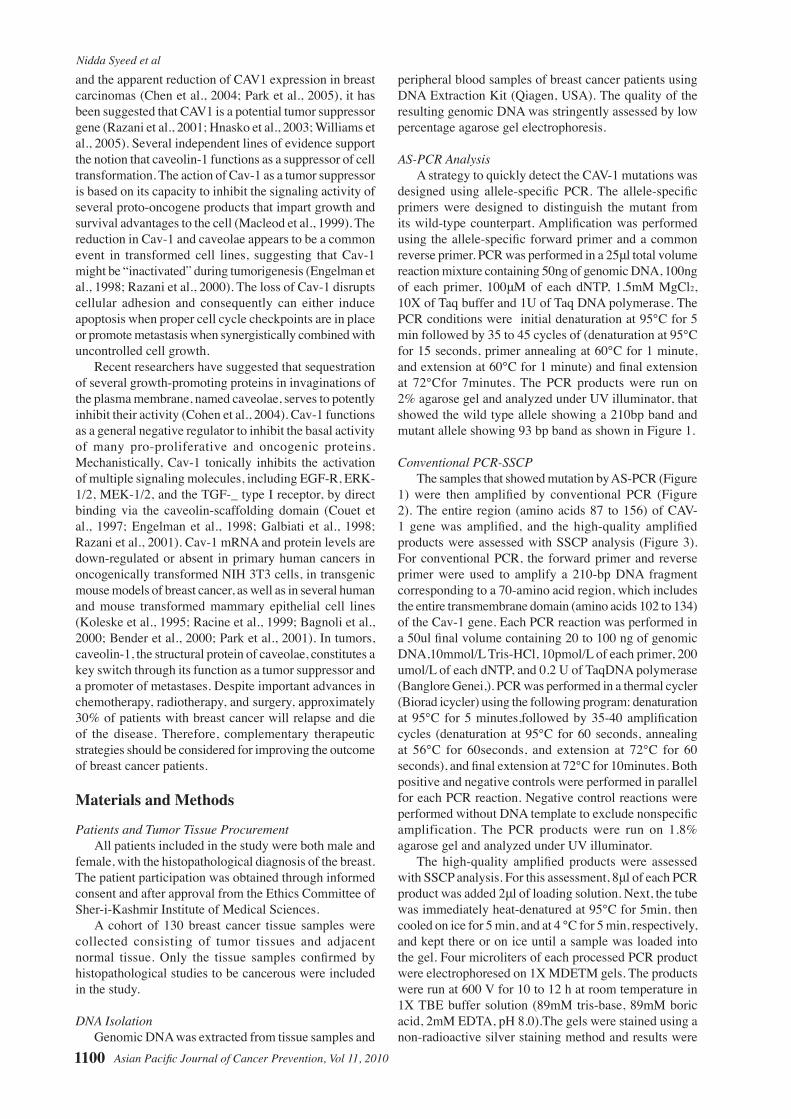
designed using allele-specific PCR. The allele-specific primers were designed to distinguish the mutant from its wild-type counterpart. Amplification was performed using the allele-specific forward primer and a common reverse primer. PCR was performed in a 25μl total volume reaction mixture containing 50ng of genomic DNA, 100ng of each primer, 100μM of each dNTP, 1.5mM MgCl2, 10X of Taq buffer and 1U of Taq DNA polymerase. The PCR conditions were initial denaturation at 95°C for 5 min followed by 35 to 45 cycles of (denaturation at 95°C for 15 seconds, primer annealing at 60°C for 1 minute, and extension at 60°C for 1 minute) and final extension at 72°Cfor 7minutes. The PCR products were run on 2% agarose gel and analyzed under UV illuminator, that showed the wild type allele showing a 210bp band and mutant allele showing 93 bp band as shown in Figure 1.
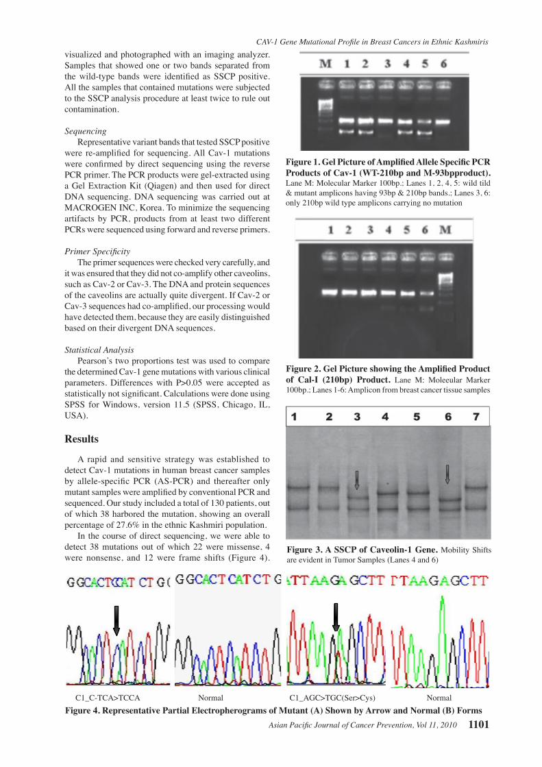
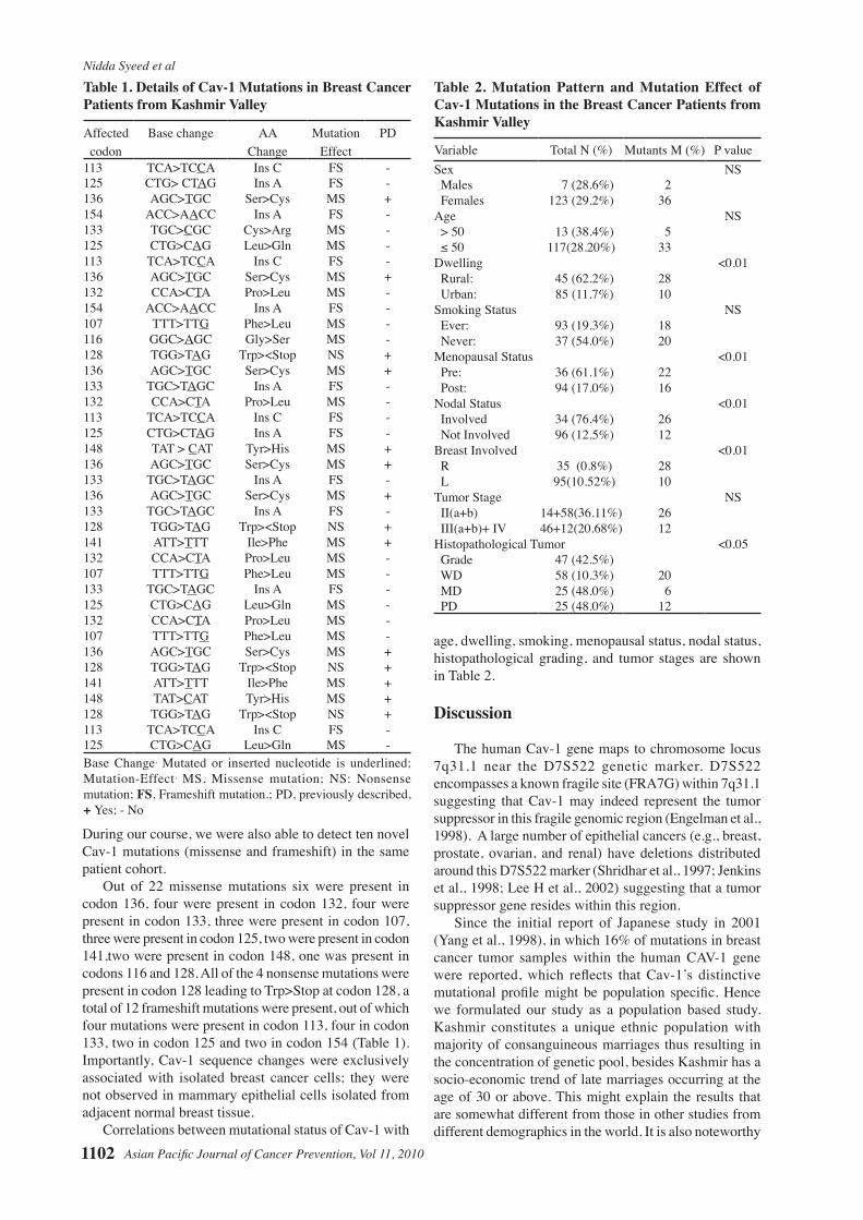
Conventional PCR-SSCPThe samples that showed mutation by AS-PCR (Figure
1) were then amplified by conventional PCR (Figure 2). The entire region (amino acids 87 to 156) of CAV-1 gene was amplified, and the high-quality amplified products were assessed with SSCP analysis (Figure 3). For conventional PCR, the forward primer and reverse primer were used to amplify a 210-bp DNA fragment corresponding to a 70-amino acid region, which includes the entire transmembrane domain (amino acids 102 to 134) of the Cav-1 gene. Each PCR reaction was performed in a 50ul final volume containing 20 to 100 ng of genomic DNA,10mmol/L Tris-HCl, 10pmol/L of each primer, 200 umol/L of each dNTP, and 0.2 U of TaqDNA polymerase (Banglore Genei,). PCR was performed in a thermal cycler (Biorad icycler) using the following program: denaturation at 95°C for 5 minutes,followed by 35-40 amplification cycles (denaturation at 95°C for 60 seconds, annealing at 56°C for 60seconds, and extension at 72°C for 60 seconds), and final extension at 72°C for 10minutes. Both positive and negative controls were performed in parallel for each PCR reaction. Negative control reactions were performed without DNA template to exclude nonspecific amplification. The PCR products were run on 1.8% agarose gel and analyzed under UV illuminator.
The high-quality amplified products were assessed with SSCP analysis. For this assessment, 8μl of each PCR product was added 2μl of loading solution. Next, the tube was immediately heat-denatured at 95°C for 5min, then cooled on ice for 5 min, and at 4 °C for 5 min, respectively, and kept there or on ice until a sample was loaded into the gel. Four microliters of each processed PCR product were electrophoresed on 1X MDETM gels. The products were run at 600 V for 10 to 12 h at room temperature in 1X TBE buffer solution (89mM tris-base, 89mM boric acid, 2mM EDTA, pH 8.0).The gels were stained using a non-radioactive silver staining method and results were
Asian Pacific Journal of Cancer Prevention, Vol 11, 2010 1101
CAV-1 Gene Mutational Profile in Breast Cancers in Ethnic Kashmiris
0
25.0
50.0
75.0
100.0
New
ly d
iagn
osed
with
out
trea
tmen
t
New
ly d
iagn
osed
with
tre
atm
ent
Pers
iste
nce
or r
ecur
renc
e
Rem
issi
on
Non
e
Chem
othe
rapy
Radi
othe
rapy
Conc
urre
nt c
hem
orad
iatio
n
10.3
0
12.8
30.025.0
20.310.16.3
51.7
75.051.1
30.031.354.2
46.856.3
27.625.033.130.031.3
23.738.0
31.3
visualized and photographed with an imaging analyzer. Samples that showed one or two bands separated from the wild-type bands were identified as SSCP positive. All the samples that contained mutations were subjected to the SSCP analysis procedure at least twice to rule out contamination.
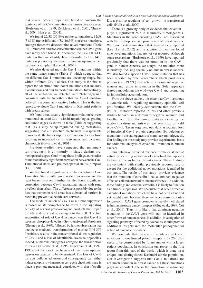
SequencingRepresentative variant bands that tested SSCP positive
were re-amplified for sequencing. All Cav-1 mutations were confirmed by direct sequencing using the reverse PCR primer. The PCR products were gel-extracted using a Gel Extraction Kit (Qiagen) and then used for direct DNA sequencing. DNA sequencing was carried out at MACROGEN INC, Korea. To minimize the sequencing artifacts by PCR, products from at least two different PCRs were sequenced using forward and reverse primers.
Primer SpecificityThe primer sequences were checked very carefully, and
it was ensured that they did not co-amplify other caveolins, such as Cav-2 or Cav-3. The DNA and protein sequences of the caveolins are actually quite divergent. If Cav-2 or Cav-3 sequences had co-amplified, our processing would have detected them, because they are easily distinguished based on their divergent DNA sequences.
Statistical AnalysisPearson’s two proportions test was used to compare
the determined Cav-1 gene mutations with various clinical parameters. Differences with P>0.05 were accepted as statistically not significant. Calculations were done using SPSS for Windows, version 11.5 (SPSS, Chicago, IL, USA).
Results
A rapid and sensitive strategy was established to detect Cav-1 mutations in human breast cancer samples by allele-specific PCR (AS-PCR) and thereafter only mutant samples were amplified by conventional PCR and sequenced. Our study included a total of 130 patients, out of which 38 harbored the mutation, showing an overall percentage of 27.6% in the ethnic Kashmiri population.
In the course of direct sequencing, we were able to detect 38 mutations out of which 22 were missense, 4 were nonsense, and 12 were frame shifts (Figure 4).
Figure 3. A SSCP of Caveolin-1 Gene. Mobility Shifts are evident in Tumor Samples (Lanes 4 and 6)
Figure 1. Gel Picture of Amplified Allele Specific PCR Products of Cav-1 (WT-210bp and M-93bpproduct). Lane M: Molecular Marker 100bp.; Lanes 1, 2, 4, 5: wild tild & mutant amplicons having 93bp & 210bp bands.; Lanes 3, 6: only 210bp wild type amplicons carrying no mutation
Figure 2. Gel Picture showing the Amplified Product of Cal-I (210bp) Product. Lane M: Moleeular Marker 100bp.; Lanes 1-6: Amplicon from breast cancer tissue samples
C1_C-TCA>TCCA Normal C1_AGC>TGC(Ser>Cys) Normal Figure 4. Representative Partial Electropherograms of Mutant (A) Shown by Arrow and Normal (B) Forms
Nidda Syeed et al
Asian Pacific Journal of Cancer Prevention, Vol 11, 20101102
During our course, we were also able to detect ten novel Cav-1 mutations (missense and frameshift) in the same patient cohort.
Out of 22 missense mutations six were present in codon 136, four were present in codon 132, four were present in codon 133, three were present in codon 107, three were present in codon 125, two were present in codon 141,two were present in codon 148, one was present in codons 116 and 128. All of the 4 nonsense mutations were present in codon 128 leading to Trp>Stop at codon 128, a total of 12 frameshift mutations were present, out of which four mutations were present in codon 113, four in codon 133, two in codon 125 and two in codon 154 (Table 1). Importantly, Cav-1 sequence changes were exclusively associated with isolated breast cancer cells; they were not observed in mammary epithelial cells isolated from adjacent normal breast tissue.
Correlations between mutational status of Cav-1 with
Table 1. Details of Cav-1 Mutations in Breast Cancer Patients from Kashmir Valley
Affected Base change AA Mutation PD codon Change Effect113 TCA>TCCA Ins C FS -125 CTG> CTAG Ins A FS -136 AGC>TGC Ser>Cys MS +154 ACC>AACC Ins A FS -133 TGC>CGC Cys>Arg MS -125 CTG>CAG Leu>Gln MS -113 TCA>TCCA Ins C FS -136 AGC>TGC Ser>Cys MS +132 CCA>CTA Pro>Leu MS -154 ACC>AACC Ins A FS -107 TTT>TTG Phe>Leu MS -116 GGC>AGC Gly>Ser MS -128 TGG>TAG Trp><Stop NS +136 AGC>TGC Ser>Cys MS +133 TGC>TAGC Ins A FS -132 CCA>CTA Pro>Leu MS -113 TCA>TCCA Ins C FS -125 CTG>CTAG Ins A FS -148 TAT > CAT Tyr>His MS +136 AGC>TGC Ser>Cys MS +133 TGC>TAGC Ins A FS -136 AGC>TGC Ser>Cys MS +133 TGC>TAGC Ins A FS -128 TGG>TAG Trp><Stop NS +141 ATT>TTT Ile>Phe MS +132 CCA>CTA Pro>Leu MS -107 TTT>TTG Phe>Leu MS -133 TGC>TAGC Ins A FS -125 CTG>CAG Leu>Gln MS -132 CCA>CTA Pro>Leu MS -107 TTT>TTG Phe>Leu MS -136 AGC>TGC Ser>Cys MS +128 TGG>TAG Trp><Stop NS +141 ATT>TTT Ile>Phe MS +148 TAT>CAT Tyr>His MS +128 TGG>TAG Trp><Stop NS +113 TCA>TCCA Ins C FS -125 CTG>CAG Leu>Gln MS -Base Change, Mutated or inserted nucleotide is underlined; Mutation-Effect, MS, Missense mutation; NS: Nonsense mutation; FS, Frameshift mutation.; PD, previously described, + Yes; - No
Table 2. Mutation Pattern and Mutation Effect of Cav-1 Mutations in the Breast Cancer Patients from Kashmir Valley
Variable Total N (%) Mutants M (%) P valueSex NS Males 7 (28.6%) 2 Females 123 (29.2%) 36Age NS > 50 13 (38.4%) 5 ≤ 50 117(28.20%) 33Dwelling <0.01 Rural: 45 (62.2%) 28 Urban: 85 (11.7%) 10Smoking Status NS Ever: 93 (19.3%) 18 Never: 37 (54.0%) 20Menopausal Status <0.01 Pre: 36 (61.1%) 22 Post: 94 (17.0%) 16Nodal Status <0.01 Involved 34 (76.4%) 26 Not Involved 96 (12.5%) 12Breast Involved <0.01 R 35 (0.8%) 28 L 95(10.52%) 10Tumor Stage NS II(a+b) 14+58(36.11%) 26 III(a+b)+ IV 46+12(20.68%) 12Histopathological Tumor <0.05 Grade 47 (42.5%) WD 58 (10.3%) 20 MD 25 (48.0%) 6 PD 25 (48.0%) 12
age, dwelling, smoking, menopausal status, nodal status, histopathological grading, and tumor stages are shown in Table 2.
Discussion
The human Cav-1 gene maps to chromosome locus 7q31.1 near the D7S522 genetic marker. D7S522 encompasses a known fragile site (FRA7G) within 7q31.1 suggesting that Cav-1 may indeed represent the tumor suppressor in this fragile genomic region (Engelman et al., 1998). A large number of epithelial cancers (e.g., breast, prostate, ovarian, and renal) have deletions distributed around this D7S522 marker (Shridhar et al., 1997; Jenkins et al., 1998; Lee H et al., 2002) suggesting that a tumor suppressor gene resides within this region.
Since the initial report of Japanese study in 2001 (Yang et al., 1998), in which 16% of mutations in breast cancer tumor samples within the human CAV-1 gene were reported, which reflects that Cav-1’s distinctive mutational profile might be population specific. Hence we formulated our study as a population based study. Kashmir constitutes a unique ethnic population with majority of consanguineous marriages thus resulting in the concentration of genetic pool, besides Kashmir has a socio-economic trend of late marriages occurring at the age of 30 or above. This might explain the results that are somewhat different from those in other studies from different demographics in the world. It is also noteworthy
Asian Pacific Journal of Cancer Prevention, Vol 11, 2010 1103
CAV-1 Gene Mutational Profile in Breast Cancers in Ethnic Kashmiristhat several other groups have failed to confirm the existence of the Cav-1 mutations in human breast cancers (Hurlstone et al., 1999; Zenklusen et al., 2001; Chen et al., 2004; Han et al., 2004).
We found 22/38 (57.8%) missense mutations, 12/38 (31.5%) frameshifts and 4/38 (10.5%) nonsense mutations, amongst theses we detected nine novel mutations (Table 01). Frameshift and nonsense mutations in the Cav-1 gene have rarely been found. Furthermore, the Cav-1 (I141T) mutation that we identify here is similar to the I141F mutation previously identified in human squamous cell carcinoma samples (Han et al., 2004).
We also detected multiple Cav-1 mutations within the same tumor sample (Table 1) which suggests that the different Cav-1 mutations are occurring singly, but within different Cav-1 alleles. Our study is the first to report the identified nine novel mutations that includes five missense and four frameshift mutations. Interestingly, all of the mutations we detected were “heterozygous,” consistent with the hypothesis that the Cav-1 mutation behaves in a dominant-negative fashion. This is the first report to evaluate Cav-1 mutations in Kashmiri patients with breast cancer.
We found a statistically significant correlation between mutational status of Cav-1 with histopathological grading and tumor stages as shown in table (Table 2) suggesting that Cav-1 may be up regulated during metastasis, suggesting that a distinctive mechanism is responsible to inactivate the tumor suppressor function of caveolin-1 resulting in increased cell-invasiveness, and increased chemotaxis (Hayashi et al., 2001).
Previous studies have suggested that mammary tumorigenesis is commonly initiated during pre-menopausal stages. Confirming these findings, our studies found statistically significant correlation between the Cav-1 mutational status and pre-menopausal status (Simpson et al., 1998).
We also found a significant correlation between Cav-1 mutation Status with lymph node involvement and the right breast involved. Further we also found significant correlation between Cav-1 mutational status with rural dwellers than urban. The difference is possibly due to the fact that women in rural areas face substantial barriers in receiving preventive health care services.
The mode of action of Cav-1 as a tumor suppressor is based on its competence to restrain the signaling activity of several proto-oncogene products that impart growth and survival advantages to the cell. The first supposition of role of Cav-1 in cancer was that Cav-1 is tyrosine-phosphorylated in v-Src–transformed fibroblasts (Glenney et al., 1989), followed by further observation that oncogene-mediated transformation of murine NIH 3T3 fibroblasts results in the transcriptional down-regulation of Cav-1 and a loss of identifiable caveolae organelles. Indeed, numerous oncogenes abrogate the transcription of Cav-1 (Koleske et al., 1995; Engelman et al., 1997; 1998), but the exact mechanism of this transcriptional repression remains to be determined. The loss of Cav-1 disrupts cellular adhesion and consequently can either induce apoptosis when proper cell cycle checkpoints are in place or promote metastasis correlated with that of cyclin
D1, a positive regulator of cell growth, in transformed cells (Hulit et al., 2000).
There is a growing body of evidence that caveolin-1 plays a significant role in mammary tumorigenesis. Mutations in the gene encoding CAV-1 are associated with the development and progression of breast cancers. We found certain mutations that were already reported (Lee H et al., 2002) and in addition to them we found nine novel mutations that are not yet reported. Although some researchers (Hurlstone et al., 1999) have reported previously that there was no mutation in the CAV-1 gene in human cancers, we sought the mutation more intensively, focusing specially on human breast cancers. We also found a specific Cav-1 point mutation that has been reported by other researchers which produces a protein (i.e., P132L) that acts in a dominant negative manner and results in retention in the Golgi apparatus thereby modulating the wild-type Cav-1 and promoting its intracellular accumulation.
From the above studies, it is clear that Cav-1 assumes a dynamic role in regulating mammary epithelial cell proliferation. We, clearly demonstrate that the Cav-1 (P132L) mutation reported in this and other previous studies behaves in a dominant-negative manner, and together with the other novel mutations causing the mislocalization and intracellular retention of wild type Cav-1. Taken together, our data associates loss of functional Cav-1 protein expression (by deletion or mutation) in the pathogenesis of mammary tumorigenesis. Our findings in this study may provide experimental basis for additional analysis of caveolin-1 mutation in human cancers.
Our data have provided evidence for the existence of naturally occurring mutations of caveolin-1 that appears to have a role in human breast cancer. These findings are consistent with similar previously reported results except for the additional novel mutations identified in our study. The results of our study provides evidence that the mutation of caveolin-1 had a dominant negative effect on cell transformation and invasiveness. In addition, these findings indicate that caveolin-1 is likely to function as a tumor suppressor. We speculate that other effective caveolin-1 mutations, which we have not been identified yet, might exist, because there are other consensus sites for caveolin. CAV1 gene promoter is heavily methylated in human prostate cancer samples (Pflug et al., 1999; Cui et al., 2001). Thus, it is likely that dominant-negative mutations in the CAV1 gene will soon be identified in other forms of human cancer. In addition, investigation of signaling pathways affected by caveolins should provide additional insights into the molecular pathogenetical action of caveolae disorders.
We conclude that the overall incidence of Cav-1 mutations in our limited patient sample is 29.2%. This needs to be corroborated by future studies with a larger patient population. In conclusion our report is the first report from this part of the world, which is done on a unique and distinguished Kashmiri ethnic population. Our investigation suggests that Cav-1 mutations are not much common in breast cancer but these mutations plays an important role in the promotion of mammary
Nidda Syeed et al
Asian Pacific Journal of Cancer Prevention, Vol 11, 20101104
tumorigenesis.The mutations in the gene encoding CAV-1 are associated with the development and progression of breast cancers. However, the exact functional role of caveolin-1 still remains controversial.
Acknowledgements
The present research was not funded by any funding agency. The collection of cancer samples used in this study was supported by the Departments of Immunology and Molecular Medicines and Department of General Surgery, Sher-I-Kashmir Institute of Medical Sciences.
The authors would like to thank all the breast cancer patients who participated in the study who are responsible for the creation and maintenance of the entire group within which this study was conducted but were not involved in the current paper.
Nidda Syeed formulated, designed and performed the lab work for the study. Akhtar Husain supervised the analysis, Safiya Abdullah helped in study design, A. Syed Sameer and Mahoor S Nanda helped in the lab work. Nissar A Chowdri procured and provided the tumor samples for the study. and Mushtaq A. Siddiqi coordinated the study ,revised the manuscript and entire work was done under his supervision. All authors have read and approved the final manuscript.
References
Arlt MF, Casper AM, Glover TW (2003). Common fragile sites. Cytogenet Genome Res, 100, 92-100.
Bagnoli M, Tomassetti A, Figini M, et al (2000). Downmodulation of caveolin-1 expression in human ovarian carcinoma is directly related to alpha-folate receptor overexpression. Oncogene, 19, 4754-63.
Bender FC, Reymond MA, Bron C, et al (2000). Caveolin-1 levels are down-regulated in human colon tumors, and ectopic expression of caveolin-1 in colon carcinoma cell lines reduces cell tumorigenicity. Cancer Res, 60, 5870-8.
Charafe-Jauffret E, Ginestier C, Monville F, et al (2005). Gene expression profiling of breast cell lines identifies potential new basal markers. Oncogene, 25, 2273-84.
Chen ST, Lin SY, Yeh KT, et al (2004). Mutational, epigenetic,and expressional analyses of caveolin-1gene in breast cancers. Int JMol Med, 14, 577-82.
Cohen AW, Hnasko R, Schubert W, et al (2004). Role of caveolae and caveolins in health and disease. Physiol Rev, 84, 1341-79.
Couet J, Sargiacomo M, Lisanti MP (1997). Interaction of a receptor tyrosine kinase, EGF-R, with caveolins. Caveolin binding negatively regulates tyrosine and serine/threonine kinase activities. J Biol Chem, 272, 30429-38.
Cui J, Rohr LR, Swanson G, et al (2001). Hypermethylation of the caveolin-1 gene promoter in prostate cancer. Prostate, 46, 249-56.
Engelman JA, Chu C, Lin A, et al (1998). Caveolin-mediated regulation of signaling along the p42/44 MAP kinase cascade in vivo. A role for the caveolin-scaffolding domain. FEBS Lett, 428, 205-11
Engelman JA, Lee RJ, Karnezis A, et al (1998). Reciprocal regulation of Neu tyrosine kinase activity and caveolin-1 protein expression in vitro and in vivo. Implications for human breast cancer. J Biol Chem, 273, 20448-55.
Engelman JA, Wykoff CC, Yasuhara S, et al (1997). Recombinant
expression of caveolin-1 in oncogenically transformed cells abrogates anchorage-independent growth. J Biol Chem, 272, 16374-81.
Engelman JA, Zhang XL, Lisanti MP (1998). Genes encoding human caveolin-1 and -2 are co-localized to the D7S522 locus (7q31.1), a known fragile site (FRA7G) that is frequently deleted in human cancers. FEBS Lett, 436, 403-10.
Engelman JA, Zhang XL, Lisanti MP (1999). Sequence and detailed organization of the human caveolin-1and -2 genes located near the D7S522 locus (7q31.1).Methylation of a CpGisland in the 5’ promoter region of the caveolin-1 gene in human breast cancer cell lines. FEBS Lett, 448, 221-30.
Galbiati F, Volonte D, Engelman JA, et al (1998). Targeted downregulation of caveolin-1 is sufficient to drive cell transformation and hyperactivate the p42/44 MAP kinase cascade. EMBO J , 17, 6633-48.
Gasser S, Raulet D (2006). The DNA damage response, immunity and cancer. Semin Cancer Biol, 16, 344-7.
Glenney, J R, Jr. Tyrosine phosphorylation of a 22-kDa protein is correlated with transformation by Rous sarcoma virus. J. Biol. Chem. 264, 20163-6 (1989).
Glenney JR Jr, Zokas L (1989). Novel tyrosine kinase substrates from Rous sarcoma virus-transformed cells are present in the membrane skeleton. J Cell Biol, 108, 2401-8.
Han SE, Park KH, Lee G, et al (2004). Mutation and aberrant expression of Caveolin-1 in human oral squamous cell carcinomas and oral cancer cell lines. Int J Oncol, 24, 435-40.
Han SY, DruckT, Huebner K (2003). Candidate tumor suppressor genes at FRA7G are coamplified with MET and do not suppress malignancy in a gastric cancer. Genomics, 81, 105-7.
Hanahan D, Weinberg RA (2000). The hallmarks of cancer. Cell, 100, 57-70.
Hayashi K, Matsuda S, Machida K, et al (2001). Invasion activating caveolin-1mutation in human scirrhous breast cancers. Cancer Res, 61, 2361-4.
Hnasko R, Lisanti MP (2003). The biology of caveolae: lessons from caveolin knockout mice and implications for human disease. Mol Interv, 3, 445-64.
Huang H, Reed CP, Mordi A, et al (1999). Frequent deletions within FRA7G at 7q31.2 in invasive epithelial ovarian cancer. Genes Chromosomes Cancer, 24, 48-55.
Hulit J, Bash T, Fu M, et al (2000). The cyclin D1 gene is transcriptionally repressed by caveolin-1. J Biol Chem, 275, 21203-9.
Hurlstone AF, Reid G, Reeves JR, et al (1999). Analysis of the CAVEOLIN-1 gene at human chromosome 7q31.1 in primary tumours and tumour-derived cell lines. Oncogene, 18, 1881-90.
Jenkins RB, Qian J, Lee HK, et al (1998). A molecular cytogenetic analysis of 7q31 in prostate cancer. Cancer Res, 58, 759-66.
Jones C, MackayA, Grigoriadis A, et al (2004). Expression profiling of purified normal human luminal and myoepithelial breast cells: identification of novel prognostic markers for breast cancer. Cancer Res, 64, 3037-45.
Kaaks R, Rinaldi S, Key TJ, et al (2005). Postmenopausal serum androgens, oestrogens and breast cancer risk: the european prospective investigation into cancerand nutrition. Endocr Relat Cancer, 12, 1071-82.
Koleske AJ, Baltimore D, Lisanti MP (1995). Reduction of caveolin and caveolae in oncogenically transformed cells. Proc Natl Acad Sci USA, 92, 1381-5.
Lee H, Park DS, Razani B, et al (2002). Caveolin-1mutations (P132L and null) and the pathogenesis of breast cancer:
Asian Pacific Journal of Cancer Prevention, Vol 11, 2010 1105
CAV-1 Gene Mutational Profile in Breast Cancers in Ethnic Kashmiriscaveolin-1 (P132L) behaves in a dominant-negative manner and caveolin-1 null mice show mammary epithelial cell hyperplasia. Am J Pathol, 161, 1357-69.
Lee SW, Reimer CL, Oh P, et al (1998). Tumor cell growth inhibition by caveolin re-expression in human breast cancer cells. Oncogene, 16, 1391-7.
Li S, Okamoto T, Chun M, et al (1995). Evidence for a regulated interaction between heterotrimeric G proteins and caveolin. J Biol Chemm, 270, 15693-701.
Macleod KF, Jacks T (1999). Insights into cancer from transgenic mouse models. J Pathol, 187, 43-60.
Nishizuka S, Tamura G, Terashima M, et al (1997).Commonly deleted region on the long armof chromosome 7 in differentiated adenocarcinoma of the stomach. Br J Cancer, 76, 1567-71.
Park DS, Razani B, Lasorella A, et al (2001). Evidence that Myc isoforms transcriptionally repress caveolin-1 gene expression via an INR-dependent mechanism. Biochemistry, 40, 3354-62.
Park SS, Kim JE, KimYA, et al (2005). Caveolin-1 is down-regulated and inversely correlated with HER2 and EGFR expression status in invasive ductal carcinoma of the breast. Histopathology, 47, 625-30.
Parkin DM, Bray F, Ferlay J, et al (2005). Global cancer statistics, 2002. CA Cancer J Clin, 55, 74-108.
Pflug BR, Reiter RE, Nelson JB (1999). Caveolin expression is decreased following androgen deprivation in human prostate cancer cell lines. Prostate, 40, 269-73.
Pinilla SM, Honrado E, Hardisson D, et al (2006). Caveolin-1expression is associated with a basal-like phenotype in sporadica nd hereditary breast cancer. Breast Cancer ResTreat, 99, 85-90.
Racine C, Belanger M, Hirabayashi H, et al (1999). Reduction of caveolin 1 gene expression in lung carcinoma cell lines. Biochem Biophys Res Commun, 255, 580-6.
Razani B, Lisanti MP (2001). Caveolin-deficient mice: insights into caveolar function human disease. J Clin Invest, 108, 1553-61.
Razani B, Zhang XL, Bitzer M, et al (2001). Caveolin-1 regulates transforming growth factor (TGF)-beta/SMAD signaling through an interaction with the TGF-beta type I receptor. J Biol Chem, 276, 6727-38.
Razani B, Altschuler Y, Zhu L, et al (2000). Caveolin-1 expression is down-regulated in cells transformed by the human papilloma virus in a p53-dependent manner. Replacement of caveolin-1 expression suppresses HPV-mediated cell transformation. Biochemistry, 39, 13916-24.
Rothberg KG, Heuser JE, Donzell WC, et al (1992). Caveolin, a protein component of caveolae membrane coats. Cell, 68, 673-82.
SagaraY, Mimori K,Yoshinaga K, et al (2004). Clinical significance of caveolin-1, caveolin-2, and HER2/neu mRNA expression inhuman breast cancer. Br J Cancer, 91, 959-65.
Scherer PE, Okamoto T, Chun M, et al (1996). Identification, sequence and expression of caveolin-2 defines a caveolin gene family. Proc Natl Acad Sci U S A, 93, 131-5.
Shridhar V, Sun QC, Miller OJ, et al (1998). Loss of heterozygosity on the long arm of human chromosome 7 in sporadic renal cell carcinomas. Oncogene, 15, 2727-33.
Simpson HW, Candlish W, Pauson AW, et al (1998). Cancer is in the premenopause. Lancet, 332, 74-6.
Sipetić S, Petrović V, Milić Z, et al (2004). Breast cancer incidence among women of Branicevo region in the period 1991-2000. Med Pregl, 57, 467-72.
Van den Eynden GG,Van Laere SJ, Van derAuwera I, et al (2006). Overexpression of caveolin-1and -2 in cell lines and in human samples of inflammatory breast cancer. Breast
Cancer ResTreat, 95, 219-28.Williams TM, Lisanti MP (2005). Caveolin-1 in oncogenic
transformation, cancer, and metastasis. Am J Physiol Cell Physiol, 288, C494-506.
Xie GS, Hou AR, Li LY, et al (2006). Aberrant p16 promoter hypermethylation in bronchial mucosae as a biomarker for the early detection of lung cancer. Chin Med J, 119, 1469-72.
Yang G, Truong LD, Timme TL, et al (1998). Elevated expression of caveolin is associated with prostate and breast cancer. Clin Cancer Res, 4, 1873-80.
Zenklusen JC, Conti CJ, Green ED (2001). Mutational and functional analyses reveal that ST7 is a highly conserved tumor-suppressor gene on human chromosome 7q31. Nat Genet, 27, 392-8.
Zou W, McDaneld L, Smith LM (2003). Caveolin-1 haploin sufficiency leads to partial transformation of human breast epithelial cells. Anticancer Res, 23, 4581-6.


