Embed Size (px)
Citation preview
A
Ca
b
a
ARRAA
KTAMM
1
cit[r(doatthere
s[
Md
0d
Mutation Research 646 (2008) 1–7
Contents lists available at ScienceDirect
Mutation Research/Fundamental and MolecularMechanisms of Mutagenesis
journa l homepage: www.e lsev ier .com/ locate /molmutCommuni ty address : www.e lsev ier .com/ locate /mutres
rsenic trioxide mutational spectrum analysis in the mouse lymphoma assay
arolina Sorianoa,b, Amadeu Creusa, Ricard Marcosa,b,∗
Grup de Mutagènesi, Departament de Genètica i de Microbiologia, Edifici Cn, Universitat Autònoma de Barcelona, 08193 Bellaterra, Cerdanyola del Vallès, SpainCIBER Epidemiología y Salud Pública, ISCIII, Spain
r t i c l e i n f o
rticle history:eceived 22 February 2008eceived in revised form 24 July 2008ccepted 26 August 2008vailable online 4 September 2008
eywords:
a b s t r a c t
It has been well documented that long-term exposure to inorganic arsenic induces cancers and vasculardiseases in a dose–response relationship. Nevertheless, arsenic has also demonstrated to have anticanceractivity; thus, arsenic trioxide (ATO, As2O3) is an inorganic trivalent arsenic form, currently used in thetreatment against acute promyelocytic leukaemia (APL). The open discussion about how arsenic com-pounds induce genotoxic damage has moved us to evaluate the mutational spectrum induced by ATO inmouse lymphoma cells. Thus, 49 Tk−/− mutant colonies obtained in the mouse lymphoma assay (MLA),
k locusrsenic trioxide (ATO, As2O3)ouse lymphoma assayutational spectrum
after treatments lasting for 4 h with 10 �M ATO, and 49 spontaneous mutant colonies from independentuntreated cultures, were used to analyse and to characterise the mutational spectrum induced by thisarsenic compound, to understand its mechanism of action. RT-PCR analysis of Tk cDNA and PCR amplifi-cations of eight selected microsatellite sequences, located on chromosome 11, were used to carry out thisscreening. Our results show that, in mouse lymphoma cells, ATO is a strong clastogenic compound induc-
mos
hbvordba
ctt1aeHA
ing large deletions, at chro11.
. Introduction
Arsenic trioxide (ATO, As2O3) is an inorganic trivalent arsenicompound, used for a long time in medicine. Compounds contain-ng arsenic have been used since the ancient Greece and Rome, inhe treatment of syphilis, amoebic dysentery, and trypanosomiasis1]. Currently, ATO has shown to be effective in inducing completeemission in treating patients with acute promyelocytic leukaemiaAPL) at therapeutic concentrations (1–2 �M) [2,3]; thus, nowa-ays arsenic trioxide treatment seems to be the most promisingption in relapsed disease [4]. This arsenic compound also inducespoptosis and inhibits growth of tumours, but the doses neededo trigger such effects are much higher (greater than 10 �M) thanhose required to inhibit haematological malignancies [5–7]. Suchigh doses are not achievable without the risk of severe sideffects due to toxicity [8–10]. This therapeutic use gives specialelevance to the studies on the genotoxic risk associated to ATO
xposure.According to epidemiological evidences, arsenic has been clas-ified by the International Agency for Research on Cancer (IARC)11,12] as a human carcinogen. Chronic exposure to this metalloid
∗ Corresponding author at: Grup de Mutagènesi, Departament de Genètica i deicrobiologia, Edifici Cn, Universitat Autònoma de Barcelona, 08193 Bellaterra, Cer-
anyola del Vallès, Spain. Tel.: +34 93 5812052; fax: +34 93 5812387.E-mail address: [email protected] (R. Marcos).
bmcaa5aea2
027-5107/$ – see front matter © 2008 Elsevier B.V. All rights reserved.oi:10.1016/j.mrfmmm.2008.08.014
omal level, covering the Tk gene, as well as other regions of chromosome
© 2008 Elsevier B.V. All rights reserved.
as been associated with different types of cancer in skin, lung,ladder and liver, although other diseases such as skin lesions,ascular health effects, diabetes, and reproductive failure, amongthers, have also been reported [13–15]. Due to the carcinogenicisk of these compounds, many studies have been carried out toetermine its genotoxic potential, but no general agreement haseen reached about its mode of action, likely related to cell type,rsenic species, and length and dose of exposure [16].
Results obtained in cytogenetic studies indicate that arsenic is alear clastogenic agent, as reviewed by Basu [14], where is reportedhat arsenite induces chromosome aberrations (CA) at concentra-ions ranging from 0.5 to 50 �M, and micronuclei (MN), from 0.5 to00 �M. Thus, several in vitro and in vivo studies have shown thatrsenic compounds are capable of inducing CA, sister chromatidxchanges (SCE) and MN in animal and human cell lines [14,17–22].owever, in bacteria and in the transgenic MutaTM mice, whereTO mutagenicity was evaluated in lung, kidney, bone marrow andladder after a dose of 10.6 mg/kg/day for 5 days, arsenic was notutagenic [23], acting weakly as a point mutagen only at toxic con-
entrations in several mammalian cell lines. In L5178Y cells, sodiumrsenite and sodium arsenate were active at concentrations of 1–2nd 10–14 �g/mL, respectively. MMA was active between 2500 and
000 �g/mL, while DMA required almost 10,000 �g/mL to inducegenotoxic response. In AS52 cells 50 and 100 �M of arsenitenhanced the frequency of spontaneous mutants; but, arsenitects as comutagen at relative non-toxic concentrations (5 �M for4 h and 10 �M for 3 h) at hprt locus in V79 cells [24–27]. In spite
2 ion Re
trba
vawtaawaAcMdtmLoc
meht
2
2
(ufip
ptw
2
TFmcm
hscwdacm(wcn
2
sp3ctda(ns
2
ahv
TP
c
TTTT
M
FRFRFRFRFRFRFRFR
P
C. Soriano et al. / Mutat
hat arsenic is considered as a clastogenic and aneugenic agent, itsole as an initiator of cancer through genotoxic mechanisms haseen questioned because the lack of clear evidence indicating thatrsenic can induce gene mutation [11,23].
Although ATO has been the subject of toxicological research, initro cytotoxicity and genotoxicity studies are not well clarified. Inrecent work carried out by us [28], a positive mutagenic responseas obtained at the concentrations of 7 and 10 �M after 4 h of
reatment, when the microwell method of the mouse lymphomassay (MLA) was employed, using the thymidine kinase (Tk) genes a reporter. In these experiments, the mutant frequency (MF)as 309 × 10−6, and 611.5 × 10⁻⁶ for 7 and 10 �M, respectively;
chieving more than two-fold the spontaneous MF (117.5 × 10⁻⁶).TO induced mostly small colony (SC) mutants, suggesting a stronglastogenic activity. In contrast to the Hprt gene mutation test, theLA additionally detects also gross genetic alterations, like large
eletions and rearrangements [29]. Due to the autosomal localiza-ion of the Tk gene, the MLA detects not only intragenic events,
ainly point mutations, but also loss of heterozygosity (LOH). ThisOH can result from entire Tk+ loss (the functional allele) to kary-typically visible deletions and rearrangements of the Tk+-bearinghromosome [30].
Hence, the purpose of the present study was to evaluate theutational spectrum generated by ATO in the MLA. Thus, ninety-
ight mutant colonies, from both induced and spontaneous origin,ave been used to analyse and characterise the mutational spec-rum induced by this arsenic compound.
. Materials and methods
.1. Chemicals
Arsenic trioxide (As2O3, CAS 1327-53-3, purity 99,99%) was obtained from SigmaSteinheim, Germany). The solutions were prepared just prior each experiment,sing 0.1 mM NaOH as solvent and reaching an ATO final concentration of 10 �M
or the treatment of cells in suspension. This concentration was clearly mutagenicn a previous study [28]. Methyl methanesulfonate (MMS, Sigma) was used as aositive control.RPMI 1640 medium, horse serum, l-glutamine, penicillin, streptomycin, sodiumyruvate and amphotericin B were purchased from PAA Laboratories (Pasching, Aus-ria). Trifluorothymidine (TFT), thymidine, hypoxanthine, methotrexate and glycineere from Sigma.
R
2
PC
able 1rimers and specific conditions used for PCRs amplification of the Tk gene and microsatel
DNA Primers Primer sequence
K1F TTCACGTAGCTGAGAGGTGGK2R GGCTCCAGTCTGCATTGAGGK3F CACGGACTCTCGGTGCTAACK4R TCTCTGAGAGTCCAACCTGG
icrosatellites Primer sequence
Tk AGGGAGGTGCCTGGCTAACTGACCGCATk GCGGCACACGGAGTGATACTTGTCGGC48 TGTCTGCTGAGCACACTTGC48 CTCCAGGTCAGCGAGATGAA59 AGCAGGTGGATGAGTTCATA59 AGAGGTCCACAGAAGCAGAG36 AGTCGTGCTTCATCCTCCAG36 GGTCCAGTGAGAGGTTCCAT29 CTGATGTGGTGCTGCTGAAC29 CCACAACATTGATGTCCAGG24 GGATCAAAGGTACATGGCAC24 GGCTACTGAGGAACAACATC19 AGCTGCTTCTAGAACCTTCC19 AAGTTGCCTTCTTGGTGCTC63 GCCCACAACTTTGTGTCCTT63 TTGACCATGCTCCTCATCAG
rimer sequences were designed using sequences available in GeneBank. FTk and RTk (D1
search 646 (2008) 1–7
.2. Cells
L5178Y/Tk+/−-3.7.2C mouse lymphoma cells were used for the mutation assay.hey were kindly provided by Dr. Olivier Gillardeux (Sanofi-Synthélabo, Paris,rance) and were cultured to prepare the master stocks. Master stocks wereaintained in liquid nitrogen, at a density of 2 × 10⁶ cells/mL, in culture medium
ontaining 5% dimethyl sulfoxide (DMSO). They were confirmed as free ofycoplasma by PCR.
Cells were cultured in suspension in RPMI 1640 medium supplemented with 10%eat-inactivated horse serum, 2 mM l-glutamine, 100 U/mL penicillin, 100 �g/mLtreptomycin, 1 mM sodium pyruvate and 2.5 �g/mL amphotericin B. Serum con-entration was lowered during the treatment to 5% and raised to 20% when cellsere dispensed into microwells. Cultures were routinely diluted at 2 × 10⁵ cells/mLaily to prevent overgrowth (>106 cells/mL). Cell density was determined withhaemocytometer. To prepare working stocks for gene mutation experiments,
ultures were purged of excess Tk−/− mutants by culturing the cells in THMGedium for 24 h. This medium contains thymidine (9 �g/mL), hypoxanthine
15 �g/mL), methotrexate (0.3 �g/mL) and glycine (22.5 �g/mL). After that, cellsere transferred to THG medium (without methotrexate) for 2 days. The purged
ultures were checked for low background Tk−/− mutants and stored in liquiditrogen.
.3. Gene mutation assay
MLA was carried out as previously described [28]. The Tk−/− mutants wereelected adding 4 �g/mL of TFT to each culture. Each TFT-treated culture was dis-ensed at 0.2 mL/well on 96-well plates. The microwell plates were incubated at7 ◦C, 5% CO2 for 12 days and scored for the number of microwells containingolonies. Just six mutant colonies were removed from each microwell plate to studyhe mutational spectrum of ATO. The putative Tk-deficient mutant clones wereiluted in 15 mL of selection medium (TFT), incubated to confirm their Tk statusnd remaining in culture until getting enough cells to carry out DNA/RNA extraction8 × 105 cells/mL). Samples of each mutant colony culture were maintained in liquiditrogen, at a density of 2 × 10⁶ cells/mL, in culture medium containing 5% dimethylulfoxide (DMSO), for future experiments.
.4. DNA and RNA isolation
For the study of the ATO mutational spectrum, RNA and DNA were isolated fromtotal of 98 mutant colonies; half of those were background mutants and the otheralf were from ATO treatments. DNA extraction was carried out by using the con-entional phenol–chloroform method. RNA isolation was carried out using Trizol®
eagent (Invitrogen, Paisley, UK), following the manufacturer’s instructions.
.5. PCR and sequencing primers for detection of Tk mutations
A reverse transcriptase polymerase chain reaction (RT-PCR) and a later nestedCR were carried out for the amplification of the Tk cDNA on an iCyclerTM Thermalycler (BioRad Laboratories, Richmond, CA, USA). Table 1 shows the primers used
lites
Annealing T◦ Mg (mM) Length (bp)
60◦ 1 100460◦ 1 100460◦ 1.5 78860◦ 1.5 788
Annealing T◦ Mg (mM) Length (bp)
65◦ 1.76 523/44365◦ 1.76 523/44359◦ 1.76 144/18059◦ 1.76 144/18059◦ 1.76 240/26559◦ 1.76 240/26559◦ 1.76 258/24059◦ 1.76 258/24059◦ 1.76 229/25059◦ 1.76 229/25060◦ 1.2 156/19560◦ 1.2 156/19560◦ 1.2 178/20060◦ 1.2 178/20060◦ 2 139/15560◦ 2 139/155
1Agl2) [33]. TK1, TK2, TK3 and TK4 [35].
ion Re
fsRRuIfawa1Gffo
mewtc2dPtfe2rat
s
2
mTaDatr
Nlos
2
s
webpr
wf
2
3
3
ammsa6awntttWAietp
omcsnt1st
3
sws
spsfm1btTton(oir
3
To settle the integrity of the chromosome and display LOH,
C. Soriano et al. / Mutat
or the amplifications, together with the corresponding annealing temperatures,pecific MgCl2 concentration and product lengths. RNA was used as template forT-PCR amplification, which was carried out using the SuperScriptTM III One-StepT-PCR System with Platinum® Taq DNA polymerase (Sigma), according to the man-facturer’s instructions. Primer concentration used was 0.25 �M of each primer.
nitial incubation for cDNA synthesis at 48 ◦C for 45 min and extension at 68 ◦Cor 2 min, was followed by 40 cycles of 94 ◦C for 30 s, temperature of annealingt 60 ◦C for 1 min, and 68 ◦C for 2 min and, finally, an extension at 68 ◦C for 5 minas done. Nested PCR was carried out using 1 �L of previous RT-PCR reaction
nd it was amplified in a 30 �L reaction mixture containing 0.2 mM of each dNTP,.5 mM MgCl2, 1 × PCR buffer, 1.5 U Taq DNA Polymerase (Bioron, Ludwigshafen,ermany) and 0.2 �M of each primer. Initial denaturation was carried out at 94 ◦C
or 3 min, and after 30 cycles of 94 ◦C for 1 min, temperature of annealing 60 ◦Cor 1 min, and 72 ◦C for 1 min, a final extension at 72 ◦C for 10 min was carriedut.
Genomic DNA was used as template for PCR amplifications of the eight selectedicrosatellite sequences located on chromosome 11 and covering virtually the
ntire chromosome. This permits to study the loss of genetic material across thehole chromosome [31,32]. Table 1 shows the primers designed and used for
he amplifications, together with the corresponding annealing temperatures, spe-ific MgCl2 concentration and product lengths. The standard conditions were:00 ng DNA were amplified in a 30 �L reaction mixture containing 0.2 mM of eachNTP, MgCl2 (variable concentration, see Table 1), 1 × PCR buffer, 1 U Taq DNAolymerase (Bioron, Ludwigshafen, Germany) and 0.25 �M of each primer. Ini-ial denaturation was carried out at 95 ◦C for 5 min, and after 35 cycles at 95 ◦Cor 30 s, temperature of annealing (variable) for 45 s, and 72 ◦C for 1 min, a finalxtension was carried out at 72 ◦C for 10 min. All PCR products were analysed on.5–3% agarose gels 1× in TAE. �-actin was used as quality control in all the PCReactions. In addition, DNA from Tk+/− L5178Y 3.2.7C mouse cells, and also from
mutant cell line that has lost all of chromosome 11b, was amplified for con-rol.
All primers used for amplifications were obtained from Isogen Life Science (IJs-elstein, The Netherlands).
.6. Sequence analysis
The selected Tk−/− mutants were subjected to sequence analysis to deter-ine the loss of Tk heterozygosis (LOH) using RT-PCR analysis of the Tk cDNA.
hey were compared to the wild-type sequence to determine point mutationsnd deletions [33]. PCR products of the 8 microsatellite polymorphisms (D11Ag12,11Mit48, D11Mit59, D11Mit36, D11Mit29, D11Mit24, D11Mit19 and D11Mit63) werelso sequenced. These microsatellite loci are distributed along the entire length ofhe chromosome with locations at 78.0, 77.0, 58.5, 47.6, 40.0, 27.8, 13.0 and 2 cM,espectively.
The clean-up of the PCR products, for cycle sequencing, was carried out usingucleoSpin® Extract Columns (Macherey-Nagel GmbH & Co., Düren, Germany), fol-
owing the instructions of the manufacturer. The sequence analysis was carried outn a 3730xl DNA analyzer (Applied Biosystems, Foster City, CA, USA) at the Macrogenequencing service (Macrogen Inc., Seoul, Korea).
.7. Statistical analysis
The statistical significance of the mutation data was evaluated with the Chi-quare test by using the SPSS version 14.0 (SPSS Inc., Chicago, USA) software.
Weighted sums of the number of large clones (LC) and small clones (SC) mutantsere used in the comparisons of LOH patterns and mutation spectra between differ-
nt groups. In this analysis, the differences in the proportion of LC and SC mutantsetween induced and spontaneous mutants were taken into account. Thus, the pro-ortion of SC mutants was 87% and 71% in the ATO-treated culture and control,espectively.
For each class of mutants, the total number including both LC and SC mutantsas calculated as the weighted sum of the number of LC and SC mutants, as
ollows:
1. For the spontaneous mutants:
Weighted sum =[(
29% × Number of LC mutants24
)
+(
71% × Number of SC mutants25
)]
. For the ATO-induced mutants:[(Number of LC mutants
)
Weighted sum = 13% ×25
+(
87% × Number of SC mutants24
)]aeDt
search 646 (2008) 1–7 3
. Results
.1. Tk mutation assay
As we previously described [28], ATO (7 �M and 10 �M) inducesclear increase in the Tk mutant frequency (MF) in L5178Y/Tk+/−
ouse lymphoma cells after 4 h of treatment, inducing more SCutants (87%) than LC mutants. The data for ATO-treated cell
urvival (RTG) were 38% at 7 �M and 25% at 10 �M, from the aver-ge of two experiments; therefore, the induced cytotoxicity was2% and 75%, respectively. It should be also noticed that the aver-ge mutant frequency (MF) induced in ATO-treated cells (10 �M)as of 611.5 × 10⁻⁶. This value is more than five-fold the sponta-eous MF (117.5 × 10⁻⁶), taking place when the toxicity is greaterhan 50%. In addition, ATO induced a dose-related cytotoxicity inhe MLA. To evaluate such results, the new criteria developed byhe Mouse Lymphoma Expert Working Group of the International
orkshop for Genotoxicity Testing (IWGT) [34] were followed.ccording to this guideline, to consider a response as positive, the
nduced mutant frequency (IMF) for any treatment must meet orxceed 126 × 10−6 the Global Evaluation Factor (GEF) in one or morereated cultures; in addition, cytotoxicity must be >10% RTG, and aositive statistical trend must be observed.
Additional experiments were carried out with the concentrationf 10 �M ATO and enough DNA/RNA was isolated from LC and SCutants. This material was kept out at −20 ◦C until its use. The
oncentration of 10 �M was chosen to establish the mutationalpectrum, since it was clearly genotoxic in the MLA and a largeumber of mutant colonies were obtained. Although ATO is usedherapeutically at a dose of 1–2 �M, concentrations greater than0 �M are used to trigger apoptosis and inhibit tumour growing inolid tumours [5–7]. Therefore, 10 �M could give an approach ofhe kind of genetic damage induced in these conditions.
.2. Non-LOH analysis
A total of ninety-eight mutant colonies were analysed in thistudy. Forty-nine colonies belong to spontaneous mutants, half ofhom were large colonies and small colonies the other half. The
ame proportion of ATO-induced mutant colonies was used.The Tk gene was analysed in all mutant colonies to detect pos-
ible gene mutations; thus, RT-PCR/cDNA sequencing of the TKrotein-coding region was carried out. The results obtained arehown in Table 2, where all the point mutations observed in theunctional Tk allele were obtained from the LC mutants; no point
utations were found in any of the SC mutants analysed. A total of2 controls and 6 ATO-induced mutations were identified. Singlease-pair substitutions were found in 5/6 (83.3%) mutants from thereated cells, and in all the mutations from untreated control cells.he most commonly induced mutation type in control and ATO-reated cells was the A:T → G:C transition, presents in 4/6 (66.7%)f the ATO-induced mutants and in 11/12 (91.7%) of the sponta-eous mutants. In addition, among the ATO-induced mutations, 1/616.7%) was a frameshift, whereas such type of mutation did notccur in the control. All obtained mutations are unique changesn the Tk sequence and result in a different amino acid, which isesponsible for the loss of protein function.
.3. LOH analysis
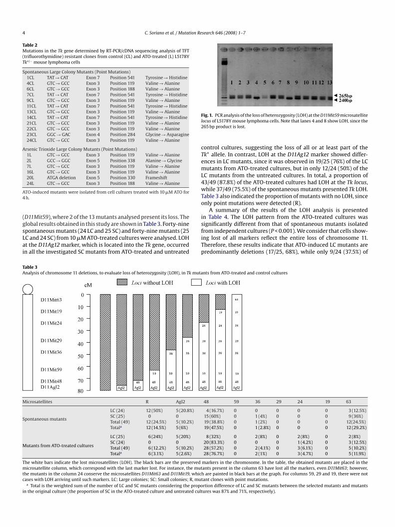
screening of the whole chromosome 11 was carried out usingight heterozygote microsatellites (D11Agl2, D11Mit48, D11Mit59,11Mit36, D11Mit29, D11Mit24, D11Mit19 and D11Mit63). In Fig. 1
here is an example of the loss of a particular microsatellite
4 C. Soriano et al. / Mutation Research 646 (2008) 1–7
Table 2Mutations in the Tk gene determined by RT-PCR/cDNA sequencing analysis of TFT(trifluorothymidine) resistant clones from control (CL) and ATO-treated (L) L5178YTk+/− mouse lymphoma cells
Spontaneous Large Colony Mutants (Point Mutations)1CL TAT → CAT Exon 7 Position 541 Tyrosine → Histidine4CL GTC → GCC Exon 3 Position 119 Valine → Alanine6CL GTC → GCC Exon 3 Position 188 Valine → Alanine7CL TAT → CAT Exon 7 Position 541 Tyrosine → Histidine9CL GTC → GCC Exon 3 Position 119 Valine → Alanine11CL TAT → CAT Exon 7 Position 541 Tyrosine → Histidine13CL GTC → GCC Exon 3 Position 119 Valine → Alanine14CL TAT → CAT Exon 7 Position 541 Tyrosine → Histidine21CL GTC → GCC Exon 3 Position 119 Valine → Alanine22CL GTC → GCC Exon 3 Position 119 Valine → Alanine23CL GGC → GAC Exon 4 Position 284 Glycine → Asparagine24CL GTC → GCC Exon 3 Position 119 Valine → Alanine
Arsenic Trioxide Large Colony Mutants (Point Mutations)1L GTC → GCC Exon 3 Position 119 Valine → Alanine2L GCC → GGC Exon 5 Position 338 Alanine → Glycine7L GTC → GCC Exon 3 Position 119 Valine → Alanine16L GTC → GCC Exon 3 Position 119 Valine → Alanine20L ATGA deletion Exon 5 Position 330 Frameshift
A4
(gsLai
Fl2
cTemL4wTo
is
TA
M
S
M
Tmtc
i
24L GTC → GCC Exon 3 Position 188 Valine → Alanine
TO-induced mutants were isolated from cell cultures treated with 10 �M ATO forh.
D11Mit59), where 2 of the 13 mutants analysed present its loss. Thelobal results obtained in this study are shown in Table 3. Forty-nine
pontaneous mutants (24 LC and 25 SC) and forty-nine mutants (25C and 24 SC) from 10 �M ATO-treated cultures were analysed. LOHt the D11Ag12 marker, which is located into the Tk gene, occurredn all the investigated SC mutants from ATO-treated and untreatedfiTp
able 3nalysis of chromosome 11 deletions, to evaluate loss of heterozygosity (LOH), in Tk muta
icrosatellites R Agl2
pontaneous mutants
LC (24) 12 (50%) 5 (20.8%)SC (25) 0 0Total (49) 12 (24.5%) 5 (10.2%)Totala 12 (14.5%) 5 (6%)
utants from ATO-treated cultures
LC (25) 6 (24%) 5 (20%)SC (24) 0 0Total (49) 6 (12.2%) 5 (10.2%)Totala 6 (3.1%) 5 (2.6%)
he white bars indicate the lost microsatellites (LOH). The black bars are the preservedicrosatellite column, which correspond with the last marker lost. For instance, the mut
he mutants in the column 24 conserve the microsatellites D11Mit63 and D11Mit19, whicases with LOH arriving until such markers. LC: Large colonies; SC: Small colonies; R, mut
a Total is the weighted sum of the number of LC and SC mutants considering the propon the original culture (the proportion of SC in the ATO-treated culture and untreated cult
ig. 1. PCR analysis of the loss of heterozygosity (LOH) at the D11Mit59 microsatelliteocus of L5178Y mouse lymphoma cells. Note that lanes 4 and 8 show LOH, since the65 bp product is lost.
ontrol cultures, suggesting the loss of all or at least part of thek+ allele. In contrast, LOH at the D11Ag12 marker showed differ-nces in LC mutants, since it was observed in 19/25 (76%) of the LCutants from ATO-treated cultures, but in only 12/24 (50%) of the
C mutants from the untreated cultures. In total, a proportion of3/49 (87.8%) of the ATO-treated cultures had LOH at the Tk locus,hile 37/49 (75.5%) of the spontaneous mutants presented Tk LOH.
able 3 also indicated the proportion of mutants with no LOH, sincenly point mutations were detected (R).
A summary of the results of the LOH analysis is presentedn Table 4. The LOH pattern from the ATO-treated cultures wasignificantly different from that of spontaneous mutants isolated
rom independent cultures (P < 0.001). We consider that cells show-ng lost of all markers reflect the entire loss of chromosome 11.herefore, these results indicate that ATO-induced LC mutants areredominantly deletions (17/25, 68%), while only 9/24 (37.5%) ofnts from ATO-treated and control cultures
48 59 36 29 24 19 63
4 (16.7%) 0 0 0 0 0 3 (12.5%)15 (60%) 0 1 (4%) 0 0 0 9 (36%)19 (38.8%) 0 1 (2%) 0 0 0 12(24.5%)19 (47.5%) 0 1 (2.8%) 0 0 0 12 (29.2%)
8 (32%) 0 2 (8%) 0 2 (8%) 0 2 (8%)20 (83.3%) 0 0 0 1 (4.2%) 0 3 (12.5%)28 (57.2%) 0 2 (4.1%) 0 3 (6.1%) 0 5 (10.2%)28 (76.7%) 0 2 (1%) 0 3 (4.7%) 0 5 (11.9%)
markers in the chromosome. In the table, the obtained mutants are placed in theants present in the column 63 have lost all the markers, even D11Mit63; however,h are painted in black bars at the graph. For columns 59, 29 and 19, there were notant clones with point mutations.rtion difference of LC and SC mutants between the selected mutants and mutantsures was 87% and 71%, respectively).
C. Soriano et al. / Mutation Research 646 (2008) 1–7 5
Table 4Comparison of large and small colonies results, using the percentages of mutants
Source of mutants Clone size Intragenic mutation Deletion Chromosome loss
Spontaneous mutants
LC (24) 12 (50%) 9 (37.5%) 3 (12.5%)SC (25) 0 16 (64%) 9 (36%)Total (49) 12 (24.5%) 25 (51%) 12 (24.5%)Totala 12 (14.5%) 25 (56.3%) 12 (29.2%)
ATO-treated cultures
LC (25) 6 (24%) 17 (68%) 2 (8%)SC (24) 0 21 (87.5%) 3 (12.5%)Total (49) 6 (12.2%) 38 (77.6%) 5 (10.2%)Totala 6 (3.1%)*** 38 (85%)*** 5 (11.9%)***
* ies.propo
c ltures
smi(btAwlt((
4
oipa
Fo
pp
imia1amah[hi
**Significantly different from controls, P < 0.001. LC: Large colonies; SC: Small colona Total is the weighted sum of the number of LC and SC mutants considering the
ulture mutants (the proportion of SC in the ATO-treated cultures and untreated cu
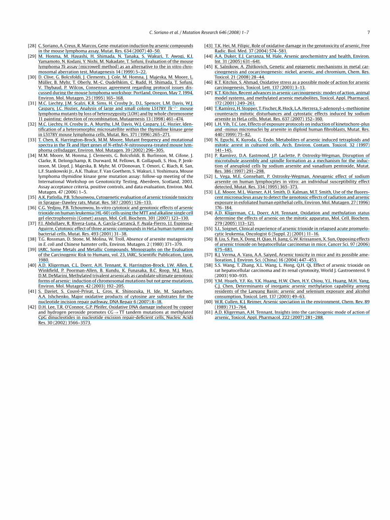
pontaneous LC mutants are due to deletions. Similarly, for SCutants, which represent the 87% of ATO-induced mutants, ATO
nduces mainly large deletions, 21/24 (87.5%), whereas only 16/2564%) was observed in spontaneous mutants. However, the num-er of chromosome losses in SC mutants is significantly higher inhose of spontaneous origin, 9/25 (36%), when compared to theTO-induced ones, 3/24 (12.5%) (P < 0.001). Taking into account theeighted sums, 85% of ATO-treated mutants are consequence of
arge deletions. Fig. 2 shows that these differences were mainly dueo the proportion of point mutations vs. deletions in LC mutantsP < 0.001), and large deletions vs. chromosome loss in SC mutantsP < 0.001).
. Discussion
The results obtained in the analysis of the mutational spectrumf ATO-induced mutants confirm that this compound is a potentnducer of large deletions, which would confirm the clastogenicotential attributable to arsenic compounds. However, taking intoccount the toxicity induced by ATO (62% and 74.5%) at 10 �M, it is
ig. 2. Comparison of the results of large and small colonies, using the percentagef mutants. ***Significantly different from background controls, P < 0.001.
[wtwnoiwtmsfSio[c[
[ciiovt
iosTiddl
rtion difference of LC and SC mutants between the selected mutants and originalwas 87% and 71%, respectively).
ossible that the mutational spectra obtained could be related inart to its toxicity.
This finding is consistent with the existing data on the ATO abil-ty to induce chromosome aberrations and micronuclei in bone
arrow cells of rats, treated for 5 days with ATO at doses rang-ng from 5 to 20 mg/kg body weight (BW) [35], and both smallnd large colony mutants in the mouse lymphoma Tk gene (7 and0 �M) [28]. Thus, it appears that when ATO is tested in mutationssays detecting also chromosome mutations, it acts as a potentutagenic/clastogenic compound. This genotoxic behaviour of ATO
grees with the results obtained in the induction of DNA breakage inuman leukaemia cells, by using the comet assay (5 and 10 �g/mL)36]. In general, previous genotoxic studies of arsenic compoundsave largely yielded negative findings for gene mutations, but pos-
tive results for chromosomal aberrations (arsenite, 0.5–50 �M)14]. Although no previous studies have been published, reportinghether ATO induces point mutations, other arsenic compounds
ested for mutagenicity were non-mutagenic in bacteria, actingeakly at high concentrations (5–10 �M) in the Hprt locus in Chi-ese hamster V79 cells [27]. The reason of the negative resultsbtained in the Hprt locus, when arsenic compounds are assayed,s due to its location in the X chromosome (single functioning),
hich makes unlikely the detection of large deletions, since a dele-ion extended past the Hprt locus is often lethal. Nevertheless, the
ouse lymphoma assay can detect the arsenic clastogenic effectsince it uses an autosomal gene target [25,28]. The negative resultsor AsIII and AsV in bacteria assays using TA98 and TA100 strains ofalmonella typhimurium (0.1–100 �g/plate of AsIII and AsV) [37] andn WP2 Escherichia coli strain (0–0.1 mM of AsIII) [38], as well as withther arsenical compounds tested for gene mutation in microbes22,39], support the view that the mouse lymphoma results indi-ate that this agent induces large deletions but not gene mutations25,28,40].
Oxidative damage predominantly induces C → T transitions41,42] that are the point mutations most represented in bothontrol and ATO-induced point mutations. Nevertheless, the lowncidence of point mutations in the ATO-induced mutants (includ-ng C → T transitions) might suggest that the assumed role ofxidative damage induced by arsenic compounds [43] would actia lesions resulting in chromosome damage instead of point muta-ions.
Arsenic genotoxicity has been explained by diverse mechanismsncluding inhibition of various DNA repair enzymes, productionf reactive oxygen species (ROS), induction of gene expression oftress response proteins [44], and also by epigenetic changes [45].
he production of ROS by arsenic usually results in oxidative stressnto the cells. Oxidative stress has been involved in mediating manyeleterious effects of arsenic including DNA damage, lipid peroxi-ation, redox enzyme activity, and decrease of antioxidant defenceevels. It has been reported that arsenic-induced oxidative stress
6 ion Re
cDciDaaatoealaos[
afiknaag1aeqtagbseaascecAag1[tdddaooavotpse
cpT1
A
fse(
R
[
[
[
[
[
[
[
[
[
[
[
[
[
[
[
[
C. Soriano et al. / Mutat
an provoke a broad spectrum of effects on the DNA includingNA single- and double-strand breaks, abasic sites, DNA–proteinross-links, and DNA base modifications [46]. This genotoxic activ-ty of ATO agrees with the results obtained in the induction ofNA breakage in human leukaemia cells, as detected in the cometssay (5 and 10 �g/mL) [36]. In addition, and as reviewed by Heind Filipic [43], evidences in support of the role of oxidative dam-ge in arsenic genotoxicity/carcinogenesis are overwhelming, sincehe modulation of the cellular redox status has a profound effectn the genotoxicity of arsenic. The induction of sister-chromatidxchanges in cultured human lymphocytes exposed to arsenite wasntagonised by the addition of superoxide dismutase and cata-ase, and the induction of micronuclei in CHO-K1 cells by 20 �M ofrsenite was antagonised by nitric oxide synthase inhibitors, super-xide dismutase or uric acid. These results support the view thatome clastogenic effects of arsenic are mediated via free radicals47].
It has been described in previous papers that arsenic is alson inducer of aneuploidy [48–53]. In in vitro studies with humanbroblasts, low doses of arsenite (1.25–10 �M) induced mainlyinetochore-positive MN, whereas high doses (5–80 �M) gaveegative results [49]. In human lymphocytes, 0.001–0.1 �M ofrsenite induced aneuploidy by disruption of microtubule assemblynd spindle formation [51] and 0.01 �M had an aneuploido-enic and a mitotic arrestant effect [52]. In humans exposed to.312 micrograms As/L in drinking water, both clastogenic and weakneuploidogenic effects of arsenic were observed in exfoliatedpithelial cells [53]; nevertheless, in the current work, the fre-uency of chromosome loss in ATO-mutants is significantly lowerhan that found in spontaneous mutants. This would indicate that,t least for the MLA assay, ATO does not seem to exert any aneu-enic potential, which would support the negative findings foundy other authors [54]. It is becoming increasingly clear that expo-ure to high doses of arsenic compounds differs from low dosexposure, with regard to genotoxic and therapeutic effects [2,55];lthough the response will depend on the cell type, its sensitivitynd the arsenic form. A good example is the therapeutically anduccessful use of ATO in treating patients with acute promyelo-ytic leukaemia at 1–2 �M therapeutic concentrations and its lessffectiveness against solid tumours, needing to use much higheroncentrations to trigger apoptosis and inhibit tumour growing.TO effectiveness against solid tumours is a narrow window of ther-peutic opportunity given that low doses of ATO stimulate tumourrowth and high doses are toxic [56]. Concentrations higher than0 �M are used to trigger apoptosis and to inhibit tumour growing5–7] and, in the present work, we show the genotoxic and clas-ogenic effects of 10 �M ATO. This would indicate that such highoses are not achievable without the risk of severe side effectsue to toxicity. As it has been previously described, ATO may pro-uce cardiac, liver and kidney toxicity, among others, in patientsnd animal models [8–10,55,57,58]. Nevertheless, inter-individualr interspecies variability in the response to arsenic compounds isften found and it has been attributed to genetic differences in thebility to metabolise arsenic [59]. Inorganic arsenic is metabolisedia successive reduction and methylation steps [60], and somef its metabolites, in particular MMAIII, have shown to be moreoxic and genotoxic than its parental forms [61]. Thus, geneticolymorphisms in genes regulating such metabolic steps are con-idered as potential risk factors, modulating the response to arsenicxposure.
The conclusion of our work is that ATO is a strong clastogenicompound, as observed in the mouse lymphoma assay, where itredominantly induces large deletions at chromosomal level in thek gene, covering such deletions also other regions of chromosome1.
[
[
search 646 (2008) 1–7
cknowledgements
Carolina Soriano is supported by a postgraduate fellowshiprom the Generalitat de Catalunya. This investigation has beenupported in part by the Spanish Ministry of Education and Sci-nce (CAICYT, SAF2008-02933) and by the Generalitat de CatalunyaCIRIT, 2005SGR-00136).
eferences
[1] K.H. Antman, Introduction: the history of arsenic trioxide in cancer therapy,Oncologist 6 (Suppl. 2) (2001) 1–2.
[2] G. Lazo, H. Kantarjian, E. Estey, D. Thomas, S. O’Brien, J. Cortes, Use of arsenic tri-oxide (As2O3) in the treatment of patients with acute promyelocytic leukemia.The M.D. Anderson experience, Cancer 97 (2003) 2218–2224.
[3] S.L. Soignet, P. Maslak, Z.G. Wang, S. Jhanwar, E. Calleja, L.J. Dardashti, D. Corso,A. DeBlasio, J. Gabrilove, D.A. Scheinberg, P.P. Pandolfi, R.P. Warrell Jr., Com-plete remission after treatment of acute promyelocytic leukemia with arsenictrioxide, N. Engl. J. Med. 339 (1998) 1341–1348.
[4] E. Lengfelder, S. Saussele, A. Weisser, T. Buchner, R. Hehlmann, Treatment con-cepts of acute promyelocytic leukemia, Crit. Rev. Oncol. Hematol. 56 (2005)261–274.
[5] H. Monzen, R.J. Griffin, B.W. Williams, M. Amano, S. Ando, T. Hasegawa, Study ofarsenic trioxide-induced vascular shutdown and enhancement with radiationin solid tumor, Radiat. Med. 22 (2004) 205–211.
[6] Z.Y. Shen, Y. Zhang, J.Y. Chen, M.H. Chen, J. Shen, W.H. Luo, Y. Zeng, Intratu-moral injection of arsenic to enhance antitumor efficacy in human esophagealcarcinoma cell xenografts, Oncol. Rep. 11 (2004) 155–159.
[7] Y.S. Pu, T.C. Hour, J. Chen, C.Y. Huang, J.Y. Guan, S.H. Lu, Arsenic trioxide as anovel anticancer agent against human transitional carcinoma-characterizingits apoptotic pathway, Anticancer Drugs 13 (2002) 293–300.
[8] K. Naito, M. Kobayashi, N. Sahara, K. Shigeno, S. Nakamura, K. Shinjo, T. Tobita, A.Takeshita, R. Ohno, K. Ohnishi, Two cases of acute promyelocytic leukemia com-plicated by torsade de pointes during arsenic trioxide therapy, Int. J. Hematol.83 (2006) 318–323.
[9] A.M. Evens, M.S. Tallman, R.B. Gartenhaus, The potential of arsenic trioxide inthe treatment of malignant disease: past, present, and future, Leuk. Res. 28(2004) 891–900.
10] Y. Li, X. Sun, L. Wang, Z. Zhou, Y.J. Kang, Myocardial toxicity of arsenic trioxidein a mouse model, Cardiovasc. Toxicol. 2 (2002) 63–73.
11] IARC, Some Drinking-water Disinfectants and Contaminants Including Arsenic.Monographs on the Evaluation of the Carcinogenic Risk to Humans, vol. 84,IARC, Scientific Publication, Lyon, 2004.
12] IARC, Arsenic and Arsenic Compounds. Monographs on the Evaluation of theCarcinogenic Risk to Humans, Suppl. 7, IARC, Scientific Publication, Lyon, 1987.
13] T.G. Rossman, Mechanism of arsenic carcinogenesis: an integrated approach,Mutat. Res. 533 (2003) 37–65.
14] A. Basu, J. Mahata, S. Gupta, A.K. Giri, Genetic toxicology of a paradoxical humancarcinogen, arsenic: a review, Mutat. Res. 488 (2001) 171–194.
15] C.O. Abernathy, Y.P. Liu, D. Longfellow, H.V. Aposhian, B. Beck, B. Fowler, R. Goyer,R. Menzer, T. Rossman, C. Thompson, M. Waalkes, Arsenic: health effects, mech-anisms of actions, and research issues, Environ. Health Perspect. 107 (1999)593–597.
16] F. Ayala-Fierro, D.S. Barber, L.T. Rael, D.E. Carter, In vitro tissue specificity forarsine and arsenite toxicity in the rat, Toxicol. Sci. 52 (1999) 122–129.
17] T.C. Lee, S.F. Tzeng, W.J. Chang, Y.C. Lin, K.Y. Jan, Post-treatments with sodiumarsenite during G2 enhance the frequency of chromosomal aberrations inducedby S-dependent clastogens, Mutat. Res. 163 (1986) 263–269.
18] T.C. Lee, N. Tanaka, P.W. Lamb, T.M. Gilmer, J.C. Barrett, Induction of gene ampli-fication by arsenic, Science 241 (1988) 79–81.
19] A.N. Jha, M. Noditi, R. Nilsson, A.T. Natarajan, Genotoxic effects of sodium arsen-ite on human cells, Mutat. Res. 284 (1992) 215–221.
20] J.K. Wiencke, J.W. Yager, Specificity of arsenite in potentiating cytogeneticdamage induced by the DNA crosslinking agent diepoxybutane, Environ. Mol.Mutagen. 19 (1992) 195–200.
21] T.S. Wang, Y.F. Shu, Y.C. Liu, K.Y. Jan, H. Huang, Glutathione peroxidaseand catalase modulate the genotoxicity of arsenite, Toxicology 121 (1997)229–237.
22] T.W. Gebel, Unanswered questions in arsenic toxicology, J. Environ. Pathol. Tox-icol. Oncol. 20 (2001) 299–309.
23] Y. Noda, T. Suzuki, A. Kohara, A. Hasegawa, T. Yotsuyanagi, M. Hayashi, T. Sofuni,K. Yamanaka, S. Okada, In vivo genotoxicity evaluation of dimethylarsinic acidin MutaMouse, Mutat. Res. 513 (2002) 205–212.
24] T.K. Hei, S.X. Liu, C. Waldren, Mutagenicity of arsenic in mammalian cells: roleof reactive oxygen species, Proc. Natl. Acad. Sci. U.S.A. 95 (1998) 8103–8107.
25] M.M. Moore, K. Harrington-Brock, C.L. Doerr, Relative genotoxic potency of
arsenic and its methylated metabolites, Mutat. Res. 386 (1997) 279–290.26] Z. Meng, A.W. Hsie, Polymerase chain reaction-based deletion analysis of spon-taneous and arsenite-enhanced gpt mutants in CHO-AS52 cells, Mutat. Res. 356(1996) 255–259.
27] J.H. Li, T.G. Rossman, Comutagenesis of sodium arsenite with ultraviolet radia-tion in Chinese hamster V79 cells, Biol. Met. 4 (1991) 197–200.
ion Re
[
[
[
[
[
[
[
[
[
[
[
[
[
[
[
[
[
[
[
[
[
[
[
[
[
[
[
[
[
[
[
[
residents of the Lanyang Basin: arsenic and selenium exposure and alcohol
C. Soriano et al. / Mutat
28] C. Soriano, A. Creus, R. Marcos, Gene-mutation induction by arsenic compoundsin the mouse lymphoma assay, Mutat. Res. 634 (2007) 40–50.
29] M. Honma, M. Hayashi, H. Shimada, N. Tanaka, S. Wakuri, T. Awogi, K.I.Yamamoto, N. Kodani, Y. Nishi, M. Nakadate, T. Sofuni, Evaluation of the mouselymphoma Tk assay (microwell method) as an alternative to the in vitro chro-mosomal aberration test, Mutagenesis 14 (1999) 5–22.
30] D. Clive, G. Bolcsfoldi, J. Clements, J. Cole, M. Homna, J. Majeska, M. Moore, L.Müller, B. Myhr, T. Oberly, M.-C. Oudelhkim, C. Rudd, H. Shimada, T. Sofuni,V. Thybaud, P. Wilcox, Consensus agreement regarding protocol issues dis-cussed during the mouse lymphoma workshop: Portland, Oregon, May 7, 1994,Environ. Mol. Mutagen. 25 (1995) 165–168.
31] M.C. Liechty, J.M. Scalzi, K.R. Sims, H. Crosby Jr., D.L. Spencer, L.M. Davis, W.J.Caspary, J.C. Hozier, Analysis of large and small colony L5178Y Tk−/− mouselymphoma mutants by loss of heterozygosity (LOH) and by whole chromosome11 painting: detection of recombination, Mutagenesis 13 (1998) 461–474.
32] M.C. Liechty, H. Crosby Jr., A. Murthy, L.M. Davis, W.J. Caspary, J.C. Hozier, Iden-tification of a heteromorphic microsatellite within the thymidine kinase genein L5178Y mouse lymphoma cells, Mutat. Res. 371 (1996) 265–271.
33] T. Chen, K. Harrington-Brock, M.M. Moore, Mutant frequency and mutationalspectra in the Tk and Hprt genes of N-ethyl-N-nitrosourea-treated mouse lym-phoma cellsdagger, Environ. Mol. Mutagen. 39 (2002) 296–305.
34] M.M. Moore, M. Honma, J. Clements, G. Bolcsfoldi, B. Burlinson, M. Cifone, J.Clarke, R. Delongchamp, R. Durward, M. Fellows, B. Gollapudi, S. Hou, P. Jenk-inson, M. Lloyd, J. Majeska, B. Myhr, M. O’Donovan, T. Omori, C. Riach, R. San,L.F. Stankowski Jr., A.K. Thakur, F. Van Goethem, S. Wakuri, I. Yoshimura, Mouselymphoma thymidine kinase gene mutation assay: follow-up meeting of theInternational Workshop on Genotoxicity Testing, Aberdeen, Scotland, 2003.Assay acceptance criteria, positive controls, and data evaluation, Environ. Mol.Mutagen. 47 (2006) 1–5.
35] A.K. Patlolla, P.B. Tchounwou, Cytogenetic evaluation of arsenic trioxide toxicityin Sprague–Dawley rats, Mutat. Res. 587 (2005) 126–133.
36] C.G. Yedjou, P.B. Tchounwou, In-vitro cytotoxic and genotoxic effects of arsenictrioxide on human leukemia (HL-60) cells using the MTT and alkaline single cellgel electrophoresis (Comet) assays, Mol. Cell. Biochem. 301 (2007) 123–130.
37] F.I. Abdullaev, R. Rivera-Luna, A. García-Carrancá, F. Ayala-Fierro, J.J. Espinosa-Aguirre, Cytotoxic effect of three arsenic compounds in HeLa human tumor andbacterial cells, Mutat. Res. 493 (2001) 31–38.
38] T.G. Rossman, D. Stone, M. Molina, W. Troll, Absence of arsenite mutagenicityin E. coli and Chinese hamster cells, Environ. Mutagen. 2 (1980) 371–379.
39] IARC, Some Metals and Metallic Compounds. Monographs on the Evaluationof the Carcinogenic Risk to Humans, vol. 23, IARC, Scientific Publication, Lyon,1980.
40] A.D. Kligerman, C.L. Doerr, A.H. Tennant, K. Harrington-Brock, J.W. Allen, E.Winkfield, P. Poorman-Allen, B. Kundu, K. Funasaka, B.C. Roop, M.J. Mass,D.M. DeMarini, Methylated trivalent arsenicals as candidate ultimate genotoxicforms of arsenic: induction of chromosomal mutations but not gene mutations,Environ. Mol. Mutagen. 42 (2003) 192–205.
41] S. Daviet, S. Couvé-Privat, L. Gros, K. Shinozuka, H. Ide, M. Saparbaev,
A.A. Ishchenko, Major oxidative products of cytosine are substrates for thenucleotide incision repair pathway, DNA Repair 6 (2007) 8–18.42] D.H. Lee, T.R. O’Connor, G.P. Pfeifer, Oxidative DNA damage induced by copperand hydrogen peroxide promotes CG → TT tandem mutations at methylatedCpG dinucleotides in nucleotide excision repair-deficient cells, Nucleic AcidsRes. 30 (2002) 3566–3573.
[
[
search 646 (2008) 1–7 7
43] T.K. Hei, M. Filipic, Role of oxidative damage in the genotoxicity of arsenic, FreeRadic. Biol. Med. 37 (2004) 574–581.
44] A.A. Duker, E.J. Carranza, M. Hale, Arsenic geochemistry and health, Environ.Int. 31 (2005) 631–641.
45] K. Salnikow, A. Zhitkovich, Genetic and epigenetic mechanisms in metal car-cinogenesis and cocarcinogenesis: nickel, arsenic, and chromium, Chem. Res.Toxicol. 21 (2008) 28–44.
46] K.T. Kitchin, S. Ahmad, Oxidative stress as a possible mode of action for arseniccarcinogenesis, Toxicol. Lett. 137 (2003) 3–13.
47] K.T. Kitchin, Recent advances in arsenic carcinogenesis: modes of action, animalmodel systems, and methylated arsenic metabolites, Toxicol. Appl. Pharmacol.172 (2001) 249–261.
48] T. Ramírez, H. Stopper, T. Fischer, R. Hock, L.A. Herrera, S-adenosyl-l-methioninecounteracts mitotic disturbances and cytostatic effects induced by sodiumarsenite in HeLa cells, Mutat. Res. 637 (2007) 152–160.
49] L.H. Yih, T.C. Lee, Effects of exposure protocols on induction of kinetochore-plusand -minus micronuclei by arsenite in diploid human fibroblasts, Mutat. Res.440 (1999) 75–82.
50] N. Eguchi, K. Kuroda, G. Endo, Metabolites of arsenic induced tetraploids andmitotic arrest in cultured cells, Arch. Environ. Contam. Toxicol. 32 (1997)141–145.
51] P. Ramírez, D.A. Eastmond, J.P. Laclette, P. Ostrosky-Wegman, Disruption ofmicrotubule assembly and spindle formation as a mechanism for the induc-tion of aneuploid cells by sodium arsenite and vanadium pentoxide, Mutat.Res. 386 (1997) 291–298.
52] L. Vega, M.E. Gonsebatt, P. Ostrosky-Wegman, Aneugenic effect of sodiumarsenite on human lymphocytes in vitro: an individual susceptibility effectdetected, Mutat. Res. 334 (1995) 365–373.
53] L.E. Moore, M.L. Warner, A.H. Smith, D. Kalman, M.T. Smith, Use of the fluores-cent micronucleus assay to detect the genotoxic effects of radiation and arsenicexposure in exfoliated human epithelial cells, Environ. Mol. Mutagen. 27 (1996)176–184.
54] A.D. Kligerman, C.L. Doerr, A.H. Tennant, Oxidation and methylation statusdetermine the effects of arsenic on the mitotic apparatus, Mol. Cell. Biochem.279 (2005) 113–121.
55] S.L. Soignet, Clinical experience of arsenic trioxide in relapsed acute promyelo-cytic leukemia, Oncologist 6 (Suppl. 2) (2001) 11–16.
56] B. Liu, S. Pan, X. Dong, H. Qiao, H. Jiang, G.W. Krissansen, X. Sun, Opposing effectsof arsenic trioxide on hepatocellular carcinomas in mice, Cancer Sci. 97 (2006)675–681.
57] R.J. Verma, A. Vasu, A.A. Saiyed, Arsenic toxicity in mice and its possible ame-lioration, J. Environ. Sci. (China) 16 (2004) 447–453.
58] S.S. Wang, T. Zhang, X.L. Wang, L. Hong, Q.H. Qi, Effect of arsenic trioxide onrat hepatocellular carcinoma and its renal cytotoxity, World J. Gastroenterol. 9(2003) 930–935.
59] Y.M. Hsueh, Y.F. Ko, Y.K. Huang, H.W. Chen, H.Y. Chiou, Y.L. Huang, M.H. Yang,C.J. Chen, Determinants of inorganic arsenic methylation capability among
consumption, Toxicol. Lett. 137 (2003) 49–63.60] W.R. Cullen, K.J. Reimer, Arsenic speciation in the environment, Chem. Rev. 89
(1989) 713–764.61] A.D. Kligerman, A.H. Tennant, Insights into the carcinogenic mode of action of
arsenic, Toxicol. Appl. Pharmacol. 222 (2007) 281–288.


