Embed Size (px)
Citation preview

Naturally-occurring anti-G-CSF antibodies produced by human cord bloodB-cell lines infected with Epstein-Barr virus
Roberto P Revoltella*,1, Leopoldo Laricchia Robbio1, Stefania Moscato1, Fabrizio Vinante2,Alessandro Fasciani3, Anna Maria Liberati4, Gigliola Reato5 and Robin Foa5
1Institute of Mutagenesis and Di�erentiation, C.N.R., Pisa, Italy; 2Department of Clinical and Experimental Medicine, Section ofHematology, University of Verona, Verona, Italy; 3Division of Gynecology and Obstetrics, University of Pisa, Pisa, Italy; 4Instituteof Internal Medicine and Oncological Sciences, Policlinico Monteluce, Perugia, Italy; 5Department of Cellular Biotechnologies andHematology, University `La Sapienza', Rome, Italy
Introduction: Naturally occurring antibodies (auto-Abs) recognizing human granulocyte-colony stimulating factor were detected with high frequency in serum samples obtained fromumbilical cord blood of newborns (12 of 65 samples screened) and maternal peripheral bloodserum samples from women at the end of gestation (seven of 56 cases tested). The aim of thispaper was to demonstrate that auto-Abs anti-G-CSF revealed in the blood of newborns wereproduced during foetal life.Materials and methods: Mononuclear cells from cord blood samples of di�erent newbornscontaining high titer anti-G-CSF Abs were infected with Epstein-Barr virus in vitro, andEBV-immortalized B-cell lines were isolated and characterized for speci®c anti-G-CSF Abproduction.Results: Six di�erent, unrelated cell lines of male origin which showed the presence ofEBNA-2 antigen in the nucleus, displayed a B-cell phenotype (CD30+, CD57, CD107,HLA-DR+, CD19+, CD20+, CD23+, CD38+, CD25+), coexpressed low intensity sIgMand sIgD, and produced only IgM with prevailing l clonal restriction and anti-rhG-CSF Abreactivity. The Ab speci®city was proven against either glycosylated or unglycosylated G-CSFby saturable binding in direct enzyme-linked immunosorbent assays, by competition bindingand Western immunoblotting assays.Conclusion: The secreted Abs did not a�ect the in vitro generation of granulocyte coloniesby human normal adult haemopoietic progenitor cells in soft agar clonogenic assays,suggesting that these Abs were not neutralizing.The Hematology Journal (2001) 2, 161 ± 171
Keywords: G-CSF; auto-antibodies; cord-blood cultures; EBV-infection
Introduction
Granulocyte-colony stimulating factor (G-CSF) is aglycoprotein that speci®cally stimulates the prolifera-tion, di�erentiation and maturation of haemopoieticmyeloid progenitors, prolongs the survival of matureneutrophils and enhances their functional properties.1 ±3 Administration of G-CSF in vivo stimulatesperipheral blood granulocytosis and induces themobilization of stem cells and progenitor cells fromthe bone marrow to the peripheral blood.4 Biological
responses induced by G-CSF occur following itsbinding to a speci®c cell membrane receptor whoseencoding gene has been cloned.5 Recombinant human(rh) G-CSF is being extensively used in patients withdi�erent diseases (reviewed in6). High levels of speci®cAbs anti-rhG-CSF are observed in lymphoma patientswho received multiple rhG-CSF injections; however,circulating anti-G-CSF Abs do not a�ect haematolo-gic recovery.7,8 Moreover, low levels of high avidity,non-neutralizing anti-G-CSF auto-Abs (both IgM andIgG) are detected in about 10% of sera from healthyadult individuals.7,8
These ®ndings stimulated us to investigate whetheranti-G-CSF Abs are produced during foetal life and toanalyse the potential functional role of these naturalfoetal Abs. In the present study, we demonstrate thepresence of natural anti-G-CSF Abs in cord blood
*Correspondence: RP Revoltella, Institute of Mutagenesis andDi�erentiation, C.N.R., Area della Ricerca di San Cataldo, Via G.Moruzzi, 1. Ghezzano ± 56100 Pisa, Italy;Tel: +39 050 3152772; Fax: +39 050 3153367;E-mail: [email protected] 9 August 2000; accepted 29 November 2000
The Hematology Journal (2001) 2, 161 ± 171ã 2001 The European Haematology Association All rights reserved 1466 ± 4680/01 $15.00
www.nature.com/thj

(CB) and provide evidence that such Abs are producedby CB B cells. In addition we show that these auto-Absdo not a�ect G-CSF functions, as demonstrated by thegeneration of granulocyte colonies in the soft agarclonogenic assay, suggesting that these Abs are non-neutralizing.
Materials and methods
Cytokines
Unglycosylated (Filgrastim) and glycosylated (Leno-grastim) rhG-CSF were obtained respectively from
Amgen GmbH (MuÈ nchen, Germany) and Rhoà ne-Poulenc, Rorer SpA (Milan, Italy). The rhG-CSF(19 KDa) used as control antigen in the Westernblotting assay was given by Santa Cruz Biotechnology,Inc. (Santa Cruz, CA, USA) Unglycosylated granulo-cyte-macrophage colony-stimulating factor (rhGM-CSF) was prepared and puri®ed by Farmitalia ±Carlo Erba Biotechnology Facility (now Pharmacia-Upjohn, Milan, Italy). Unglycosylated rhIL-1b, rhIL-2,rhIL-3, rhIL-8 and rhStem Cell Factor (rhSCF) wereall from Sigma Chemical (St. Louis, MO, USA); rhIL-4 was from DNAX, Palo Alto, CA; rh Interferon-a 2A(rhIFNa2A) was from Ho�man La Roche (Basel,Switzerland).
Figure 1 Flow chart of the procedure adopted (steps A to F) for e�cient immortalization of human cord blood B cells using EBV, followedby selection and characterization of cell lines with the desired phenotypic characteristics.
Human anti-G-CSF auto-antibodiesRP Revoltella et al
162
The Hematology Journal

Sera and plasma isolation
Five to 10 ml of CB from newborns and maternalperipheral blood samples were collected and processed.Informed consent was obtained from all donors.Positive serum samples from two myeloma patientswho developed anti-G-CSF antibodies after repetitiveinjections with rhG-CSF (DM and GD)7 were used ascontrol.
Precautions adopted to avoid non-speci®c bindingby human serum antibodies
Rheumatoid factor and contaminating traces of Abreacting with E. Coli antigens were absorbed fromserum specimens as previously described.9± 11 Allproteins, antibodies and serum samples were pre-tested to exclude endotoxin contamination.7 ± 9
Cord blood-mononuclear cell immortalization anddevelopment of cell lines
Cord blood-mononuclear cells (CB-MNC) were in-fected with Epstein-Barr virus (EBV) and immortalized(Figure 1), as previously described.12,13 Figure 1 shows a¯ow chart of the procedure adopted. EBV for infectionwas obtained from B95-8 cells (American Type CultureCollection, Rockville, MD, USA). Fresh mediumsupernatant from two-day culture of B95-8 cells wascollected, centrifuged (500 g, 10 min), ®ltered (0.45 mm®lters) and stored at 7808C. CB-MNC were obtainedby Ficoll-Paque (Pharmacia-Biotech, Milan, Italy)separation.7 Approximately 106106 viable cells in1 ml of complete RPMI-1640 with 10% foetal bovineserum (FBS) (Hyclone Laboratories, Inc., Logan, UT,USA) were incubated in 5% CO2 in air atmosphere at378C with 1 ml EBV containing supernatant from B95-8 cell culture. Cell preparations were occasionallyshaken. Three ml of complete medium were subse-quently added, and the cells centrifuged, resuspendedin RPMI 1640 complete medium supplemented with20% FBS and antibiotics. Cells were then plated in 96-well plates (¯at-bottom) (Falcon, Becton Dickinson &Co., Oxnard, CA, USA) at a concentration of about100 000 to 200 000 cells/0.1 ml/well. After three daysof culture, 0.1 ml complete RPMI-1640 mediumcontaining 0.2 mg Cyclosporin A (CsA) (Sandoz, EastHannover, NJ, USA) was added to each well to inhibitT-cell activity and to activate B-cell di�erentiation.14
CsA was kept only for the ®rst four weeks of culture.Culture supernatants were tested (see below) forhuman Ig and anti-G-CSF Ab production 7 ± 15 daysfrom culture seeding and every 10 days thereafter. Cellspresent in the wells containing high anti-G-CSF Abtiters were subcultured (about 10 000 cells/0.1-ml/well)twice into new 96-well plates. Ten thousand lethallyirradiated (7500 r) CB-MNC/0.1-ml/well from the sameindividual were recovered from Ab-negative wells andused as feeder layer. Cells from wells showing thehighest anti-rhG-CSF Ab amount in the supernatantwere then further selected by limiting dilution, into 96-
well plates ®rst with and subsequently expandedwithout feeder layer. Established cell lines did notrequire feeder cells, provided that a high densityseeding was accomplished. All autonomous cell lineswere judged to be free of Mycoplasma using selectivegrowth media and the Mycoplasma Primer Set(Stratagene, La Jolla, CA, USA). At intervals ofabout 10 cell population doublings, aliquots of cellsfrom each cell line were frozen and stored at 7808C orin liquid nitrogen.
Cytogenetic studies
Chromosome studies were performed on logarithmi-cally growing cells after approximately six subsequentpassages from the establishment of the autonomouslines. After 4 h exposure to Demecolcine (Colcemid)(Sigma) (0.05 mg/ml), cells were washed, treated with0.075 M KCl for 15 min at room temperature and then®xed with methanol-acetic acid (3 : 1). Chromosomepreparations were made by the air-drying method andwere stained by the Q(QFQ) banding technique(resolution : 320 bands) as routine procedure. At least10 to 20 metaphases were scored.
Epstein-Barr virus content
Samples from cell lines immortalized with EBV werecytocentrifuged, allowed to air-dry and ®xed with coldacetone: methanol (1 : 1). Each cell preparation wasincubated with monoclonal antibody (mAb) PE2(mouse IgG1) anti-EBNA-2 (Biogenesis, Poole, UK)(1 : 5) at 378C for 1 h, washed and subsequentlyexposed to biotinylated-goat IgG anti-mouse IgG1
(BIOT-Goat anti-mIgG1) (Vector Laboratories, Inc.,Burlingame, CA, USA) (1 : 25) for 30 min followed by¯uorescein-streptavidin (Vector) (1 : 50). Ab controlsincluded incubation with an unrelated primary Ab orthe secondary Ab alone to exclude non-speci®creactivity. Controls always included the EBV positiveB95-8 cells, the EBV negative MOLT-4 cell line, PB-MNC from healthy adult donors, and CB-MNC. Slideswere examined with a 256 or a 406 objective of aLeitz ¯uorescence microscope.
Immunophenotypic analysis
The following FITC- or PE- conjugated or un-conjugated polyclonal or monoclonal Abs were used:PECD3, FITCCD5, PECD19, FITCCD45, PECD23, PECD20,PECD14, FITCCD10, PECD25, PECD38, (Becton Dick-inson, Mountain View, CA, USA); FITCCD30,FITCCD79a (Dako, GolstruÈ p, Denmark); anti-surfacehIg (sIg), anti-sIgD, anti-sIgM, anti-Igk, anti-Igl, anti-HLA-DR (Coulter, Hialeah, FL, USA). In the directimmuno¯uorescence assays, cells growing logarithmi-cally were harvested, washed and incubated with 50 mlof the desired mAb (106 cells per reaction volume) for30 min at 48C in PBS pH 7.4 with 0.1% bovine serumalbumin (BSA) (Sigma), 0.02 mmol/l NaN3. In theindirect staining assays the cells, ®xed in 1%
The Hematology Journal
Human anti-G-CSF auto-antibodiesRP Revoltella et al
163

formaldehyde in PBS, after incubation with theappropriate unlabelled mAb, were washed, resus-pended in FITC-sheep F(ab')2 anti-mIg (1 : 100) andheld on ice for 30 min at 48C. After three washes, thelabelled cells were analysed on a FACS scan (BectonDickinson). For cyDD79 detection, cell permeabiliza-tion was performed as previously described.15
Detection of anti-G-CSF antibodies
Sera and cell culture supernatants were stored at 7208Cor7708C until assayed. Single aliquots were thawed justbefore use and screened for the presence of anti-rhG-CSF Abs. When whole sera were tested, samples were®rst decomplemented (at 568C for 30 min).
Enzyme-linked immunosorbent assay (ELISA) Directand competition ELISA were performed as previouslydescribed.16,17 Brie¯y, in direct ELISA rhG-CSF wascoupled to the plastic surface of 96-well plates (0.3 mg/50 ml carbonate bu�er, pH 9.6/well) for 6 h at 48C. Afterwashing, the plates were blocked with 2% bovinegelatine diluted in PBS, pH 7.4, for 2 h, then incubatedfor 4 h at room temperature or overnight at 48C withhuman serum, culture supernatant or puri®ed Ig. Afterwashing, 50 ml of alkaline-phosphatase (AP)-labelledgoat anti-human polyvalent IgG Abs (AP-goat anti-hIgor alternatively, AP-goat anti human IgG or IgM F(ab')2IgGs) (Sigma) (1 : 1,000 dilution) were added for 2 h atroom temperature. The enzyme reaction was developedusing 4-nitrophenyl-phosphate (PNPP) (Merck AG,Darmstadt, Germany) as substrate17 and was evaluatedby absorbance at 405 nm wavelength, with an automaticELISA reader. Rabbit anti- rhG-CSF polyclonal serumantibodies were included in the assays as a positivestandard control, using AP-labelled goat anti-rabbit Ig-IgG (Sigma) as second Ab. In competition ELISAaliquots (100 ml) of an Ab concentration correspondingto *80% maximum Ab binding capacity to rhG-CSFimmobilized onto plastic, were pre-incubated in amicrofuge tube for 0.5 h at 378C and 3 h at roomtemperature with increasing concentrations of solublerhG-CSF or an unrelated protein as inhibitor. Aftercentrifugation (14 000 g for 5 min) 50 ml of the mixturesupernatant were added to the plate and incubated for2 ± 4 h at room temperature. After washing, Abs boundwere measured as above described.
Ig subclass determination rhG-CSF was immobilizedby incubating microtitration plates for 4 h at 48C.After washing, 50 ml/well of culture supernatant or acalibration reference hIg was added for a furtherincubation of 4 h at 48C. Plates were washed and50 ml/well of peroxidase labelled anti-hFcm (or Fcg, oranti-human IgG subclass) goat speci®c Abs (Sigma)(1 : 1,000) in PBS were added. After 2 h at roomtemperature, the plates were washed again and o-phenylenediamine-dihydrochloride (OPD) (Sigma) wasadded as substrate.17 The resulting OD was read at490 nm. In some experiments IgG subclasses in cell linesupernatants were further determined using microtitra-
tion plates coated with puri®ed mouse IgG1 Absspeci®c for human IgG1, IgG2, IgG3 and IgG4
following the above mentioned procedure.
Abs isolation, partial puri®cation and measurement
Conditioned culture medium from each cell line wascollected from 5108 cells. Igs were ®rst isolated by(NH4)2SO4 precipitation (at 40% concentration of thesalt), dialysed and then ®ltered through a Sephadex G-100 column and/or through a protein- G-Sepharosecolumn (Pharmacia-Upjohn, Uppsala, Sweden). Abactivity in eluted fractions was monitored by ELISA.Pooled positive Ab fractions were then concentratedapproximately 10 times the original volume by ®ltrationthrough 100 000 mw cut-o� ®lters Centri/Por (SpectrumMedical Industries Inc., Houston, TX, USA), dialysedagainst PBS, pH 7.4, centrifuged twice in a microfuge(14 000 g, 5 min) to remove aggregates and then thesupernatant tested at varying dilutions for the presenceof anti-G-CSF Abs. Ab content was measured fromScatchard plots of data obtained in competition ELISA.
Gel electrophoresis and Western immunoblotting assaysProtein gel electrophoresis was performed underdenaturing conditions, using protein samples (5 to10 mg/lane) in 10% sodium dodecyl sulphate gel bu�erand 12% acrylamide gel.18 Immunoblotting wasperformed essentially as described,19 except thattransfer was done at 48C for 75 min at 130 V andthat bound Abs were detected on PVDF membraneswith 1 : 5000 dilution of AP-labelled-goat anti-rabbit oranti-human Igs.
Granulocyte-colony forming assays in soft agar
Mononuclear cells were separated from peripheral bloodby centrifugation on Ficoll-Paque gradient.7 Residualred blood cells were lysed and the resulting cell fractionwas depleted of adherent cells by incubation on plasticplates at 378C for 1 h. Non-adherent cells were culturedat a concentration of 1.56105/ml in a 5% CO2-in airatmosphere at 378C on 0.9% agar containing 20%decomplemented (568C for 30 min) FBS in RPMImedium, with 10% 5637-cells three-day conditionedmedium for 14 days to assay for the presence of totalprogenitors forming colonies (colony 530 cells). Agardishes were then transferred onto glass slides, covered bya ring of bibulus paper and air-dried. Adherent cells wereidenti®ed by May-GruÈ ndwald-Giemsa staining. Puri®edAbs were tested at varying dilutions for the presence ofneutralizing anti-G-CSF Abs.
Results
Serum antibody detection by direct and competitionELISA and in Western blot analysis
In a preliminary screening Abs able to speci®callyrecognize rhG-CSF were detected in 12 of 65 CB
The Hematology Journal
Human anti-G-CSF auto-antibodiesRP Revoltella et al
164

specimens from newborns and in seven of 56 serumsamples obtained from women at the end of pregnancy(Table 1). These Abs speci®cally bound to glycosylatedor unglycosylated rhG-CSF in a dose-dependentmanner in both ELISA and w.b. assays, but not toseveral other cytokines (eg, rhIL-3, rhIL-4, rhIL-8,rhSCF, rhIL-1b, rhIFNa2A) or unrelated proteins (eg,BSA, E. Coli lysate). They did not reveal anysigni®cant competition for rhGM-CSF binding activ-ity (Figures 2 and 3).
Abs were more easily detected in Ig serum fractions(enriched after (NH4)2SO4 precipitation at 40% of thesalt concentration and Sephadex G-100 columnchromatography), than in whole serum. In CB, Abswere predominantly of the IgM isotype, although IgG(IgG4 and IgG1 primarily) were also detected. Pepsintreatment of Protein G Sepharose a�nity-puri®ed
serum Igs showed that rhG-CSF bound strongly tointact serum Ig-Abs as well as to their F(ab')2fragments, but not to their Fc fragments isolated bygel ®ltration chromatography or to a heat aggregatednormal human Ig preparation.
EBV-immortalization of cord blood B-cells
The MNC obtained from the cord blood samplescontaining high titer of anti-rhG-CSF Abs wereinfected with EBV in vitro. From each CB sample,approximately 40 (+14) of 600 cell culture super-natants analysed contained detectable human Igs andnine (+4) of them revealed the presence of speci®canti-rhG-CSF Abs which were only of the IgM type.Subsequent expansion of EBV-infected cells from the12 positive CB samples allowed to select six stableunrelated autonomous cell lines (GP4, GP6, G4E,G5D, G11DE, G11AC from ®ve di�erent male CBspecimens) of immunologically-committed B-cellsproducing anti-rhG-CSF IgM Abs (Table 2).
Morphologic and immunophenotypic features ofEBV-immortalized cell lines
The six cell lines established, all expressed EBNA-2antigen. Thus, immortalization was EBV speci®c. Allsix cell lines were identi®ed by chromosomal analysisto be of male origin (data not shown). Morphologi-cally, the cell lines were characterized as homogeneousround cells growing in suspension (doubling time ofabout 24 h) and forming large clumps, a typical featureof EBV-infected B cells.
Table 1 Characteristics of naturally-occurring anti-rhG-CSF serumauto-antibodies
Newborns cord bloodWomen at the end of
pregnancy peripheral blood
Frequencya 12 of 65 seven of 56Ig class of Ab IgM>IgG IgM and IgG
(IgG1, IgG4)Reactivityb Igs fraction>
whole serumIgs fraction>whole serum
rhG-CSF bindingwith whole Ig yes yeswith Fab yes yeswith Fc no no
aNumber of positive out of total samples tested. Requirement forpositivity was binding to rhG-CSF in both ELISA (51.0 OD405nm, at1/200 serum dilution) and w.b. assays. bIg serum fractions wereobtained by (NH4)2SO4 precipitation (at 40% concentration of thesalt), resuspended in PBS, dialyzed, ®ltered through a Sephadex G-100 column, concentrated 10 times the original volume. Whole Igsand pepsin-cleaved fragments separated by gel chromatography, werethen tested (see Materials and methods).
Figure 2 Serum Ab binding to rhG-CSF by ELISA. Anti-rhG-CSF Ig-Ab binding activity in the sera from one pregnant woman(~, G5M) and from CB of four neonates (&, GN9; *, G12C;~, G42C; *, GPL6). Di�erent dilutions of these sera were testedin plates coated with unglycosylated rhG-CSF (0.3 mg/50 ml perwell).
Figure 3 Western immunoblotting analysis for the detection ofhuman anti-G-CSF serum auto-antibodies. rhG-CSF (5 mg/lane)was run on a 12% polyacrylamide gel under denatured andreduced conditions. After protein transfer on PVDF membrane®lter and blocking with bovine gelatine (2% in bu�er), antibodiesfrom a representative human serum (GN9) were incubated andsubsequently detected using a�nity puri®ed polyvalent AP-goat-anti hIg F(ab')2 IgG Abs (1 : 1,000). Glycosylated rhG-CSF wasapplied in lanes 1, 3 and 4, whereas unglycosylated rhG-CSF wasapplied in lanes 2, 5 and 6. Lanes 1 and 2, rabbit anti-rhG-CSFAbs as positive control (1 : 500); lanes 3 and 5, serum GN9; lanes4 and 6 serum GN9 pre-adsorbed on plates coated with rhG-CSF(5 mg). Human serum was tested at 1 : 50 dilution. The molecularmass standards are shown.
The Hematology Journal
Human anti-G-CSF auto-antibodiesRP Revoltella et al
165

Immunophenotyping of cell lines was performed atapproximately 10 cell population doubling levels fromtheir establishment as autonomous cultures. Theanalysis revealed that they were exclusively com-posed of B cells, co-expressing low-intensity sIgMand sIgD (Table 3). Over 99% of positive cells in thenon-apoptotic population (about 65%) showed anantigenic pro®le typical of B-cell lineage withactivated naive phenotype. When these cells wereexamined at about 10 population doubling level, theywere 100% negative for: CD3, CD5, CD10 andCD14. In contrast, they were over 99% positive forCD19, CD19/CD30, CD45, CD23 (with very strong¯uorescence intensity), CD20, HLA-DR+ (withstrong ¯uorescence intensity) and CD38 (withrelatively intensive ¯uorescence intensity). All cellswere negative for the CD30 ligand and only a littlesubset (about 3%) expressed poorly CD122 molecule(p75, IL-2R). A transient membrane expression ofcyCD79 antigen was noted during a limited numberof early passages. A cell subset (ranging from about6 ± 8% in the GP4, GP6 and G4E cell lines to about20 and 30% in the G5D and G11DE and G11ACcell lines, respectively) which coexpressed myeloidmarkers (CD13 and CD33) disappeared rapidly withtime. All cell lines always secreted Igs of exclusivelyM type with a prevailing l clonal restriction. Figure4 shows the ¯ow-cytrometric pro®les for the mostrepresentative surface antigens expressed by G4Ecells, as a representative example of all the otherlines.
The immunophenotype did not change after ®ve-day-exposure of cells (eg, G5D and GP6 lines) to 12-O-tetradecanoyl-phorbol-13-acetate (TPA) (Midland Co,Brewster, NY, USA) (1077 M) or lipopolysaccharide(LPS) (Sigma) (1.0 mg/ml) and further four-week-culture in absence of di�erentiation-inducing agents.In fact, the B-cell immunophenotype was maintainedand the production of anti-rhG-CSF auto-Abs of theIgM isotype retained (data not shown).
Production of Abs anti-rhG-CSF byEBV-immortalized cell lines
The antigenic speci®city of the Abs anti-G-CSFproduced by the six cell lines (GP4, GP6, G4E, G5D,
G11DE, G11AC) was con®rmed by direct andcompetition ELISA and Western blotting assays(Table 2). IgM-Abs from culture supernatants of thesecell lines strongly reacted with rhG-CSF, but not withother human cytokines (ie, rhIL-3, rhIL-4, rhIL-8,rhSCF, rhIL-1b, rhIFN-a2A), E. Coli lysate orunrelated proteins (ie, BSA, HSA).
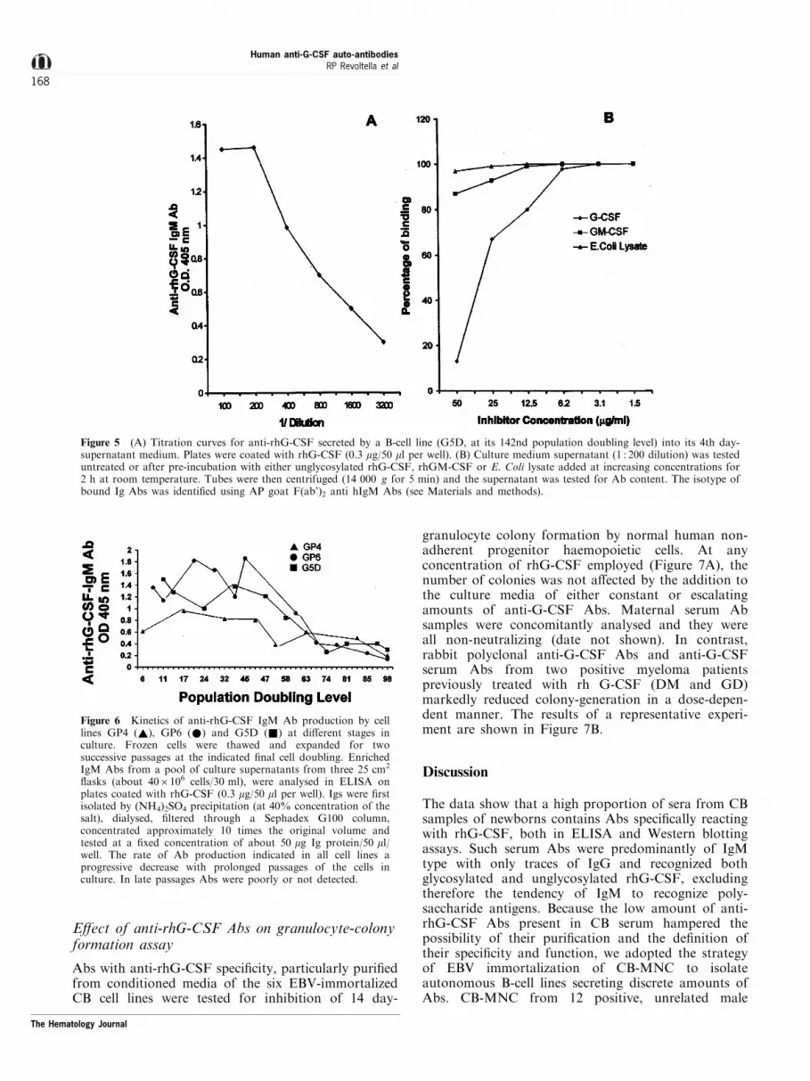
However, two (G4E and G5D) of the six cell linesproduced IgM Abs which speci®cally recognized bothrhG-CSF and rhGM-CSF. Because these two cell lineswere isolated by limiting dilution, but not by cloningtechnique, we could not exclude the presence of multiplecell clones producing Abs recognizing each of the twoproteins. Indeed Ab reactivity against either rhG-CSFor rhGM-CSF adsorbed separately on plates, wasinhibited by adding in excess the speci®c cytokine, butnot other molecules. The results of a representativeexperiment performed with one of these two cell lines(G5D) are shown in Figure 5. Further experiments arerequired to ascertain the clonal speci®city of these twolines.
Loss of anti-G-CSF Ab production with theprogress of culture
The Ig secretion by all six cell lines was examined fromearly to late passages. IgM Abs were secreted in earlypassages and signi®cant amounts were produced untillate stages. Transient phases of higher or lower anti-
Table 2 Binding speci®city of Abs from six EBV-immortalized CB-derived B cell lines
Cytokinesa
rhIL-3, rhIL-4, Other proteinsrhG-CSF rhIL-8, rhSCF, (BSA, HSA,
Cell lines Unglycosylated Glycosylated rhGM-CSF rhIL-1b, rhIFN-a2A E. Coli lysate)
GP4 + + ± ± ±GP6 + + ± ± ±G4E + + + ± ±G5D + + + ± ±G11DE + + ± ± ±G11AC + + ± ± ±
Cell lines were isolated from ®ve di�erent male newborns. Cell lines G11DE and G11AC were obtained from the same CB sample. aPositive andnegative antibody binding, by combined direct and competition ELISA and Western blot analysis.
Table 3 Cell surface immunoglobulin expression by six EBV-immortalized CB-derived human B cell lines producing anti-rhG-CSF IgM Abs
Immunoglobulin expressionb
L chainCell lines PDLa sIg sIgM sIgD k l
GP4 12 96.5 95.0 88.3 5.3 76.0GP6 11 93.5 96.0 78.5 16.4 81.7G4E 10 91.2 86.7 76.4 23.0 56.3G5D 15 95.1 92.4 92.3 18.6 87.5G11DE 10 89.2 93.1 93.1 7.0 44.2G11AC 10 89.3 89.8 89.8 9.2 83.9aPDL, population doubling level (four days each), at the analysis.bResults are expressed as the percentage of positive cells. Cells werestained with saturing amounts of the desired ¯uorescein-conjugatedmAb and analysed by ¯ow cytometry (see Materials and methods).
The Hematology Journal
Human anti-G-CSF auto-antibodiesRP Revoltella et al
166

rhG-CSF Ab titers could be detected in culturesupernatants. However, a gradual decrease of Ab
secretion by all cell lines was noted prolonging thepassages (Figure 6).
Figure 4 Flow cytometric pro®les for the most signi®cant di�erent surface antigens (marked lines) expressed on the EBV-immortalizedB-lymphoblastoid cell line G4E. The vertical axis represents the relative number of cells, while the horizontal axis represents the intensity of¯uorescence. Irrelevant controls (light lines) are superimposed on each histogram.
The Hematology Journal
Human anti-G-CSF auto-antibodiesRP Revoltella et al
167
E�ect of anti-rhG-CSF Abs on granulocyte-colonyformation assay
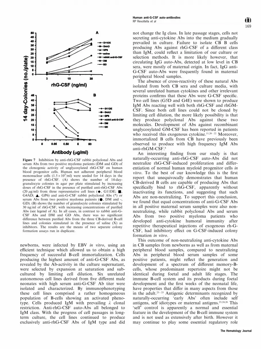
Abs with anti-rhG-CSF speci®city, particularly puri®edfrom conditioned media of the six EBV-immortalizedCB cell lines were tested for inhibition of 14 day-
granulocyte colony formation by normal human non-adherent progenitor haemopoietic cells. At anyconcentration of rhG-CSF employed (Figure 7A), thenumber of colonies was not a�ected by the addition tothe culture media of either constant or escalatingamounts of anti-G-CSF Abs. Maternal serum Absamples were concomitantly analysed and they wereall non-neutralizing (date not shown). In contrast,rabbit polyclonal anti-G-CSF Abs and anti-G-CSFserum Abs from two positive myeloma patientspreviously treated with rh G-CSF (DM and GD)markedly reduced colony-generation in a dose-depen-dent manner. The results of a representative experi-ment are shown in Figure 7B.
Discussion
The data show that a high proportion of sera from CBsamples of newborns contains Abs speci®cally reactingwith rhG-CSF, both in ELISA and Western blottingassays. Such serum Abs were predominantly of IgMtype with only traces of IgG and recognized bothglycosylated and unglycosylated rhG-CSF, excludingtherefore the tendency of IgM to recognize poly-saccharide antigens. Because the low amount of anti-rhG-CSF Abs present in CB serum hampered thepossibility of their puri®cation and the de®nition oftheir speci®city and function, we adopted the strategyof EBV immortalization of CB-MNC to isolateautonomous B-cell lines secreting discrete amounts ofAbs. CB-MNC from 12 positive, unrelated male
Figure 5 (A) Titration curves for anti-rhG-CSF secreted by a B-cell line (G5D, at its 142nd population doubling level) into its 4th day-supernatant medium. Plates were coated with rhG-CSF (0.3 mg/50 ml per well). (B) Culture medium supernatant (1 : 200 dilution) was testeduntreated or after pre-incubation with either unglycosylated rhG-CSF, rhGM-CSF or E. Coli lysate added at increasing concentrations for2 h at room temperature. Tubes were then centrifuged (14 000 g for 5 min) and the supernatant was tested for Ab content. The isotype ofbound Ig Abs was identi®ed using AP goat F(ab')2 anti hIgM Abs (see Materials and methods).
Figure 6 Kinetics of anti-rhG-CSF IgM Ab production by celllines GP4 (~), GP6 (*) and G5D (&) at di�erent stages inculture. Frozen cells were thawed and expanded for twosuccessive passages at the indicated ®nal cell doubling. EnrichedIgM Abs from a pool of culture supernatants from three 25 cm2
¯asks (about 406106 cells/30 ml), were analysed in ELISA onplates coated with rhG-CSF (0.3 mg/50 ml per well). Igs were ®rstisolated by (NH4)2SO4 precipitation (at 40% concentration of thesalt), dialysed, ®ltered through a Sephadex G100 column,concentrated approximately 10 times the original volume andtested at a ®xed concentration of about 50 mg Ig protein/50 ml/well. The rate of Ab production indicated in all cell lines aprogressive decrease with prolonged passages of the cells inculture. In late passages Abs were poorly or not detected.
The Hematology Journal
Human anti-G-CSF auto-antibodiesRP Revoltella et al
168
newborns, were infected by EBV in vitro, using ane�cient technique which allowed us to obtain a highfrequency of successful B-cell immortalization. Cellsproducing the highest amount of anti-G-CSF Abs, asrevealed by the Ab-activity in the culture supernatant,were selected by expansion at saturation and sub-cultured by limiting cell dilution. Six unrelatedautonomous cell lines derived from ®ve di�erent maleneonates with high serum anti-G-CSF Ab titer wereisolated and characterized. By immunophenotypingthese cell lines comprised a rather homogeneouspopulation of B-cells showing an activated pheno-type. Cells produced IgM with prevailing l clonalrestriction. Anti-rhG-CSF auto-Abs all belonged toIgM class. With the progress of cell passages in long-term culture, the cell lines continued to produceexclusively anti-rhG-CSF Abs of IgM type and did
not change the Ig class. In late passage stages, cells notsecreting anti-cytokine Abs into the medium graduallyprevailed in culture. Failure to isolate CB B cellsproducing Abs against rhG-CSF of a di�erent classthan IgM, could re¯ect a limitation of our culture orselection methods. It is more likely however, thatcirculating IgG auto-Abs, detected at low level in CBsera, were mostly of maternal origin. In fact, IgG anti-G-CSF auto-Abs were frequently found in maternalperipheral blood samples.
The absence of cross-reactivity of these natural Absisolated from both CB sera and culture media, withseveral unrelated human cytokines and other irrelevantproteins con®rms that these Abs were G-CSF speci®c.Two cell lines (G5D and G4E) were shown to produceIgM Abs reacting well with both rhG-CSF and rhGM-CSF. Since both cell lines could not be cloned bylimiting cell dilution, the more likely possibility is thatthey produce polyclonal Abs against these twomolecules. Development of Abs against recombinantunglycosylated GM-CSF has been reported in patientswho received this exogenous cytokine.11,20 ± 22 Moreover,immortalized B cells from CB have previously beenobserved to produce with high frequency IgM Absanti-rhGM-CSF.8
An interesting ®nding from our study is thatnaturally-occurring anti-rhG-CSF auto-Abs did notneutralize rhG-CSF-induced proliferation and differ-entiation of normal human myeloid progenitor cells invitro. To the best of our knowledge this is the ®rstreport that unequivocally demonstrates that humanCB-derived B cells are capable of producing Abs thatspeci®cally bind to rhG-CSF, apparently withoutinactivating its functions, and suggesting that suchAbs are non-neutralizing. To support this conclusion,we found that equal concentrations of anti-G-CSF Absin all positive maternal serum samples were also non-neutralizing, while rabbit polyclonal Abs and serumAbs from two positive myeloma patients whodeveloped anti-cytokine humoral immunity afterrepetitive therapeutical injections of exogenous rh-G-CSF, had inhibitory e�ect on G-CSF-induced colonyformation in vitro.
This outcome of non-neutralizing anti-cytokine Absin CB samples from newborns as well as from maternalperipheral blood samples, compared to neutralizingAbs in peripheral blood serum samples of somepositive patients, might re¯ect the generation anddevelopment of a spectrum of di�erent memory-B-cells, whose predominant repertoire might not beidentical during foetal and adult life stages. Theimmune B-cell system and its products during foetaldevelopment and the ®rst weeks of the neonatal life,have properties that di�er in many aspects from thosein the adult.23 ± 25 Antigenic determinants recognized bynaturally-occurring `early Abs' often include selfantigens, self idiotypes or maternal antigens.23,24,26 This`self' control is apparently a normal and essentialfeature in the development of the B-cell immune systemand is not used as extensively after birth. However itmay continue to play some essential regulatory role
Figure 7 Inhibition by anti-rhG-CSF rabbit polyclonal Abs andserum Abs from two positive myeloma patients (DM and GD) ofthe clonogenic activity of unglycosylated rhG-CSF on humanblood progenitor cells. Human not adherent peripheral bloodmononuclear cells (1.56105/ml) were seeded for 14 days in thepresence of rhG-CSF. (A) shows the number of 14-day-granulocyte colonies in agar per plate stimulated by increasingdoses of rhG-CSF in the presence of puri®ed anti-rhG-CSF Abs(20 mg/ml) from three representative cell lines (^, G11DE; &,G4AD; ~, GP6) and anti-G-CSF rabbit polyclonal Abs (*) orserum Abs from two positive myeloma patients (*, DM and -,GD). (B) shows the number of granulocyte colonies stimulated by30 ng/ml of rhG-CSF, with increasing concentrations of puri®edAbs (see legend of A). In all cases, in contrast to rabbit anti-G-CSF Abs and DM and GD Abs, there was no signi®cantdi�erence between puri®ed Abs from the three CB-derived B-celllines and colonies stimulated in the presence of saline (X) asinhibitors. The results are the means of two separate colonyformation assays run in duplicate.
The Hematology Journal
Human anti-G-CSF auto-antibodiesRP Revoltella et al
169

throughout life, for example particularly during post-transplant period, pregnancy, or severe infections,assuring proper tissue homeostasis, and balancingimmunity, auto-immunity and self-tolerance.27 Auto-Abs, perhaps reacting with di�erent a�nity or todi�erent epitopes of the molecule, may form solubleimmune complexes with neutralization of the cytokinein excess, or remain longer in circulation, acting ascarriers and providing a sustained supply of thecytokine without inhibiting its biological activity.Alternatively, Abs could induce changes on G-CSFand reduce its binding capacity with the receptor onblood and endothelial target cells, a�ecting activity.28
Transitory subtraction of Abs following immunecomplex formation, could also result with increasingproduction of the growth factor during a variety ofphysiological and pathological conditions.29,30
Thus, rather than being considered simply asinhibitors or antagonists, naturally occurring anti-G-
CSF antibodies are likely to be advantageous, acting asbene®cial physiological carriers and regulators of G-CSF activities. In contrast, the outcome of neutralizingAbs (occurring, for example, by cross-reaction withhomologous oligopeptides present in exogenous anti-gens,31 or abnormal anti-idiotypic interactions32,33) bydetermining a change in patterns of `physiological'autoreactivity, could lead to transform the `self'antigen to become an autoaggressive antigen, withthe development of a new status of `pathological'reactivity. All these possibilities are now open to futureinvestigation.
AcknowledgementsContract grant sponsors: CNR-P.B. Italy-Germany; CNR-P.B. Italy-Sweden; CNR-P.F. M.A.D.E.S.S. 28; IstitutoSuperiore di SanitaÁ , Italy-USA Project on `Therapy ofTumors'.
References
1 Nagata S, Tsuchiya M, Asano S, Yamamoto O, Hirata Y,Kubata N, Oheda M, Nomura H, Yamazaki T. Thechromosomal gene structure and two mRNAs for humangranulocyte colony stimulating factor. The EMBOJournal 5: 575, 1986.
2 Asano S. Human granulocyte-colony stimulating factor:Its basic aspects and clinical applications. AmericanJournal of Pediatrical and Hematological Oncology 13:400, 1991.
3 Nicola N. Granulocyte-colony stimulating factor, p. 77In: Dexter TM, Garland JM, Testa NG (eds) ColonyStimulating Factors: Molecular and Cellular Biology.Marcel Dekker, New York, 1990.
4 Nemunaitis J. Cytokine-mobilized peripheral bloodprogenitor cells. Seminars in Oncology 23: 9, 1996.
5 Fukunaga R, Seto Y, Mizushima S, Nagata S. Threedi�erent mRNAs encoding for human granulocyte-colony stimulating factor receptor. Proceedings of theNational Academy of Sciences USA 87: 8702, 1990.
6 American Society of Clinical Oncology. Recommenda-tion for the use of hemopoietic colony-stimulatingfactors: Evidence-based, clinical practice guidelines.American Society of Clinical Oncology 12: 2471, 1994.
7 Laricchia-Robbio L, Moscato S, Genua A, Liberati AM,Revoltella RP. Naturally occurring and therapy-inducedantibodies to human granulocyte colony stimulatingfactor (G-CSF) in human serum. Journal of CellularPhysiology 173: 219, 1997.
8 Revoltella RP, Laricchia Robbio L, Moscato S, GenuaA, Liberati AM. Natural and therapy-induced anti-GM-CSF and anti-G-CSF antibodies in human serum.Leukemia and Lymphoma 26 (1): 29, 1997.
9 Webster ABD, Efter T, Asheron GL. Escherichia Coliantibody: A screening test for immunode®ciency. BritishMedical Journal 3: 16, 1974.
10 Ragnhammer P, Frie sen HJ, FroÈ din JE, Lefvert AK,Hassan M, OÈ stenborg A, Mellstedt H. Induction of anti-recombinant granulocyte-macrophage colony stimulat-ing factor (Escherichia Coli derived) antibodies andclinical e�ects in non-immunocompromised patients.Blood 584: 4078, 1994.
11 Revoltella RP, Laricchia Robbio L, Liberati AM.Natural and therapy-induced anti-GM-CSF antibodiesin human serum, p. 463 In: Waxman S (ed) Challenges ofModern Medicine, Vol. 10. Ares Serono Symposia Publ.,Rome, New York, 1995.
12 Lam KMC, Crawford DH. Method for generation ofhuman B lymphoblastoid cell lines using Epstein-Barrvirus. Methods in Cell Science 17: 67, 1995.
13 Wall FE, Henkel RD, Stern MP, Jenson HB, Moyer MP.An e�cient method for routine Epstein-Barr virusimmortalization of human B lymphocytes. In vitroCellular and Developmental Biology 31: 156, 1995.
14 Bird AG, McLachlan SM, Britton S. Cyclosporin Apromotes spontaneous outgrowth in vitro of Epstein-Barrvirus-induced B-cell lines. Nature 289: 300, 1981.
15 Vinante F, Rampe AM,Morosato L, Meo A, RomagnaniS, and Pizzolo G. Peripheral T lymphocytes cytokinepro®le (IFNg, IL-2, IL-4) and CD30 expression/releaseduring measles infection. Haematologica 84: 683, 1999.
16 Be�y P, Di Bartolo V, Laricchia Robbio L, Pegoraro S,Chiello E, Rovero P, Caracciolo L, Revoltella RP. Smallsynthetic peptides of human GM-CSF require di�erentconditions for immobilization, epitope density andpresentation in ELISA. Fundamental of Clinical Immu-nology 2: 53, 1994.
17 Be�y P, Rovero P, Di Bartolo V, Laricchia Robbio L,Dane A, Pegoraro S, Bertolero F, Revoltella RP. Animmunodominant epitope in a functional domain nearthe N-terminus of human granulocyte-macrophagecolony-stimulating factor identi®ed by cross-reaction ofsynthetic peptides with neutralizing anti-protein andanti-peptide antibodies. Hybridoma 13: 457, 1994.
18 Laemmli UK. Cleavage of structural proteins duringassembly of the head of bacteriophage T4. Nature 227:680, 1970.
19 Towbin H, Staehelin T Gordon J. Electrophoretictransfer of proteins from polyacrylamide gels tonitrocellulose sheets: Procedure and some applications.Proceedings of the National Academy of Sciences USA 76:4350, 1979.
The Hematology Journal
Human anti-G-CSF auto-antibodiesRP Revoltella et al
170

20 Gribben JG, Devereux S, Thomas NSB, Klein M, JonesHM, Goldstone AH, Linch DC. Development ofantibodies to unprotected glycosylation sites on recom-binant human GM-CSF The Lancet 335: 434, 1990.
21 Mellstedt H. Induction of anti-granulocyte-macrophagecolony-stimulating factor antibodies against exogenousnon-glycosylated GM-CSF: biological implications.Journal of Interferon Research 14: 179, 1994.
22 Ragnhammer P, Fayerberg J, FroÈ din JE, WersaÈ ll P,Hansson LO, Mellstedt H. Granulocyte-macrophagecolony-stimulating factor augments the induction ofantibodies, especially anti-idiotypic antibodies, to ther-apeutic monoclonal antibodies. Cancer Immunology andImmunotherapy 40: 367, 1995.
23 Vakil M, Kearney JF. Regulatory in¯uences of neonatalmultispeci®c antibodies on the developing B-cell repe-toire. International Immunology 3: 117, 1988.
24 Kearney JF. Formation of autoantibodies, includinganti-cytokine antibodies, is a hallmark of the immuneresponse of early B-cells. Journal of Interferon Research14: 151, 1994.
25 Kearney JF, Berthels J, Hamilton AM, Lehuen A,Solvason N, Vakil M. Development and function of theearly B-cell repertoire. International Review of Immunol-ogy 8: 247, 1992.
26 Bendtzen K, Hansen MB, Ross C, Poulsen LK, SvensonM. Cytokines and autoantibodies to cytokines. StemCells 13: 206, 1995.
27 Goodnow CC. Balancing immunity, autoimmunity, andself-tolerance, p. 55 In: Chiovazzi N, Lahita RG, PavekaK, Ferrarini M (eds) B lymphocytes and Autoimmunity.Am. N.Y. Acad. Sci., N.Y., 1997.
28 Dighiero G. Natural autoantibodies, tolerance andautoimmunity. p. 182 In: Chiovazzi N, Lahita RG,Paveka K Ferrarini M (eds) B lymphocytes andAutoimmunity. Am. N.Y. Acad. Sci., N.Y., 1997.
29 Bendtzen K, Svenson M, Jùnsson V, Hippe E. Auto-antibodies to cytokines: friends or foes? ImmunologyToday 11: 167, 1990.
30 Revoltella RP. Naturally and therapeutically-inducedantibodies to cytokines. Biotherapy 10: 321, 1998.
31 Bost K, Hahn BH, Saag MS, Shaw GM, Weigent DA,Blalock JE. Individuals infected with HIV possessantibodies against Il-2. Immunology 65: 611, 1988.
32 Avrameas S. Natural antibodies. From `horror autotox-icus' to `gnothi sauton'. Immunology Today 12: 154, 1991.
33 Kazatchkine MD. Natural IgG autoantibodies in the seraof healthy individuals. Journal of Interferon Research 14:165, 1994.
The Hematology Journal
Human anti-G-CSF auto-antibodiesRP Revoltella et al
171


