Embed Size (px)
Citation preview

Immunol. Cell Biol. (1988)66. 159-165
Role of the CD21 antigen in lymphocyte transformation byEpstein-Barr virus
Andrew W. Boyd and John Fecondo
The Lions Clinical Cancer Research Laboratory, The Walter and Eliza Hall Institute of MedicalResearch, The Royal Melbourne Hospital, Parkville, Vic. 3050, Australia
(Submitted 8 October 1987. Accepted for publication 24 December 1987.)
Summary The human CD21 membrane antigen has been shown to bind Epstein-Barr Virus (EBV) andsome binding and functional data imply a role for this interaction in the B cell tropism of EBV. In thisreport the technique of limit dilution analysis was used to provide evidence that B cell transformation byEBV only occurred when CD21 antigen was present and that all cells expressing CD21 weretransformation-competent. The rate of transformation varied, being increased if B cells were activatedby an additional B cell mitogen. and being dependent on. although not strictly proportional to, theintensity of CD21 expression. The possibility that EBV might enter by other means was examined byselectively deleting CD21 antigen from the cell surface. This was accompanied by a profound decrease inthe transformation frequency. It is concluded that CD21 antigen is necessary for EBV transformationand that CD21 expression is the major limiting factor in these events.
INTRODUCTION
The CD21 cluster of monoclonal antibodies(1) defines a 140 000 dalton glycoprotein onthe membrane of human B cells (2). Thismolecule has now been intensively investi-gated and three functional properties iden-tified. The first function characterized wasthe highly specific binding of the C3dg frag-ment of complement factor C3 (3,4). Thereceptor also binds other cleavage fragmentsof C3, but with less affinity (5). Thesestudies identified this molecule as the CR2receptor for complement (4). Another line ofinvestigation of the CD21 has focused onthe capacity of some antibodies to thismolecule to augment B cell activating sig-nals (6,7). The possibility that CD21 isimportant in activation is supported by dataobtained in the mouse demonstrating thatC3 acts as a cofactor in mouse B cell activa-tion (8) and by the observation that stronglyCD21-positive cells are most readily acti-vated by B cell mitogens (9). The third line
Abbreviations used in this paper: C3, complementfragment 3; CD, cluster designation; EBV, Epstein-Barrvirus; Ig, immunoglobulin; PEC, peritoneal exudatecells.
of investigation has focused on the role ofthis antigen as the receptor for Epstein-Barrvirus (EBV). It has now been clearly demon-strated that both monoclonal and hetero-antisera to the CD21 antigen inhibit EBV-induced B cell mitogenesis (10,11). One ofthe monoclonals (B2) was shown to competefor virus binding to the B cell (10). Otherstudies have shown that the purified CD21molecule specifically absorbs EBV (12).
Phenotypic analysis at various stages of Bcell maturation demonstrates a characteris-tic pattern of antigen expression of CD21antigen. It is undetectable on pre-B cellpopulations and tumours, but is expressedon resting, surface Ig-positive B cells (13).After activation by a panel of B cell mitog-ens the antigen intensity decreases to unde-tectable levels by about 72 h after activation(9), and is then absent from all subsequentstages including the end cell stage (plasmacell). In part this study aims to determinehow transformation by EBV is correlatedwith and determined by CD21 -antigenexpression.
In this report we investigate the relativeimportance of CD21 expression using alimit-dilution cloning assay. We will showthat infectivity by EBV correlates strongly

160 A. W. BOYD AND J. FECONDO
with CD21 antigen expression at all stages inboth in vitro and in vivo (freshly isolated) Blymphocytes. Moreover, we will demon-strate that results obtained for EBV-inducedmitogenesis also hold true for EBV-inducedcell transformation and indicate that CD21expression is both the necessary and limit-ing factor in EBV infection.
MATERIALS AND METHODS
Human lymphoid cellsPreparation of human splenocytes has been de-
scribed in detail elsewhere (9). Splenic tissue wasobtained from operative specimens according to proto-cols approved by institutional ethics committees. Thesplenic tissue was gently dissociated into Eisen's bal-anced salt solution to form a single cell suspension.Mononuclear cells were isolated on Ficoll-Hypaquegradients and cryopreserved in liquid nitrogen.
Foetal spleen and bone marrow cells were obtainedfrom approximately 20 week old aborted humanfoetuses. The tissues were prepared as single cell sus-pensions and further purified on Ficoll-Hypaquegradients.
Foetal blood was obtained from placental tissueimmediately post partum. Only intact placenta wereused to minimize possible contamination with mater-nal blood. Mononuclear cells were prepared on Ficoll-Hypaque gradients. The B cell fraction was then pre-pared by E rosetting.
Epstein-Barr virusEBV used in this study was obtained from the super-
natants from dense cultures of the marmoset cell lineB95-8. Supernatants were frozen at - 7 0 ^ in aliquots.An aliquot was tested for mitogenic activity on splenicB cells by measuring tritiated thymidine incorporationafter 1 week of co-culture with different dilutions of thesupernatant. In most experiments frozen aliquots ofvirus from a single batch of B95-8 supernatant wereused. Other batches were used at the same titre as deter-mined by the mitogenesis assay.
Monoclonal antibodiesA panel of monoclonal antibodies specific for the
monocyte/macrophage lineage, natural killer cells, andB and T lymphocytes were used in this study. Bl{CD20), B2 (CD21), T4 {CD4), T8 (CD8) and Mol(CDlib) were obtained from Coulter Immunology(Hialeah, FL), THB5 (CD21) and OKTl 1 (CD2) wereobtained from the American Tissue Type Culture Col-lection. The WEHI-B2 (CD21) and WEHI-Bl (CD20)antibodies were prepared in our own laboratory bystandard methods.
Anti-Ig antibody reagentsAffinity purified rabbit anti-human Fab2 antibody
was coupled to Affigel 702 beads (Bio-rad, Richmond,CA) as described previously (9).
Fluorescent stainingIndirect Cells were prepared in 5% pooled human
serum from individuals with type AB blood group (ABserum) in human tonicity phosphate buffered saline,and aliquots of 10̂ cells were incubated with each anti-body (generally a 1:100 to 1:200 dilution of ascites) for10 min at room temperature. After washing, the cellswere incubated with a 1:50 dilution of fiuorescein-conjugated sheep anti-mouse Ig antibody (Silenus,Melbourne) for 20 min at 4°C. The cells were washedand either analysed immediately or fixed in 1% formal-dehyde for subsequent analysis.
Direct and dual fluorescence staining Direct fluor-escein- and biotin-conjugated monoclonal antibodies(Bl-FITC and B2-biotin) were obtained from CoulterImmunology (Hialeah, FL). Binding of biotin-conjugated antibodies was detected by incubation withTexas red TM (Molecular Probes, Junction City, OR)conjugated to avidin (Calbiochem, La JoUa, CA),
Cytofluorimetry and cell sortingAnalysis and cell sorting was performed on a modi-
fied FACS II (Becton Dickinson) or on EPICS cellsorter (Coulter Electronics, Hialeah, FL).
B cell culturesMicro-cultures B cells were purified by cell sorting
or by two treatments with anti-T4, T8, and Mol anti-bodies and complement. The cells were cultured in96-well round bottom microtitre trays (Costar, Cam-bridge, MA) at 10-50 000 per well in RPMI-1640/10%foetal calf serum/2 mmol/1 glutamine/1 mmol/1 sod-ium pyruvate. Medium containing B95-8 supernatantwas added to yield a final culture volume (per well) of200 |jl.
Thymidine uptake assayThymidine uptake was used as an index of mitogenic
activity. Microcultures were pulsed with 0 2 nCi of[^H]-thymidine (Amersham, Eastbourne, England) perwell and harvested 15 h later. Dried filters werecounted on a Packard Td-carb scintillation counter(Downers Grove, IL).
Limit dilution culturesEeeder layers Most experiments described
employed 100 000 mouse peritoneal exudate cells(PEC) per microwell. In other experiments the samenumber of irradiated human peripheral blood mono-nuclear cells or human thymocytes was used. All thesecell types gave uniform responses over the range of Bcells/well tested. Cultures without filler cells or withmurine 3T3 cells failed to support B cell growth at low Bcell densities.
Microcultures The feeder cells were plated out anda number (300-30 000/well) of the test B cell popu-lations added to each well. For each cell number tested60-96 identical culture wells were established. EBVand in some cases anti-Ig was then added. The cultureswere fed weekly with fresh medium, and after about 3weeks the number of transformants was counted underan inverted phase contrast microscope. Considerable

CD21 ANTIGEN EXPRESSION LIMITS EBV TRANSFORMATION 161
variability was still noted from experiment to experi-ment. These variations may in part be due to vari-ability in feeder cells and to patient variability as other-wise all features were constant.
Limit dilution analysisThe statistical methods used in this study have been
outlined in detail previously (14). In brief, three ormore experimental points were obtained as follows: foreach cell number plated in 60-90 wells, the number ofwells containing no transformants (negative wells) wasdetermined. The number of negatives versus cells/wellwas used to calculate the slope (frequency of transfor-mation) and y intercept (spontaneous transformants)with 95% confidence limits for each value.
RESULTS
The eflect on EBV transformation of the lossofCDH antigen by prior activationThe CD21 antigen is rapidly lost after B cellactivation (9). To determine the effect ofthese events on EBV-induced transforma-tion, purified splenic B cells were preparedand activated with anti-Ig beads. The cellswere harvested at varying times andanalysed for CD21 expression. The cellswere also cultured (100-10 000 B cells/well)with EBV (B95-8 supernatant) and mousePEC cells (100 000/well). Table 1 shows thepercentage CD21 antigen expression andthe frequency of transformation at differenttime points after anti-Ig-induced activation.There was a decline in the frequency oftransformation from 8 per 1000 cells at day0 to less than 2 per 10 000 at day 5, except fora consistent increase in transformation ratewhen day 1 activated cells were culturedwith EBV. Similarly when day 0 (unstimu-lated spleen) cells were co-cultured withEBV alone (Fig. 1) or with both anti-Ig andEBV (Fig. 1) the rate of EBV transformationwas increased when both mitogens were
Table 1. The effect of prior activation on EBV-induced transformation.
Duration ofpre-activation
(days)
Frequency transformation
CD21-positive% (X 10-3)
()91 (++)47 (0-+ + )23 (0- + )19(0- + )
8 4 (5 9-10 9)19-7(15 2-24-2)2-9(2 0-3-8)
0-39(0 24-0 54)0 1 7 (0 07-0-25)
Fig. 1. EBV transformation is facilitated by otherBcell antigens: (•) transformation with EBV alone;(o) transformation with EBV when both EBV andantigen Ig beads are present.
present. This increased transformation ratefollowing activation was not explained byCD21 expression as no enhancement ofCD21 staining was observed. This increaserequired co-culture with both mitogen andEBV. As shown in Table 2, if resting B cellswere incubated with EBV for 4 h, washedand recultured with or without anti-Ig, littledifference in transformation rate wasobserved.
Antigenically distinct resting B cellsubpopulations differ in rates of EBVtransformationIt has been shown that the level of CD21antigen expression is heterogeneous on rest-ing splenic B cells (9). By contrast, otherantigens (e.g. CD 19, CD20, HLA-DR) showquite uniform expression. This suggestedanother way to test the relative importanceof CD21 antigen. Spleen cells were labelledwith Bl(CD20)-FITC and B2(CD21)-bio-tin/Texas red avidin and sorted into cellsexpressing high, low or undetectable levelsof CD21 antigen as described previously (9).Re-analysis of these groups confirmed less

162 A. W. BOYD AND J. FECONDO
Table 2. Pre-incubation with EBV followed by anti-Iginduced stimulation does not increase transform-ation.
Pre-incubation*
IsStimulus
Transformationupdate
(ct/min)t (X
- - 319± 42Anti-Ig 8963±291
EBV - 1579± 63Anti-lg 9423 ±319
NTNT
0-7 (0-49-0-91)0-83(0-6-0-96)
*The purified spleen cells were incubated at100 000/ml for 4 h with EBV, washed twice and recul-tured at 30 000/well in microtitre trays for 3 days forthymidine uptake studies or at limit dilutions for fre-quency determination with additions as shown.
t Thymidine uptake was determined after 3 days.tCells were plated at 30 000/3000 per well and
300/well as described and frequencies determined at 14days.
than 10% overlap of the three groups. Thecells were incubated for 2 h with EBV thenre-cultured at limit dilution. As shown inFig. 2, the frequency of transformationincreased with increasing CD21 antigendensity. The difference between thefrequencies of transformants in the low andhigh CD21 groups was only marginally sig-nificant ( 0 . 0 5 < P < 0 . 1 ) . However both
Fig. 2. EBV transformation correlates with CD21expression. Spleen cells were sorted into high (o), low(x) and negative CD21 (o) groups and cultured withEBV for 14 days at limit dilution.
these groups differed significantly from theCD21-negative group {P < 0.01).
EBV infectivity of B cells of differingontogenyThe importance of CD21 antigen in EBVtransformation was also examined in devel-oping B cells. CD21 antigen is known to beabsent or only weakly expressed on bonemarrow pre-B cells. Other antigens such asla (HLA-DR), which have been implicatedin EBV binding, are strongly expressed onpre-B cells (13). Foetal splenocytes, bonemarrow, cord blood B cells and adult spleenB cells were cultured at limit dilution (300-10 000/well) with 100 000 mouse PEC andthe frequency of transformation determinedafter 2-3 weeks. In Table 3, the frequency oftransformation in each tissue was tabulatedwith the expression of CD20 and CD21antigens in each tissue. Calculated on a per Bcell (CD20-positive) basis the frequency oftransformation in all of the other tissues,where most cells co-express both CD20 andCD21 antigens is comparable. In foetal bonemarrow the somewhat higher percentage ofCD20 cells probably reflects the presence ofCD21-negative pre-B cells.
Antigenic modulation of CD21 antigendramatically inhibits transformation byEBVThe above results all provide evidence forthe importance of the CD21 antigen in EBVtransformation. However, it could beargued that the differences were due to otherfactors which tend to segregate with the levelof CD21 antigen expression. One way toaddress this was to modulate CD21 from thecell surface and thus test a single cell popu-
Table3. EBV transformation of B cells at differentstages of ontogeny.
Tissue
Foetal bonemarrow
Foetal spleen cellsCord bloodAdult spleen B
cells
CD21-positive
{%)
8864778
CD20-positive
(%)
16843983
Frequency oftransformation
(X 10--̂ )
0-7(0-4-l-0)10-3(7-8-12-8)2-8 (1-9-3-7)8-4(7-0-9-8)
CD21 ANTIGEN EXPRESSION LIMITS EBV TRANSFORMATION 163
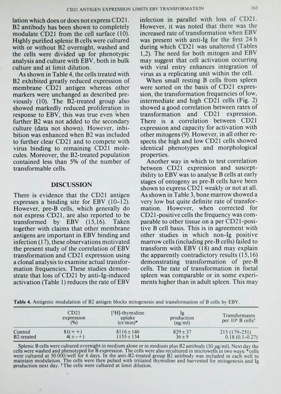
lation which does or does not express CD21.B2 antibody has been shown to completelymodulate CD21 from the cell surface (10).Highly purified splenic B cells were culturedwith or without B2 overnight, washed andthe cells were divided up for phenotypicanalysis and culture with EBV, both in bulkculture and at limit dilution.
As shown in Table 4, the cells treated withB2 exhibited greatly reduced expression ofmembrane CD21 antigen whereas othermarkers were unchanged as described pre-viously (10). The B2-treated group alsoshowed markedly reduced proliferation inresponse to EBV, this was true even whenfurther B2 was not added to the secondaryculture (data not shown). However, inhi-bition was enhanced when B2 was includedto further clear CD21 and to compete withvirus binding to remaining CD21 mole-cules. Moreover, the B2-treated populationcontained less than 5% of the number oftransformable cells.
DISCUSSION
There is evidence that the CD21 antigenexpresses a binding site for EBV (10-12).However, pre-B cells, which generally donot express CD21, are also reported to betransformed by EBV (15,16). Takentogether with claims that other membraneantigens are important in EBV binding andinfection (17), these observations motivatedthe present study of the correlation of EBVtransformation and CD21 expression usinga clonal analysis to examine actual transfor-mation frequencies. These studies demon-strate that loss of CD21 by anti-Ig-inducedactivation (Table 1) reduces the rate of EBV
infection in parallel with loss of CD21.However, it was noted that there was theincreased rate of transformation when EBVwas present with anti-Ig for the first 24 hduring which CD21 was unaltered (Tables1,2). The need for both mitogen and EBVmay suggest that cell activation occurringwith viral entry enhances integration ofvirus as a replicating unit within the cell.
When small resting B cells from spleenwere sorted on the basis of CD21 expres-sion, the transformation frequencies of low,intermediate and high CD21 cells (Fig. 2)showed a good correlation between rates oftransformation and CD21 expression.There is a correlation between CD21expression and capacity for activation withother mitogens (9). However, in all other re-spects the high and low CD21 cells showedidentical phenotypes and morphologicalproperties.
Another way in which to test correlationbetween CD21 expression and suscept-ibility to EBV was to analyse B cells at earlystages of ontogeny as pre-B cells have beenshown to express CD21 weakly or not at all.As shown in Table 3, bone marrow showed avery low but quite definite rate of transfor-mation. However, when corrected forCD21 -positive cells the frequency was com-parable to other tissue on a per CD21-posi-tive B cell basis. This is in agreement withother studies in which non-Ig positivemarrow cells (including pre-B cells) failed totransform with EBV (18) and may explainthe apparently contradictory results (15,16)demonstrating transformation of pre-Bcells. The rate of transformation in foetalspleen was comparable or in some experi-ments higher than in adult spleen. This may
Table 4. Antigenic modulation of B2 antigen blocks mitogenesis and transformation of B cells by EBV.
CD21expression
%
[^H]-thymidineuptake
(ct/min)*
Igproduction
(ng/ml)
Transformantsper lO-* Bcellst
ControlB2-treated
8116±1461155±134
829 ±3736±9
215(179-251)0.18 (0.1-0.27)
Splenic B cells were cultured overnight in medium alone or in medium plus B2 antibody (50 /Jg/ml). Next day thecells were washed and phenotyped for B expression. The cells were also recultured in microwells in two ways: * cellswere cultured at 50 000/well for 6 days. In the anti-B2-treated group B2 antibody was included in each well tomaintain modulation. The cells were then pulsed with tritiated thymidine and harvested for mitogenesis and Igproduction next day. tThe cells were cultured al limit dilution.

164 A. W. BOYD AND J. FECONDO
reflect the relatively greater expression ofCD21 on foetal spleen cells when comparedwith post-natal B cells.
While these studies showed a good corre-lation between CD21 expression and EBV-infectibility they did not directly indicate anecessary role for CD21 antigen in transfor-mation. To approach this question we usedthe observation that CD21 antigen can becleared from the B cell in a highly selectivemanner by treatment with the B2 antibody(10). When this procedure was performed ahighly significant inhibition of EBV trans-formation occurred. As the capacity of thesecells to be activated by mitogens (anti-Ig)was not impaired by this procedure, itwould appear that the basic properties andprocesses of the cell are intact and unim-paired. Hence, the inhibition of EBV trans-formation appears to provide good evidencefor an obligatory role of CD21 in efficientvirus entry and transformation.
In summary the findings presented hereprovide evidence for the requirement ofCD21 expression for EBV transformation.The data suggest that lack of CD21 makestransformation at least inefficient and prob-ably ineffective. One determinant of theefficiency of transformation was entry intocell cycle induced by other mitogens. Theother determinant of susceptibility was theoverall level of CD21 antigen expression,higher expression giving higher transforma-tion frequencies. That CD21 antigen wasdirectly responsible was addressed by clear-ing CD21 by B2 antibody treatment. Thisprofoundly inhibited EBV transformation.
Acknowledgments The authors acknowledge theexcellent technical assistance of Filomena Micozziand Karen Welch, and thank the Lions Clubs andAnti-Cancer Council of Victoria for their generoussupport.
REFERENCES
1. Boyd, A. W. 1987. Human leukocyte antigens. Anupdate on structure, function and nomenclature.Pathology 19: 329-337.
2. Nadler, L. M., Stashenko, P., Harding, R., vanAgthoven, A., Terhorst, C. and Schlossman, S. F.1981. Characterization of a human B cell specificantigen(B2) distinct from Bl. J, Immunol. 126:1941-1947.
3. Iida, K., Nadler, L. M. and Nussensweig, V. 1981.Identification of the membrane receptor for thecomplement fragment C3d by means of a mono-clonal antibody. J. Exp. Med. 138: 1021-1034.
4. Weiss. J., Tedder, T. F. and Fearon, D. T. 1984.Identification of a 145,000 Mr membrane proteinas a C3d receptor (CD2) of human B lymphocytes.Proc. Natl Acad. Sci. USA 81: 881-885.
5. Fearon, D. T. 1984. Cellular receptors for frag-ments of the third component of complement.Immunoi Today 5: 105-110.
6. Wilson, B. S., Platt. J. L. and Kay, N. E. 1985.Monoclonal antibodies to the 140,000 mol. wt.glycoprotein of B lymphocyte membranes (CR2receptor) initiates proliferation of B cells in vitro.Blood 66: 824-829.
7. Nemorow, G. R., McNaughton, M. E. and Cooper,N. R. 1985. Binding ofmonoclonal antibody to theEpstein-Barr virus (EBV)/C3d complement recep^tor {CR2) induces activation and differentiation ofhuman B lymphocytes. ./, Immunol. 135: 3068-3073.
8 Melchers, F. and Lenhardt, W. 1985. Three restric-tion points in the cell cycle of activated murine B
lymphocytes. Proc. Natl Acad. Sci. USA 82: 7681-7685.
9. Boyd, A. W., Anderson, K. C, Freedman. A. S.et ai 1985. Studies on the in vitro activation anddifferentiation of normal human B cells. I. Pheno-typic and functional characterization of the B cellresponse to anti-Ig antibody. J. Immunol 134:1516-1522.
10. Nadler, L. M., Boyd, A. W., Park, E. et al. 1984.The B cell-restricted glycoprotein (B2) is the recep-tor for Epstein-Barr virus. In Leukocyte Typing U,Vol. 2, E. L. Reinherz, B. F. Haynes.L. M. Nadler,and I. D. Bernstein, (eds). Springer-Verlag, NewYork, pp. 509-516.
11. Frade, R., Barel, M., Ehlin-Henriksson, B. andKlein, G. 1985. gpl40, the C3d receptor of humanB lymphocytes is also the Epstein-Barr virus recep-tor. Proc. Natl Acad. Sci. USA 82:1490-1495.
12. Fingeroth, J., Weiss, J., Tedder, T. F., Strominger,J., Biro, P. and Fearon, D. 1984. Epstein-Barr virusreceptor of human B lymphocytes is the C3d recep-tor CR2. Proc. Natl Acad. Sci USA 81: 4500-4514.
13. Nadler, L. M., Korsmeyer, S. J., Anderson, K. C. etal. 1984. The B ceil origin of non-T cell acutelymphoblastic leukaemia: a model for discretestages of neoplastic and normal Pre B cell differen-tiation. J. Clin. Invest. 74: 332-347.
14. Good, M. F., Boyd, A. W. and Nossal, G. J. V.1983. Analysis of true anti-hapten cytotoxic clonesin limit dilution microcultures after correction for"anti-self activity: precursor frequencies, Ly-2

CD21 ANTIGEN EXPRESSION LIMITS EBV TRANSFORMATION 165
and Thy-1 phenotypes, specificity and statisticalmethods. / Immunoi 130: 2046-2053.
15. Hansson, M., Falk, K. and Emberg, I. 1983.Epstein-Barr virus transformation of human pre-Bcells. / Exp. Med. 158: 616-626.
16. Katamine, S., Otsu, M., Tada, K. et ai 1984.Epstein-Barr virus transforms precursor B cellseven before immunoglobulin gene rearrange-ments. Nature :iO9: 369-372.
17. Trowbridge, I. S., Hyman, R. and Klein, G. 1977.Human B cell lines deficient in the expression of Bcell specific cytoproteins (gp27/35). Eur. J.Immunoi 1: 640-645.
18. Hibi, T., Chan, M.A., Petsche, D. and Dosch, H-M. 1986. Phenotype, frequency and EBV respon-siveness of human marrow B and pre B cells.J. Immunoi 136: 3211-3216.