Embed Size (px)
Citation preview

4932–4943 Nucleic Acids Research, 2009, Vol. 37, No. 15 Published online 15 June 2009doi:10.1093/nar/gkp497
Translation of intronless RNAs is stronglystimulated by the Epstein–Barr virus mRNAexport factor EB2Emiliano P. Ricci1,2,3, Fabrice Mure1,2,3, Henri Gruffat1,2,3, Didier Decimo1,2,3,
Cahora Medina-Palazon1,2,3, Theophile Ohlmann1,2,3 and Evelyne Manet1,2,3,*
1INSERM U758, Unite de Virologie Humaine, 2Ecole Normale Superieure de Lyon, Lyon F-69007 and3IFR128 Biosciences Gerland-Lyon Sud, Lyon F-69364, France
Received November 28, 2008; Revised May 18, 2009; Accepted May 21, 2009
ABSTRACT
The Epstein–Barr virus protein (EB2) allows thenuclear export of a particular subset of early andlate viral RNAs derived from intronless genes. EB2is conserved among most herpesvirus members andits presence is essential for the production of infec-tious particles. Here we show that, besides its roleas a nuclear export factor, EB2 strongly stimulatestranslation of unspliced mRNAs without affectingoverall cellular translation. Interestingly, this effectcan be reversed by the addition of an intron withinthe gene. The spliced mRNA is then efficientlyexported and translated even in the absence ofEB2. Moreover, we show that EB2 associates withtranslating ribosomes and increases the proportionof its target RNA in the polyribosomal fraction.Finally, testing of EB2 homolog proteins derivedfrom EBV-related herpesviruses, shows that, evenif they play similar roles within the replication cycleof their respective virus, their mechanisms of actionare different.
INTRODUCTION
In eukaryotic cells, gene expression is tightly controlledfrom the biogenesis of messenger RNAs (mRNAs)within the cell nucleus, until their export and translationin the cytoplasm (1). In particular, the control of mRNAtranslation is a multi-step complex mechanism mediatedby a large number of factors. Translation initiationappears to be the rate-limiting and most regulated stepof the overall translation mechanism (2,3). Regulation of
translation initiation is mediated mostly by initiationfactors, which recruit the 40S ribosomal subunit to the50 cap of the mRNA, allow scanning to the initiationcodon and then the recruitment of the 60S ribosomalsubunit (4).
Even though maturation of pre-mRNAs occurs in adifferent cellular compartment than translation, proteinsthat participate in the former process can also play arole in the latter. Indeed, translation stimulation ofintron-containing genes has been observed in several sys-tems and is linked to proteins that participate either insplicing or in the export of spliced mRNAs (5–12).Among these, the exon junction complex (EJC), which isdeposited during splicing and plays an important role inmRNA surveillance, is able to modulate translation ofspliced mRNAs through the mTOR pathway (7). Otherproteins involved in translation stimulation of splicedmRNAs comprise the Ser-Arg-rich (SR) proteins thatplay a role not only in pre-mRNA splicing and spliceo-some assembly but also in splice-site recognition and selec-tion (13,14). Conversely, recent data have also shownthat some of the SR proteins, which shuttle from thenucleus to the cytoplasm together with the splicedmRNA, can be associated with translating ribosomesto stimulate the translation of spliced mRNAs (11,15).This would allow the cell to ensure that only fully splicedRNAs are expressed as opposed to unspliced or incom-pletely spliced RNAs that could result in translation ofaberrant proteins.
Viruses have evolved different mechanisms to efficientlyexport and translate unspliced RNAs. One example is theconstitutive transport element (CTE) present in simpleretroviruses, such as the Mason–Pfizner monkey virus(MPMV), which interacts with the TAP/NXF1 exportprotein and the cellular protein NXT1/p15 to promote
*To whom correspondence should be addressed. Tel: +33 472 728 176; Fax: +33 472 728 137; Email: [email protected] address:Cahora Medina-Palazon, Westmead Institute for Cancer Research, University of Sydney, Westmead Millenium Institute, Westmead, New SouthWales 2145, Australia.
The authors wish it to be known that, in their opinion, the first two authors should be regarded as joint First Authors.
� 2009 The Author(s)This is an Open Access article distributed under the terms of the Creative Commons Attribution Non-Commercial License (http://creativecommons.org/licenses/by-nc/2.0/uk/) which permits unrestricted non-commercial use, distribution, and reproduction in any medium, provided the original work is properly cited.
by guest on April 6, 2016
http://nar.oxfordjournals.org/D
ownloaded from

export and translation of unspliced genomic RNA(15–18). Again, translation stimulation of unsplicedRNAs containing the CTE seems to rely on SR proteinssuch as 9G8 (15). For complex retroviruses, such as lenti-viruses, the unspliced genomic RNA is exported by theviral protein Rev which interacts with cis-acting sequenceslocated within the envelope coding region of the RNA,allowing export and translation stimulation (19–22).
The Epstein–Barr virus (EBV) early protein EB2 (alsoknown as BMFL1, Mta or SM) shares several featureswith mRNA export factors. It is able to interact withRNA both in vitro and in vivo (23–25), it shuttles betweenthe nucleus and the cytoplasm and it allows the cytoplas-mic accumulation of unspliced RNAs generated fromintronless and intron-containing genes, probably by therecruitment of REF and TAP/NXF1 (24,26–28). EB2 isessential for the production of viral particles and promotesthe nuclear export of some early and most late viralmRNAs generated from EBV intronless genes (28).Moreover, like EBV many other herpesviruses code fora protein similar to EB2, i.e. ICP27 from herpes simplexvirus type 1 (HSV1) (29–31), UL69 from cytomegalovirus(CMV) (32) ORF57 from Kaposi’s sarcoma-associatedherpesvirus (KSHV) (33) and ORF57 from herpesvirussaimiri (HVS) (34). All these proteins act as nuclearmRNA export factors but surprisingly their functioncannot be trans-complemented between each other(27,35).
As cellular mRNA export factors, EB2 shuttles fromthe nucleus to the cytoplasm, probably associated withits target mRNAs, suggesting that it could also affecttheir translation. We thus tested the effect of EB2 ontranslation from an intronless reporter construct codingfor the Renilla luciferase. In this system, translation stim-ulation was measured by analyzing the expression levelsof Renilla luciferase normalized to the amount of cyto-plasmic mRNAs determined by quantitative PCR. Ourresults show that EB2 strongly and specifically stimulatestranslation of intronless mRNAs without affecting overallcellular translation or protein stability. Introduction of anintron in this construct stimulates the efficiency of exportand translation of the luciferase mRNA and at the sametime abrogates the effect of EB2. Interestingly, the increasein translation of the luciferase mRNA generated fromthe intronless construct in the presence of EB2, correlateswith an increase in the proportion of luciferase mRNAassociated with polyribosomes. Moreover, we show thatEB2 itself is associated with polyribosomes. Finally, acomparison between EB2 homologs from EBV-relatedviruses (HSV-1, CMV, KSHV) led us to conclude thattheir mechanisms of action are different.
MATERIALS AND METHODS
Cell lines and transfections
HeLa and HEK293T cells were grown in Dulbecco’s mod-ified Eagle medium supplemented with penicillin, strepto-mycin and 5% fetal calf serum (Invitrogen). NIH3T3PKR-deficient cells (36) and NIH3T3 wild-type cellswere grown as described for HeLa cells. For experiments
using EBV gene-derived constructs, transfections wereperformed in 100mm plates using calcium phosphatewith 15 mg of total DNA (0.5 mg of pTRE2-Flag.BDLF1,0.25mg of pCI-Flag.EB2 and 0.5mg of pTet-On or pTet-Off expression vectors (Clontech) and pUC18 up to 15 mg).When necessary, doxycyclin was added at a concentrationof 1 mg/ml. For experiments using the luciferase reporterplasmids, transfections were carried out in 60mmplates using Lipofectamine 2000 (Invitrogen) followingthe manufacturer’s protocol. For the metabolic labelingexperiments, transfection of HeLa cells with the EB2expression plasmid was carried out in six-well platesusing Lipofectamine 2000 (Invitrogen). The efficiency oftransfection was evaluated by transfecting the pEGFP-C1plasmid (Clontech) in the same conditions and countingthe number of green fluorescent cells by FACS. Over 60%of the cells expressed GFP. For polysomal fractionation,HEK293T cells were seeded at 2� 106 on a 150mm cul-ture dish 3 days before polysomal fractionation. Forty-eight hours before harvesting, cells were transfected with1.25mg of luciferase-coding plasmid together or not with6.25mg of pCI-Flag.EB2, using PEI reagent.
Plasmids
The pCI-Flag.EB2 construct has been previouslydescribed (28). For pTRE2-Flag.BDLF1, the EBVBDLF1 open reading frame tagged with the Flag epitopewas amplified by PCR and cloned into the BamHI andXbaI sites of the pTRE2 expression plasmid (Clontech).The pTet-On vector was supplied by Clontech (Tet-Offand Tet-On gene expression systems). The pcDNAGlobinRen reporter plasmid was constructed by cloningthe human b-globin 50UTR (with the authentic initiationcodon) followed by the Renilla luciferase reporter gene,amplified by PCR from the p-Globin Renilla vector (37)into the double digested (XbaI/AflII) pcDNA3.1 expres-sion vector. For the pcDNAIntron-GlobinRen, thesequence corresponding to the intron of the humanb-globin gene was amplified by PCR and cloned into thepcDNAGlobinRen vector previously digested by XbaI.pCI-mycORF57 contained the complete ORF57 codingsequence (first exon included) cloned in frame with themyc epitope, in pCI (Promega). The expression plasmidfor UL69 (pCMV-UL69) was kindly provided byT. Stamminger (38). The expression plasmid for ICP27(pCI-FlagICP27) has been previously described (27).
RNA extraction and real-time quantitative PCR(RT-PCR) from cytoplasmic RNAs
Cells were first scraped from the dish and then resus-pended in 200 ml of cold RLNa buffer (10mM Tris–HCl(pH 8), 10mM NaCl, 3mM MgCl2, 1mM DTT, 0.5%NP40 and 10U/ml of RNaseOUT (Invitrogen). After5min incubation on ice, lysed cells were centrifuged for2min at 400g at 48C and the supernatant was then recov-ered. One milliliter of Trizol (Invitrogen) was thenadded to the supernatant and RNAs were extracted fol-lowing the Trizol protocol provided by the manufacturer.Cytoplasmic RNAs (1mg) were treated with RQ1 DNAse(Promega) to avoid DNA contamination and reversed
Nucleic Acids Research, 2009, Vol. 37, No. 15 4933
by guest on April 6, 2016
http://nar.oxfordjournals.org/D
ownloaded from

transcribed with (dT)16 and 1 ml of Superscript II enzyme(Invitrogen) in a 20 ml reaction mix at 428C for 1 h.PCRs were performed using a Taq core kit (Q-Biogen)
with a set of specific primer pairs (BDLF1 forward/BDLF1 reverse: Table 1) on various amounts of the RTreaction mixtures (0.05, 0.4, 1 or 2 ml) to have a linearlyincreasing signal after 25 PCR cycles. The PCR-amplifiedfragments were then analyzed on 2% agarose gels. Weevaluated the endogenous expression of b-actin mRNAby RT-PCR (b-actin forward/b-actin reverse: Table 1).Amplification of a 690-bp DNA fragment correspondingto the b-actin mature mRNA showed that no DNA con-tamination was present in our RNA preparations. Wetested for the presence of U6 snRNA using RT-PCR(U6 forward/U6 reverse: Table 1).For RT-qPCR, a 20 ml reaction was prepared with 5 ml
of template cDNA (1/20 diluted), 10 ml of MESA greenSYBR premix (Eurogentec), 0.2 mM of each primer andsubjected to amplification using a fluorescence thermocy-cler (Applied Biosystems 7000 RT-PCR, Foster City, CA)under the following conditions: 10min at 948C for initialdenaturation, followed by 40 cycles of denaturation at958C for 15 s, annealing at 608C for 15 s and elongationat 728C for 30 s. This program was followed by a meltingcurve analysis in order to verify the specificity of the PCRproduct. Renilla luciferase was amplified in parallel withthe housekeeping gene GAPDH (for HeLa cells) or HPRT(for mouse cells) and relative copy numbers of RenillacDNAs were compared to GAPDH using x–�Ct (wherex corresponds to the experimentally calculated amplifica-tion efficiency of each primer couple). The primersequences used in this study (presented in Table 1) weredesigned using Beacon designer software (fromPREMIER Biosoft).
Western blotting analysis
Cells were collected by centrifugation, lysed on ice for30min in 100ml of HNTG buffer (50mM HEPES pH7.5; 150mM NaCl; 1% Triton X-100; 10% glycerol;1mM EDTA; 1mM phenylmethylsulfonyl fluoride).
Proteins were separated on 10% sodium dodecyl sulfate-polyacrylamide electrophoresis gels and then transferredto a nitrocellulose membrane by electroblotting (Hybond-ECL; Amersham Biosciences). Membranes wereincubated with, respectively, anti-Flag M2 (Sigma) oranti a-tubulin (T5168, Sigma) monoclonal antibodies oran anti-PABP (kind gift from S. Morley) rabbit polyclonalantibody (39). Goat anti-mouse horseradish peroxidaseconjugate or goat anti-rabbit horseradish peroxidase con-jugate (Amersham) were used at a dilution of 1:5000 assecondary antibodies. For immunoblot detection, theECL system (Amersham) was used.
Luciferase assays
Renilla activity from transfected cells was measured in aVeritasTM Luminometer (Turner Biosystems) using theRenilla luciferase assay system (Promega Madison Co).Luciferase activity was measured for identical amountsof total protein as evaluated by Bradford assay.
Polyribosome fractionation
Polyribosome fractionation was performed essentially asdescribed previously (40,41). Forty-eight hours after trans-fection, HEK293T cells were treated with 100 mg/ml cyclo-heximide at 378C for 5min and harvested by scrapingfrom the plate. In some experiments, EDTA was addedto the cell lysate at a final concentration of 15mM to dis-rupt the polysomes. The gradient was collected fromthe top using a Piston Gradient Fractionator (BioComp,New Brunswick, Canada) with concomitant measurementof the absorbance at 254 nm using an AKTA purifier (GEHealthcare, Amersham Biosciences, Piscataway, NJ)coupled to a fraction collector. For western blotting,20 ml of each fraction were separated on 12% sodiumdodecyl sulfate-polyacrylamide electrophoresis gels andthen transferred to a PVDF membrane (Amersham bio-sciences) by electroblotting. The membrane was then incu-bated with various antibodies as described above inthe ‘western blotting analysis’ section. RNA extractionwas performed as described (15). RNAs were then reversetranscribed and analyzed by quantitative PCR asdescribed above.
RESULTS
EB2 stimulates protein expression from EBV-derivedmRNAs
It has been shown that the EBV-encoded viral protein EB2acts as a nuclear export factor for a particular subset ofmRNAs generated from intronless genes and for someunspliced mRNAs derived from intron-containing genes(24,26–28). However, although EB2 probably shuttlesfrom the nucleus to the cytoplasm together with theexported RNA, the effect of EB2 on mRNA translationhas never been reported. This prompted us to test whetherexpression of EB2 could have an influence on translationof its target viral mRNAs. We thus tested the effect of EB2on translation of an EBV-encoded mRNA whose cyto-plasmic accumulation depends on EB2. In order to control
Table 1. PCR primers used in this studya
Primer name Primer sequence
BDLF1 forward CAGATTTGAAAGTGGTAGTGTCBDLF1 reverse TTATCTTAACCAGCAAGTGGCCGb-actin forward (Human) GCTGCGTGTGGCTCCCGAGGAGb-actin reverse (Human) ATCTTCATTGTGCTGGGTGCCAGGAPDH forward (Human) TCCACCACCCTGTTGCTGTAGGAPDH reverse (Human) ACCCACTCCTCCACCTTTGACHPRT forward (Mouse) TCATTATGCCGAGGATTTGGAHPRT reverse (Mouse) CAGAGGGCCACAATGTGATGRenilla forward AGGTGAAGTTCGTCGTCCAACATTATCRenilla reverse GAAACTTCTTGGCACCTTCAACAATAGC18S rRNA forward (Human) GTGGAGCGATTTGTCTGGTT18S rRNA reverse (Human) CGCTGAGCCAGTCAGTGTAG28S rRNA forward (Human) TGGGTTTTAAGCAGGAGGTG28S rRNA reverse (Human) AACCTGTCTCACGACGGTCTU6 forward (Human) CGCTTCGGCAGCACATATACU6 reverse (Human) AAAATATGGAACGCTTCACGA
aPrimers for real-time PCR were designed using Beacon designersoftware (from PREMIER Biosoft).
4934 Nucleic Acids Research, 2009, Vol. 37, No. 15
by guest on April 6, 2016
http://nar.oxfordjournals.org/D
ownloaded from

transcription of the target mRNA, we used the Tet-Ongene expression system (Clontech) to express the EBVBDLF1 late mRNA (Figure 1A). For this, thepTRE2-Flag-BDLF1 plasmid was transfected into HeLacells together with the pTet-On vector (Clontech) and withor without the EB2 expression plasmid. Expression of EB2and the BDLF1 protein was then monitored by westernblotting using an anti-Flag antibody (Figure 1B). Theamount of cytoplasmic BDLF1 mRNAs was quantifiedby semi-quantitative RT-PCR (Figure 1C). In cellscotransfected with the BDLF1 reporter plasmid and thepTet-On vector, in the absence of doxycycline, no BDLF1protein was detected (Figure 1B, lane 4) and only a lowamount of BDLF1 mRNAs were detected by RT-PCR(Figure 1C, lane 4). In these conditions, the expressionof EB2 enhanced the cytoplasmic accumulation of thecorresponding BDLF1 mRNA (Figure 1C, lane 6) butagain no BDLF1 protein was detected (Figure 1B, lane6). When doxycycline was added, the transcription ofBDLF1 mRNAs was strongly stimulated (Figure 1C,lane 3) although only low levels of BDLF1 protein weredetected by western blot in the absence of EB2 (Figure 1B,lane 3). However, in the presence of EB2 there was a large
increase in the amount of BDLF1 protein expressed(Figure 1B, lane 5) although the amount of BDLF1mRNA detected in the cytoplasm was comparable tothat in the absence of EB2 (Figure 1C, compare lanes 3and 5). Indeed, in these conditions, transcription ofBDLF1 triggered by doxycycline was so strong thatit appeared to compensate for the otherwise poor cyto-plasmic accumulation observed in the absence of EB2.This allowed us to focus just on the effect of EB2 onBDLF1 translation without introducing a bias with theamount of mRNA. Finally, upon addition of doxycycline,only a weak signal corresponding to the BDLF1 mRNAexpression was detected by RT-PCR in the absence orpresence of EB2 (Figure 1C, lanes 1 and 2) and noBDLF1 protein expression was detected (Figure 1B,lanes 1 and 2) thus ruling out any non-specific effect ofdoxyxycline on BDLF1 expression in the absence of thepTet-On vector. Essentially similar results were obtainedusing the Tet-Off system for which transcription ofBDLF1 was triggered by the absence of doxycycline(data not shown).These results strongly suggest that EB2 is able to
stimulate protein expression from EBV-derived mRNAs.
EB2 stimulates both export and translation independentlyof an EBV-specific sequence without affecting globalmRNA translation
In order to quantify the relative effects of EB2 on mRNAexport and translation we designed a reporter constructcontaining the 50UTR of the human b-Globin gene fol-lowed by the Renilla luciferase coding region under thecontrol of the cytomegalovirus (CMV) immediate earlypromoter (Figure 2A). It is important to note that thisconstruct does not contain any EBV-related sequence.This construct was cotransfected together with the EB2expression vector in HeLa cells and total luciferase activitywas analyzed 24 h after transfection (Figure 2D, toppanel), while expression of EB2 was measured by westernblotting (Figure 2B). Cytoplasmic RNAs were extractedand we first quantified the amount of U6 snRNA in orderto make sure that our cytoplasmic fractions were notcontaminated by nuclear material (Figure 2C). Then cyto-plasmic luciferase RNAs were quantified by relative quan-titative RT-PCR using the housekeeping gene GAPDH asan internal control (Supplementary Figure 1).Interestingly, EB2 had no effect on GAPDH mRNAexport thus ruling out any bias in the relative quantifica-tion of luciferase-coding mRNAs. As expected, expres-sion of EB2 promoted the cytoplasmic accumulation ofluciferase-coding RNAs in a dose-dependent manner(Figure 2D, middle panel) thus leading to a stimulationof luciferase expression (Figure 2D, top panel). The luci-ferase activity measured in each assay was then normal-ized to the amount of luciferase cytoplasmic RNAs. Thisgives an exact measure of the luciferase activity per RNAmolecule, and thus can be considered as a quantitativerepresentation of the translation rate. Interestingly, usingthis quantification we found that an average 7-fold stim-ulation of luciferase mRNA translation in the presence ofEB2 was observed (Figure 2D, bottom panel).
Figure 1. EB2 stimulates expression of the late EBV viral proteinBDLF1. (A) Schematic representation of the BDLFI-encoding con-struct pTRE2-Flag-BDLF1. (B) Western analysis of Flag-BDLF1expression in HeLa cells transfected with pTRE2-Flag-BDLF1,together with pTet-On and an expression plasmid for Flag-EB2 (pCI-FEB2) as indicated in the figure. Doxycycline was added as indicated.The M2 anti-Flag MAb was used to visualize both Flag-BDLF1 andFlag-EB2 proteins. (C) Quantification of the BDLF1 cytoplasmic RNAby semi-quantitative RT-PCR in HeLa cells transfected as describedabove.
Nucleic Acids Research, 2009, Vol. 37, No. 15 4935
by guest on April 6, 2016
http://nar.oxfordjournals.org/D
ownloaded from

To test any impact EB2 could have on the stability ofthe neosynthesized luciferase protein we analyzed the sta-bility of the luciferase protein in the absence and presenceof EB2. For this, we blocked cellular translation by addingcycloheximide and measured luciferase decay activity overtime on cells expressing, or not, EB2. As shown inFigure 3A, EB2 did not affect the stability of the Renillaluciferase protein, which had a 30-min half-life both in theabsence and presence of EB2.Furthermore, the lack of EBV-derived sequences on the
luciferase reporter construct prompted us to test the effectof EB2 on global cellular mRNA translation. We thusperformed a metabolic labeling of cells expressing (ornot) EB2 (Figure 3B). For this, cells were pulsed in thepresence of radiolabeled methionine for 30min. Cells werethen lysed and proteins resolved on SDS–PAGE to quan-tify the overall cellular translation rates. As shown inFigure 3B, we did not detect any significant difference in
translation rates between cells expressing EB2 and thosenot expressing EB2.
We also wanted to exclude the possibility that translationstimulation driven by EB2 depends on protein kinase R(PKR). PKR is the principal cellular factor involved in theinterferon-mediated inhibition of viral translation. Thiskinase is activated by double-stranded RNA and this acti-vation leads to the phosphorylation of initiation factoreIF2a, thus inhibiting translation initiation of all cellularand viral RNAs (42,43). We thus monitored luciferasetranslation both in NIH3T3 PKR-deficient cells (36)and wt NIH3T3 in the presence or absence of EB2.We found that EB2 was able to stimulate translation ofluciferase codingmRNAs even in the absence of PKR (Sup-plementaryDataFigure2),whichargues foramechanismoftranslation stimulation independent of the PKR pathway.
Finally, to rule out the possibility that translation rateswere affected by the amount of cytoplasmic mRNAs, thus
Figure 2. EB2 stimulates expression of a Renilla luciferase intronless gene at the translational level, independently of any viral-derived sequence. (A)Schematic representation of the luciferase intronless coding vector used in this study (pcDNAGlobinRen) showing positions of the CMV promoterand BGH polyadenylation signal. (B) Immunoblot of HeLa cells cotransfected with pcDNAGlobinRen together with the empty pCI vector orincreasing amounts of the FlagEB2-encoding plasmid, pCI-FlagEB2 (250 and 500 ng). The M2 anti-Flag MAb was used to visualize Flag-EB2.Asterisk denotes an unspecific band detected by the M2 anti-Flag antibody. (C) Quantification of the amount of U6 snRNA present respectively inthe nuclear and cytoplasmic fractions of cellular extracts used in D. U6 snRNA was amplified by RT-PCR using the specific primer set indicated inTable 1, and analyzed on a 2% agarose gel. (D) Measure of luciferase activity and quantification of cytoplasmic luciferase-encoding mRNAs byquantitative RT-PCR using GAPDH as an internal control. Total luciferase activity was measured 24 h post-transfection (top panel) and the amountof cytoplasmic luciferase coding mRNAs was quantified (middle panel). Translational efficiency (bottom panel) was calculated by normalizing thetotal luciferase activity by reference to the amount of cytoplasmic luciferase mRNA. AU: arbitrary units.
4936 Nucleic Acids Research, 2009, Vol. 37, No. 15
by guest on April 6, 2016
http://nar.oxfordjournals.org/D
ownloaded from

leading to a stimulation of translation which would beindependent of EB2, we transfected increasing amountsof luciferase expression vector in HeLa cells expressing,or not, EB2 (Figure 3C). As EB2 expression leads to a4–5-fold increase of mRNA levels in the cytoplasm, itwas of interest to test if increasing the amount of luciferasemRNAs in the absence of EB2 could lead to a stimulationof translation. For this, we measured luciferase translation
rates (luciferase activity/amount of luciferase mRNA) forincreasing amounts of cytoplasmic mRNAs either in thepresence or absence of EB2 (Figure 3C). As presented inthe top panel, luciferase expression increased proportion-ally with the amount of cytoplasmic mRNAs, both in thepresence and absence of EB2. However, when luciferaseactivity was normalized to the amount of cytoplasmicmRNAs (which corresponds to the translation rate perarbitrary unit of mRNA) there was no significant changein luciferase translation rates for increasing amountsof cytoplasmic luciferase mRNAs in control (pCI) andEB2-expressing cells (bottom panel). Nevertheless, transla-tion rates in the presence of EB2 were systematically4–5-fold more than those in the absence of EB2, andthis for identical mRNA amounts measured in the cyto-plasm of the cells.Taken together, these results show that EB2 stimulates
translation of mRNAs without affecting the stability ofthe neosynthesized protein. Interestingly, EB2 expressiondoes not affect global mRNA translation. Moreover,translation stimulation does not depend on the amountof cytoplasmic mRNA available for translation.
Addition of an intron within the reporter constructimpairs translation stimulation driven by EB2
Our results show that EB2 is able to stimulate translationof viral genes and that of a non-related reporter gene with-out affecting global cellular translation. Consequently,we focused on understanding the lack of an effect ofEB2 on cellular mRNA translation. Interestingly, EB2has been shown to specifically stimulate the nuclearexport of mRNA generated from intronless genes (23).This prompted us to test the translation of an mRNAgenerated from an intron-containing gene in the presenceof EB2. We have thus introduced the b-globin intronwithin the 50UTR of the luciferase reporter gene(Figure 4A). In order to test the efficiency of splicing ofthe corresponding mRNA, an RT-PCR from cytoplasmicmRNAs was performed using a forward and reverseprimer flanking the intron (Figure 4B). We observedthat transfection of the intronless construct in HeLacells led to the expression of an unspliced mRNA which,upon RT-PCR, yielded a band of the same size to thatfrom the control PCR, performed directly using the DNAplasmid as a template (Figure 4B, lanes 2, 3 and 4). On thecontrary, transfection of the intron-containing constructled to the expression of an mRNA which, upon RT-PCR,yielded a band slightly longer than that of the intronlessmRNA, but shorter than that of the control PCRobtained from the intron-containing DNA plasmid(Figure 4B, lanes 5–7). This band corresponds to thespliced form of the luciferase-coding mRNA. Thus themRNA transcribed from this intron-containing reportergene was efficiently spliced both in the presence or absenceof EB2.We then quantified the exact amount of cytoplasmic
luciferase mRNA by quantitative RT-PCR and measuredthe corresponding luciferase activities (SupplementaryFigure 3). When translation rates were calculated bynormalizing the luciferase activity to the amount of
Figure 3. EB2 does not affect protein stability or global cellular mRNAtranslation and its effect on translation is independent of the amount ofcytoplasmic luciferase coding mRNA. (A) Time-lapse measure of totalluciferase activity from HeLa cells mock transfected (pCI) or trans-fected with an EB2 expression vector (pCI-FlagEB2) (250 ng) afteraddition of cycloheximide to block translation. Luciferase activitywas measured 0, 15, 30, 60, 120, 180 and 240min after addition ofcycloheximide to the cell medium. (B) Metabolic labeling of HeLacells mock transfected (pCI) or transfected with the EB2-encodingplasmid (250 ng), using 35S-labeled methionine. After a 30-min pulselabeling, cells were lyzed and total cellular proteins resolved on 12%SDS-PAGE. Total translation was quantified by phosphorimagingusing a Fujifilm FLA5100. (C) Top panel: Luciferase activity was plot-ted against the amount of cytoplasmic luciferase coding mRNAs inHeLa cells transfected with increasing amounts of luciferase-encodingplasmid in the absence (pCI) or presence (pCI-FlagEB2) of EB2 (250and 500 ng). Bottom panel: Translation rates per unit of luciferase-encoding mRNAs (calculated by normalizing luciferase activity by ref-erence to the amount of cytoplasmic luciferase-encoding mRNAs) inHeLa cells transfected with increasing amounts of luciferase-encodingplasmid in the absence or presence of EB2 expression plasmid. AU:arbitrary units.
Nucleic Acids Research, 2009, Vol. 37, No. 15 4937
by guest on April 6, 2016
http://nar.oxfordjournals.org/D
ownloaded from

luciferase-coding mRNA (Figure 4C), we observed a10-fold more efficient translation of the spliced mRNAcompared to unspliced in the absence of EB2. As expected,in the presence of EB2 the translation rate of the unsplicedmRNA was increased by a factor 6. However, expressionof EB2 did not further stimulate translation of the splicedluciferase mRNA but rather led to a mild inhibition.
It is noteworthy, that we also observed an inhibitoryeffect of EB2 on the accumulation of luciferase mRNAgenerated from the intron-containing construct (Supple-mentary Figure 3) which corroborates previously pub-lished data from Ruvolo et al. (44).
These results indicate that EB2 specifically stimulatestranslation of intronless mRNAs without significantlyaffecting translation of spliced mRNAs.
EB2 co-sediments with polyribosomes and increases theutilization of reporter mRNA by the translation machinery
To determine whether EB2 is associated with the transla-tion machinery and to find out whether it increases theassociation of its target mRNAs with polyribosomes,we performed sucrose gradient analysis to separate poly-ribosomes from monoribosomes and uncomplexed riboso-mal subunits. HEK293T cells were transfected withpcDNAIntron-GlobinRen alone, pcDNAGlobinRenalone or pcDNAGlobinRen together with pCI-Flag.EB2. Cell extracts were prepared and fractionatedon 10–50% sucrose gradients and fractions were first ana-lyzed by western blotting. An example of the UV absor-bance profile of the gradients is shown in Figure 5A andthe corresponding 18S and 28S RNA profile, determinedby RT-PCR, in Figure 5B. UV absorbance and the 18S/28S RNA profiles from all gradients were very similar.The polyribosome distribution of EB2 was comparedwith that of PABP, a general translation factor forpolyA+ mRNA (45,46) (Figure 5A). As expected,PABP was present across the gradient from mRNPs topolyribosomal fractions. Interestingly, EB2 cosedimentedwith the 80S ribosome but was also found in the lighterpolysomal fractions. As a control, a-tubulin was onlyfound associated with the uncomplexed ribosomal subunitfractions. Furthermore, treatment of cytoplasmic extractswith EDTA, which is known to induce a dissociation ofmono- and polyribosomes into ribosomal subunits,induced a redistribution of EB2 to the top of the gradient(Figure 5D and E).
We then analyzed the distribution of the reportermRNAs throughout our sucrose gradients by quantitativeRT-PCR (Figure 5C). Interestingly, the proportion of theRenilla luciferase mRNA generated from the intron-lessconstruct, which is found associated with the polyriboso-mal fractions, is greatly increased in the presence of EB2(compare the middle and top panels). Furthermore, in thelatter case, the profile of repartition of the Renilla lucifer-ase mRNA throughout the gradient is very similar to thatobtained with the luciferase mRNA generated from theintron-containing construct (bottom panel). As expected,Renilla luciferase mRNA moved to the lighter fractionsof the gradient following EDTA treatment (Figure 5F).Taken together, these data suggest that EB2 directlyincreases the utilization of its target reporter mRNA bythe translation machinery.
EB2 viral homolog proteins exhibit different effects onmRNA generated from intronless genes
Herpesviruses code for EB2 homolog proteins that alsoserve as viral mRNA export factors. Interestingly, despite
Figure 4. Translation stimulation does not occur with spliced mRNAs.(A) Schematic representation of the intronless (pcDNAGlobinRen)and the intron-containing (pcDNAIntronGlobinRen) vectors encodingthe Renilla luciferase showing positions of the human b-globin intronwithin the 50UTR of the luciferase construct (gray arrows correspond topositions of the PCR primers used to test efficient splicing of the intron).(B) RT-PCR (using primers shown in A) from cells cotransfected withpcDNAGlobinRen and pCI or the EB2-encoding plasmid pCI-FlagEB2(lanes 2 and 3) or from cells cotransfected with pcDNAIntronGlobinRenand pCI or pCI-FlagEB2 (lanes 5 and 6), or directly from the purifiedDNA vector (lanes 4 and 7). (C) Luciferase activity normalized byreference to the amount of cytoplasmic luciferase-encoding mRNAsfrom HeLa cells cotransfected with pcDNAGlobinRen and pCI or pCI-FlagEB2, or pcDNAIntronGlobinRen and pCI or pCI-FlagEB2.The amount of luciferase-encoding mRNAs was monitored by quantita-tive RT-PCR.
4938 Nucleic Acids Research, 2009, Vol. 37, No. 15
by guest on April 6, 2016
http://nar.oxfordjournals.org/D
ownloaded from
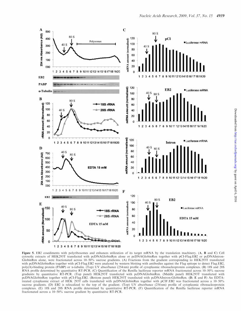
Figure 5. EB2 cosediments with polyribosomes and enhances utilization of its target mRNA by the translation machinery. (A, B and C) Cellcytosolic extracts of HEK293T transfected with pcDNAGlobinRen alone or pcDNAGlobinRen together with pCI-Flag.EB2 or pcDNAIntron-GlobinRen alone, were fractionated across 10–50% sucrose gradients. (A) Fractions from the gradient corresponding to HEK293T transfectedwith pcDNAGlobinRen together with pCI-Flag.EB2 were analyzed by western blotting with antibodies against the Flag epitope to detect Flag.EB2,poly(A)-binding protein (PABP) or a-tubulin. (Top) UV absorbance (254 nm) profile of cytoplasmic ribonucleoprotein complexes. (B) 18S and 28SRNA profile determined by quantitative RT-PCR. (C) Quantification of the Renilla luciferase reporter mRNA fractionated across 10–50% sucrosegradients by quantitative RT-PCR. (Top panel) HEK293T transfected with pcDNAGlobinRen. (Middle panel) HEK293T transfected withpcDNAGlobinRen together with pCI-Flag.EB2. (Bottom panel) HEK293T transfected with pcDNAIntron-GlobinRen. (D, E and F) An EDTA-treated cytoplasmic extract of HEK 293T cells transfected with pcDNAGlobinRen together with pCIF.EB2 was fractionated across a 10–50%sucrose gradients. (D) EB2 is relocalized to the top of the gradient. (Top) UV absorbance (254 nm) profile of cytoplasmic ribonucleoproteincomplexes. (E) 18S and 28S RNA profile determined by quantitative RT-PCR. (F) Quantification of the Renilla luciferase reporter mRNAfractionated across a 10–50% sucrose gradient by quantitative RT-PCR.
Nucleic Acids Research, 2009, Vol. 37, No. 15 4939
by guest on April 6, 2016
http://nar.oxfordjournals.org/D
ownloaded from

their similarities, EB2 homolog proteins cannot trans-complement each other for viral production (35,47).Thus, it was of interest to test the effect of EB2-relatedproteins in our system. For this, we used proteins fromeach herpesvirus sub-family: ICP27 from HSV1 (herpessimplex virus 1), an a-herpesvirus, ORF57 from KSHV(Kaposi’s sarcoma-associated herpesvirus), like EBV ag-herpesvirus and UL69 from CMV (cytomegalovirus), ab-herpesvirus. Among these proteins, only ICP27 has beenpreviously shown to stimulate translation of specific viralmRNAs (48,49). In order to monitor the effect of theseproteins in our luciferase reporter system, we cotrans-fected HeLa cells with the intronless luciferase constructand expression plasmids for the different EB2-related her-pesvirus proteins and we quantified both cytoplasmicmRNA accumulation (Figure 6A) and luciferase activity(not shown). Luciferase expression was again normalizedto the amount of cytoplasmic luciferase-encoding mRNAsin order to specifically measure the impact of each viralprotein on translation (Figure 6B). Expression of each ofthe viral proteins was verified by western blotting (datanot shown). Unexpectedly, the effect observed with EB2was not conserved for all of the homolog proteins. Indeed,only UL69 led to a strong luciferase translation stimula-tion similar to that of EB2 (i.e. 4–5-fold stimulation oftranslation), whereas ICP27 and ORF57 did not have asignificant effect (Figure 6B). This was probably due to thefact that neither ICP27 nor ORF57 appear to export the
luciferase mRNA (expression of ICP27 led in fact to a40% reduction of the amount of luciferase cytoplasmicmRNA) contrary to EB2 and UL69, which provoked a4–5-fold increase of luciferase cytoplasmic mRNA levels(Figure 6A).
This result shows that EB2 homologs derived fromrelated viruses have differential effects on a heterologousmRNA and suggests that export of the mRNA and stim-ulation of its translation are strongly linked.
DISCUSSION
Although the role of EB2 in the nuclear export ofunspliced RNAs has been extensively studied, its effecton translation has never been evaluated. With the growingevidence that cellular mRNA splicing and export factorsare also able to modulate translation of spliced mRNAs,we decided to test whether the viral protein EB2 could alsoaffect translation. Indeed, most of the EBV early and lategenes do not contain any intron, suggesting that bothexport and translation of the corresponding mRNAsshould be very inefficient. However, this defect is over-come by expression of the early viral protein EB2, whichinteracts with the viral mRNAs to facilitate their cytoplas-mic accumulation. This tight interaction of EB2 with theexported mRNA and its transit to the cytoplasm stronglysuggested that EB2 could also affect translation. In fact,expression of EB2 in cells coding for an EBV-derivedunspliced RNA (BDLF1) led to a strong stimulation ofBDLF1 accumulation that did not depend on an increasein cytoplasmic levels of the corresponding mRNA. Thisresult indicates that, besides its role as a nuclear exportfactor, EB2 can also stimulate translation of EBVunspliced RNAs. Interestingly, the effect of EB2 on pro-tein accumulation from unspliced mRNAs did not dependon any EBV-derived cis-acting sequence since expressionof an artificial unspliced mRNA encoding for the Renillaluciferase was also strongly stimulated without affectingprotein stability. Surprisingly, even though EB2 lacked arequirement for a specific RNA sequence, its expressiondid not affect global cellular mRNA translation suggestinga role for EB2 on translation of only a specific subset ofmRNAs.
Since EB2 has been shown to export mostly mRNAsgenerated from intronless genes (23) including its specificEBV-encoded target genes we tested the effect of the intro-duction of intronic sequences within the 50UTR of ourreporter gene. Interestingly, we found that after additionof an intron within the luciferase reporter construct, EB2was no longer able to stimulate translation, whereas it hada strong effect on the same RNA transcribed from anintronless construct.
These results together with previous results showingthat EB2 can interact with mRNA independently of anyspecific sequence (50) suggest that EB2 can bind to bothspliced and unspliced mRNAs. However, in the case ofunspliced mRNAs, which do not recruit the normal setof splicing factors, EB2 would allow their export and stim-ulate their translation to levels similar to those of splicedmRNAs. In the case of intron-containing genes, EB2
Figure 6. Differential effects on translation from EB2-related proteinsderived from different herpesviruses. (A) Cytoplasmic luciferase mRNAlevels monitored by quantitative PCR in HeLa cells mock transfected(pCI) or transfected with 500 ng of EBV EB2, HSV-1 ICP27, HKSVORF57 and HCMV UL69-encoding plasmids. (B) Translation rate.Luciferase activity was normalized by the amount of cytoplasmicmRNAs from cells mock transfected (pCI) or transfected with EBVEB2, HSV-1 ICP27, HKSV ORF57 and HCMV UL69-encodingvectors, together with the reporter plasmid pcDNAGlobinRen. AU:arbitrary units.
4940 Nucleic Acids Research, 2009, Vol. 37, No. 15
by guest on April 6, 2016
http://nar.oxfordjournals.org/D
ownloaded from

could also bind to the mRNA but we suggest that either itis excluded from the mRNA by a cellular splicing orexport factor, or it is exported to the cytoplasm with thespliced mRNA. In the latter case, its effect on translationwould be redundant in the presence of splicing proteins.Indeed, it is possible that EB2 recruited cotranscription-ally to nascent mRNAs is able to interact with an as yetunknown cellular factor necessary for translation stimula-tion of spliced mRNAs. This would explain the stimula-tion of the translation of intronless mRNAs in thepresence of EB2. On the contrary, if EB2 also interactswith spliced mRNAs, it could interfere with the cellularfactors that normally stimulate their export and transla-tion. This would explain the mild inhibition of export andtranslation that we observed upon addition of an intron inour luciferase reporter construct.
In accordance with the fact that EB2 specifically stimu-lates export and translation of mRNAs generated fromintronless genes we have shown that cellular mRNAs,which are, in the majority, generated from intron-contain-ing genes, are not globally affected by expression of EB2.There are however few cellular genes which are known fortheir absence of introns. It would be interesting to look atthe effect of EB2 on the export and translation of mRNAsgenerated from such genes. In the case of EBV, most ofthe viral mRNAs of the productive cycle are intronless.We have previously shown that EB2 is necessary for theefficient export of the majority of these, but it is interestingto note that some are efficiently exported even in theabsence of EB2, suggesting that they use an alternativeexport pathway, independent of splicing. Such an alterna-tive pathway has been previously reported with the SRproteins, 9G8 and ASF/SF2, which have been found topromote the recruitment of TAP to mRNPs (51). Anotherinteresting example of mRNA generated from an intron-less gene and which is not affected by EB2 is the fireflyluciferase mRNA expressed from an intronless construct(25). However, although there is no effect of EB2 on fireflyluciferase expression, EB2 bound efficiently to its mRNAin vivo (25). Again, it is likely that this mRNA uses analternative pathway for its export, independent of splicing.Thus, even if EB2 is associated with these mRNAs in vivo,its effect is probably redundant as discussed above in thecase of spliced mRNAs.
In order to definitively conclude on a direct effect ofEB2 on translation efficiency we studied the associationof Renilla luciferase mRNAs with polyribosomes. Thedata clearly showed that the proportion of Renilla lucifer-ase mRNAs associated with polyribosomes is largelyincreased in the presence of EB2. Moreover, we foundthat there is also an association of EB2 with polyribo-somes, suggesting that EB2 binds the mRNPs in thenucleus, where it stimulates their export and then remainsassociated with the mRNPs as far as the polyribosomes.Taken together, these results argue for a role of EB2 in cissimilar to that of the EJC proteins responsible for thetranslation stimulation of cellular mRNAs. One proposedmechanism involves an interaction between the EJC andthe 48S preinitiation complex mediated by an interactionbetween Y14:Magoh and the protein PYM (52). Anothermechanism involves the EJC-dependent recruitment of the
40S ribosomal protein S6 kinase 1 (S6K1) which is a cen-tral player in the TOR signaling cascade (7). When acti-vated by the TOR pathway, S6K1 enhances translationinitiation both by activating stimulatory factors andby inactivating inhibitory factors bound at the 50 cap ofmRNAs. The SR protein ASF/SF2 has also recently beenreported to enhance translation initiation via recruitmentof mTOR (8).Finally, we tested several EB2 homologous proteins
from other herpesviruses for their ability to stimulatetranslation of the intronless luciferase gene. Among theproteins tested, only UL69 from human cytomegalovirus(hCMV) behaves like EB2, while ICP27 and ORF57 wereunable to stimulate luciferase translation. For UL69, thisis the first report suggesting that this protein plays a role instimulation of translation. On the contrary, it has beenpreviously shown that ICP27 (from the herpes simplexvirus type 1) plays a role in regulating translation of asubset of late viral mRNAs (48,49,53). However, the inter-action between ICP27 and the viral mRNAs has beenshown to depend on specific RNA sequences distributedalong the viral genome (54). In addition, ICP27 has beenshown to be able to stimulate translation of a luciferasemRNA only if it was previously tethered to it (53). Thus, itis not surprising that in our system, ICP27 does not stim-ulate Renilla luciferase mRNA translation nor cytoplas-mic Renilla luciferase mRNA accumulation, suggestingthat translation stimulation is probably dependent onthe binding of the herpes simplex virus proteins to themRNA. Since all the herpesvirus EB2 homologous pro-teins have been shown to shuttle from the nucleus to thecytoplasm (31,38,55) it is tempting to speculate that theyfirst interact with their unspliced mRNA targets inside thenucleus, and then shuttle to the cytoplasm bound to thesemRNAs where they play a role in translation. Accordingly,it is interesting to notice that ICP27 has also been shownto be associated with polyribosomes (53). Taken together,these results suggest firstly that both EB2 and the herpes-virus EB2 homologous proteins have a direct role on trans-lation of the mRNA they interact with, and secondly thatprotein–mRNA interaction, mRNA export and transla-tion stimulation are strongly linked.EB2 expression is essential for viral particle production
and its absence leads to very poor viral DNA replicationprobably because of the low expression of early viralmRNAs that depend on EB2 for their export (i.e.BALF5 and BALF2) (27). An even more drastic effectwas seen on most of the late viral mRNAs (28,56). Wehave shown here that EB2 expression leads to a 25-foldstimulation of renilla luciferase reporter gene expressionwith a cytoplasmic accumulation of the correspondingmRNA stimulated 3.5-fold and the translation itselfstimulated from 5- to 7-fold. This suggests that the essen-tial role of EB2 during the EBV productive cycle couldbe explained by a combined role on mRNA export andtranslation stimulation.
SUPPLEMENTARY DATA
Supplementary Data are available at NAR Online.
Nucleic Acids Research, 2009, Vol. 37, No. 15 4941
by guest on April 6, 2016
http://nar.oxfordjournals.org/D
ownloaded from

ACKNOWLEDGEMENTS
We thank B. Blanquier from the ‘Plateau d’analyse genet-ique’ of IFR128 for advice on quantitative PCR. Wethank A. Chaboud and Y. Tauran from the ‘Plateau deproduction et d’analyse des proteines’ of IFR 128 for useof the AKTA purifier. We thank Dr S. Morley for provid-ing anti-human PABP antibodies and Dr D. Levy andS. Delpeut for providing PKR-deficient cells. We alsothank the Reseau Herpesvirus and Cancer for its support.Finally, we thank Dr R. Buckland for reading themanuscript.
FUNDING
‘Institut National de la Sante et de la Recherche Medicale’(INSERM), the ‘Agence Nationale pour la Recherche’(ANR) [ANR MIME: grant number RPV06120CSA(to E.M.) and ANR blanche: grant number 06-0290-01(to T.O.)] and the pole of competitivity Lyon Biopole.Fellowship from the ‘Association pour la Recherchecontre le cancer’ (ARC) (to C.M.-P.); fellowship fromthe ‘Ministere de la Recherche et de la Technologie’(MRT to E.R.). E.M. is a CNRS scientist. Funding foropen access charge: INSERM.
Conflict of interest statement. None declared.
REFERENCES
1. Moore,M.J. and Proudfoot,N.J. (2009) Pre-mRNA processingreaches back to transcription and ahead to translation. Cell, 136,688–700.
2. Gingras,A.C., Raught,B. and Sonenberg,N. (1999) eIF4 initiationfactors: effectors of mRNA recruitment to ribosomes and regulatorsof translation. Annu. Rev. Biochem., 68, 913–963.
3. Prevot,D., Darlix,J.L. and Ohlmann,T. (2003) Conducting theinitiation of protein synthesis: the role of eIF4G. Biol. Cell, 95,141–156.
4. Pestova,T.V., Kolupaeva,V.G., Lomakin,I.B., Pilipenko,E.V.,Shatsky,I.N., Agol,V.I. and Hellen,C.U. (2001) Molecularmechanisms of translation initiation in eukaryotes. Proc. Natl Acad.Sci. USA, 98, 7029–7036.
5. Le Hir,H., Nott,A. and Moore,M.J. (2003) How introns influenceand enhance eukaryotic gene expression. Trends Biochem. Sci., 28,215–220.
6. Lu,S. and Cullen,B.R. (2003) Analysis of the stimulatory effectof splicing on mRNA production and utilization in mammaliancells. RNA, 9, 618–630.
7. Ma,X.M., Yoon,S.O., Richardson,C.J., Julich,K. and Blenis,J.(2008) SKAR links pre-mRNA splicing to mTOR/S6K1-mediatedenhanced translation efficiency of spliced mRNAs. Cell, 133,303–313.
8. Michlewski,G., Sanford,J.R. and Caceres,J.F. (2008) The splicingfactor SF2/ASF regulates translation initiation by enhancingphosphorylation of 4E-BP1. Mol. Cell, 30, 179–189.
9. Nott,A., Meislin,S.H. and Moore,M.J. (2003) A quantitativeanalysis of intron effects on mammalian gene expression. RNA, 9,607–617.
10. Pfeifer,I., Elsby,R., Fernandez,M., Faria,P.A., Nussenzveig,D.R.,Lossos,I.S., Fontoura,B.M., Martin,W.D. and Barber,G.N. (2008)NFAR-1 and -2 modulate translation and are required for efficienthost defense. Proc. Natl Acad. Sci. USA, 105, 4173–4178.
11. Sanford,J.R., Gray,N.K., Beckmann,K. and Caceres,J.F. (2004)A novel role for shuttling SR proteins in mRNA translation.Genes Dev., 18, 755–768.
12. Wiegand,H.L., Lu,S. and Cullen,B.R. (2003) Exon junctioncomplexes mediate the enhancing effect of splicing on mRNAexpression. Proc. Natl Acad. Sci. USA, 100, 11327–11332.
13. Sanford,J.R., Longman,D. and Caceres,J.F. (2003) Multiple rolesof the SR protein family in splicing regulation. Prog. Mol. SubcellBiol., 31, 33–58.
14. Tacke,R. and Manley,J.L. (1999) Determinants of SR proteinspecificity. Curr. Opin. Cell Biol., 11, 358–362.
15. Swartz,J.E., Bor,Y.C., Misawa,Y., Rekosh,D. andHammarskjold,M.L. (2007) The shuttling SR protein 9G8 plays arole in translation of unspliced mRNA containing a constitutivetransport element. J. Biol. Chem., 282, 19844–19853.
16. Braun,I.C., Herold,A., Rode,M., Conti,E. and Izaurralde,E. (2001)Overexpression of TAP/p15 heterodimers bypasses nuclear retentionand stimulates nuclear mRNA export. J. Biol. Chem., 276,20536–20543.
17. Jin,L., Guzik,B.W., Bor,Y.C., Rekosh,D. and Hammarskjold,M.L.(2003) Tap and NXT promote translation of unspliced mRNA.Genes Dev., 17, 3075–3086.
18. Levesque,L., Guzik,B., Guan,T., Coyle,J., Black,B.E., Rekosh,D.,Hammarskjold,M.L. and Paschal,B.M. (2001) RNA exportmediated by tap involves NXT1-dependent interactions with thenuclear pore complex. J. Biol. Chem., 276, 44953–44962.
19. Hadzopoulou-Cladaras,M., Felber,B.K., Cladaras,C.,Athanassopoulos,A., Tse,A. and Pavlakis,G.N. (1989) The rev(trs/art) protein of human immunodeficiency virus type 1 affectsviral mRNA and protein expression via a cis-acting sequence inthe env region. J. Virol., 63, 1265–1274.
20. Malim,M.H., Hauber,J., Le,S.Y., Maizel,J.V. and Cullen,B.R.(1989) The HIV-1 rev trans-activator acts through a structuredtarget sequence to activate nuclear export of unspliced viral mRNA.Nature, 338, 254–257.
21. Perales,C., Carrasco,L. and Gonzalez,M.E. (2005) Regulation ofHIV-1 env mRNA translation by Rev protein. Biochim. Biophys.Acta, 1743, 169–175.
22. Balvay,L., Lopez Lastra,M., Sargueil,B., Darlix,J.L. andOhlmann,T. (2007) Translational control of retroviruses. Nat. Rev.Microbiol., 5, 128–140.
23. Buisson,M., Hans,F., Kusters,I., Duran,N. and Sergeant,A. (1999)The C-terminal region but not the Arg-X-Pro repeat of Epstein–Barr virus protein EB2 is required for its effect on RNA splicingand transport. J. Virol., 73, 4090–4100.
24. Hiriart,E., Farjot,G., Gruffat,H., Nguyen,M.V., Sergeant,A. andManet,E. (2003) A novel nuclear export signal and a REF inter-action domain both promote mRNA export by the Epstein–Barrvirus EB2 protein. J. Biol. Chem., 278, 335–342.
25. Ruvolo,V., Gupta,A.K. and Swaminathan,S. (2001) Epstein–Barrvirus SM protein interacts with mRNA in vivo and mediates agene-specific increase in cytoplasmic mRNA. J. Virol., 75,6033–6041.
26. Farjot,G., Buisson,M., Duc Dodon,M., Gazzolo,L., Sergeant,A.and Mikaelian,I. (2000) Epstein–Barr virus EB2 protein exportsunspliced RNA via a Crm-1-independent pathway. J. Virol., 74,6068–6076.
27. Gruffat,H., Batisse,J., Pich,D., Neuhierl,B., Manet,E.,Hammerschmidt,W. and Sergeant,A. (2002) Epstein–Barr virusmRNA export factor EB2 is essential for production of infectiousvirus. J. Virol., 76, 9635–9644.
28. Batisse,J., Manet,E., Middeldorp,J., Sergeant,A. and Gruffat,H.(2005) Epstein–Barr virus mRNA export factor EB2 is essentialfor intranuclear capsid assembly and production of gp350. J. Virol.,79, 14102–14111.
29. Chen,I.H., Sciabica,K.S. and Sandri-Goldin,R.M. (2002) ICP27interacts with the RNA export factor Aly/REF to direct herpessimplex virus type 1 intronless mRNAs to the TAP export pathway.J. Virol., 76, 12877–12889.
30. Koffa,M.D., Clements,J.B., Izaurralde,E., Wadd,S., Wilson,S.A.,Mattaj,I.W. and Kuersten,S. (2001) Herpes simplex virus ICP27protein provides viral mRNAs with access to the cellular mRNAexport pathway. EMBO J., 20, 5769–5778.
31. Sandri-Goldin,R.M. (1998) ICP27 mediates HSV RNA export byshuttling through a leucine-rich nuclear export signal and bindingviral intronless RNAs through an RGG motif. Genes Dev., 12,868–879.
4942 Nucleic Acids Research, 2009, Vol. 37, No. 15
by guest on April 6, 2016
http://nar.oxfordjournals.org/D
ownloaded from

32. Lischka,P., Toth,Z., Thomas,M., Mueller,R. and Stamminger,T.(2006) The UL69 transactivator protein of human cytomegalovirusinteracts with DEXD/H-Box RNA helicase UAP56 to promotecytoplasmic accumulation of unspliced RNA. Mol. Cell Biol., 26,1631–1643.
33. Malik,P., Blackbourn,D.J. and Clements,B. (2004) The evolutiona-rily conserved Kaposi’s sarcoma-associated herpesvirus ORF57protein interacts with REF protein and acts as an mRNA exportfactor. J. Biol. Chem., 279, 33001–33011.
34. Williams,B.J.L., Boyne,J.R., Goodwin,D.J., Roaden,L.,Hautbergue,G.M., Wilson,S.A. and Whitehouse,A. (2005) Theprototype gamma 2 herpesvirus nucleocytoplasmic shuttlingprotein, ORF57, transports viral RNA through the cellular mRNAexport pathway. Biochem. J., 387, 295–308.
35. Han,Z. and Swaminathan,S. (2006) Kaposi’s sarcoma-associatedherpesvirus lytic gene ORF57 is essential for infectious virionproduction. J. Virol., 80, 5251–5260.
36. Smith,E.J., Marie,I., Prakash,A., Garcia-Sastre,A. and Levy,D.E.(2001) IRF3 and IRF7 phosphorylation in virus-infected cells doesnot require double-stranded RNA-dependent protein kinase R orIkappa B kinase but is blocked by Vaccinia virus E3L protein.J. Biol. Chem., 276, 8951–8957.
37. Soto Rifo,R., Ricci,E.P., Decimo,D., Moncorge,O. and Ohlmann,T.(2007) Back to basics: the untreated rabbit reticulocyte lysate as acompetitive system to recapitulate cap/poly(A) synergy and theselective advantage of IRES-driven translation. Nucleic Acids Res.,35, e121.
38. Lischka,P., Rosorius,O., Trommer,E. and Stamminger,T. (2001)A novel transferable nuclear export signal mediates CRM1-independent nucleocytoplasmic shuttling of the human cytomega-lovirus transactivator protein pUL69. EMBO J., 20, 7271–7283.
39. Fraser,C.S., Pain,V.M. and Morley,S.J. (1999) The association ofinitiation factor 4F with poly(A)-binding protein is enhanced inserum-stimulated Xenopus kidney cells. J. Biol. Chem., 274,196–204.
40. Li,Y., Bor,Y.C., Misawa,Y., Xue,Y., Rekosh,D. andHammarskjold,M.L. (2006) An intron with a constitutive transportelement is retained in a Tap messenger RNA. Nature, 443, 234–237.
41. Bor,Y.C., Swartz,J., Li,Y., Coyle,J., Rekosh,D. andHammarskjold,M.L. (2006) Northern blot analysis of mRNAfrom mammalian polyribosomes. Nat. Protoc., doi:10.1038/nprot.2006.216. Available at http://www.natureprotocols.com/2006/09/15/northern_blot_analysis_of_mrna.php.
42. Meurs,E.F., Watanabe,Y., Kadereit,S., Barber,G.N., Katze,M.G.,Chong,K., Williams,B.R. and Hovanessian,A.G. (1992) Constitutiveexpression of human double-stranded RNA-activated p68 kinase inmurine cells mediates phosphorylation of eukaryotic initiationfactor 2 and partial resistance to encephalomyocarditis virusgrowth. J. Virol., 66, 5805–5814.
43. Barber,G.N., Wambach,M., Wong,M.L., Dever,T.E.,Hinnebusch,A.G. and Katze,M.G. (1993) Translationalregulation by the interferon-induced double-stranded-RNA-activated 68-kDa protein kinase. Proc. Natl Acad. Sci. USA, 90,4621–4625.
44. Ruvolo,V., Wang,E., Boyle,S. and Swaminathan,S. (1998) TheEpstein–Barr virus nuclear protein SM is both a post-transcriptionalinhibitor and activator of gene expression. Proc. Natl Acad. Sci.USA, 95, 8852–8857.
45. Gorgoni,B. and Gray,N.K. (2004) The roles of cytoplasmic poly(A)-binding proteins in regulating gene expression: a developmentalperspective. Brief. Funct. Genomic Proteomic, 3, 125–141.
46. Mangus,D.A., Evans,M.C. and Jacobson,A. (2003) Poly(A)-bindingproteins: multifunctional scaffolds for the post-transcriptional con-trol of gene expression. Genome Biol., 4, 223.
47. Sergeant,A., Gruffat,H. and Manet,E. (2008) The Epstein–Barrvirus (EBV) protein EB2 is an mRNA export factor essential forvirus production. Frontiers Biosci., 13, 3798–3813.
48. Ellison,K.S., Maranchuk,R.A., Mottet,K.L. and Smiley,J.R. (2005)Control of VP16 translation by the herpes simplex virus type 1immediate-early protein ICP27. J. Virol., 79, 4120–4131.
49. Fontaine-Rodriguez,E.C. and Knipe,D.M. (2008) Herpes simplexvirus ICP27 increases translation of a subset of viral late mRNAs.J. Virol., 82, 3538–3545.
50. Hiriart,E., Bardouillet,L., Manet,E., Gruffat,H., Penin,F.,Montserret,R., Farjot,G. and Sergeant,A. (2003) A region of theEpstein–Barr virus (EBV) mRNA export factor EB2 containing anarginine-rich motif mediates direct binding to RNA. J. Biol. Chem.,278, 37790–37798.
51. Huang,Y., Gattoni,R., Stevenin,J. and Steitz,J.A. (2003) SR splicingfactors serve as adapter proteins for TAP-dependent mRNA export.Mol. Cell, 11, 837–843.
52. Diem,M.D., Chan,C.C., Younis,I. and Dreyfuss,G. (2007) PYMbinds the cytoplasmic exon-junction complex and ribosomes toenhance translation of spliced mRNAs. Nat. Struct. Mol. Biol., 14,1173–1179.
53. Larralde,O., Smith,R.W., Wilkie,G.S., Malik,P., Gray,N.K. andClements,J.B. (2006) Direct stimulation of translation by the mul-tifunctional herpesvirus ICP27 protein. J. Virol., 80, 1588–1591.
54. Sokolowski,M., Scott,J.E., Heaney,R.P., Patel,A.H. andClements,J.B. (2003) Identification of herpes simplex virus RNAsthat interact specifically with regulatory protein ICP27 in vivo.J. Biol. Chem., 278, 33540–33549.
55. Bello,L.J., Davison,A.J., Glenn,M.A., Whitehouse,A.,Rethmeier,N., Schulz,T. and Clements,J.B. (1999) The humanORF57 gene and its properties. J. Gen. Virol., 80, 3207–3215.
56. Han,Z., Marendy,E., Wang,Y.D., Yuan,J., Sample,J.T. andSwaminathan,S. (2007) Multiple roles of Epstein–Barr virus SMprotein in lytic replication. J. Virol., 81, 4058–4069.
Nucleic Acids Research, 2009, Vol. 37, No. 15 4943
by guest on April 6, 2016
http://nar.oxfordjournals.org/D
ownloaded from