Embed Size (px)
Citation preview
Shukrun et al. Oncogenesis (2019) 8:48 https://doi.org/10.1038/s41389-019-0156-9 Oncogenesis
ART ICLE Open Ac ce s s
NCAM1/FGF module serves as a putativepleuropulmonary blastoma therapeutic targetRachel Shukrun1,2, Hana Golan1,2,3, Revital Caspi1,2, Naomi Pode-Shakked 1,2,4, Oren Pleniceanu1,2, Einav Vax1,2,Dekel D. Bar-Lev1, Sara Pri-Chen5, Jasmine Jacob-Hirsch2,6, Ginette Schiby2,7, Orit Harari-Steinberg1,Michal Mark-Danieli1, Benjamin Dekel1,2,8 and Amos Toren2,3
AbstractPleuropulmonary blastoma (PPB) is a rare pediatric lung neoplasm that recapitulates developmental pathways of earlyembryonic lungs. As lung development proceeds with highly regulated mesenchymal-epithelial interactions, a DICER1mutation in PPB generates a faulty lung differentiation program with resultant biphasic tumors composed of aprimitive epithelial and mesenchymal stroma with early progenitor blastomatous cells. Deciphering of PPB progressionhas been hampered by the difficulty of culturing PPB cells, and specifically progenitor blastomatous cells. Here, weshow that in contrast with in-vitro culture, establishment of PPB patient-derived xenograft (PDX) in NOD-SCID miceselects for highly proliferating progenitor blastoma overexpressing critical regulators of lung development andmultiple imprinted genes. These stem-like tumors were sequentially interrogated by gene profiling to show a FGFmodule that is activated alongside Neural cell adhesion molecule 1 (NCAM1). Targeting the progenitor blastoma andthese transitions with an anti-NCAM1 immunoconjugate (Lorvotuzumab mertansine) inhibited tumor growth andprogression providing new paradigms for PPB therapeutics. Altogether, our novel in-vivo PPB xenograft modelallowed us to enrich for highly proliferating stem-like cells and to identify FGFR and NCAM1 as two key players thatcan serve as therapeutic targets in this poorly understood and aggressive disease.
IntroductionPleuropulmonary blastoma (PPB) is the most common
primary malignancy of the lungs in children1. It is a rareand highly aggressive tumor of the Pleuropulmonarymesenchyme that arises during fetal lung developmentand occurs most often in infants and children youngerthan 10 years1,2. Despite a multimodality treatmentapproach, outcomes are uniformly poor with an overall 2-year survival rate of 63%3. Therefore, there is an urgentneed to uncover novel therapeutic strategies. Thisembryonal tumor of the lung is characterized by a multi-step tumor progression from a less aggressive to a more
aggressive phase via a sequence of morphological changes,reflecting biological progression and predicting clinicaloutcome4. The early stage of PPB (Type I PPB) is char-acterized by the presence of epithelial cysts and smallnumbers of uncommitted mesenchymal cells. In laterstages of tumorigenesis, the mesenchymal cells expandand overgrow the epithelial cysts, forming an overtlymalignant cystic and solid (Type II) or purely solid sar-coma (Type III)5. The malignant cellular component ofPPB is a high-grade sarcoma that derives from animmature lung mesenchymal cell that has the capacity todifferentiate into multiple mesenchymal lineages. It hasbeen shown that in approximately 70% of cases, PPBappears to develop as the result of inherited germlinemutations in the microRNA-processing enzymeDICER15–8.The use of patient derived xenograft (PDX) model
systems for studying cancer has gained great popularity in
© The Author(s) 2019OpenAccessThis article is licensedunder aCreativeCommonsAttribution 4.0 International License,whichpermits use, sharing, adaptation, distribution and reproductionin any medium or format, as long as you give appropriate credit to the original author(s) and the source, provide a link to the Creative Commons license, and indicate if
changesweremade. The images or other third partymaterial in this article are included in the article’s Creative Commons license, unless indicated otherwise in a credit line to thematerial. Ifmaterial is not included in the article’s Creative Commons license and your intended use is not permitted by statutory regulation or exceeds the permitted use, you will need to obtainpermission directly from the copyright holder. To view a copy of this license, visit http://creativecommons.org/licenses/by/4.0/.
Correspondence: Benjamin Dekel ([email protected])1Pediatric Stem Cell Research Institute, Safra Children’s Hospital, Sheba MedicalCenter, 5262000 Ramat-Gan, Israel2Sackler School of Medicine, Tel Aviv University, 6997801 Tel Aviv, IsraelFull list of author information is available at the end of the article.These authors contributed equally: Rachel Shukrun, Hana Golan, Revital CaspiThese authors jointly supervised this work: Benjamin Dekel, Amos Toren
Oncogenesis
1234
5678
90():,;
1234
5678
90():,;
1234567890():,;
1234
5678
90():,;
recent years. These in-vivo models provide uniqueopportunities to uncover and explain important cancer-related cellular pathways, and have therefore become thereference model for functional validation of discoveries inthe field of tumor biology and for preclinical evaluation ofanticancer therapy. The robustness and reproducibilityof the assay, together with the remarkable preservation ofthe characteristics of the tumor of origin, make PDX atrustable surrogate of patient tumor for many types ofcancers. In our lab we have used a PDX model to studycancer stem cells (CSCs) populations and define processesinvolved in tumor initiation and progression of severalrare pediatric tumors, including Wilms’ tumor (WT)9–11,Angiomyolipma12 and malignant rhabdoid tumors13. Wehave shown that serial PDX propagation in mice sig-nificantly enriches for CSC function and results in a moreaggressive phenotype in late passage xenograft (Xn),thereby unveiling responsible pathways and moleculesthat can serve as new therapeutic targets10,11,14–17.Herein, we have established a PPB PDX model, which
simulates the natural history of PPB progression, therebyproviding a unique platform to uncover and explain bio-logical processes that occur during disease progression.We used this model to dissect tumor biology anduncovered a novel therapeutic target for PPB, namelyNeural cell adhesion molecule 1 (NCAM1). In addition,this model can serve as a renewable laboratory resourcefor discovering and testing potential therapeutic targets inthis rare pediatric malignancy.
ResultsPPB serial propagation is associated with increased tumoraggressivenessA human Pleuropulmonary blastoma sample was
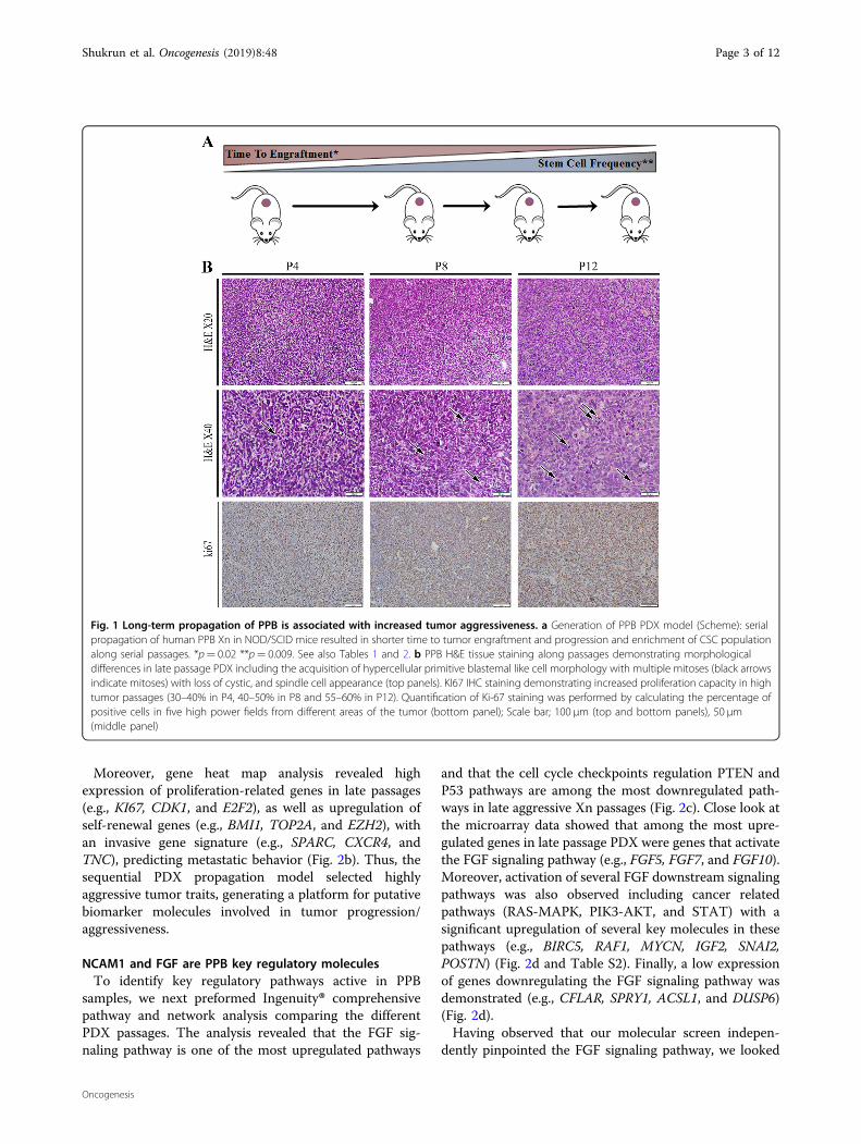
transplanted into NOD/SCID mice, thereby generating aPDX. Sequential propagation of PPB Xn in mice wasperformed by 1 × 10⁶ PDX derived cells injections. Serialpropagation allowed us to establish early (P1–P7) andlate-passage (P7-P14) PPB PDX that were studied forpathogenic pathways associated with PPB-initiatingcapacity. Sequential propagation of PPB PDX correlatedwith significantly shorter time to tumor engraftment andaccelerated tumor growth (Table 1 and Fig. 1a), indicatingthe promotion of tumor aggressiveness along passages.We next queried whether CSC capacity is functionallyenhanced with PPB propagation. We performed limitingdilution xenotransplantation experiments with PPB cellsderived from early-passage and late-passage PDX. Thisanalysis showed significant positive selection for CSCfrequency in late-passage PDX (Table 2 and Fig. 1a), asevident by the significantly increased engraftment fre-quency. Having observed that high-passage PPB PDXselects for the CSC population, we analyzed histologicaland immunohistochemical changes that accompany the
acquisition of the CSC phenotype along passages. H&Estaining revealed that PDX derived tumors maintain thebasic mesenchymal PPB cellular morphology. Never-theless, some morphological differences were observed inlate passage PDX including the acquisition of a hyper-cellular primitive blastemal like cell morphology withmultiple mitoses while losing cystic and spindle cellappearance. (Fig. 1b). In addition, we carried out KI67immunohistochemical (IHC) staining, demonstratingincreased proliferation in high-passage tumors (Fig. 1b).Mouse cell tumor contamination was ruled out by HLAIHC staining and FACS analysis of the mice specificantigen H2K (Fig. S1A and S1B accordingly). In addition,STR analysis demonstrated identical genetic signaturecomparing several samples including primary tumor, P2,P8, and P12 (Fig. S1C).
Sequential PDX propagation model generated putativebiomarker molecules involved in tumor aggressivenessWe next sought to characterize the global molecular
profile of sequential PPB PDX. For this purpose, we per-formed a microarray gene expression analysis comparingseveral different samples: (1) primary PPB (PT); (2) Pas-sage 4 (P4); (3) Passage 8 (P8); (4) Passage 12 (P12); (5)Adult lung (AL); (6) Fetal lung (FL). Unsupervised hier-archical clustering revealed a resemblance between PTand it’s derived Xn samples in comparison to adult andfetal normal healthy lung tissues (Fig. 2a). Gene expres-sion analysis of the samples demonstrated an earlymesenchymal developmental signature in late Xn pas-sages (P12) including the upregulation of critical reg-ulators of lung formation (e.g., GLI2, GLI3, PITX2,LEFTY, SOX11, SOX8) alongside paternally expressedgenes (e.g., PEG1/MEST, PEG3, PEG10, NNAT,KCNQ1OT1, DLK1, and IGF2) as previously shown forthe WT blastemal14 (Table S1). In addition, Ingenuity®functional analysis comparing late passage Xn vs. normaladult lung, demonstrated that among the most upregu-lated pathways in P12 are several developmental pathwaysincluding embryonic and respiratory development(Fig. S2A).
Table 1 PPB PDX frequency and characteristics duringpropagation
Engraftmentrate (%)
Time toengraftment(Days)*
Time toresection (Days)
Averageweight
Averagevolume
Early 63.16 58.08 77.57 1.43 0.88
Late 87.50 33.85 51.94 1.83 1.03
Table summarizing the frequency and characteristics of secondary tumorformation from PPB PDX and further propagation. During serial propagation ofPPB PDX shorter time to tumor engraftment and accelerated tumor growth werenoticed. In the comparison between early and late PDX passages time toengraftment*p= 0.02, Mann–Whitney U test
Shukrun et al. Oncogenesis (2019) 8:48 Page 2 of 12
Oncogenesis
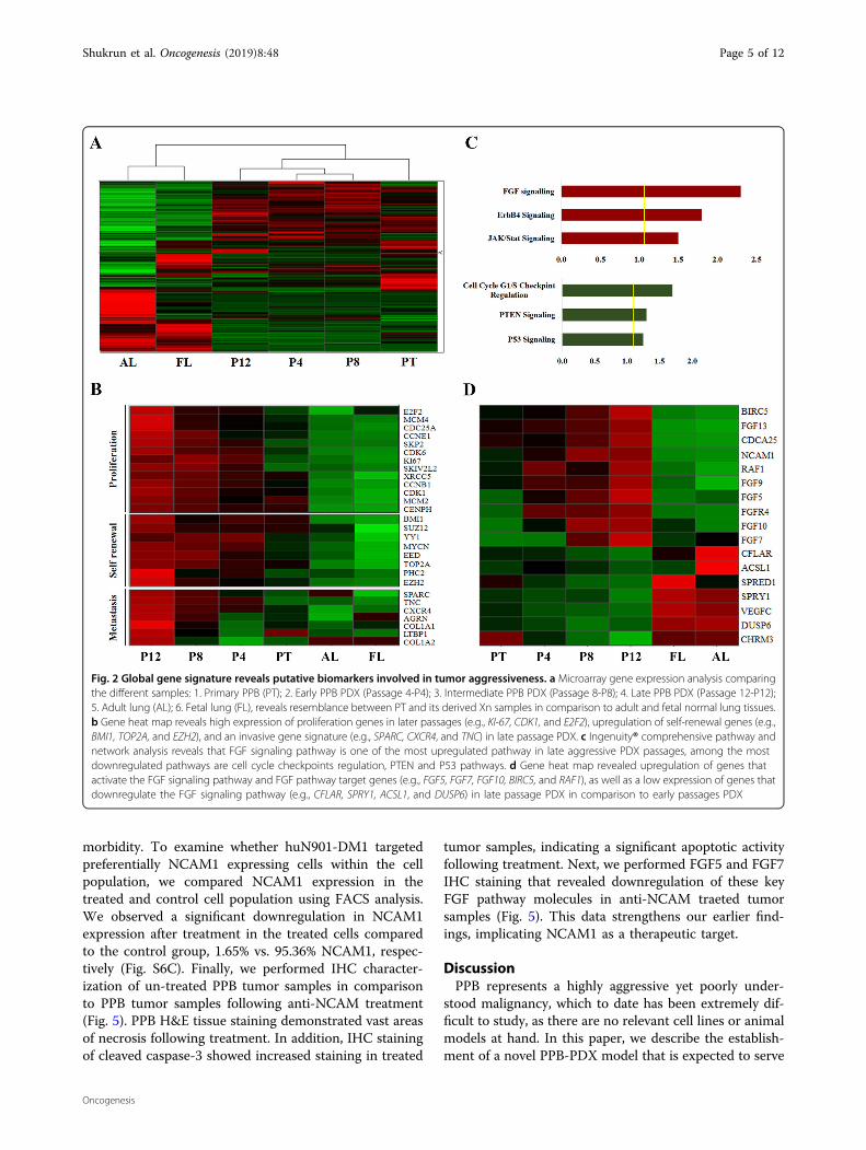
Moreover, gene heat map analysis revealed highexpression of proliferation-related genes in late passages(e.g., KI67, CDK1, and E2F2), as well as upregulation ofself-renewal genes (e.g., BMI1, TOP2A, and EZH2), withan invasive gene signature (e.g., SPARC, CXCR4, andTNC), predicting metastatic behavior (Fig. 2b). Thus, thesequential PDX propagation model selected highlyaggressive tumor traits, generating a platform for putativebiomarker molecules involved in tumor progression/aggressiveness.
NCAM1 and FGF are PPB key regulatory moleculesTo identify key regulatory pathways active in PPB
samples, we next preformed Ingenuity® comprehensivepathway and network analysis comparing the differentPDX passages. The analysis revealed that the FGF sig-naling pathway is one of the most upregulated pathways
and that the cell cycle checkpoints regulation PTEN andP53 pathways are among the most downregulated path-ways in late aggressive Xn passages (Fig. 2c). Close look atthe microarray data showed that among the most upre-gulated genes in late passage PDX were genes that activatethe FGF signaling pathway (e.g., FGF5, FGF7, and FGF10).Moreover, activation of several FGF downstream signalingpathways was also observed including cancer relatedpathways (RAS-MAPK, PIK3-AKT, and STAT) with asignificant upregulation of several key molecules in thesepathways (e.g., BIRC5, RAF1, MYCN, IGF2, SNAI2,POSTN) (Fig. 2d and Table S2). Finally, a low expressionof genes downregulating the FGF signaling pathway wasdemonstrated (e.g., CFLAR, SPRY1, ACSL1, and DUSP6)(Fig. 2d).Having observed that our molecular screen indepen-
dently pinpointed the FGF signaling pathway, we looked
Fig. 1 Long-term propagation of PPB is associated with increased tumor aggressiveness. a Generation of PPB PDX model (Scheme): serialpropagation of human PPB Xn in NOD/SCID mice resulted in shorter time to tumor engraftment and progression and enrichment of CSC populationalong serial passages. *p= 0.02 **p= 0.009. See also Tables 1 and 2. b PPB H&E tissue staining along passages demonstrating morphologicaldifferences in late passage PDX including the acquisition of hypercellular primitive blastemal like cell morphology with multiple mitoses (black arrowsindicate mitoses) with loss of cystic, and spindle cell appearance (top panels). KI67 IHC staining demonstrating increased proliferation capacity in hightumor passages (30–40% in P4, 40–50% in P8 and 55–60% in P12). Quantification of Ki-67 staining was performed by calculating the percentage ofpositive cells in five high power fields from different areas of the tumor (bottom panel); Scale bar; 100 μm (top and bottom panels), 50 μm(middle panel)
Shukrun et al. Oncogenesis (2019) 8:48 Page 3 of 12
Oncogenesis
for related membrane expressed molecule that couldserve as a putative therapeutic target. As previouslydemostrated, FGFRs can also be activated by non FGFligands such as NCAM1. The functional interactionbetween NCAM1 and FGF signaling pathway was ori-ginally reported in neurons18. Thereafter, extensive evi-dence of a physical association between the two proteinsin different, non‐neural cell types, was demonstrated19–22.Accordingly, Ingenuity analysis of our microarray datademonstrated an interaction between NCAM1, FGFreceptors and several of their downstream targets (Fig.S2B). Importantly, NCAM1 has been suggested as a CSCmarker and a therapeutic target in other pediatric solidtumors11, and could therefore validate our approach.Analysis of the microarray data showed an increased
NCAM1 expression along the passages (Fig. S3). In orderto validate our results, we next preformed qRT-PCRanalysis that revealed high NCAM1 expression in pri-mary PPB tumor compared to healthy adult lung controlsamples (top) and in late passages PPB-Xn in comparisonto primary PPB (bottom) (Fig. 3a). Next, we carried outFACS analysis to quantify the proportion of theNCAM1+ population in the different stages of PDXpropagation. This analysis revealed that in early passagePDX cells (P2), only 20% of cells were NCAM1+ incomparison to 76% of cells in late passage (P10) (Fig. 3b).Finally, immunohistochemistry staining demonstratedincreased expression of NCAM1 and several FGF sig-naling molecules (i.e., FGF5 and FGF7) along passages,emphasizing the relevance of our approach (Fig. 3c andFig. S4 accordingly).
We next sorted NCAM1 positive cells in order toexamine gene expression patterns in NCAM1+ vs.NCAM1− sorted cells. qRT-PCR analysis revealed highexpression of several self-renewal genes (e.g., BMI1,EZH2, OCT4, and KLF4) in the NCAM1+ population,supporting their stem cell phenotype (Fig. 3d). We nextsought to examine whether NCAM1 and FGF key sig-naling molecules are expressed in other primary PPBtumors. Indeed, IHC staining demonstrated positiveNCAM1 and FGF5, FGF7, and FGF10 expression in sev-eral PPB primary tumors, validating the relevance of ourfindings (Fig. 3e and Fig. S5 accordingly).
NCAM1 as a PPB therapeutic targetBased on our observation that NCAM1 expression
increased during Xn propagation, we next sought toestablish NCAM1 as a therapeutic target in an in-vitromodel. To do so, we examined the therapeutic effects oflorvotuzumab mertansine (huN901-DM1), a humanizedanti-NCAM1 antibody-cytotoxic drug conjugate, on PPBPDX cell morphology and gene expression. huN901-DM1is an antibody-drug conjugate (ADC), consisting of ahumanized anti-CD56 antibody to which the tubulin-binding maytansinoid DM1 is covalently conjugated via astable disulfide linker23. huN901-DM1 targets CD56 atthe cell surface and, upon antigen binding, becomesinternalized, resulting in the intracellular release ofDM124, which in turn promotes disruption of micro-tubule assembly, G2/metaphase arrest, and ultimatelyapoptosis25,26.First, cells were treated in-vitro with anti-NCAM1
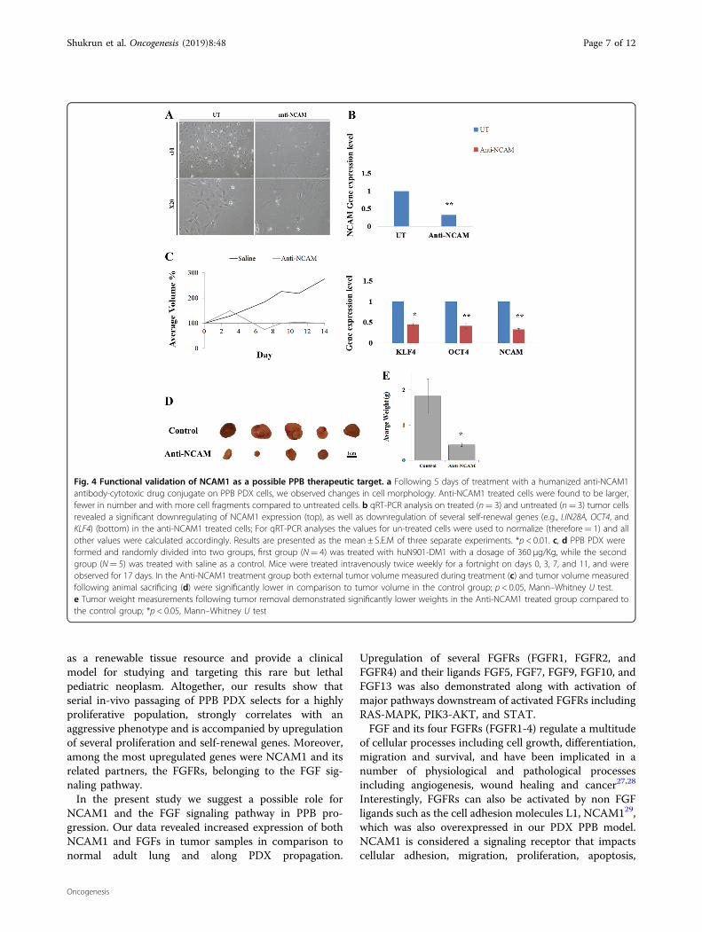
antibody-cytotoxic drug conjugate for 5 days. Followingthe treatment, cell morphology was examined and com-pared to untreated cells (Fig. 4a). Anti-NCAM1 treatmentresulted in increased cell death and apoptotic cell features,including cell swelling and fragmentation. Next, we per-formed a qRT-PCR analysis on treated and untreatedtumor cells revealing significant downregulating ofNCAM1 expression, as well as downregulation of severalself-renewal genes (e.g., LIN28A, OCT4, and KLF4) in theanti-NCAM1 treated cells (Fig. 4b). We then turned toexamine the therapeutic effects of anti-NCAM1 antibody-cytotoxic drug conjugate on PPB PDX progression capa-city in-vivo. For this purpose, nine PPB Xn were formedand randomly divided into two groups, 4 were treatedwith huN901-DM1, while 5 were treated with saline as acontrol. We observed a significant difference in tumorgrowth rate in anti-NCAM1 antibody treated mice vs. thecontrol group following treatment (Fig. 4c–e and Fig.S6A). A minor decrease in weight was observed in thehuN901-DM1 treated group, while an eight percentweight gain was observed in the control group, most likelyattributed to the significant increase in tumor volume.(Fig. S6B). There was no effect on viability nor on
Table 2 Limiting dilution xenotransplantation summaryrepresenting tumor CSC frequency during propagation
Number of cells
injected
Engraftment rate Stem cell
frequency*
Early 1000 3/4 1/1581
500 0/4
100 0/4
Late 1000 3/4 1/304
500 4/4
100 2/4
50 /14
10 0/4
Summary of CSC frequency estimated by limiting dilution xenotransplantationof cells isolated from early and late PDX passages. The data is presented as theratio of injections that formed tumors within 12 weeks. Bold numbers representthe estimated CSC frequency. In the comparison between early and late PDXpassages. Estimation of the relative frequency of cancer propagating cells wascalculated online using the ELDA software online (http://bioinf.wehi.edu.au/software/elda/)*p= 0.009
Shukrun et al. Oncogenesis (2019) 8:48 Page 4 of 12
Oncogenesis
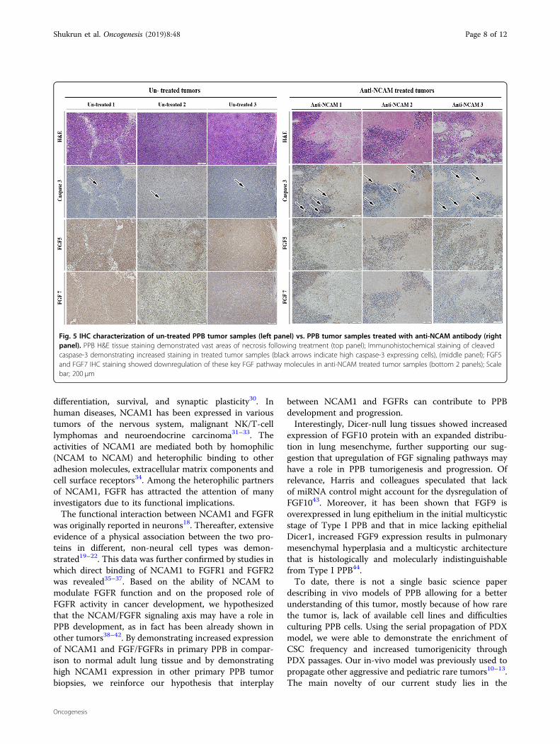
morbidity. To examine whether huN901-DM1 targetedpreferentially NCAM1 expressing cells within the cellpopulation, we compared NCAM1 expression in thetreated and control cell population using FACS analysis.We observed a significant downregulation in NCAM1expression after treatment in the treated cells comparedto the control group, 1.65% vs. 95.36% NCAM1, respec-tively (Fig. S6C). Finally, we performed IHC character-ization of un-treated PPB tumor samples in comparisonto PPB tumor samples following anti-NCAM treatment(Fig. 5). PPB H&E tissue staining demonstrated vast areasof necrosis following treatment. In addition, IHC stainingof cleaved caspase-3 showed increased staining in treated
tumor samples, indicating a significant apoptotic activityfollowing treatment. Next, we performed FGF5 and FGF7IHC staining that revealed downregulation of these keyFGF pathway molecules in anti-NCAM traeted tumorsamples (Fig. 5). This data strengthens our earlier find-ings, implicating NCAM1 as a therapeutic target.
DiscussionPPB represents a highly aggressive yet poorly under-
stood malignancy, which to date has been extremely dif-ficult to study, as there are no relevant cell lines or animalmodels at hand. In this paper, we describe the establish-ment of a novel PPB-PDX model that is expected to serve
Fig. 2 Global gene signature reveals putative biomarkers involved in tumor aggressiveness. a Microarray gene expression analysis comparingthe different samples: 1. Primary PPB (PT); 2. Early PPB PDX (Passage 4-P4); 3. Intermediate PPB PDX (Passage 8-P8); 4. Late PPB PDX (Passage 12-P12);5. Adult lung (AL); 6. Fetal lung (FL), reveals resemblance between PT and its derived Xn samples in comparison to adult and fetal normal lung tissues.b Gene heat map reveals high expression of proliferation genes in later passages (e.g., KI-67, CDK1, and E2F2), upregulation of self-renewal genes (e.g.,BMI1, TOP2A, and EZH2), and an invasive gene signature (e.g., SPARC, CXCR4, and TNC) in late passage PDX. c Ingenuity® comprehensive pathway andnetwork analysis reveals that FGF signaling pathway is one of the most upregulated pathway in late aggressive PDX passages, among the mostdownregulated pathways are cell cycle checkpoints regulation, PTEN and P53 pathways. d Gene heat map revealed upregulation of genes thatactivate the FGF signaling pathway and FGF pathway target genes (e.g., FGF5, FGF7, FGF10, BIRC5, and RAF1), as well as a low expression of genes thatdownregulate the FGF signaling pathway (e.g., CFLAR, SPRY1, ACSL1, and DUSP6) in late passage PDX in comparison to early passages PDX
Shukrun et al. Oncogenesis (2019) 8:48 Page 5 of 12
Oncogenesis
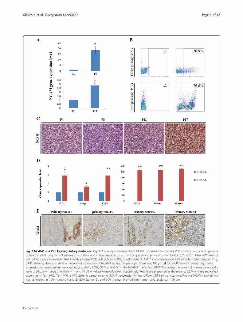
Fig. 3 NCAM1 is a PPB key regulatory molecule. a qRT-PCR analysis revealed high NCAM1 expression in primary PPB tumor (n= 3) in comparisonto healthy adult lung control sample (n= 3) (top) and in late passages. (n= 3) in comparison to primary tumor (bottom); *p < 0.01; Mann–Whitney Utest. b FACS analysis revealed that in early passage PDX cells (P2), only 20% of cells were NCAM1+ in comparison to 76% of cells in late passage (P10).c IHC staining demonstrating an increased expression of NCAM1 along the passages; Scale bar, 100 μm. d qRT-PCR analysis reveals high geneexpression of several self-renewal genes (e.g., BMI1, EZH2, OCT4 and KLF4) in the NCAM1+ cells; For qRT-PCR analyses the values of primary tumor cellswere used to normalize (therefore= 1) and all other values were calculated accordingly. Results are presented as the mean ± S.E.M of three separatedexperiments. *p < 0.05, **p < 0.01. e IHC staining demonstrating NCAM1 expression in four different PPB primary tumors; Positive NCAM1 expressionwas estimated at 10% (tumors 1 and 2), 20% (tumor 3), and 30% (tumor 4) of primary tumor cells. Scale bar, 100 μm
Shukrun et al. Oncogenesis (2019) 8:48 Page 6 of 12
Oncogenesis
as a renewable tissue resource and provide a clinicalmodel for studying and targeting this rare but lethalpediatric neoplasm. Altogether, our results show thatserial in-vivo passaging of PPB PDX selects for a highlyproliferative population, strongly correlates with anaggressive phenotype and is accompanied by upregulationof several proliferation and self-renewal genes. Moreover,among the most upregulated genes were NCAM1 and itsrelated partners, the FGFRs, belonging to the FGF sig-naling pathway.In the present study we suggest a possible role for
NCAM1 and the FGF signaling pathway in PPB pro-gression. Our data revealed increased expression of bothNCAM1 and FGFs in tumor samples in comparison tonormal adult lung and along PDX propagation.
Upregulation of several FGFRs (FGFR1, FGFR2, andFGFR4) and their ligands FGF5, FGF7, FGF9, FGF10, andFGF13 was also demonstrated along with activation ofmajor pathways downstream of activated FGFRs includingRAS-MAPK, PIK3-AKT, and STAT.FGF and its four FGFRs (FGFR1-4) regulate a multitude
of cellular processes including cell growth, differentiation,migration and survival, and have been implicated in anumber of physiological and pathological processesincluding angiogenesis, wound healing and cancer27,28
Interestingly, FGFRs can also be activated by non FGFligands such as the cell adhesion molecules L1, NCAM129,which was also overexpressed in our PDX PPB model.NCAM1 is considered a signaling receptor that impactscellular adhesion, migration, proliferation, apoptosis,
Fig. 4 Functional validation of NCAM1 as a possible PPB therapeutic target. a Following 5 days of treatment with a humanized anti-NCAM1antibody-cytotoxic drug conjugate on PPB PDX cells, we observed changes in cell morphology. Anti-NCAM1 treated cells were found to be larger,fewer in number and with more cell fragments compared to untreated cells. b qRT-PCR analysis on treated (n= 3) and untreated (n= 3) tumor cellsrevealed a significant downregulating of NCAM1 expression (top), as well as downregulation of several self-renewal genes (e.g., LIN28A, OCT4, andKLF4) (bottom) in the anti-NCAM1 treated cells; For qRT-PCR analyses the values for un-treated cells were used to normalize (therefore= 1) and allother values were calculated accordingly. Results are presented as the mean ± S.E.M of three separate experiments. *p < 0.01. c, d PPB PDX wereformed and randomly divided into two groups, first group (N= 4) was treated with huN901-DM1 with a dosage of 360 µg/Kg, while the secondgroup (N= 5) was treated with saline as a control. Mice were treated intravenously twice weekly for a fortnight on days 0, 3, 7, and 11, and wereobserved for 17 days. In the Anti-NCAM1 treatment group both external tumor volume measured during treatment (c) and tumor volume measuredfollowing animal sacrificing (d) were significantly lower in comparison to tumor volume in the control group; p < 0.05, Mann–Whitney U test.e Tumor weight measurements following tumor removal demonstrated significantly lower weights in the Anti-NCAM1 treated group compared tothe control group; *p < 0.05, Mann–Whitney U test
Shukrun et al. Oncogenesis (2019) 8:48 Page 7 of 12
Oncogenesis
differentiation, survival, and synaptic plasticity30. Inhuman diseases, NCAM1 has been expressed in varioustumors of the nervous system, malignant NK/T-celllymphomas and neuroendocrine carcinoma31–33. Theactivities of NCAM1 are mediated both by homophilic(NCAM to NCAM) and heterophilic binding to otheradhesion molecules, extracellular matrix components andcell surface receptors34. Among the heterophilic partnersof NCAM1, FGFR has attracted the attention of manyinvestigators due to its functional implications.The functional interaction between NCAM1 and FGFR
was originally reported in neurons18. Thereafter, extensiveevidence of a physical association between the two pro-teins in different, non‐neural cell types was demon-strated19–22. This data was further confirmed by studies inwhich direct binding of NCAM1 to FGFR1 and FGFR2was revealed35–37. Based on the ability of NCAM tomodulate FGFR function and on the proposed role ofFGFR activity in cancer development, we hypothesizedthat the NCAM/FGFR signaling axis may have a role inPPB development, as in fact has been already shown inother tumors38–42. By demonstrating increased expressionof NCAM1 and FGF/FGFRs in primary PPB in compar-ison to normal adult lung tissue and by demonstratinghigh NCAM1 expression in other primary PPB tumorbiopsies, we reinforce our hypothesis that interplay
between NCAM1 and FGFRs can contribute to PPBdevelopment and progression.Interestingly, Dicer-null lung tissues showed increased
expression of FGF10 protein with an expanded distribu-tion in lung mesenchyme, further supporting our sug-gestion that upregulation of FGF signaling pathways mayhave a role in PPB tumorigenesis and progression. Ofrelevance, Harris and colleagues speculated that lackof miRNA control might account for the dysregulation ofFGF1043. Moreover, it has been shown that FGF9 isoverexpressed in lung epithelium in the initial multicysticstage of Type I PPB and that in mice lacking epithelialDicer1, increased FGF9 expression results in pulmonarymesenchymal hyperplasia and a multicystic architecturethat is histologically and molecularly indistinguishablefrom Type I PPB44.To date, there is not a single basic science paper
describing in vivo models of PPB allowing for a betterunderstanding of this tumor, mostly because of how rarethe tumor is, lack of available cell lines and difficultiesculturing PPB cells. Using the serial propagation of PDXmodel, we were able to demonstrate the enrichment ofCSC frequency and increased tumorigenicity throughPDX passages. Our in-vivo model was previously used topropagate other aggressive and pediatric rare tumors10–13.The main novelty of our current study lies in the
Fig. 5 IHC characterization of un-treated PPB tumor samples (left panel) vs. PPB tumor samples treated with anti-NCAM antibody (rightpanel). PPB H&E tissue staining demonstrated vast areas of necrosis following treatment (top panel); Immunohistochemical staining of cleavedcaspase-3 demonstrating increased staining in treated tumor samples (black arrows indicate high caspase-3 expressing cells), (middle panel); FGF5and FGF7 IHC staining showed downregulation of these key FGF pathway molecules in anti-NCAM treated tumor samples (bottom 2 panels); Scalebar; 200 μm
Shukrun et al. Oncogenesis (2019) 8:48 Page 8 of 12
Oncogenesis
contribution of our in-vivo model to unravel the NCAM1molecule and its suggested interplay with FGFR in theprogression of PPB, pinpointing these molecules as pos-sible novel therapeutic targets in PPB.Indeed, targeting PPB PDX with the anti-NCAM1
antibody-cytotoxic drug conjugate huN901-DM1 resul-ted in a reduction in PPB PDX propagation rate anddifference in gene signature between treated anduntreated cells. Since the antitumorigenic effect ofhuN901-DM1 has been attributed to its cytotoxic con-ugate DM1, a member of the maytansinoids mitoticinhibitors23, our results probably reflect the killing ofNCAM1-high cells by the cytotoxic component ratherthan direct effect of anti CD56 on downregulation of self-renewal genes including FGF pathway. Nevertheless, dueto the known role of NCAM in regulating the FGFpathway through its association with FGFR that results inFGFR recycling at the cell surface45,46, sustained signal-ing36, and cancer progression47, we believe that down-regulation of NCAM1 by the antibody interferes with theNCAM/FGFR interplay which may result in reduction oftumor aggressiveness and metastatic properties as wasshown in other cancers47. Finally, our work highlights thetranslational relevance of this model in identifying targe-table genes in general and of the anti-NCAM1 strategy inparticular, implicating NCAM1 as a therapeutic target.Moreover, from the translational perspective, huN901-DM1 is currently being evaluated as monotherapy inchildren with various NCAM1-expressing solid tumorsincluding: Wilms tumor, rhabdomyosarcoma, neuro-blastoma, malignant peripheral nerve sheath tumor andsynovial sarcoma. Among patients eligible for this studyare also children with relapsed or refractory PPB (http://clinicaltrials.gov/show/NCT02452554).Our results point to a possible NCAM/FGFR interplay
as a novel mechanism underlying PPB malignancy thatmay represent a valuable therapeutic target. We intendto further explore this axis and its role in PPB. More-over, there are several available FGFR inhibitors that arecurrently in early phases of clinical development48, andintegration of these drugs with the anti-NCAM anti-bodies can serve as a novel and promising therapeuticapproach. Furthermore, to validate our findings, we willestablish additional PPB PDX, and we will use our in-vivo model to unravel other regulatory moleculesand pathways that may have a role in PPB developmentand progression in order to find new therapeutictargets.In summary, we have successfully generated a new in-
vivo model for PPB, a rare and fatal childhood malig-nancy, and have shown that this model can be usedeffectively to pinpoint key molecular pathways drivingtumorigenesis and propagation. In turn, these can beharnessed to formulate targeted therapies for better
tumor eradication. Specifically, we identified FGFR andNCAM1 as two key players, which could have significantimplications on the effectiveness of treatment in thispoorly understood disease.
Materials and methodsEthics statementThis study was conducted according to the principles
expressed in the Declaration of Helsinki and wasapproved by the Institutional Review Board of the ShebaMedical Center.
Primary PPB and healthy pulmonary samplesPrimary PPB sample was obtained from a lung tumor
mass of a pediatric patient within 1 h of surgery. Informedconsent was given by the legal guardians of the patientinvolved according to the declaration of Helsinki.Fetal lung tissues for microarray expression analysis
were taken from healthy aborted fetuses in 22 weekgestational age and were kindly provided to us by DrChava Rosen (Weizmann Institute). Total RNA fromnormal human adult lung tissue was purchased fromBioChain®.
In vivo xenograft formationThe animal experiments were performed in accordance
with the Guidelines for Animal Experiments of ShebaMedical Center. Initial PPB engrafting to 5–8 weeks old,female, nonobese diabetic immunodeficient (NOD/SCID)mice was performed as previously described14. Briefly, pri-mary PPB tissue was cut into 2–5mm pieces and implantedsubcutaneously in the flank of the mouse. Tumors wereharvested approximately 1–3 months post implantation orwhen they reached a size of 1.5 cm diameter. Time toengraftment, time to resection, weight and volume for eachengrafted Xn were recorded. Xn tissue was immediately cutinto small pieces and processed for further experiments asfollows: (i) flash freezing for subsequent molecular char-acterization of extracted analyses; (ii) formalin fixation andparaffin embedding for future IHC studies. In addition, Xnserial passages were formed in two methods: (1) PPB Xntissue was cut into 2–5mm pieces and implanted sub-cutaneously in the flank of the mouse; (2) For the in-vivoexperiments compering anti-NCAM1 antibody-cytotoxicdrug conjugate vs. control and for the estimation of relativefrequency of tumor propagating cells we injected dis-sociated cells from freshly retrieved PPB Xn; Cells wereinjected in 100 μl 1:1 serum free medium/Matrigel (BDBiosciences, San Jose, CA). Preparation of single cell sus-pensions was used for subsequent Xn propagation asdescribed and for in-vitro experiments.Primary tumor and propagated PDX (P2, P8, and P14)
were authenticated by short tandem repeat profiling usingseveral markers, including: D3S1358, vWa, D16S539,
Shukrun et al. Oncogenesis (2019) 8:48 Page 9 of 12
Oncogenesis
D2S1338, D8S1179, D21S11, D18S51, D19S433, TH01,and FGA.
In vivo animal experimentsEstimation of the relative frequency of tumor propagatingcellsTo evaluate and compare the tumorigenic activity of Xn
cells from early and late passages, serial dilutions (50−1 ×10⁶ cells) of cells suspended in 100 μl of PBS and 100 μlMatrigel were injected subcutaneously to the flank ofNOD/SCID mice. Estimation of the relative frequency ofcancer propagating cells was calculated online using theELDA software online (http://bioinf.wehi.edu.au/software/elda/).
In vivo xenograft experiments using anti-NCAM1 antibody1 × 106 cells derived from late passage PPB PDX (P12)
suspended in 100 μl 1:1 serum free medium/Matrigelwere injected subcutaneously to the flank of NOD/SCIDmice to generate tumors. Of the nine mice which devel-oped a tumor, 4 were treated with 360 µg/Kg huN901-DM123 while the other 5 were treated with saline as acontrol. Mice were treated intravenously twice weekly fora fortnight on days 0, 3, 7, and 11, and were observed for17 days. Mice were monitored for tumor growth on days0, 3, 7, 9, 11, 14. The investigator assessing Xn volume wasblinded to the group allocation. On the 17th day the micewere euthanized, tumors were harvested and measured.Tissue RNA was produced and used for qRT-PCRexperiments.
Fluorescence-activated cell sorting (FACS) analysisFACS analysis of the primary PPB cells and subsequent
fresh Xn derived cells was performed as previouslydescribed14. Surface markers antigens [CD24 (eBiosience,120247-42), CD34 (Miltenyi, 3008100), CD56 (eBiosience,1205942), CD90 (Beckman Coulter, IM3600U), H2K(eBiosience, 12-5958-82)] were labeled by incubation withfluorochrome conjugated antibody at a concentration of5 µg antibody per 106 cells for 30 min, in the dark, at 4 °Cto prevent internalization of antibodies. In addition, weused 7-amino-actinomycin-D (7AAD; eBioscience, SanDiego, CA) for viable cell gating. All washing steps wereperformed in FACS buffer. All Quantitative measure-ments were made in comparison to IgG isotype antibody.
FACS sortingFACS Aria was used to enrich for cells expressing sur-
face markers. A 100-µm nozzle (BD Biosciences, San Jose,CA), sheath pressure of 20–25 pounds per square inch(PSI), and an acquisition rate of 1000–3000 eventsper second were used as conditions optimized for PPB cellsorting. Single viable cells were gated based on 7AAD, andthen physically sorted according to NCAM1 expression
into collection tubes for all subsequent experiments. Datawas additionally analyzed and presented using FlowJosoftware.
MicroarrayThe microarray data is deposited in publicly library
(GEO); accession number GSE97236. All experimentswere performed using Affymetrix HU GENE1.0st oligo-nucleotide arrays49. Total RNA from each sample wasused to prepare biotinylated target cDNA, according tothe manufacturer’s recommendations. The target cDNAgenerated from each sample was processed as per man-ufacturer’s recommendation using an Affymetrix GeneChip Instrument System. Details of quality control mea-sures can be found online. Significantly changed geneswere filtered as changed by at least twofold (p-value: 0.05).
Quantitative real-time reverse transcription PCRanalysis–Gene expression analysisQuantitative reverse transcription PCR (qRT-PCR) was
carried out to determine fold changes in expression of aselection of genes. Total RNA from cells was isolatedusing an RNeasy Micro Kit (Qiagen GmbH, Hilden,Germany) according to the manufacturer's instructions.cDNA was synthesized using a High Capacity cDNAReverse Transcription kit (Applied Biosystems, CaliforniaUSA) on total RNA. Real-time PCR was performed usingan ABI7900HT sequence detection system (Perkin-Elmer/Applied Biosystems, California, USA) in the presence ofTaqMan Gene Expression Master Mix (Applied Biosys-tems, California, USA). PCR amplification was performedusing gene specific TaqMan Gene Expression Assay-Pre-Made kits (Applied Biosystems, California, USA). Eachanalysis reaction was performed in triplicate. HPRT1 orGAPDH were used as an endogenous control throughoutthe experimental analyses. PCR results were analyzedusing SDS RQ Manager 1.2 software. Statistical analysiswas performed using a non-paired 2-tails T-test. Statis-tical significance was considered at P < 0.05.
H&E stainingH&E staining of 4 µm sections of paraffin-embedded
tissues from primary PPB and propagated PDX weremounted on super frost/plus glass and incubated at 60 °Cfor 40min. After deparaffinization, slides were incubatedin Mayer's Hematoxylin solution (Sigma-Aldrich) andincubated with 1% HCl in 70% ethanol for 1 min. Slideswere then incubated for 10 sec in Eosin (Sigma-Aldrich).Images were produced using Olympus BX51TF.
Immunohistochemical staining of primary PPB and PPB XnSections, 4 µm thick, were cut from primary PPB and
PPB Xn for immunohistochemistry. Immunostainingswere performed as previously described50. Briefly, the
Shukrun et al. Oncogenesis (2019) 8:48 Page 10 of 12
Oncogenesis
sections were processed to avoid oxidation of antigens.Before immunostaining, sections were treated with10mM citrate buffer, PH 6.0 for 10min at 97 °C forantigen retrieval, followed by 3% H2O2 for 10 min. Theslides were subsequently stained using the labeledstrepavidin-biotin (LAB-SA) method using a Histostainplus kit (Zymed, San Francisco, CA, USA). The immu-noreaction was visualized by an HRP-based chromogen/substrate system (liquid DAB substrate kit–Zymed, SanFrancisco, CA, USA). All antibody dilutions were carriedout as recommended by the manufacturers of the stainingantibodies. NCAM (Epitomics, cat #2433-1, 1:250),human HLA (Abcam, ab52922, 1:200), KI67 (Abcam,ab15580, 1:200), FGF5 (Abcam, ab89279, 1:50), FGF7(Abcam, ab90259, 1 µg/ml), FGF10 (Abcam, ab80064,1:20), Active caspase 3 antibody (Epitomics, cat #1476-1,1:10).
PPB cell culture and in-vitro anti-NCAM1 experimentSingle cell suspension from PPB PDX tissues were
grown in Bronchial Epithelial Growth medium (BEGM)(Lonza Walkersville, Inc. USA) and Iscove's Modificationof Dulbecco's medium (IMDM) supplemented with 10%FBS and the following growth factors: EGF, FGF, and SCF(ratio 3:1 accordingly). Cells were treated with huN901-DM123, a humanized anti-NCAM1 antibody-cytotoxicdrug conjugate (ImmunoGen Inc., Waltham, Massachu-setts) at a concentration of 0.18 µM. Following five days oftreatment cells were harvested and RNA was derived forqRT-PCR experiments.
Statistical analysisResults are expressed as the mean ± S.E.M, unless other-
wise indicated. Statistical differences in gene expressionbetween PPB cell populations were evaluated using theStudent's T-test. For animal studies, sample size was esti-mated to be at least four mice per group to ensure powerwith statistical confidence Statistical differences in the in-vivo experiments were calculated using Mann–Whitney Utest. For all statistical analysis, the level of significance wasset as p < 0.05 unless otherwise indicated.
AcknowledgementsWe thank Dr. Chava Rosen (Weizmann Institute of Science) for provision ofembryonic lung tissue. We thank Dr. Victoria Marcu-Malina and Prof. NinetteAmariglio for performing short tandem repeat analysis. We thank Victor S.Goldmacher, ImmunoGen, Inc. for providing the conjugated antibody. Wethank Dr. Marina Perelman for pathological assistant. This work is part of therequirements towards a PhD degree, Sackler School of Medicine, Tel AvivUniversity (H.G., R.S., and R.C.). This study was supported by the NationalInstitutes of Health RO1 AR47901P30 AR42687, Robert Margolis Foundation,Rabinowitch‐Davis Foundation for Melanoma Research, Betty MinskFoundation for Melanoma Research, Reynolds Sarcoma Foundation, ZieringFamily Foundation, Israel Cancer Research Fund (ICRF) PG‐14‐112, ICRFinnovation grant (BD) and the Israel Cancer Association (ICA) 20150916. VictorS. Goldmacher, ImmunoGen, Inc. provided the conjugated antibody.
Author details1Pediatric Stem Cell Research Institute, Safra Children’s Hospital, Sheba MedicalCenter, 5262000 Ramat-Gan, Israel. 2Sackler School of Medicine, Tel AvivUniversity, 6997801 Tel Aviv, Israel. 3Pediatric Hematology Oncology ResearchLaboratory, Safra Children’s Hospital, Sheba Medical Center, 5262000 Ramat-Gan, Israel. 4Dr. Pinchas Borenstein Talpiot Medical Leadership Program 2013,Sheba Medical Center, Tel Hashomer, 5262000 Ramat-Gan, Israel. 5The Mauriceand Gabriela Goldschleger Eye Research Institute, Sheba Medical Center,5262000 Ramat-Gan, Israel. 6Cancer Research Center and the Wohl Institute ofTranslational Medicine, Sheba Medical Center, 5262000 Ramat-Gan, Israel.7Department of Pathology, Sheba Medical Center, 5262000 Ramat-Gan, Israel.8Division of Pediatric Nephrology, Safra Children’s Hospital, Sheba MedicalCenter, 5262000 Ramat-Gan, Israel
Author's contributionsConception and design: B.D., A.T., H.G., R.S.; development of methodology: B.D., A.T., H.G., R.S.; acquisition of data: H.G., R.S., R.C., N.P.S., O.P., J.J.H., G.S., I.T., D.D.B.L., M.M.D., E.P., E.V., O.H.S., D.B. Analysis and interpretation of data: B.D., A.T., H.G., R.S.;writing, review, and/or revision of the paper: B.D., A.T., H.G., R.S., R.C.; administrative,technical, or material support: H.G., R.S.; study supervision: B.D. and A.T.
Conflict of interestThe authors declare that they have no conflict of interest.
Publisher’s noteSpringer Nature remains neutral with regard to jurisdictional claims inpublished maps and institutional affiliations.
Supplementary Information accompanies this paper at (https://doi.org/10.1038/s41389-019-0156-9).
Received: 17 September 2018 Revised: 22 April 2019 Accepted: 6 May 2019
References1. Dishop, M. K. & Kuruvilla, S. Primary and metastatic lung tumors in the
pediatric population: a review and 25-year experience at a large children'shospital. Arch. Pathol. Lab. Med. 132, 1079–1103 (2008).
2. Christosova, I. R. et al. Diagnosis and treatment of pleuropulmonary blastoma-single center experience. Pediatr. Pulmonol. 50, 698–703 (2015).
3. Manivel, J. C. et al. Pleuropulmonary blastoma the so-called pulmonary blas-toma of childhood. Cancer 62, 1516–1526 (1988).
4. Hill, D. A. et al. Type I pleuropulmonary blastoma: pathology and biologystudy of 51 cases from the international pleuropulmonary blastoma registry.Am. J. Surg. Pathol. 32, 282–295 (2008).
5. Schultz, K. A. et al. DICER1-pleuropulmonary blastoma familial tumor predis-position syndrome: a unique constellation of neoplastic conditions. PathologyCase Rev. 19, 90–100 (2014).
6. Doros, L. et al. in DICER1-related disorders (eds Adam M. P., Ardinger H. H.,Pagon R. A., Wallace S. E., Bean L. J. H., Stephens K., et al.) (GeneReviews®,Seattle, 1993).
7. Messinger, Y. H. et al. Pleuropulmonary blastoma: a report on 350 centralpathology-confirmed pleuropulmonary blastoma cases by the internationalpleuropulmonary blastoma registry. Cancer 121, 276–285 (2015).
8. Dehner, L. P. et al. Pleuropulmonary blastoma: evolution of an entity as anentry into a familial tumor predisposition syndrome. Pedia. Dev. Pathol. 18,504–511 (2015).
9. Pode-Shakked, N. & Dekel, B. Wilms tumor–a renal stem cell malignancy?Pediatr. Nephrol. 26, 1535–1543 (2011).
10. Shukrun, R. et al. Wilms' tumor blastemal stem cells dedifferentiate to pro-pagate the tumor bulk. Stem Cell Rep. 3, 24–33 (2014).
11. Pode-Shakked, N. et al. The isolation and characterization of renal cancerinitiating cells from human Wilms' tumour xenografts unveils new therapeutictargets. EMBO Mol. Med. 5, 18–37 (2013).
12. Pleniceanu, O. et al. Peroxisome proliferator-activated receptor gamma(PPARgamma) is central to the initiation and propagation of human angio-myolipoma, suggesting its potential as a therapeutic target. EMBO Mol. Med. 9,508–530 (2017).
Shukrun et al. Oncogenesis (2019) 8:48 Page 11 of 12
Oncogenesis
13. Golan, H. et al. In vivo expansion of cancer stemness affords novel cancer stemcell targets: malignant rhabdoid tumor as an example. Stem Cell Rep. 11,795–810 (2018).
14. Dekel, B. et al. Multiple imprinted and stemness genes provide a link betweennormal and tumor progenitor cells of the developing human kidney. CancerRes. 66, 6040–6049 (2006).
15. Metsuyanim, S. et al. Accumulation of malignant renal stem cells is associatedwith epigenetic changes in normal renal progenitor genes. Stem Cells 26,1808–1817 (2008).
16. Pode-Shakked, N. et al. Resistance or sensitivity of Wilms' tumor to anti-FZD7antibody highlights the Wnt pathway as a possible therapeutic target.Oncogene 30, 1664–1680 (2011).
17. Shukrun, R., Pode Shakked, N. & Dekel, B. Targeted therapy aimed atcancer stem cells: Wilms' tumor as an example. Pediatr. Nephrol. 29,815–823 (2014).
18. Williams, E. J., Furness, J., Walsh, F. S. & Doherty, P. Activation of the FGFreceptor underlies neurite outgrowth stimulated by L1, N-CAM, and N-cadherin. Neuron 13, 583–594 (1994).
19. Cavallaro, U., Niedermeyer, J., Fuxa, M. & Christofori, G. N-CAM modulatestumour-cell adhesion to matrix by inducing FGF-receptor signalling. Nat. CellBiol. 3, 650–657 (2001).
20. Francavilla, C. et al. Neural cell adhesion molecule regulates the cellularresponse to fibroblast growth factor. J. Cell Sci. 120, 4388–4394 (2007).
21. Kos, F. J. & Chin, C. S. Costimulation of T cell receptor-triggered IL-2 productionby Jurkat T cells via fibroblast growth factor receptor 1 upon its engagementby CD56. Immunol. Cell Biol. 80, 364–369 (2002).
22. Sanchez-Heras, E., Howell, F. V., Williams, G. & Doherty, P. The fibroblast growthfactor receptor acid box is essential for interactions with N-cadherin and all ofthe major isoforms of neural cell adhesion molecule. J. Biol. Chem. 281,35208–35216 (2006).
23. Whiteman, K. R. et al. Lorvotuzumab mertansine, a CD56-targeting antibody-drug conjugate with potent antitumor activity against small cell lung cancerin human xenograft models. MAbs 6, 556–566 (2014).
24. Tijink, B. M. et al. A phase I dose escalation study with anti-CD44v6 bivatu-zumab mertansine in patients with incurable squamous cell carcinoma of thehead and neck or esophagus. Clin. Cancer Res. 12, 6064–6072 (2006).
25. Chari, R. V. et al. Immunoconjugates containing novel maytansinoids: pro-mising anticancer drugs. Cancer Res. 52, 127–131 (1992).
26. Erickson, H. K. & Lambert, J. M. ADME of antibody-maytansinoid conjugates.AAPS J. 14, 799–805 (2012).
27. Ahmad, I., Iwata, T. & Leung, H. Y. Mechanisms of FGFR-mediated carcino-genesis. Biochim. et. Biophys. Acta 1823, 850–860 (2012).
28. Tenhagen, M., van Diest, P. J., Ivanova, I. A., van der Wall, E. & van der Groep, P.Fibroblast growth factor receptors in breast cancer: expression, downstreameffects, and possible drug targets. Endocr. Relat. Cancer 19, R115–R129 (2012).
29. Kochoyan, A., Poulsen, F. M., Berezin, V., Bock, E. & Kiselyov, V. V. Structural basisfor the activation of FGFR by NCAM. Protein science : a publication of theProtein. Society 17, 1698–1705 (2008).
30. Amoureux, M. C., Cunningham, B. A., Edelman, G. M. & Crossin, K. L. N-CAMbinding inhibits the proliferation of hippocampal progenitor cells andpromotes their differentiation to a neuronal phenotype. J. Neurosci. 20,3631–3640 (2000).
31. Sugimoto, K. J. et al. CD56-positive adult T-cell leukemia/lymphoma: a casereport and a review of the literature. Med. Mol. Morphol. 48, 54–59 (2015).
32. Takata, K. et al. Primary cutaneous NK/T-cell lymphoma, nasal type and CD56-positive peripheral T-cell lymphoma: a cellular lineage and clinicopathologicstudy of 60 patients from Asia. Am. J. Surg. Pathol. 39, 1–12 (2015).
33. Etzell, J. E., Keet, C., McDonald, W. & Banerjee, A. Medulloblastoma simulatingacute myeloid leukemia: case report with a review of "myeloid antigen"expression in nonhematopoietic tissues and tumors. J. Pediatr. Hematol./Oncol.28, 703–710 (2006).
34. Hinsby, A. M., Berezin, V. & Bock, E. Molecular mechanisms of NCAM function.Front. Biosci. 9, 2227–2244 (2004).
35. Christensen, C., Lauridsen, J. B., Berezin, V., Bock, E. & Kiselyov, V. V. The neuralcell adhesion molecule binds to fibroblast growth factor receptor 2. FEBS Lett.580, 3386–3390 (2006).
36. Francavilla, C. et al. The binding of NCAM to FGFR1 induces a specific cellularresponse mediated by receptor trafficking. J. Cell Biol. 187, 1101–1116 (2009).
37. Kiselyov, V. V. et al. Structural basis for a direct interaction between FGFR1 andNCAM and evidence for a regulatory role of ATP. Structure 11, 691–701 (2003).
38. Davidson, B. et al. The clinical role of epithelial-mesenchymal transition andstem cell markers in advanced-stage ovarian serous carcinoma effusions. Hum.Pathol. 46, 1–8 (2015).
39. Flippot, R., Kone, M., Magne, N. & Vignot, S. [FGF/FGFR signalling: Implication inoncogenesis and perspectives]. Bull. du Cancer 102, 516–526 (2015).
40. He, Q., Gong, Y., Gower, L., Yang, X. & Friesel, R. E. Sef regulates epithelial-mesenchymal transition in breast cancer cells. J. Cell. Biochem. 117, 2346–2356(2016).
41. Mori, S. et al. Enhanced expression of integrin alphavbeta3 induced by TGF-beta is required for the enhancing effect of Fibroblast Growth Factor 1 (FGF1)in TGF-beta-induced Epithelial-Mesenchymal Transition (EMT) in mammaryepithelial cells. PLoS ONE 10, e0137486 (2015).
42. Takase, N. et al. NCAM- and FGF-2-mediated FGFR1 signaling in the tumormicroenvironment of esophageal cancer regulates the survival and migrationof tumor-associated macrophages and cancer cells. Cancer Lett. 380, 47–58(2016).
43. Harris, K. S., Zhang, Z., McManus, M. T., Harfe, B. D. & Sun, X. Dicer function isessential for lung epithelium morphogenesis. Proc. Natl Acad. Sci. USA 103,2208–2213 (2006).
44. Yin, Y. et al. Fibroblast growth factor 9 regulation by MicroRNAs controls lungdevelopment and links DICER1 loss to the pathogenesis of pleuropulmonaryblastoma. PLoS Genet. 11, e1005242 (2015).
45. Zamai, M. et al. Number and brightness analysis reveals that NCAM and FGF2elicit different assembly and dynamics of FGFR1 in live cells. J. Cell Sci. 132,jcs220624 (2019).
46. Francavilla, C. et al. Correction: The binding of NCAM to FGFR1 induces aspecific cellular response mediated by receptor trafficking. J. Cell Biol. 218,1422 (2019).
47. Zecchini, S. et al. The adhesion molecule NCAM promotes ovarian cancerprogression via FGFR signalling. EMBO Mol. Med. 3, 480–494 (2011).
48. Turner, N. & Grose, R. Fibroblast growth factor signalling: from development tocancer. Nat. Rev. Cancer 10, 116–129 (2010).
49. Pode-Shakked, N. et al. Developmental tumourigenesis: NCAM as a putativemarker for the malignant renal stem/progenitor cell population. J. Cell. Mol.Med. 13, 1792–1808 (2009).
50. Dekel, B. et al. Isolation and characterization of nontubular sca-1+lin- multi-potent stem/progenitor cells from adult mouse kidney. J. Am. Soc. Nephrol. 17,3300–3314 (2006).
Shukrun et al. Oncogenesis (2019) 8:48 Page 12 of 12
Oncogenesis


![FIF [Fibroblast Growth Factor - 2 (FGF-2)-Interacting-Factor], a Nuclear Putatively Antiapoptotic Factor, Interacts Specifically with FGF-2](https://img.pdfslide.net/doc/110x75/636296899b985d7ef50271c1/fif-fibroblast-growth-factor-2-fgf-2-interacting-factor-a-nuclear-putatively.jpg)


