Embed Size (px)
Citation preview

Ns
HCJa
b
c
d
a
ARRAA
KECSEEf
1
cOiaifpida2M
MT
0d
Neuropsychologia 48 (2010) 1813–1822
Contents lists available at ScienceDirect
Neuropsychologia
journa l homepage: www.e lsev ier .com/ locate /neuropsychologia
eural correlates of using distancing to regulate emotional responses to socialituations
arold W. Koenigsberga,b,∗, Jin Fana, Kevin N. Ochsnerc, Xun Liua, Kevin Guisea, Scott Pizzarellod,hristine Dorantesa, Lucia Tecutaa, Stephanie Guerreri a, Marianne Goodmana,b, Antonia Newa,
anine Florya, Larry J. Sievera,b
Mount Sinai School of Medicine, New York, NY, United StatesJames J Peters VA Medical Center, Bronx, NY, United StatesDepartment of Psychology, Columbia University, New York, NY, United StatesUniversity of Florida, Tallahasse, FL, United States
r t i c l e i n f o
rticle history:eceived 21 July 2009eceived in revised form 3 March 2010ccepted 3 March 2010vailable online 11 March 2010
eywords:motionognitive reappraisal
a b s t r a c t
Cognitive reappraisal is a commonly used and highly adaptive strategy for emotion regulation that hasbeen studied in healthy volunteers. Most studies to date have focused on forms of reappraisal that involvereinterpreting the meaning of stimuli and have intermixed social and non-social emotional stimuli. Herewe examined the neural correlates of the regulation of negative emotion elicited by social situations usinga less studied form of reappraisal known as distancing. Whole brain fMRI data were obtained as partic-ipants viewed aversive and neutral social scenes with instructions to either simply look at and respondnaturally to the images or to downregulate their emotional responses by distancing. Three key findingswere obtained accompanied with the reduced aversive response behaviorally. First, across both instruc-
ocial cognitive neurosciencemotional distancingmotion regulationMRI
tion types, aversive social images activated the amygdala. Second, across both image types, distancingactivated the precuneus and posterior cingulate cortex (PCC), intraparietal sulci (IPS), and middle/superiortemporal gyrus (M/STG). Third, when distancing one’s self from aversive images, activity increased in dor-sal anterior cingulate (dACC), medial prefrontal cortex (mPFC), lateral prefrontal cortex, precuneus andPCC, IPS, and M/STG, meanwhile, and decreased in the amygdala. These findings demonstrate that dis-
ial cupectiv
tancing from aversive socin social perception, pers
. Introduction
Many of our most important emotions arise in interpersonalontexts (Adolphs, 2003; Minzenberg, Poole, & Vinogradov, 2006;lsson & Ochsner, 2008). This suggests that one of the most
mportant self-regulatory challenges is how we voluntarily anddaptively regulate our emotional responses to social cues. Fail-ng to regulate such emotions may have serious consequencesor mental and physical health (Gross, 2002), and may underliesychiatric disorders in which there are serious disturbances in
nterpersonal functioning, such as borderline personality disor-
er (BPD) (Gunderson, 2007; Meyer, Pilkonis, & Beevers, 2004),voidant personality disorder (AvPD) (Leising, Sporberg, & Rehbein,006) or schizophrenia spectrum disorders (SSD) (Ballon, Kaur,arks, & Cadenhead, 2007; Meyer & Shean, 2006), among others.∗ Corresponding author at: Mount Sinai School of Medicine, James J. Peters VAedical Center, 130 West Kingsbridge Road, 116A, Bronx, NY 10468, United States.
el.: +718 584 9000x5757.E-mail address: [email protected] (H.W. Koenigsberg).
028-3932/$ – see front matter. Published by Elsevier Ltd.oi:10.1016/j.neuropsychologia.2010.03.002
es modulates amygdala activity via engagement of networks implicatede-taking, and attentional allocation.
Published by Elsevier Ltd.
Individuals with BPD, for example, have particularly intense emo-tional reactions to social situations of perceived abandonment orabuse (Gunderson & Lyons-Ruth, 2008). Understanding the mecha-nisms by which we can effectively regulate these socially triggeredemotional responses is therefore an important goal of both basicand translational research (Ochsner, 2008; Phillips, Drevets, Rauch,& Lane, 2003; Roffman, Marci, Glick, Dougherty, & Rauch, 2005).
Recently, much progress in understanding these mechanismshas been made using functional imaging to identify the neural basesof cognitive forms of emotion regulation (Ochsner & Gross, 2005).Much of this work has examined the neural dynamics underlyingcognitive reappraisal, which involves reinterpreting the meaning ofa stimulus or situation in ways that alter one’s emotional response.Behavioral studies have shown that reappraisal is one of the mostflexible, adaptive and commonly employed strategies for down-regulating negative emotional responses (Gross, 2002). Imaging
studies have shown that reappraisal activates prefrontal and cin-gulate systems implicated in various kinds of cognitive controlprocesses, which in turn appear to modulate activity in neuralsystems associated with emotional responding, such as the amyg-dala (Banks, Eddy, Angstadt, Nathan, & Phan, 2007; Beauregard,
1 opsyc
L222
EttdpnvDaremtwt(2iudaoife
dovsaia(so&AnKOrhnfiaeuHeiaTa
iprcpa
ment, or inanimate objects that have been intermixed with socialstimuli in prior reappraisal studies. The negative interpersonalscenes selected included pictures of people in situations of lossor grief, of abuse or of physical threat.1 Neutral images depicted
1 The following IAPS pictures were employed: Negative: 2053,2490,9810,6821,6360,2691,6838,3550,3500,3181,6311,2710,2900,9433,6313,2800,3160,2095,
814 H.W. Koenigsberg et al. / Neur
evesque, & Bourgouin, 2001; Kalisch et al., 2005; Kim & Hamann,007; Levesque et al., 2003; Ochsner, Bunge, Gross, & Gabrieli,002; Ochsner et al., 2004b; Ochsner & Gross, 2005; Phan et al.,005; Urry et al., 2006).
For present purposes, two aspects of this work are noteworthy.xamining neural systems implicated in the regulation of responseo emotionally evocative social cues is particularly important givenhe relevance of regulating social-emotional responses in clinicalisorders (Ochsner, 2008; Phillips et al., 2003) and the fact thatrocessing socially cued emotion engages different networks thanon-socially cued emotion (Britton et al., 2006; Harris, McClure,an den Bos, Cohen, & Fiske, 2007; Lestou, Pollick, & Kourtzi, 2008).espite the importance of such interpersonal emotional cues, rel-tively few studies have examined the regulation of emotion inesponse to emotionally evocative social interactions (Beauregardt al., 2001; Harenski & Hamann, 2006; Levesque et al., 2003). Theajority of reappraisal studies have used image-based paradigms
hat intermix stimuli depicting emotionally charged social cuesith non-social stimuli, making it difficult to identify networks
hat may be involved in regulating socially evoked responses per see.g. Kim & Hamann, 2007; Ochsner et al., 2002, 2004b; Phan et al.,005; Urry et al., 2006). The first goal of the present study was to
dentify neural correlates of the cognitive reappraisal of social sit-ations that elicit negative emotional responses. Since emotionalysregulation in reaction to interpersonal events is a prominentnd disabling feature of a number of emotional disorders, we focusn negative emotions generated from social situations, which cannclude a variety of emotional responses, rather than limiting ourocus to one or more specific emotions that are considered inher-ntly social (Burnett, Bird, Moll, Frith, & Blakemore, 2009).
Second, although it is clear that prefrontal regions are engageduring reappraisal, most research to date has focused on only onef two main types of reappraisal. The most commonly studiedariant of reappraisal is the situation-focused or reinterpretationtrategy, which involves re-thinking the meaning of the actionsnd events depicted in an image. Another variant of reappraisals the self-focused or distancing strategy, which involves viewingn image from the perspective of a detached and distant observerOchsner & Gross, in press; Ochsner et al., 2004b). In behavioraltudies, distancing appears to be adaptive in reducing the intensityf angry and depressive affect and blood pressure responses (AydukKross, 2008; Kross & Ayduk, 2008; Kross, Ayduk, & Mischel, 2005).lthough both strategies have been effective for down-regulatingegative emotion (Kalisch et al., 2005; Kross & Ayduk, in press;ross et al., 2005; Kross, Egner, Ochsner, Hirsch, & Downey, 2007;chsner et al., 2004b), to date, fewer studies have examined the
egulation of responses to aversive images using distancing. Theandful of studies examining distancing have used a heteroge-eous array of emotionally evocative stimuli, including photos,lms, shock, and risky choices (Beauregard et al., 2001; Eippert etl., 2007; Kalisch et al., 2005; Koenigsberg et al., 2009; Ochsnert al., 2004b). By contrast, the reinterpretation strategy has beensed in many studies and primarily with image stimuli (e.g. Kim &amann, 2007; Ochsner et al., 2002, 2004b; Phan et al., 2005; Urryt al., 2006). The second goal of this study was to further exam-ne of the use of distancing to down-regulate negative emotion invariant of a commonly used image-based reappraisal paradigm.hus we focus on the use of the distancing reappraisal strategy aspplied specifically to emotions evoked by social cues.
With these goals in mind, we formulated three predictions. First,n keeping with prior work, we expected that carrying out reap-
raisal of social-emotional cues by distancing would involve theecruitment of prefrontal and cingulate systems previously impli-ated in the top–down cognitive control of limbic, attentional andrimary sensory regions. In particular, because distancing involveschange in the perceived self-relevance of images as well as with-hologia 48 (2010) 1813–1822
drawal of attention from emotional cues, we expected to observeactivity in medial frontal and medial parietal regions associatedwith self-referential processing and perspective-taking, as well asprefrontal and parietal regions implicated in the top–down controlof attention (Knight, 2007; Pessoa, Kastner, & Ungerleider, 2002).Second, we expected that the selective presentation of aversivesocial-emotional cues would activate systems associated with pro-cessing aversive stimuli, such as the amygdala, as well as regionsimplicated in processing the relevance of social cues, such as thesuperior temporal sulcus (Allison, Puce, & McCarthy, 2000; Kourtzi& Kanwisher, 2000). Third and last, we expected amygdala activityto be modulated by engagement of the distancing strategy, as hasbeen observed in prior work.
To test these predictions we used an experimental design thatcrossed factors of instruction type (look, a baseline condition vs.distance, the regulation condition) with stimulus type (aversive vs.neutral). Unlike the unbalanced experimental designs of most priorreappraisal studies using images that have not fully crossed instruc-tion and stimulus type (e.g. Ochsner et al., 2002, 2004b; Phan etal., 2005; Urry et al., 2006; van Reekum et al., 2007), this designallowed us to distinguish regions generally engaged by attemptsat distancing (the main effect of instruction) or by socially cuednegative emotion (the main effect of stimulus type), as opposed tobeing specifically engaged by attempts to distance one’s self fromnegative events (the instruction × stimulus type interaction).
2. Methods and materials
2.1. Participants
Sixteen (7 male/9 female, mean age: 31.8 ± 7.7; range: 18–50)healthy volunteers participated this study. Potential participantswere screened to insure the absence of present or past Axis I orAxis II disorders using the Structured Clinical Interview for DSM-IV (SCID-I/P) to evaluate Axis I diagnoses and the Schedule forInterviewing DSM-IV Personality Disorders-IV (SIDP-IV) for Axis IIdisorders. Participants who had first degree relatives with an axis Idisorder were excluded as were those with significant head trauma,CNS neurological disease, significant medical illness, pregnancy orcontraindications to MRI. This study was approved by the Institu-tional Review Boards of the Mount Sinai School of Medicine andthe James J. Peters VA Medical Center and all participants providedwritten informed consent. A comparison of the responses of thesehealthy subjects to those of patients with borderline personalitydisorder is reported elsewhere (Koenigsberg et al., 2009).
2.2. Materials
The emotional stimuli for the reappraisal task were selectedimages from the International Affective Pictures System (IAPS;(Lang and Cuthbert, 2001)), depicting negative and neutral inter-personal situations, specifically excluding non-social IAPS picturessuch as images of fearsome animals or insects, human disfigure-
9910,3530,6312,9800,3350,8485, 3180,6315,6200,6242,6510,2455,3301,6230,6530,6350,9252,3170,3230,9050,6370,2683,2205,3022,6212,9040,6020,6540,3300.Neutral: 5875,4605,2215,2393,2870,4000,8160,2780,2749,2575,2810,2518,2570,2441,9210,2210,2385,2487,2516,8232,2394,8311,2410,9070,2495,8060,5455,2372,2890,2745.1,2580,2635,2702,2880,2514,2850,2493,2440,8010,2485,2499,2830,2235,2980,2383,5410,2515,7550,2020.
opsychologia 48 (2010) 1813–1822 1815
pprit9tht(vpi
s(1s5aNst3(
2
vtrpt
2
nFitaocFeiatdftdnttgetfptiei
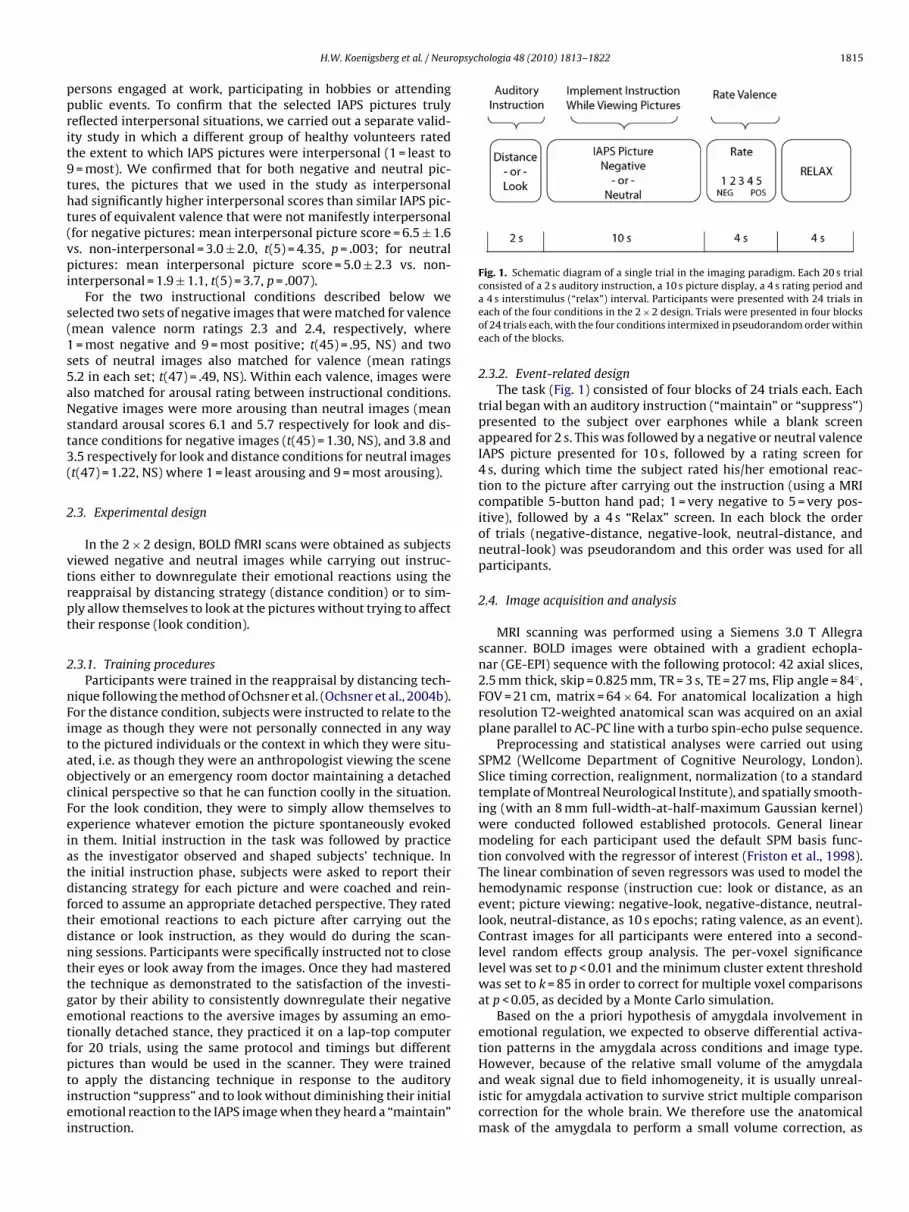
Fig. 1. Schematic diagram of a single trial in the imaging paradigm. Each 20 s trialconsisted of a 2 s auditory instruction, a 10 s picture display, a 4 s rating period and
H.W. Koenigsberg et al. / Neur
ersons engaged at work, participating in hobbies or attendingublic events. To confirm that the selected IAPS pictures trulyeflected interpersonal situations, we carried out a separate valid-ty study in which a different group of healthy volunteers ratedhe extent to which IAPS pictures were interpersonal (1 = least to= most). We confirmed that for both negative and neutral pic-
ures, the pictures that we used in the study as interpersonalad significantly higher interpersonal scores than similar IAPS pic-ures of equivalent valence that were not manifestly interpersonalfor negative pictures: mean interpersonal picture score = 6.5 ± 1.6s. non-interpersonal = 3.0 ± 2.0, t(5) = 4.35, p = .003; for neutralictures: mean interpersonal picture score = 5.0 ± 2.3 vs. non-
nterpersonal = 1.9 ± 1.1, t(5) = 3.7, p = .007).For the two instructional conditions described below we
elected two sets of negative images that were matched for valencemean valence norm ratings 2.3 and 2.4, respectively, where= most negative and 9 = most positive; t(45) = .95, NS) and two
ets of neutral images also matched for valence (mean ratings.2 in each set; t(47) = .49, NS). Within each valence, images werelso matched for arousal rating between instructional conditions.egative images were more arousing than neutral images (mean
tandard arousal scores 6.1 and 5.7 respectively for look and dis-ance conditions for negative images (t(45) = 1.30, NS), and 3.8 and.5 respectively for look and distance conditions for neutral imagest(47) = 1.22, NS) where 1 = least arousing and 9 = most arousing).
.3. Experimental design
In the 2 × 2 design, BOLD fMRI scans were obtained as subjectsiewed negative and neutral images while carrying out instruc-ions either to downregulate their emotional reactions using theeappraisal by distancing strategy (distance condition) or to sim-ly allow themselves to look at the pictures without trying to affectheir response (look condition).
.3.1. Training proceduresParticipants were trained in the reappraisal by distancing tech-
ique following the method of Ochsner et al. (Ochsner et al., 2004b).or the distance condition, subjects were instructed to relate to themage as though they were not personally connected in any wayo the pictured individuals or the context in which they were situ-ted, i.e. as though they were an anthropologist viewing the scenebjectively or an emergency room doctor maintaining a detachedlinical perspective so that he can function coolly in the situation.or the look condition, they were to simply allow themselves toxperience whatever emotion the picture spontaneously evokedn them. Initial instruction in the task was followed by practices the investigator observed and shaped subjects’ technique. Inhe initial instruction phase, subjects were asked to report theiristancing strategy for each picture and were coached and rein-orced to assume an appropriate detached perspective. They ratedheir emotional reactions to each picture after carrying out theistance or look instruction, as they would do during the scan-ing sessions. Participants were specifically instructed not to closeheir eyes or look away from the images. Once they had masteredhe technique as demonstrated to the satisfaction of the investi-ator by their ability to consistently downregulate their negativemotional reactions to the aversive images by assuming an emo-ionally detached stance, they practiced it on a lap-top computer
or 20 trials, using the same protocol and timings but differentictures than would be used in the scanner. They were trainedo apply the distancing technique in response to the auditorynstruction “suppress” and to look without diminishing their initialmotional reaction to the IAPS image when they heard a “maintain”nstruction.a 4 s interstimulus (“relax”) interval. Participants were presented with 24 trials ineach of the four conditions in the 2 × 2 design. Trials were presented in four blocksof 24 trials each, with the four conditions intermixed in pseudorandom order withineach of the blocks.
2.3.2. Event-related designThe task (Fig. 1) consisted of four blocks of 24 trials each. Each
trial began with an auditory instruction (“maintain” or “suppress”)presented to the subject over earphones while a blank screenappeared for 2 s. This was followed by a negative or neutral valenceIAPS picture presented for 10 s, followed by a rating screen for4 s, during which time the subject rated his/her emotional reac-tion to the picture after carrying out the instruction (using a MRIcompatible 5-button hand pad; 1 = very negative to 5 = very pos-itive), followed by a 4 s “Relax” screen. In each block the orderof trials (negative-distance, negative-look, neutral-distance, andneutral-look) was pseudorandom and this order was used for allparticipants.
2.4. Image acquisition and analysis
MRI scanning was performed using a Siemens 3.0 T Allegrascanner. BOLD images were obtained with a gradient echopla-nar (GE-EPI) sequence with the following protocol: 42 axial slices,2.5 mm thick, skip = 0.825 mm, TR = 3 s, TE = 27 ms, Flip angle = 84◦,FOV = 21 cm, matrix = 64 × 64. For anatomical localization a highresolution T2-weighted anatomical scan was acquired on an axialplane parallel to AC-PC line with a turbo spin-echo pulse sequence.
Preprocessing and statistical analyses were carried out usingSPM2 (Wellcome Department of Cognitive Neurology, London).Slice timing correction, realignment, normalization (to a standardtemplate of Montreal Neurological Institute), and spatially smooth-ing (with an 8 mm full-width-at-half-maximum Gaussian kernel)were conducted followed established protocols. General linearmodeling for each participant used the default SPM basis func-tion convolved with the regressor of interest (Friston et al., 1998).The linear combination of seven regressors was used to model thehemodynamic response (instruction cue: look or distance, as anevent; picture viewing: negative-look, negative-distance, neutral-look, neutral-distance, as 10 s epochs; rating valence, as an event).Contrast images for all participants were entered into a second-level random effects group analysis. The per-voxel significancelevel was set to p < 0.01 and the minimum cluster extent thresholdwas set to k = 85 in order to correct for multiple voxel comparisonsat p < 0.05, as decided by a Monte Carlo simulation.
Based on the a priori hypothesis of amygdala involvement inemotional regulation, we expected to observe differential activa-tion patterns in the amygdala across conditions and image type.
However, because of the relative small volume of the amygdalaand weak signal due to field inhomogeneity, it is usually unreal-istic for amygdala activation to survive strict multiple comparisoncorrection for the whole brain. We therefore use the anatomicalmask of the amygdala to perform a small volume correction, as
1816 H.W. Koenigsberg et al. / Neuropsyc
Fs
iRe
3
3
osad
stwpcwtnn(
TMc
ig. 2. Mean valence ratings after carrying out look/distance instruction during thecan.
mplemented in SPM2. The resampled voxel size was 2 × 2 × 2 mm3.egression weights (betas) were used to quantify signal change forach instructional condition and picture type in regions of interest.
. Results
.1. Behavioral results
In post-scan debriefing, participants reported that they carriedut the reappraisal by distancing strategy as instructed during thecan. Participants confirmed that they did not close their eyes orvert their gaze during the viewing periods in either the look oristance conditions.
The self-report ratings of emotional valence obtained during thecan demonstrated an image type (negative vs. neutral) × instruc-ion (look vs. distance) interaction (F(1,14) = 50.35, p = .000005) asell as main effects for image type and instruction (F(1,14) = 66.78,= .000001, and F(1,14) = 14.52, p = .002, respectively). Plannedomparisons showed that negative affect was greater (lower score)
hen simply looking at (mean score: 1.82 ± 0.33) as comparedo distancing from (mean: 2.59 ± 0.51; t(14) = 5.73, p = .00005)egative images (Fig. 2). Responses to neutral images were not sig-ificantly different in the look (mean score: 3.20 ± .23) and distancemean score 3.07 ± .11) conditions (t(14) = 1.99, p = .07).
able 1ain effect of picture valence k is cluster size in 2 × 2 × 2 mm voxels, (x, y, z) are MNI coor
orrected p < .05.
Region k
Negative ≥ neutralR fusiform G. (BA19) 10969R amygdala 571R inferior parietal (BA7) 150L posterior cingulate (BA30) 89R superior medial frontal G. (BA10) 783R inferior frontal G., pars triangularis (BA45) 925L inferior frontal G., pars triangularis (BA45) 206L thalamus 1159
Neutral ≥ negativeR middle frontal G. (BA11) 225R superior frontal G. (BA6) 86R cerebellum 134Cerebellum vermis 161R caudate 106
hologia 48 (2010) 1813–1822
3.2. Imaging results
3.2.1. Main effect of picture valenceWe first examined the effect of picture valence independent of
instruction, computing the (negative > neutral) and (neutral > neg-ative) contrasts (Table 1). As expected, there was greater amygdalaactivation to negative as compared to neutral pictures.
3.2.2. Main effect of instruction typeTo examine the main effect of distancing, we computed the
(distance > look) and (look > distance) contrasts, collapsing acrossimage type (Fig. 3 and Table 2). Regions with significantly greateractivation in the distance compared to look condition included themiddle and superior temporal gyri bilaterally (BA21/22; the regionbordering the superior temporal sulcus; STS), the posterior cin-gulate cortex (PCC; BA31)/precuneus (BA7), and inferior parietallobules (IPL: BA40) bilaterally. During the look compared to dis-tance condition there was greater activation in visual areas (cuneus,middle occipital gyrus; BA17), in the left fusiform gyrus (BA37),the left middle temporal gyrus extending to the occipital gyrus(BA37/39/19), the right superior parietal lobule (BA7), and medialprefrontal regions (BA8/9/10).
3.2.3. Interaction between instructional condition and image typeTo explore the interaction between instructional condition and
picture valence we constructed SPM maps of the double differencesof BOLD activation: (1) distancing (negative pictures − neutral pic-tures) − look (negative pictures − neutral pictures), and (2) look(negative pictures − neutral pictures) − distancing (negative pic-tures − neutral pictures) (Table 3 and Fig. 4).
Contrast (1) identified regions where the effects of distanc-ing were greater for negative as compared to neutral images, andincluded the dorsal anterior cingulate gyrus (dACC; BA32), themiddle cingulate gyrus (BA23), medial prefrontal cortex (mPFC,BA10), right middle and superior frontal gyrus (BA10), left, infe-rior frontal gyrus/insula (BA45/47/13), the middle occipital (BA19)and middle and superior temporal regions (BA39/19) bilaterally.Inspection of the regression weights (betas) for condition and pic-ture type (see Fig. 5) show condition effects for both picture types.We carried out post hoc repeated measures ANOVAs to determinewhether the effects of condition were significant for the nega-
tive pictures alone. For negative pictures there was significantlygreater activation in the Distance vs. Look condition in the dorsalACC (BA32/9) (F(1,15) = 6.71, p = .02), the right medial prefrontalcortex (mPFC,BA10) (F(1,15) = 4.93, p = .04), the middle cingulate(BA23) (F(1,15) = 19.40, p = .0005), and the left superior occipitaldinates of peak voxel in cluster. Individual voxel p < .01, with k > 85 voxels to obtain
MNI coordinates Z-score
x y z
36 −70 −12 5.5920 −2 −10 3.9832 −48 56 3.42
0 −50 26 3.444 54 28 4.78
50 32 12 4.17−50 26 18 3.12−4 −16 2 3.64
26 48 4 3.6922 4 56 2.8834 −60 −34 3.87−2 −68 −20 3.1812 22 −4 3.37

H.W. Koenigsberg et al. / Neuropsychologia 48 (2010) 1813–1822 1817
Table 2Group activations for distancing > looking and looking > distancing for all pictures collapsed across valence (negative/neutral) k is cluster size in 2 × 2 × 2 mm3 voxels,(x, y, z) are MNI coordinates of peak voxel in cluster. Individual voxel p < .01, with k > 85 voxels to obtain corrected p < .05.
Region k MNI coordinates Z-score
x y z
Distancing ≥ lookingPosterior cingulate (BA31)/precuneus (BA7) 3035 4 −24 34 4.19L inferior parietal lobule/supramarginal G (BA40/7) 1560 −40 −52 36 6.00R inferior parietal lobule (BA40) 416 50 −54 42 3.84R middle/superior temporal G (BA21/22) 330 56 −18 −2 3.61L middle temporal gyrus (BA21) 193 −62 −42 −4 3.57
Looking ≥ distancingL & R cuneus/middle occipital G (BA17) 4632 −6 −88 4 4.43L middle temporal/occipital G. (BA37/39/19) 1446 −54 −60 10 4.90L postcentral G (BA2) 282 −50 −32 56 3.62
gcnfra(rF
oi
FaL
L superior/medial frontal G (BA8/9/10) 298R superior parietal lobule (BA7) 151L fusiform G. (BA37) 127R thalamus 164
yrus (BA7) (F(1,15) = 10.22, p = .006). Although we had no spe-ific a priori hypotheses about regions involved in distancing fromeutral pictures, for completeness we report the post hoc finding
or these pictures as well. In the left and right middle tempo-al/occipital regions (BA39/19) there is a significant decrease inctivation to neural pictures when distancing compared to lookingpost hoc F(1,15) = 42.35, p = .00001, left; F(1,15) = 23.99, p = .00002,
ight) and no change for negative pictures (F(1,15) = 0.04, NS and(1,15) = 0.34, NS, respectively).Contrast (2) identified regions where the modulatory effectsf distancing were greater for negative as compared to neutralmages, and included the right lingual gyrus (Fig. 4d) and cuneus
ig. 3. Statistical parametric maps of the main effects of reappraisal (distancing vs. lookin.) Distancing > looking showing greater activity in the posterior cingulate cortex (PCC),ooking > distancing showing greater activity in the occipital visual regions (BA17/19) an
−6 56 26 3.4624 −70 44 3.85
−42 −46 −14 3.0316 −6 10 3.65
(BA17/18), the right and left precentral gyri (BA43/BA6), as well asthe left superior temporal gyrus (BA38). In addition these mod-ulatory effects were also observed in the amygdala (Fig. 4c) atp < .05 (two-tailed) threshold, applying a small volume correctionas described above. This interaction effect is accounted for by theresponse to negative pictures (Fig. 5b) as demonstrated in the posthoc analysis of the region of interest (F(1,15) = 4.94, p = .04).
3.2.4. Simple effect of instruction type for negative images onlyGiven that the behavioral data demonstrated that distancing
reduced the negative emotion experienced when viewing nega-tive pictures, but had little effect for neutral pictures, we examined
g).precuneus and intraparietal sulcus (IPS) when distancing compared to looking. b.)d left fusiform gyrus (Fus) when looking compared to distancing.

1818 H.W. Koenigsberg et al. / Neuropsychologia 48 (2010) 1813–1822
Fig. 4. Statistical parametric maps of the effects of distancing as a function of image type.a . a.) Sal le/supi to loC areda
tdFva>rda
4
o
.) and b.) Effects of distancing (negative − neutral) − looking (negative − neutral)ooking in the dorsal anterior cingulate cortex (dACC), medial PFC (mPFC) and middn the region of the superior temporal sulcus (MTG/STG) when distancing comparedoronal view showing increased right amygdala (AMY) activity when looking compctivity under the same conditions.
he (distance > look) contrast and the reversed contrast (look >istance) for the negative pictures alone. The results (Table 4 andig. 6) were similar to those for the main effect of instruction acrossalence, suggesting the distancing finding arose primarily from theversive pictures, as would be expected. In addition, the distancelook contrast for negative pictures also identified a locus in the
ight middle frontal gyrus (BA10) and the reverse contrast of look >istance identified loci in the right inferior temporal gyrus (BA19)nd left precentral gyri (BA6).
. Discussion
To our knowledge, this study is the first to focus specificallyn the use of distancing to regulate responses to aversive social
gittal and coronal views showing greater activation when distancing compared toerior frontal gyrus (SFG). b.) Sagittal and coronal views showing greater activation
oking. c.) and d.) Looking (negative − neutral) − distancing (negative − neutral). c.)to distancing. d.) Sagittal view showing increased visual cortex (lingual gyrus; LIN)
cues employing an event-related fMRI design. As detailed below,this study extends previous work by identifying regions that mayplay a special role in regulating emotional responses to social cuesusing distancing, and replicates the findings of prefrontal and cin-gulate participation in distancing that have been demonstrated byother studies using reappraisal of emotional cues that have mixedcontent.
4.1. Behavioral and brain correlates of distancing from aversive
social cues4.1.1. Behavioral performanceParticipants reported less negative emotion when distancing
themselves from, as compared to looking at, negative images,

H.W. Koenigsberg et al. / Neuropsychologia 48 (2010) 1813–1822 1819
Table 3Group activations for interaction between instruction (distancing vs. looking) and image type (negative vs. neutral) k is cluster size in 2 × 2 × 2 mm3 voxels, (x, y, z) are MNIcoordinates of peak voxel in cluster. Individual voxel p < .01, with k > 85 voxels to obtain corrected p < .05 *p < .05 (two-tailed) with small volume correction.
Region k MNI coordinates Z-score
x y z
Distancing (neg − neu) ≥ looking (neg − neu)R middle & superior temporal G/middle occipital G (BA19) 963 46 −64 12 4.94L middle & superior temporal G/middle occipital G (BA39/19) 539 −50 −60 12 4.10L superior occipital G. (BA7) 126 −16 −66 40 3.07Anterior cingulate G./L medial frontal G. (BA32/9) 162 −2 28 38 3.57Middle cingulate G. (BA23) 103 0 −20 40 3.00L inferior frontal G./insula (BA45/47/13) 102 −38 18 2 3.88R middle/superior frontal G. (BA10) 341 32 54 20 4.20R medial frontal G. (BA10) 132 8 54 2 3.24L medial/superior frontal G. (BA10) 200 −10 56 10 3.45
Looking (neg − neu) ≥ distancing (neg − neu)R lingual G/cuneus (BA17/18) 505 4 −84 2 3.19L superior temporal G. (BA38) 120 −44 0 −16 3.68
f righ
btsn
Fnd*a
R precentral G (BA43) 93L precentral G (BA6) 239R amygdala* 37
* p(FDR) < .05 (two-tailed) using small volume correction with anatomical mask o
ut reported similarly low levels of negative emotion in responseo neutral images in both the distance and look conditions. Thisuggests that participants were able to reduce their subjectiveegative reactions to aversive social scenes by employing the dis-
ig. 5. Comparison of regression weights in the look and distance conditions foregative and neutral pictures. a.) Anterior cingulate cortex (ACC), b.) Right amyg-ala.Significant difference between look and distance condition (p < .02 ACC; p < .05mygdala, see text).
−58 −4 24 3.41−40 −14 58 3.12
28 −6 −10 3.34
t amygdala.
tancing strategy, but that this strategy had little effect, as wouldbe expected, upon neutral scenes that evoked little emotion tobegin with. While we can not rule out the possibility that sub-ject responses were influenced by the demand characteristics ofthe task or the possibility that subjects closed their eyes or lookedat non-emotional parts of the pictures, during debriefing par-ticipants indicated that they followed instructions. Furthermore,as noted below, we observed decreased amygdala activation inthe distancing condition, which suggests that participants werein fact decreasing their emotional reactions as they reported(Kim & Hamann, 2007; Ochsner et al., 2002, 2004b; Phan et al.,2005).
4.1.2. Imaging findingsThe main effect of distancing the self from interpersonal scenes
was to activate a network of regions important for perspective-taking, attentional control, and assessment of social cues. Withregard to perspective-taking, we found activation of posterior cin-gulate and precuneus regions implicated in processes importantfor assuming a distanced perspective, including being unengagedin (i.e. remote from) witnessed social interactions (Schilbach etal., 2006), turning away from external sources of informationto a more inward focused state of consciousness (Binder et al.,1999; Kjaer, Nowak, & Lou, 2002), and with assessing the self-relevance of stimuli (Kelley et al., 2002; Ochsner et al., 2005; Vogt,2005). The IPL engagement we found is consistent with its rolein top–down control of the allocation of attention to visual cues(Corbetta, Kincade, Ollinger, McAvoy, & Shulman, 2000; Hopfinger,Buonocore, & Mangun, 2000; Pessoa et al., 2002). The activation weobserved in the region of the superior and middle temporal gyriis consistent with the purported role of this region in social per-ception (Allison et al., 2000; Kourtzi & Kanwisher, 2000) as wellas in attributing intention and mental states to others (theory ofmind) (Allison et al., 2000; Frith & Frith, 2003; Gallagher et al., 2000;Kourtzi & Kanwisher, 2000).
The interaction of instruction type by image type highlightsthose regions that are more engaged when distancing from neg-ative as compared to neutral social cues. Our findings suggest thatregulatory demands when distancing from negative social cues
recruits a network of regions, some active for distancing in gen-eral, as well as additional regions that appear to support distancingfrom aversive stimuli per se. The latter regions are of greatest inter-est because they are critical to the emotion regulatory and not justthe perspective-taking aspects of distancing.
1820 H.W. Koenigsberg et al. / Neuropsychologia 48 (2010) 1813–1822
Table 4Group activations for distancing vs. looking when viewing negative valence pictures k is cluster size in 2 × 2 × 2 mm3 voxels, (x, y, z) are MNI coordinates of peak voxel incluster. Individual voxel p < .01, with k > 85 voxels to obtain corrected p < .05.
Region k MNI coordinates Z-score
x y z
Distancing ≥ lookingPosterior cingulate G/precuneus (BA23/31) 3154 4 −24 36 4.60L inf parietal lobule/angular G/supramarginal G (BA40) 1450 −40 −56 38 4.04R inf parietal lobule/supramarginal G/Angular G (BA40) 470 50 −56 40 3.66L middle temporal G. (BA21) 210 −58 −52 −6 3.94R middle temporal G. (BA21/22) 225 62 −36 2 3.64R middle frontal G. (BA10) 89 36 56 4 2.85
Looking ≥ distancingL middle temporal/middle occipital G (BA19) 486 −40 −78 14 4.79
aci(TPadabflifdstpcthAemmcaoaC
FNm
R inferior temporal G. (BA19) 219L & R cuneus/lingual G. (BA17) 3173L postcentral G. (BA1) 95L precentral G. (BA6) 304
Three regions were selectively engaged during distancing toversive social cues. The first was the dACC, which is typi-ally engaged in monitoring conflict between opposing tasks andt recruits cognitive control processes to resolve such conflictBotvinick, Cohen, & Carter, 2004; Fan, Flombaum, McCandliss,homas, & Posner, 2003; Fan, McCandliss, Fossella, Flombaum, &osner, 2005; Liu, Banich, Jacobson, & Tanabe, 2004; Mohanty etl., 2007; Ochsner et al., 2002). In keeping with prior work showingACC activity during reappraisal (Kim & Hamann, 2007; Ochsner etl., 2002, 2004b; Phan et al., 2005), we would expect this region toe activated in the distancing task because there would be a con-ict between a prepotent tendency to attend to emotionally salient
mages and the conscious effort to distance oneself from them. Inact, as observed here, there should be greater recruitment of theACC in the reappraisal of negative compared to neutral imagesince the viewing of aversive images should heighten look vs. dis-ance conflict because of the tendency to attend more strongly tootentially threatening aversive situations, with the dACC beingalled into play to resolve the conflict by enlisting top–down con-rol. The second was the mPFC, which is recruited in monitoringow emotional cues affect the self (Fossati et al., 2003; Gusnard,kbudak, Shulman, & Raichle, 2001; Lane & McRae, 2004; Ochsnert al., 2004a), information that would be important for keepingatters emotionally distant from the self. The third was the rightiddle frontal gyrus, which was expected since this region is impli-
ated in the selection and control of behavioral strategies andctions, keeping these strategies in mind throughout performancef a task, inhibiting prepotent responses, and regulating selectivettention (Garavan, Ross, Murphy, Roche, & Stein, 2002; Miller &ohen, 2001).
ig. 6. Statistical parametric maps for distancing > looking when viewing negative picturote increased activation when distancing compared to looking in the posterior cingulaiddle temporal gyrus (MTG).
44 −72 −6 3.202 −86 0 4.53
−50 −32 56 2.93−40 −12 62 3.31
Taken together these findings fit with prior studies implicat-ing the lateral PFC in reappraisal (Kim & Hamann, 2007; Ochsneret al., 2002, 2004a; Phan et al., 2005) and the mPFC in distancingin particular (Kalisch et al., 2005; Ochsner et al., 2004b). We can-not rule out the possibility that activation of these regions duringdistancing may reflect the engagement of cognitive effort in gen-eral rather than processes specific to distancing (Urry, van Reekum,Johnstone, & Davidson, 2009). Our two-by-two factorial design,however, makes this interpretation less likely because distancingfrom neutral pictures (a somewhat counter-intuitive task) couldbe expected to impose an equal or even greater general cogni-tive demand compared to distancing from negative pictures. Inaddition, our observations extend previous work by demonstrat-ing that regions generally implicated in perspective-taking, suchas the precuneus, are involved in distancing in general, whereasother regions implicated in emotion regulation and social percep-tion, such as the STS, are engaged in distancing from aversive socialcues in particular. These findings have not been reported in priorwork on distancing which involved shock (Kalisch et al., 2005) ormixed aversive images (Kim & Hamann, 2007; Ochsner et al., 2002,2004b; Phan et al., 2005; Urry et al., 2006). This difference also sug-gests that activation of these regions is not simply a reflection ofgeneral cognitive demand during reappraisal.
The instruction type by image type interaction also identifiedregions that are modulated more strongly when distancing from
negative as compared to neutral images, presumably because theseare the regions supporting the generation of the negative emotionalresponse from which one must distance one’s self. These regionsinclude the amygdala and visual areas. This is consistent with priorwork showing modulation of amygdala (Kim & Hamann, 2007;es only.te cortex (PCC), the precuneus (Precun), the intraparietal sulcus (IPS) and the left
opsych
Oa
4
toriutstn
eaicatit(icoaibaTaar
alaosuaa
A
tMRC
R
A
A
A
B
B
H.W. Koenigsberg et al. / Neur
chsner et al., 2002, 2004b; Urry et al., 2006) and visual cortexctivity (Ochsner et al., 2002) by various forms of reappraisal.
.2. Limitations and future directions
Although this study sheds new light on the neural bases of dis-ancing, it is important to acknowledge its limitations as well. As inther studies of cognitive reappraisal, we relied upon participants’eports that they carried out the task as directed. We selected IAPSmages that exclusively depicted persons in socially evocative sit-ations and excluded images of bodily mutilation, disfigurement,hreatening animals that have been included as aversive emotionaltimuli in other studies. Further studies are called for to replicatehese findings and to directly contrast distancing from social andon-social images.
Future work might also further clarify the specific processesngaged during the two conditions in this experiment – lookingnd distancing. Looking at pictures with facial emotional content istself a complex task that likely involves a number of psychologi-al operations that encode the perceptual characteristics of stimulis well as their valence and intensity, and support recognition ofhem. These processes may call upon distributed neural networksncluding the ventromedial and dorsolateral prefrontal cortex,he ventrolateral and dorsomedial prefrontal cortex and the ACCGrimm et al., 2006). As noted in the introduction, distancing, too,nvolves a number mental operations in addition to those impli-ated in looking at emotional stimuli, including the maintenancef a regulatory goal, attentional allocation, self-monitoring andttributions about one’s emotional state. In future work, it will bemportant to further clarify which regions specifically are engagedy distancing, as opposed to looking, other variants of reappraisal,nd other types of emotion judgments and regulatory strategies.his is important, given that even seemingly simple cognitive oper-tions such as making a judgment about one’s emotional responses opposed to passively looking at a stimulus can modulateesponses in the amygdala (Taylor, Phan, Decker, & Liberzon, 2003).
As this research progresses and normative patterns of neuralctivation in distancing from aversive social cues are firmly estab-ished, this work could be extended to contrast the patterns ofctivation in healthy volunteers to those of individuals with dis-rders characterized by disturbances in interpersonal relatednessuch as BPD, AvPD and SSD (Koenigsberg et al., 2009). This can helps to better understand the neural correlates of these disordersnd suggest psychotherapeutic and pharmacologic approaches toddress interpersonal disturbances.
cknowledgements
Completion of this work was supported in part by grants fromhe National Institute of Mental Health (RO1 MH077813, PI: H.W.K.;
H076137, PI: K.N.O.) and from the National Center for Researchesources, National Institutes of Health for the Mount Sinai Generallinical Research Center (5MO1 RR00071).
eferences
dolphs, R. (2003). Cognitive neuroscience of human social behaviour. NatureReviews. Neuroscience, 4(3), 165–178.
llison, T., Puce, A., & McCarthy, G. (2000). Social perception from visual cues: Roleof the STS region. Trends in Cognitive Sciences, 4(7), 267–278.
yduk, O., & Kross, E. (2008). Enhancing the pace of recovery: Self-distanced analysisof negative experiences reduces blood pressure reactivity. Psychological Science,
19(3), 229–231.allon, J. S., Kaur, T., Marks, I. I., & Cadenhead, K. S. (2007). Social functioning in youngpeople at risk for schizophrenia. Psychiatry Research, 151(1–2), 29–35.
anks, S. J., Eddy, K. T., Angstadt, M., Nathan, P. J., & Phan, K. L. (2007). Amygdala-frontal connectivity during emotion regulation. Society Cognitive & AffectiveNeuroscience, 2(4), 303–312.
ologia 48 (2010) 1813–1822 1821
Beauregard, M., Levesque, J., & Bourgouin, P. (2001). Neural correlates of consciousself-regulation of emotion. Journal of Neuroscience, 21(18), RC165.
Binder, J. R., Frost, J. A., Hammeke, T. A., Bellgowan, P. S., Rao, S. M., & Cox, R. W.(1999). Conceptual processing during the conscious resting state. A functionalMRI study. Journal of Cognitive Neuroscience, 11(1), 80–95.
Botvinick, M. M., Cohen, J. D., & Carter, C. S. (2004). Conflict monitoring and anteriorcingulate cortex: An update. Trends in Cognition Science, 8(12), 539–546.
Britton, J. C., Phan, K. L., Taylor, S. F., Welsh, R. C., Berridge, K. C., & Liberzon, I. (2006).Neural correlates of social and nonsocial emotions: An fMRI study. NeuroImage,31(1), 397–409.
Burnett, S., Bird, G., Moll, J., Frith, C., & Blakemore, S. J. (2009). Development dur-ing adolescence of the neural processing of social emotion. Journal of CognitiveNeuroscience, 21(9), 1736–1750.
Corbetta, M., Kincade, J. M., Ollinger, J. M., McAvoy, M. P., & Shulman, G. L. (2000). Vol-untary orienting is dissociated from target detection in human posterior parietalcortex. Nature Neuroscience, 3(3), 292–297.
Eippert, F., Veit, R., Weiskopf, N., Erb, M., Birbaumer, N., & Anders, S. (2007). Reg-ulation of emotional responses elicited by threat-related stimuli. Human BrainMapping, 28(5), 409–423.
Fan, J., Flombaum, J. I., McCandliss, B. D., Thomas, K. M., & Posner, M. I. (2003).Cognitive and brain consequences of conflict. NeuroImage, 18(1), 42–57.
Fan, J., McCandliss, B. D., Fossella, J., Flombaum, J. I., & Posner, M. I. (2005). Theactivation of attentional networks. NeuroImage, 26(2), 471–479.
Fossati, P., Hevenor, S. J., Graham, S. J., Grady, C., Keightley, M. L., Craik, F., et al.(2003). In search of the emotional self: An FMRI study using positive and negativeemotional words. American Journal of Psychiatry, 160(11), 1938–1945.
Friston, K. J., Fletcher, P., Josephs, O., Holmes, A., Rugg, M. D., & Turner, R.(1998). Event-related fMRI: characterizing differential responses. Neuroimage, 7,30–40.
Frith, U., & Frith, C. D. (2003). Development and neurophysiology of mentalizing.Philosophical Transactions of the Royal Society of London. Series B, Biological Sci-ences, 358(1431), 459–473.
Gallagher, H. L., Happe, F., Brunswick, N., Fletcher, P. C., Frith, U., & Frith, C. D. (2000).Reading the mind in cartoons and stories: An fMRI study of ‘theory of mind’ inverbal and nonverbal tasks. Neuropsychologia, 38(1), 11–21.
Garavan, H., Ross, T. J., Murphy, K., Roche, R. A., & Stein, E. A. (2002). Dissociable exec-utive functions in the dynamic control of behavior: Inhibition, error detection,and correction. NeuroImage, 17(4), 1820–1829.
Grimm, S., Schmidt, C. F., Bermpohl, F., Heinzel, A., Dahlem, Y., Wyss, M., et al.(2006). Segregated neural representation of distinct emotion dimensions in theprefrontal cortex-an fMRI study. NeuroImage, 30(1), 325–340.
Gross, J. J. (2002). Emotion regulation: Affective, cognitive, and social consequences.Psychophysiology, 39(3), 281–291.
Gunderson, J. G. (2007). Disturbed relationships as a phenotype for borderline per-sonality disorder. American Journal of Psychiatry, 164(11), 1637–1640.
Gunderson, J. G., & Lyons-Ruth, K. (2008). BPD’s interpersonal hypersensitivityphenotype: A gene-environment-developmental model. Journal of PersonalityDisorders, 22(1), 22–41.
Gusnard, D. A., Akbudak, E., Shulman, G. L., & Raichle, M. E. (2001). Medial prefrontalcortex and self-referential mental activity: Relation to a default mode of brainfunction. Proceedings of the National Academy of Sciences USA, 98(7), 4259–4264.
Harenski, C. L., & Hamann, S. (2006). Neural correlates of regulating negative emo-tions related to moral violations. NeuroImage, 30(1), 313–324.
Harris, L. T., McClure, S. M., van den Bos, W., Cohen, J. D., & Fiske, S. T. (2007). Regionsof the MPFC differentially tuned to social and nonsocial affective evaluation.Cognitive Affective & Behavioral Neuroscience, 7(4), 309–316.
Hopfinger, J. B., Buonocore, M. H., & Mangun, G. R. (2000). The neural mechanismsof top–down attentional control. Nature Neuroscience, 3(3), 284–291.
Kalisch, R., Wiech, K., Critchley, H. D., Seymour, B., O’Doherty, J. P., Oakley, D. A., et al.(2005). Anxiety reduction through detachment: Subjective, physiological, andneural effects. Journal of Cognitive Neuroscience, 17(6), 874–883.
Kelley, W. M., Macrae, C. N., Wyland, C. L., Caglar, S., Inati, S., & Heatherton, T.F. (2002). Finding the self? An event-related fMRI study. Journal of CognitiveNeuroscience, 14(5), 785–794.
Kim, S. H., & Hamann, S. (2007). Neural correlates of positive and negative emotionregulation. Journal of Cognitive Neuroscience, 19(5), 776–798.
Kjaer, T. W., Nowak, M., & Lou, H. C. (2002). Reflective self-awareness and consciousstates: PET evidence for a common midline parietofrontal core. NeuroImage,17(2), 1080–1086.
Knight, R. T. (2007). Neuroscience. Neural networks debunk phrenology. Science,316(5831), 1578–1579.
Koenigsberg, H. W., Fan, J., Ochsner, K. N., Liu, X., Guise, K. G., Pizzarello, S., etal. (2009). Neural correlates of the use of psychological distancing to regulateresponses to negative social cues: A study of patients with borderline personalitydisorder. Biological Psychiatry, 66(9), 854–863.
Kourtzi, Z., & Kanwisher, N. (2000). Activation in human MT/MST by static imageswith implied motion. Journal of Cognitive Neuroscience, 12(1), 48–55.
Kross, E., & Ayduk, O. (2008). Facilitating adaptive emotional analysis: Distinguish-ing distanced-analysis of depressive experiences from immersed-analysis anddistraction. Personality and Social Psychology Bulletin, 34(7), 924–938.
Kross E, Ayduk O. Facilitating adaptive emotional analysis: Distinguishing distanced-analysis of depressive experiences from immersed-analysis and distraction.Personality and Social Psychology Bulletin, in press.
Kross, E., Ayduk, O., & Mischel, W. (2005). When asking “why” does not hurt.Distinguishing rumination from reflective processing of negative emotions. Psy-chological Science, 16(9), 709–715.
1 opsyc
K
L
L
L
L
L
L
M
M
M
M
M
O
O
O
O
822 H.W. Koenigsberg et al. / Neur
ross, E., Egner, T., Ochsner, K., Hirsch, J., & Downey, G. (2007). Neural dynamics ofrejection sensitivity. Journal of Cognitive Neuroscience, 19(6), 945–956.
ane, R., & McRae, K. (2004). Neural substrates of conscious emotional experience:A cognitive-neuroscientific perspective. In M. Beauregard (Ed.), Consciousness,emotional self-regulation and the brain (pp. 87–122). Amsterdam: John Ben-jamins.
ang, P. J. B. M., & Cuthbert, B. N. (2001). International affective pictures system (IAPS):Technical manual and affective ratings. NIMH Center for the Study of Emotion andAttention.
eising, D., Sporberg, D., & Rehbein, D. (2006). Characteristic interpersonal behaviorin dependent and avoidant personality disorder can be observed within veryshort interaction sequences. Journal of Personality Disorders, 20(4), 319–330.
estou, V., Pollick, F. E., & Kourtzi, Z. (2008). Neural substrates for action under-standing at different description levels in the human brain. Journal of CognitiveNeuroscience, 20(2), 324–341.
evesque, J., Eugene, F., Joanette, Y., Paquette, V., Mensour, B., Beaudoin, G., et al.(2003). Neural circuitry underlying voluntary suppression of sadness. BiologicalPsychiatry, 53(6), 502–510.
iu, X., Banich, M. T., Jacobson, B. L., & Tanabe, J. L. (2004). Common and distinct neuralsubstrates of attentional control in an integrated Simon and spatial Stroop taskas assessed by event-related fMRI. NeuroImage, 22(3), 1097–1106.
eyer, B., Pilkonis, P. A., & Beevers, C. G. (2004). What’s in a (neutral) face? Person-ality disorders, attachment styles, and the appraisal of ambiguous social cues.Journal of Personality Disorders, 18(4), 320–336.
eyer, J., & Shean, G. (2006). Social-cognitive functioning and schizotypal charac-teristics. Journal of Psychology, 140(3), 199–207.
iller, E. K., & Cohen, J. D. (2001). An integrative theory of prefrontal cortex function.Annual Review of Neuroscience, 24, 167–202.
inzenberg, M. J., Poole, J. H., & Vinogradov, S. (2006). Social-emotion recognitionin borderline personality disorder. Comprehensive Psychiatry, 47(6), 468–474.
ohanty, A., Engels, A. S., Herrington, J. D., Heller, W., Ho, M. H., Banich, M. T., etal. (2007). Differential engagement of anterior cingulate cortex subdivisions forcognitive and emotional function. Psychophysiology, 44(3), 343–351.
chsner, K. (2008). The social-emotional processing stream: Five core constructs andtheir translational potential for schizophrenia and beyond. Biological Psychiatry,64(1), 48–61.
chsner K., & Gross J. Cognitive emotion regulation: Insights from social cognitiveand affective neuroscience. Current Directions in Psychological Science, in press.
chsner, K. N., Beer, J. S., Robertson, E. R., Cooper, J. C., Gabrieli, J. D., Kihsltrom, J.F., et al. (2005). The neural correlates of direct and reflected self-knowledge.NeuroImage, 28(4), 797–814.
chsner, K. N., Bunge, S. A., Gross, J. J., & Gabrieli, J. D. (2002). Rethinking feel-ings: An FMRI study of the cognitive regulation of emotion. Journal of CognitiveNeuroscience, 14(8), 1215–1229.
hologia 48 (2010) 1813–1822
Ochsner, K. N., & Gross, J. J. (2005). The cognitive control of emotion. Trends inCognitive Sciences, 9(5), 242–249.
Ochsner, K. N., Knierim, K., Ludlow, D. H., Hanelin, J., Ramachandran, T., Glover, G., etal. (2004). Reflecting upon feelings: An fMRI study of neural systems supportingthe attribution of emotion to self and other. Journal of Cognitive Neuroscience,16(10), 1746–1772.
Ochsner, K. N., Ray, R. D., Cooper, J. C., Robertson, E. R., Chopra, S., Gabrieli, J. D., et al.(2004). For better or for worse: Neural systems supporting the cognitive down-and up-regulation of negative emotion. NeuroImage, 23(2), 483–499.
Olsson, A., & Ochsner, K. N. (2008). The role of social cognition in emotion. Trends inCognitive Sciences, 12(2), 65–71.
Pessoa, L., Kastner, S., & Ungerleider, L. G. (2002). Attentional control of the pro-cessing of neural and emotional stimuli. Brain Research Cognitive Brain Research,15(1), 31–45.
Phan, K. L., Fitzgerald, D. A., Nathan, P. J., Moore, G. J., Uhde, T. W., & Tancer,M. E. (2005). Neural substrates for voluntary suppression of negative affect:A functional magnetic resonance imaging study. Biological Psychiatry, 57(3),210–219.
Phillips, M. L., Drevets, W. C., Rauch, S. L., & Lane, R. (2003). Neurobiology of emotionperception II: Implications for major psychiatric disorders. Biological Psychiatry,54(5), 515–528.
Roffman, J. L., Marci, C. D., Glick, D. M., Dougherty, D. D., & Rauch, S. L. (2005). Neu-roimaging and the functional neuroanatomy of psychotherapy. PsychologicalMedicine, 35(10), 1385–1398.
Schilbach, L., Wohlschlaeger, A. M., Kraemer, N. C., Newen, A., Shah, N. J., Fink, G. R.,et al. (2006). Being with virtual others: Neural correlates of social interaction.Neuropsychologia, 44(5), 718–730.
Taylor, S. F., Phan, K. L., Decker, L. R., & Liberzon, I. (2003). Subjective ratingof emotionally salient stimuli modulates neural activity. NeuroImage, 18(3),650–659.
Urry, H. L., van Reekum, C. M., Johnstone, T., & Davidson, R. J. (2009). Individual dif-ferences in some (but not all) medial prefrontal regions reflect cognitive demandwhile regulating unpleasant emotion. NeuroImage, 47(3), 852–863.
Urry, H. L., van Reekum, C. M., Johnstone, T., Kalin, N. H., Thurow, M. E., Schae-fer, H. S., et al. (2006). Amygdala and ventromedial prefrontal cortex areinversely coupled during regulation of negative affect and predict the diurnalpattern of cortisol secretion among older adults. Journal of Neuroscience, 26(16),4415–4425.
van Reekum, C. M., Johnstone, T., Urry, H. L., Thurow, M. E., Schaefer, H. S.,Alexander, A. L., et al. (2007). Gaze fixations predict brain activation duringthe voluntary regulation of picture-induced negative affect. Neuroimage, 36,1041–1055.
Vogt, B. A. (2005). Pain and emotion interactions in subregions of the cingulate gyrus.Nature Reviews. Neuroscience, 6(7), 533–544.


![[Adolescents, risk situations and road safety]](https://img.pdfslide.net/doc/110x75/6355c4db922cbb7c550cc729/adolescents-risk-situations-and-road-safety.jpg)


