Embed Size (px)
Citation preview
Neural precursor cells possess multiple p53-dependent apoptotic pathways
RS Akhtar1,2, Y Geng1, BJ Klocke1 and KA Roth*,1,2
1 Department of Pathology, Division of Neuropathology, University of Alabamaat Birmingham, SC 961, 1530 3rd Avenue South, Birmingham,AL 35294-0017, USA
2 Department of Neurobiology, University of Alabama at Birmingham,Birmingham, AL, USA
* Corresponding author: KA Roth, Department of Pathology, Division ofNeuropathology, University of Alabama at Birmingham, SC 961, 1530 3rdAvenue South, Birmingham, AL 35294-0017, USA.Tel: þ 205-934-5802; Fax: þ 205-934-6700; E-mail: [email protected]
Received 23.8.05; revised 22.11.05; accepted 21.12.05; published online 03.3.06Edited by V De Laurenzi
AbstractNeural precursor cells (NPCs) are markedly sensitive toapoptotic insults. p53-dependent transcriptional activation ofproapoptotic genes has been hypothesized to regulate NPCdeath in response to DNA damage. Recent studies of non-NPCs have also indicated that p53 may directly interact withBcl-2 molecules and thereby regulate death independently oftranscription. The contribution of transcription-independentp53 activation in NPC death has not been characterized. Inthis study, we found that apoptosis caused by chemo-therapeutic agents in NPCs required p53 expression and newmacromolecular synthesis. In contrast, NPC death induced bystaurosporine, a broad kinase inhibitor, is regulated by p53 inthe absence of macromolecular synthesis. The apoptosiseffector molecules Bax and Bak, Apaf-1, and caspase-9 wereshown to be downstream of p53 in both pathways. Thesefindings indicate that p53 is in a unique position to regulate atleast two distinct signaling portals that activate the intrinsicapoptotic death pathway in NPCs.Cell Death and Differentiation (2006) 13, 1727–1739.doi:10.1038/sj.cdd.4401879; published online 3 March 2006
Keywords: caspase; neural stem cells; genotoxic injury; Bax;
transcription
Abbreviations: AraC, cytosine arabinoside; BAF, BOC-aspar-
tyl(Ome)-fluoromethyl ketone; CHX, cycloheximide; NPC, neural
precursor cell; STS, staurosporine
Introduction
Neural precursor cells (NPCs) are operationally defined asmultipotent neural stem cells and mitotically active lineage-restricted neural progenitor cells.1 NPCs reside in thetelencephalic ventricular zone and ganglionic eminences ofthe embryonic forebrain and the external granule cell layer ofthe neonatal cerebellum and play a critical role in normal braindevelopment.2
Injury to the developing brain has serious consequences forbrain morphogenesis and function.3 NPCs are exquisitelysensitive to DNA damage and undergo apoptosis followinggenotoxic insult. DNA damage-induced death is regulated bymultidomain members of the Bcl-2 family, caspases, andcomponents of the intrinsic mitochondrial apoptotic path-way.4,5 In vivo, genotoxic injury causes the activation ofcaspase-9 and -3, and produces extensive apoptotic death ofNPCs in embryonic day 13 (E13) mice.6–9 These pathologicalchanges are also seen in freshly isolated NPCs treated ex vivowith genotoxic agents.10 Bax and Bak are known to regulateentry into apoptosis in NPCs,11 but the molecules positionedupstream of Bax and Bak have not been as well characterized,and it is unclear how the intrinsic pathway is activated in NPCs.
In many cell types, genotoxic stress leads to both cell cyclearrest and apoptosis that is regulated by the tumor suppressorprotein p53.12,13 p53 is highly expressed in NPCs,14 and p53-deficient NPCs show dramatically reduced caspase-3 activa-tion and cell death following exposure to cytosine arabinoside(AraC) or g-irradiation.7 The precise mechanism of p53 actionin NPCs exposed to genotoxic injury is incompletely defined,but may require p53-dependent transcription of proapoptoticBH3-domain-only molecules, such as Noxa and PUMA.15,16
In addition to having transcription-dependent functions, p53may have a transcription-independent role in regulatingapoptosis.17 Several reports have described a novel role forp53 protein in activating the intrinsic apoptotic pathway.18–20 Itis unclear whether a p53-dependent, transcription-indepen-dent apoptotic pathway exists in NPCs.
To better define the role of p53 and other apoptosis-associated molecules in regulating NPC apoptosis, wecharacterized the death responses, caspase-3 activation,and genetic requirements for NPC apoptosis followingexposure to several DNA-damaging agents and staurospor-ine (STS), a broad kinase inhibitor and potent apoptosisinducer.21 Using NPCs prepared from mice with genedisruptions of p53, bax and bak, apaf-1, or caspase-9, weinvestigated whether the molecular requirements for STS-induced death were similar to the requirements for genotoxicinjury-induced death. Our results indicate that specific deathstimuli activate selective upstream signaling molecules thatconverge at the level of p53, which leads to Bax/Bak-dependent caspase activation and NPC death through eithertranscription-dependent or -independent mechanisms. Takentogether, we conclude that p53 activates the intrinsicapoptotic pathway in NPCs through at least two differentmechanisms.
Results
NPCs exhibit caspase activation and die after DNAdamage or exposure to STS
Our previous studies on freshly isolated NPCs determinedthat these cells are sensitive to several forms of genotoxic
Cell Death and Differentiation (2006) 13, 1727–1739& 2006 Nature Publishing Group All rights reserved 1350-9047/06 $30.00
www.nature.com/cdd
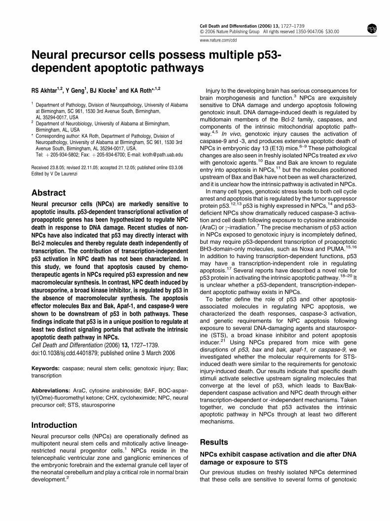
injury, including exposure to AraC, etoposide, ethylnitrosour-ea, or g-irradiation.7,8,10 Here, we used mitogenically ex-panded telencephalic NPCs in order to make mechanisticstudies of NPC death more feasible. Primary telencephaliccells were isolated from E13 mice and incubated in achemically defined, serum-free medium containing fibroblastgrowth factor-2 (FGF2) (see Materials and Methods). FGF2-expanded NPCs retain immunoreactivity for nestin, anintermediate filament expressed in neural progenitors(97.771.6% positive cells, n¼ 3 wells). In addition, FGF2-expanded NPC cultures contained only very rare NeuN-immunoreactive (0.2570.1%, n¼ 3 wells) or GFAP-immuno-reactive (0.7070.5%, n¼ 4 wells) cells, markers for matureneurons or astrocytes, respectively.
An assessment of baseline cell proliferation in our experi-mental system is important because DNA damage can induceboth cell cycle arrest and cell death,12,13 and thus decreasedviability in response to cytotoxic injury may result from cellcycle arrest rather than cell death. To determine the extent ofcell proliferation that occurs during the experimental period,we assessed the cell viability of untreated NPCs at 0, 6, and24 h. Our viability assay detected a 2.5774.4% increase incell number over 6 h and an 11.172.1% increase over 24 h(n¼ 8 wells from three independent experiments; additionaldata not shown). These results demonstrate that themagnitude of cell proliferation is relatively small during ourdrug treatment periods, accounting for less than a 15%increase in cell number over a 24 h period.
Previous reports showed that exposure to the nucleosideanalog AraC induces caspase-dependent cell death in freshly
isolated NPCs.7,8,10,22 We confirmed that FGF2-expandedNPCs are also sensitive to AraC in a concentration and time-dependent manner (Figure 1a and data not shown). Todetermine whether other genotoxic agents that producedifferent forms of DNA damage induce similar responses,we exposed FGF2-expanded NPCs to the chemotherapeuticagents bleomycin, etoposide, or camptothecin. Bleomycinsulfate, a streptomyces product, causes single- and double-stranded DNA breaks.23 Etoposide forms complexes withDNA topoisomerase and also induces single- and double-stranded DNA breaks.24 Camptothecin binds to DNA–topoisomerase complexes and produces DNA breaks.25 Wedetermined that these agents also potently induced cell deathin NPCs (Figure 1b and c, and data not shown). These resultsdemonstrate that FGF2-expanded NPCs have a conservedsensitivity to a number of genotoxic agents with differentmechanisms of action.
We have previously reported that freshly isolated telence-phalic NPCs were responsive to the death-inducing effects ofSTS; however, the specific molecules regulating this re-sponse were not investigated.10 NPC-derived cell lines thatlack Apaf-1 are protected from STS-induced death,26 sug-gesting that the intrinsic pathway may be activated by thisagent. To confirm and extend these findings, we exposedFGF2-expanded NPCs to increasing concentrations of STSand detected a significant decrease in cell viability following6 h (Figure 1d). Although this result demonstrates that STScan induce death of NPCs, it does not indicate whether FGF2-expanded NPCs die via an apoptotic pathway following thisstimulus. Immunocytochemical staining for the cleaved p17
Figure 1 Exposure to genotoxic agents or STS induces cell death in FGF2-expanded NPCs. Exposure to 24 h AraC (a), 36 h bleomycin (b), or 24 h camptothecin (c)produces a significant decrease in cell viability in a concentration-dependent manner. Similar results are seen following 24 h etoposide exposure (data not shown).Exposure to STS for 6 h also reduces cell viability in a concentration-dependent manner (d). Cell viability is normalized to untreated control (UT). Data points representmean7S.E.M. with n¼ 8. (*Po0.001 by one-way ANOVA/Bonferroni post-test versus UT)
p53 regulation of NPC apoptosisRS Akhtar et al
1728
Cell Death and Differentiation
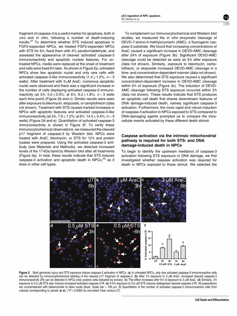
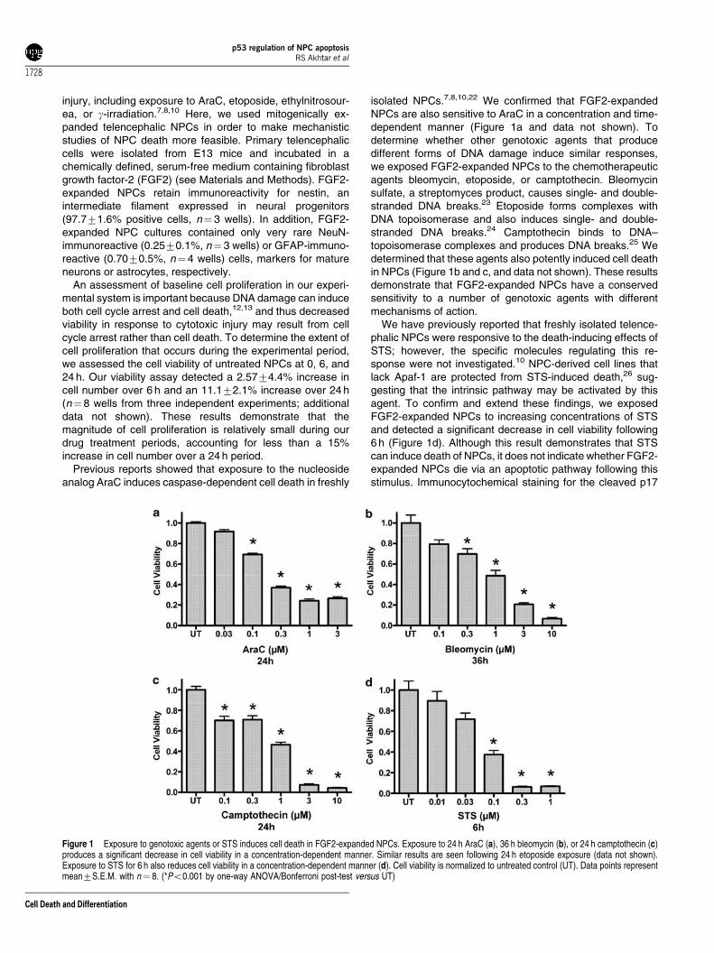
fragment of caspase-3 is a useful marker for apoptosis, both invivo and in vitro, following a number of death-inducinginsults.27 To determine whether STS induced apoptosis inFGF2-expanded NPCs, we treated FGF2-expanded NPCswith STS for 6 h, fixed them with 4% paraformaldehyde, andassessed the appearance of cleaved ‘activated’ caspase-3immunoreactivity and apoptotic nuclear features. For un-treated NPCs, media were replaced at the onset of treatmentand cells were fixed 6 h later. As shown in Figure 2a, untreatedNPCs show few apoptotic nuclei and only rare cells withactivated caspase-3-like immunoreactivity (1.471.2%, n¼ 3wells). After treatment with 3 mM AraC, numerous apoptoticnuclei were observed and there was a significant increase inthe number of cells displaying activated caspase-3 immuno-reactivity (at 3 h, 5.072.0%; at 6 h, 9.271.8%; n¼ 3 wellseach time point) (Figure 2b and c). Similar results were seenafter exposure to bleomycin, etoposide, or camptothecin (datanot shown). Treatment with STS caused marked increases inNPCs with apoptotic features and activated caspase-3-likeimmunoreactivity (at 3 h, 7.971.2%; at 6 h, 14.573.4%; n¼ 3wells) (Figure 2d and e). Quantitation of activated caspase-3immunoreactivity is shown in Figure 2f. To verify theseimmunocytochemical observations, we measured the cleavedp17 fragment of caspase-3 by Western blot. NPCs weretreated with AraC, bleomycin, or STS for 12 h and proteinlysates were prepared. Using the activated caspase-3 anti-body (see Materials and Methods), we detected increasedlevels of the 17 kDa band by Western blot after all treatments(Figure 3a). In total, these results indicate that STS inducescaspase-3 activation and apoptotic death in NPCs,26 as itdoes in other cell types.
To complement our immunocytochemical and Western blotstudies, we measured the in vitro enzymatic cleavage ofDEVD-7-amino-4-methylcoumarin (AMC), a fluorogenic cas-pase-3 substrate. We found that increasing concentrations ofAraC caused a significant increase in DEVD-AMC cleavageafter 24 h of exposure (Figure 3b). Significant DEVD-AMCcleavage could be detected as early as 6 h after exposure(data not shown). Similarly, exposure to bleomycin, camp-tothecin, or etoposide increased DEVD-AMC cleavage in atime- and concentration-dependent manner (data not shown).We also determined that STS exposure caused a significantconcentration-dependent increase in DEVD-AMC cleavagewithin 6 h of exposure (Figure 3c). The induction of DEVD-AMC cleavage following STS exposure occurred within 3 h(data not shown). These results indicate that STS producesan apoptotic cell death that shares downstream features ofDNA damage-induced death, namely significant caspase-3activation. Furthermore, the more rapid and robust inductionof caspase-3 activation in NPCs exposed to STS compared toDNA-damaging agents prompted us to compare the intra-cellular events activated by these different death stimuli.
Caspase activation via the intrinsic mitochondrialpathway is required for both STS- and DNAdamage-induced death in NPCs
To begin to identify the upstream mediators of caspase-3activation following STS exposure or DNA damage, we firstinvestigated whether caspase activation was required fordeath in NPCs exposed to these stimuli. We selected the
Figure 2 Both genotoxic injury and STS exposure induce caspase-3 activation in NPCs. (a) In untreated NPCs, only rare activated caspase-3 immunoreactive cellscan be detected by immunocytochemical labeling of the cleaved p17 fragment of caspase-3. (b) After 3 h exposure to 3mM AraC, increased cleaved caspase-3immunoreactivity (IR) can be detected in NPCs (red; positive cells indicated by arrows). (c) This effect increases after 6 h of exposure to 3 mM AraC. (d) Similarly, 3 hexposure to 0.5 mM STS also induces increased activated caspase-3 IR. (e) A 6 h exposure to 0.5 mM STS induces widespread cleaved caspase-3 IR. All preparationsare counterstained with bisbenzimide to label nuclei (blue). Scale bar¼ 100 mm. (f) Quantitation of the number of activated caspase-3 immunoreactive cells fromcultures corresponding to panels (a–e). (*Po0.0005 by one-tailed t-test versus UT)
p53 regulation of NPC apoptosisRS Akhtar et al
1729
Cell Death and Differentiation
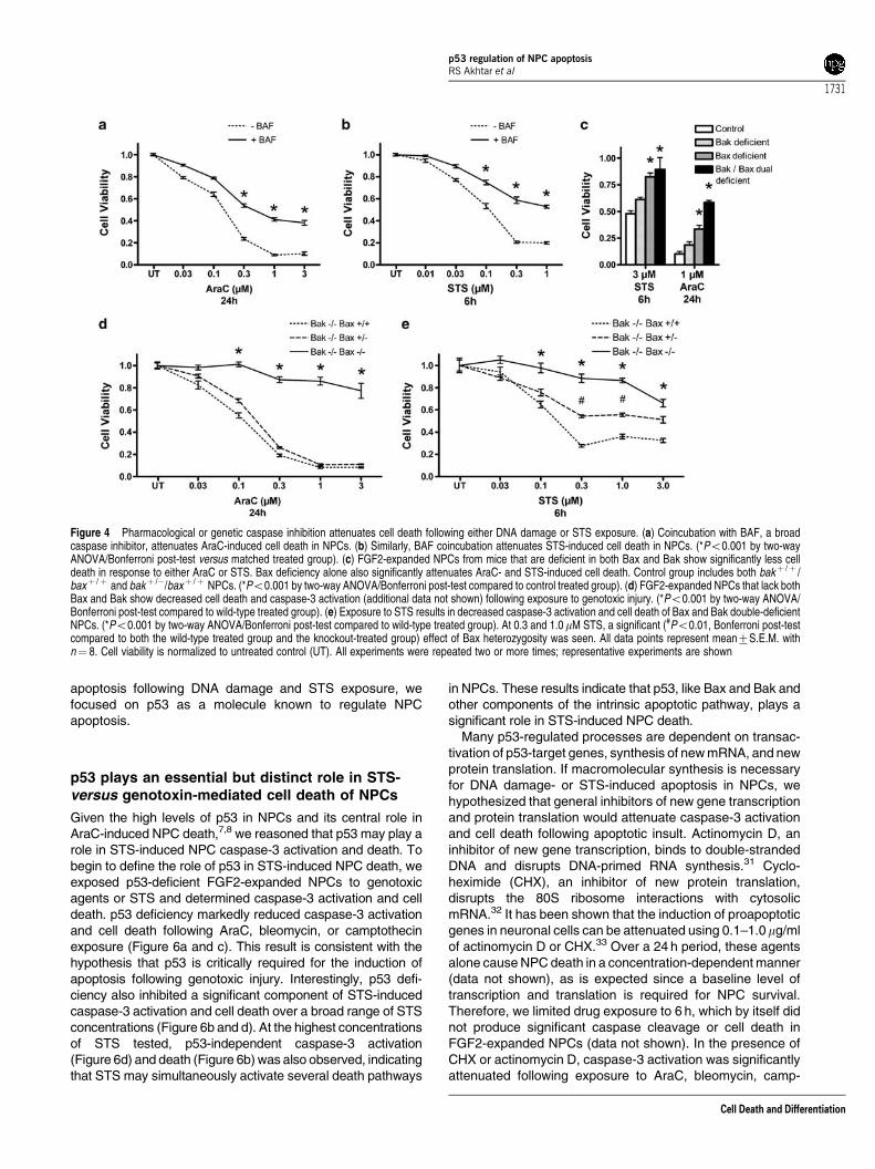
broad caspase inhibitor BOC-aspartyl(Ome)-fluoromethylketone (BAF) to test the caspase requirement of STS- andgenotoxin-induced NPC death.10 We found that DNAdamage-induced NPC death is significantly inhibited bycoincubation with 150mM BAF (Figure 4a). Similarly, 150mMBAF attenuated STS-induced cell death (Figure 4b), indicat-ing that caspase activation is important for both DNA damage-and STS-induced NPC death.
To further define the molecular pathways regulating STS-and DNA damage-induced death, we analyzed NPCs derivedfrom mice deficient in critical mediators of the intrinsicapoptotic pathway, such as Bax and Bak, Apaf-1, andcaspase-9. The proapoptotic molecules Bax and Bak playoverlapping functions in the regulation of apoptosis, and itappears that the significance of either molecule is highlydependent on cell type.11,28–30 We reasoned that either Bax orBak might be critical for the activation of the intrinsic apoptoticpathway in NPCs. To test this hypothesis, we crossed micethat were heterozygous for both bax and bak and preparedFGF2-expanded NPCs from E13 embryos. We then com-pared the death responsiveness of Bax or Bak single-deficientNPCs and Bax and Bak double-deficient NPCs to theirlittermate control NPCs. We found that Bak deficiency alonedid not significantly alter cell death following exposure to AraCor STS (Figure 4c). However, Bax deficiency alone providedsignificant protection against both AraC- and STS-inducedNPC death (Figure 4c). Quantitatively, deficiency of bothmolecules provided the highest level of protection followingeither stimulus. We confirmed these results by investigatingthe effect of Bax expression, in the context of Bak deficiency,on AraC- and STS-induced death. Dual deficiency led to asignificant attenuation of death following exposure to multipleconcentrations of AraC (Figure 4d). We also found that death
following exposure to bleomycin, etoposide, or camptothecinwas also attenuated by dual Bax and Bak deficiency (data notshown). Similarly, dual deficiency markedly reduced deathfollowing STS exposure (Figure 4e). Interestingly, a gene–dosage effect of Bax was seen for STS-induced NPC death.Dual Bax and Bak deficiency virtually eliminated caspase-3activation, as measured by DEVD-AMC cleavage, followingeither stimulus (data not shown). Taken together, our resultsdemonstrate that STS-induced NPC death is regulated by thecombined action of Bax and Bak, but that Bax appears to playa more significant role, similar to previous findings for DNAdamage.8,11 Dual Bax- and Bak-deficient NPCs exposed toeither AraC or STS have a small decrease in cell viability ascompared to untreated NPCs, which may result from cell cyclearrest and/or nonapoptotic death.
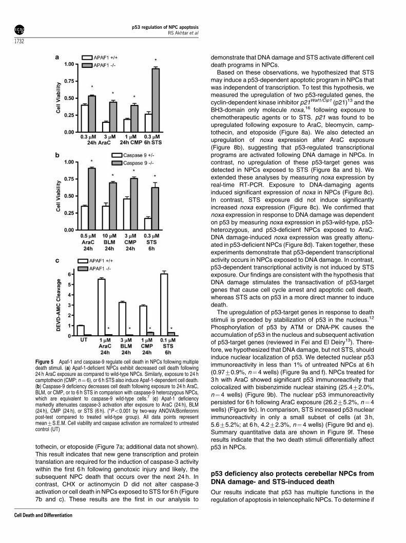
Given Bax and Bak involvement in both DNA damage- andSTS-induced death, we proposed that deficiency of eithercaspase-9 or Apaf-1 would also lead to diminished NPC deathfollowing treatment with these two stimuli. As expected, AraC-induced death was significantly decreased by deficiency ofApaf-1 (Figure 5a). This protective effect was also seen afterexposure to either camptothecin or STS (Figure 5a). We alsoexposed caspase-9-deficient NPCs to AraC, bleomycin,camptothecin or STS, and found that deficiency of caspase-9 significantly attenuated death (Figure 5b). Caspase-3activation in caspase-9 or Apaf-1-deficient NPCs was virtuallyeliminated following exposure to either genotoxic agents or toSTS (Figure 5c and data not shown). Thus, disruption ofapoptosome formation prevents caspase-3 activation anddeath in NPCs exposed to DNA damage or to STS. Theseresults suggest that both STS and genotoxic agents engage acommon apoptotic pathway downstream of Bax and Bakactivation. To investigate the upstream events leading to NPC
Figure 3 Biochemical detection of caspase-3 cleavage following either DNA damage or exposure to STS. (a) Exposure to either AraC, bleomycin (BLM), or STS for12 h induces cleavage of caspase-3 as detected by Western blot. b-Tubulin indicated as loading control. (b) AraC and (c) STS induce significant enzymatic cleavage ofDEVD-AMC, a synthetic caspase-3 substrate, in a concentration-dependent manner. Similar results are seen after exposure to bleomycin, camptothecin, or etoposide(data not shown). Caspase-3 activity is normalized to untreated control (UT). Data points represent mean7S.E.M. with n¼ 8. (*Po0.001 by one-way ANOVA/Bonferroni post-test versus UT)
p53 regulation of NPC apoptosisRS Akhtar et al
1730
Cell Death and Differentiation
apoptosis following DNA damage and STS exposure, wefocused on p53 as a molecule known to regulate NPCapoptosis.
p53 plays an essential but distinct role in STS-versus genotoxin-mediated cell death of NPCs
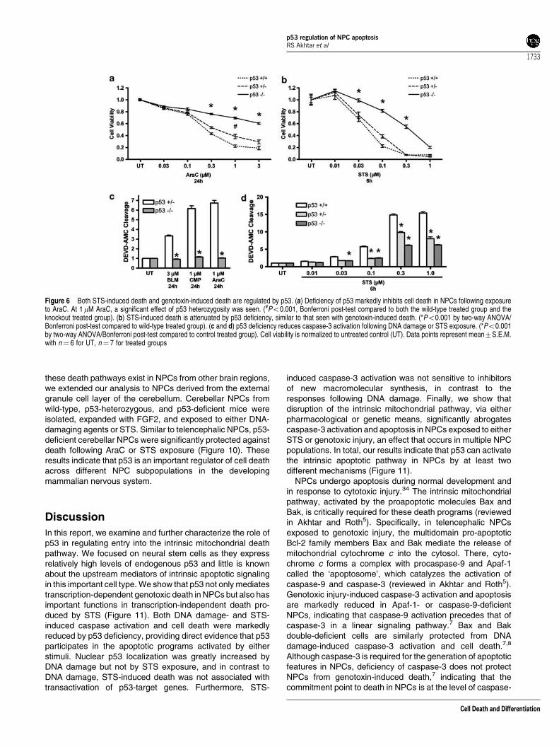
Given the high levels of p53 in NPCs and its central role inAraC-induced NPC death,7,8 we reasoned that p53 may play arole in STS-induced NPC caspase-3 activation and death. Tobegin to define the role of p53 in STS-induced NPC death, weexposed p53-deficient FGF2-expanded NPCs to genotoxicagents or STS and determined caspase-3 activation and celldeath. p53 deficiency markedly reduced caspase-3 activationand cell death following AraC, bleomycin, or camptothecinexposure (Figure 6a and c). This result is consistent with thehypothesis that p53 is critically required for the induction ofapoptosis following genotoxic injury. Interestingly, p53 defi-ciency also inhibited a significant component of STS-inducedcaspase-3 activation and cell death over a broad range of STSconcentrations (Figure 6b and d). At the highest concentrationsof STS tested, p53-independent caspase-3 activation(Figure 6d) and death (Figure 6b) was also observed, indicatingthat STS may simultaneously activate several death pathways
in NPCs. These results indicate that p53, like Bax and Bak andother components of the intrinsic apoptotic pathway, plays asignificant role in STS-induced NPC death.
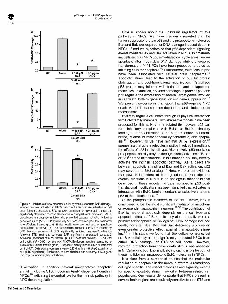
Many p53-regulated processes are dependent on transac-tivation of p53-target genes, synthesis of new mRNA, and newprotein translation. If macromolecular synthesis is necessaryfor DNA damage- or STS-induced apoptosis in NPCs, wehypothesized that general inhibitors of new gene transcriptionand protein translation would attenuate caspase-3 activationand cell death following apoptotic insult. Actinomycin D, aninhibitor of new gene transcription, binds to double-strandedDNA and disrupts DNA-primed RNA synthesis.31 Cyclo-heximide (CHX), an inhibitor of new protein translation,disrupts the 80S ribosome interactions with cytosolicmRNA.32 It has been shown that the induction of proapoptoticgenes in neuronal cells can be attenuated using 0.1–1.0mg/mlof actinomycin D or CHX.33 Over a 24 h period, these agentsalone cause NPC death in a concentration-dependent manner(data not shown), as is expected since a baseline level oftranscription and translation is required for NPC survival.Therefore, we limited drug exposure to 6 h, which by itself didnot produce significant caspase cleavage or cell death inFGF2-expanded NPCs (data not shown). In the presence ofCHX or actinomycin D, caspase-3 activation was significantlyattenuated following exposure to AraC, bleomycin, camp-
Figure 4 Pharmacological or genetic caspase inhibition attenuates cell death following either DNA damage or STS exposure. (a) Coincubation with BAF, a broadcaspase inhibitor, attenuates AraC-induced cell death in NPCs. (b) Similarly, BAF coincubation attenuates STS-induced cell death in NPCs. (*Po0.001 by two-wayANOVA/Bonferroni post-test versus matched treated group). (c) FGF2-expanded NPCs from mice that are deficient in both Bax and Bak show significantly less celldeath in response to either AraC or STS. Bax deficiency alone also significantly attenuates AraC- and STS-induced cell death. Control group includes both bakþ /þ /baxþ /þ and bakþ /�/baxþ /þ NPCs. (*Po0.001 by two-way ANOVA/Bonferroni post-test compared to control treated group). (d) FGF2-expanded NPCs that lack bothBax and Bak show decreased cell death and caspase-3 activation (additional data not shown) following exposure to genotoxic injury. (*Po0.001 by two-way ANOVA/Bonferroni post-test compared to wild-type treated group). (e) Exposure to STS results in decreased caspase-3 activation and cell death of Bax and Bak double-deficientNPCs. (*Po0.001 by two-way ANOVA/Bonferroni post-test compared to wild-type treated group). At 0.3 and 1.0 mM STS, a significant (#Po0.01, Bonferroni post-testcompared to both the wild-type treated group and the knockout-treated group) effect of Bax heterozygosity was seen. All data points represent mean7S.E.M. withn¼ 8. Cell viability is normalized to untreated control (UT). All experiments were repeated two or more times; representative experiments are shown
p53 regulation of NPC apoptosisRS Akhtar et al
1731
Cell Death and Differentiation
tothecin, or etoposide (Figure 7a; additional data not shown).This result indicates that new gene transcription and proteintranslation are required for the induction of caspase-3 activitywithin the first 6 h following genotoxic injury and likely, thesubsequent NPC death that occurs over the next 24 h. Incontrast, CHX or actinomycin D did not alter caspase-3activation or cell death in NPCs exposed to STS for 6 h (Figure7b and c). These results are the first in our analysis to
demonstrate that DNA damage and STS activate different celldeath programs in NPCs.
Based on these observations, we hypothesized that STSmay induce a p53-dependent apoptotic program in NPCs thatwas independent of transcription. To test this hypothesis, wemeasured the upregulation of two p53-regulated genes, thecyclin-dependent kinase inhibitor p21Waf1/Cip1 (p21)13 and theBH3-domain only molecule noxa,16 following exposure tochemotherapeutic agents or to STS. p21 was found to beupregulated following exposure to AraC, bleomycin, camp-tothecin, and etoposide (Figure 8a). We also detected anupregulation of noxa expression after AraC exposure(Figure 8b), suggesting that p53-regulated transcriptionalprograms are activated following DNA damage in NPCs. Incontrast, no upregulation of these p53-target genes wasdetected in NPCs exposed to STS (Figure 8a and b). Weextended these analyses by measuring noxa expression byreal-time RT-PCR. Exposure to DNA-damaging agentsinduced significant expression of noxa in NPCs (Figure 8c).In contrast, STS exposure did not induce significantlyincreased noxa expression (Figure 8c). We confirmed thatnoxa expression in response to DNA damage was dependenton p53 by measuring noxa expression in p53-wild-type, p53-heterozygous, and p53-deficient NPCs exposed to AraC.DNA damage-induced noxa expression was greatly attenu-ated in p53-deficient NPCs (Figure 8d). Taken together, theseexperiments demonstrate that p53-dependent transcriptionalactivity occurs in NPCs exposed to DNA damage. In contrast,p53-dependent transcriptional activity is not induced by STSexposure. Our findings are consistent with the hypothesis thatDNA damage stimulates the transactivation of p53-targetgenes that cause cell cycle arrest and apoptotic cell death,whereas STS acts on p53 in a more direct manner to inducedeath.
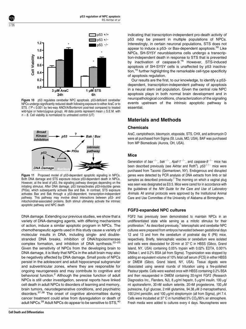
The upregulation of p53-target genes in response to deathstimuli is preceded by stabilization of p53 in the nucleus.12
Phosphorylation of p53 by ATM or DNA-PK causes theaccumulation of p53 in the nucleus and subsequent activationof p53-target genes (reviewed in Fei and El Deiry13). There-fore, we hypothesized that DNA damage, but not STS, shouldinduce nuclear localization of p53. We detected nuclear p53immunoreactivity in less than 1% of untreated NPCs at 6 h(0.9770.9%, n¼ 4 wells) (Figure 9a and f). NPCs treated for3 h with AraC showed significant p53 immunoreactivity thatcolocalized with bisbenzimide nuclear staining (25.472.0%,n¼ 4 wells) (Figure 9b). The nuclear p53 immunoreactivitypersisted for 6 h following AraC exposure (26.275.2%, n¼ 4wells) (Figure 9c). In comparison, STS increased p53 nuclearimmunoreactivity in only a small subset of cells (at 3 h,5.675.2%; at 6 h, 4.272.3%, n¼ 4 wells) (Figure 9d and e).Summary quantitative data are shown in Figure 9f. Theseresults indicate that the two death stimuli differentially affectp53 in NPCs.
p53 deficiency also protects cerebellar NPCs fromDNA damage- and STS-induced death
Our results indicate that p53 has multiple functions in theregulation of apoptosis in telencephalic NPCs. To determine if
Figure 5 Apaf-1 and caspase-9 regulate cell death in NPCs following multipledeath stimuli. (a) Apaf-1-deficient NPCs exhibit decreased cell death following24 h AraC exposure as compared to wild-type NPCs. Similarly, exposure to 24 hcamptothecin (CMP; n¼ 6), or 6 h STS also induce Apaf-1-dependent cell death.(b) Caspase-9 deficiency decreases cell death following exposure to 24 h AraC,BLM, or CMP, or to 6 h STS in comparison with caspase-9 heterozygous NPCs,which are equivalent to caspase-9 wild-type cells.7 (c) Apaf-1 deficiencymarkedly attenuates caspase-3 activation after exposure to AraC (24 h), BLM(24 h), CMP (24 h), or STS (6 h). (*Po0.001 by two-way ANOVA/Bonferonnipost-test compared to treated wild-type group). All data points representmean7S.E.M. Cell viability and caspase activation are normalized to untreatedcontrol (UT)
p53 regulation of NPC apoptosisRS Akhtar et al
1732
Cell Death and Differentiation
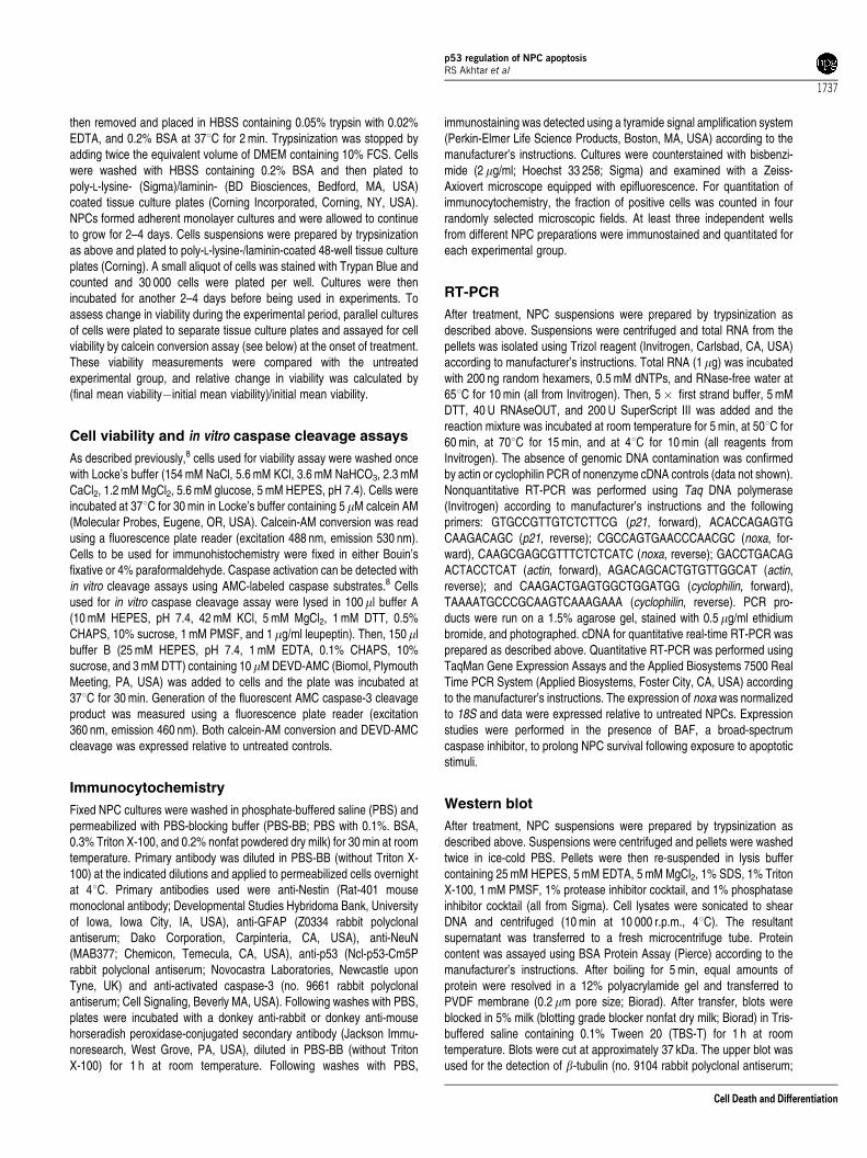
these death pathways exist in NPCs from other brain regions,we extended our analysis to NPCs derived from the externalgranule cell layer of the cerebellum. Cerebellar NPCs fromwild-type, p53-heterozygous, and p53-deficient mice wereisolated, expanded with FGF2, and exposed to either DNA-damaging agents or STS. Similar to telencephalic NPCs, p53-deficient cerebellar NPCs were significantly protected againstdeath following AraC or STS exposure (Figure 10). Theseresults indicate that p53 is an important regulator of cell deathacross different NPC subpopulations in the developingmammalian nervous system.
Discussion
In this report, we examine and further characterize the role ofp53 in regulating entry into the intrinsic mitochondrial deathpathway. We focused on neural stem cells as they expressrelatively high levels of endogenous p53 and little is knownabout the upstream mediators of intrinsic apoptotic signalingin this important cell type. We show that p53 not only mediatestranscription-dependent genotoxic death in NPCs but also hasimportant functions in transcription-independent death pro-duced by STS (Figure 11). Both DNA damage- and STS-induced caspase activation and cell death were markedlyreduced by p53 deficiency, providing direct evidence that p53participates in the apoptotic programs activated by eitherstimuli. Nuclear p53 localization was greatly increased byDNA damage but not by STS exposure, and in contrast toDNA damage, STS-induced death was not associated withtransactivation of p53-target genes. Furthermore, STS-
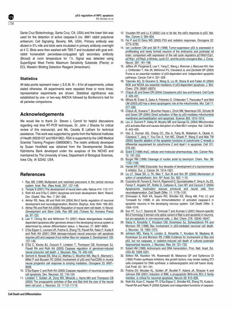
induced caspase-3 activation was not sensitive to inhibitorsof new macromolecular synthesis, in contrast to theresponses following DNA damage. Finally, we show thatdisruption of the intrinsic mitochondrial pathway, via eitherpharmacological or genetic means, significantly abrogatescaspase-3 activation and apoptosis in NPCs exposed to eitherSTS or genotoxic injury, an effect that occurs in multiple NPCpopulations. In total, our results indicate that p53 can activatethe intrinsic apoptotic pathway in NPCs by at least twodifferent mechanisms (Figure 11).
NPCs undergo apoptosis during normal development andin response to cytotoxic injury.34 The intrinsic mitochondrialpathway, activated by the proapoptotic molecules Bax andBak, is critically required for these death programs (reviewedin Akhtar and Roth5). Specifically, in telencephalic NPCsexposed to genotoxic injury, the multidomain pro-apoptoticBcl-2 family members Bax and Bak mediate the release ofmitochondrial cytochrome c into the cytosol. There, cyto-chrome c forms a complex with procaspase-9 and Apaf-1called the ‘apoptosome’, which catalyzes the activation ofcaspase-9 and caspase-3 (reviewed in Akhtar and Roth5).Genotoxic injury-induced caspase-3 activation and apoptosisare markedly reduced in Apaf-1- or caspase-9-deficientNPCs, indicating that caspase-9 activation precedes that ofcaspase-3 in a linear signaling pathway.7 Bax and Bakdouble-deficient cells are similarly protected from DNAdamage-induced caspase-3 activation and cell death.7,8
Although caspase-3 is required for the generation of apoptoticfeatures in NPCs, deficiency of caspase-3 does not protectNPCs from genotoxin-induced death,7 indicating that thecommitment point to death in NPCs is at the level of caspase-
Figure 6 Both STS-induced death and genotoxin-induced death are regulated by p53. (a) Deficiency of p53 markedly inhibits cell death in NPCs following exposureto AraC. At 1mM AraC, a significant effect of p53 heterozygosity was seen. (#Po0.001, Bonferroni post-test compared to both the wild-type treated group and theknockout treated group). (b) STS-induced death is attenuated by p53 deficiency, similar to that seen with genotoxin-induced death. (*Po0.001 by two-way ANOVA/Bonferroni post-test compared to wild-type treated group). (c and d) p53 deficiency reduces caspase-3 activation following DNA damage or STS exposure. (*Po0.001by two-way ANOVA/Bonferroni post-test compared to control treated group). Cell viability is normalized to untreated control (UT). Data points represent mean7S.E.M.with n¼ 6 for UT, n¼ 7 for treated groups
p53 regulation of NPC apoptosisRS Akhtar et al
1733
Cell Death and Differentiation
9 activation. In addition, several nongenotoxic apoptoticstimuli, including STS, induce an Apaf-1-dependent death inNPCs,26 indicating the central role for the intrinsic pathway inNPC death regulation.
Little is known about the upstream regulators of thispathway in NPCs. We have previously reported that thetumor suppressor protein p53 and the proapoptotic moleculesBax and Bak are required for DNA damage-induced death inNPCs,7,8 and we hypothesize that p53-dependent signalingevents mediate Bax and Bak activation in NPCs. In proliferat-ing cells such as NPCs, p53-mediated cell cycle arrest and/orapoptosis after irreparable DNA damage inhibits oncogenictransformation.12,13 NPCs have been proposed to serve asinitiating cells for neoplasia.35 Furthermore, mutations in p53have been associated with several brain neoplasms.12
Apoptotic stimuli lead to the activation of p53 by proteinstabilization and post-translational modification.12 Stabilizedp53 protein may interact with both pro- and antiapoptoticmolecules. In addition, p53 and homologous proteins p63 andp73 regulate the expression of several target genes involvedin cell death, both by gene induction and gene suppression.12
We present evidence in this report that p53-regulate NPCdeath via both transcription-dependent and -independentmechanisms.
P53 may regulate cell death through its physical interactionwith Bcl-2 family members. Two alternative models have beenproposed for this activity. In irradiated thymocytes, p53 canform inhibitory complexes with Bcl-xL or Bcl-2, ultimatelyleading to permeabilization of the outer mitochondrial mem-brane, release of mitochondrial cytochrome c, and apopto-sis.18 However, NPCs have minimal Bcl-xL expression,34
suggesting that other molecules must be involved in mediatingthe effects of p53 in this cell type. Alternatively, p53-mediatedproapoptotic activity may be through direct activation of Bax19
or Bak20 at the mitochondria. In this manner, p53 may directlyactivate the intrinsic apoptotic pathway. As a direct linkbetween apoptotic stimuli and Bax and Bak activation, p53may serve as a ‘BH3 analog’.17 Here, we present evidencethat p53, independent of its regulation of transcriptionalevents, functions in NPCs in an analogous manner to thatdescribed in these reports. To date, no specific p53 post-translational modification has been identified that activates itsinteraction with Bcl-2 family members or selectively targetsp53 to the mitochondria.36
Of the proapoptotic members of the Bcl-2 family, Bax isconsidered to be the most significant mediator of mitochon-dria-dependent apoptosis in neurons.29,30 The contribution ofBak to neuronal apoptosis depends on the cell type andapoptotic stimulus.28 Bax deficiency alone partially protectsprimary telencephalic NPCs against DNA damage-induceddeath; however, dual Bax and Bak deficiency provides aneven greater protective effect against this apoptotic stimu-lus.7,8 In this study, we found that Bax deficiency alone, butnot Bak deficiency alone, significantly protected NPCs fromeither DNA damage- or STS-induced death. However,maximal protection from these death stimuli was observedin NPCs lacking both Bax and Bak, indicating a role for both ofthese multidomain proapoptotic Bcl-2 molecules in NPCs.
It is clear from a number of studies that the molecularregulation of apoptosis in the nervous system is remarkablycell-type specific. The critical molecules and signaling eventsfor specific apoptotic stimuli may differ between related cellpopulations. Our results demonstrate that NPCs present inseveral brain regions are exquisitely sensitive to both STS and
Figure 7 Inhibitors of new macromolecular synthesis attenuate DNA damage-induced caspase activation in NPCs but do not alter caspase activation or celldeath following exposure to STS. (a) CHX, an inhibitor of new protein translation,significantly attenuated caspase-3 activation following 6 h AraC exposure. BAF, abroad-spectrum caspase inhibitor, also prevented caspase activation followinggenotoxic injury. (*Po0.001 by one-way ANOVA/Bonferroni post-test comparedto AraC-alone treated group). Similar results were seen using other genotoxicagents (data not shown). (b) CHX does not alter caspase-3 activation induced bySTS. No concentration of CHX significantly inhibited caspase-3 activationfollowing STS treatment, whereas BAF significantly decreased caspase-3activation (additional data not shown). (c) CHX does not prevent STS-inducedcell death. (*Po0.001 by one-way ANOVA/Bonferroni post-test compared toAraC- or STS-alone treated group). Caspase-3 activity is normalized to untreatedcontrol (UT). Data points represent mean7S.E.M. with n¼ 8 (AraC experiment)or 6 (STS experiment). Similar results were obtained with actinomycin D, a genetranscription inhibitor (data not shown)
p53 regulation of NPC apoptosisRS Akhtar et al
1734
Cell Death and Differentiation
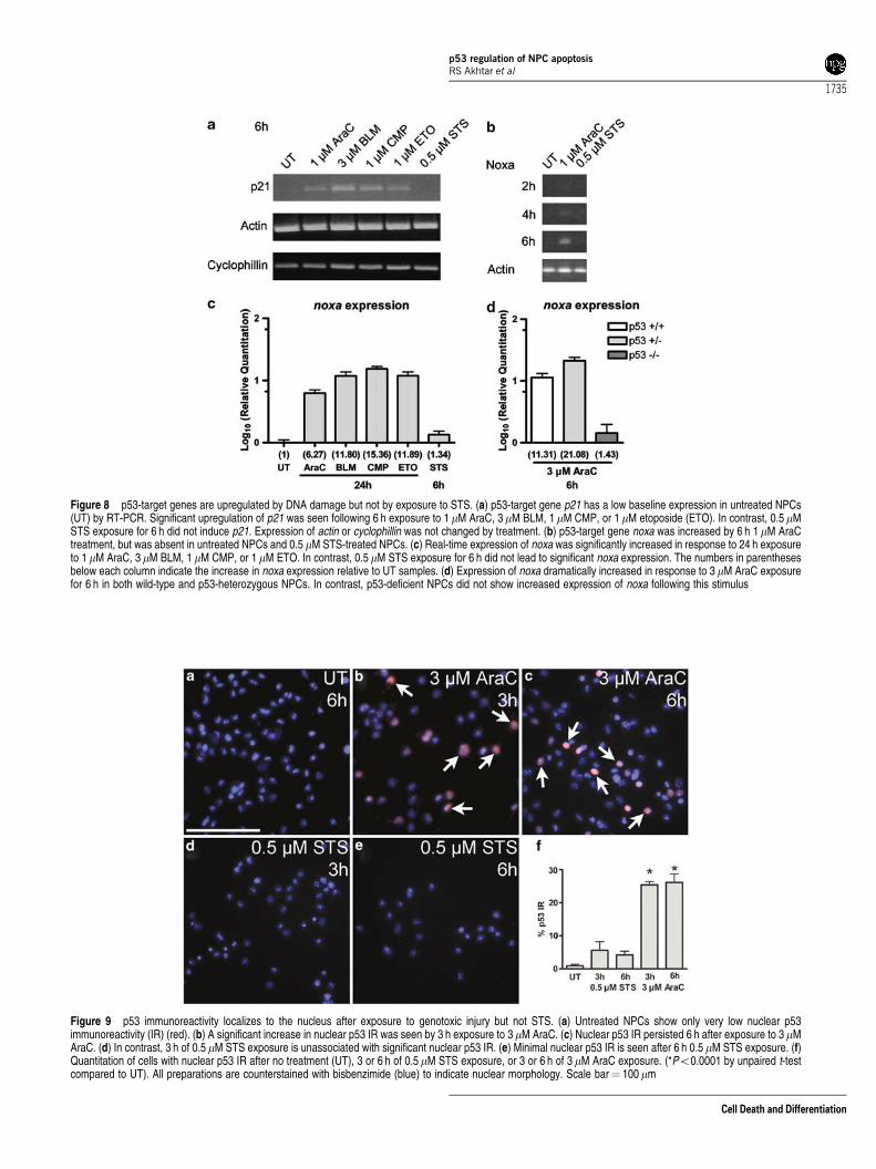
Figure 8 p53-target genes are upregulated by DNA damage but not by exposure to STS. (a) p53-target gene p21 has a low baseline expression in untreated NPCs(UT) by RT-PCR. Significant upregulation of p21 was seen following 6 h exposure to 1mM AraC, 3mM BLM, 1mM CMP, or 1mM etoposide (ETO). In contrast, 0.5 mMSTS exposure for 6 h did not induce p21. Expression of actin or cyclophillin was not changed by treatment. (b) p53-target gene noxa was increased by 6 h 1mM AraCtreatment, but was absent in untreated NPCs and 0.5 mM STS-treated NPCs. (c) Real-time expression of noxa was significantly increased in response to 24 h exposureto 1 mM AraC, 3mM BLM, 1 mM CMP, or 1 mM ETO. In contrast, 0.5 mM STS exposure for 6 h did not lead to significant noxa expression. The numbers in parenthesesbelow each column indicate the increase in noxa expression relative to UT samples. (d) Expression of noxa dramatically increased in response to 3 mM AraC exposurefor 6 h in both wild-type and p53-heterozygous NPCs. In contrast, p53-deficient NPCs did not show increased expression of noxa following this stimulus
Figure 9 p53 immunoreactivity localizes to the nucleus after exposure to genotoxic injury but not STS. (a) Untreated NPCs show only very low nuclear p53immunoreactivity (IR) (red). (b) A significant increase in nuclear p53 IR was seen by 3 h exposure to 3mM AraC. (c) Nuclear p53 IR persisted 6 h after exposure to 3 mMAraC. (d) In contrast, 3 h of 0.5 mM STS exposure is unassociated with significant nuclear p53 IR. (e) Minimal nuclear p53 IR is seen after 6 h 0.5 mM STS exposure. (f)Quantitation of cells with nuclear p53 IR after no treatment (UT), 3 or 6 h of 0.5 mM STS exposure, or 3 or 6 h of 3 mM AraC exposure. (*Po0.0001 by unpaired t-testcompared to UT). All preparations are counterstained with bisbenzimide (blue) to indicate nuclear morphology. Scale bar¼ 100mm
p53 regulation of NPC apoptosisRS Akhtar et al
1735
Cell Death and Differentiation
DNA damage. Extending our previous studies, we show that avariety of DNA-damaging agents, with differing mechanismsof action, induce a similar apoptotic program in NPCs. Thechemotherapeutic agents used in this study cause a variety ofmolecular insults in DNA, including single- and double-stranded DNA breaks, inhibition of DNA/topoisomerasecomplex formation, and inhibition of DNA synthesis.22–25
Given the sensitivity of NPCs from the developing brain toDNA damage, it is likely that NPCs in the adult brain may alsobe negatively affected by DNA damage. Small pools of NPCspersist in the adolescent and adult hippocampal subgranularand subventricular zones, where they are responsible forongoing neurogenesis and may contribute to cognitive andbehavioral function.2 Although the precise function of adultNPCs is still under investigation, several reports have linkedcell death in adult NPCs to disorders of learning and memory,brain tumors, neurodegenerative conditions, and psychiatricdisorders.37,38 The onset of cognitive abnormalities duringcancer treatment could arise from dysregulation or death ofadult NPCs.39 Adult NPCs do appear to be sensitive to STS,40
indicating that transcription-independent pro-death activity ofp53 may be present in multiple populations of NPCs.Interestingly, in certain neuronal populations, STS does notappear to induce a p53- or Bax-dependent apoptosis.30 LikeNPCs, SH-SY5Y neuroblastoma cells undergo a transcrip-tion-independent death in response to STS that is preventedby inactivation of caspase-9.16 However, STS-inducedapoptosis of SH-SY5Y cells is unaffected by p53 inactiva-tion,16 further highlighting the remarkable cell-type specificityof apoptosis regulation.
Our results are the first, to our knowledge, to identify a p53-dependent, transcription-independent pathway of apoptosisin a neural stem cell population. Given the central role NPCapoptosis plays in both normal brain development and inneuropathological conditions, characterization of the signalingevents upstream of the intrinsic apoptotic pathway isessential.
Materials and Methods
Chemicals
AraC, camptothecin, bleomycin, etoposide, STS, CHX, and actinomycin Dwere all purchased from Sigma (St. Louis, MO, USA). BAF was purchasedfrom MP Biomedicals (Aurora, OH, USA).
Mice
Generation of bax�/�, bak�/�, Apaf-1�/�, and caspase-9�/� mice hasbeen described previously (see Akhtar and Roth5). p53þ /� mice werepurchased from Taconic (Germantown, NY). Endogenous and disruptedgenes were detected by PCR analysis of DNA extracts from limb or tailsamples as described previously.7 The morning on which a vaginal plugwas seen was designated as E0.5. Mice were cared for in accordance withthe guidelines of the NIH Guide for the Care and Use of LaboratoryAnimals. All animal protocols were approved by the Institutional AnimalCare and Use Committee of the University of Alabama at Birmingham.
FGF2-expanded NPC cultures
FGF2 has previously been demonstrated to maintain NPCs in anundifferentiated state while serving as a mitotic stimulus for theirproliferation.7 As described previously,7 telencephalic and cerebellar NPCcultures were prepared from embryos harvested between gestational days12 and 13 and from the cerebellum of postnatal day 6 (P6) mice,respectively. Briefly, telencephalic vesicles or cerebellum were isolatedand cells were dissociated for 20 min at 371C in HBSS (Gibco, GrandIsland, NY, USA) containing 0.05% trypsin with 0.02% EDTA, 0.001%DNAse I, and 0.2% BSA (all from Sigma). Trypsinization was stopped byadding an equivalent volume of 10% fetal calf serum (FCS) in either HBSSor DMEM (Gibco, Grand Island, NY, USA). Tissue digests weredissociated using several rounds of trituration using a fire-polishedPasteur pipette. Cells were washed once with HBSS containing 0.2% BSAand then resuspended in DMEM containing 20 ng/ml FGF2 (ResearchDiagnostics Inc., Flanders, NJ), 8 mg/ml heparin, 5 mg/ml insulin, 100 mg/ml apotransferrin, 30 nM sodium selenite, 20 nM progesterone, 100 mMputrescine, 6 g/l glucose, 2 mM glutamine, 94.38mM 2-mercaptoethanol,100 U/ml penicillin, and 100 mg/ml streptomycin (all from Sigma), pH 7.4.Cells were incubated at 371C in humidified 5% CO2/95% air atmosphere.Fresh media were added to cultures every 4 days. Neurospheres were
Figure 10 p53 regulates cerebellar NPC apoptosis. p53-deficient cerebellarNPCs undergo significantly reduced death following exposure to either AraC or toSTS. (*Po0.001 by two-way ANOVA/Bonferroni post-test compared to treatedwild-type or heterozygous group). All data points represent mean7S.E.M. withn¼ 8. Cell viability is normalized to untreated control (UT)
Figure 11 Proposed model of p53-dependent apoptotic signaling in NPCs.Both DNA damage and STS exposure induce p53-dependent death in NPCs.However, at the level of p53, the signaling pathway diverges depending on theinitiating stimulus. After DNA damage, p53 transactivates p53-inducible genes(PIGs), which subsequently activate Bax and Bak. In contrast, STS exposureactivates Bax and Bak through a p53-dependent, transcription-independentpathway. This pathway may involve direct interactions between p53- andmitochondrial-associated proteins. Both stimuli ultimately activate the intrinsicapoptotic pathway and NPC death
p53 regulation of NPC apoptosisRS Akhtar et al
1736
Cell Death and Differentiation
then removed and placed in HBSS containing 0.05% trypsin with 0.02%EDTA, and 0.2% BSA at 371C for 2 min. Trypsinization was stopped byadding twice the equivalent volume of DMEM containing 10% FCS. Cellswere washed with HBSS containing 0.2% BSA and then plated topoly-L-lysine- (Sigma)/laminin- (BD Biosciences, Bedford, MA, USA)coated tissue culture plates (Corning Incorporated, Corning, NY, USA).NPCs formed adherent monolayer cultures and were allowed to continueto grow for 2–4 days. Cells suspensions were prepared by trypsinizationas above and plated to poly-L-lysine-/laminin-coated 48-well tissue cultureplates (Corning). A small aliquot of cells was stained with Trypan Blue andcounted and 30 000 cells were plated per well. Cultures were thenincubated for another 2–4 days before being used in experiments. Toassess change in viability during the experimental period, parallel culturesof cells were plated to separate tissue culture plates and assayed for cellviability by calcein conversion assay (see below) at the onset of treatment.These viability measurements were compared with the untreatedexperimental group, and relative change in viability was calculated by(final mean viability�initial mean viability)/initial mean viability.
Cell viability and in vitro caspase cleavage assays
As described previously,8 cells used for viability assay were washed oncewith Locke’s buffer (154 mM NaCl, 5.6 mM KCl, 3.6 mM NaHCO3, 2.3 mMCaCl2, 1.2 mM MgCl2, 5.6 mM glucose, 5 mM HEPES, pH 7.4). Cells wereincubated at 371C for 30 min in Locke’s buffer containing 5 mM calcein AM(Molecular Probes, Eugene, OR, USA). Calcein-AM conversion was readusing a fluorescence plate reader (excitation 488 nm, emission 530 nm).Cells to be used for immunohistochemistry were fixed in either Bouin’sfixative or 4% paraformaldehyde. Caspase activation can be detected within vitro cleavage assays using AMC-labeled caspase substrates.8 Cellsused for in vitro caspase cleavage assay were lysed in 100ml buffer A(10 mM HEPES, pH 7.4, 42 mM KCl, 5 mM MgCl2, 1 mM DTT, 0.5%CHAPS, 10% sucrose, 1 mM PMSF, and 1 mg/ml leupeptin). Then, 150 mlbuffer B (25 mM HEPES, pH 7.4, 1 mM EDTA, 0.1% CHAPS, 10%sucrose, and 3 mM DTT) containing 10mM DEVD-AMC (Biomol, PlymouthMeeting, PA, USA) was added to cells and the plate was incubated at371C for 30 min. Generation of the fluorescent AMC caspase-3 cleavageproduct was measured using a fluorescence plate reader (excitation360 nm, emission 460 nm). Both calcein-AM conversion and DEVD-AMCcleavage was expressed relative to untreated controls.
Immunocytochemistry
Fixed NPC cultures were washed in phosphate-buffered saline (PBS) andpermeabilized with PBS-blocking buffer (PBS-BB; PBS with 0.1%. BSA,0.3% Triton X-100, and 0.2% nonfat powdered dry milk) for 30 min at roomtemperature. Primary antibody was diluted in PBS-BB (without Triton X-100) at the indicated dilutions and applied to permeabilized cells overnightat 41C. Primary antibodies used were anti-Nestin (Rat-401 mousemonoclonal antibody; Developmental Studies Hybridoma Bank, Universityof Iowa, Iowa City, IA, USA), anti-GFAP (Z0334 rabbit polyclonalantiserum; Dako Corporation, Carpinteria, CA, USA), anti-NeuN(MAB377; Chemicon, Temecula, CA, USA), anti-p53 (Ncl-p53-Cm5Prabbit polyclonal antiserum; Novocastra Laboratories, Newcastle uponTyne, UK) and anti-activated caspase-3 (no. 9661 rabbit polyclonalantiserum; Cell Signaling, Beverly MA, USA). Following washes with PBS,plates were incubated with a donkey anti-rabbit or donkey anti-mousehorseradish peroxidase-conjugated secondary antibody (Jackson Immu-noresearch, West Grove, PA, USA), diluted in PBS-BB (without TritonX-100) for 1 h at room temperature. Following washes with PBS,
immunostaining was detected using a tyramide signal amplification system(Perkin-Elmer Life Science Products, Boston, MA, USA) according to themanufacturer’s instructions. Cultures were counterstained with bisbenzi-mide (2 mg/ml; Hoechst 33 258; Sigma) and examined with a Zeiss-Axiovert microscope equipped with epifluorescence. For quantitation ofimmunocytochemistry, the fraction of positive cells was counted in fourrandomly selected microscopic fields. At least three independent wellsfrom different NPC preparations were immunostained and quantitated foreach experimental group.
RT-PCR
After treatment, NPC suspensions were prepared by trypsinization asdescribed above. Suspensions were centrifuged and total RNA from thepellets was isolated using Trizol reagent (Invitrogen, Carlsbad, CA, USA)according to manufacturer’s instructions. Total RNA (1 mg) was incubatedwith 200 ng random hexamers, 0.5 mM dNTPs, and RNase-free water at651C for 10 min (all from Invitrogen). Then, 5� first strand buffer, 5 mMDTT, 40 U RNAseOUT, and 200 U SuperScript III was added and thereaction mixture was incubated at room temperature for 5 min, at 501C for60 min, at 701C for 15 min, and at 41C for 10 min (all reagents fromInvitrogen). The absence of genomic DNA contamination was confirmedby actin or cyclophilin PCR of nonenzyme cDNA controls (data not shown).Nonquantitative RT-PCR was performed using Taq DNA polymerase(Invitrogen) according to manufacturer’s instructions and the followingprimers: GTGCCGTTGTCTCTTCG (p21, forward), ACACCAGAGTGCAAGACAGC (p21, reverse); CGCCAGTGAACCCAACGC (noxa, for-ward), CAAGCGAGCGTTTCTCTCATC (noxa, reverse); GACCTGACAGACTACCTCAT (actin, forward), AGACAGCACTGTGTTGGCAT (actin,reverse); and CAAGACTGAGTGGCTGGATGG (cyclophilin, forward),TAAAATGCCCGCAAGTCAAAGAAA (cyclophilin, reverse). PCR pro-ducts were run on a 1.5% agarose gel, stained with 0.5 mg/ml ethidiumbromide, and photographed. cDNA for quantitative real-time RT-PCR wasprepared as described above. Quantitative RT-PCR was performed usingTaqMan Gene Expression Assays and the Applied Biosystems 7500 RealTime PCR System (Applied Biosystems, Foster City, CA, USA) accordingto the manufacturer’s instructions. The expression of noxa was normalizedto 18S and data were expressed relative to untreated NPCs. Expressionstudies were performed in the presence of BAF, a broad-spectrumcaspase inhibitor, to prolong NPC survival following exposure to apoptoticstimuli.
Western blot
After treatment, NPC suspensions were prepared by trypsinization asdescribed above. Suspensions were centrifuged and pellets were washedtwice in ice-cold PBS. Pellets were then re-suspended in lysis buffercontaining 25 mM HEPES, 5 mM EDTA, 5 mM MgCl2, 1% SDS, 1% TritonX-100, 1 mM PMSF, 1% protease inhibitor cocktail, and 1% phosphataseinhibitor cocktail (all from Sigma). Cell lysates were sonicated to shearDNA and centrifuged (10 min at 10 000 r.p.m., 41C). The resultantsupernatant was transferred to a fresh microcentrifuge tube. Proteincontent was assayed using BSA Protein Assay (Pierce) according to themanufacturer’s instructions. After boiling for 5 min, equal amounts ofprotein were resolved in a 12% polyacrylamide gel and transferred toPVDF membrane (0.2 mm pore size; Biorad). After transfer, blots wereblocked in 5% milk (blotting grade blocker nonfat dry milk; Biorad) in Tris-buffered saline containing 0.1% Tween 20 (TBS-T) for 1 h at roomtemperature. Blots were cut at approximately 37 kDa. The upper blot wasused for the detection of b-tubulin (no. 9104 rabbit polyclonal antiserum;
p53 regulation of NPC apoptosisRS Akhtar et al
1737
Cell Death and Differentiation
Santa Cruz Biotechnology, Santa Cruz, CA, USA) and the lower blot wasused for the detection of active caspase-3 (no. 9661 rabbit polyclonalantiserum; Cell Signaling, Beverly, MA, USA). Primary antibody wasdiluted in 5% milk and blots were incubated in primary antibody overnightat 41C. Blots were then washed with TBS-T and incubated with goat anti-rabbit horseradish peroxidase-conjugated IgG secondary antibody(Biorad) at room temperature for 1 h. Signal was detected usingSuperSignal West Femto Maximum Sensitivity Substrate (Pierce) orECL Western Blotting Detection Reagent (Amersham).
Statistics
All data points represent mean7S.E.M. N¼ 8 for all experiments, unlessstated otherwise. All experiments were repeated three or more times;representative experiments are shown. Statistical significance wasestablished by one- or two-way ANOVA followed by Bonferroni’s test forall pairwise comparisons.
Acknowledgements
We would like to thank Dr. Steven L Carroll for helpful discussionsregarding real time RT-PCR techniques, Dr. John J Shacka for criticalreview of this manuscript, and Ms. Cecelia B Latham for technicalassistance. This work was supported by grants from the National Institutesof Health (NS35107 and NS41962). RSA is supported by the UAB MedicalScientist Training Program (GM008361). The nestin antibody developedby Susan Hockfield was obtained from the Developmental StudiesHybridoma Bank developed under the auspices of the NICHD andmaintained by The University of Iowa, Department of Biological Sciences,Iowa City, IA 52242, USA.
References
1. Rao MS (1999) Multipotent and restricted precursors in the central nervoussystem. Anat. Rec. (New Anat). 257: 137–148.
2. Temple S (2001) The development of neural stem cells. Nature 414: 112–117.3. Roth KA and D’Sa C (2001) Apoptosis and brain development. Ment. Retard.
Dev. Disabil. Res. Rev. 7: 261–266.4. Akhtar RS, Ness JM and Roth KA (2004) Bcl-2 family regulation of neuronal
development and neurodegeneration. Biochim. Biophys. Acta 1644: 189–203.5. Akhtar RS and Roth KA (2006) Regulation of neural stem cell death. In Neural.
Development and Stem Cells, Rao MS (ed) (Totowa NJ: Humana Press)pp. 97–122.
6. Lee Y, Chong MJ and McKinnon PJ (2001) Ataxia telangiectasia mutated-dependent apoptosis after genotoxic stress in the developing nervous system isdetermined by cellular differentiation status. J. Neurosci. 21: 6687–6693.
7. D’Sa-Eipper C, Leonard JR, Putcha G, Zheng TS, Flavell RA, Rakic P, Kuida Kand Roth KA (2001) DNA damage-induced neural precursor cell apoptosisrequires p53 and caspase 9 but neither Bax nor caspase 3. Development 128:137–146.
8. D’Sa C, Klocke BJ, Cecconi F, Lindsten T, Thompson CB, Korsmeyer SJ,Flavell RA and Roth KA (2003) Caspase regulation of genotoxin-inducedneural precursor cell death. J. Neurosci. Res. 74: 435–445.
9. Semont A, Nowak EB, Silva LC, Mathieu C, Mouthon MA, May E, Allemand I,Millet P and Boussin FD (2004) Involvement of p53 and Fas/CD95 in murineneural progenitor cell response to ionizing irradiation. Oncogene 23: 8497–8508.
10. D’Sa-Eipper C and Roth KA (2000) Caspase regulation of neuronal progenitorcell apoptosis. Dev. Neurosci. 22: 116–124.
11. Lindsten T, Golden JA, Zong WX, Minarcik J, Harris MH and Thompson CB(2003) The proapoptotic activities of Bax and Bak limit the size of the neuralstem cell pool. J. Neurosci. 23: 11112–11119.
12. Vousden KH and Lu X (2002) Live or let die: the cell’s response to p53. Nat.Rev. Cancer 2: 594–604.
13. Fei P and El Deiry WS (2003) P53 and radiation responses. Oncogene 22:5774–5783.
14. van Lookeren CM and Gill R (1998) Tumor-suppressor p53 is expressed inproliferating and newly formed neurons of the embryonic and postnatal ratbrain: comparison with expression of the cell cycle regulators p21Waf1/Cip1,p27Kip1, p57Kip2, p16Ink4a, cyclin G1, and the proto-oncogene Bax. J. Comp.Neurol. 397: 181–198.
15. Jeffers JR, Parganas E, Lee Y, Yang C, Wang J, Brennan J, MacLean KH, HanJ, Chittenden T, Ihle JN, McKinnon PJ, Cleveland JL and Zambetti GP (2003)Puma is an essential mediator of p53-dependent and -independent apoptoticpathways. Cancer Cell 4: 321–328.
16. Yakovlev AG, Di Giovanni S, Wang G, Liu W, Stoica B and Faden AI (2004)BOK and NOXA are essential mediators of p53-dependent apoptosis. J. Biol.Chem. 279: 28367–28374.
17. Chipuk JE and Green DR (2004) Cytoplasmic p53: bax and forward. Cell Cycle3: 429–431.
18. Mihara M, Erster S, Zaika A, Petrenko O, Chittenden T, Pancoska P and MollUM (2003) p53 has a direct apoptogenic role at the mitochondria. Mol. Cell 11:577–590.
19. Chipuk JE, Kuwana T, Bouchier-Hayes L, Droin NM, Newmeyer DD, Schuler Mand Green DR (2004) Direct activation of Bax by p53 mediates mitochondrialmembrane permeabilization and apoptosis. Science 303: 1010–1014.
20. Leu JI, Dumont P, Hafey M, Murphy ME and George DL (2004) Mitochondrialp53 activates Bak and causes disruption of a Bak-Mcl1 complex. Nat. Cell Biol.6: 443–450.
21. Hao Z, Duncan GS, Chang CC, Elia A, Fang M, Wakeham A, Okada H,Calzascia T, Jang Y, You-Ten A, Yeh WC, Ohashi P, Wang X and Mak TW(2005) Specific ablation of the apoptotic functions of cytochrome C reveals adifferential requirement for cytochrome C and Apaf-1 in apoptosis. Cell 121:579–591.
22. Grant S (1998) AraC: cellular and molecular pharmacology. Adv. Cancer Res.72: 197–233.
23. Burger RM (1998) Cleavage of nucleic acids by beomycin. Chem. Rev. 98:1153–1169.
24. Hande KR (1998) Etoposide: four decades of development of a topoisomeraseII inhibitor. Eur. J. Cancer 34: 1514–1521.
25. Liu LF, Desai SD, Li TK, Mao Y, Sun M and Sim SP (2000) Mechanism ofaction of camptothecin. Ann. NY Acad. Sci. 922: 1–10.
26. Cozzolino M, Ferraro E, Ferri A, Rigamonti D, Quondamatteo F, Ding H, Xu ZS,Ferrari F, Angelini DF, Rotilio G, Cattaneo E, Carri MT and Cecconi F (2004)Apoptosome inactivation rescues proneural and neural cells fromneurodegeneration. Cell Death Differ. 11: 1179–1191.
27. Srinivasan A, Roth KA, Sayers RO, Shindler KS, Wong AM, Fritz LC andTomaselli KJ (1998) In situ immunodetection of activated caspase-3 inapoptotic neurons in the developing nervous system. Cell Death Differ. 5:1004–1016.
28. Sun YF, Yu LY, Saarma M, Timmusk T and Arumae U (2001) Neuron-specificBcl-2 homology 3 domain-only splice variant of Bak is anti-apoptotic in neurons,but pro-apoptotic in non-neuronal cells. J. Biol. Chem. 276: 16240–16247.
29. Xiang H, Kinoshita Y, Knudson CM, Korsmeyer SJ, Schwartzkroin PA andMorrison RS (1998) Bax involvement in p53-mediated neuronal cell death.J. Neurosci. 18: 1363–1373.
30. Johnson MD, Xiang H, London S, Kinoshita Y, Knudson M, Mayberg M,Korsmeyer SJ and Morrison RS (1998) Evidence for involvement of Bax andp53, but not caspases, in radiation-induced cell death of cultured postnatalhippocampal neurons. J. Neurosci. Res. 54: 721–733.
31. Sobell HM (1985) Actinomycin and DNA transcription. Proc. Natl. Acad. Sci.USA 82: 5328–5331.
32. Setkov NA, Kazakov VN, Rosenwald IB, Makarova GF and Epifanova OI(1992) Protein synthesis inhibitors, like growth factors, may render resting 3T3cells competent for DNA synthesis: a radioautographic and cell fusion study.Cell Prolif. 25: 181–191.
33. Putcha GV, Moulder KL, Golden JP, Bouillet P, Adams JA, Strasser A andJohnson EM (2001) Induction of BIM, a proapoptotic BH3-only BCL-2 familymember, is critical for neuronal apoptosis. Neuron 29: 615–628.
34. Roth KA, Kuan C, Haydar TF, D’Sa-Eipper C, Shindler KS, Zheng TS, Kuida K,Flavell RA and Rakic P (2000) Epistatic and independent functions of caspase-
p53 regulation of NPC apoptosisRS Akhtar et al
1738
Cell Death and Differentiation

3 and Bcl-X(L) in developmental programmed cell death. Proc. Natl. Acad. Sci.USA 97: 466–471.
35. Chen D, Livne-Bar I, Vanderluit JL, Slack RS, Agochiya M and Bremner R(2004) Cell-specific effects of RB or RB/p107 loss on retinal developmentimplicate an intrinsically death-resistant cell-of-origin in retinoblastoma. CancerCell 5: 539–551.
36. Erster S and Moll UM (2005) Stress-induced p53 runs a transcription-independent death program. Biochem. Biophys. Res. Commun. 331: 843–850.
37. Monje ML, Mizumatsu S, Fike JR and Palmer TD (2002) Irradiation inducesneural precursor-cell dysfunction. Nat. Med. 8: 955–962.
38. Amano T, Inamura T, Wu CM, Kura S, Nakamizo A, Inoha S, Miyazono M andIkezaki K (2002) Effects of single low dose irradiation on subventricular zonecells in juvenile rat brain. Neurol. Res. 24: 809–816.
39. Rola R, Raber J, Rizk A, Otsuka S, Vandenberg SR, Morhardt DR andFike JR (2004) Radiation-induced impairment of hippocampal neurogenesisis associated with cognitive deficits in young mice. Exp. Neurol. 188:316–330.
40. Sleeper E, Tamm C, Frisen J, Zhivotovsky B, Orrenius S andCeccatelli S (2002) Cell death in adult neural stem cells. Cell Death Differ. 9:1377–1378.
p53 regulation of NPC apoptosisRS Akhtar et al
1739
Cell Death and Differentiation


