Embed Size (px)
Citation preview

OPEN
Stabilization of apoptotic cells: generationof zombie cells
M Oropesa-Avila1, Y Andrade-Talavera2, J Garrido-Maraver1, MD Cordero3, M de la Mata1, D Cotan1, MV Paz1, AD Pavon1,E Alcocer-Gomez1, I de Lavera1, R Lema1, AP Zaderenko4, A Rodrıguez-Moreno2 and JA Sanchez-Alcazar*,1
Apoptosis is characterized by degradation of cell components but plasma membrane remains intact. Apoptotic microtubulenetwork (AMN) is organized during apoptosis forming a cortical structure beneath plasma membrane that maintains plasmamembrane integrity. Apoptotic cells are also characterized by high reactive oxygen species (ROS) production that can bepotentially harmful for the cell. The aim of this study was to develop a method that allows stabilizing apoptotic cells for diagnosticand therapeutic applications. By using a cocktail composed of taxol (a microtubule stabilizer), Zn2þ (a caspase inhibitor) andcoenzyme Q10 (a lipid antioxidant), we were able to stabilize H460 apoptotic cells in cell cultures for at least 72 h, preventingsecondary necrosis. Stabilized apoptotic cells maintain many apoptotic cell characteristics such as the presence of apoptoticmicrotubules, plasma membrane integrity, low intracellular calcium levels and mitochondrial polarization. Apoptotic cellstabilization may open new avenues in apoptosis detection and therapy.Cell Death and Disease (2014) 5, e1369; doi:10.1038/cddis.2014.332; published online 14 August 2014
Apoptosis, also known as programmed cell death, is central tohomoeostasis and normal development and physiology inmulticellular organisms, including humans.1 The dysregula-tion of apoptosis can lead to the destruction of normal tissuesin a variety of disorders, including autoimmune and neuro-degenerative diseases (increased apoptosis) or cancer(reduced apoptosis). In addition, effective therapy of tumorsrequires the iatrogenic induction of apoptosis by radiation,chemotherapy or both. In particular, many antineoplasic drugssuch as campothecin, a topoisomerase I inhibitor, kill tumorcells by inducing apoptosis.
Apoptosis is thought to be physiologically advantageousbecause apoptotic cells are removed by phagocytosis beforethey lose their permeability barrier, thus preventing inductionof an inflammatory response to the dying cells and potentialharmful secondary effects. However, when massive cell deathoverwhelms macrophage clearance, as for example in earlypostchemotherapy or viral infection,2 apoptotic cells mayprogress to secondary necrosis characterized by cell mem-brane degradation with spillage of intracellular contents to theextracellular milieu.3 Similarly, cells undergoing apoptosisin vitro cannot usually be cleared by phagocytes and undergoa late process of secondary necrosis.4
In the execution phase of apoptosis, effector caspasescleave vital cellular proteins, leading to the morphologicalchanges that characterize apoptosis. These changes includedestruction of the nucleus and other organelles, DNAfragmentation, chromatin condensation, cell shrinkage, cell
detachment and membrane blebbing.5 In apoptosis, all thedegradative processes are isolated from the extracellularspace by the plasma membrane that remains impermeable.However, the mechanisms involved in plasma membrane andassociated protein protection from the action of caspases arenot completely understood. In contrast, necrosis is accom-panied by disruption of plasma membrane integrity with thesubsequent release of all intracellular compounds to theintercellular space, thus inducing inflammation and more toxiceffects to adjacent cells.6,7
To allow the dramatic morphological changes that accom-pany the execution phase, an apoptotic cell undergoes aseries of profound cytoskeletal breakdowns/rearrangements.Previous evidence suggests that the actomyosin cytoskeletonplays an essential role in apoptotic cell remodeling during theearly events of the execution phase, whereas all othercytoskeleton elements (microtubules and intermediate fila-ments) are dismantled.8 However, during the course of theexecution phase and after actininomyosin ring contraction, theactomyosin filaments are also depolymerized by a caspase-dependent mechanism. In this situation, the apoptotic cellforms a network of apoptotic microtubules that becomes themain cytoskeleton element of the apoptotic cell. The presenceof microtubules in apoptotic cells has previously beenreported.9,10 Moreover, more recent results indicate thatmicrotubules during apoptosis assist in the dispersal ofnuclear and cellular fragments,11,12 and may help to preservethe integrity of plasma membrane of the dying cell.13
1Centro Andaluz de Biologıa del Desarrollo (CABD-CSIC-Universidad Pablo de Olavide), and Centro de Investigacion Biomedica en Red Enfermedades Raras, Institutode Salud Carlos III, Sevilla, Spain; 2Laboratorio de Neurociencia Celular y Plasticidad, Universidad Pablo de Olavide, Sevilla, Spain; 3Facultad de Odontologıa,Universidad de Sevilla, Sevilla, Spain and 4Sistemas Fısicos, Quımicos y Naturales-Universidad Pablo de Olavide, Sevilla, Spain*Corresponding author: JA Sanchez-Alcazar, Centro Andaluz de Biologıa del Desarrollo (CABD), Consejo Superior de Investigaciones Cientıficas, Universidad Pablo deOlavide, Carretera de Utrera Km 1, Sevilla 41013, Spain. Tel: +34 95 4978071; Fax: +34 95 4349376; E-mail: [email protected]
Received 27.3.14; revised 27.6.14; accepted 03.7.14; Edited by R Johnstone
Abbreviations: AMN, apoptotic microtubule network; CoQ, coenzyme Q10; CPT, camptothecin; Dcm, mitochondrial membrane potential; FAK, focal adhesion kinase;GADPH, glyceraldehyde-3-phosphate dehydrogenase; LDH, lactic dehydrogenase; PMCA-4, plasma membrane Ca2þ ATPase; PS, phosphatidylserine; ROCK-1,Rho-associated, coiled-coil-containing protein kinase 1
Citation: Cell Death and Disease (2014) 5, e1369; doi:10.1038/cddis.2014.332& 2014 Macmillan Publishers Limited All rights reserved 2041-4889/14
www.nature.com/cddis

Reactive oxygen species (ROS) are also important media-tors of apoptosis. ROS have been shown to play a major rolein apoptosis signaling.14–16 Electron leak in the presence ofoxygen during the process of oxidative phosphorylation makemitochondria the major endogenous source of ROS in the cell.Although mitochondria have been identified as a key player,the mechanism connecting ROS and apoptosis remainsunclear.17 It has been debated whether increased ROSduring apoptosis is a cause or a consequence of impairedmitochondrial function, and whether ROS are a death signal tothe mitochondria or are produced as effector molecules by themitochondria in response to apoptosis signal.18,19 Hyperpro-duction of ROS in execution stages of apoptosis is thought tobe caused by the disruption of the mitochondrial respiratorychain after release of cytochrome c into the cytosol.20
The main objective of this work was to develop a method forthe stabilization of apoptotic cells for proper apoptosisdetection or safer potential therapeutic applications. Ourresults show that apoptotic cells can be stabilized by a cocktailof a microtubule stabilizer (taxol), a caspase inhibitor such(Zn2þ ) and an antioxidant (coenzyme Q10 (CoQ)).
Results
Plasma membrane and the cellular cortex are preservedduring apoptosis. To examine the arrangement of micro-tubules during the execution phase of apoptosis and itsrelationship with plasma membrane, control and camptothe-cin (CPT)-induced apoptotic H460 cells were fixed andstained for b-tubulin and active caspase-3. Wheat germagglutinin fluorescent conjugated that selectively recognizessialic acid and N-acetylglucosaminyl sugar residues wasused to reveal plasma membrane (Figures 1a and b). Incontrol cells, caspase-3 was not activated and microtubulesshowed their typical interphase radial organization. Inapoptotic cells, caspase-3 was activated and the microtu-bules were reorganized beneath plasma membrane formingthe apoptotic microtubule network (AMN). Apoptotic micro-tubules seemed to work as a physical barrier impeding theaccess of active caspases to the cellular cortex and plasmamembrane (Figure 1b). The cellular cortex that appears as anarrow area between the plasma membrane and theapoptotic microtubules was free of active caspase-3.Altogether, these results suggest that if the cellular cortexis maintained free of the action of caspases by AMNstabilization and/or caspase inhibition, apoptotic cell integritycould be preserved for longer periods of time.
Increased ROS generation during apoptosis. To examineROS generation during the execution phase of apoptosis,control and apoptotic H460 cells were stained with MitoSOX,a mitochondrial superoxide fluorescent indicator, and ana-lyzed by fluorescence microscopy and flow cytometry.Figures 1c and d clearly show a notable increase of ROSproduction in apoptotic cells. These data suggest thatantioxidants could be useful for protection and stabilizationof apoptotic cells.
Stabilization of apoptotic cells. Previously, we havedemonstrated that the execution phase in CPT-induced
apoptosis lasts between 50 and 158 min and is characterizedby the presence of the AMN, high mitochondrial polarizationand low intracellular calcium levels.21 When apoptotic cellsundergo secondary necrosis, AMN is disorganized, mito-chondrial potential collapses and calcium levels increase,suggesting the loss of plasma membrane integrity(Supplementary Figure 1a).
For the temporal stabilization and longer preservation ofcells in apoptosis, apoptotic H460 cells were treated with 1 mMtaxol, a microtubule-stabilizing agent to prevent AMN depo-lymerization, 50 mM Zn2þ , a caspase inhibitor to preventextensive degradation of cellular components and caspase-dependent cleavage of cellular cortex and plasma membraneproteins or 50 mM CoQ, a lipid-derived antioxidant to protectmembranes from oxidative stress or a combination of thethree compounds (Supplementary Figure 1b).
Stabilization of apoptotic cells preserves cell attachmentand cell content release. First, we obtained a homoge-neous synchronized population of H460 apoptotic cells forsubsequent stabilization. To do this, we used the apoptoticcell enrichment assay previously described by our group.13
Once we had a homogeneous apoptotic cell population, cellswere treated with taxol (1 mM), Zn2þ (50mM) or CoQ (50mM)or with a cocktail composed of the three compounds. Afterthe stabilization treatment, apoptotic cells were incubated for72 h and then we examined the number of apoptotic cells thatremained attached to the coverslip by light microscopy andthe intracellular content release to the medium by measuring(lactate dehydrogenase (LDH) activity by using spectro-photometry (Figures 2a–c). After 72 h of incubation, most ofapoptotic cells without stabilization were detached (Figures2a and b) from the coverslip and underwent secondarynecrosis that was confirmed by the significant increase ofLDH release (Figure 2c). On the contrary, stabilization ofapoptotic cells by taxol, Zn2þ or CoQ and more significantlythe combined treatment with all the three compoundsreduced cell detachment from the coverslip and a decreasedLDH release to the medium, suggesting that plasmamembrane integrity was maintained after 72 h of incubation.These results indicated that an apoptotic-stabilizing cocktailcomposed of taxol, Zn2þ and CoQ (apoptotic stabilizer) canbe used for apoptotic cell preservation. Stabilized apoptoticcells could be maintained in culture for 96 h (SupplementaryFigures 2a and b). After 96 h of incubation, stabilizedapoptotic cells underwent secondary necrosis that wascharacterized by a marked increase of cell detachment andLDH release. Apoptotic cell stabilization was also confirmedin CPT-induced apoptotic HeLa cells (SupplementaryFigures 3a–c).
Stabilization of apoptotic cells prevents plasma mem-brane permeability. To confirm apoptotic cell stabilizationby the combined treatment of taxol, Zn2þ and CoQ after 72 hof incubation, we examined plasma membrane permeabilityin both stabilized apoptotic cells and apoptotic cells withoutstabilization. Cells were examined using the Dead Redreagent, a red fluorescent nucleic acid stain that only labelspermeable cells, thus testing plasma membrane integrity.22
These observations were quantified by scoring the proportion
AMN stabilizationM Oropesa-Avila et al
2
Cell Death and Disease

Active Caspase 3β -Tubulin Agglutinin Hoechst
Ap
op
toti
cC
on
tro
l
Merge
Active caspase free area
Mitosox Actin Hoechst
Ap
op
toti
cC
on
tro
l
Merge
0,00
10,00
20,00
30,00
40,00
50,00
60,00
70,00
80,00
Control
Mit
oS
OX
( a
rbit
rary
un
its)
*
β -Tubulin
Apoptotic cells
Figure 1 AMN, caspase activation and ROS production during the execution phase of apoptosis. (a) Fluorescence microscopy of microtubules, plasma membrane andactive caspase-3 in H460 control and apoptotic cells. H460 cells were grown on glass coverslips and apoptosis was induced as described in the enriched apoptotic assay in theMaterials and Methods. Then, cells were fixed and immunostained with anti b-tubulin (green), anti-active caspase-3. Plasma membrane was revealed by staining with WGagglutinin. Nuclear morphology was revealed by staining with Hoechst 33342 (1mg/ml). (b) Magnification of H460 apoptotic cells. (c) Fluorescence microscopy of ROSproduction in H460 control and apoptotic cells. H460 cells were grown on glass coverslips and apoptosis was induced as described in the enriched apoptotic assay in theMaterials and Methods. Then, cells were incubated with MitoSOX. After fixation, cells were immunostained with anti b-tubulin (green). Nuclear morphology was revealed bystaining with Hoechst 33342 (1mg/ml). Bar¼ 15mm. (d) ROS levels in H460 control and apoptotic cells were determined by flow cytometry using MitoSOX staining, asdescribed in the Materials and Methods. The y axis of the MitoSOX graph represents the fluorescence intensities of MitoSOX cell samples relative to unstained cells. Datarepresent means±S.D. of three separate experiments. **Po0.01 versus control
AMN stabilizationM Oropesa-Avila et al
3
Cell Death and Disease

Co
ntr
ol
Apo
ptot
ic C
ells
Apo
ptot
ic C
ells
+T
axol
+CoQ
+Zn2+
0 hours 72 hours
Quantification of attached apoptotic cells
0200400600800
100012001400160040004200
Control Apoptotic Apoptotic+Taxol
Apoptotic+CoQ
Apoptotic+Zn2+
Apoptotic+Taxol+CoQ+Zn2+
Control Apoptotic Apoptotic+Taxol
Apoptotic+CoQ
Apoptotic+Zn2+
Apoptotic+Taxol+CoQ+Zn2+
Initial cell counting
Final cell countinga
Cel
l nu
mb
er
LDH release
Arb
itra
ry u
nit
s
0123456789
10
a
Figure 2 Effect of apoptotic cell stabilization on cell attachment and intracellular content release. Attached cells (a and b) and LDH release (c) in control cells, apoptoticcells without stabilization and apoptotic cells plus 1 mM taxol or 50mM Zn2þ or 50mM CoQ or a combination of all of them after 72 h of incubation. H460 cells were grown onglass coverslips and apoptosis was induced as described in the enriched apoptotic assay in the Materials and Methods. For apoptotic cell stabilization, apoptotic cells weretreated with taxol, Zn2þ and CoQ. Attached cells were scored by microscopy examination. LDH determination in the cultured medium was assessed as described in theMaterials and Methods. *Po0.01, significant differences with respect to control cells; aPo0.01, significant differences with respect to apoptotic cells without stabilization
AMN stabilizationM Oropesa-Avila et al
4
Cell Death and Disease

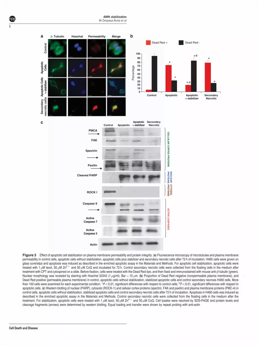
of permeable and nonpermeable cells. We found thatstabilized apoptotic cells maintained the AMN and were alsononpermeable to the supravital dye (Figures 3a and b). Onthe contrary, we observed that membrane integrity wasimpaired in apoptotic cells without stabilization and in controlsecondary necrotic cells in which AMN was clearly disorga-nized (Figures 3a and b). As expected, control cells remainednonpermeable.
Plasma membrane and cell cortex proteins are protectedfrom caspase-mediated cleavage by apoptotic stabilizer.To verify the integrity of cellular cortex and plasmamembrane proteins in stabilized apoptotic cells by thecombined treatment of taxol, Zn2þ and CoQ (apoptoticstabilizer) for 72 h, we examined the integrity of well-knowncellular cortex and plasma membrane proteins that arecommon substrates of effector caspases when apoptoticcells undergo secondary necrosis.23 Western blotting analy-sis revealed that initiator caspase-9 and effector caspases-3and -7 were activated and that nuclear (PARP) and cytosolicproteins (Rho-associated, coiled-coil containing proteinkinase 1 (ROCK-1)) were cleaved in apoptotic cells withand without stabilization treatment and also in controlsecondary necrotic cells, thus indicating that active caspaseshad degraded nuclear and cytosolic components during theexecution phase of apoptosis (Figure 3c). However, cellularcortex and transmembrane proteins such as a-spectrin,paxilin and plasma membrane Ca2þ ATPase (PMCA-4)were only cleaved when AMN was disorganized in apoptoticcells without stabilization or in control secondary necroticcells. On the contrary, cellular cortex and plasma membraneproteins were intact in stabilized apoptotic cells that maintainthe AMN (Figure 3c). Together, these data indicate thatapoptotic cells can be stabilized maintaining the integrity ofcellular cortex and plasma membrane proteins.
Apoptotic cell stabilization prevents PMCA-4 cleavageand calcium overload. To confirm plasma membraneprotein integrity, we performed immunofluorescence analysisof PMCA-4, a plasma membrane protein involved in calciumextrusion. PMCA-4 integrity was visualized in stabilizedapoptotic cells by the combined treatment of taxol, Zn2þ
and CoQ (apoptotic stabilizer) versus nonstabilized apoptoticcells after 72 h of incubation. Previously, we have demon-strated that PMCA-4 remains intact during the executionphase of apoptosis and is only degraded when cells undergosecondary necrosis.23 As shown in Figure 4a, stabilizedapoptotic cells maintain the AMN and the integrity of PMCA-4.On the contrary, the fluorescent signal of PMCA-4disappears in apoptotic cells without stabilization and incontrol secondary necrotic cells in which the AMN wasdisorganized.
To confirm plasma membrane integrity and PMC-4 func-tionality, we measured intracellular calcium levels by FLUO-4staining. Stabilized apoptotic cells showed a moderateincrease of FLUO-4 fluorescence with respect to control cells.However, apoptotic cells without stabilization or controlsecondary necrotic cells showed a notable increase ofFLUO-4 fluorescence suggesting increased plasma mem-brane permeability and calcium overload (Figure 4b).
Mitochondrial membrane potential (Dwm), ATP levelsand ROS production in stabilized apoptotic cells. Wehave previously demonstrated that AMN during apoptosisdepends on energized mitochondria.21 Here, we have foundthat in stabilized apoptotic cells by the combined treatment oftaxol, Zn2þ and CoQ (by the action of the describedapoptotic stabilizer), mitochondria maintain a degree ofpolarization, although this was significantly reduced com-pared with control cells (Figure 4c). On the contrary, whenAMN was disorganized in apoptotic cells without stabilizationor in control secondary necrotic cells, a marked mitochondrialdepolarization was found (Figure 4c). These results suggestthat mitochondrial large depolarization observed duringsecondary necrosis was significantly prevented in stabilizedapoptotic cells. Stabilized apoptotic cells also showedincreased ATP levels with respect to secondary necroticcells, although they were significantly reduced with respect tocontrol cells (Figure 4d). ROS production was also signifi-cantly reduced in stabilized apoptotic cells with respect togenuine apoptotic and necrotic cells (Figure 4e).
High phosphatidylserine exposure in stabilized apopto-tic cells. Phosphatidylserine (PS) is actively localized on theinner leaflet of the plasma membrane in healthy cells. Theasymmetry of its distribution is lost in apoptotic cells. Toexamine PS exposure in stabilized apoptotic cells by thecombined treatment of taxol, Zn2þ and CoQ (apoptoticstabilizer), we analyzed the binding of Annexin V-FITC to PSby using immunofluorescence and flow cytometry analysis incontrol, apoptotic cells without stabilization, stabilized apop-totic cells and control secondary necrotic cells. We havefound that PS was highly translocated in stabilized apoptoticcells. However, PS translocation was significantly reduced inapoptotic cells without stabilization or in cells sufferingsecondary necrosis. As expected, control cells did not showPS externalization (Figures 5a and b).
Stabilized apoptotic cells interact with macrophages. Asapoptotic cell stabilization effectively allows high PS exter-nalization, we also tested their binding and uptake byphagocytes. In control, apoptotic cells without stabilizationor in control secondary necrotic cells, that is, associated withlow PS externalization, the proportion of macrophagesinteracting with and engulfing apoptotic cells was markedlyreduced as compared with stabilized apoptotic cells by thecombined treatment of taxol, Zn2þ and CoQ with high PSexternalization (Figures 5c and d ).
Stabilized apoptotic cells maintain plasma membranepotential. To evaluate the effect of apoptotic stabilization oncell plasma membrane potentials, we performed patch-clamprecordings in H460 control, genuine apoptotic cells beforestabilization and stabilized apoptotic cells (stabilized asindicated above by the combined treatment with taxol,Zn2þ and CoQ). To acquire information of baseline and tomonitor membrane potential (Vm) at rest, cells were recordedfor periods of 20 min using the current-clamp mode of thewhole-cell configuration of the patch-clamp technique(Figure 6a). Vm was very stable during recordings. H460control cells showed a Vm of � 52±2 and � 55±2 mV at 0
AMN stabilizationM Oropesa-Avila et al
5
Cell Death and Disease
β- Tubulin Hoechst Permeability Merge
Apo
ptot
icC
ells
Sec
on
dar
yn
ecro
tic
cells
Apo
ptot
ic C
ells
+ st
abili
zer
Dead Red + Dead Red -
01020304050
60
7080
90
100
Control Apoptotic Apoptotic+ stabilizer
SecondaryNecrotic
Per
cent
age
a
a
Control ApoptoticApoptotic+ stabilizer
Secondary Necrotic
Cleaved PARP
Spectrin
PMCA
Caspase 9
Actin
Active Caspase 7
ROCK I
NU
CL
EA
R
PR
OT
EIN
SC
YT
OS
OL
P
RO
TE
INS
AC
TIV
E C
AS
PA
SE
SC
EL
LU
LA
R C
OR
TE
X P
RO
TE
INSPaxilin
Active Caspase 3
FAK
Co
ntr
ol
Figure 3 Effect of apoptotic cell stabilization on plasma membrane permeability and protein integrity. (a) Fluorescence microscopy of microtubules and plasma membranepermeability in control cells, apoptotic cells without stabilization, apoptotic cells plus stabilizer and secondary necrotic cells after 72 h of incubation. H460 cells were grown onglass coverslips and apoptosis was induced as described in the enriched apoptotic assay in the Materials and Methods. For apoptotic cell stabilization, apoptotic cells weretreated with 1mM taxol, 50mM Zn2þ and 50mM CoQ and incubated for 72 h. Control secondary necrotic cells were collected from the floating cells in the medium aftertreatment with CPT and cytospined on a slide. Before fixation, cells were treated with the Dead Red dye, and then fixed and immunostained with mouse anti-b-tubulin (green).Nuclear morphology was revealed by staining with Hoechst 33342 (1mg/ml). Bar¼ 15mm. (b) Proportion of Dead Red negative (nonpermeable plasma membrane), andDead Red positive (permeable plasma membrane) in control, apoptotic cells without stabilization, stabilized apoptotic cells and control secondary necrosis H460 cells. Morethan 100 cells were examined for each experimental condition. *Po0.01, significant differences with respect to control cells; aPo0.01, significant differences with respect toapoptotic cells. (c) Western blotting of nuclear (PARP), cytosolic (ROCK-1) and cellular cortex proteins (spectrin, FAK and paxilin) and plasma membrane proteins (PMC-4) incontrol cells, apoptotic cells without stabilization, stabilized apoptotic cells and control secondary necrotic cells after 72 h of incubation. Apoptosis in H460 cells was induced asdescribed in the enriched apoptotic assay in the Materials and Methods. Control secondary necrotic cells were collected from the floating cells in the medium after thetreatment. For stabilization, apoptotic cells were treated with 1 mM taxol, 50mM Zn2þ and 50mM CoQ. Cell lysates were resolved by SDS-PAGE and protein levels andcleavage fragments (arrows) were determined by western blotting. Equal loading and transfer were shown by repeat probing with anti-actin
AMN stabilizationM Oropesa-Avila et al
6
Cell Death and Disease
and 20 min after the beginning of the recordings, respectively(n¼ 11). In genuine apoptotic cells before stabilization, theVm was significantly depolarized compared with control cells(� 16±3 and � 17±2 mV at 0 and 20 min, n¼ 6). Instabilized apoptotic cells, the Vm was significantly depolar-ized compared with control cells but very similar to apoptoticcells before stabilization, maintaining a membrane potentialof � 17±3 and � 14±3 mV at 0 and 20 min after thebeginning of the recordings, respectively (n¼ 7; Figure 6b).Secondary necrotic cells had a membrane potential close to0 mV (� 1±0.5 and � 2±0.1 mV at 0 and 20 min, n¼ 3). Tobetter characterize the electrical properties of the cells,series resistance was monitored and measured duringrecordings. No changes in series resistance were foundduring the course of the experiments. Average seriesresistances were 26±2, 24±1 23±2 and 25±1 MO forcontrol, apoptotic cells before stabilization, stabilized apop-totic cells and secondary necrotic cells, respectively, indicat-ing that the apoptotic treatment did not induce any change inthis parameter (Figure 6c). These results indicate thatstabilization of apoptotic cells helps to maintain the valuesof series resistance, thus contributing to the maintaining of amembrane potential by apoptotic cells.
We next studied the passive membrane properties ofcontrol, apoptotic cells before stabilization and stabilizedapoptotic cells. For this purpose, a series of test pulses wasapplied with 10 mV increases in the pulse amplitude per step.According to nonexcitable cell behavior, there was no actionpotential occurrence at depolarizing potentials (Figure 6d).Furthermore, no occurrence of local potential or otherspontaneous activity was observed at very depolarized orhyperpolarized membrane potentials ranging from � 100 toþ 40 mV (Figure 6e).
It is clear from our results that the induction of apoptosis inH460 cells by CPT produces changes in the Vm of the cells.The fact that the stabilized apoptotic cells still maintain thesame level of Vm than apoptotic cells before stabilization isindicating that the cells are not physiologically dead and thatsome of the physiological processes of the cell involving thecrossing of ions (sodium and potassium ions) and the plasmamembrane pump are most probably still working, alsoindicating that stabilization of apoptotic cells may delay theloss of membrane potential.
Stabilization of apoptotic cells prevents secondarynecrosis and allows reliable apoptosis quantificationby flow cytometry. It is well known that reliable apoptoticquantification in adherent cell cultures by flow cytometry isdifficult and unreliable because of the fact that during theprocess of cell harvesting and manipulation before andduring flow cytometry assays many apoptotic cells undergosecondary necrosis. Therefore, we decided to examine theeffect of the stabilizer cocktail in apoptosis quantification byflow cytometry.
With the aim of stabilizing apoptotic cells for reliablediagnostic detection and quantification of apoptosis, we firstinduced a high population of apoptotic H460 cells by treatmentwith 10 mM CPT for 48 h. Subsequently, apoptotic cells wereincubated in the absence and presence of apoptotic stabilizer(taxolþZn2þ þCoQ) for 10 min before harvesting. Then, we
quantified apoptotic cells and cells in secondary necrosis byflow cytometry by staining with Annexin-FITC and7-aminoactinomycin (7-AAD) following the standard protocoldescribed in the Materials and Methods section(Supplementary Figures 4a and b). Results showed thatduring the process of cell harvesting and manipulation duringthe flow cytometry assay, many apoptotic cells withoutstabilization underwent secondary necrosis (42%). However,the treatment of apoptotic cells with the stabilizer 10 minbefore harvesting significantly prevented plasma membranedisruption and secondary necrosis (8%). These data suggestthat stabilization of apoptotic cells may be a very useful tool forreliable apoptosis quantification by flow cytometry assays.
Discussion
In the present study, we have developed and described aninnovative method aimed to the temporal stabilization andpreservation of apoptotic cells. This method consists of thecombined treatment of apoptotic cells with taxol, Zn2þ andCoQ. This experimental approach guarantees apoptotic cellintegrity by preventing plasma membrane permeability andsecondary necrosis.
The rationale for using this stabilizing combination was: (1)the use of taxol that, as a microtubule- stabilizing agent,24,25
prevents AMN depolymerization and subsequently the accessof active caspases to the cellular cortex; (b) the use of Zn2þ
that, as a caspase inhibitor,26–29 prevents extensive degrada-tion of cellular components and caspase-dependent cleavageof cellular cortex and plasma membrane proteins; and (3) theuse of CoQ that, as an antioxidant,30 protects againstoxidative membrane damage that is increased in apoptoticcells.
Stabilized apoptotic cells can be considered as dying cellsin which the cellular cortex and plasma membrane aremaintained intact or alive. In a metaphorical sense, we canconsider them as ‘living dead’ or ‘zombie cells’. Stabilizedapoptotic retain many of the hallmark characteristics ofgenuine apoptotic cells such as plasma membrane imperme-ability, integrity of plasma membrane proteins, low intracel-lular calcium levels, plasma membrane potential, PSexternalization and ability of being phagocytosed.
Interest in apoptosis research has increased considerablyrecently for a number of reasons including development oftherapeutic treatments, cell culture technology development,metabolic engineering of mammalian cells and gene therapy.Furthermore, apoptotic cells play important roles in biomedi-cine because of their characteristics, and they are beingwidely used to evaluate the cytotoxic effects of variouscompounds by quantifying apoptotic cells by flow cytometry.31
However, this determination is often affected by the process ofcell manipulation (treatments for harvesting, cell centrifuga-tion, cell pipetting and so on), especially in adherent cellcultures required for flow cytometry assays that disruptsplasma membrane permeability and leads apoptotic cells tosecondary necrosis.32 Therefore, accurate and reliableapoptosis quantifications are particularly difficult in adherentcell cultures. Stabilization of apoptotic cells before cellharvesting allows a more accurate and reliable quantificationof the actual number of apoptotic cells or the correct
AMN stabilizationM Oropesa-Avila et al
7
Cell Death and Disease

determination of biochemical parameters of genuine apoptoticcells such as mitochondrial membrane potential, intracellularcalcium concentration, pH, caspase activity and many others,without the interference of plasma membrane disruption andsubsequent secondary necrosis.
Moreover, apoptotic cells are currently used for variousforms of therapy, primarily with the aim of inducing immuno-logical tolerance in the recipient individual.33 The stabilizationof apoptotic cells before administration may help to ensurethat the inoculated apoptotic cells will retain their
Mit
och
on
dri
a M
emb
ran
e P
ote
nti
all
(arb
itra
ryu
nit
s u
nit
s)
0
20
40
60
80
100
120
140
a
Intr
acel
lula
r c
alci
um
(arb
itra
ry u
nit
s)
Control Apoptotic Apoptotic+ stabilizer
SecondaryNecrotic
0
500
1000
1500
2000
2500
a
Control Apoptotic Apoptotic+ stabilizer
SecondaryNecrotic
0
20
40
60
80
100
120
ATP
lev
els
Control Apoptotic Apoptotic+ stabilizer
SecondaryNecrotic
a
Mit
oS
OX
(ar
bit
rary
un
its)
0
40
80
120
160
180
220
* *
Control GenuineApoptotic
Apoptotic+ stabilizer
SecondaryNecrotic
Apoptoticwithout
stabilization
a
β-Tubulin PMCA-4 Hoechst Merge
Co
ntr
ol
Ap
op
totic
Cel
lsS
eco
nd
ary
Nec
roti
c ce
llsA
po
pto
tic C
ells
+ st
abili
zer
AMN stabilizationM Oropesa-Avila et al
8
Cell Death and Disease

Annexin V Hoechst MergeA
popt
otic
Cel
lsS
eco
nd
ary
Nec
roti
c ce
llsA
popt
otic
Cel
ls+
stab
ilize
rC
on
tro
l
Apo
ptot
ic c
ells
+ st
abili
zer
C
on
tro
l M
C
Cell Tracker Hoechst Merge/Light
0
5
10
15
20
25
Control Apoptotic Apoptotic+ stabilizer
Secondarynecrotic
a
Ann
exin
V s
igna
l (ar
bitr
ary
units
)
0
20
40
60
80
100
120
Control Apoptotic Apoptotic+stabilizer
Secondarynecrotic
a
% M
acro
ph
ages
Normal
Contact
Engulfment
AM
β-Tubulin
Figure 5 Effect of apoptosis stabilization on PS externalization and interaction with phagocytes. (a) Fluorescence microscopy of microtubules and PS externalization in controlcells, apoptotic cells without stabilization, stabilized apoptotic cells and control secondary necrotic cells after 72 h of incubation. H460 cells were grown on glass coverslips andapoptosis was induced as described in the enriched apoptotic assay in the Materials and Methods. Secondary necrotic cells were collected from the floating cells in the medium aftertreatment and cytospined onto a slide. Stabilized apoptotic cells were treated with 1 mM taxol, 50mM Zn2þ and 50mM CoQ. Then, cells were fixed and immunostained with antib-tubulin (red), and PS externalization was revealed by staining with Annexin V-FITC. Nuclear morphology was revealed by staining with Hoechst 33342 (1mg/ml). Bar¼ 15mm).(b) PS externalization was quantified by flow cytometry as described in the Materials and Methods. Data represent the means±S.D. of three independent experiments.(c) Wide-field images of CellTracker Green-labeled control and stabilized apoptotic H460 cells (white arrows) interacting with THP-1 macrophages (red arrows). Bars¼ 15mm.(d) Proportion of THP-1 macrophages interacting and engulfing control and stabilized apoptotic H460 cells. CellTracker Green-labeled H460 cells were induced into apoptosis byCPT/cytochalasin as described in the Materials and Methods. Stabilized apoptotic cells were treated with 1mM taxol, 50mM Zn2þ and 50mM CoQ. Control and stabilized apoptoticcells were then co-incubated with THP-1 macrophages. Nuclear morphology was revealed by staining with Hoechst 33342 (1mg/ml). Data represent the means±S.D. of threeindependent experiments. *Po0.01, significant differences with respect to control cells; aPo0.01, significant differences with respect to apoptotic cells without stabilization
Figure 4 PMCA-4 integrity, mitochondrial membrane potential, ATP and ROS levels after apoptosis stabilization. (a) Fluorescence microscopy of PMCA-4 in control cells,apoptotic cells without stabilization, stabilized apoptotic cells and control secondary necrotic cells after 72 h of incubation. H460 cells were grown on glass coverslips and apoptosiswas induced as is described in the enriched apoptotic assay in the Materials and Methods. Secondary necrotic cells were collected from the floating cells in the medium aftertreatment and cytospined on a slide. For stabilization, apoptotic cells were treated with 1mM taxol, 50mM Zn2þ and 50mM CoQ. Then, cells were fixed and immunostained with antiPMCA-4 (green) and anti b-tubulin (red). Nuclear morphology was revealed by staining with 1mg/ml Hoechst 33342. Bar¼ 15mm. (b) Intracellular calcium levels were measured byflow cytometry using the calcium indicator FLUO-4 (1mM) in control cells, apoptotic cells without stabilization, stabilized apoptotic cells and control secondary necrotic cells after 72 hof incubation. The y axis of the calcium graph represents the fluorescence intensity of the samples relative to the background intensity. (c) Mitochondrial membrane potential instabilized apoptotic cells. Mitochondrial membrane potential was measured in control cells, apoptotic cells without stabilization, stabilized apoptotic cells and control secondarynecrotic cells after 72 h of incubation. Apoptosis was induced as described in the enriched apoptotic assay in the Materials and Methods. Secondary necrotic cells were collected fromthe floating cells in the medium after treatment. Stabilized apoptotic cells were treated with 1mM taxol, 50mM Zn2þ and 50mM CoQ. Mitochondrial membrane potential (Dcm) wasdetermined by MitoTracker Red staining and flow cytometry analysis as described in the Materials and Methods. The y axis of the MitoTracker graph represents the fluorescenceintensity of the samples relative to the background intensity. (d) ATP levels were measured in control cells, apoptotic cells without stabilization, stabilized apoptotic cells and controlsecondary necrotic cells after 72 h of incubation. ATP levels were measured as described in the Materials and Methods. Results were expressed as percentage with respect to ATPlevels in control cells. (e) ROS levels were measured in control cells, genuine apoptotic cells, apoptotic cells without stabilization, stabilized apoptotic cells and control secondarynecrotic cells after 72 h of incubation. ROS levels were measured by MitoSOX staining coupled to flow cytometry analysis as described in the Materials and Methods. The y axis ofthe MitoSOX graph represents the fluorescence intensities of MitoSOX cell samples relative to unstained cells. Data represent the means±S.D. of three independent experiments.*Po0.01, significant differences with respect to control cells; aPo0.01, significant differences with respect to apoptotic cells without stabilization
AMN stabilizationM Oropesa-Avila et al
9
Cell Death and Disease

-60
-40
-20
0
(11)
(6) (7)
(3)
a
2 mV
50 ms
- 10 mV
- 50 mV
- 100 mV
Control Apoptotic+ stabilizer
ControlApoptotic+ stabilizer
Apoptoticbefore stabilization
Secondary necrotic
Time (min)
0 5 10 15 20-100
-80
-60
-40
-20
0
Control
Apoptotic before stabilization
Apoptotic + stabilizer
Secondary necrotic
Vm
(mV
)
Vm
(mV
)
10 mV
50 ms
10 mV
50 ms
- 30 mV- 60 mV
- 60 mV
10 mV- 20 mV
10 mV10 mV
ControlApoptotic before
stabilization
- 100 mV
Apoptotic + stabilizer
Apoptotic before stabilization
Figure 6 Plasma membrane potential after apoptotic cell stabilization. (a) The pictures show control, genuine apoptotic cells before stabilization and stabilized apoptotic andnecrotic cells under recording. (b) Graph shows Vm of control (circles), genuine apoptotic cells before stabilization (squares) and stabilized apoptotic (rhombus) and necrotic cells(triangles). Note that stabilized apoptotic cells maintain the same membrane potential as genuine apoptotic cells before stabilization. (c) Quantification of results from (b).*Po0.01. (d) Passive membrane properties of control, genuine apoptotic before stabilization and stabilized apoptotic H460 cells. Protocol shows series of test pulses with 10 mVincreasing amplitude each step. Representative traces for control (left panel) and apoptotic (genuine before stabilization and stabilized) cells (right panel) are shown.(e) Representative samples showing the lack of activity of control and apoptotic cells (before stabilization and stabilized) in response to depolarizing and hyperpolarizing pulses
AMN stabilizationM Oropesa-Avila et al
10
Cell Death and Disease

characteristic features until they are phagocytosed by macro-phages. The administration of stabilized apoptotic cells canalso be used for delivering substances of interest such astherapeutic proteins (for protein replacement therapy) ordrugs to recipient macrophages.34
There are forms of death that by their nature impair thecorrect formation of the AMN (e.g., mitochondrial toxics andcold exposure)21 and as a result apoptotic cells are not able tomaintain plasma membrane integrity and undergo secondarynecrosis that may cause serious side effects. Apoptotic cellstabilization may allow the development of therapies for thecorrect formation and stabilization of AMN, thus promoting amore physiological and controlled type of cell death. Based onthe above arguments, there is a need to provide compositionsand methods for the stabilization of apoptotic cells.
In summary, we have demonstrated that apoptotic cells canbe stabilized for reliable detection and quantification ofapoptosis in cultured cells. Stabilization of apoptotic cellsmay also allow a safer administration of apoptotic cells inclinical applications and opens new avenues in the functionalreconstruction of apoptotic cells for longer preservation.
Materials and MethodsReagents. CPT was purchased from Alexis Corporation (Nottingham, UK).Cytochalasin, taxol, ZnSO4, Phalloidin-coumarin, trypsin, anti-ROCK-1, anti-actin,coenzyme Q10 and anti-a-tubulin were purchased from Sigma Chemical Co. (St.Louis, MO, USA). Anti-b-tubulin antibodies were from Chemicon International(Temecula, CA, USA). Hoechst 33342, FITC-labeled goat anti-mouse andtetramethyl rhodamine goat anti-rabbit antibodies, Dead Red reagent and FLUO-4were purchased from Molecular Probes (Eugene, OR, USA). Anti-active caspase-3,anti-active caspase-7, anti-caspase-9 and anti-cleaved PARP were obtained fromCell Signaling Technology (Beverly, MA, USA). Anti- focal adhesion kinase (FAK),anti-PMCA-4, anti-a-spectrin and anti-paxilin were purchased from Santa CruzBiotechnology (Santa Cruz, CA, USA). Anti-GADPH was purchased fromCalbiochem Merck Millipore (Darmstadt, Germany).
Cell culture. Non-small lung cancer cell line, H-460, was a gift from Dr. PJWoll (CRC Department of Clinical Oncology, City Hospital, Nottingham, UK). Cellswere cultured in RPMI medium supplemented with penicillin, streptomycin and10% fetal bovine serum at 371C.
Enriched apoptotic assay. Apoptosis was induced to cells by treatmentwith 10mM CPT, an anticancer drug that inhibits the DNA enzyme topoisomerase I.To increase the proportion of attached apoptotic cells with AMN formation, H460cells were treated with CPTþ cytochalasin (2 mM) for 48 h. To prevent cell cycleeffects, cytochalasin was added 24 h after CPT treatment. Inhibiting actinpolymerization, and consequently actin contraction with cytochalasin, CPT inducedapoptosis but apoptotic cells maintained the spread state and remained attachedto the flask or slide.35 Secondary necrotic cells were collected from the floatingcells in the medium after CPT treatment.
Apoptotic stabilizer. Apoptotic cell stabilization was performed by treatingapoptotic cells with: 1 mM taxol, 50mM Zn2þ (ZnSO4) or 50mM CoQ, or thecombined treatment of the three compounds.
Analysis of apoptosis. Apoptosis was defined by the occurrence of cellswith nuclear condensation and fragmentation by Hoechst staining, but retainingplasma membrane integrity (measured by exclusion of the membrane-imperme-able dye, Dead Red reagent). Apoptosis was also assessed by caspase activation,cytochrome c release, lack of actin cytoskeleton, presence of apoptoticmicrotubules and/or cell morphology by phase-contrast microscopy. Primarynecrosis was defined as a loss of plasma membrane integrity without signs ofnuclear condensation or fragmentation. Secondary necrosis was determined byscoring over time apoptotic cells that became permeable to the Dead Red reagent.Cells were counted for up to 48 h after CPT/cytochalasin exposure. In each case,
10 random fields and more than 100 cells were counted. To differentiate betweengenuine apoptotic cells and cells in secondary necrosis, Dead Red reagent wasadded to the cells 10 min before fixation.
Immunofluorescence studies. H460 cells were grown on 1 mm (ThermoScientific Gold Seal, Waltham, MA, USA) glass coverslips for 24–48 h in RPMIcontaining 10% FBS. After treatments, cells were rinsed once with PBS, fixed in3.8% paraformaldehyde for 5 min at room temperature and permeabilized in 0.1%saponin for 5 min. For immunostaining, glass coverslips were incubated withprimary antibodies diluted 1 : 100 in PBS for 1–2 h at 371C in a humidifiedchamber. Excess antibody binding was removed by washing the coverslips withPBS (three times, 5 min). The secondary antibody, a FITC-labeled goat anti-mouse antibody or a tetramethyl rhodamine goat anti-rabbit (Molecular Probes),diluted 1 : 100 in PBS, were added and incubated for 1 h at 371C. Coverslips werethen rinsed with PBS for 3 min, incubated for 1 min with PBS containing Hoechst33342 (1mg/ml) and washed with PBS (three 5-min washes). Finally, thecoverslips were mounted onto microscope slides using Vectashield MountingMedium (Vector Laboratories, Burlingame, CA, USA) and analyzed using anupright fluorescence microscope (Leica DMRE, Leica Microsystems GmbH,Wetzlar, Germany). Deconvolution studies and 3D projections were performedusing a DeltaVision system (Applied Precision, Issaquah, WA, USA) with anOlympus IX-71 microscope with � 100 objective/1.35 NA and � 60 objective/1.40NA (Olympus, Tokyo, Japan) and filters set for DAPI, fluorescein isothiocyanate,and rhodamine provided by Applied Precision. Acquired z planes were separatedby 0.3mm, and an average of 50 planes was taken for each nucleus. The 3Dstacks were deconvolved using the Softworx software algorithm (conservative ratiomethod, 10 iterations; Applied Precision).
Western blotting analysis. Whole cellular lysates were prepared in abuffer, with gentle shaking, composed of 0.9% NaCl, 20 mM Tris-HCl, pH 7.6,0.1% Triton X-100, 1 mM phenylmethylsulfonylfluoride and 0.01% leupeptine.Electrophoresis was carried out in a 10–15% acrylamide SDS-PAGE. Proteinswere transferred to Immobilon membranes (Amersham Pharmacia, Buckingham-shire, UK). Proteins were electrophorezed, transferred to nitrocellulosemembranes and, after blocking overnight at 41C, incubated with the respectiveantibody solution at 1 : 1000 dilutions. Then, membranes were probed with theirrespective secondary antibody (1 : 2500). Immunolabeled proteins were detectedby using a chemioluminiscence method (Bio-Rad, Hercules, CA, USA). Proteinwas determined by the Bradford method.36
Cell content release: LDH determination. Plasma membrane integrityand cell content release was monitored by measuring LDH activity into theincubation medium. This was done using a commercial kit (CytoTox 96 Non-Radioactive Cytotoxicity Assay from Promega (Madison, WI, USA).
Determination of intracellular calcium levels by flow cytometry.H460 cells were incubated for 60 min at 371C in medium supplemented with 1mMFLUO-4, AM. Cells were then washed three times with RPMI medium, fixed with3.8% paraformaldehyde and analyzed by flow cytometry using an EPICS Elite flowcytometer (Beckman-Coulter, Hialeah, FL, USA).
Measurement of Dwm by flow cytometry. H460 cells were incubatedfor 60 min at 371C in medium supplemented with 1mm Mitotracker Red (MolecularProbes). Cells were then washed three times with RPMI medium, fixed with 3.8%paraformaldehyde and analyzed by flow cytometry.
ATP determination. ATP levels were determined by using a luciferase-based assay according to the directions of Molecular Probes, using a TunerDesigns Luminometer TD 20/20 (Sunnyvale, CA, USA).
Measurement of intracellular generation of ROS. MitochondrialROS generation in fibroblasts was assessed by MitoSOX, a red mitochondrialsuperoxide indicator. Once in the mitochondria, MitoSOX red reagent is oxidizedby superoxide and exhibits red fluorescence. Approximately 1� 106 cells wereincubated with 1 mM MitoSOX for 30 min at 371C, washed twice with PBS,resuspended in 500ml of PBS and analyzed by flow cytometry (excitation at510 nm and fluorescence detection at 580 nm). Specificity of MitoSOX forsuperoxide has been shown by the manufacturer, and its mitochondrial localizationwas tested by co-staining with MitoTracker Green (data not shown).
AMN stabilizationM Oropesa-Avila et al
11
Cell Death and Disease

Electrophysiological recordings. Somatic whole-cell patch-clamprecordings were made from healthy (control), nonstabilized and stabilizedapoptotic H460 cells and necrotic cells. These cells were visually identified byIR-DIC (infrared-differential interference contrast) microscopy using a 40� waterimmersion objective. Cells were perfused at a flow rate of 3.5 ml/min with asolution containing (in mM): NaCl 126; KCl 3; NaH2PO4 1.25; MgSO4 2; CaCl2 2;NaHCO3 26; glucose 10, pH 7.2–7.4; bubbled with carbogen gas (95% O2/5%CO2). All experiments were performed at 371C. Patch electrodes were made fromborosilicate glass, and had a resistance of 4–7 MO when filled with (in mM):potassium gluconate 110; HEPES 40; NaCl 4; ATP-Mg 4; GTP 0.3 (pH 7.2,290 mOsm). Cells were voltage clamped, using a Multiclamp 700B amplifier(Molecular Devices, Foster City, CA, USA). Access resistance was regularlymonitored during recordings, and cells were rejected if it changed 415% duringthe experiment. Data were filtered at 2 kHz, digitized at 10 kHz and stored on acomputer using pClamp software (Molecular Devices). Membrane potentials (Vm)of the cells were recorded by using the current-clamp mode of the amplifier. Vm
were measured continuously during the experiments. Average values indicated inthe text correspond to Vm at 5 and 15 min after the beginning of the recordings.Potentials were obtained by applying a series of test pulses from a holdingpotential of � 60 mV (control healthy cells) and � 20 mV (apoptotic cells) with10 mV increases in pulse amplitude per step.
Determination of PS externalization. PS translocation from the inner tothe outer leaflet of the plasma membrane is one of the earliest apoptotic features.We used the PS-binding protein Annexin V conjugated with FITC to identify PSexposure in H460 cells. The binding of annexin V to cell surface PS was detectedwith a commercial Annexin Apoptosis Kit (Santa Cruz Biotechnology) according tothe manufacturer’s instructions. To distinguish cells that had lost membraneintegrity, propidium iodide (PI) was added to a final concentration of 10 mg/mlimmediately before analysis. Apoptotic cells were identified either by fluorescencemicroscopy or by flow cytometry. Hoechst 33342 (1 mg/ml) staining was used toreveal nuclear condensation/fragmentation.
Phagocytosis assays. CellTracker-labeled H460 cells (Molecular Probes)that adhered to glass coverslips were induced into apoptosis by CPT/cytochalasintreatment and then were incubated for 1 h in the absence or presence ofcolchicine or colchicine plus z-VAD. Then, cells were washed and co-incubatedwith PMA-differentiated THP-1 macrophages (150 000 cells/well). Floatingapoptotic cells representing secondary necrotic cells were cytospined ontocoverslips and also co-incubated with PMA-differentiated THP-1 macrophages.After 8 h of co-incubation at 371C, coverslips were washed extensively in PBS andcells were fixed in 3.8% paraformaldehyde. The number of THP-1 macrophagesinteracting (bound and engulfed) and engulfing cell fragments was calculated in 10random fields in triplicate by fluorescence and phase-contrast microscopy.
Apoptosis flow cytometry assay. We followed a standard protocolsupplied by Santa Cruz Biotechnology (Annexin V-FITC: sc-4252 FITC). Controland apoptotic H460 cells were trypsinized and transferred to a 15 ml conical tube.Cells were pelleted by low speed centrifugation at 1500 r.p.m. for 5 min. Gentlywash in medium containing serum. Next, cells were washed once with PBS andresuspended in 1� Assay Buffer at a concentration of 1� 106 cells/ml. Then,100ml aliquot of cells (1� 105 cells) was transferred to a 5-ml culture tube and1mg/ml of Annexin V-FITC and 1mg/ml 7-ADD was added. Recommendednegative controls for flow cytometry include: (1) no Annexin V-FITC and no 7-ADD;(2) Annexin V-FITC alone and (3) 7-ADD alone. Subsequently, samples weregently vortexed and incubated for 15 min at room temperature in the dark.Immediately, samples were analyzed by flow cytometry.
Statistical analysis. All results are expressed as means±S.D. unlessstated otherwise. Two-tailed Student’s t-tests (paired and unpaired, accordingto samples) or ANOVA variance analysis were used as appropriate forstatistical analysis. Only P-values of o0.05 were considered as statisticallysignificant.
Conflict of InterestThe authors declare no conflict of interest.
Acknowledgements. This work was supported by FIS PI10/00543 grant,Ministerio de Sanidad, Spain, and Fondo Europeo de Desarrollo Regional (FEDER-Union Europea), SAS 111242 grant, Servicio Andaluz de Salud-Junta de Andalucıa,Proyecto de Investigacion de Excelencia de la Junta de Andalucıa CTS-5725,BFU2012-38208 and by AEPMI (Asociacion de Enfermos de PatologıaMitocondrial).
1. Kerr JF, Wyllie AH, Currie AR. Apoptosis: a basic biological phenomenon withwide-ranging implications in tissue kinetics. Br J Cancer 1972; 26: 239–257.
2. Wyllie AH. The biology of cell death in tumours. Anticancer Res 1985; 5: 131–136.3. Wyllie AH. Apoptosis: an overview. Br Med Bull 1997; 53: 451–465.4. Afford S, Randhawa S. Apoptosis. Mol Pathol 2000; 53: 55–63.5. Fischer U, Janicke RU, Schulze-Osthoff K. Many cuts to ruin: a comprehensive update of
caspase substrates. Cell Death Differ 2003; 10: 76–100.6. Savill J, Dransfield I, Gregory C, Haslett C. A blast from the past: clearance of apoptotic
cells regulates immune responses. Nat Rev Immunol 2002; 2: 965–975.7. Zong WX, Thompson CB. Necrotic death as a cell fate. Genes Dev 2006; 20: 1–15.8. Mills JC, Stone NL, Pittman RN. Extranuclear apoptosis. The role of the cytoplasm in the
execution phase. J Cell Biol 1999; 146: 703–708.9. Pittman S, Geyp M, Fraser M, Ellem K, Peaston A, Ireland C. Multiple centrosomal
microtubule organising centres and increased microtubule stability are early features ofVP-16-induced apoptosis in CCRF-CEM cells. Leuk Res 1997; 21: 491–499.
10. Pittman SM, Strickland D, Ireland CM. Polymerization of tubulin in apoptotic cells is not cellcycle dependent. Exp Cell Res 1994; 215: 263–272.
11. Moss DK, Betin VM, Malesinski SD, Lane JD. A novel role for microtubules in apoptoticchromatin dynamics and cellular fragmentation. J Cell Sci 2006; 119(Pt 11): 2362–2374.
12. Moss DK, Lane JD. Microtubules: forgotten players in the apoptotic execution phase.Trends Cell Biol 2006; 16: 330–338.
13. Sanchez-Alcazar JA, Rodriguez-Hernandez A, Cordero MD, Fernandez-Ayala DJ,Brea-Calvo G, Garcia K et al. The apoptotic microtubule network preservesplasma membrane integrity during the execution phase of apoptosis. Apoptosis 2007;12: 1195–1208.
14. Iverson SL, Orrenius S. The cardiolipin-cytochrome c interaction and the mitochondrialregulation of apoptosis. Arch Biochem Biophys 2004; 423: 37–46.
15. Nishimura G, Proske RJ, Doyama H, Higuchi M. Regulation of apoptosis by respiration:cytochrome c release by respiratory substrates. FEBS Lett 2001; 505: 399–404.
16. Simon HU, Haj-Yehia A, Levi-Schaffer F. Role of reactive oxygen species (ROS) inapoptosis induction. Apoptosis 2000; 5: 415–418.
17. Petrosillo G, Ruggiero FM, Paradies G. Role of reactive oxygen species and cardiolipin inthe release of cytochrome c from mitochondria. FASEB J 2003; 17: 2202–2208.
18. Hampton MB, Orrenius S. Redox regulation of apoptotic cell death in the immune system.Toxicol Lett 1998; 102-103: 355–358.
19. Slater AF, Stefan C, Nobel I, van den Dobbelsteen DJ, Orrenius S. Intracellular redoxchanges during apoptosis. Cell Death Differ 1996; 3: 57–62.
20. Cai J, Jones DP. Superoxide in apoptosis. Mitochondrial generation triggered bycytochrome c loss. J Biol Chem 1998; 273: 11401–11404.
21. Oropesa M, de la Mata M, Maraver JG, Cordero MD, Cotan D, Rodriguez-Hernandez A et al.Apoptotic microtubule network organization and maintenance depend on high cellular ATPlevels and energized mitochondria. Apoptosis 2011; 16: 404–424.
22. Sanchez-Alcazar JA, Khodjakov A, Schneider E. Anticancer drugs induce increasedmitochondrial cytochrome c expression that precedes cell death. Cancer Res 2001; 61:1038–1044.
23. Oropesa-Avila M, Fernandez-Vega A, de la Mata M, Maraver JG, Cordero MD, Cotan D et al.Apoptotic microtubules delimit an active caspase free area in the cellular cortex during theexecution phase of apoptosis. Cell Death Dis 2013; 4: e527.
24. Schiff PB, Fant J, Horwitz SB. Promotion of microtubule assembly in vitro by taxol.Nature 1979; 277: 665–667.
25. Schiff PB, Horwitz SB. Taxol stabilizes microtubules in mouse fibroblast cells. Proc NatlAcad Sci USA 1980; 77: 1561–1565.
26. Huber KL, Hardy JA. Mechanism of zinc-mediated inhibition of caspase-9. Protein Sci2012; 21: 1056–1065.
27. Perry DK, Smyth MJ, Stennicke HR, Salvesen GS, Duriez P, Poirier GG et al. Zinc is apotent inhibitor of the apoptotic protease, caspase-3. A novel target for zinc in the inhibitionof apoptosis. J Biol Chem 1997; 272: 18530–18533.
28. Smith AF, Longpre J, Loo G. Inhibition by zinc of deoxycholate-induced apoptosis inHCT-116 cells. J Cell Biochem 2012; 113: 650–657.
29. Stennicke HR, Salvesen GS. Biochemical characteristics of caspases-3, -6, -7, and -8.J Biol Chem 1997; 272: 25719–25723.
30. Bentinger M, Brismar K, Dallner G. The antioxidant role of coenzyme Q. Mitochondrion2007; 7(Suppl): S41–S50.
31. Darzynkiewicz Z, Bruno S, Del Bino G, Gorczyca W, Hotz MA, Lassota P et al. Features ofapoptotic cells measured by flow cytometry. Cytometry 1992; 13: 795–808.
32. van Engeland M, Ramaekers FC, Schutte B, Reutelingsperger CP. A novel assay tomeasure loss of plasma membrane asymmetry during apoptosis of adherent cells inculture. Cytometry 1996; 24: 131–139.
AMN stabilizationM Oropesa-Avila et al
12
Cell Death and Disease

33. Saas P, Kaminski S, Perruche S. Prospects of apoptotic cell-based
therapies for transplantation and inflammatory diseases. Immunotherapy 2013; 5:
1055–1073.
34. Perez B, Paquette N, Paidassi H, Zhai B, White K, Skvirsky R et al. Apoptotic cells can
deliver chemotherapeutics to engulfing macrophages and suppress inflammatory cytokine
production. J Biol Chem 2012; 287: 16029–16036.
35. Huot J, Houle F, Rousseau S, Deschesnes RG, Shah GM, Landry J. SAPK2/
p38-dependent F-actin reorganization regulates early membrane blebbing during
stress-induced apoptosis. J Cell Biol 1998; 143: 1361–1373.
36. Bradford MM. A rapid and sensitive method for the quantitation of microgram
quantities of protein utilizing the principle of protein-dye binding. Anal Biochem 1976;
72: 248–254.
Cell Death and Disease is an open-access journalpublished by Nature Publishing Group. This work is
licensed under a Creative Commons Attribution-NonCommercial-NoDerivs 3.0 Unported License. The images or other third partymaterial in this article are included in the article’s Creative Commonslicense, unless indicated otherwise in the credit line; if the material isnot included under the Creative Commons license, users will need toobtain permission from the license holder to reproduce the material. Toview a copy of this license, visit http://creativecommons.org/licenses/by-nc-nd/3.0/
Supplementary Information accompanies this paper on Cell Death and Disease website (http://www.nature.com/cddis)
AMN stabilizationM Oropesa-Avila et al
13
Cell Death and Disease

SECONDARY
NECROSIS2.2
0.8
1.0
1.2
1.4
1.8
-60 -50 -40 -30 -20 -10 0 10
Time (min)
2.0
0.6
20
AMN depolymerization
AMN polymerization
2.2
0.8
1.0
1.2
1.4
1.8
2.0
0.6
TM
RM
(F
/F0)
Cal
ciu
m s
ign
al (
F/F
0)
EXECUTION PHASE
2.2
0.8
1.0
1.2
1.4
1.8
-60 -50 -40 -30 -20 -10 0 10
Time (min)
2.0
0.6
20
D
AMN polymerization
2.2
0.8
1.0
1.2
1.4
1.8
2.0
0.6
TM
RM
(F
/F0)
Cal
ciu
m s
ign
al (
F/F
0)
ZOMBIE CELL
hours/days
Microtubule stabilization: Taxol
Caspase inhibition: Zn2+
Antioxidant: Coenzyme Q
Apoptotic stabilizer(zombie cocktail)
EXECUTION PHASE
1 hour
a
b
Supplementary Figure 1
10

a
b
Supplementary Figure 2
Quantification of attached apoptotic cells
0200400600800
100012001400160018002000
Apoptoticwithout stabilization
0h 48h 72h 96h 120h 144h24h 0h 48h 72h 96h 120h 144h24h
Apoptotic+Taxol+CoQ+Zn2+
** * *
Cel
l nu
mb
er
Apoptotic+Taxol+CoQ+Zn2+
Apoptoticwithout stabilization
Control+digi
LDH release
Arb
itra
ry u
nit
s
0
1
2
3
4
5
6
7
8
9
10
*

Co
ntr
ol
ApoptoticCells
Ap
op
toti
c C
ells
+
Taxo
l+C
oQ
+Z
n2+
0 hours 72 hoursa
0
Cel
l nu
mb
er
Control Apoptotic Apoptotic+Taxol+CoQ+Zn2+
72 hours48 hours24 hours0 hours
200
400
600
800
1000
1200
*a
***
Quantification of attached HeLa apoptotic cells
0
2
4
6
8
10
12LDH release
Control Apoptotic Apoptotic+Taxol+CoQ+Zn2+
Control+digi
72 hours24 hours0 hours
b
Arb
itra
ry u
nit
s
c
**
*a
Supplementary Figure 3

4%96%
91%
3%
5%
1%
42%8%
20%30%
Primary Necrosis Secondary Necrosis
Viables Apoptotics
Apoptotic + stabilizer
8%0,5%
52%39,5%
Apoptotic cells
Control
0
10
20
30
40
50
60
Viable Apoptotic PrimaryNecrosis
SecondaryNecrosis
Apoptotic cells
Apoptotic cells + stabilizerPer
cen
tag
e
Supplementary Figure 4
*
*
*
*
Annexin-FITC Annexin-FITC
Annexin-FITCAnnexin-FITC
7-A
min
oac
tin
om
ycin
D
7-A
min
oac
tin
om
ycin
D
7-A
min
oac
tin
om
ycin
D
7-A
min
oac
tin
om
ycin
D
a
b
Secondary Necrotic cells


