Embed Size (px)
Citation preview

Ž .Molecular Brain Research 71 1999 149–158www.elsevier.comrlocaterbres
Research report
Neurokinin-1 expression and co-localization with glutamate and GABAin the hypothalamus of the cat
Ruihong Yao a, Pranela Rameshwar c, Robert J. Donnelly d, Allan Siegel a,b,)
a Department of Neurosciences, UniÕersity of Medicine and Dentistry, NJ Medical School, 185 South Orange AÕe., MSB, Rm. H-512, Newark, NJ 07103,USA
b Department of Psychiatry, UniÕersity of Medicine and Dentistry, NJ Medical School, 185 South Orange AÕe., MSB, Rm. H-512, Newark, NJ 07103, USAc Department of Medicine–Hematology, UniÕersity of Medicine and Dentistry, NJ Medical School, Newark, NJ 07103, USA
d Department of Laboratory Medicine and Pathology, UniÕersity of Medicine and Dentistry, NJ Medical School, Newark, NJ 07103, USA
Accepted 4 May 1999
Abstract
Ž .Recent behavioral studies using pharmacological techniques have demonstrated that the high affinity substance P SP receptor,Ž .neurokinin-1 receptor NK-1 , in the medial hypothalamus could be important in mediating defensive rage behavior in the cat. These
observations prompted us to use molecular techniques to determine the distribution of NK-1 in the hypothalamus and in other regions ofthe forebrain relevant to the control of rage behavior. We cloned a 650 bp fragment of the cat NK-1 cDNA. Partial DNA sequence
Ž .analyses of this fragment indicate 90% homology with the human cDNA. By in situ hybridization ISH , we showed that NK-1 mRNAwas localized in the cytoplasm but not nuclei of cat forebrain neurons. Furthermore, NK-1 mRNA was co-localized in neurons thatdisplayed positive immunolabeling for glutamate or GABA. Moderate labeling was visualized in the anterior medial hypothalamus whichreceives significant SP input via the stria terminalis from the medial amygdala. Strong labeling was also observed in the basal amygdaloidcomplex. The functional significance of this labeling pattern is suggested from the observation that both the medial and basal complex ofamygdala serve as powerful modulators of defensive rage behavior. Weaker labeling was seen over the posterior medial and lateralhypothalamus. The distribution of NK-1 in the hypothalamus was matched by that of SP-immunoreactive axons and pre-terminals thatwere observed in the hypothalamus. The overall findings provide anatomical evidence to show that the high affinity SP receptor, NK-1, islinked to glutamate and GABA neurons in the anterior medial hypothalamus and further suggests its likely role in the regulation of felineaggression. q 1999 Elsevier Science B.V. All rights reserved.
Keywords: Aggressive behavior; In situ hybridization; g-Aminobutyric acid; Glutamate; Medial hypothalamus; Immunocytochemistry; NK-1; Substance P
1. Introduction
Ž .The neuropeptide, substance P SP , is a member of thetachykinin family that shares a common COOH-terminal.SP has been characterized as an excitatory neurotrans-mitter andror neuromodulator in the peripheral and central
w xnervous systems 18 . The tachykinins are involved inmediating various biological functions such as smooth
w xmuscle contraction, hematopoiesis 20,21 , inflammation,neuronal excitation, pain transmission and nerve regenera-
) Corresponding author. Fax: q1-973-972-5059; E-mail:[email protected]
w xtion 19 . Tachykinins interact with G-protein coupledŽ . w xreceptors, neurokinin NK -1, NK-2 and NK-3 13 . SP has
a preference for NK-1 and interacts with less affinity withw xNK-2 and NK-3 22 . Molecular cloning of NK-1 indicates
sequence conservation across species: rat, human and thew xrecently characterized NK-1 from bull frog 15,25 .
The availability of NK-1 cDNA allows quantitation ofits mRNA in various rat brain regions. By a sensitive
w xsolution hybridization assay, Whitty et al. 27 demon-strated that the highest level of NK-1 mRNA is located inthe caudate–putamen; relatively high levels are found inthe superior colliculus and locus coeruleus; and moderateto low levels are present in the inferior colliculus, olfactorybulb, hypothalamus and hippocampus. NK-1 mRNA wasnot detected in the cerebral cortex. In situ hybridizationŽ .ISH , utilized to study gene expression and cellular local-
0169-328Xr99r$ - see front matter q 1999 Elsevier Science B.V. All rights reserved.Ž .PII: S0169-328X 99 00173-4

( )R. Yao et al.rMolecular Brain Research 71 1999 149–158150
ization of NK-1 mRNA, revealed that NK-1-positive cellswere identified in such brain regions as the hypothalamus,
w xthalamus, basal ganglia and amygdala 2,17,27 . Utilizingalternative methods, which included immunocytochemistryw x w x26 and receptor ligand binding 6,9 , moderate levels ofNK-1 were noted in the hypothalamus of the rat.
The rat has been the species of choice for studying theexpression of tachykinins and their cognate receptors.Therefore, most information about the localization andfunction of the tachykinins and their receptors have beenlinked to the rat nervous system. Similar information in thecat is not available. The need to develop a better under-standing of the anatomical localization of NK-1 in the catwas prompted by the findings obtained from previousbehavioral studies performed by pharmacological tech-
w xniques in our laboratory 11,12,24 . These studies showedthat the medial amygdala modulates defensive rage andpredatory attack behavior through NK-1 in the medial
w xhypothalamus 11,24 . Although receptor autoradiographyw xwas employed in one of these studies 11 , which provided
indirect support for the presence of NK-1 in the cat medialhypothalamus, this method lacks specificity.
It is a generally accepted view that ISH provides analternative method for receptor localization. Therefore, thepresent study was designed to detect and localize NK-1mRNA in the hypothalamus and related regions of theforebrain of the cat by utilizing ISH. However, the se-quence of the cloned NK-1 was unavailable. Thus, in orderto perform these studies, it was necessary to clone the catNK-1 cDNA.
w xIt has recently been shown that glutamate 16,23 andw xGABA 7,12 neurons are situated in the hypothalamus of
the cat and play important roles in the expression andregulation of defensive rage and predatory attack behavior.Accordingly, a second objective of this study sought todetermine whether NK-1 is expressed in glutamate andGABA neurons in the hypothalamus of the cat. Thisobjective required an approach which utilized double-labeling methods to identify the presence of either GABAor glutamate neurons as well as the presence of NK-1 inthis region of the forebrain.
2. Materials and methods
2.1. Animals
Eight adult cats of either sex were employed in thepresent study. The protocol for the use of cats was ap-proved by the institutional animal care and use committeeof UMDNJ-New Jersey Medical School.
2.2. Reagents
SP, streptavidin, proteinase K, RNase, paraformal-Ž .dehyde, bovine serum albumin BSA and diethyl pyrocar-
Ž . Žbonate DEPC were purchased from Sigma St. Louis,. Ž .MO . Phosphate buffered saline PBS , pH 7.4, was pur-
Ž .chased from Cellgro, Mediatech Herndon, VA . For-mamide, phenol, chloroform and iso-amyl alcohol were
Ž .purchased from Life Technologies Grand Island, NY .
2.3. Isolation of RNA
Total RNA was isolated from the brain of seven adultcats of both sexes. RNA was extracted either immediatelyafter dissection of brain tissue or from frozen tissue. Braintissues were immediately dipped into dry ice and thenstored at y808C until ready for RNA extraction. RNA wasisolated by the method of acid guanidinium thiocyanatew x Ž .8 . Cat brain tissue 100 mg was minced into small pieceswith a razor blade and then homogenized in 0.5 ml dena-turing solution that contained 4 M guanidinium thio-
Ž .cyanate, 25 mM sodium acetate pH 7.0 , 0.5% sarcosyland 0.1 M beta-mercaptoethanol. All reagents were pur-chased from Sigma. The following solutions were added
Ž .sequentially: 50 ml of 2 M sodium acetate pH 4.0 , 500ml water saturated phenol, and 100 ml chloroform: iso-amyl
Ž .alcohol 49:1 . Tubes were vigorously shook after additionof each reagent. The aqueous and organic phases wereseparated by centrifugation at 10,000=g for 30 min at48C. The RNA within the aqueous phase was precipitatedwith equal volumes of isopropanol at y208C for 1 h andthen peletted by centrifugation at 10,000=g for 20 min.The pellet was washed with 75% ethanol, dried and thendissolved in DEPC-treated water. Cautionary measureswere exerted to prevent RNA degradation by performingthe extraction procedures in RNase-free reagents andequipment.
2.4. ReÕerse transcriptionrpolymerase chain reaction( )RT-PCR for NK-1 cDNA
Double stranded DNA was generated by RT-PCR.First-strand cDNA was synthesized by RT with 1 mg totalRNA in a total volume of 20 ml reaction mixture thatcontained the following: 5 mM MgCl , 1=MMLV buffer2Ž .II Perkin Elmer, Norwalk, CT , 1 mM dNTP, 1 U RNasin,
Ž .2.5 U MuLV reverse transcriptase Perkin Elmer and 0.5Ž .mg oligo d T . Before RT, RNA was denatured at 658C for
5 min and then chilled immediately on ice for 5 min. RTwas performed at room temperature for 10 min. Thesecond-strand cDNA was synthesized by PCR in a totalvolume of 20 ml in the following buffer: 4 ml of 25 mMMgCl , 8 ml 10=PCR buffer II, 65.5 ml water, 1 ml each2
of 50 mM primers and 0.5 ml of Taq DNA polymerase.PCR was performed for 35 cycles using the followingprofile: 948C for 30 s, 608C for 30 s, and 728C for 1 minwith a final extension at 728C for 5 min. PCR productswere electrophoresed on 1.5% agarose and the sizes evalu-

( )R. Yao et al.rMolecular Brain Research 71 1999 149–158 151
Žated based on the comparison with 1 kb DNA ladder Life.Technologies . PCR products were purified with select
Ž .spin column III 5 Prime™3 Prime, Boulder, CO . Frag-Ž .ments were inserted into pNotArT7 5 Prime™3 Prime
and then screened for inserts corresponding to approxi-mately 650 bp. Fragments were subcloned into pGEM-
Ž . Ž .7Z fq Promega, Madison, WI . This vector was used toprepare sense and antisense probes for ISH.
2.5. Preparation and labeling of NK-1 riboprobe
ŽDNA was linearized with HindIII and BamH1 both.from Boehringer Mannheim, Indianapolis, IN for prepara-
tion of antisense and sense riboprobes, respectively. Invitro transcription was performed with 1.0 mg linearizedDNA in 20 ml mixture that contained the following:1=RNA polymerase transcription buffer, 4 mM DTT, 20
Ž . Ž .U RNasin Promega , 0.4 mM each of NTP Promega , 5Ž .ml of digoxigenin-CTP Boehringer Mannheim and 100
Ž .mM CTP Promega . Transcription was initiated with 20 UŽ .of SP6 RNA polymerase Promega for antisense ribo-
Ž .probe and T7 RNA polymerase Promega for senseriboprobe. Reaction was performed at 378C for 60 min.Following the transcription reaction, DNA template was
Ž .removed by digestion with RQ DNase Promega . The1
labeled probes were stored at y208C until ready to beused.
2.6. In situ hybridization
ŽCats were sedated with sodium pentobarbital 40.mgrkg by i.v. injection. Animals were perfused with 4%
Ž .paraformaldehyde in 0.1 M phosphate buffer pH 7.4 .Brain tissue was blocked, further fixed in 4% paraformal-dehyde fixative at 48C overnight, cryoprotected with 30%sucrose in 0.1 M phosphate buffer, and then frozen on dry
Ž . Žice. Frozen sections 20 mm on probe-plus slides Fisher.Scientific, Springfield, NJ were baked at 558C for 5 min
and then post-fixed with ice-cold 4% paraformaldehyde in0.1 M phosphate buffer for 10 min. Tissue was washed in0.5=SSC and then microwaved in 10 mM citrate bufferŽ . Ž .pH 6.0 for 5 min =2 . This was followed by anotherwash with 0.5=SSC. Sections were hybridized with catNK-1 riboprobes at 508C overnight in the following solu-tion: 40% formamide, 10% dextran sulfate, 1=Denhardt’ssolution, 4=SSC, 1 mgrml tRNA and 1 mgrml salmonsperm DNA and digoxigenin-labeled riboprobe at 10ngrml. After hybridization, sections were washed with2=SSC for 10 min at room temperature. Single strandRNA was digested with 100 mgrml RNase A. Slides were
Ž .washed =3 with 2=SSC at 458C for 1 h, 0.1=SSC for10 min at room temperature. Hybrids were detected by
Žincubation with sheep anti-digoxigenin Boehringer.Mannheim Biochemicals , followed by a second incuba-
tion with biotin-conjugated anti-sheep IgG. Biotin wasŽdeveloped with the ABC reagent kit Vector Laboratories,
.Burlingame, CA . Reactions were visualized after sectionswere developed in the HRP substrate. Two types of con-trols were used: RNase A pre-treated tissues and hybridiza-tion with sense riboprobe.
2.7. Immunohistochemistry for SP, GABA and glutamate
Frozen cat brain sections were stored at y708C. Priorto processing, sections were thawed at room temperaturefor 30 min. Sections stained for SP and GABA were bakedat 508C for 5 min and then post-fixed with cold 4%
Ž .paraformaldehyde in 0.1 M PBS pH 7.2 for 10 min.Sections stained for glutamate were post-fixed in cold3.8% paraformaldehyde and 0.1% glutaraldehyde in 0.1 M
Ž .PBS pH 7.2 . Sections were washed in PBS for 10 minŽ .=3 and then blocked for 1 h at room temperature in
Žblocking solution 2% BSA, 4% normal goat serum in. ŽPBS . The primary antibodies rabbit anti-GABA: Sigma;
rabbit anti-glutamate: Biogenesis, Sandown, NH; rabbit.anti-SP: Incstar, Stillwater, Minnesota were incubated
Ž . Ž .Fig. 1. Comparison of cat and human NK-1 cDNA sequence. Computer analysis indicated a high 90% percentage of homology between the cat f andŽ . Ž .human h NK-1 acc.mh1797 sequences.

( )R. Yao et al.rMolecular Brain Research 71 1999 149–158152
Ž .Fig. 2. Agar gel electrophoresis demonstrating: A the RT-PCR productŽ .with human NK-1 specific primers and cDNA from cat brain, and B
DNA from PCR inserted in pZEM7zf. Lane 1A and Lane 1B: 1 kb DNAŽ .ladder; Lane 2A: RT-PCR product; Lane 2B: DNA insert arrow .
overnight at 48C at a final concentration of 1:1000. Sec-Ž .tions were washed in PBS for 10 min =3 and then
incubated for 60 min with secondary Texas red-conjugatedŽdonkey anti-rabbit IgG 1:100 final concentration, Jackson
.Immuno Research Laboratory, West Grove, PA . Bothprimary and secondary antibodies were diluted in PBS.
Ž .Sections were washed =3 , mounted with 95% glycerolin PBS.
2.8. Double-labeling of sections with NK-1 mRNArGABAor NK-1 mRNArglutamate
Frozen cat brain sections were processed as describedabove. Sections were initially labeled by ISH for NK-1mRNA. Tissues were then subjected to a second labelingfor either GABA or glutamate as described above. Sectionstreated for ISH and immunohistochemistry were examinedunder an Olympus BH-2 microscope with both bright fieldand fluorescence illumination, respectively. For identifyingNK-1 mRNA, bright field illumination was used. Foridentifying SP axons, GABA and glutamate neurons, a
ŽTexas red filter excitation wavelengths554 nm, emission.wavelengths573 nm was used.
Two controls were employed in this aspect of the studyfor purposes of demonstrating the specificity of the im-munohistochemical staining. The first control was carried
Žout by pre-incubating the primary antibody i.e., anti-.GABA or anti-glutamate with the corresponding ligand
Ž .i.e., GABA or glutamate . The tissue was then incubatedwith the pre-absorbed antibody which was followed bytreatment with the appropriate secondary antibody. For thesecond control procedure, the standard procedures de-scribed above were employed except that the primaryantibody was omitted.
Ž . Ž .Fig. 3. ISH detecting NK-1 mRNA in cat brain. Positive staining of NK-1 mRNA in cytoplasm of neurons in: A cerebral cortex and B basal complex ofŽ . Ž .amygdala. Note the absence of staining in the nuclei of these neurons; C and D absence of labeled signals for NK-1 mRNA after ‘sense’ and RNase
pre-treatment controls, respectively.

( )R. Yao et al.rMolecular Brain Research 71 1999 149–158 153
3. Results
3.1. Partial clone of the cat NK-1 cDNA
The strategy used to clone the cat NK-1 cDNA wasbased on the high degree of DNA sequence homology
w xamong species 10 . PCR primers were designed fromw xregions unique to the cloned human NK-1 cDNA 10 and
then used in PCR with cDNA prepared from cat’s braintissues. The following primers were synthesized at theMolecular Resource Facility, UMDNJ-New Jersey Medical
Ž .School Newark, NJ with an ABI model 392 DNArRNAŽ . Xsynthesizer ABI, Foster City, CA : 5 -AGG ACA GTG
X Ž . XACG AAC TAT TTT CTG G-3 upstream ; 5 -CTG CTGX Ž .GAT AAA CTT CTT CAG GTA G-3 downstream . PCR
products were equivalent to 650 bp, the predicted size forw xhuman cDNA 10 . Partial sequence analysis of this frag-
Ž .ment 125 bp , shown in Fig. 1, indicate 90% homologyw xwith the cloned human NK-1 cDNA 14 . Fig. 2 shows a
Ž . Ž .650 bp fragment of PCR product before A and after Bits insertion into pNotArT7 PCR cloning vector.
3.2. In situ hybridization for NK-1 mRNA in the cat
By ISH, our results indicate that NK-1 mRNA is local-ized in the cytoplasm of neurons while the nuclei of
Ž .neurons displayed negative staining Fig. 3A . The inten-sity of labeled signals were characterized as either strong,moderate or weak. NK-1-mRNA-positive cells were notedthroughout the basal amygdaloid complex and were abun-dant in both numbers and intensity of labeling. In particu-lar, the most intense labeling was present in the magnocel-
Ž .lular division of the basal amygdala Fig. 3B . Less intenselabeling was detected in the adjoining lateral nucleus andlittle or no labeled signals could be identified in thecortical or medial nuclei as well as in the anterior amyg-daloid area.
Moderate positive signals identified in the anterior me-dial hypothalamus are shown in Fig. 4A. The distributionpattern of NK-1 mRNA in the medial and lateral aspects ofcat hypothalamus is depicted in Fig. 4C and D. As shownin these illustrations, stronger and greater intensities oflabeling were noted in the anterior medial hypothalamusrelative to that observed in more posterior aspects of the
Ž . w xFig. 4. ISH detecting NK-1 mRNA in cat hypothalamus. A Cytoplasm of neurons arrows display signal for NK-1 mRNA in medial hypothalamus. Thew x Ž . Ž . Ž .nuclei of the neurons are unlabeled arrowheads ; B sparse labeling of signal for NK-1 mRNA in lateral hypothalamus; C and D maps depict extent of
Ž . Ž .label for NK-1 mRNA in cat hypothalamus as shown in A . Note the presence of more intense labeling in the anterior medial hypothalamus C than atŽ .more caudal levels D . F, fornix; IC, internal capsule; LH, lateral hypothalamus; MH, medial hypothalamus; OT, optic tract.

( )R. Yao et al.rMolecular Brain Research 71 1999 149–158154
medial hypothalamus. In addition to the anterior medialhypothalamus, moderate positive signals were also de-tected in the dorsomedial hypothalamus and ventromedialnucleus. Strong positive signals were particularly noted inan area containing a small band of cells situated in theventral aspect of the hypothalamus adjoining the tuberalregion.
However, only weak signals were observed throughoutŽthe rostral–caudal extent of the lateral hypothalamus Fig.
.4B . Likewise, other regions of hypothalamus also showedlittle evidence of neurons that displayed positive signals.These areas included principally the mammillary bodies,premammillary region and posterior hypothalamus.
Control brain tissues pre-treated with RNase and hy-bridized with a sense riboprobe showed no evidence of
Ž .detectable signal Fig. 3C and D .
3.3. Immunocytochemical labeling for SP axons in the catbrain
In order to provide additional support for the relevanceof NK-1 in the hypothalamus, we next determined whether
the ligand is present within this region. To this end, weŽ .determined the distribution of SP-immunoreactive SP-IR
axons andror preterminals within the hypothalamus. Wealso compared the distribution of SP-IR neurons with thelocation of NK-1. The data, shown in Fig. 5A, reveals thepresence of high densities of SP-IR-labeled axons andpreterminals in the anterior medial hypothalamus. Themost intense labeling was in the anterior of the medial
Ž . Žhypothalamus Fig. 5C and D . In contrast, few if any or.sparse labeled axons were noted in the region of the
Ž .lateral hypothalamus Fig. 5B . The data indicate that thedistribution patterns of SP-IR axons and preterminals weresimilar to that described above for NK-1 receptors.
3.4. Co-localization of GABA, glutamate and NK-1 mRNA
In the last phase of this study, we determined theneurochemical properties of the NK-1-expressing neuronsin the medial hypothalamus. Previous anatomical and phar-
w xmacological studies in our laboratory 7,11,23 and else-
Ž .Fig. 5. SP immunohistochemical staining in cat hypothalamus. A depicts large quantities of SP-labeled axons and preterminals in the anterior medialŽ . Ž . Ž .hypothalamus; B relative absence of SP labeling of axons in the adjacent lateral hypothalamus; C and D maps depict axons and preterminals labeled
Ž . Ž . Ž .for SP as shown in A . Note the more intense labeling of SP axons in the anterior medial hypothalamus D than at more caudal levels C . AC, anteriorcommissure; BNST, bed nucleus of stria terminalis; F, fornix; IC, internal capsule; OT, optic tract.

( )R. Yao et al.rMolecular Brain Research 71 1999 149–158 155
w xwhere 3–5 have demonstrated both the presence of GABAand glutamate-positive neurons in the anterior hypothala-mus. Thus, brain tissues from regions of the anteriorhypothalamus were stained for GABA and glutamate. The
results, shown in Fig. 6, indicate that both glutamate aswell as GABA-positive neurons also contained NK-1mRNA. It should be noted, however, that not all glutamateand GABA-positive neurons contained NK-1 mRNA
w x Ž w x. Ž .Fig. 6. Double-labeling of GABA left panels or glutamate right panels and NK-1 mRNA in medial hypothalamic neurons: A GABA-immunoposi-Ž . Ž . Ž . Ž .tive-labeled neurons shown in bright red; B neurons containing NK-1 mRNA shown in bright green; and C superimposition of A and B indicating
Ž . Ž . Ž . Ž X .double-labeled neurons arrows , GABA neurons alone arrowheads and neurons containing NK-1 mRNA alone stars ; A glutamate-immunopositive-Ž X . Ž X . Ž X . Ž X .labeled neurons shown in bright red; B neurons containing NK-1 mRNA shown in bright green; and C superimposition of A and B indicating
Ž . Ž .double-labeled neurons arrows , glutamate neurons alone arrowheads .
( )R. Yao et al.rMolecular Brain Research 71 1999 149–158156
Ž X .Fig. 6C and C . Likewise, it was also observed that NK-1mRNA was also present over neurons that were not la-
Ž X.beled by either glutamate or GABA Fig. 6B and B .ŽExamination of control sections i.e., where the primary
antibody was pre-absorbed or where primary antibody was.omitted did not reveal any positive anti-GABA or anti-
glutamate immunohistochemical staining.
4. Discussion
The present study addressed two issues. The first relatesto the development of a method that would allow themolecular analysis of NK-1 in the cat’s brain. The secondrepresent’s our attempt to begin to determine the distribu-tion of NK-1 mRNA in the cat hypothalamus and toidentify the neurochemical properties of the hypothalamicneurons containing NK-1 mRNA.
4.1. EÕidence in support of detection of NK-1 mRNA in thecat brain
The first line of evidence was derived from the cloningof the partial sequence of the cat cDNA. This was per-formed by RT-PCR with RNA from cat brain using human
w xNK-1 specific primers 10,20 . As shown in Fig. 2, a singleband of cDNA with molecular weight of 650 bp wasamplified. The molecular mass was verified by comparingit with 1 kb DNA ladder. The size of the amplifiedfragment was within the range of the predicted size of
human cDNA. A second line of evidence was that thepartial DNA sequence of the amplified fragment was 90%
w xhomologous with the human NK-1 14 . Collectively, thesedata indicate that the riboprobe generated from the catpartial NK-1 cDNA is specific for detection of the NK-1mRNA.
A third line of evidence was derived from the intra-cellular staining patterns by ISH for NK-1 mRNA. Inparticular, it should be noted that the staining for NK-1
ŽmRNA was localized in the cytoplasm but not in the.nuclei of neurons where mRNA is specifically present. If
the probe and staining were non-specific, then it would beanticipated that label would be present in nuclei as well.
Ž .However, as indicated above see Fig. 3A , this was notthe case.
A fourth element of supporting data related to theregional distribution of NK-1 mRNA within the cat hypo-thalamus. As indicated in Fig. 4, the distribution of NK-1mRNA revealed that the greatest intensity of labeling waslimited to the anterior third of the medial hypothalamus.This pattern closely paralleled those findings recently de-
w xscribed by receptor autoradiography in the cat 11 . More-over, this distribution pattern is also consistent with thedistribution pattern described for NK-1 in the rat hypo-
w xthalamus 17 .An additional line of supporting data was gained by
comparing the distribution pattern of NK-1 mRNA in thehypothalamus with the one observed from the immuncyto-chemical labeling of SP-positive axons and preterminals in
Ž .the cat hypothalamus Fig. 5 . As shown above in Sec-
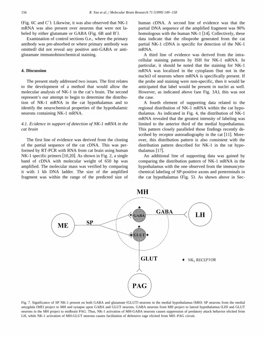
Ž . Ž .Fig. 7. Significance of SP NK-1 present on both GABA and glutamate GLUT neurons in the medial hypothalamus MH : SP neurons from the medialŽ . Ž .amygdala ME project to MH and synapse upon GABA and GLUT neurons. GABA neurons from MH project to lateral hypothalamus LH and GLUT
neurons in the MH project to midbrain PAG. Thus, NK-1 activation of MH-GABA neurons causes suppression of predatory attack behavior elicited fromLH, while NK-1 activation of MH-GLUT neurons causes facilitation of defensive rage elicited from MH–PAG circuit.

( )R. Yao et al.rMolecular Brain Research 71 1999 149–158 157
tion 3, the distribution of cells expressing NK-1 matchedclosely the distribution pattern of the SP-positive axonsand preterminals in the medial hypothalamus. The func-tional implications of this matching distribution are dis-cussed in Section 4.2.
4.2. Functional implications of the distribution of NK-1mRNA in the cat hypothalamus and its relationship toGABA and glutamate neurons
The second phase of this study provided evidence thatNK-1 is situated on both GABA and glutamate neuronswithin the cat anterior medial hypothalamus. Since it is awidely accepted view that SP receptors mediate excitatory
w xfunctions within the central nervous system 1,18 , it isreasonable to conclude that SP neurons that synapse uponthese neurons will normally have excitatory effects uponthese neurons. In a series of recent studies conducted inour laboratory, it was demonstrated that SP neurons situ-ated in the medial amygdala project to the anterior medial
w xhypothalamus via the stria terminalis 11,24 . These inves-tigators further showed that medial amygdaloid stimulationfacilitated the occurrence of defensive rage behavior
w xelicited from the medial hypothalamus 24 , while stimula-tion of this region of amygdala suppressed predatory attack
w xbehavior elicited from the lateral hypothalamus 11,12 .The modulating actions of the medial amygdala upon eachof these forms of aggressive behavior were further shownto be mediated by SP receptor activation in the medialhypothalamus. With respect to defensive rage behavior, wehave further shown that the descending pathway mediatingthis form of aggression arises from glutamatergic neuronsin the medial hypothalamus. This descending pathway
Ž .projects directly to the midbrain periaqueductal gray PAGand the excitatory functions of this pathway are mediated
w xby NMDA receptors 16,23 . Thus, the overall effects ofthese relationships can be summarized as follows: themedial amygdala facilitates defensive rage behavior byexciting neurons in the medial hypothalamus via an SPreceptor mechanism, which, in turn, drives the descendingglutamatergic pathway from the medial hypothalamus tothe PAG whose functions are mediated by NMDA recep-tors. In contrast, the suppressive effects of medial amyg-daloid stimulation upon predatory attack behavior involvesa disynaptic circuit, the first limb of which involves anexcitatory SP mediated pathway to the medial hypothala-mus. The second limb of this pathway involves an in-hibitory GABAergic neuron projecting from the medial tolateral hypothalamus whose functions are mediated byGABA receptors, therefore, accounting for the inhibitoryA
effects of medial amygdaloid stimulation upon predatoryw xattack behavior 12 . In this manner, the differential effects
of medial amygdaloid stimulation upon defensive rage andpredatory attack are mediated through SP receptors locatedon both glutamate and GABA neurons within the medialhypothalamus. These relationships are depicted in a sum-
mary diagram shown in Fig. 7. One of the significantfeatures of the present study is that it provides anatomicalevidence for these conclusions. Ongoing studies in ourlaboratory are currently attempting to further define andcharacterize the relationships between NK-1 and glutamateand GABA neurons in the hypothalamus.
Acknowledgements
This research was supported by NIH grants NS 07941-27, HL-57675 and grants from the following: Foundationof the University of Medicine and Dentistry of NewJersey, the Violence Institute of the University of Medicineand Dentistry of New Jersey, the Alcoholic Medical Bev-erage Foundation and Ruth Estrin Goldberg Memorial forCancer Research. The authors wish to thank Xiao-FengWang for his excellent assistance in generating the com-puterized representation of the photomicrographs and JingQian for her input in cloning of the NK-1 cDNA.
References
w x1 B.E. Alger, R.A. Nicoll, Epileptiform burst afterhyperpolarization:calcium-dependent potassium potential in hippocampal CA1 pyrami-
Ž .dal cells, Science 210 1980 1122–1124.w x2 M.J. Bannon, C.J. Whitty, Neurokinin receptor gene expression in
substantia nigra: localization, regulation, and potential physiologicalŽ .significance, Can. J. Physiol. Pharmacol. 73 1995 866–870.
w x3 P.M. Beart, L.S. Nicolopoulos, D.C. West, P.M. Headley, Anexcitatory amino acid projection from ventromedial hypothalamus toperiaqueductal gray in the rat: autoradiographic and electrophysio-
Ž .logical evidence, Neurosci. Lett. 85 1988 205–211.w x4 P.M. Beart, R.J. Summers, J.A. Stephenson, C.J. Cook, M.J. Christie,
Excitatory amino acid projections to the periaqueductal gray in thew3 xrat: a retrograde transport study utilizing d H aspartate and
w3 x Ž .H GABA, Neuroscience 34 1990 163–176.w x5 A.J. Beitz, Possible origin of glutamatergic projections to the mid-
brain periaqueductal gray and deep layer of the superior colliculus ofŽ .the rat, Brain Res. Bull. 23 1989 25–35.
w x6 S.H. Buck, C.J. Helke, E. Burcher, C.A. Shults, T.L. O’Donahue,Pharmacologic characterization and autoradiographic distribution ofbinding sites for iodinated tachykinins in the rat central nervous
Ž .system, Peptides 7 1986 1109–1120.w x7 J.W. Cheu, A. Siegel, GABA receptor mediated suppression of
defensive rage behavior elicited from the medial hypothalamus ofŽ .the cat: role of the lateral hypothalamus, Brain Res. 783 1998
293–304.w x8 P. Chomczynski, N. Sacchi, Single-step method of RNA isolation by
acid guanidinium thiocyanate–phenol–chloroform extraction, Anal.Ž .Biochem. 162 1987 156–159.
w x9 T.V. Dam, B. Martinelli, R. Quirion, Autoradiographic distributionof brain neurokinin-1rsubstance P receptors using a highly selective
w3 x w 9 Ž .11 x Ž .ligand H - Sar ,Met O -substance P, Brain Res. 531 19902
330–337.w x10 N.P. Gerard, L.A. Garraway, R.L. Eddy Jr., B.S. Thomas, H. Iijima,
Ž .J.-L. Paquet, C. Gerard, Human substance P receptor NK-1 : orga-nization of the gene, chromosome localization, and functional ex-
Ž .pression of cDNA clones, Biochemistry 30 1991 10640–10646.w x11 Y. Han, M.B. Shaikh, A. Siegel, Medial amygdaloid suppression of
predatory attack behavior in the cat: I. Role of a substance P

( )R. Yao et al.rMolecular Brain Research 71 1999 149–158158
pathway from the medial amygdala to the medial hypothalamus,Ž .Brain Res. 716 1996 59–71.
w x12 Y. Han, M.B. Shaikh, A. Siegel, Medial amygdaloid suppression ofpredatory attack behavior in the cat: II. Role of a GABAergicpathway from the medial to the lateral hypothalamus, Brain Res. 716Ž .1996 72–83.
w x13 A.D. Hershey, J.E. Krause, Molecular characterization of a func-tional cDNA encoding the rat substance P receptor, Science 247Ž .1990 958–962.
w x14 B. Hopkins, S.J. Powell, P. Danks, I. Briggs, A. Graham, Isolationand characterization of the human lung NK-1 receptor cDNA,
Ž .Biochem. Biophys. Res. Commun. 182 1992 1514.w x15 J.E. Krause, J.M. Chirgwin, M.S. Carter, Z.S. Xu, A.D. Hershey,
Three rat preprotachykinin mRNAs encode the neuropeptides sub-Ž .stance P and Neurokinin A, Proc. Natl. Acad. Sci. U.S.A. 84 1997
881–885.w x16 C.-L. Lu, M.B. Shaikh, A. Siegel, Role of NMDA receptors in
hypothalamic facilitation of feline defensive rage elicited from theŽ .midbrain periaqueductal gray, Brain Res. 581 1992 123–132.
w x17 H. Maeno, H. Kiyama, M. Tohyama, Distribution of the substance PŽ .receptor NK-1 receptor in the central nervous system, Mol. BrainŽ .Res. 18 1993 43–58.
w x18 M. Otsuka, K. Yoshioka, Neurotransmitter functions of mammalianŽ .tachykinins, Physiol. Rev. 73 1993 229–308.
w x19 L. Quartara, C.A. Maggi, The tachykinin NK receptor: Part I.1
Ligands and mechanisms of cellular activation, Neuropeptides 31Ž .1997 537–563.
w x20 P. Rameshwar, Substance P: a regulatory neuropeptide for hemato-
poiesis and immune functions, Clin. Immunol. Immunopathol. 85Ž .1997 129–133.
w x21 P. Rameshwar, P. Gascon, Induction of negative hematopoieticŽ .regulators by neurokinin-A in bone marrow stroma, Blood 88 1996
98–106.w x22 D. Regoli, A. Boudon, J.L. Fauchere, Receptors and antagonists for
Ž .substance P and related peptides, Pharmacol. Rev. 46 1994 551–599.
w x23 K. Schubert, M.B. Shaikh, A. Siegel, NMDA receptors in themidbrain periaqueductal gray mediate hypothalamically evoked hiss-
Ž .ing behavior in the cat, Brain Res. 726 1996 80–90.w x24 M.B. Shaikh, A. Steinberg, A. Siegel, Evidence that substance P is
utilized in medial amygdaloid facilitation of defensive rage behaviorŽ .in the cat, Brain Res. 625 1993 283–294.
w x25 Y. Takeda, K.B. Chou, J. Takeda, B.S. Sachais, J.E. Krause, Molec-ular cloning, structural characterization and functional expression ofthe human substance P receptor, Biochem. Biophys. Res. Commun.
Ž .179 1991 1232–1240.w x26 S.R. Vigna, J.J. Bowden, D.M. McDonald, J. Fisher, A. Okamoto,
D.C. McVey, D.G. Payan, N.W. Bunnett, Characterization of anti-Ž .bodies to the rat substance P NK-1 receptor and to a chimeric
substance P receptor expressed in mammalian cells, J. Neurosci. 14Ž .1994 834–845.
w x27 C.J. Whitty, P.D. Walker, D.J. Goebel, M.S. Poosch, M.J. Bannon,Quantitation, cellular localization and regulation of neurokinin re-ceptor gene expression within the rat substantia nigra, Neuroscience
Ž .64 1995 419–425.


![Modulation Of [35S]Tert Butylbicyclophosphorothionate Binding By Somatostatin In Rat Hypothalamus](https://img.pdfslide.net/doc/110x75/631ba0d07abff1d7c20b0671/modulation-of-35stert-butylbicyclophosphorothionate-binding-by-somatostatin-in.jpg)


![Glutamate receptor agonists release [3H]GABA preferentially from horizontal cells](https://img.pdfslide.net/doc/110x75/631d0b22665120b3330c268c/glutamate-receptor-agonists-release-3hgaba-preferentially-from-horizontal-cells.jpg)


